characterization of a 27 kda fibrinolytic enzyme from … 7 (' j. microbiol. biotechnol....
TRANSCRIPT

ACCEPTED
J. Microbiol. Biotechnol. (2009), 19(2), 000–000doi: 10.4014/jmb.0811.600First published online 26 March 2009
Characterization of a 27 kDa Fibrinolytic Enzyme from Bacillus amyloliquefaciensCH51 Isolated from Cheonggukjang
Kim, Gyoung Min1, Ae Ran Lee
1, Kang Wook Lee
1, Jae-Young Park
1, Mee-Ryung Lee
1, Jiyeon Chun
3,
Jaeho Cha4, Young-Sun Song
5, and Jeong Hwan Kim
1,2*
1Division of Applied Life Science (BK21 Program), Graduate School and 2Institute of Agriculture and Life Science, GyeongsangNational University, Jinju 660-701, Korea3Department of Food Science and Technology, Sunchon National University, Sunchon 540-742, Korea4Department of Microbiology, Pusan National University, Busan 609-735, Korea5School of Food and Life Science, Inje University, Gimhae 621-749, Korea
Received: November 3, 2008 / Revised: February 5, 2009 / Accepted: February 12, 2009
Bacillus amyloliquefancies CH51 isolated from cheonggukjang,
a traditional Korean fermented soy food, has strong
fibrinolytic activity and produces several fibrinolytic enzymes.
Among four different growth media, tryptic soy broth was
the best in terms of supporting cell growth and fibrinolytic
activity of this strain. A protein with fibrinolytic activity
was partially purified from the culture supernatant by CM-
Sephadex and Phenyl Sepharose column chromatographies.
Tandem mass spectrometric analysis showed that this
protein is a homolog of AprE from B. subtilis and it was
accordingly named AprE51. The optimum pH and
temperature for partially purified AprE51 activity were
6.0 and 45o
C, respectively. A gene encoding AprE51, aprE51,
was cloned from B. amyloliquefaciens CH51 genomic
DNA. The aprE51 gene was overexpressed in heterologous
B. subtilis strains deficient in fibrinolytic activity using an
E. coli-Bacillus shuttle vector, pHY300PLK.
Keywords: Fibrinolytic enzymes, bacilli, subtilisins, Bacillus
amyloliquefaciens, cheonggukjang
Strains of Bacillus amyloliquefaciens are some of the most
common bacilli isolated from fermented soybean foods.
Some of these strains possess fibrinolytic activity [4, 18]
conferred by certain proteolytic enzymes that are secreted
by the bacilli, nattokinase being the most well-known
example [20]. Fibrinolytic enzymes are able to resolve
fibrin clots, thus being responsible for an important
functionality of soybean foods fermented by bacilli.
Cheonggukjang, a traditional Korean fermented soy food,
is an important source of bacilli with fibrinolytic activity
[11]. In traditional cheonggukjang preparation, cooked
soybeans are covered with rice straw, which is a rich
source of Bacillus spores. During fermentation for 2 or 3
days at around 40oC, soybean proteins are digested, releasing
oligopeptides and amino acids. In some cases, sticky
materials are produced on the surface of the soybeans.
After fermentation, cheonggukjang is usually served as a
side dish with rice after being boiled with other seasonings,
such as garlic and pepper.
Cheonggukjang is known to possess several health-
promoting properties, such as anticancer activity, abilities to
lower blood pressure and prevent myocardial and cerebral
infarction, antioxidant activity, and probiotic activity [15].
B. amyloliquefaciens CH51, previously isolated from
cheonggukjang, showed higher fibrinolytic activity than
other isolates when examined by the fibrin plate method
[9]. It produces several fibrinolytic enzymes (see Fig. 1)
but these have not yet been characterized. In an effort to
understand the fibrinolytic system of B. amyloliquefaciens
CH51, we attempted to purify fibrinolytic enzymes from
its culture supernatants. As a result, the major fibrinolytic
enzyme, AprE51 was partially purified, its properties were
examined, and its structural gene was cloned.
MATERIALS AND METHODS
Bacterial Strains and Culture Conditions
B. amyloliquefaciens CH51 was previously isolated from cheonggukjang
and identified by molecular methods including 16S rRNA gene and
recA sequencing [12].
B. amyloliquefaciens CH51 was grown in tryptic soy broth (TSB;
Becton, Dickinson and Company, Sparks, MD, U.S.A.) or LB at
*Corresponding authorPhone: +82-55-751-5481; Fax: +82-55-753-4630;E-mail: [email protected]
ACCEPTED
2 Kim et al.
37oC with aeration. Nutrient broth (NB) and brain heart infusion
(BHI) were used for comparision. E. coli and B. subtilis strains were
grown in LB at 37oC with shaking. Cells containing pHY300PLK
(Takara, Shiga, Japan) or pHY51 were cultivated in LB supplemented
with 10 µg/ml tetracycline (LBTc).
Assay of Fibrinolytic Activity
Bacillus cells were cultivated in TSB at 37oC with shaking. At the
appropriate growth stage, the culture was centrifuged at 5,000 ×g for
20 min at 4oC. The supernatant was filtered and assayed for fibrinolytic
activity. If necessary, the supernatant was further concentrated by
ammonium sulfate precipitation (80%, w/v). The ammonium sulfate
pellet was redissolved in a small volume of buffer A (20 mM Tris-
HCl, pH 7.0). Protein concentration was determined according to the
Bradford method [2] using bovine serum albumin (BSA) as a standard.
Fibrinolytic activity was determined using the fibrin plate method
[1]. In a Petri dish, 7 ml of 1.2% (w/v) fibrinogen (Sigma, St. Louis,
MO, U.S.A.) solution in 1 M phosphate-buffered saline (PBS) was
mixed with an equal volume of 2% (w/v) agarose solution along
with 0.1 ml of thrombin solution (100 NIH units/ml; Sigma). The
Petri dish was left at room temperature for 1 h to allow a fibrin clot
layer to form. Twenty µl of sample was dropped into a hole that had
been made in a fibrin plate using a capillary glass tube, and the
plate was incubated at 37oC for 6 h. The size of the clear zone that
formed was converted into plasmin units (U) by comparing it to zones
formed by known quantities of plasmin. A standard curve showing
the relationship between the area of the clear zone formed and the
number of plasmin units was prepared in the range of 1.5-30 mU.
SDS-PAGE and Fibrin Zymography
Culture supernatant was concentrated by ammonium sulfate precipitation
(80%, w/v), and then the pellet was resuspended in a small volume of
buffer A and dialyzed against the same buffer for 24 h at 4oC with four
buffer changes. SDS-PAGE was performed according to Laemmli’s
method [13], and fibrin zymography was carried out as previously
described [6, 10]. The separating gel solution (12%, w/v) contained
fibrinogen (0.12%, w/v) and 100 µl of thrombin (10 NIH units/ml).
Partial Purification of AprE51
Culture supernatant (1 l) was concentrated by ammonium sulfate
precipitation (80%, w/v). The precipitate was resuspended in 10 ml
of buffer A and then dialyzed against 20 volumes of the same buffer
for 24 h with four buffer changes. The protein sample was loaded onto
a CM-Sephadex (Amersham Pharmacia Biotech, Uppsala, Sweden)
column (2.5×8 cm), and proteins were eluted by sequential application
of six 200-ml volumes of buffer A containing NaCl at a concentration
of 0 to 1 M, increased stepwise in 0.2 M increments. Twenty µl of
each 15-ml fraction was spotted onto a fibrin plate. Fractions
showing fibrinolytic activity were pooled, dialyzed against buffer A,
and then lyophilized. Hydrophobic interaction chromatography using
Phenyl Sepharose 6 Fast Flow (Amersham Pharmacia Biotech, Uppsala,
Sweden) was used for further purification. The column (2.5×12 cm)
was washed and pre-equilibrated with buffer A containing 1 M
(NH4)2SO4. Proteins were eluted by sequential application of 200-ml
volumes of buffer A containing (NH4)2SO4 at a concentration of 1 to
0 M, decreased stepwise in 0.2 M increments. Active 15 ml fractions
were pooled, dialyzed against buffer A, and lyophilized prior to
analysis by SDS-PAGE and zymography. Protein bands were excised
from the stained SDS gel. Trypsin digestion and mass spectrometric
analysis of peptides were performed at Genomine (Pohang, Gyungbuk,
Korea).
Properties of Partially Purified AprE51
Effects of pH, temperature, metal ions, and inhibitors on the
fibrinolytic activity of AprE51 were examined by the fibrin plate
method. Eight mg of AprE51 was incubated in either 0.1 M citrate-
NaOH buffer (pH 3.0 to 5.0), 0.1 M sodium phosphate buffer (pH 6.0
to 7.0), 0.1 M Tris-HCl (pH 8.0 to 9.0), or 0.1 M glycine-NaOH buffer
(pH 10.0) at 37oC for 2 h, and then the amount of fibrinolytic activity
remaining was measured. Heat stability was examined by incubating
8 µg of AprE51 in 0.1 M sodium phosphate buffer (pH 6.0) at 37-
55oC for 30 min and measuring the activity that remained. For the
effects of metal ions and inhibitors, AprE51 was incubated in 0.1 M
sodium phosphate buffer (pH 6.0) containing 5 mM metal ions (KCl,
CaCl2, CoCl2, MgCl2, CuSO4, MnCl2, or ZnCl2) or 1 mM inhibitor
(phenylmethylsulfonylfluoride [PMSF], SDS, or EDTA) at 45oC for
1 h and the activity that remained was measured.
Cloning of the aprE51 Gene
The primer pair CH51-F (5'-AGGATCCCAAGAGAGCGATTGCG
GCTGTGTAC-3', BamHI site underlined) and CH51-R (5'-AGAAT
TCTTCAGAGGGAGCCACCCGTCGATCA-3', EcoRI site underlined),
was designed based on the sequence of aprE (Accession No.
CP000560.1) from B. amyloliquefaciens FZB42. PCR was performed
using a GeneAmp 2400 PCR system (PerkinElmer, Waltham, MA,
U.S.A.). The reaction mixture (50µl) contained 1 ml of template DNA,
1 µl of each primer (10 µM), 1 µl of deoxynucleoside triphosphates
(0.25 mM), and 0.5 µl of ExTaq DNA polymerase (Takara, Shiga,
Japan). The amplification conditions were as follows: 94oC for 5 min;
30 cycles of 94oC for 30 sec, 64
oC for 30 sec, 72
oC for 40 sec; a
final extension at 72oC for 5 min. The amplified fragment was
ligated into pGEM-T Easy Vector (Promega, Madison, WI, U.S.A.)
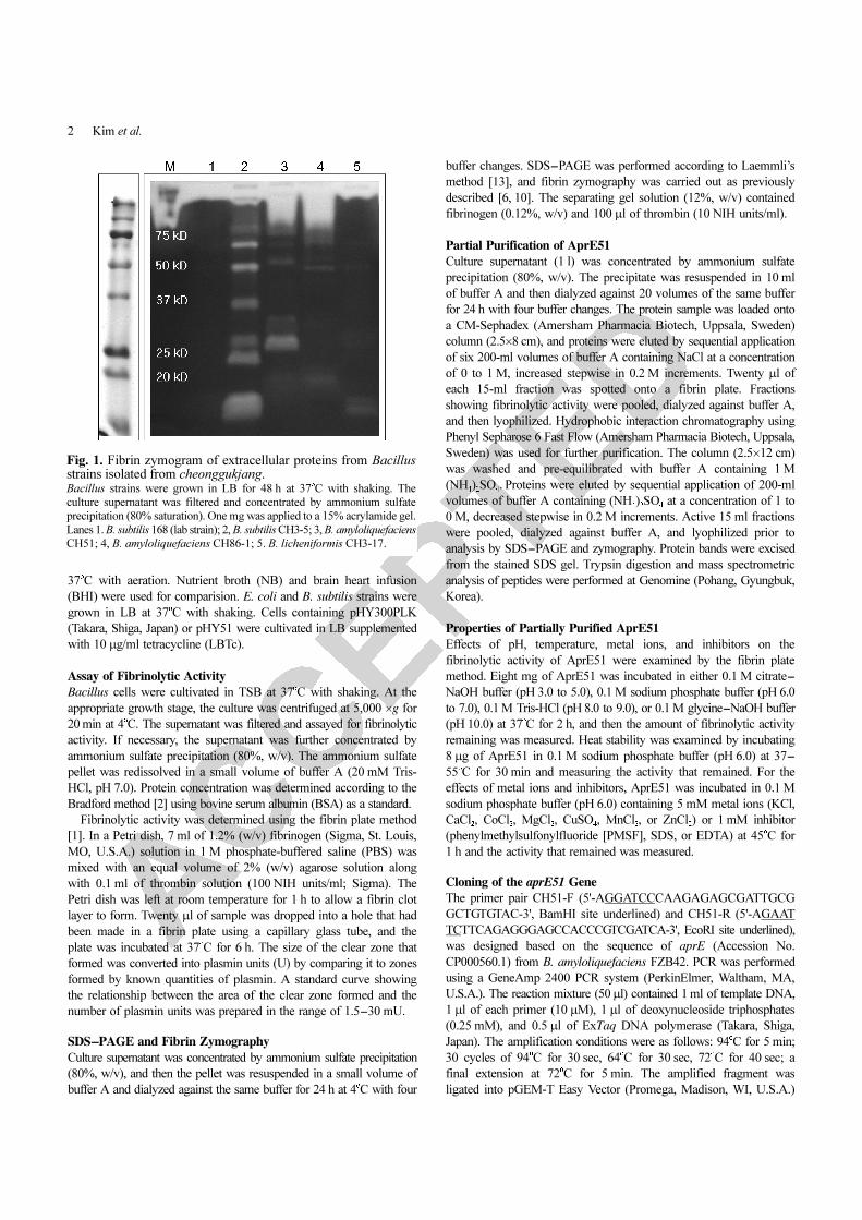
Fig. 1. Fibrin zymogram of extracellular proteins from Bacillusstrains isolated from cheonggukjang. Bacillus strains were grown in LB for 48 h at 37
o
C with shaking. The
culture supernatant was filtered and concentrated by ammonium sulfate
precipitation (80% saturation). One mg was applied to a 15% acrylamide gel.
Lanes 1. B. subtilis 168 (lab strain); 2, B. subtilis CH3-5; 3, B. amyloliquefaciens
CH51; 4, B. amyloliquefaciens CH86-1; 5. B. licheniformis CH3-17.

ACCEPTED
CHARACTERIZATION OF APRE51 3
and E. coli DH5α cells were transformed with the ligation mixture.
Preparation and electroporation of E. coli competent cells were
performed as previously described [9]. Plasmid DNA preparation,
restriction enzyme digestion, and agarose gel electrophoresis were
performed according to published methods [19].
Overexpression of aprE51 in Protease-Deficient B. subtilis Strains
aprE51 was amplified using primer pair CH51-F and CH51-R, and
then digested with BamHI and EcoRI. The 1.5 kb fragment was
ligated into pHY300PLK, resulting in pHY51. Preparation and
electroporation of Bacillus competent cells were carried out as
previously described [9]. B. subtilis WB600 [21] and ISW 1214
(Takara) were used as hosts for aprE51 gene expression. B. subtilis
transformants (TFs) were grown in LBTc at 37oC for 2 days. Cells
were recovered by centrifugation at 5,000 ×g for 20 min at 4oC and
the supernatant was used for the enzyme assay.
For SDS-PAGE and zymography, protein samples were prepared
from B. subtilis TFs using different methods. Culture supernatant
was mixed with an equal volume of cold 20% (w/v) trichloroacetic
acid (TCA) solution and kept on ice for 15 min prior to centrifugation
[7]. The protein pellet was washed four times with 100% ethyl
alcohol, dried, and then resuspended in 50 mM Tris-HCl (pH 7.4).
Ten µg of protein was subjected to SDS-PAGE (12% acrylamide)
as described above. For zymography, the supernatant was concentrated
by ammonium sulfate precipitation (80%, w/v) and 1 mg was applied
to the gel.
RESULTS AND DISCUSSION
Fibrinolytic Activity of B. amyloliquefaciens CH51
We previously isolated bacilli exhibiting fibrinolytic
activity from cheonggukjang prepared by traditional methods
in Sunchang County, North Jeolla Province, Korea [12].
Among these strains, CH51 and CH86-1, which were
identified as B. amyloliquefaciens, showed the highest
fibrinolytic activities. Analysis of culture supernatant by
zymography on a fibrin gel revealed that B. amyloliquefaciens
CH51 produces 6 fibrinolytic enzymes (see Fig. 1, lane 3).
Distinct bands with sizes of 70, 66, 50, 32, and 27 kDa were
observed. In addition, a large smear was observed at the
top of the separating gel. The smear was caused by either
binding of fibrinolytic enzymes to fibrin in the gel [14] or
by their high pI values [4], which could have prevented
them from migrating to the points in the gel that correspond
to their molecular masses. It will be necessary to characterize
each component of the fibrinolytic system and evaluate the
contribution of each enzyme to the observed fibrinolytic
activity of B. amyloliquefaciens CH51 if this strain is to be
fully utilized for the production of functional foods or medicines.
The growth and fibrinolytic activity of B. amyloliquefaciens
CH51 in four different media were compared. Each medium
was inoculated with 2% (w/v) overnight culture and incubated
at 37oC with shaking. Growth in TSB showed the best
result, reaching an absorbance value of 1.9 by the end of
cultivation at 48 h (Fig. 2). Growth in other media was less
efficient than in TSB, reaching about 1.5. Cells grown in
TSB showed the highest fibrinolytic activity, followed by
those grown in NB and LB. Interestingly, cells did not
show any significant fibrinolytic activity when grown in
BHI although the medium supported cell growth as well as
NB and LB did (Fig. 2). This result, the cause of which is
unknown, indicates that the choice of growth medium and
possibly cultivation conditions are important for the
production of fibrinolytic enzymes by B. amyloliquefaciens
CH51.
Partial Purification of AprE51
Fibrinolytic enzymes in the culture supernatant were
purified by CM-Sephadex and Phenyl-Sepharose column
chromatographies. A fibrin plate assay was used to detect
active fractions. Purification results are summarized in Table 1
and the overall purification was 20.3-fold. When active
fractions from the Phenyl-Sepharose column were pooled
Fig. 2. Changes in absorbance (A) and fibrinolytic activity (B) ofB. amyloliquefaciens CH51 during growth on four different media.●, BHI; ○, LB; ▼ , NB; ▽, TSB. Filtered culture supernatant (20 µl) at
each time point was applied to a fibrin plate and the fibrinolytic activity
was expressed as U/ml.

ACCEPTED
4 Kim et al.
and examined by SDS-PAGE, four major bands (45, 27,
23, and 19 kDa in size), in addition to a few minor bands,
were detected when the gel was stained with Coomassie
blue (Fig. 3). The four major bands were excised from the
gel and subjected to trypsin digestion and mass spectrometric
analysis. The 27 kDa protein (AprE51 in Fig. 3) was
identified as a homolog of subtilisin DJ-4 from Bacillus sp.
DJ-4 [5], subtilisin DFE from B. amyloliquefaciens [18],
hypothetical protein AprE from B. amyloliquefaciens FZB42
(protein id=ABS73414), and AprE from B. subtilis (protein
id=ABY83469).
These results confirmed that the 27 kDa protein is a
subtilisin-type protease with fibrinolytic activity; it has
been accordingly named AprE51. B. subtilis secretes several
proteases into the culture medium, subtilisin being the most
predominant [3]. Therefore, AprE51 is the major protease
with fibrinolytic activity secreted by B. amyloliquefaciens
CH51. The 23 kDa protein (BglS in Fig. 3) was identified
as a β-1,3-1,4-endoglucanase precursor, and the 19 kDa
protein (XynA in Fig. 3) as endo-1,4-beta-xylanhydrolase.
The 45 kDa protein (YnfF in Fig. 3) might be a homolog of
YnfF, a precursor of dimethyl sulfoxide reductase, from
Bacillus species. Zymography results (Fig. 3B) also support
the conclusion that AprE51 is the major fibrinolytic
enzyme of B. amyloliquefaciens CH51. In lane 4, two
minor fibrinolytic enzymes were observed that were later
removed by CM-Sephadex column chromatography. These
minor fibrinolytic enzymes will need to be characterized
in the future. Cultivation conditions for the production of
fibrinolytic enzymes must be chosen carefully since they
may affect the activity of each enzyme differently.
Properties of Partially Purified AprE51
The effects of pH, temperature, and metal ions on the
fibrinolytic activity of AprE51 were examined using the fibrin
Table 1. Partial purification of AprE51.
Step Total protein (mg) Total activity (U) Specific activity (U/mg) Fold purification (χ fold) Yield (%)
Culture supernatant 425.2 7156.2 16.8 1.0 100
80% Ammonium sulfate precipitation
71.2 3851.2 54.1 3.2 54
CM-Sephadex 5.2 1,173.5 227.5 13.5 16
Phenyl-Sepharose 2.7 940.1 342.4 20.4 13
Fig. 3. SDS-PAGE (A) and zymography (B) of partially purifiedAprE51. A 10-µg boiled sample was subjected to SDS-PAGE and a 2-µg unboiled
sample to zymography. Lanes 1 and 4, 80% ammonium sulfate precipitation
of supernatant; lanes 2 and 5, active fractions from CM-Sephadex column;
lanes 3 and 6, active fractions from Phenyl-Sepharose 6 Fast Flow column;
M, Precision Plus protein standard (BioRad, U.S.A.). Four bands in lane 3
were identified and the name marked on the right side of each band: Yn
(=YnfF), Ap (=AprE51), Bg (=BglS), and Xn (=XynA).
Fig. 4. Effects of pH and temperature on the fibrinolytic activityof AprE51.
ACCEPTED
CHARACTERIZATION OF APRE51 5
plate method. The optimal pH was 6.0 (Fig. 4). The enzyme
activity was maintained at 45oC but was decreased at higher
temperatures. At 50oC, 62% activity remained, but at 55oC,
only 12% activity remained. Enzyme activity was not inhibited
by K+, Ca2+, or Mg2+ but was inhibited by Co2+, Mn2+, Cu2+,
and Zn2+ ions (Table 2). Among the metal ions tested, Cu2+
showed the greatest inhibition (39.5%). PMSF, a well-known
inhibitor of serine-type proteases, inhibited fibrinolytic activity
by 66.6%. EDTA also inhibited the activity by 17%,
indicating that inhibition of another minor protease, probably
a metalloprotease, might be responsible for this loss of activity,
even though its presence was not confirmed by SDS-PAGE.
Cloning and Sequencing of aprE51
The aprE51 gene encoding AprE51 was cloned from B.
amyloliquefaciens CH51 genomic DNA by PCR. The
amplified 1.5 kb DNA fragment was ligated into pGEM-T
Easy Vector and introduced into E. coli DH5α cells. TFs
harboring the recombinant plasmid pTaprE51 were obtained,
Table 2. Effect of metal ions and inhibitors on the fibrinolyticactivity of AprE51.
Metal ions (5 mM)/Inhibitors (1 mM) Activity (%)
None 100
KCl 102.2
CaCl2
104.5
CoCl2
93.4
MgCl2 103.4
CuSO4
60.5
MnCl2
95.6
ZnCl2 87.1
PMSF 33.4
SDS 69.4
EDTA 83.0
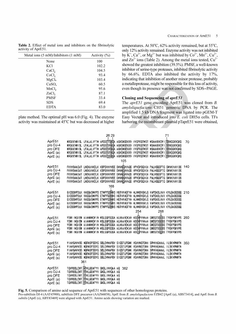
Fig. 5. Comparison of amino acid sequence of AprE51 with sequences of other homologous proteins. Pro-subtilisin DJ-4 (AAT45900), subtilisin DFE precursor (AAZ66858), AprE from B. amyloliquefaciens FZB42 [AprE (a), ABS73414], and AprE from B.
subtilis [AprE (s), ABY83469] were aligned with AprE51. Amino acids showing variation are marked.

ACCEPTED
6 Kim et al.
and DNA sequencing confirmed that the cloned gene is
indeed a homolog of aprE. The sequence was deposited
in GenBank (Accession No. EU414203). Fig. 5 shows the
translated amino acid sequence of AprE51 and its alignment
with the sequences of other subtilisins. These proteins were
found to be 99% identical to AprE51 when conservative
amino acid substitutions are taken into consideration. When
compared with AprE from B. amyloliquefaciens FZB42,
AprE51 has four different amino acids, located at positions
26 (T in AprE51, S in AprE), 27 (S in AprE51, P in AprE),
105 (Q in AprE51, K in AprE), and 168 (N in AprE51, Y
in AprE). Two changes are conservative (positions 26 and
105), leaving only two significant differences in amino acid
sequence (99% positive matches) between the two proteins.
AprE51 is synthesized as a preproprotein, as are all
subtilisins. It consists of 382 amino acids, with the 107
amino acids at the N-terminus corresponding to the prepro
sequence; tandem mass spectrometric results showed that
the active 27 kDa protein sequence begins with AQS--,
corresponding to position 108 of AprE51 as inferred from
the nucleotide sequence (data not shown). The calculated
molecular weight of the active enzyme is 27,446.57, which
corresponds well to the SDS-PAGE result. The first 30
amino acids seem to constitute the prepeptide (signal peptide)
when comparing the sequence to that of other homologous
proteins. This signal peptide is longer than that of AprE2
from Bacillus subtilis CH3-5 by one amino acid [9].
Subtilisins BPN’ and DJ-4 have signal peptides consisting
of 30 amino acids, whereas subtilisins E [16], NAT [17],
and J [8] have signal peptides consisting of 29 amino acids.
Although an active ca. 27 kDa band was observed on a
zymogram of B. amyloliquefaciens CH51 culture supernatant
(Fig. 1, lane 3), it was not AprE51. Under nondenaturing
conditions, AprE51 failed to migrate through the separating
gel to the point corresponding to its size (27 kDa). When
slices corresponding to the apparent molecular masses of
70 and 80 kDa were excised from the gel and analyzed by
mass spectrometry, one band (80 kDa) turned out to be
AprE51 (data not shown). When the eluted protein sample
was analyzed by SDS-PAGE after boiling, a 27 kDa band
appeared instead of an 80 kDa band (data not shown).
These results indicate that, under nondenaturing conditions,
AprE51 molecules are not resolved according to size but
remain near the top of the acrylamide gel, forming a large
smear; this behavior has also been reported for other
subtilisins secreted by Bacillus species [3, 4]. The same
result was obtained when aprE51 was overexpressed in
heterologous B. subtilis hosts (see Fig. 6 B, lanes 3 and 6).
Zymography cannot clearly distinguish between each enzyme
component, especially if their activity has been destroyed
by denaturing samples in order to resolve protein components.
In many cases, a large smear is observed at the top of a
zymogram instead of individually separated active bands.
More efficient methods of resolving fibrinolytic enzymes
will certainly need to be developed in order to facilitate the
understanding of the fibrinolytic capacity of bacilli.
Overexpression of aprE51 in Protease-Deficient B. subtilis
Strains
The PCR-amplified 1.5 kb aprE51 gene was ligated into
pHY300PLK, generating pHY51, which was introduced
into B. subtilis WB600 and ISW1214. Expression of aprE51
in these hosts was confirmed by SDS-PAGE and zymography
(Fig. 6). In Fig. 6A, an arrow on the right side of the gel
indicates the position of AprE51, and the results show that
only cells harboring pHY51 produced this protein. In B.
subtilis WB600, a strain deficient in six extracellular
proteases [21], the lack of these proteases was probably
responsible for the efficient production of AprE51. It is
suspected that B. subtilis ISW1214 has a similar genetic
background to WB600 since similar levels of aprE51
expression were observed in both strains (Fig. 6B).
Results obtained for cell growth and fibrinolytic activity
of B. subtilis TFs are summarized in Fig. 7. The fibrinolytic
Fig. 6. SDS-PAGE (A) and fibrin zymography (B) of culture supernatant of B. subtilis TFs. Lanes 1, B. subtilis WB600; 2, B. subtilis WB600 [pHY300PLK]; 3, B. subtilis WB600 [pHY51]; 4, B. subtilis ISW1214; 5, B. subtilis ISW1214
[pHY300PLK]; 6, B. subtilis ISW1214 [pHY51]; M, Precision Plus protein standard. A 15% acrylamide gel was used. A 10-µg protein sample (boiled) was
subjected to SDS-PAGE and a 1-µg (unboiled) sample to zymography.

ACCEPTED
CHARACTERIZATION OF APRE51 7
activities of both B. subtilis TFs were lower than that of B.
amyloliquefaciens CH51. It could be interpreted that
AprE51 alone might not be sufficient for B. subtilis hosts
to be as highly fibrinolytic as B. amyloliquefaciens CH51
because the latter synthesizes additional fibrinolytic enzymes.
If aprE51 were to be overexpressed in B. amyloliquefaciens
CH51 or B. amyloliquefaciens CH86-1, another highly
fibrinolytic isolate, it might enhance the fibrinolytic
activity of the host cells. It is desirable that each component
of the fibrinolytic system of B. amyloliquefaciens CH51 be
characterized and used for the construction of Bacillus
strains with improved fibrinolytic activity.
Acknowledgments
This work was supported by a research grant (Project No.
R01-2007-000-20024-0) from the Korea Science and
Engineering Foundation (KOSEF). G. M. Kim, A. R. Lee,
K. W. Lee, M. R. Lee, and J. Chun were supported by the
2nd Stage Brain Korea 21 Program of the Korean Ministry
of Education and Human Resources Development. The
authors are grateful for the financial support.
REFERENCES
1. Astrup, T. and S. Mullertz. 1952. The fibrin plate method for
estimating fibrinolytic activity. Arch. Biochem. Biophys. 40:
346-351.
2. Bradford, M. M. 1976. Rapid and sensitive methods for the
quantification of microgram quantities of protein utilizing the
principle of protein-dye binding. Anal. Biochem. 72: 248-254.
3. Choi, N.-S., D.-M. Chung, C. R. Ryu, K.-S. Yoon, P. J. Maeng,
and S.-H. Kim. 2006. Identification of three extracellular
proteases from Bacillus subtilis KCTC 3014. J. Microbiol.
Biotechnol. 16: 457-464.
4. Choi, N.-S., S.-K. Ju, T. Y. Lee, K.-S. Yoon, K.-T. Chang, P. J.
Maeng, and S.-H. Kim. 2005. Miniscale identification and
characterization of subtilisins from Bacillus sp. strains. J.
Microbiol. Biotechnol. 15: 537-543.
5. Choi, N.-S., K.-T. Chang, P. J. Maeng, and S.-H. Kim. 2004.
Cloning, expression, and fibrin (ogen)olytic properties of a
subtilisin DJ-4 gene from Bacillus sp. DJ-4. FEMS Microbiol.
Lett. 236: 325-331.
6. Choi, N. S. and S. H. Kim. 2001. The effect of sodium chloride
on the serine-type fibrinolytic enzymes and the thermostability
of extracellular protease from Bacillus amyloliquefaciens DJ-4.
J. Biochem. Mol. Biol. 34: 134-138.
7. Coligan, J. E., B. M. Dunn, D. W. Speicher, and P. T.
Wingfield. (eds.) 2003. Short Protocols in Protein Science, pp.
3-31-3-32. Wiley.
8. Jang, J. S., D. O. Kang, M. J. Chun, and S. M. Byun. 1992.
Molecular cloning of a subtilisin J gene from Bacillus
stearothermophilus and its expression in Bacillus subtilis.
Biochem. Biophys. Res. Commun. 184: 277-282.
9. Jeong, S. J., G. H. Kwon, J. Chun, J. S. Kim, C. S. Park, D. Y.
Kwon, and J. H. Kim. 2007. Cloning of fibrinolytic enzyme
gene from Bacillus subtilis isolated from Cheonggukjang and its
expression in protease-deficient Bacillus subtilis strains. J.
Microbiol. Biotechnol. 17: 1018-1023.
10. Kim, S. H. and N. S. Choi. 1999. Electrophoretic analysis of
protease inhibitors in fibrin zymography. Anal. Biochem. 270:
179-181.
11. Kim, W., K. Choi, Y. Kim, H. Park, J. Choi, Y. Lee, H. Oh, I.
Kwon, and S. Lee. 1996. Purification and characterization of a
fibrinolytic enzyme produced from Bacillus sp. strain CK11-4
screened from Chungkook-Jang. Appl. Environ. Microbiol. 62:
2482-2488.
12. Kwon, G.-H., H.-A. Lee, J.-Y. Park, J. S. Kim, J. Lim, C.-S.
Park, D. Y. Kwon, and J. H. Kim. 2009. Development of
a RAPD-PCR method for identification of Bacillus species
isolated from Chunggukjang. Int. J. Food Microbiol. 129: 282-
287.
13. Laemmli, U. K. 1970. Cleavage of structural proteins during the
assembly of the head of bacteriophage T4. Nature 227: 680-
685.
Fig. 7. Growth (A) and fibrinolytic activity (B) of B. subtilis TFs. ●, B. amyloliquefaciens CH51; ○, B. subtilis ISW1214; ▼ , B. subtilis
ISW1214 [pHY300PLK]; ▽, B. subtilis ISW1214 [pHY51].

ACCEPTED
8 Kim et al.
14. Lantz, M. S. and P. Ciborowski. 1994. Zymographic techniques
for detection and characterization of microbial proteases. Methods
Enzymol. 235: 563-594.
15. Lee, J.-O., S.-D. Ha, A.-J. Kim, C.-S. Yuh, I.-S. Bang, and S.-
H. Park. 2005. Industrial application and physiological functions
of Chungkukjang. Food Sci. Indus. 38: 69-78.
16. Nakamura, H., H. Takagi, and M. Inouye. 1987. Requirement of
pro-sequence for the production of active subtilisin E in
Escherichia coli. J. Biol. Chem. 262: 7859-7864.
17. Nakamura, T., Y. Tamagata, and E. Ichishima. 1992. Nucleotide
sequence of the subtilisin NAT gene, aprN, of Bacillus subtilis
(Natto). Biosci. Biotech. Biochem. 56: 1869-1871.
18. Peng, Y., Q. Huang, R. H. Zhang, and Y. Z. Zhang. 2003.
Purification and characterization of a fibrinolytic enzyme
produced by Bacillus amyloliquefaciens DC-4 screened from
douchi, a traditional Chinese soybean food. Comp. Biochem.
Physiol. part B 134: 45-52.
19. Sambrook, J. and D. W. Russel. 2001. Molecular Cloning: A
Laboratory Manual, 3rd ed. Cold Spring Harbor Laboratory
Press, Cold Spring Harbor, NY, U.S.A.
20. Sumi, H., H. Hamada, H. Tsushima, H. Mihara, and H. Muraki.
1987. A novel fibrinolytic enzyme (nattokinase) in the vegetable
cheese Natto; a typical and popular soybean food in the
Japanese diet. Experientia 43: 1110-1111.
21. Wu, X. C., W. Lee, L. Tran, and S. L. Wong. 1991. Engineering
a Bacillus subtilis expression-secretion system with a strain
deficient in six extracellular proteases. J. Bacteriol. 173: 4952-
4958.