characterization of a novel salivary immunosuppressive ... · means of cona-unstimulated wells were...
TRANSCRIPT

Ixodes ricinus immunosuppressive protein (Iris) 1
Characterization of a novel salivary immunosuppressive protein from Ixodes ricinus
ticks.
Gérard Leboulle*, Mara Crippa , Yves Decrem*, Naceur Mejri , Michel Brossard , Alex
Bollen* and Edmond Godfroid*
*Applied Genetics, Université Libre de Bruxelles, Rue des Professeurs Jeener et Brachet, 12,
B-6041 Gosselies, Belgium.
Institut de Zoologie, Université de Neuchâtel, Rue Emile Argand, 9, CH-2007 Neuchâtel,
Switzerland.
Running title : Ixodes ricinus immunosuppressive protein (Iris).
Correspondence to : Godfroid Edmond, Applied Genetics, Université Libre de Bruxelles, Rue
des Professeurs Jeener et Brachet, 12, B-6041 Gosselies, Belgium. Tél:2.2.650.99.34
Fax:32.2.650.99.00 E-Mail address: [email protected]
Copyright 2002 by The American Society for Biochemistry and Molecular Biology, Inc.
JBC Papers in Press. Published on January 15, 2002 as Manuscript M111391200 by guest on June 13, 2018
http://ww
w.jbc.org/
Dow
nloaded from
Ixodes ricinus immunosuppressive protein (Iris) 2
SUMMARY
In tick salivary glands, several genes are induced during the feeding process leading to the
expression of new proteins. These proteins are typically secreted in tick saliva and are
potentially involved in the modulation of the host immune and haemostatic responses. In a
previous study, the construction and the analysis of a subtractive library led to the
identification of Ixodes ricinus immunosuppressor (Iris), a novel protein, differentially
expressed in I. ricinus salivary glands during the blood meal. In the present study, the data
strongly suggest that this protein is secreted by tick salivary glands into the saliva. In addition,
Iris is also found to modulate T lymphocyte and macrophage responsiveness by inducing a
Th2 type response and by inhibiting the production of pro-inflammatory cytokines. In
conclusion, these results suggest that Iris is an immunosuppressor, which might play an
important role in the modulation of host immune response.
by guest on June 13, 2018http://w
ww
.jbc.org/D
ownloaded from
Ixodes ricinus immunosuppressive protein (Iris) 3
INTRODUCTION
In contrast to most hematophagous arthropods that feed to repletion rapidly, Ixodid female
ticks feed for an extended period of over 2 weeks (1).
Completion of the blood meal is dependent on the relationships of ticks with hosts species (2).
Resistance to tick infestation implicates both innate and acquired immunity, and is
characterized by reduced feeding, molting, and mating capabilities that may lead to the death
of the parasite. Acquired immunity of resistant hosts is mediated by a polarized Th1-type
immune response, involving IFN-γ production and delayed type hypersensitivity reaction (3,
4).
Some hosts are unable to counteract the tick infestation (4). Indeed, during their blood meal,
ticks circumvent host defenses via pharmacologically active components secreted in their
saliva. These factors of major importance for tick survival can modulate innate and acquired
immunity. In this way, the leukocyte responsiveness is modified during tick feeding (5). For
example, cytokine production is modulated inducing a polarized Th2 immune response (4, 6).
Therefore, the complex tick-host molecular interactions can be considered as a competition
between host defenses raised against the parasite and the tick evasion strategies, facilitating
feeding for an extended period.
Although, there is extensive information about the effects of tick bioactive factors on host
immune response, little is known about the mechanisms of their actions. However, it has been
observed that a wide range of new proteins are expressed during the blood meal (7). Several
of them might be essential for the completion of the tick feeding process. At present time,
only a few of them have been characterized (8, 9).
In order to identify cDNAs encoding proteins specifically expressed during the blood meal in
the salivary glands of I. ricinus female ticks, a representational difference analysis (RDA)
subtractive library was set up using mRNAs extracted from salivary glands of unfed and 5
by guest on June 13, 2018http://w
ww
.jbc.org/D
ownloaded from

Ixodes ricinus immunosuppressive protein (Iris) 4
day fed female I. ricinus ticks. One clone, formerly named seq24 (accession number :
AJ269658), was selected for further characterization of its recombinant protein (previously
Seq24/MBP and Seq24/His), due to its similarity to the pig leukocyte elastase inhibitor (10).
Elastase inhibitors have been identified in many mammalian species but also in arthropods
like the leech Hirudo nipponia (11). The immunological properties of such inhibitors are not
clearly established. However, these proteins are secreted by human macrophages, monocytes,
and neutrophils (12). Recently, it has been also shown that the elastase inhibitor mRNA is
abundantly expressed during delayed type hypersensibility responses (13). Based on the
results, we have decided to call this tick protein “Iris”1, for “Ixodes ricinus
Immunosuppressor”.
In this study, we describe the characterization of Iris. We demonstrate that Iris is induced
during the tick feeding process but is also secreted into the saliva. Moreover, the
characterization of its properties shows that Iris has the unique capacity to modulate both the
innate and the acquired immunity of the host.
1 Abbreviations used in this paper : Iris, Ixodes ricinus immunosuppressor; NEG, negative control; rIris,
recombinant Iris; SGE, salivary gland extracts.
by guest on June 13, 2018http://w
ww
.jbc.org/D
ownloaded from

Ixodes ricinus immunosuppressive protein (Iris) 5
EXPERIMENTAL PROCEDURES
Biological materials.
Unfed and five day fed tick salivary gland proteinic extracts were obtained from 300 and 70 I.
ricinus salivary glands, respectively. These salivary glands were crushed for 10 min using a
potter and a pestle in extraction buffer (PBS 1X, pH 7,4; EDTA 10 mM, AEBSF 1mM -
Sigma-Aldrich, Bornem, Belgium). The samples were centrifuged at 10,000g for 8 min, and
the supernatants were stored at –20°C.
Saliva was collected from ticks fed for 5 days on the ears of a rabbit. Engorging ticks were
removed, washed, and a 0,2% Dopamine (Sigma-Aldrich, Bornem, Belgium) solution in PBS
1X pH 7,2 was injected in the tick body. A finely drawn capillary tube fitted over the
mouthparts of each tick permitted to collect saliva.
Production of recombinant Iris (rIris) protein in bacterial and mammalian expression
systems.
Recombinant Iris (rIris) protein was obtained as described by Leboulle et al (10). Briefly, iris
cDNA was recovered by analyzing a RDA subtractive library. This library was constructed as
described by Hubank and Schatz (14) by using mRNA populations extracted from unfed and
5 day engorged tick salivary glands. The complete iris cDNA sequence was recovered by
performing the RACE methodology as described by Frohman (15). The recombinant
rIris/MBP protein was expressed in E. coli by using the pMALC2-E vector (NEB, Hitchin,
UK) containing MBP fusion partner. The rIris/His protein was expressed in CHO-KI cells by
using the pCDNA3.1/V5-His A vector containing the Epitag V5/6X His tag fusion partner
(10).
The rIris/His protein was purified in batch by Ni-chelate chromatography (Ni-NTA superflow
resin - Qiagen, Hilden, Germany), according to manufacturer’s instructions. Different buffers
were used to purify the rIris/His protein : the lysis buffer (PBS 1X, NaCl 500mM, Zwittergent
by guest on June 13, 2018http://w
ww
.jbc.org/D
ownloaded from
Ixodes ricinus immunosuppressive protein (Iris) 6
3.12 0,1%, pH7,5) ; the washing buffer (PBS 1X, NaCl 500 mM, Zwittergent 3.12 0,1%,
imidazole 17,25 mM, pH 7,5) and the elution buffer (PBS 1X, NaCl 500 mM, Zwittergent
3.12 0,1%, imidazole 103 mM, pH 7,5). The eluate was dialyzed (in a 7.000 Da cut-off
membrane) in PBS 1X, NaCl 500 mM, pH 7,5. The concentration of the protein was
evaluated on a Commassie blue stained acrylamide gel, at ~10 ng/µl (250 nM), by comparison
with known quantities of BSA. This result was confirmed by performing a micro
bicinchoninic acid (BCA) protein assay (Pierce, Rockford, IL; data not shown).
Immunodetection of Iris.
To examine the expression of native proteins in salivary glands and to detect rIris/His, the
same quantities of fed and unfed tick salivary glands, and a rIris/His sample were subjected to
SDS-PAGE and transferred onto nitrocellulose membranes. The membranes were probed
with diluted sera directed against rIris/MBP (1:1,000) (10) and revealed with NBT-BCIP.
Confocal microscopy.
I. ricinus salivary glands were dissected from unfed, 3 day or 5 day fed ticks and,
immobilized on silanated slides (Biorad, Nazareth EKE, Belgium). Salivary glands were fixed
in a 4% paraformaldehyde solution. After a treatment with 0.5% Triton X-100, the samples
were incubated in PBS 1X containing 5% FCS. The anti-rIris/MBP serum was used at a 1:10
dilution, and the secondary antibody, a FITC Anti-Mouse IgG (H+L) (ICN, Asse-Relegem,
Belgium) at a 1:32 dilution. The slides were mounted in Vectashield mounting medium
(Vector Lab, Peterborough, UK) and observed with a Leica confocal laser microscope using a
Leica TCS 4D operating system (Leica, Wetzlar, Germany).
Preparation of rIris/His cellular extract and rIris negative control (NEG) extracts used
in activity tests.
CHO-KI cells expressing rIris/His protein, obtained from a confluent culture in five 150 cm²
flasks, were resuspended in 1 ml of RPMI-1640 complete medium. The sample was frozen
by guest on June 13, 2018http://w
ww
.jbc.org/D
ownloaded from
Ixodes ricinus immunosuppressive protein (Iris) 7
and thaw 3 times before being centrifuged at 50.000 g for 1 hour at 4°C. The supernatant was
recovered and used in the different activity tests. The negative control (NEG) was a proteinic
extract of CHO-KI cells resistant to G418 that do not express the recombinant protein and
prepared as the rIris/His extract. The concentration of rIris/His in the cellular extract was
evaluated at ~4 ng/µl (~100 nM), by comparing rIris/His contained in cellular extracts and
purified rIris/His, on Western blot revealed with anti-V5 antibody (Invitrogen, Groningen,
The Netherlands). The toxicity of the different samples for human PBMCs was evaluated by
using the 7-amino actinomycin D (7-AAD) viability dye (Immunotech, Marseille, France),
according to manufacturer’s instructions
Cell Culture.
Naive Balb/c spleen cells : A suspension of spleen cells (SC) was isolated from naive
Balb/c mice. 106 lymph node cells per well were cultivated with serial dilutions of rIris/His or
NEG cellular extracts for 2 hours in 100 µl of complete culture medium containing RPMI-
1640 (Gibco, Basel, Switzerland) supplemented with 10% fetal calf serum (v/v), 2mM L-
glutamin, 1mM sodium pyruvate, 1mM non-essential amino acids (Sigma, St Louis, MO),
0,05 mM mercaptoethanol, 100 U/ml penicillin/streptomycin (Gibco, Basel, Switzerland) and
25 µg/ml Funigizone (Gibco, Basel, Switzerland). Cells were then stimulated with 1 µg/ml of
ConA in a final volume of 200 µl for 15 hours. 1 µCi/well of [3H]thymidine (Amersham Int.,
Amersham, UK) was added 24 hours before harvesting the cells. Tritiated thymidine
incorporation was determined by liquid scintillation counting. Results show the means (+/-
S.D.) of duplicate rIris/His or NEG stimulated wells realized in 2 independent experiments.
Means of ConA-unstimulated wells were previously subtracted (net 10³ c.p.m.).
Naive human PBMCs : Experiments were realized with PBMCs, obtained from 5
different donors, used separately. PBMCs were resuspended in RPMI-1640 medium
supplemented with FCS 10% (v/v), L-glutamine 2 mM, penicilline-streptomycine (100U/ml)
by guest on June 13, 2018http://w
ww
.jbc.org/D
ownloaded from

Ixodes ricinus immunosuppressive protein (Iris) 8
and IL-2 (20 U/ml). 2.106 cells were pre-cultivated in 1 ml of culture medium. Cells were
then diluted at different concentrations in 96 well plates (2.105 cells/100 µl for PPD
stimulation and 5.104 cells/100 µl for LPS stimulation for the ELISPOT technique and 2.105
cells/100 µl for the ELISA technique). Finally, PBMCs were incubated, for 72 hours at 37°C,
with different dilutions of rIris/His or NEG cellular extract or anti-rIris/MBP serum and the
different activators : PHA (at a final concentration of 1 µg/ml), LPS (1 µg/ml), anti-
CD3/CD28 mAb, (500 ng/ml), PMA/CD28 (PMA 25 ng/ml – CD28 500 ng/ml) and PPD (5
µg/ml).
Detection of cytokines.
ELISPOT assay : 96 well nitrocellulose bottom coated plates (Multiscreen-HA
Mahan, Millipore, Brussels, Belgium) were coated with coating antibodies directed against
IFN-γ (clone C1-D16 MAB 1-D1K, Nodia, Antwerp, Belgium) and IL-10 (Clone JES3-9D7,
BD Pharmingen, San Diego, CA). Cells were stimulated with PHA or LPS and rIris/His or
NEG cellular extracts for 72 hours at 37°C. Supernatants were recovered and stored at –20°C.
The cytokines were detected, in the 96 well plates, with biotinylated anti-IFN-γ antibody
(clone JES3-5A10 MAB 7-B6-1, Nodia, Antwerp, Belgium) and anti-IL10 antibody (clone
JES3-12G8, BD Pharmingen, San Diego, CA) diluted in PBS Tween 0,25% (1µg/ml).
Finally, the captured antibodies were revealed with extravidine peroxydase and AEC substrate
(Sigma-Aldrich, Bornem, Belgium). Results show the mean of triplicate wells (+/- S.D.).
Means of unstimulated wells were previously subtracted. The rIris/His and NEG protein
extracts do not affect the cell viability (data not shown).
ELISA technique : Cytokine-specific ELISAs were realized using the Flexia-human kit
(Biosource, Nivelles, Belgium) for the detection of IFN-γ, IL-10, TNF-α, IL-6, IL-1β and IL-
8, and the IL-5 kit (Endogene,Woburn, Massachusetts) for the detection of IL-5. The assays
were carried out using manufacturer’s instructions and were revealed using TMB substrate.
by guest on June 13, 2018http://w
ww
.jbc.org/D
ownloaded from
Ixodes ricinus immunosuppressive protein (Iris) 9
The concentration of the different cytokines in the experimental samples (pg/ml) was
calculated against a standard curve generated with recombinant cytokines. Results show the
mean of rIris/His or NEG cellular extracts stimulated wells realized in 5 independent
experiments (+/- S.D.). Means of unstimulated wells were previously subtracted.
Statistical Analysis.
The significance of the differences obtained between NEG and rIris/His-treated groups was
evaluated by Student t-test using the Pooled or Satterhwaite method.
by guest on June 13, 2018http://w
ww
.jbc.org/D
ownloaded from
Ixodes ricinus immunosuppressive protein (Iris) 10
RESULTS
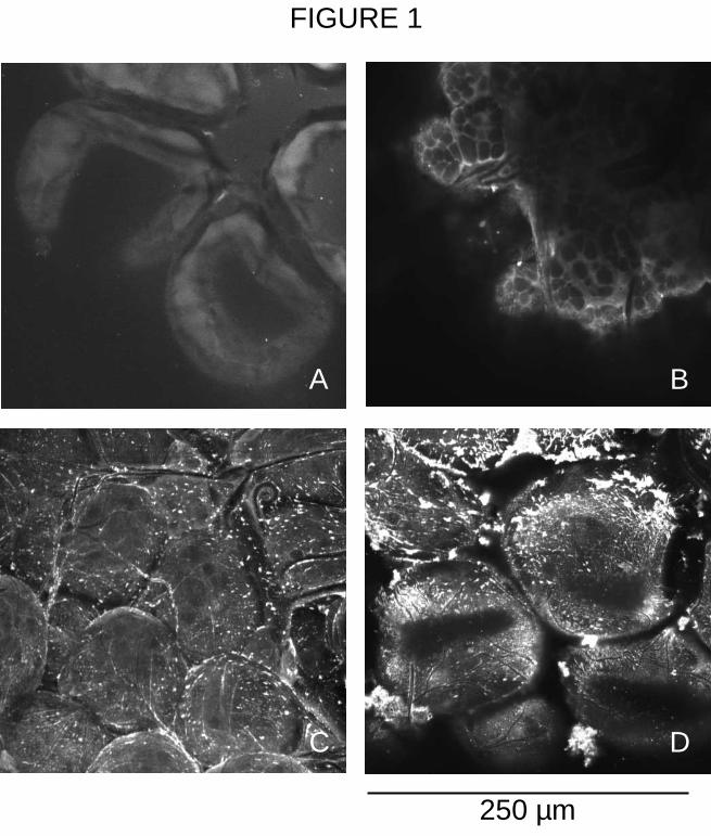
Detection of Iris in I. ricinus salivary glands and saliva.
Immune sera recovered from mice immunized with rIris/MBP (formerly Seq24/MBP) were
used to detect the native Iris protein in the salivary glands and saliva from unfed or engorged
female I. ricinus ticks. Iris was revealed by Western blot in tick saliva at a molecular weight
of 43 kDa (data not shown). By using confocal microscopy, it was found in 3 day and, more
abundantly, in 5 day fed tick salivary glands on the external surface of salivary acini, within
the cells and also in the acini’s light; but was not detected in unfed tick salivary glands
(Figure 1). These results suggest that the expression of Iris is induced in the salivary glands
during the tick feeding process process and that Iris is secreted in tick saliva.
Fig. 1 inserted here
Characterization of the immunomodulatory properties of Iris.
Based on its homology to a leukocyte elastase inhibitor, the immunomodulatory properties of
Iris were examined by using different activity tests that were mainly performed with soluble
proteinic extracts of CHO-KI cells expressing rIris/His (formerly named Seq24/His) at a
concentration of ~4 ng/µl (100 nM). Proteinic extracts of CHO-KI cells, which do not express
rIris/His, were used as a negative control (NEG).
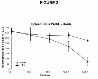
In vitro proliferation of Balb/c naive spleen cells. The proliferation of naive Balb/c spleen
cells (SC) was analyzed in vitro by pre-incubating them with various dilution (1 :6.25 to
1 :50) of rIris/His cellular extracts, followed by stimulation with concanavalin-A (ConA). As
shown on figure 2, the proliferative response of SC was strongly diminished (81% of
inhibition at a 1 :6.25 dilution) in a dose dependent manner. The negative control had no
significant effect on ConA-stimulated SC (average inhibition of 15%) ; even if it inhibited by
25% the SC proliferation at a 1 :6.25 dilution.
.
by guest on June 13, 2018http://w
ww
.jbc.org/D
ownloaded from

Ixodes ricinus immunosuppressive protein (Iris) 11
Fig. 2 inserted here
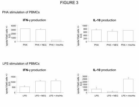
In vitro stimulation of human peripheral blood mononuclear cells (PBMCs). The effect of
rIris/His on cytokine production was studied on PBMCs stimulated with different activators.
The number of PBMCs secreting IFN-γ and IL-10, after stimulation either with LPS or
phytohaemagglutinin-A (PHA), was assayed by the ELISPOT technique (Figure 3). After
PHA stimulation, in presence of rIris/His cellular extract, a reduced number of PBMCs (more
than 80%) secreted IFN-γ while the number of cells producing IL-10 remained unchanged.
Furthermore, the NEG protein extract had no effect on the production of both cytokines by
PHA-stimulated PBMCs. In contrast, after LPS stimulation, no difference in the number of
cells producing IFN-γ was observed between PBMCs incubated with rIris/His and the NEG
cellular extract. On the other hand, rIris/His extract enhanced by 400% the number of PBMCs
expressing IL-10, while stimulation with NEG cellular extract did not enhance the number of
cells producing IL-10.
Fig 3. inserted here
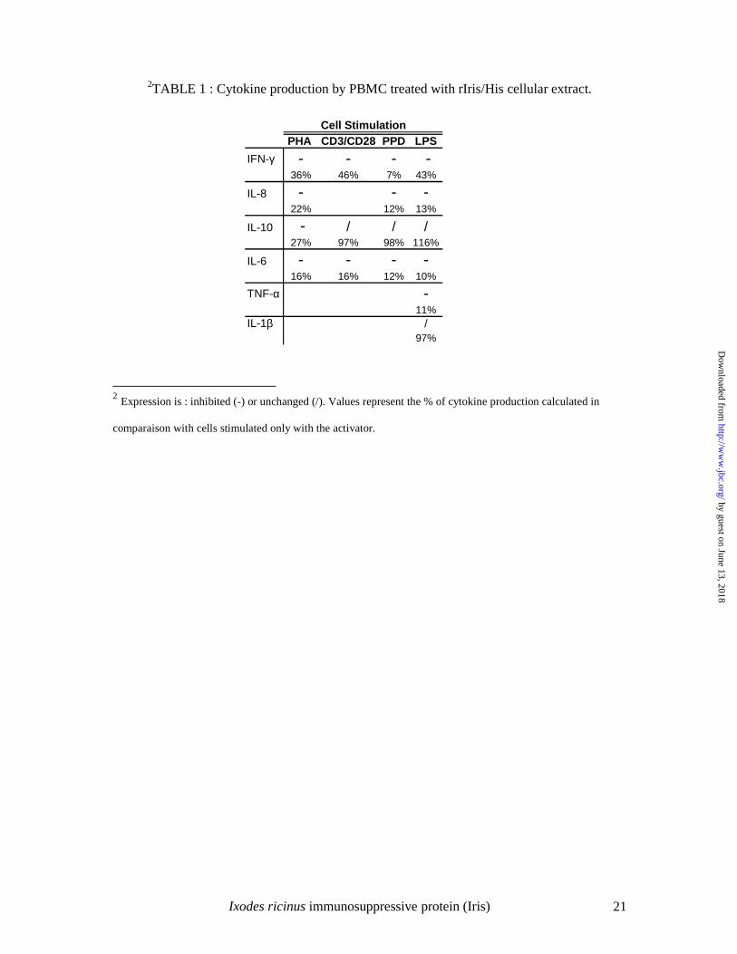
The effect of rIris/His cellular extract (used at a 1:5 dilution) on the production of cytokines
(IFN-γ, IL-8, IL-10, IL-6, TNF-α, and IL-1β) by PBMCs stimulated with a set of activators
(PHA, CD3/CD28, LPS and protein purified derivative - PPD) was also evaluated by ELISA
(Table 1).
The results indicated that the production of almost all tested cytokines (IFN-γ, IL-6, TNF-α,
and IL-8) was inhibited by the rIris/His cellular extract, except for the IL-1β production that
was unaffected after LPS stimulation (Table 1). rIris/His cellular extract inhibited IL-10
production by PMBCs after PHA stimulation, had no effect on PMBCs stimulated with
CD3/CD28, and LPS. The NEG cellular extract had no significant effect on the cytokine
production except after LPS stimulation that inhibited IL-10 production and enhanced IL-6
by guest on June 13, 2018http://w
ww
.jbc.org/D
ownloaded from
Ixodes ricinus immunosuppressive protein (Iris) 12
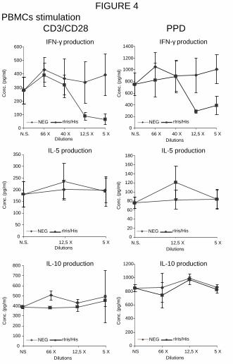
production. The dose-dependent effect of rIris/His was examined by analyzing the IFN-γ, IL-5
and IL-10 production under CD3/CD28 and PPD stimulation (Figure 4). In these cases, the
maximum inhibition of IFN-γ production by rIris/His was of ~65% (the difference between
rIris/His and NEG cellular extracts were statistically significant : P<0.01) and ~75% (P<0.05)
after CD3/CD28 and PPD stimulation, respectively. This inhibition was still effective at a
1:12.5 dilution. In contrast, no difference in the production of IL-5 and IL-10 was observed
between PBMCs incubated with rIris/His and NEG cellular extracts.
Fig. 4 inserted here
To confirm the role of Iris in the modulation of the production of some cytokines, PBMCs
stimulated by CD3/CD28 activator were incubated with various dilutions (from 1:250 to
1:4,000) of anti-rIris/MBP serum (Figure 5A). It was observed that PBMCs treated with
rIris/His cellular extract (at dilution 1:12.5) in the presence of anti-rIris/MBP serum restored
the IFN-γ production in a dose-dependent manner (Figure 5A); while this antiserum itself had
no immuno-stimulating effect on cytokine production by PBMCs (data not shown). At
dilution 1:250, the antiserum re-established the IFN-γ production to a level similar to that
obtained by CD3/CD28-stimulated PBMCs without rIris/His. To assert the specificity of the
neutralizing activity of the anti-rIris/MBP serum, its effect was measured on the activity of
cyclosporine-A (CsA) (Figure 5B), an immunosuppressive drug. The effect of a serum
specific to an unrelated MBP fusion protein was also measured on rIris/His cellular extracts
activity (Figure 5A). The anti-rIris/MBP serum (at a 1:250 dilution) did not affect the activity
of 400 nM CsA, and the unrelated antiserum had no effect on the rIris/His
immunomodulatory activity.
Fig. 5 inserted here
Finally, a small amount of rIris/His was purified from the rIris/His CHO-K1 cellular extract.
This purified rIris/His protein, at a ~25 nM concentration, completely inhibited the IFN-γ
by guest on June 13, 2018http://w
ww
.jbc.org/D
ownloaded from
Ixodes ricinus immunosuppressive protein (Iris) 13
production by CD3/CD28-stimulated PBMCs (Figure 5C), which was partially restored (50 %
of the IFN-γ production by CD3/CD28-stimulated PBMCs) by using anti-rIris/MBP serum (at
1:250 dilution). Interestingly, it was found that the level of inhibition of ~25 nM of purified
rIris/His was comparable to that of 400 nM CsA. The incubation of CD3/CD28-stimulated
PBMCs with either purified NEG and anti-rIris/MBP serum (at 1:250 dilution) or purified
NEG alone had no influence on the IFN-γ production.
by guest on June 13, 2018http://w
ww
.jbc.org/D
ownloaded from
Ixodes ricinus immunosuppressive protein (Iris) 14
DISCUSSION
It is now well established that the modulation of host immunity by tick saliva is of major
importance both in the successful accomplishment of the blood meal and in the transmission
of tick-borne pathogens such as Borrelia burgdorferi, the causal agent of Lyme disease (16).
Although extensive information is available on the effects of tick feeding on host immune
defenses, little is known about the nature of the immunomodulatory molecules expressed by
tick salivary glands. Tick salivary gland extracts (SGE) modulates host immune response by
modifying both lymphocyte and monocyte activities. For example, T lymphocyte proliferation
in response to mitogens (17) and the production of Th1 cytokines as IFN-γ and IL-2 are
inhibited by SGE (18). Moreover, Th2 type cytokine production such as IL-10, IL-5 and IL-4
is in general slightly enhanced (4, 5). Tick SGE also inhibits the production of several
cytokines (IFN-γ, IL-8, IL-6, TNF-α, …) by human macrophages (19). Studies indicated that
some of those immunomodulating phenomena are induced by proteins (20), (21).
In this work, we have characterized the properties of a protein induced during the tick feeding
process, which is called Iris for “I. ricinus immunosuppressor”, due to its exceptional immune
properties on leukocytes.
The corresponding mRNA sequence was first recovered by analyzing a RDA subtractive
library, and by using the RACE method (10). In this report, we show by Western blot and
confocal microscopy using a specific antiserum that Iris is not only specifically expressed
during the blood meal in female I. ricinus salivary glands but is also present in saliva with an
increasing expression from day 3 to day 5 of engorgement. In order to determine the
immunomodulatory properties of Iris, we studied the effect of the corresponding recombinant
protein (rIris/His) on naive Balb/c spleen cells proliferation, and on human PBMCs cytokine
production, using specific T-lymphocytes (PHA, ConA, CD3/CD28 and PMA/CD28),
macrophages (LPS) and APC (PPD) activators. The results indicated that rIris/His cellular
by guest on June 13, 2018http://w
ww
.jbc.org/D
ownloaded from
Ixodes ricinus immunosuppressive protein (Iris) 15
extracts inhibited on a dose dependent manner the proliferation of murine lymphocytes.
ELISA and ELISPOT assays showed that the rIris/His cellular extracts suppressed the
production of IFN-γ by human T cell and APC, while IL-5 and IL-10 levels remained
unchanged or were inhibited. Indeed, rIris/His cellular extract inhibited IL-10 production by
T-lymphocytes stimulated with PHA, but had no effect on CD3/CD28 stimulated cells. In
contrast, rIris/His extract enhanced the number of macrophages producing
IL-10 (see Figure 3) and had no effect on the expression of this cytokine by macrophages
(Table 1). This difference could be explained by the low level of IL-10 detected by ELISA,
the high sensitivity (up to 200 times) of the ELISPOT technique and the variety in shape and
size of the spots observed with ELISPOT. Nevertheless, , these results suggest Iris does not
inhibit the IL-10 production by macrophages. It was also shown that the expression of the
pro-inflammatory cytokine IL-6 was inhibited by macrophages, T cell, and APC. In addition,
the production of TNF-α by macrophages was inhibited while IL-1β expression remained
unaffected. Furthermore, by neutralizing completely rIris/His cellular extract activity with a
specific anti-rIris serum, and by showing that purified rIris/His protein inhibited IFN-γ
production by T cell, we clearly established that the recombinant protein was responsible for
the immunomodulation. Importantly, the inhibitory effect of ~25 nM rIris/His on IFN-γ
production (inhibition of 94%) is comparable to 400 nM CsA activity (inhibition of 99%).
This inhibition of IFN-γ production suggests that the T cell proliferation is also inhibited. This
hypothesis was confirmed by showing that rIris/His protein inhibited murine T cell
proliferation. These observations indicate that Iris is a novel immunosuppressor induced in
the salivary glands of engorged ticks during the blood meal. It suppresses T lymphocytes
proliferation, and induces a Th2 type immune response that is characterized by the inhibition
of IFNγ production, an unaffected expression of IL-5 and IL-10 by T lymphocytes and APC.
Moreover, IL-10 production by macrophages is not inhibited by Iris, confirming the pro-Th2
by guest on June 13, 2018http://w
ww
.jbc.org/D
ownloaded from
Ixodes ricinus immunosuppressive protein (Iris) 16
activity of Iris. In addition, Iris modulates the mechanisms of innate immunity by inhibiting
the production of pro-inflammatory cytokines (IL-6 and TNF-α).
It is known that several immunomodulatory factors are secreted in saliva at various times of
the feeding process. Indeed, SGE, prepared daily from engorging Dermacentor andersoni,
suppressed IL-1 secretion by macrophages only from day 0 to day 5 of engorgement while
TNF-α production was suppressed during the entire blood meal by these cells (18). Except for
the fact that IL-1 expression is not inhibited, the modulating effect of Iris on T cell and
macrophage responsiveness seems to mimic the SGE properties, suggesting that Iris is an
important salivary factor in the modulation of the local host immune response.
Finally, it is known that IL-4 produced following I. ricinus tick bites strongly influences the
immune response against B. burgdorferi (22). Moreover, Th1 cytokines are down regulated
during the initial spirochete transmission period in mice infested with Ixodes scapularis
infected ticks (23). IFN-γ, TNF-α and IL-2 given at the time of tick feeding suppress
spirochetes transmission by I. scapularis (16). In addition, it has been recently shown that this
Th2 type immune response makes possible the transmission of the human granulocytic
ehrlichiosis agent (24). Consequently, based on its immune properties, Iris could be a key
factor facilitating the transmission of a large number of tick borne pathogens.
ACKNOWLEDGEMENTS
We thank Fabienne Millecamp, Sarah Rorive, Cécile Dubois and Louis Delhaye, for
providing invaluable help in the realization of this work. We are also thankful to Dr Lise Gern
(Institut de Zoologie, Neuchâtel, Switzerland) for her critical analysis of this manuscript. G.L.
is supported by grants from the FRIA (Fonds de la Recherche Industrielle et Agricole) and
from the “Fondation Alice et David Van Buuren”. This work was supported by an
“International Brachet Stiftung” grant (GR97-1/6).
by guest on June 13, 2018http://w
ww
.jbc.org/D
ownloaded from

Ixodes ricinus immunosuppressive protein (Iris) 17
REFERENCES
1. Sauer, J. R., McSwain, J. L., Bowman, A. S., and Essenberg, R. C. (1995)
Annu.Rev.Entomol. 40, 245-267
2. Brossard, M. and Wikel, S. K. (1997) Med.Vet.Entomol. 11, 270-276
3. Allen, J. R. (1973) Int.J.Parasitol. 3, 195-200
4. Ganapamo, F., Rutti, B., and Brossard, M. (1995) Immunology 85, 120-124
5. Ganapamo, F., Rutti, B., and Brossard, M. (1996) Immunology 87, 259-263
6. Kopecky, J. and Kuthejlova, M. (1998) Parasite Immunol. 20, 169-174
7. Wang, H. and Nuttall, P. A. (1994) Parasitology 109 ( Pt 4), 517-523
8. Bergman, D. K., Ramachandra, R. N., and Wikel, S. K. (1998) J.Med.Entomol. 35, 505-
509
9. Valenzuela, J. G., Charlab, R., Mather, T. N., and Ribeiro, J. M. (2000) J.Biol.Chem.
275, 18717-18723
10. Leboulle, G., Rochez, C., Louahed, J., Rutti, B., Brossard, M., Bollen, A., and Godfroid,
E. (2001) Am.J.Trop.Med.Hyg.
11. Jung, H. I., Kim, S. I., Ha, K. S., Joe, C. O., and Kang, K. W. (1995) J.Biol.Chem. 270,
13879-13884
12. Remold-O'Donnell, E., Nixon, J. C., and Rose, R. M. (1989) J.Exp.Med. 169, 1071-
1086
by guest on June 13, 2018http://w
ww
.jbc.org/D
ownloaded from

Ixodes ricinus immunosuppressive protein (Iris) 18
13. Yang, D., Nakada-Tsukui, K., Ohtani, M., Goto, R., Yoshimura, T., Kobayashi, Y., and
Watanabe, N. (2001) J.Biochem.(Tokyo) 129, 561-568
14. Hubank, M. and Schatz, D. G. (1994) Nucleic Acids Res. 22, 5640-5648
15. Frohman, B. H. (1995) Rapid amplification of cDNA ends. In Dieffenbach C.W. and
Dveksler G.S., editors. PCR primer a laboratory manual, Cold Spring Harbor
Laboratory Press, New York
16. Zeidner, N., Dreitz, M., Belasco, D., and Fish, D. (1996) J.Infect.Dis. 173, 187-195
17. Wikel, S. K. (1982) Ann.Trop.Med.Parasitol. 76, 627-632
18. Ramachandra, R. N. and Wikel, S. K. (1992) J.Med.Entomol. 29, 818-826
19. Fuchsberger, N., Kita, M., Hajnicka, V., Imanishi, J., Labuda, M., and Nuttall, P. A.
(1995) Exp.Appl.Acarol. 19, 671-676
20. Urioste, S., Hall, L. R., Telford, S. R., III, and Titus, R. G. (1994) J.Exp.Med. 180,
1077-1085
21. Mejri, N., Franscini, N., Rutti, B., and Brossard, M. (2001) Parasite Immunol. 23, 61-69
22. Christe, M., Rutti, B., and Brossard, M. (2000) Parasitol.Res. 86, 491-496
23. Zeidner, N., Mbow, M. L., Dolan, M., Massung, R., Baca, E., and Piesman, J. (1997)
Infect.Immun. 65, 3100-3106
24. Zeidner, N. S., Dolan, M. C., Massung, R., Piesman, J., and Fish, D. (2000) Parasite
Immunol. 22, 581-588
by guest on June 13, 2018http://w
ww
.jbc.org/D
ownloaded from

Ixodes ricinus immunosuppressive protein (Iris) 19
LEGENDS TO FIGURES
Figure 1 Confocal microscopy of female I. ricinus salivary glands. Negative control
corresponding to 5 day fed tick salivary glands incubated only with the secondary antibody
(A). Unfed (B), 3 day (C) and 5 day (D) fed tick salivary glands incubated with anti-
rIris/MBP serum.
Figure 2 Balb/c spleen cells stimulated with ConA. SC were incubated only with ConA
(N.S.) or with different dilutions of rIris/His and NEG cellular extracts. Tritiated thymidine
incorporation was determined by liquid scintillation counting (10-³ c.p.m. +/- S.D.).
Figure 3 IFN-γ and IL-10 ELISPOT of human PBMCs. The number of activated cells
producing the cytokines upon treatment with PHA or LPS was evaluated (spots/106 cells +/-
S.D.). Activated cells were counted after treatment with rIris/His or NEG cellular extracts. A
positive control was realised by stimulating the cells only with the activator (PHA or LPS).
Figure 4 IFN-γ and IL-5 production by human PBMCs. Cells were incubated only with the
activator (N.S.) or with various dilutions of rIris/His and NEG cellular extracts. The
production of the cytokines (Conc. - pg/ml +/- S.D.) was evaluated by ELISA after treatment
with CD3/CD28 or PPD.
Figure 5 IFN-γ production by human PBMCs stimulated with CD3/CD28. The cells were
stimulated with NEG and rIris/His cellular extracts at a 1:12.5 dilution. All the assays were
realised by stimulating the cells: only with CD3/CD28 (P.C.), with CD3/CD28 in the presence
of NEG cellular extract (NEG), or with CD3/CD28 in the presence of rIris/His cellular extract
(rIris/His). A. CD3/CD28 stimulated cells were incubated with rIris/His cellular extract in the
by guest on June 13, 2018http://w
ww
.jbc.org/D
ownloaded from

Ixodes ricinus immunosuppressive protein (Iris) 20
presence of various dilutions of either anti-rIris/MBP serum (Anti-rIris/MBP+rIris/His) or a
non-specific serum (ns Ab+rIris/His). B. CD3/CD28-stimulated cells were incubated with 400
nM CsA (CsA), with 400 nM CsA in the presence of anti-rIris/MBP serum (CsA + Anti-
rIris/MBP), or with 400 nM CsA and the non-specific serum (CsA + NS AB); both antisera
were used at a 1:250 dilution. C. CD3/CD28 stimulated PBMCs were also incubated with
purified NEG (pNEG) or rIris/His (pIris/His) protein, and with purified NEG or rIris/His
proteins in the presence of anti-rIris/MBP serum at a 1:250 dilution(pNEG + Anti-rIris/MBP
and pIris/His + Anti-rIris/MBP).
by guest on June 13, 2018http://w
ww
.jbc.org/D
ownloaded from
Ixodes ricinus immunosuppressive protein (Iris) 21
2TABLE 1 : Cytokine production by PBMC treated with rIris/His cellular extract.
2 Expression is : inhibited (-) or unchanged (/). Values represent the % of cytokine production calculated in
comparaison with cells stimulated only with the activator.
PHA CD3/CD28 PPD LPS
IFN-γ - - - -36% 46% 7% 43%
IL-8 - - -22% 12% 13%
IL-10 - / / /27% 97% 98% 116%
IL-6 - - - -16% 16% 12% 10%
TNF-α -11%
IL-1β /97%
Cell Stimulation
by guest on June 13, 2018http://w
ww
.jbc.org/D
ownloaded from
250 µm
FIGURE 1
A B
C D
by guest on June 13, 2018http://w
ww
.jbc.org/D
ownloaded from
Spleen Cells Prolif. - ConA
0
50
100
150
200
250
300
350
400
N.S. 50 X 25 X 12,5 X 6,25 X
Dilutions
Tri
tiu
m U
pta
ke (
10-e
3 c.
p.m
. +/-
S.D
.)
rIris/HisNEG
FIGURE 2
by guest on June 13, 2018http://w
ww
.jbc.org/D
ownloaded from
IFN-γ production
IFN-γ production
IL-10 production
IL-10 production
spot
s/10
e6 c
ells
+/-
S
.D.
spot
s/10
e6 c
ells
+/-
S
.D.
spot
s/10
e6 c
ells
+/-
S
.D.
spot
s/10
e6 c
ells
+/-
S
.D.
FIGURE 3
PHA stimulation of PBMCs
LPS stimulation of PBMCs
0
1000
2000
3000
4000
PHA PHA + NEG PHA + rIris/His0
200400
600800
1000
PHA PHA + NEG PHA + rIris/His
0
500
1000
1500
LPS LPS + NEG LPS + rIris/His
0500
10001500200025003000
LPS LPS + NEG LPS + rIris/His
by guest on June 13, 2018http://w
ww
.jbc.org/D
ownloaded from
0
100
200
300
400
500
600
N.S. 66 X 40 X 12,5 X 5 XDilutions
0
200
400
600
800
1000
1200
1400
N.S. 66 X 40 X 12,5 X 5 XDilutions
0
50
100
150
200
250
300
350
N.S. 12,5 X 5 XDilutions
0
20
40
60
80
100
120
140
160
180
N.S. 12,5 X 5 XDilutions
0
200
400
600
800
1000
1200
NS 66 X 12,5 X 5 XDilutions
0
100
200
300
400
500
600
700
800
NS 66 X 12,5 X 5 XDilutions
NEG rIris/HisNEG rIris/His
NEG rIris/HisNEG rIris/His
NEG rIris/HisNEG rIris/His
FIGURE 4PBMCs stimulation CD3/CD28 PPD
Con
c. (
pg/m
l)
Con
c. (
pg/m
l)
Con
c. (
pg/m
l)
Con
c. (
pg/m
l)
Con
c. (
pg/m
l)
Con
c. (
pg/m
l)
IFN-γ production IFN-γ production
IL-5 production IL-5 production
IL-10 production IL-10 production
by guest on June 13, 2018http://w
ww
.jbc.org/D
ownloaded from

A.
0100200300400500600700
P.C. NEG rIris/His 1:250 1:500 1:1000 1:2000 1:4000 1:250 1:500 1:1000 1:2000 1:4000
B.
P.C. NEG rIris/His CsA CsA +Anti-rIris/MBP
CsA +NS AB
600
500
400
300
200
100
0
Con
c. (
pg/m
l)C
onc.
(pg
/ml)
C.
0
100
200
300
400
500
600
P.C. NEG rIris/His pNEG pNEG +Anti-
rIris/MBP
pIris/His pIris/His +Anti-
rIris/MBP
Anti-rIris/MBP + rIris/His ns AB + rIris/His
FIGURE 5C
onc.
(pg
/ml)
by guest on June 13, 2018http://w
ww
.jbc.org/D
ownloaded from
and Edmond GodfroidGérard Leboulle, Mara Crippa, Yves Decrem, Naceur Mejri, Michel Brossard, Alex Bollen
ticksCharacterization of a novel salivary immunosuppressive protein from Ixodes ricinus
published online January 15, 2002J. Biol. Chem.
10.1074/jbc.M111391200Access the most updated version of this article at doi:
Alerts:
When a correction for this article is posted•
When this article is cited•
to choose from all of JBC's e-mail alertsClick here
by guest on June 13, 2018http://w
ww
.jbc.org/D
ownloaded from