characterization of a squalene synthase from the ... open reading frame of the gene is 1,164 bp in...
TRANSCRIPT
J. Microbiol. Biotechnol. (2013), 23(6), 759–765http://dx.doi.org/10.4014/jmb.1212.12023First published online April 12, 2013pISSN 1017-7825 eISSN 1738-8872
Characterization of a Squalene Synthase from the Thraustochytrid MicroalgaAurantiochytrium sp. KRS101
Hong, Won-Kyung1, Sun-Yeon Heo
1, Hye-Mi Park
1, Chul Ho Kim
1, Jung-Hoon Sohn
2, Akihiko Kondo
3,
and Jeong-Woo Seo1*
1Applied Microbiology Research Center, Bio-Materials Research Institute, Korea Research Institute of Bioscience andBiotechnology (KRIBB), Jeonbuk 580-185, Korea2Systems and Synthetic Biology Research Center, Korea Research Institute of Bioscience and Biotechnology (KRIBB), Daejeon 305-333, Korea3Department of Chemical Science and Engineering, Graduate School of Engineering, Kobe University, Kobe 657-8501, Japan
Received: December 10, 2012 / Revised: February 27, 2013 / Accepted: February 28, 2013
The gene encoding squalene synthase (SQS) of the lipid-
producing heterotrophic microalga Aurantiochytrium sp.
KRS101 was cloned and characterized. The krsSQS gene
is 1,551 bp in length and has two exons and one intron.
The open reading frame of the gene is 1,164 bp in length,
yielding a polypeptide of 387 predicted amino acid residues
with a molecular mass of 42.7 kDa. The deduced krsSQS
sequence shares at least four conserved regions known to
be required for SQS enzymatic activity in other species.
The protein, tagged with His6, was expressed into soluble
form in Escherichia coli. The purified protein catalyzed
the conversion of farnesyl diphosphate to squalene in
the presence of NADPH and Mg2+
. This is the first report
on the characterization of an SQS from a Thraustochytrid
microalga.
Key words: Aurantiochytrium, squalene synthase, gene
analysis, functional characterization
Squalene (2,6,10,15,19,23-hexamethyltetracosa-2,6,10,14,18,22-
hexaene) is a dehydrotriterpenic hydrocarbon (C30H50)
containing six double bonds. Squalene is a key precursor
of cholesterol, bile acids, and steroids in plants and
animals, and is also a potential antioxidant [9, 16] used in
the cosmetic industry as a moisturizing agent and an
emollient; it serves as a natural antioxidant protecting cells
from free radicals and reactive oxygen species. Squalene is
also used in the food industry [14]. Squalene has been
extensively investigated by the medical and pharmaceutical
sectors because many studies have shown that the chemical
effectively inhibits chemically induced colon, lung, and
skin cancers; has bactericidal and antifungal properties;
markedly increases both cellular and nonspecific immune
functions; and reduces serum cholesterol levels [7].
To date, the major commercial sources of squalene are
the liver oils of deep-sea sharks, and plant seeds [5]. The
consistent availability of shark liver oils is doubtful.
International concern for marine ecology has grown, and
seas are becoming contaminated with environmentally
derived organic pollutants and heavy metals. Squalene is
also found in the oils of plant seeds (e.g., olive oil [approx.
7 mg squalene/g] and the oil of Amaranthus seed [0-
5.65 mg/g]) [5]. Not only are the levels of squalene low
(1-61 mg/g), but local and seasonal variations in crop
production increase industrial costs. Although yeasts have
been investigated as a potential source of squalene, the
levels of the chemical are generally very low (<0.43 mg/g),
with the exception of Pseudozyma sp. (70.3 mg/g) [1, 6].
Microalgae are currently under intensive investigation
as potential sources of useful natural products, including
squalene [2]. The green microalga Botryococcus braunii has
been reported to produce squalene when cultured under
photoautotrophic conditions [13]. However, it is difficult to
achieve high biomass levels under such conditions owing to
light limitation. From an industrial viewpoint, a high cell
density is required if microbial production processes are to
be successfully commercialized. Microalgae that grow
heterotrophically do not require light and attain high cell
densities, and exhibit good productivities, when cultured
on commercial scales. Thraustochytrids are a group of
microalgae widely distributed in mangroves; the microbes
grow well in heterotrophic culture. Such microbes are
currently being used to produce polyunsaturated fatty
acids, and some species contain relatively high amounts of
*Corresponding authorPhone: +82-63-570-5160; Fax: +82-63-570-5109;E-mail: [email protected]
760 Hong et al.
squalene, which can be easily purified by counter-current
chromatography [5]. Thraustochytrid in the Aurantiochytrium
genus-heterotrophic microbes belonging to the Stramenopiles
is being investigated as a possible source of squalene [12].
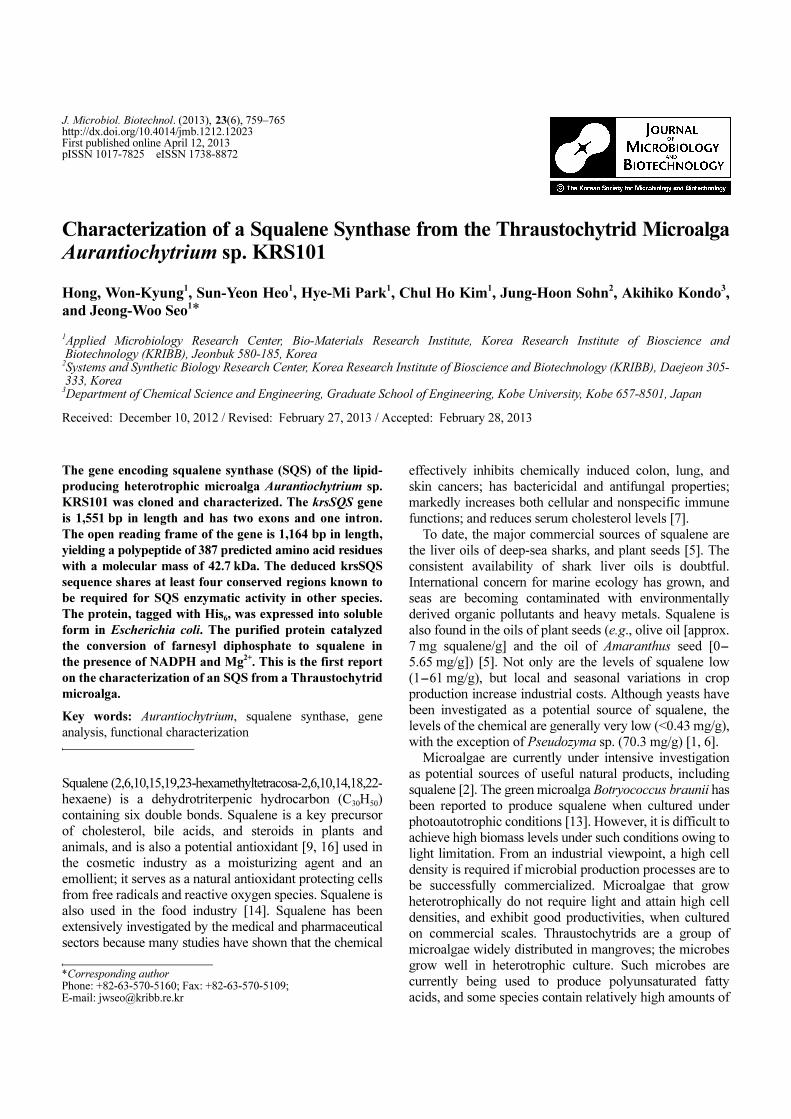
Squalene synthase (SQS, E.C. 2.5.1.21) is a membrane-
bound enzyme catalyzing the first dedicated step in the
biosynthesis of sterols and other triterpenoids, and SQS is
thought to play an important role in regulating isoprenoid
biosynthesis in eukaryotes. SQS is a bifunctional enzyme
initially catalyzing the condensation of two molecules of
farnesyl diphosphate (FPP) to form presqualene diphosphate
(PSPP), and next converting PSPP to squalene in a reaction
requiring NADPH and Mg2+ (Fig. 1) [17]. In bacteria, these
reactions are the first pathway-specific steps in hopanoid
biosynthesis; hopanoids are pentacyclic triterpene lipids of
bacterial membranes that exert stabilizing effects similar to
those characteristic of membrane sterols in eukaryotes [10].
As SQS is a key enzyme regulating isoprenoid biosynthesis,
the genes encoding the enzyme have been cloned from
bacteria [10], yeasts [11], Ganoderma lucidum, protozoa,
various animals, humans, and plants [15]. However, very
little is known regarding the SQS gene of the Thraustochytrid
Aurantiochytrium. In the present report, we describe the
cloning, gene organization, heterologous expression, and
functional analysis of SQS from the strain Aurantiochytrium
sp. KRS101 (krsSQS).
MATERIALS AND METHODS
Microalgal Strain and Growth Conditions
Details of the Thraustochytrid microalgal strain Aurantiochytrium
sp. KRS101 (KCTC 11686BP) have been reported previously [4]. The
microalga was cultivated in 500 ml baffled flasks, each containing
50 ml of basal medium [glucose, 60 g/l; yeast extract, 10 g/l; dried
natural sea salt (CJ Co., Seoul, Korea), 10 g/l] at 28oC, with shaking
at 125 rpm, for 2 days. Escherichia coli BL21 cells harboring plasmid
pET28a-krsSQS or pET-28a (control) were grown overnight at 37oC
in Luria-Bertani (LB) medium containing ampicillin (100 µg/ml).
Aliquots (500 µl) of overnight cultures were added to 50 ml of fresh
LB medium supplemented with 100 µg/ml ampicillin. When the OD600
values reached 0.5, recombinant gene expression was induced by
addition of isopropylthio-β-galactoside (IPTG) to a final concentration
of 1 mM; growth was continued further for 16 h at 23oC.
Cloning of the Aurantiochytrium sp. KRS101 krsSQS Gene and
Synthesis of cDNA
Whole genome of Aurantiochytrium sp. KRS101 was read with
16.9X coverage by Hiseq, which resulted in a draft whole genome
sequence. The Hiseq reads were aligned to reference genome using
BWA’s short read aligner at default settings and the SAM (Sequence
Alignment/Map) generic format tool for storing the nucleotide sequence
alignment (Macrogen, Seoul, Korea). The whole genome sequencing
allowed alignment of the deduced sequence of Aurantiochytrium sp.
KRS101 genes. To identify the krsSQS gene of Aurantiochytrium sp.
KRS101, we examined the whole-genome sequence database and
identified a candidate gene of 1,551 bp in length, with two exons and
one intron. The krsSQS gene (GenBank Accession No. JX684107)
has an open reading frame of 1,164 bp, encoding a polypeptide of
387 predicted amino acid residues, and our clone (please see below)
carries an artificially introduced terminal NcoI in the N-terminus and
BamHI restriction enzyme sites in the C-terminus.
Genomic DNA was extracted using a “mini-prep” kit (Invitrogen,
Carlsbad, CA, USA) and total RNA was isolated with the aid of the
Isol-RNA Lysis Reagent (5Prime, Gaithersburg, MD, USA). Single-
stranded cDNA was synthesized by reverse transcription of total
RNA (at 25oC for 5 min in nuclease-free water) using an iScript
cDNA synthesis kit (Bio-Rad, Hercules, CA, USA). Genomic DNA
containing the krsSQS gene was amplified using the forward primer
P1 (5'-GCTAGCATGCCTAACAAGCCT-3') and the reverse primer
P2 (5'-GGATCCGTCAGAGTGGGTTTGGC-3'). Each 50 µl PCR
mixture contained 5 µl of 10× PCR buffer, 2.5 mM of each
deoxyribonucleotide triphosphate, 0.5 µM of either primer, 2 U Taq
polymerase (Takara, Otsu, Japan), and 1 µg of genomic DNA. The
PCR protocol featured 1 min of denaturation at 94oC; followed by
25 cycles each of 0.5 min at 94oC, 0.5 min at 55
oC, and 1.5 min at
72oC. The final extension was performed over 7 min at 72
oC. PCR
products were visualized by agarose gel electrophoresis.
Comparison of krsSQS Protein Sequences and Phylogenetic
Analysis
The krsSQS protein sequences of representative species, including
Aurantiochytrium sp. KRS101 (the microalga), Aphanomyces euteiches
Fig. 1. The squalene biosynthesis pathway. A simplified version of the steroid synthesis pathway with the intermediates
isopentenyl pyrophosphate (IPP), dimethylallyl pyrophosphate (DMAPP),
geranyl pyrophosphate (GPP), and squalene shown. Some intermediates
are omitted.

SQUALENE SYNTHASE FROM AURANTIOCHYTRIUM SP. KRS101 761
(GenBank Accession No. CAQ55982), Phaeodactylum tricornutum
CCAP 1055 (GenBank Accession No. XP_002180940), Saccharomyces
cerevisiae (GenBank Accession No. ACD03847), Candida albicans
WO-1 (GenBank Accession No. BAA13995), and Ogataea
parapolymorpha DL-1 (GenBank Accession No. EFW95406), were
obtained from the NCBI protein sequence database and aligned
using MultiAlign ClustalW 2.0 of EMBL-EBI.
Expression of Aurantiochytrium sp. KRS101 krsSQS in E. coli
cDNA encoding krsSQS was excised from the pGEM-T-Easy vector
using NcoI and BamHI; the eluted DNA fragment was subcloned
between the NcoI and BamHI sites of the cloning cassette region of
pET-28a (Novagen, Darmstadt, Germany); the recombinant plasmid
thus obtained (pET-krsSQS) was transformed into E. coli BL21.
Cells with either pET-krsSQS or pET-28a (control) were cultivated
at 37oC in LB medium (100 µg/ml kanamycin) to the OD600 value
of 0.5, gene expression was then induced by addition of IPTG to a
final concentration of 1 mM, and growth was further continued for
16 h at 23oC. The recombinant protein was purified using a His-Tag
Ni-NTA spin column (Qiagen, Hilden, Germany) according to the
manufacturer’s instructions. A non-denaturing buffer [50 mM NaH2PO4,
300 mM NaCl, 0.05% (v/v) Tween 20; pH 8.0] with a gradient of
10-250 mM imidazole was used for elution.
SDS-PAGE and Western Blotting
The recombinant bacterial cells were collected by centrifugation at
10,000 ×g for 5 min and washed with 50 mM phosphate buffer, pH
7.2. Each bacterial pellet was resuspended in phosphate buffer and
sonicated for 5 min (each pulse was 10 s in duration followed by
10 s on ice). Supernatants were obtained by centrifugation at 10,000
×g for 15 min; protein concentrations were measured using the BCA
protein assay kit (Pierce, Rockford, IL, USA) and proteins were
subjected to SDS-PAGE. Next, separated proteins were transferred
to nitrocellulose membranes with the aid of a tank transfer unit
(Hoefer, Holliston, MA, USA) at 100 V and 10oC for 1.5 h.
Membranes were blocked with 5% (w/v) skim milk powder in TBS
for 1 h at room temperature and next incubated overnight at 4oC
with a 1/1,000 dilution of mouse monoclonal anti-His-Tag antibody
(Novagen) in wash buffer [TBS with 0.1% (v/v) Tween 20].
Unbound primary antibody was removed by washing three times
(each wash duration was 10 min) in wash buffer, and each blot was
next incubated for 2 h at room temperature in a 1/5,000 dilution of
anti-mouse Ig-conjugate (Promega, Madison, WI, USA) in wash
buffer. Unbound secondary antibody was removed by washing three
times (each wash duration was 10 min) in wash buffer. The membranes
were incubated with a reagent (Pierce, Rockford, IL, USA) that
caused bound secondary antibody to emit chemiluminescence, and
protein bands were visualized using a detection kit (Novagen)
following the instructions of the manufacturer.
Activity of the krsSQS of Aurantiochytrium sp. KRS101 and
Analysis of Fatty Acid Composition
The krsSQS activity of the purified recombinant protein was measured
using an assay based on conversion of FPP to squalene in the
presence of 5 mM NADPH and 25 mM Mg2+. Following incubation
with purified protein (or lysate) at 37oC for 6 h, reaction mixtures
were extracted two times with 500 µl amounts of n-hexane. These
solutions were concentrated overnight at room temperature to final
volumes of about 60µl. Squalene was detected by gas chromatography-
mass spectrometry (GC-MS). An Agilent GC-MS-7890A/5975C
(Agilent, Santa Clara, CA, USA) as fitted with a J&W HP 5ms
column (Agilent P/N 19091S-433; 0.25 mm × 930 m; film thickness
of 0.25 mm) that was subjected to helium carrier flow at 1.2 ml/min,
and that was operated over the scan range m/z 20-550. Each sample
volume was 1 µl and the split ratio was 10:1. The injector temperature
was 250oC. The column temperature was maintained at 120oC for
3 min, next elevated to 180oC at 15
oC per minute, and next to 260
oC
at 25oC per minute; the column was maintained at that temperature
for 25 min. Authentic squalene (Sigma, St. Louis, MO, USA) served
as a standard. Dried cells were resuspended in 3 ml of 4% (v/v)
methanolic sulfuric acid and heated at 90oC for 1 h in sealed vials.
Fatty acid methyl esters (FAMEs) were extracted into 0.3 ml of
hexane and analyzed via gas chromatography (Hewlett Packard
6890N; Ramsey, MN, USA); the instrument was equipped with a
flame-ionization detector and an HP-5 (30 m × 0.32 mm; 0.25 mm;
Agilent Technologies, Santa Clara, CA, USA). The column temperature
was raised from 150oC (after 2 min of holding) to 270
oC (with a
further 2 min of holding) at a rate of 7oC per minute.
Dry Cell Weight
Dry cell weight (DCW) was estimated by harvesting cells at 4,500
×g at 4oC for 20 min. Each supernatant was discarded and each
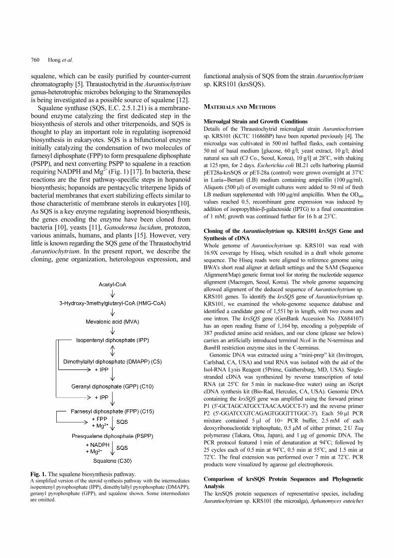
Fig. 2. Structure of the krsSQS gene of Aurantiochytrium sp. KRS101. Exon/intron boundaries are shown in italics; the splice sites are between the TG and TT doublets (nts 2–3 and 4–5, respectively) and the TG and CC doublets
(nts 389–390 and 391–392, respectively).

762 Hong et al.
Fig. 3. Alignment of the amino acid sequence of the krsSQS of Aurantiochytrium sp. KRS101 with those of other SQSs. At least four very similar regions are evident. Six sequences of SQSs of different species were retrieved from GenBank. The accession numbers are
indicated. EFW95406, Ogataea parapolymorpha; ACD03847, Saccaromyces cerevisiae; CAQ55982, Aphanomyces euteiches. XP_002180940,
Phaeodactylum tricornutum; JX684107, Aurantiochytrium krs101; ABE97915, Aurantiochytrium limacinum.

SQUALENE SYNTHASE FROM AURANTIOCHYTRIUM SP. KRS101 763
pellet washed three times with phosphate-buffered saline (PBS, pH
7.2). Resuspended cells were again harvested by centrifugation at
4,500 ×g at 4oC for 20 min. Each pellet was resuspended in 600 µl
of distilled water and transferred to a pre-weighed vial. Cell pellets
were dried at 60oC for 12 h using a speed vacuum concentrator
(Biotron 4080C; Bucheon, Korea). Each vial was weighed and the
DCW value obtained.
RESULTS
Identification of the krsSQS of Aurantiochytrium sp.
KRS101
The whole-genome sequence database indicated that the
SQS gene of Aurantiochytrium sp. KRS101 was 1,551 bp
in length, and included two exons and one intron. The SQS
protein was synthesized from an open reading frame
1,164 bp in length, yielding a polypeptide with a predicted
387 amino acid residues, a predicted MW of 42.7 kDa, and
a predicted pI value of pH 5.03. RT-PCR was employed to
isolate the open reading frame of krsSQS cDNA (Fig. 4).
Comparison of the full gene sequence with that of the
putative coding region indicated that the gene had two
exons and one intron. It is noteworthy that the krsSQS gene
contains an intron; the splice sites lie between TG and TT
(nts 2-3 and 4-5, respectively) and TG and CC (nts 389-
390 and 391-392, respectively) (Fig. 2).
Molecular Characterization of the krsSQS of
Aurantiochytrium sp. KRS101
An NCBI BLAST search revealed that the deduced krsSQS
sequence of Aurantiochytrium sp. KRS101 is 99.2% identical
to that of a hypothetical protein of Aurantiochytrium
limacinum, but is much less similar to krsSQS proteins of
other microalgae, yeast, and fungal species, including S.
cerevisiae (28%), Candida albicans (33%), and Ogataea
parapolymorpha (33%) (Fig. 3). Alignment of the
Aurantiochytrium krsSQS with those of other species
revealed at least four regions that are highly conserved in
microalgae, yeast, and fungi. Among these species, Regions
II, III, and IV are nearly identical.
Region I is relatively well conserved, although only a
partial sequence from Aphanomyces euteiches is available.
This region harbors a highly conserved aspartate-rich
motif (DXXXD) (24DTVED28 in our enzyme). Region III
is also very highly conserved among the superfamily of
isoprenoid biosynthesis enzymes (class 1), as revealed by
an NCBI BLAST protein search. Region III also contains
an aspartate-rich motif (193DYLED197). The two aspartate-
rich regions mediate binding of prenyl phosphates, and
contain Mg2+-binding sites and substrate-binding pockets.
Region II is also conserved in the class 1 superfamily of
enzymes involved in isoprenoid biosynthesis. Region IV
harbors motifs responsible for catalysis of the second
reaction; the likely NADPH-binding motifs are shown in
bold in the Aurantiochytrium sequence from this region:
FCALPQVMAIATLSVCFDNQKVFQGVVKIRKG. In
Zhao et al. [17], this motif was essential for performance
of the second enzymatic activity. C-Terminal SSQ amino
acid sequences vary greatly among microalgal, yeast, and
fungal proteins. A peroxisome-targeting signal 1 (PTS1)
motif is evident; this is a tripeptide with the consensus
sequence (S/A/C)-(K/R/H)-(L/A). The most common PTS1
Fig. 4. RT-PCR of krsSQS. M, marker; 1, Aurantiochytrium KRS101 cDNA; 2, Aurantiochytrium
KRS101 gDNA; 3, No template.
Fig. 5. SDS-PAGE (A and C) and Western blotting (B) of Aurantiochytrium sp. KRS101 krsSQS expressed in E. coli. M, protein markers; SS, soluble SQS (BL21/pET-krsSQS); SC, soluble control (BL21/pET-28a); IS, insoluble SQS (BL21/pET-krsSQS); IC, insoluble
control (BL21/pET-28a); PS, purified recombinant SQS.
764 Hong et al.
is serine-lysine-leucine (SKL). However, a peroxisome-
targeting signal of type 2 (PTS2) is a head region nonapeptide
with the consensus sequence (R/K)-(L/V/I)-XXXXX-(H/
Q)-(L/A/F) (where X is any amino acid); such a sequence
may be found in the RIMLDMSPMEHPTIFELQQNYLG
FVLPPLTAAFGKIIKVVDSKLQCLQESGRPLGQTHSD
(the residues of interest are shown in bold) run of the
krsSQS C-terminal region. This sequence is hydrophobic,
and some reports have indicated that the sequence may
function to anchor the protein to the membrane [3, 8, 11].
Heterologous Expression of Aurantiochytrium sp. KRS101
krsSQS in E. coli
E. coli BL21 cells harboring plasmid pET-kraSQS or pET-
28a (control) were cultured as described in Materials and
Methods. Both the protein mix including recombinant
krsSQS protein (prepared from BL21/pET-krsSQS) and a
control pET-28a protein mix were subjected to SDS-PAGE
and Western blotting (Figs. 5A and 5B). krsSQS was not
expressed in the cytosol of the recombinant but was
evident in the insoluble fraction. Western blotting showed
that the protein size (thus including the His-tag) was
correct. No such band was observed in the control strain.
Western immunoblotting showed that both soluble and
insoluble recombinant krsSQSs specifically bound with
anti-His-tag antibody, although the insoluble recombinant
krsSQS expressed very low level. The recombinant protein
was purified on an Ni-NTA affinity spin column under
non-denaturing conditions, and the purified protein with
approximately 43.3 kDa was detected (Fig. 5C).
Functional Characterization of the krsSQS of
Aurantiochytrium sp. KRS101
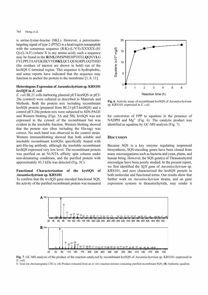
To confirm that the krsSQS gene encoded functional SQS,
the activity of the purified recombinant protein was measured
for conversion of FPP to squalene in the presence of
NADPH and Mg2+ (Fig. 6). The catalytic product was
identified as squalene by GC-MS analysis (Fig. 7).
DISCUSSION
Because SQS is a key enzyme regulating isoprenoid
biosynthesis, SQS-encoding genes have been cloned from
many microorganisms such as bacteria and yeast, plants, and
human being. However, the SQS gene(s) of Thraustochytrid
microalgae have been poorly studied. In the present report,
we first identified the SQS gene of Aurantiochytrium sp.
KRS101, and next characterized the krsSQS protein in
both molecular and functional terms. Our results show that
further work on Aurantiochytrium strains, and on gene
expression systems in thraustochytrids, may render it
Fig. 6. Activity assay of recombinant krsSQS of Aurantiochytriumsp. KRS101 expressed in E. coli.
Fig. 7. GC-MS analysis of the product of the reaction catalyzed by recombinant krsSQS of Aurantiochytrium sp. KRS101 expressed inE. coli. S: Total ion chromatograms (TICs). (A) Product extracted from an in vitro reaction mixture containing purified recombinant SQS; (B) Authentic squalene.

SQUALENE SYNTHASE FROM AURANTIOCHYTRIUM SP. KRS101 765
possible to use such strains for commercial production of
squalene.
The krsSQS gene of Aurantiochytrium sp. KRS101 has
one intron and two exons. A BLAST search revealed that
the deduced SQS sequence of Aurantiochytrium sp.
KRS101 is similar to those of other microalgae, yeast, and
fungi; four protein regions are highly conserved in all
krsSQS proteins. The SQS C-terminal region is hydrophobic
and may function to anchor the protein to the membrane.
Earlier reports suggested that deletion of the hydrophobic
C-terminal SQS region facilitated expression of soluble
protein [3]. However, the krsSQS protein was expressed to
some extent in the cytosol, although most protein was
insoluble. It is possible that addition of a His-tag to the C-
terminus of krsSQS facilitated expression of the protein in
a soluble form; histidine is a polar amino acid.
Recently, Thraustochytrid microalgae strains have been
reported to produce high levels of squalene [12]. In the
present report, we have gathered molecular and biochemical
information relevant to squalene production by such
microalgae. However, unfortunately, we could not detect
squalene production in the Aurantiochytrium strain, although
the active squalene synthase gene was expressed. Failure
in the observation of squalene production might be due to
production of compounds competing with fatty acids
biosynthesis for the common precursor acetyl-CoA. The
fatty acid production level is very high, being over 30%
(w/w) of the dried biomass in the microalgal strain. We
expect that a metabolic engineering strategy for increasing
the expression level of krsSQS along with decreasing the
fatty acid biosynthesis level could enhance squalene
production in the microalgal strain.
Acknowledgments
This work was supported by a National Research Foundation
of Korea (NRF) grant funded by the Korea government
(MEST) (No. 2011-0027171) and by the Ministry of Food,
Agriculture, Forestry, and Fisheries, of the Republic Korea.
REFERENCES
1. Chang, M. H., H. J. Kim, K. Y. Jahng, and S. C. Hong. 2008.
The isolation and characterization of Pseudozyma sp. JCC 207,
a novel producer of squalene. Appl. Microbiol. Biotechnol. 78:
963-972.
2. Chen, G., K. W. Fan, F. P. Lu, Q. Li, T. Aki, F. Chen, and Y.
Jiang. 2010. Optimization of nitrogen source for enhanced
production of squalene from thraustochytrid Aurantiochytrium
sp. N. Biotechnol. 27: 382-389.
3. Gupta, N., P. Sharma, R. J. Santosh Kumar, R. K. Vishwakarma,
and B. M. Khan. 2012. Functional characterization and differential
expression studies of squalene synthase from Withania somnifera.
Mol. Biol. Rep. 39: 8803-8812.
4. Hong, W. K., D. Rairakhwada, P. S. Seo, S. Y. Park, B. K. Hur,
C. H. Kim, and J. W. Seo. 2011. Production of lipids containing
high levels of docosahexaenoic acid by a newly isolated microalga,
Aurantiochytrium sp. KRS101. Appl. Biochem. Biotechnol. 164:
1468-1480.
5. Jiang, Y., K. W. Fan, R. T. Wong, and F. Chen. 2004. Fatty acid
composition and squalene content of the marine microalga
Schizochytrium mangrovei. J. Agric. Food Chem. 52: 1196-
1200.
6. Kaya, K., A. Nakazawa, H. Matsuura, D. Honda, I. Inouye, and
M. M. Watanabe. 2011. Thraustochytrid Aurantiochytrium sp.
18W-13a accummulates high amounts of squalene. Biosci.
Biotechnol. Biochem. 75: 2246-2248.
7. Kim, S. K. and F. Karadeniz. 2012. Biological importance and
applications of squalene and squalane. Adv. Food Nutr. Res. 65:
223-233.
8. Kim, T. D., J. Y. Han, G. H. Huh, and Y. E. Choi. 2011.
Expression and functional characterization of three squalene
synthase genes associated with saponin biosynthesis in Panax
ginseng. Plant Cell Physiol. 52: 125-137.
9. Ko, T. F., Y. M. Weng, and R. Y. Chiou. 2002. Squalene content
and antioxidant activity of Terminalia catappa leaves and seeds.
J. Agric. Food Chem. 50: 5343-5348.
10. Lee, S. and C. D. Poulter. 2008. Cloning, solubilization, and
characterization of squalene synthase from Thermosynechococcus
elongatus BP-1. J. Bacteriol. 190: 3808-3816.
11. LoGrasso, P. V., D. A. Soltis, and B. R. Boettcher. 1993.
Overexpression, purification, and kinetic characterization of a
carboxyl-terminal-truncated yeast squalene synthetase. Arch.
Biochem. Biophys. 307: 193-199.
12. Nakazawa, A., H. Matsuura, R. Kose, S. Kato, D. Honda, I.
Inouye, et al. 2012. Optimization of culture conditions of the
thraustochytrid Aurantiochytrium sp. strain 18W-13a for squalene
production. Bioresour. Technol. 109: 287-291.
13. Okada, S., T. P. Devarenne, and J. Chappell. 2000. Molecular
characterization of squalene synthase from the green microalga
Botryococcus braunii, race B. Arch. Biochem. 373: 307-317.
14. Tikekar, R. V., R. D. Ludescher, and M. V. Karwe. 2008.
Processing stability of squalene in amaranth and antioxidant
potential of amaranth extract. J. Agric. Food Chem. 56: 10675-
10678.
15. Uchida, H., H. Yamashita, M. Kajikawa, K. Ohyama, O.
Nakayachi, R. Sugiyama, et al. 2009. Cloning and characterization
of a squalene synthase gene from a petroleum plant, Euphorbia
tirucalli L. Planta 229: 1243-1252.
16. Wei, A. and T. Shibamoto. 2007. Antioxidant activities of
essential oil mixtures toward skin lipid squalene oxidized by
UV irradiation. Cutan. Ocul. Toxicol. 26: 227-233.
17. Zhao, R. Y., W. Xiao, H. L. Cheng, P. Zhu, and K. D. Cheng.
2010. Cloning and characterization of squalene synthase gene
from Fusarium fujikuroi (Saw.) Wr. J. Ind. Microbiol. Biotechnol.
37: 1171-1182.