characterization of chromosomal and megaplasmid ... · characterization of chromosomal and...
TRANSCRIPT

Li et al. BMC Genomics (2015) 16:317 DOI 10.1186/s12864-015-1523-3
RESEARCH ARTICLE Open Access
Characterization of chromosomal andmegaplasmid partitioning loci in Thermusthermophilus HB27Haijuan Li, Angel Angelov, Vu Thuy Trang Pham, Benedikt Leis and Wolfgang Liebl*
Abstract
Background: In low-copy-number plasmids, the partitioning loci (par) act to ensure proper plasmid segregationand copy number maintenance in the daughter cells. In many bacterial species, par gene homologues are encodedon the chromosome, but their function is much less understood. In the two-replicon, polyploid genome of thehyperthermophilic bacterium Thermus thermophilus, both the chromosome and the megaplasmid encode par genehomologues (parABc and parABm, respectively). The mode of partitioning of the two replicons and the role of thetwo Par systems in the replication, segregation and maintenance of the genome copies are completely unknown inthis organism.
Results: We generated a series of chromosomal and megaplasmid par mutants and sGFP reporter strains andanalyzed them with respect to DNA segregation defects, genome copy number and replication origin localization.We show that the two ParB proteins specifically bind their cognate centromere-like sequences parS, and that bothParB-parS complexes localize at the cell poles. Deletion of the chromosomal parAB genes did not apparently affectthe cell growth, the frequency of cells with aberrant nucleoids, or the chromosome and megaplasmid replication.In contrast, deletion of the megaplasmid parAB operon or of the parB gene was not possible, indicating essentialityof the megaplasmid-encoded Par system. A mutant expressing lower amounts of ParABm showed growth defects,a high frequency of cells with irregular nucleoids and a loss of a large portion of the megaplasmid. The truncatedmegaplasmid could not be partitioned appropriately, as interlinked megaplasmid molecules (catenenes) could bedetected, and the ParBm-parSm complexes in this mutant lost their polar localization.
Conclusions: We show that in T. thermophilus the chromosomal par locus is not required for either the chromosomalor megaplasmid bulk DNA replication and segregation. In contrast, the megaplasmid Par system of T. thermophilus isneeded for the proper replication and segregation of the megaplasmid, and is essential for its maintenance. The twoPar sets in T. thermophilus appear to function in a replicon-specific manner. To our knowledge, this is the first analysisof Par systems in a polyploid bacterium.
Keywords: Partitioning genes (par), Thermus thermophilus, Chromosome, Megaplasmid, ParB
BackgroundAll living cells have mechanisms ensuring the faithfulsegregation of the replicated genomes to the daughtercells. While the tubulin-based mitotic apparatus forDNA segregation used by eukaryotes is well studied, themechanisms that mediate chromosome segregation inprokaryotic cells are less well understood. Evidence from
* Correspondence: [email protected] für Mikrobiologie, Technische Universität München,Emil-Ramann-Straße 4, D-85354 Freising-Weihenstephan, Germany
© 2015 Li et al.; licensee BioMed Central. ThisAttribution License (http://creativecommons.oreproduction in any medium, provided the orDedication waiver (http://creativecommons.orunless otherwise stated.
several groups demonstrates that bacterial chromosomesare also actively segregated and this segregation does notrely on cell growth [1-4]. Also, it has been shown thatcytoskeletal proteins are also present in prokaryotic cellsand they form mitotic-like apparatuses that provideforce for active chromosome segregation [5,6].Several elements have been proposed which may make
contributions to the dynamic movement of bacterialchromosomes [5,7]. For example, it has been suggestedthat DNA polymerase can provide force for bidirectionalchromosome segregation in Bacillus subtilis cells [8,9].
is an Open Access article distributed under the terms of the Creative Commonsrg/licenses/by/4.0), which permits unrestricted use, distribution, andiginal work is properly credited. The Creative Commons Public Domaing/publicdomain/zero/1.0/) applies to the data made available in this article,

Li et al. BMC Genomics (2015) 16:317 Page 2 of 17
Likewise, RNA polymerase has also been implicated toafford both incentive force and directionality for segre-gation through interacting with origin-proximal regions[10,11]. MreB is a chromosomally encoded actin homo-log and in some rod-shaped bacteria it has been shownthat MreB not only determines the cell shape, but isalso involved in chromosome segregation [12-14].Partitioning (par) genes have been known for a long
time to play a pivotal role in the maintenance of low-copy-number plasmids. Plasmid par locus usually containthree components: two ORFs encoding an ATPase (ParA)and a DNA-binding protein (ParB), and a centromere-likespecific DNA sequence (parS). ParB binds its corre-sponding parS sequence, forming a large nucleoproteincomplex. Low-copy-number plasmids with disruptedpar loci localize improperly and are thus readily elimi-nated from host cells [15,16]. The molecular mecha-nisms by which par loci segregate plasmids have beenstudied to some extent. It has been suggested that ParAcan form filaments which interact with ParB-parS com-plexes and provide force for segregation [17-20].Many bacterial chromosomes encode orthologs of the
plasmid partitioning proteins (Par) near their origin re-gions [21]. The first ParB-binding chromosomal parSsites were discovered in B. subtilis, where 10 pseudopa-lindromic 16-bp sequences were identified in the 20%origin-proximal region of its chromosome. The pres-ence of merely one such site could prevent the loss ofan otherwise unstable plasmid from the host cell in aParAB-dependent manner [22,23]. The consensus forthe 16-bp sequence is 5′-TGTTNCACGTGAAACA-3′.Recently, this 16-bp sequence has been found in a largevariety of bacteria, and in most cases, these sequencesare origin-proximally located [24,25]. Normally, the cor-responding parAB genes can also be identified in suchparS-containing chromosomes. While the crucial roleof the par loci in plasmid partitioning has been wellstudied, their chromosomal counterparts are relativelypoor investigated and the role of these Par systems inchromosome segregation is still disputable. The avail-able data [26] support a model called diffusion ratchetmechanism where ParA uses the nucleoid as a matrixand ori-parS-ParB foci move by following a retractingParA cloud.Mutations introduced in chromosomal par loci usu-
ally have pleiotropic effects. In B. subtilis the chromo-somal parAB orthologs are not essential genes, butthey are involved in chromosome replication and seg-regation, chromosome origin localization and separ-ation, and developmental gene regulation [27-29]. TheparAB genes in Caulobacter crescentus on the otherhand are essential and their depletion or overexpres-sion results in defects in cell-cycle progression, celldivision and chromosome segregation [30,31]. Each of
the two chromosomes (chrI and chrII) of Vibrio cho-lerae contains a par locus (parABS1 and parABS2). Ithas been shown that parABS1 is probably involved inthe segregation of the origin regions of chrI, but not ofthe bulk DNA of chrI or chrII [32,33]. In contrast,parABS2 can promote accurate subcellular localizationand maintenance of the bulk DNA of chrII but not ofchrI [34].Although these diverse functions of chromosomal Par
systems have been revealed to some extent in a fewmodel organisms, the situation in other bacteria re-mains largely unknown, especially in bacteria containingmore than one replicon. In addition to V. cholerae, thereis only one study related to the par loci in bacteria pos-sessing multiple replicons. Burkholderia cenocepaciahas three chromosomes and a low-copy-number plas-mid. Dubarry and co-workers [35] identified parS siteson the four replicons, and showed that the respectiveparABS systems are independent of each other.Thermus thermophilus, which belongs to the phylogen-
etically deeply branching Deinococcus-Thermus phylum,has been established as a model organism for studyingthermophilic bacteria. The genome of T. thermophilusconsists of a chromosome (1.89 Mb) and a megaplasmid(0.23 Mb). It has been recently shown that T. thermophi-lus strains are polyploid and the chromosomal and mega-plasmid copy number of the HB8 strain has beenestimated to be four or five [36]. There are no reports inthe literature regarding the chromosome and megaplas-mid segregation in this organism, and the chromosomesegregation mechanisms in polyploid bacteria have onlyrecently begun to be addressed, e.g. in some polyploidcyanobacterial species [37,38]. In T. thermophilus, parABgene homologues (termed here parABc and parABm, re-spectively) are also present both on the chromosomeand on the megaplasmid [39]. Two studies have investi-gated the biochemical and structural properties of thechromosomal ParAc and ParBc proteins of T. thermo-philus [40,41]. In this work, we address the functions ofthe chromosomal and megaplasmid par loci in T. ther-mophilus. We performed in vitro DNA binding assayswith heterologously expressed ParB proteins, and gener-ated a series of chromosomal and megaplasmid par mu-tants and sGFP reporter strains for subsequent analysiswith respect to growth and DNA segregation defects,genome copy number and replication origin localization.The results from these experiments give first insights intohow the two Par systems function in parallel in thisthermophilic and polyploid bacterium.
ResultsGenetic organization of the par loci in T. thermophilusBoth the chromosome and megaplasmid of T. thermo-philus contain par loci [39], termed here parABc and
Li et al. BMC Genomics (2015) 16:317 Page 3 of 17
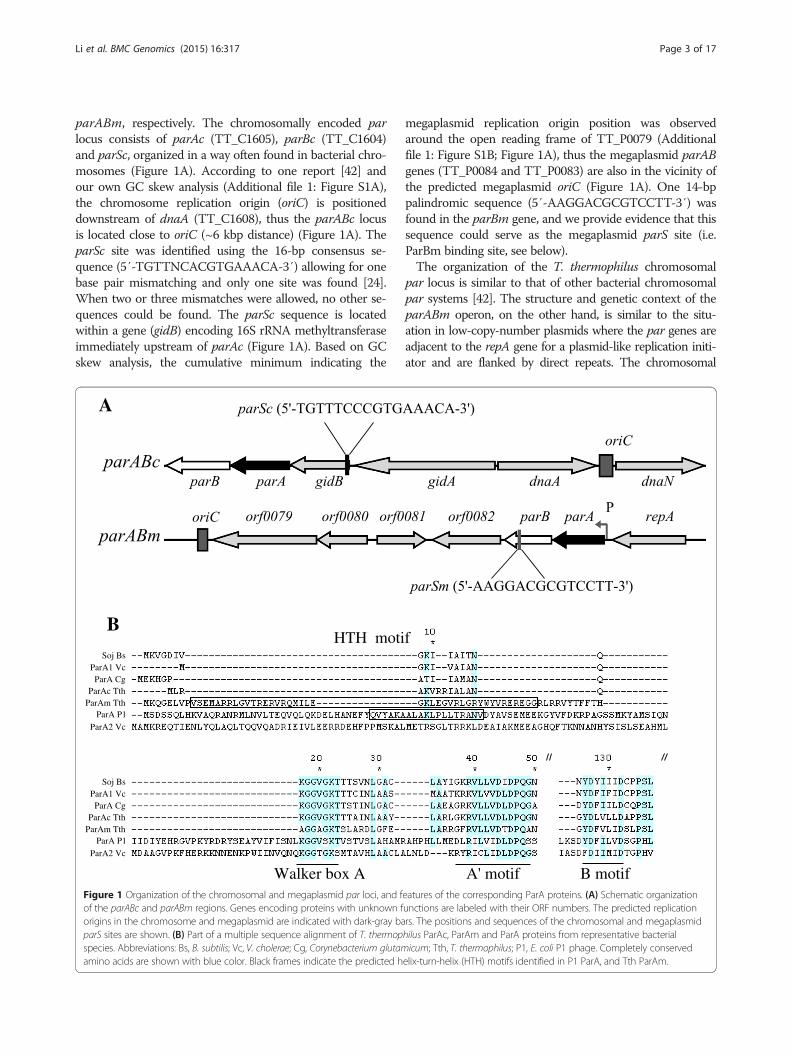
parABm, respectively. The chromosomally encoded parlocus consists of parAc (TT_C1605), parBc (TT_C1604)and parSc, organized in a way often found in bacterial chro-mosomes (Figure 1A). According to one report [42] andour own GC skew analysis (Additional file 1: Figure S1A),the chromosome replication origin (oriC) is positioneddownstream of dnaA (TT_C1608), thus the parABc locusis located close to oriC (~6 kbp distance) (Figure 1A). TheparSc site was identified using the 16-bp consensus se-quence (5′-TGTTNCACGTGAAACA-3′) allowing for onebase pair mismatching and only one site was found [24].When two or three mismatches were allowed, no other se-quences could be found. The parSc sequence is locatedwithin a gene (gidB) encoding 16S rRNA methyltransferaseimmediately upstream of parAc (Figure 1A). Based on GCskew analysis, the cumulative minimum indicating the
Soj BsParA1 Vc ParA Cg
ParAc Tth ParAm Tth
ParA P1 ParA2 Vc
A
BHTH moti
Walker box A
Soj BsParA1 Vc ParA Cg
ParAc Tth ParAm Tth
ParA P1 ParA2 Vc
Figure 1 Organization of the chromosomal and megaplasmid par loci, and fof the parABc and parABm regions. Genes encoding proteins with unknown forigins in the chromosome and megaplasmid are indicated with dark-gray baparS sites are shown. (B) Part of a multiple sequence alignment of T. thermopspecies. Abbreviations: Bs, B. subtilis; Vc, V. cholerae; Cg, Corynebacterium glutamamino acids are shown with blue color. Black frames indicate the predicted h
megaplasmid replication origin position was observedaround the open reading frame of TT_P0079 (Additionalfile 1: Figure S1B; Figure 1A), thus the megaplasmid parABgenes (TT_P0084 and TT_P0083) are also in the vicinity ofthe predicted megaplasmid oriC (Figure 1A). One 14-bppalindromic sequence (5′-AAGGACGCGTCCTT-3′) wasfound in the parBm gene, and we provide evidence that thissequence could serve as the megaplasmid parS site (i.e.ParBm binding site, see below).The organization of the T. thermophilus chromosomal
par locus is similar to that of other bacterial chromosomalpar systems [42]. The structure and genetic context of theparABm operon, on the other hand, is similar to the situ-ation in low-copy-number plasmids where the par genes areadjacent to the repA gene for a plasmid-like replication initi-ator and are flanked by direct repeats. The chromosomal
f
A' motif B motif eatures of the corresponding ParA proteins. (A) Schematic organizationunctions are labeled with their ORF numbers. The predicted replicationrs. The positions and sequences of the chromosomal and megaplasmidhilus ParAc, ParAm and ParA proteins from representative bacterialicum; Tth, T. thermophilus; P1, E. coli P1 phage. Completely conserved
elix-turn-helix (HTH) motifs identified in P1 ParA, and Tth ParAm.

Li et al. BMC Genomics (2015) 16:317 Page 4 of 17
and megaplasmid Par proteins also possess differentfeatures. While both ParAc and ParAm are Walker-type ATPases which contain a conserved P-loop ATPbinding motif, they differ in their sizes (249 aa forParAc and 322 aa for ParAm) as well as in the presenceof an N-terminal helix-turn-helix motif (HTH) inParAm (Figure 1B), a feature that normally appears inplasmid ParAs but not in their chromosomal counter-parts [21].
8.0
6.0
5.0
4.0
3.5
kBp parABc WT
A C
B
parAmN-1 par
Rel
ativ
e ex
pres
sion
(lo
g2-t
rans
form
ed)
D
blm
parAm parBm
blm parBm
parAmN
I
WT
parAmN-1
relative parAm exprelative parBm exp
Figure 2 Generation and genotype confirmation of the chromosomal and meby Southern blot. The genomic DNA was digested with BamHI and hybridizatipredicted sizes are 5.13 kbp for the wild type and 4.68 kbp for ΔparABc. (B) Schof ParAm (amino acid positions 1–40) with blm. Gray arrowhead denotes t(C) Genotype confirmation of the ΔparAmN-1 and ΔparAmN-2 mutants bypredicted sizes are 2.26 kbp for the wild type and 2.73 kbp for ΔparAmN-1 angenes in ΔparAmN-1 and ΔparAmN-2 relative to those of the wild type determtruncated parAm, white bar represents that of parBm. The average values and
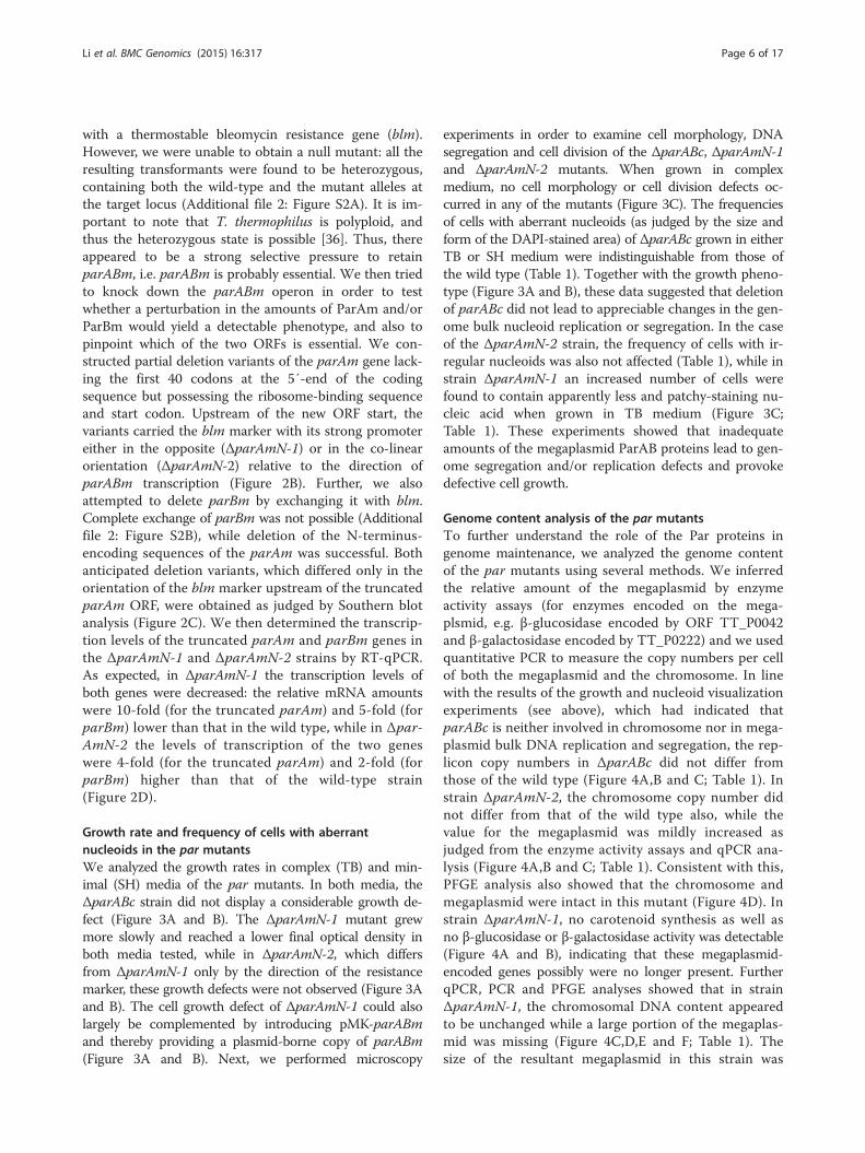
Generation of par mutants in T. thermophilusWe initiated the investigation of the two distinct parloci in T. thermophilus by attempting to generate dele-tion mutants. The chromosomal parAB genes were re-placed by a thermostable kanamycin resistance genecassette (kat). Southern blot analysis showed completedeletion of the parABc operon in the resulting mutant,ΔparABc (Figure 2A). In the same way, we initially triedto replace the whole parABm operon in the megaplasmid
WT
2.0
2.5
3.5
3.0
4.0
kBp
AmN-2
parAmN-1 parAmN-2
blm
parAm parBm
blm parBm
parAmN
II
WT
parAmN-2
ressionression
gaplasmid par mutants. (A) Genotype confirmation of the parABc mutanton was performed with a 992-bp biotin-labeled DNA fragment. The in silicoematic diagrams showing exchange of the N-terminus-encoding regionhe promoter region of parABm; black arrowhead, promoter of blm.Southern blot. The genomic DNA was digested with PstI, thed ΔparAmN-2. (D) Transcription levels of the truncated parAm and parBmined by RT-qPCR. Gray bar represents the relative expression level of theSDs shown are from three experiments.

A
B
C
Figure 3 Growth phenotypes, cell shape and nucleoid morphology observations of the par mutant strains. (A and B) The cultures of themutants were grown in antibiotic-free complex medium (TB) (A) and minimal medium (SH) (B). For complementation experiments, the wildtype and the ΔparAmN-1 strains carrying the plasmid pMK-parABm were grown in the presence of kanamycin (20 μg/ml). One representative ofthree independent experiments is shown. (C) Microscopic analysis of the cell shape, cell division and DNA morphology of the wild type and parmutants grown in complex medium (TB). Shown are representative phase-contrast (Phase) and fluorescence images (Membrane, DNA) and amerge between the membrane and DNA images (Overlay). The cells were stained with carboxyfluorescein (for membranes) and with DAPI (forDNA) before imaging. White arrows show aberrant nucleoids. Scale bars, 2 μm.
Li et al. BMC Genomics (2015) 16:317 Page 5 of 17
Li et al. BMC Genomics (2015) 16:317 Page 6 of 17
with a thermostable bleomycin resistance gene (blm).However, we were unable to obtain a null mutant: all theresulting transformants were found to be heterozygous,containing both the wild-type and the mutant alleles atthe target locus (Additional file 2: Figure S2A). It is im-portant to note that T. thermophilus is polyploid, andthus the heterozygous state is possible [36]. Thus, thereappeared to be a strong selective pressure to retainparABm, i.e. parABm is probably essential. We then triedto knock down the parABm operon in order to testwhether a perturbation in the amounts of ParAm and/orParBm would yield a detectable phenotype, and also topinpoint which of the two ORFs is essential. We con-structed partial deletion variants of the parAm gene lack-ing the first 40 codons at the 5′-end of the codingsequence but possessing the ribosome-binding sequenceand start codon. Upstream of the new ORF start, thevariants carried the blm marker with its strong promotereither in the opposite (ΔparAmN-1) or in the co-linearorientation (ΔparAmN-2) relative to the direction ofparABm transcription (Figure 2B). Further, we alsoattempted to delete parBm by exchanging it with blm.Complete exchange of parBm was not possible (Additionalfile 2: Figure S2B), while deletion of the N-terminus-encoding sequences of the parAm was successful. Bothanticipated deletion variants, which differed only in theorientation of the blm marker upstream of the truncatedparAm ORF, were obtained as judged by Southern blotanalysis (Figure 2C). We then determined the transcrip-tion levels of the truncated parAm and parBm genes inthe ΔparAmN-1 and ΔparAmN-2 strains by RT-qPCR.As expected, in ΔparAmN-1 the transcription levels ofboth genes were decreased: the relative mRNA amountswere 10-fold (for the truncated parAm) and 5-fold (forparBm) lower than that in the wild type, while in Δpar-AmN-2 the levels of transcription of the two geneswere 4-fold (for the truncated parAm) and 2-fold (forparBm) higher than that of the wild-type strain(Figure 2D).
Growth rate and frequency of cells with aberrantnucleoids in the par mutantsWe analyzed the growth rates in complex (TB) and min-imal (SH) media of the par mutants. In both media, theΔparABc strain did not display a considerable growth de-fect (Figure 3A and B). The ΔparAmN-1 mutant grewmore slowly and reached a lower final optical density inboth media tested, while in ΔparAmN-2, which differsfrom ΔparAmN-1 only by the direction of the resistancemarker, these growth defects were not observed (Figure 3Aand B). The cell growth defect of ΔparAmN-1 could alsolargely be complemented by introducing pMK-parABmand thereby providing a plasmid-borne copy of parABm(Figure 3A and B). Next, we performed microscopy
experiments in order to examine cell morphology, DNAsegregation and cell division of the ΔparABc, ΔparAmN-1and ΔparAmN-2 mutants. When grown in complexmedium, no cell morphology or cell division defects oc-curred in any of the mutants (Figure 3C). The frequenciesof cells with aberrant nucleoids (as judged by the size andform of the DAPI-stained area) of ΔparABc grown in eitherTB or SH medium were indistinguishable from those ofthe wild type (Table 1). Together with the growth pheno-type (Figure 3A and B), these data suggested that deletionof parABc did not lead to appreciable changes in the gen-ome bulk nucleoid replication or segregation. In the caseof the ΔparAmN-2 strain, the frequency of cells with ir-regular nucleoids was also not affected (Table 1), while instrain ΔparAmN-1 an increased number of cells werefound to contain apparently less and patchy-staining nu-cleic acid when grown in TB medium (Figure 3C;Table 1). These experiments showed that inadequateamounts of the megaplasmid ParAB proteins lead to gen-ome segregation and/or replication defects and provokedefective cell growth.
Genome content analysis of the par mutantsTo further understand the role of the Par proteins ingenome maintenance, we analyzed the genome contentof the par mutants using several methods. We inferredthe relative amount of the megaplasmid by enzymeactivity assays (for enzymes encoded on the mega-plsmid, e.g. β-glucosidase encoded by ORF TT_P0042and β-galactosidase encoded by TT_P0222) and we usedquantitative PCR to measure the copy numbers per cellof both the megaplasmid and the chromosome. In linewith the results of the growth and nucleoid visualizationexperiments (see above), which had indicated thatparABc is neither involved in chromosome nor in mega-plasmid bulk DNA replication and segregation, the rep-licon copy numbers in ΔparABc did not differ fromthose of the wild type (Figure 4A,B and C; Table 1). Instrain ΔparAmN-2, the chromosome copy number didnot differ from that of the wild type also, while thevalue for the megaplasmid was mildly increased asjudged from the enzyme activity assays and qPCR ana-lysis (Figure 4A,B and C; Table 1). Consistent with this,PFGE analysis also showed that the chromosome andmegaplasmid were intact in this mutant (Figure 4D). Instrain ΔparAmN-1, no carotenoid synthesis as well asno β-glucosidase or β-galactosidase activity was detectable(Figure 4A and B), indicating that these megaplasmid-encoded genes possibly were no longer present. FurtherqPCR, PCR and PFGE analyses showed that in strainΔparAmN-1, the chromosomal DNA content appearedto be unchanged while a large portion of the megaplas-mid was missing (Figure 4C,D,E and F; Table 1). Thesize of the resultant megaplasmid in this strain was

Table 1 Frequencies of cells with aberrant nucleoids and relative genome copy numbers in par mutants and ParAm/ParBm overexpression strains
Strain Cells with aberrant nucleoids (%) Relative TT_P0043 copies Relative term copies Relative oriCc copies Relative terc copies
WT 1.24 1 1 1 1
ΔparABc 3.05 1.13 ± 0.04 1.09 ± 0.05 1.13 ± 0.15 1.14 ± 0.08
ΔparAmN-1 33.02 / 1.52 ± 0.05 0.91 ± 0.13 0.93 ± 0.23
ΔparAmN-2 2.28 1.24 ± 0.09 1.26 ± 0.16 1.08 ± 0.07 1.12 ± 0.02
TMP01 1.26 3.06 ± 0.23 2.45 ± 0.19 0.93 ± 0.17 0.85 ± 0.12
TMP02 2.12 2.84 ± 0.14 2.29 ± 0.35 0.92 ± 0.09 0.89 ± 0.07
TMP0 1.15 0.84 ± 0.26 0.95 ± 0.05 0.97 ± 0.07 1.05 ± 0.03
The frequencies of cells with aberrant nucleoids were measured in cultures grown in TB medium; approximately 300 cells were analyzed for each strain. Relativegenome copy numbers were determined by quantitative PCR. The mean values and the standard deviations of three independent experiments are shown. “/”indicates undetectable.
Li et al. BMC Genomics (2015) 16:317 Page 7 of 17
approximately 125–130 kbp in contrast to 232.6 kbp forthe wild type (Figure 4D), and the coordinates of theeliminated region could be roughly mapped (Figure 4E).It seemed that this smaller megaplasmid could not beresolved properly after replication, as duplicated, tripli-cated, and even quadruplicated megaplasmid sizes couldbe observed (Figure 4D). Because the above effects werenot present in strain ΔparAmN-2, which differs fromΔparAmN-1 only by the orientation of the resistancemarker and by the expression levels of the truncatedParAm and ParBm proteins (see above), we concludethat inadequate amounts of ParABm in ΔparAmN-1 ledto the loss of large portions of the megaplasmid accom-panied by megaplasmid resolution and segregationdefects.
Overexpression of ParAm and ParBm in T. thermophilusA slight but repeatedly detectable increase of the mega-plasmid copy number was observed in the ΔparAmN-2strain (Figure 4B and C; Table 1), which is characterizedby a higher expression level of ParABm. For furtherclarification if the megaplasmid copy number is relatedto the amounts of ParAm and/or ParBm, we constructedtwo strains (TMP01 and TMP02) in which the parAmand parBm genes were expressed from plasmids (pMK-parAm and pMK-parBm, respectively). Both strains didnot display obvious cell growth, cell morphology, celldivision or DNA segregation defects. However, TMP01and TMP02 were found to synthesize increased levels ofcarotenoids and displayed higher β-glucosidase activities(Figure 4A and B). Further, qPCR experiments demon-strated that both strains had 2.5 to 3.5 fold more mega-plasmid copies compared to the control TMP0 strain(carrying the empty pMK18 vector), while the chromo-somal copy number was unaffected (Figure 4C; Table 1).Moreover, entangled forms (catenenes) of the megaplas-mids could be observed by PFGE analysis of TMP01 andTMP02, indicating that megaplasmid replication speedprobably exceeded that of DNA separation and cell
division (Figure 4D). Thus, it seems that both ParAmand ParBm act to promote megaplasmid replication.
In vivo localization of the ParB proteins in T. thermophiluscellsStudies of ParB localization patterns in other bacteria haveshown that fusions of fluorescent proteins to ParB pro-teins form punctate fluorescent foci representing ParB-parS nucleoprotein complexes in the cells [32,34,43]. Toinvestigate the in vivo localization pattern of the T. ther-mophilus ParB proteins, we constructed C-terminal sGFPfusions of ParBc and ParBm. The sGFP variant used by ushas been reported before [44], and it has been shown thatit is able to fold and fluoresce properly when expressed inT. thermophilus growing at high temperatures (about 60°C). When the ParBc-sGFP and ParBm-sGFP constructswere expressed in T. thermophilus TL-1 (a carotenoid syn-thesis deficient strain isolated in our group, permittingbetter observation of sGFP fluorescence), well-definedfluorescent foci could be observed (Figure 5A and F) pro-viding in vivo evidence that the two ParB proteins canbind parS sites. Obviously, the polyploid nature of thecells is unproblematic with respect to distinct foci forma-tion. In both cases, the majority of cells contained 2–6 fociwith two of them localized at the cell poles (“old” poles),and the rest localized at positions of septum formation(“future” poles) (Figure 5A and F). For better illustration,the positions of the two most pole-proximal foci weremeasured from the nearest poles and expressed as frac-tions of the cell lengths. The plot of these measure-ments (approximately 120 cells were randomly selectedfor each strain) showed that the nearest-to-pole foci ofParBc-sGFP and ParBm-sGFP were on average in only7.2% and 5.5% distance from the cell poles, indicatingParBc and ParBm are extremely polar localized (Figure 5Eand I). Assuming that the subcellular locations of ParBcand ParBm actually also mark the positions of the corre-sponding parS regions and thus the respective chromo-somal and megaplasmid origin regions (see Figure 1A), it

Gen
ome
copy
num
ber
rela
tive
to W
T
WT parABc parAmN-1 parAmN-2 TMP01 TMP02 TMP0
TB
TB-XGlc_XGal
A
WT bgl
parABc
parAmN-1
parAmN-2
TMP01
TMP02
TMP0
B
C
-glu
cosi
dase
act
ivity
(un
its/O
D60
0)
parABc
parAmN-1
parAmN-2
TMP01
TMP02
TMP0
97
145.5
194
242.5
291
339.5
388
436.5
485
WT TMP01 TMP02
kBp
parAmN-2 WT parAmN-1
0.5
kBp
0.4
0.3
0.25
0.2
0.15
0.1
0.5
kBp
0.4
0.3
0.2
0.15
1.0
kBp
0.7
0.5
0.6
0.7
0.4
0.25
0.15
primer pairs 1-3 (+ control) primer pairs 4-7 (+ control) primer pairs 8-10 (+ control)
parAmN-2 WT parAmN-1
parAmN-2 WT parAmN-1
D
F
control
control
control
1
2
345
67
8910
parAmN-2
97
145.5
194
242.5
291
339.5
388
436.5
485
kBp
parAmN-1 L L
12
3
4
5
67
8
9
10
ter
oriC
oriC
TT_P0057
TT_P0042-TT_P0043
TT_P0222
TT_P0195/ter
1-10 The 1-10 loci tested by PCR
The eliminated region in parAmN-1
E
Figure 4 Characterization of genome features of the chromosomal and megaplasmid parmutants and ParAm/ParBm overexpression strains. (A)Phenotypes of the strains on complex media (TB) and on TB supplemented with the chromogenic substrates XGlc and XGal. (B) Intracellular β-glucosidaseactivity measurements of the strains. The Δbgl strain was used as a negative control. The means and the SDs of three independent experiments are shown.(C) Relative chromosome and megaplasmid copy numbers of the individual mutants determined by qPCR. The means and SDs are from threeexperiments. (D) Pulsed field gel electrophoresis visualizing chromosome and megaplasmid. “L”, lambda ladder; the positions of the chromosome andmegaplasmid are indicated with black and white arrows. (E) Schematic drawing of the megaplasmid pTT27. The positions of the primer pairs used fordetecting the megaplasmid sequence loss in ΔparAmN-1 are indicated with short black lines and numbers from 1 to 10. The loci on the megaplasmidthat have been investigated are indicated with different bars, and their names are on the right panel of the figure. (F) PCR amplification results for the10 loci indicated in (E) from wild type, ΔparAmN-1 and ΔparAmN-2. The primer pairs 1 to 3, 4 to 7 and 8 to 10 were mixed into three pools, and ineach reaction amplification of a chromosomal gene locus (pyrF) was used as a control. The predicted sizes of the PCR products 1 to 10 are 87, 164,247, 346, 400, 498, 610, 699, 898 and 1014 bp. The size of the control amplicon is 460 bp (white frame). The bands of the 10 PCR products are indicatedwith numbers 1–10 on the right side of the corresponding figure panel. The gray arc in (E) indicates the megaplasmid region estimated to be lostin ΔparAmN-1.
Li et al. BMC Genomics (2015) 16:317 Page 8 of 17
can be concluded that the origin regions of both repliconsshare a cellular localization near the cell poles. The factthat foci sometimes could also be detected at the cell cen-ters or septum formation positions (“future” poles) indi-cated that ParB-parS (i.e., ParB-origin) nucleoproteincomplexes might travel from cell poles to cell division po-sitions, thus their cellular localization is dynamic.As described above (Figure 4D), the ΔparAmN-1
strain lacking adequate ParABm amounts displayedmegaplasmid segregation defects. We tested if also thesubcellular location of ParBm (i.e. the subcellular loca-tion of parSm) was altered in this strain by expressing
the ParBm-sGFP fusion in the ΔparAmN-1 back-ground. ParBm-sGFP also formed discrete foci in thisstrain (Figure 5G). However, the foci were mostly dis-sociated from the cell poles (Figure 5G), i.e. most ofthe cells contained randomly positioned fluorescentfoci. The average pole-proximal focus position (mea-sured from the nearest poles) in cells that containedone focus was drastically increased compared with thatin the wild-type cells (Figure 5J). This experimentshowed that the decrease of the ParABm amounts andespecially that of the ParAm amount (see Figure 2D),caused by reduced parABm expression, led to mislocalization

C
Phase DNA ParBc-sGFP Overlay
TL-1/
XL-1/
parSc)
XL-1/
parSc)
A
B
D XL-1/
Cell length (µm)
Tth TL-1/ParBc-sGFP
(n = 128 cells)
Foci
pos
ition
s (%
cel
l len
gth)
7.2%
foci positions nearest to poles
foci positions farthest from the same poles
E
Phase DNA ParBm-sGFP Overlay
Ec XL-1/ ParBm-sGFP
Tth parAmN-1/ ParBm-sGFP
F
G
H
Cell length (µm)
Tth TL-1/ParBm-sGFP
Foci
pos
ition
s (%
cel
l len
gth)
foci positions nearest to poles
foci positions farthest from the same poles(n = 113 cells)
5.5%
I
Tth parAmN-1/ParBm-sGFP
Cell length (µm)
Foci
pos
ition
s (%
cel
l len
gth)
(n = 120 cells)
29.7%
foci positions nearest to poles
J
Tth TL-1/ParBm-sGFP
Figure 5 Subcellular localizations of ParBc-sGFP and ParBm-sGFP in T. thermophilus and E. coli cells. Representative cells are shown with a gallery viewof phase-contrast (Phase), DNA, ParBc-sGFP or ParBm-sGFP signal, and merged images (Overlay). Scale bars, 2 μm. (A and F) Subcellular localization ofParBc-sGFP and ParBm-sGFP in the T. thermophilus TL-1 strain grown in complex medium. (B, C, D and H) Expression of T. thermophilus ParBc-sGFP orParBm-sGFP in E. coli XL-1. In the absence of the parSc site, ParBc-sGFP was found as patches (B); when parSc sites were provided from a plasmid,ParBc-sGFP localized as discrete foci (C), and foci were not observed in the presence of the empty vector (D); ParBm-sGFP formed discrete foci in E. coli(H). (G) Representative image of mislocalized foci formed by ParBm-sGFP expressed in the ΔparAmN-1 strain. (E and I) Relative positions of the twomost pole-proximal foci of ParBc-sGFP (E) and ParBm-sGFP (I) expressed in T. thermophilus TL-1. Black diamonds represent the nearest-to-pole focipositions in individual cells, white diamonds represent the foci positions that are farthest from these poles. The mean position of the nearest-to-polefoci is shown with a dotted line. (J) The relative foci positions of 120 ΔparAmN-1/ParBm-sGFP cells containing one focus are shown.
Li et al. BMC Genomics (2015) 16:317 Page 9 of 17
of the parSm sites and thus of the megaplasmid originregions.
In vivo localization of T. thermophilus ParBc and ParBm inE. coli cellsIn order to better understand the factors that influencethe ParB proteins’ localization patterns, we expressedcomponents of the T. thermophilus Par systems in E.coli, a host that does not encode chromosomal parABSsystem homologues. When T. thermophilus ParBc-sGFP was expressed in E. coli, the fluorescence signalwas spread over the nucleoid and no foci were formed(Figure 5B). Discrete fluorescent foci, which wererandomly localized in the cells, could be observed only
after the T. thermophilus parSc site was introducedinto this strain (from a plasmid pUC-ΔparABc::kat)(Figure 5C), and this effect was not observed in theempty vector control (pUC18) (Figure 5D). This meansthe ParBc subcellular localization pattern is dependenton the specific chromosomal parS site. On the contrary,we found that the megaplasmid ParB protein (expressedas ParBm-sGFP) formed foci when expressed alone in E.coli cells (Figure 5H), suggesting that there were ParBmbinding sites contributed by E.coli or by the parBm cod-ing sequence itself. Since the 14-bp palindromic se-quence could be readily identified in the parBm gene(see Figure 1A), we favored the latter option. In sum-mary, the different localization patterns of ParBc and

Li et al. BMC Genomics (2015) 16:317 Page 10 of 17
ParBm in E. coli cells suggested that the two ParBs binddifferent parS sites.
In vitro DNA binding assays of the ParB proteins to parSsitesThe above experiments demonstrated that the parABcand parABm systems seem to play different cellularroles, and that the two ParBs also tend to only associatewith their cognate parS sites. To further verify these ob-servations, in vitro binding of recombinant ParB pro-teins to the 16-bp chromosomal parSc site and/or thepredicted 14-bp megaplasmid parSm site were assayedby EMSA. These assays showed that ParBc could bindparSc and ParBm could bind the predicted parSm(Additional file 3: Figure 3SA and B). Almost all DNA-binding proteins contain more than one nucleic acidbinding site, and during in vitro DNA binding assaysthey possibly bind any DNA non-specifically [45]. To
A
B
Figure 6 Specificity of ParB proteins binding to parS sites in vitro, tested by comSpecificity of the ParB proteins binding to cognate parS sites (ParBc-parSc and P(C and D) Specificity of the ParB proteins binding to non-cognate parS sites (PaDNA probes. The 25-bp DNA probes containing the wild-type and mutant parSprobes containing the wild-type and mutant parSm sequences are indicated aswild-type parSc or parSm DNA probes, and purified ParBc or ParBm proteins we
test binding specificity, competition experiments withunlabeled probes were performed. In both cases, the un-labeled wild-type parSc/parSm probe competed muchbetter than the unlabeled mutant parSc/parSm probe(Figure 6A and B). Thus the 16-bp parSc sequence andthe 14-bp parSm sequence were bound specifically byParBc and ParBm, respectively. Further, we performedEMSA of the ParB proteins with their non-cognate parSsites. These assays showed that ParBc did not bind spe-cifically to parSm and ParBm did not bind specifically toparSc in vitro, as the respective mutant parS probecompeted even better than the wild-type parS probe(Figure 6C and D). Thus, we conclude that the two ParBproteins bind parS sites in a replicon-specific manner.
DiscussionThere are multiple copies of the chromosome and themegaplasmid in T. thermophilus [36] and whether their
C
D
petition with unlabeled DNA probes in gel mobility shift assays. (A and B)arBm-parSm) tested by the addition of unlabeled competitor DNA probes.rBc-parSm and ParBm-parSc) tested by the addition of unlabeled competitorc sequences are indicated as WT parSc and Mu parSc, and the 18-bp DNAWT parSm and Mu parSm. All reactions contained 15 pmol FAM-labeledre added with a concentration of either 0 (no protein) or 200 pmol.
Li et al. BMC Genomics (2015) 16:317 Page 11 of 17
segregation is stringently regulated is not clear. Both thechromosome and the megaplasmid sequences of T.thermophilus strain HB27 revealed par loci. In thisstudy, we investigate the characteristics and functions ofthe two par systems, thereby providing first insights inthe mechanisms which may be involved in the genomepartitioning in T. thermophilus.
Characteristics of the chromosomal par locusChromosomal Par orthologs seem to possess variousfunctions in different bacterial species, and their role inchromosome segregation is suggested to be less pivotalcompared with their counterparts in plasmids. Althoughdeletion of spo0J (parB) of B. subtilis leads to a consid-erable increase of anucleate cells during vegetativegrowth, the rest of the cells still exhibit a normalchromosome segregation pattern [27]; moreover, dele-tion of soj (parA) has no significant effect on chromo-some segregation [46]. Similar observations have beenmade in some Gram-negative bacteria. In Pseudomonasputida, the parAB genes are not essential, and parA andparB mutations did not influence cell growth orchromosome segregation in rich medium [47]. In V.cholerae, deletion of parA1 does not alter cell growthand chromosome I (chrI) partitioning; however, thepolar localization pattern of the origin region is abro-gated, indicating that the parABS1 system functions tomediate the localization and segregation of the chrI ori-gin region but not of the bulk nucleoid [32]. Our resultsshow that in T. thermophilus the role of the chromo-somal Par system is similar to that of the parABS1 sys-tem of V. cholerae. The parABc null mutant generatedby us did not display apparent defects with respect tocell growth rate or frequency of cells with aberrantnucleoids (Figure 3; Table 1). Further observations fromexperiments targeting the copy numbers of the repliconsshowed that the chromosomal par locus was probably notrequired for either chromosome or megaplasmid bulkDNA replication and segregation (Figure 4A,B and C;Table 1). It is likely that the T. thermophilus chromo-somal bulk nucleoid segregation is accomplished byother mechanisms. This conclusion is in line with theview that separate and redundant mechanisms may beinvolved to regulate the bacterial chromosome replica-tion and segregation [48].However, the chromosomal par locus may play other
roles. Indeed, the in vitro DNA-binding assays showed thatParBc could bind the parSc site specifically (Additionalfile 3: Figure S3A; Figure 6A), indicating it is a func-tional ParB protein. In vivo, the ParBc-parSc complexeslocalized to the poles of wild-type T. thermophilus cells(Figure 5A and E) and this localization was apparentlydynamic, indicating that the origin regions were boundby ParBc, and the nucleoprotein complexes were driven
from “old” poles to “new” poles. It has been shown thatin vitro the T. thermophilus ParAc can form dimers andthen associate with DNA, forming nucleoprotein fila-ments, suggesting that ParAc has the capacity to medi-ate DNA movement [41]. Thus, similar to some otherPar systems [25,32], the ParBc-origin complexes couldpossibly be anchored to the poles via ParAc filaments.Taken together, our data indicate that parABc isprobably involved in the chromosomal origin regionlocalization.
Characteristics of the megaplasmid par locusThe megaplasmid par locus is structured differentlythan the chromosomal par region. The parABm locusis also located in the megaplasmid origin-proximal re-gion, and has a genetic set-up very similar to thatfound in some low-copy-number plasmids (Figure 1A).We could not obtain deletion mutants of the parABmoperon or of the parBm gene, suggesting an essentialrole of parABm. Essentiality of par genes for bacterialcells has been observed for some chromosomal parsystems. The null mutant of parB in C. cresentus is le-thal [31], and direct deletion of the parAB2 genes in V.cholerae chromosome II is also not feasible [34]. Toour knowledge, our work shows for the first time that a(mega)plasmid par locus is essential for its host organism.However, a parallel can be drawn from the case of the V.cholerae parAB2 locus on chromosome II, because thisappears to be a megaplasmid-derived chromosome [49].In the T. thermophilus ΔparAmN-1 mutant, which
expressed less ParABm, both the cell growth rate andthe frequency of cells with irregular nucleoids were af-fected (Figure 3; Table 1). Furthermore, a substantialpart (about 100 kbp) of the 232 kbp megaplasmid, cov-ering the region between approximately 11 kbp and 111kbp distance from one side of the megaplasmid origin,was lost in this mutant (Figure 4). This was not a spuri-ous observation for just one clone, as all of 10 randomlyselected ΔparAmN-1 colonies picked up directly fromthe transformation plates were found to have lost thesame region of the megaplasmid. These phenotypes ofthe ΔparAmN-1 strain were not observed in the iso-genic mutant ΔparAmN-2 that differed from Δpar-AmN-1 only by the direction of the antibiotic resistancecassette (and thus the transcription levels of the trun-cated parABm), suggesting that they were caused by in-adequate amounts of ParABm in the ΔparAmN-1 cells.Moreover, the truncated megaplasmid in ΔparAmN-1seemed not to be decatenated properly, as multimericforms of the megaplasmid could be observed by PFGEanalysis (Figure 4D). In addition, the parSm sites (i.e.the megaplasmid origin regions) were dissociated fromthe cell poles and drastically mislocalized, as judged bythe subcellular locations of the ParBm-sGFP fusion

Li et al. BMC Genomics (2015) 16:317 Page 12 of 17
(Figure 5G and J). These findings suggest that parABmprobably mediates the accurate subcellular localizationand segregation of the megaplasmid, resembling thefunction of the Par systems in most low-copy-numberplasmids and some chromosomes of other bacteria.The observation that only part of the megaplasmid
was missing is in agreement with the conclusion thatparBm is essential in T. thermophilus. It is likely thatnot only parBm but also other megaplasmid regions areessential and it seems that elimination of the entiremegaplasmid is lethal to T. thermophilus. In support ofthis, we were not able to cure the megaplasmid from T.thermophilus despite various attempts (own unpublishedwork). The precise reason why the megaplasmid loss isnot tolerated is currently unknown.When we overexpressed either ParAm or ParBm in
wild-type T. thermophilus cells (TMP01 and TMP02),the megaplasmid but not the chromosomal copy num-bers were increased (Figure 4A,B,C and D; Table 1).This points to a role of parABm in megaplasmid repli-cation initiation and/or copy number maintenance. Itis possible that the ParABm proteins can activate thefactors (e.g. RepA initiator) that are involved in themegaplasmid replication. A role of the ParA and ParBproteins in genome replication has been recently pro-posed also for other bacteria. In B. subtilis, Spo0J (ParB)was found to recruit a SMC condensin protein to replica-tion origin regions, thereby promoting chromosome seg-regation [50,51]; the same phenomenon was also observedin Streptococcus pneumoniae [52]. ParB2 encoded by theV. cholerae chromosome II (chrII) was also found to influ-ence the replication of chrII, in which ParB2 appeared topromote the replication by activating RctB protein thatinitiates chrII replication [53]. In the chromosome of B.subtilis and chromosome I of V. cholerae, ParA was foundto directly interact with the chromosome replication initi-ator DnaA, thereby participating in the regulation ofchromosome replication [54-56]. Taken together, it is con-ceivable that the ParABm system in T. thermophilus is im-portant for maintaining the megaplasmid throughregulating its replication and segregation. Interestingly,it also seems that segregation of the T. thermophilusmegaplasmid is coordinated with its decatenation,which is reminiscent of the situations found in the E.colior Streptomyces coelicolor chromosomes. Mutations ofthe parE gene (encoding one of the subunits of topo-isomerase IV) in E.coli or S. coelicolor lead to chromo-some catenation and fragmentation, thereby affectingthe chromosome segregation [57,58]. It is possible thatin the T. thermophilus ΔparAmN-1 cells, the megaplas-mid could not be decatenated properly due to the inad-equate ParABm amounts, thus the megaplasmid wasguillotined during separation into the daughter cells,and only those cells that recombined the essential
portions of the megaplasmid would then survive. Appar-ently, further experiments (e.g. FISH) are needed in orderto define whether the irregular nucleoid cells of the Δpar-AmN-1 strain were cells that lacked the entire megaplas-mid and thus were essentially dead cells or were cells thatcontained the chromosome and the “mini” megaplasmid.
Chromosomal and megaplasmid Par are two independentsystemsIn vitro, the T. thermophilus chromosomal ParBc andthe megaplasmid ParBm could bind their correspond-ing parS site in a specific manner, and the Par proteins’binding to non-cognate parS sites was unspecific (Figure 6).These findings suggested that the two ParBs act only withtheir cognate parS sequences. This is supported by thein vivo ParB localization investigations in E. coli cells, asthe two ParBs seemed to localize differently in thisheterologous system (Figure 5B,C and H). In E. colicells, we found that ParBm-sGFP could form foci, andthis further confirmed the conclusion drawn from thein vitro ParBm-parSm binding experiments, that isparBm contains its own binding site parSm.The results of the in vitro ParB-parS bindings and of
the in vivo ParB localization experiments, together withthe fact that perturbation of the expression of parABmonly affected the replication and/or segregation of themegaplasmid but not that of the chromosome, supportthe hypothesis that the two Par systems function inde-pendently. The phenomenon that parAB function in areplicon-specific manner has also been observed inother bacteria containing more than one Par system, forexample the ParAB1 and ParAB2 systems of chromo-somes I and II in V. cholerae [59] and the Par systems ofthe four replicons in B. cenocepacia [35]. From the bac-teria with multiple replicons studied so far it seems likea common theme that their Par systems behave inde-pendently of each other rather than forming a networksystem with shared components.
ConclusionsOne T. thermophilus cell contains multiple copies of thechromosome and megaplasmid. Like many bacteria,both the chromosome and the megaplasmid of T. ther-mophilus encode orthologs of the plasmid partitioning(par) genes, however their role in genome segregation isnot known. In this study, we investigate the functions ofthese two Par systems in T. thermophilus through ana-lysis of chromosomal and megaplasmid par gene mu-tants and ParAm/ParBm overexpression strains, as wellas by using in vitro DNA binding assays of heterolo-gously expressed ParB proteins and in vivo ParB proteinlocalization observations. We show that in T. thermo-philus the chromosomal ParAB system is not requiredfor either the chromosomal or megaplasmid bulk DNA

Li et al. BMC Genomics (2015) 16:317 Page 13 of 17
replication and segregation. It is however involved in thepolar localization and separation of the chromosomal ori-gin region. In contrast, the megaplasmid ParAB system inT. thermophilus probably functions to regulate the mega-plasmid replication and segregation, thereby maintainingthe megaplasmid. The two Par systems in T. thermophilusappear to function in a replicon-specific manner. Ourstudy provides the first insights of the mode of operationof Par systems in a two-replicon, polyploid bacterium.
MethodsBacterial strains and growth conditionsEscherichia coli XL-1 Blue (Agilent Technologies, SantaClara, USA) was used as a host for DNA manipulationsand was grown in LB medium (10 g/l tryptone, 5 g/lyeast extract, 5 g/l NaCl) at 37°C. T. thermophilus HB27(DSM 7039) and its derivative strains were grown at60°C or 70°C with vigorous shaking in rich medium(TB) or nutritionally defined medium (SH). TB mediumhad a pH of 7.5 and contained per litre 8 g trypticasepeptone, 4 g yeast extract, and 3 g NaCl, and was pre-pared with a high-carbonate mineral water (Purania,DRINKPOOL GmbH, Germany). SH medium was pre-pared as described in [60]. The growth media weresupplemented with ampicillin (100 μg/ml for E. coli),kanamycin (20 μg/ml for E. coli and T. thermophilus),bleomycin (“Bleocin”, Calbiochem, 15 μg/ml), chloram-phenicol (12.5 μg/ml for E. coli), XGlc (5-bromo-4-chloro-3-indolyl-β-D-glucopyranoside, 50 μg/ml) or XGal(5-bromo-4-chloro-3-indolyl-β-D-galactopyranoside, 50 μg/ml) when appropriate. All reagents were purchased fromSigma-Aldrich (Schnelldorf, Germany) except for growthmedia components which were obtained from BD Biosci-ences (Heidelberg, Germany).
Strains and plasmidsAll strains and plasmids used are listed in Table 2. Theoligonucleotides used are summarized in Additionalfile 4: Table S1. All allele exchange vectors (Table 2)for generating par mutants were derived from pUC18,and the constructs were obtained by Gibson assembly(New England Biolabs) [61]. In general, the upstreamand downstream sequences (approximately 1 kbp each)of the target regions, and the gene cassette encodingthermostable resistance to kanamycin (kat) or bleo-mycin (blm) (chemically synthesized using sequencedata from [62]) were PCR-amplified using primers thatgenerated sufficient overlaps. The purified PCR prod-ucts of the two flanking regions and the kat/blm cas-sette were introduced into XbaI-digested pUC18 viafour-fragment Gibson assembly reactions.All the replicative vectors (Table 2) in T. thermophilus
were derived from the E. coli/T. thermophilus shuttle
vector pMK18 [63]. The constructs pMK-parAm, pMK-parBm and pMK-parABm, which were generated byGibson assembly as described above, carry parAm,parBm or the entire parABm operon, respectively,transcriptionally fused to kat of pMK18. In the samemanner, the plasmid pMK-sgfp was obtained by addingthe sgfp coding sequence, which was chemically syn-thesized using sequence data from [44], to kat ofpMK18. The constructs pMKparBc-sgfp and pMKparBm-sgfp represent translationally fused parBc and parBm tothe sgfp gene in pMK-sgfp. Codons encoding four glycineresidues (poly-glycine linker) were introduced betweenparB and sgfp, and the ParB-sGFP fusions were expressedunder the same promoter of the kat gene in pMK18.The plasmids pET21a-parBc and pET21a-parBm were
obtained by introducing purified parBc and parBm PCRfragments into XhoI, NdeI linearized pET21a by Gibsonassembly.
Quantitative PCRThe quantitative PCR method for measuring the relativegenome copies was performed as described in [64]. Thechosen sites of the chromosome were near the origin(oriCc) and terminus (terc) regions, and those of themegaplasmid were the TT_P0043 locus (approximately32 kbp from the megaplasmid origin) and TT_P0195locus (near the megaplasmid terminus (term)). Standardfragments used for quantification for each chosen locuswere amplified by PCR using T. thermophilus genomicDNA as the template. The fragments were then purifiedfrom agarose gels and photometrically quantified. Aseries of dilutions containing defined numbers of thestandard molecules were then used as templates forqPCR to generate standard curves. Cell extracts of thestrains for qPCR were prepared by harvesting definedcell numbers (determined by spectrophotometry andwith a Neubauer counting chamber) from exponentiallygrowing cultures and resuspending in cell lysis buffer(Epicentre Biotechnologies, Germany); the cell lysisefficiency was determined by cell counting. After dialysis,dilutions were prepared from the cell lysates and aliquotswere used as templates for qPCR. The sizes of the targetamplicons were between 100 and 200 bp, and PCR wasperformed using qPCR Mastermix plus with fluorescein(Eurogentec, Germany) based on the protocol pro-vided by the manufacturer. Three independent ex-periments were carried out for each strain. Standardcurves were constructed from the CT values of thestandard fragments and were later used to quantitatethe genome copy numbers in the cell lysates.
RT-qPCRFor determining the relative expression levels of the trun-cated parAm and parBm genes, reverse transcription-
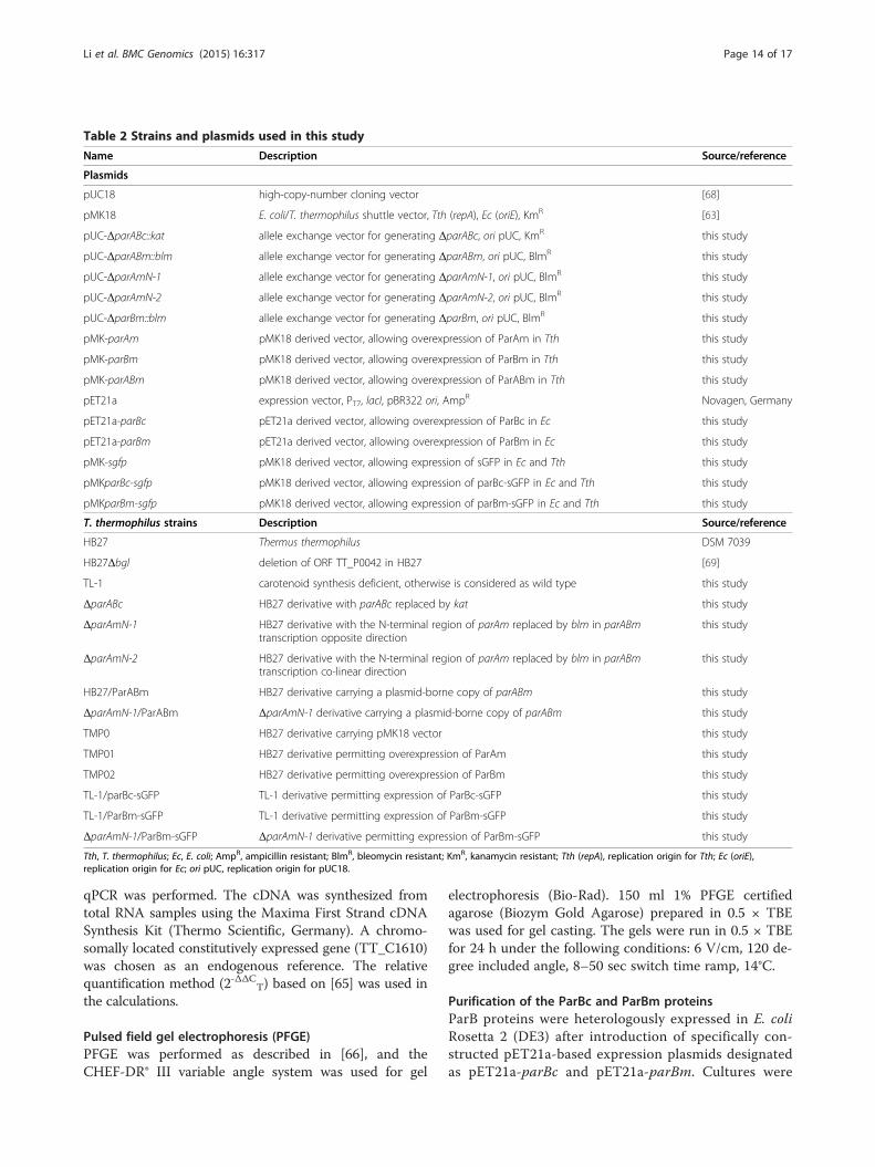
Table 2 Strains and plasmids used in this study
Name Description Source/reference
Plasmids
pUC18 high-copy-number cloning vector [68]
pMK18 E. coli/T. thermophilus shuttle vector, Tth (repA), Ec (oriE), KmR [63]
pUC-ΔparABc::kat allele exchange vector for generating ΔparABc, ori pUC, KmR this study
pUC-ΔparABm::blm allele exchange vector for generating ΔparABm, ori pUC, BlmR this study
pUC-ΔparAmN-1 allele exchange vector for generating ΔparAmN-1, ori pUC, BlmR this study
pUC-ΔparAmN-2 allele exchange vector for generating ΔparAmN-2, ori pUC, BlmR this study
pUC-ΔparBm::blm allele exchange vector for generating ΔparBm, ori pUC, BlmR this study
pMK-parAm pMK18 derived vector, allowing overexpression of ParAm in Tth this study
pMK-parBm pMK18 derived vector, allowing overexpression of ParBm in Tth this study
pMK-parABm pMK18 derived vector, allowing overexpression of ParABm in Tth this study
pET21a expression vector, PT7, lacI, pBR322 ori, AmpR Novagen, Germany
pET21a-parBc pET21a derived vector, allowing overexpression of ParBc in Ec this study
pET21a-parBm pET21a derived vector, allowing overexpression of ParBm in Ec this study
pMK-sgfp pMK18 derived vector, allowing expression of sGFP in Ec and Tth this study
pMKparBc-sgfp pMK18 derived vector, allowing expression of parBc-sGFP in Ec and Tth this study
pMKparBm-sgfp pMK18 derived vector, allowing expression of parBm-sGFP in Ec and Tth this study
T. thermophilus strains Description Source/reference
HB27 Thermus thermophilus DSM 7039
HB27Δbgl deletion of ORF TT_P0042 in HB27 [69]
TL-1 carotenoid synthesis deficient, otherwise is considered as wild type this study
ΔparABc HB27 derivative with parABc replaced by kat this study
ΔparAmN-1 HB27 derivative with the N-terminal region of parAm replaced by blm in parABmtranscription opposite direction
this study
ΔparAmN-2 HB27 derivative with the N-terminal region of parAm replaced by blm in parABmtranscription co-linear direction
this study
HB27/ParABm HB27 derivative carrying a plasmid-borne copy of parABm this study
ΔparAmN-1/ParABm ΔparAmN-1 derivative carrying a plasmid-borne copy of parABm this study
TMP0 HB27 derivative carrying pMK18 vector this study
TMP01 HB27 derivative permitting overexpression of ParAm this study
TMP02 HB27 derivative permitting overexpression of ParBm this study
TL-1/parBc-sGFP TL-1 derivative permitting expression of ParBc-sGFP this study
TL-1/ParBm-sGFP TL-1 derivative permitting expression of ParBm-sGFP this study
ΔparAmN-1/ParBm-sGFP ΔparAmN-1 derivative permitting expression of ParBm-sGFP this study
Tth, T. thermophilus; Ec, E. coli; AmpR, ampicillin resistant; BlmR, bleomycin resistant; KmR, kanamycin resistant; Tth (repA), replication origin for Tth; Ec (oriE),replication origin for Ec; ori pUC, replication origin for pUC18.
Li et al. BMC Genomics (2015) 16:317 Page 14 of 17
qPCR was performed. The cDNA was synthesized fromtotal RNA samples using the Maxima First Strand cDNASynthesis Kit (Thermo Scientific, Germany). A chromo-somally located constitutively expressed gene (TT_C1610)was chosen as an endogenous reference. The relativequantification method (2-ΔΔCT) based on [65] was used inthe calculations.
Pulsed field gel electrophoresis (PFGE)PFGE was performed as described in [66], and theCHEF-DR® III variable angle system was used for gel
electrophoresis (Bio-Rad). 150 ml 1% PFGE certifiedagarose (Biozym Gold Agarose) prepared in 0.5 × TBEwas used for gel casting. The gels were run in 0.5 × TBEfor 24 h under the following conditions: 6 V/cm, 120 de-gree included angle, 8–50 sec switch time ramp, 14°C.
Purification of the ParBc and ParBm proteinsParB proteins were heterologously expressed in E. coliRosetta 2 (DE3) after introduction of specifically con-structed pET21a-based expression plasmids designatedas pET21a-parBc and pET21a-parBm. Cultures were

Li et al. BMC Genomics (2015) 16:317 Page 15 of 17
grown in 1 l LB medium (supplemented with chloram-phenicol and ampicillin) at 37°C. When the OD600
reached a value between 0.7 and 0.8, protein expressionwas induced by the addition of IPTG at a final concen-tration of 1 mM and the cultures were agitated at 30°Cfor 4 h. The cells were harvested and lysed by sonic-ation (UP200S, Hilscher, Teltow, Germany). After son-ication, the crude cell extracts were centrifuged at 4°Cat 15,000 g for 30 min and the supernatants were sub-jected to affinity purification using Protino Ni-IDA2000 columns (Macherey Nagel, Germany).
Electrophoretic mobility shift assay (EMSA)For DNA binding assays, a 25-bp or a 18-bp DNA frag-ment that contained the chromosomal or megaplasmidparS sequence was used as the probe. The probes were6-carboxyfluorescein (FAM)-labeled and were generatedby hybridization of two complementary oligonucleo-tides. The chromosomal parS probe had the sequence:5′-TGTTTCCCGTGAAACATCAGGCGCC-3′(WT
parSc), and the megaplasmid parS probe had the se-quence: 5′-GCAAGGACGCGTCCTTCA-3′ (WT parSm).The binding reactions (25 μl) were performed in 50 mMKCl, 10 mM Tris–HCl (pH 7.0), 1 mM EDTA, 1 mMDTT, 4% glycerol, 0.02 μg/μl Poly (dI-dC), and con-tained 15 pmol of FAM-labeled probe and varyingamounts of ParB proteins. The reactions were incubatedat 25°C for 30 min and then applied on 1% agarose gelsprepared in 1 × TBE buffer. The ParB-parS binding com-petition experiments were performed using both theunlabeled probes containing the wild-type parSc or thewild-type parSm sequence (wild-type competitor), andunlabeled probes that contained seven base-pair andeight base-pair changes in the parSc and parSm sitesrespectively (mutant competitor). The mutant parScprobe had the sequence: 5′-cGTgcCCaGgGAgACcTCAGGCGCC-3′ (Mu parSc), and the mutant parSmprobe had the sequence: 5′-GCtgtGtgcaGgCCTTCA-3′(Mu parSm). They were also generated by hybridizationof two complementary oligonucleotides.
Fluorescence microscopyFor fluorescence microscopy, the cells from liquid cul-tures were collected by centrifugation (5000 g, 10 min),washed once with 1 × PBS buffer and resuspended inthe same volume of 1 × PBS buffer. Staining was per-formed by the addition of DAPI (4′,6-diamidino-2-phe-nylindole-dihydrochloride) with a final concentration of0.2 μg/ml, and if necessary, by the addition of 10 μg/ml6-carboxyfluorescein (CFS), followed by incubation atRT for 20 min. The residual dyes were washed off andthe cells were resuspended in 1 × PBS buffer. Fluores-cence microscopy was performed with a Zeiss Axio-Imager M1 microscope using filter sets “DAPI” for
DAPI, “AF488” for CFS and for the sGFP fluorescenceof strains that expressing ParB-sGFP, respectively. Themicrographic images were taken with an AxioCamMRm camera and analyzed with the Image J (NIH,USA) and AxioVision software (Carl Zeiss, Germany).
β-glucosidase activity assay for T. thermophilusβ-glucosidase activity was measured with exponentiallygrowing cells as described in [67]. The enzyme assayswere performed with three independently grown cultures.
Additional files
Additional file 1: Figure S1. Predictions of the chromosomal andmegaplasmid origin and terminus regions in T. thermophilus HB27. TheGenSkew software (http://genskew.csb.univie.ac.at/) was used tocompute the normal and cumulative GC skew for the chromosome andthe megaplasmid. The windowsize and stepsize for the chromosomalsequence were both set to 1000 bp, and those for the megaplasmidsequence were both set to 100 bp. (A) Cumulative GC skew of thechromosomal sequence. The maximum indicating the chromosomalterminus position is at 558, 731 bp, the minimum representing thechromosomal origin region position is at 1, 524, 671 bp. (B) CumulativeGC skew of the megaplasmid sequence. The maximum and minimumvalues are at the megaplasmid sequence positions 189, 545 bp and 71,689 bp, indicating the positions of the megaplasmid terminus andorigin regions, respectively.
Additional file 2: Figure S2. Genotype confirmation of the parABm andparBm mutants in T. thermophilus. (A) Genotype confirmation of the parABmmutants by PCR using genomic DNA as template and primers flanking thedeleted region (primer pairs parm-F/parm-R). The in silico predicted sizes are3.99 kbp for the wild-type allele and 2.73 kbp for the ΔparABm allele.(B) Genotype confirmation of the parBm mutants by PCR (primer pairsparm-F/parm-R-2). The predicted sizes for the PCR products are 3.39 kbpfor the wild type and 3.13 kbp for the ΔparBm allele.
Additional file 3: Figure S3. In vitro DNA binding of ParBc to parSc,and of ParBm to parSm measured by gel mobility shift assays. Allreactions were performed under the same condition as described in theMethods section. Shifted DNA species were labeled with “bound”, freeDNA species were labeled with “free DNA”. (A) Gel shift assays wereperformed with 15 pmol FAM-labeled DNA probe containing the 16-bpparSc site (probe sequence: 5′-TGTTTCCCGTGAAACATCAGGCGCC-3′), andwith various concentrations of ParBc. (B) Gel shift assays were performedwith 15 pmol FAM-labeled DNA probe containing the predicted 14-bpparSm site (probe sequence: 5′- GCAAGGACGCGTCCTTCA-3′) and withvarious concentrations of ParBm.
Additional file 4: Table S1. Primers used in this study.
Competing interestsThe authors declare that they have no competing interests.
Authors’ contributionsHL, AA and WL designed the experiments, HL and AA performed theexperiments; VTTP and BL contributed to the construction of strains; HL, AAand WL wrote the manuscript. All authors read and approved the finalmanuscript.
AcknowledgmentsWe thank Maria Übelacker and Beate Schumacher for providing excellenttechnical assistance.This work was supported by the Bundesministerium für Bildung,Wissenschaft, Forschung und Technologie (BMBF) within the framework ofthe GenoMik (Genomforschung an Mikroorganismen) funding measure andby the German Research Foundation (DFG) and the Technische UniversitätMünchen within the funding programme Open Access Publishing.

Li et al. BMC Genomics (2015) 16:317 Page 16 of 17
Received: 4 December 2014 Accepted: 10 April 2015
References1. Glaser P, Sharpe ME, Raether B, Perego M, Ohlsen K, Errington J. Dynamic,
mitotic-like behavior of a bacterial protein required for accurate chromosomepartitioning. Genes Dev. 1997;11:1160–8.
2. Gordon GS, Sitnikov D, Webb CD, Teleman A, Straight A, Losick R, et al.Chromosome and low copy plasmid segregation in E. coli: visual evidencefor distinct mechanisms. Cell. 1997;90:1113–21.
3. Webb CD, Graumann PL, Kahana JA, Teleman AA, Silver PA, Losick R. Use oftime-lapse microscopy to visualize rapid movement of the replication originregion of the chromosome during the cell cycle in Bacillus subtilis. MolMicrobiol. 1998;28:883–92.
4. Viollier PH, Thanbichler M, McGrath PT, West L, Meewan M, McAdams HH,et al. Rapid and sequential movement of individual chromosomal loci tospecific subcellular locations during bacterial DNA replication. Proc NatlAcad Sci. 2004;101:9257–62.
5. Sharpe ME, Errington J. Upheaval in the bacterial nucleoid: an activechromosome segregation mechanism. Trends Genet. 1999;15:70–4.
6. Gerdes K, Møller-Jensen J, Ebersbach G, Kruse T, Nordström K. Bacterialmitotic machineries. Cell. 2004;116:359–66.
7. Leonard TA, Møller-Jensen J, Löwe J. Towards understanding the molecularbasis of bacterial DNA segregation. Philos Trans R Soc Lond B Biol Sci.2005;360:523–35.
8. Lemon KP, Grossman AD. Localization of bacterial DNA polymerase:evidence for a factory model of replication. Science. 1998;282:1516–9.
9. Lemon KP, Grossman AD. Movement of replicating DNA through astationary replisome. Mol Cell. 2000;6:1321–30.
10. Dworkin J, Losick R. Does RNA polymerase help drive chromosomesegregation in bacteria? Proc Natl Acad Sci. 2002;99:14089–94.
11. Kruse T, Blagoev B, Løbner-Olesen A, Wachi M, Sasaki K, Iwai N, et al. Actinhomolog MreB and RNA polymerase interact and are both required forchromosome segregation in Escherichia coli. Genes Dev. 2006;20:113–24.
12. Defeu Soufo HJ, Graumann PL. Actin-like proteins MreB and Mbl fromBacillus subtilis are required for bipolar positioning of replication origins.Curr Biol. 2003;13:1916–20.
13. Kruse T, Møller-Jensen J, Løbner-Olesen A, Gerdes K. Dysfunctional MreBinhibits chromosome segregation in Escherichia coli. EMBO J. 2003;22:5283–92.
14. Gitai Z, Dye NA, Reisenauer A, Wachi M, Shapiro L. MreB actin-mediatedsegregation of a specific region of a bacterial chromosome. Cell.2005;120:329–41.
15. Austin S, Abeles A. Partition of unit-copy miniplasmids to daughter cells. II.The partition region of miniplasmid P1 encodes an essential protein and acentromere-like site at which it acts. J Mol Biol. 1983;169:373–87.
16. Li Y, Dabrazhynetskaya A, Youngren B, Austin S. The role of Par proteins inthe active segregation of the P1 plasmid. Mol Microbiol. 2004;53:93–102.
17. Ebersbach G, Gerdes K. The double par locus of virulence factor pB171: DNAsegregation is correlated with oscillation of ParA. Proc Natl Acad Sci.2001;98:15078–83.
18. Møller-Jensen J, Jensen RB, Löwe J, Gerdes K. Prokaryotic DNA segregationby an actin-like filament. EMBO J. 2002;21:3119–27.
19. Barillà D, Rosenberg MF, Nobbmann U, Hayes F. Bacterial DNA segregationdynamics mediated by the polymerizing protein ParF. EMBO J.2005;24:1453–64.
20. Ringgaard S, van Zon J, Howard M, Gerdes K. Movement andequipositioning of plasmids by ParA filament disassembly. Proc Natl AcadSci. 2009;106:19369–74.
21. Gerdes K, Møller-Jensen J, Bugge Jensen R. Plasmid and chromosomepartitioning: surprises from phylogeny. Mol Microbiol. 2000;37:455–66.
22. Lin DC, Grossman AD. Identification and characterization of a bacterialchromosome partitioning site. Cell. 1998;92:675–85.
23. Breier AM, Grossman AD. Whole-genome analysis of the chromosomepartitioning and sporulation protein Spo0J (ParB) reveals spreading andorigin-distal sites on the Bacillus subtilis chromosome. Mol Microbiol.2007;64:703–18.
24. Livny J, Yamaichi Y, Waldor MK. Distribution of centromere-like parS sites inbacteria: insights from comparative genomics. J Bacteriol. 2007;189:8693–703.
25. Gerdes K, Howard M, Szardenings F. Pushing and pulling in prokaryoticDNA segregation. Cell. 2010;141:927–42.
26. Reyes-Lamothe R, Nicolas E, Sherratt DJ. Chromosome replication andsegregation in Bacteria. Annu Rev Genet. 2012;46:121–43.
27. Ireton K, Gunther 4th NW, Grossman AD. spo0J is required for normalchromosome segregation as well as the initiation of sporulation in Bacillussubtilis. J Bacteriol. 1994;176:5320–9.
28. Lee PS, Lin DC, Moriya S, Grossman AD. Effects of the chromosomepartitioning protein Spo0J (ParB) on oriC positioning and replicationinitiation in Bacillus subtilis. J Bacteriol. 2003;185:1326–37.
29. Lee PS, Grossman AD. The chromosome partitioning proteins Soj (ParA) andSpo0J (ParB) contribute to accurate chromosome partitioning, separation ofreplicated sister origins, and regulation of replication initiation in Bacillussubtilis. Mol Microbiol. 2006;60:853–69.
30. Mohl DA, Gober JW. Cell cycle-dependent polar localization of chromosomepartitioning proteins in Caulobacter crescentus. Cell. 1997;88:675–84.
31. Mohl DA, Easter Jr J, Gober JW. The chromosome partitioning protein, ParB, isrequired for cytokinesis in Caulobacter crescentus. Mol Microbiol. 2001;42:741–55.
32. Fogel MA, Waldor MK. A dynamic, mitotic-like mechanism for bacterialchromosome segregation. Genes Dev. 2006;20:3269–82.
33. Saint-Dic D, Frushour BP, Kehrl JH, Kahng LS. A parA homolog selectivelyinfluences positioning of the large chromosome origin in Vibrio cholerae.J Bacteriol. 2006;188:5626–31.
34. Yamaichi Y, Fogel MA, Waldor MK. par genes and the pathology ofchromosome loss in Vibrio cholerae. Proc Natl Acad Sci. 2007;104:630–5.
35. Dubarry N, Pasta F, Lane D. ParABS systems of the four replicons ofBurkholderia cenocepacia: new chromosome centromeres confer partitionspecificity. J Bacteriol. 2006;188:1489–96.
36. Ohtani N, Tomita M, Itaya M. An extreme thermophile, Thermusthermophilus, is a polyploid bacterium. J Bacteriol. 2010;192:5499–505.
37. Hu B, Yang G, Zhao W, Zhang Y, Zhao J. MreB is important for cell shapebut not for chromosome segregation of the filamentous cyanobacteriumAnabaena sp. PCC 7120. Mol Microbiol. 2007;63:1640–52.
38. Schneider D, Fuhrmann E, Scholz I, Hess WR, Graumann PL. Fluorescencestaining of live cyanobacterial cells suggest non-stringent chromosomesegregation and absence of a connection between cytoplasmic andthylakoid membranes. BMC Cell Biol. 2007;8:39.
39. Henne A, Brüggemann H, Raasch C, Wiezer A, Hartsch T, Liesegang H, et al.The genome sequence of the extreme thermophile Thermus thermophilus.Nat Biotechnol. 2004;22:547–53.
40. Leonard TA, Butler PJ, Löwe J. Structural analysis of the chromosome segregationprotein Spo0J from Thermus thermophilus. Mol Microbiol. 2004;53:419–32.
41. Leonard TA, Butler PJ, Löwe J. Bacterial chromosome segregation: structureand DNA binding of the Soj dimer–a conserved biological switch. EMBO J.2005;24:270–82.
42. Nardmann J, Messer W. Identification and characterization of the dnaAupstream region of Thermus thermophilus. Gene. 2000;261:299–303.
43. Li Y, Austin S. The P1 plasmid in action: time-lapse photomicroscopy revealssome unexpected aspects of plasmid partition. Plasmid. 2002;48:174–8.
44. Cava F, de Pedro MA, Blas-Galindo E, Waldo GS, Westblade LF, Berenguer J.Expression and use of superfolder green fluorescent protein at hightemperatures in vivo: a tool to study extreme thermophile biology.Environ Microbiol. 2008;10:605–13.
45. Hellman LM, Fried MG. Electrophoretic mobility shift assay (EMSA) fordetecting protein-nucleic acid interactions. Nat Protoc. 2007;2:1849–61.
46. Marston AL, Errington J. Dynamic movement of the ParA-like Soj protein ofB. subtilis and its dual role in nucleoid organization and developmentalregulation. Mol Cell. 1999;4:673–82.
47. Lewis RA, Bignell CR, Zeng W, Jones AC, Thomas CM. Chromosome lossfrom par mutants of Pseudomonas putida depends on growth medium andphase of growth. Microbiology. 2002;148:537–48.
48. Errington J, Murray H, Wu LJ. Diversity and redundancy in bacterialchromosome segregation mechanisms. Philos Trans R Soc Lond B Biol Sci.2005;360:497–505.
49. Heidelberg JF, Eisen JA, Nelson WC, Clayton RA, Gwinn ML, Dodson RJ, et al.DNA sequence of both chromosomes of the cholera pathogen Vibriocholerae. Nature. 2000;406:477–83.
50. Gruber S, Errington J. Recruitment of condensin to replication origin regionsby ParB/Spo0J promotes chromosome segregation in B. subtilis. Cell.2009;137:685–96.
51. Sullivan NL, Marquis KA, Rudner DZ. Recruitment of SMC by ParB-parS organizesthe origin region and promotes efficient chromosome segregation. Cell.2009;137:697–707.
Li et al. BMC Genomics (2015) 16:317 Page 17 of 17
52. Minnen A, Attaiech L, Thon M, Gruber S, Veening JW. SMC is recruited tooriC by ParB and promotes chromosome segregation in Streptococcuspneumoniae. Mol Microbiol. 2011;81:676–88.
53. Yamaichi Y, Gerding MA, Davis BM, Waldor MK. Regulatory cross-talk linksVibrio cholerae chromosome II replication and segregation. PLoS Genet.2011;7:e1002189.
54. Murray H, Errington J. Dynamic control of the DNA replication initiationprotein DnaA by Soj/ParA. Cell. 2008;135:74–84.
55. Kadoya R, Baek JH, Sarker A, Chattoraj DK. Participation of chromosomesegregation protein ParAI of Vibrio cholerae in chromosome replication.J Bacteriol. 2011;193:1504–14.
56. Scholefield G, Errington J, Murray H. Soj/ParA stalls DNA replication byinhibiting helix formation of the initiator protein DnaA. EMBO J.2012;31:1542–55.
57. Kato J, Nishimura Y, Imamura R, Niki H, Hiraga S, Suzuki H. Newtopoisomerase essential for chromosome segregation in E. coli. Cell.1990;63:393–404.
58. Huang TW, Hsu CC, Yang HY, Chen CW. Topoisomerase IV is required forpartitioning of circular chromosomes but not linear chromosomes inStreptomyces. Nucleic Acids Res. 2013;41:10403–13.
59. Yamaichi Y, Fogel MA, McLeod SM, Hui MP, Waldor MK. Distinctcentromere-like parS sites on the two chromosomes of Vibrio spp.J Bacteriol. 2007;189:5314–24.
60. Tanaka T, Kawano N, Oshima T. Cloning of 3-isopropylmalate dehydrogenasegene of an extreme thermophile and partial purification of the gene product.J Biochem. 1981;89:677–82.
61. Gibson DG, Young L, Chuang RY, Venter JC, Hutchison 3rd CA, Smith HO.Enzymatic assembly of DNA molecules up to several hundred kilobases. NatMethods. 2009;6:343–5.
62. Brouns SJ, Wu H, Akerboom J, Turnbull AP, de Vos WM, van der Oost J.Engineering a selectable marker for hyperthermophiles. J Biol Chem.2005;280:11422–31.
63. De Grado M, Castan P, Berenguer J. A high-transformation-efficiency cloningvector for Thermus thermophilus. Plasmid. 1999;42:241–5.
64. Breuert S, Allers T, Spohn G, Soppa J. Regulated polyploidy in halophilicArchaea. PLoS One. 2006;1:e92.
65. Livak KJ, Schmittgen TD. Analysis of relative gene expression data usingreal-time quantitative PCR and the 2(−Delta Delta C(T)). Method.2001;25:402–8.
66. Herschleb J, Ananiev G, Schwartz DC. Pulsed-field gel electrophoresis. Nat Protoc.2007;2:677–84.
67. Ohta T. Glucosidase as a reporter for the gene expression studies inThermus thermophilus and constitutive expression of DNA repair genes.Mutagenesis. 2006;21:255–60.
68. Yanisch-Perron C, Vieira J, Messing J. Improved M13 phage cloning vectorsand host strains: nucleotide sequences of the M13mp18 and pUC19vectors. Gene. 1985;33:103–19.
69. Angelov A, Li H, Geissler A, Leis B, Liebl W. Toxicity of indoxyl derivativeaccumulation in bacteria and its use as a new counterselection principle.Syst Appl Microbiol. 2013;36:585–92.
Submit your next manuscript to BioMed Centraland take full advantage of:
• Convenient online submission
• Thorough peer review
• No space constraints or color figure charges
• Immediate publication on acceptance
• Inclusion in PubMed, CAS, Scopus and Google Scholar
• Research which is freely available for redistribution
Submit your manuscript at www.biomedcentral.com/submit