characterization of conventional one-step sodium
TRANSCRIPT
Saverot et al. Nanoscale Research Letters (2015) 10:241 DOI 10.1186/s11671-015-0940-1
NANO EXPRESS Open Access
Characterization of Conventional One-StepSodium Thiosulfate Facilitated Gold NanoparticleSynthesisScott-Eugene Saverot1†, Laura M Reese2,3†, Daniela Cimini4, Peter J Vikesland3,5 and Lissett Ramirez Bickford2,3,6*
Abstract
Gold-gold sulfide nanoparticles are of interest for drug delivery, biomedical imaging, and photothermal therapyapplications due to a facile synthesis method resulting in small particles with high near-infrared (NIR) absorptionefficiency. Previous studies suggest that the NIR sensitivity of these nanoparticles was due to hexagonally shapedmetal-coated dielectric nanoparticles that consist of a gold sulfide core and gold shell. Here, we illustrate that theconventional synthesis procedure results in the formation of polydisperse samples of icosahedral gold particles,gold nanoplates, and small gold spheres. Importantly, through compositional analysis, via UV/vis absorptionspectrophotometry, transmission electron microscopy (TEM), and energy dispersive x-ray spectroscopy (EDS), weshow that all of the nanoparticles exhibit identical face center cubic (FCC) gold crystalline structures, thus suggestingthat sulfide is not present in the final fabricated nanoparticles. We show that icosahedrally shaped nanoparticlesresult in a blue-shifted absorbance, with a peak in the visible range. Alternatively, the nanoplate nanoparticles resultin the characteristic NIR absorbance peak. Thus, we report that the NIR-contributing species in conventional gold-goldsulfide formulations are nanoplates that are comprised entirely of gold. Furthermore, polydisperse gold nanoparticlesamples produced by the traditional one-step reduction of HAuCl4 by sodium thiosulfate show increased in vitrotoxicity, compared to isolated and more homogeneous constituent samples. This result exemplifies the importance ofdeveloping monodisperse nanoparticle formulations that are well characterized in order to expedite the developmentof clinically beneficial nanomaterials.
Keywords: Gold-gold sulfide; Nanoplates; Photothermal therapy; Theranostic nanoparticles
BackgroundGold-based nanoparticles have received immense researchattention due to their unique optical properties, biocom-patibility, and ease of surface modification [1]. Carefulmanipulation of the resonance oscillation of conductiveelectrons, or surface plasmon resonance (SPR), enablesthese particles to be utilized in an array of biomedicalapplications including photothermal therapy and imaging[2]. Gold nanocages, nanorods, nanoshells, and nanoplateshave all been investigated due to the location of their SPRin the near-infrared (NIR) region, where tissue attenuation
* Correspondence: [email protected]†Equal contributors2Department of Biomedical Engineering and Mechanics, Virginia Tech, 325Stanger Street, 310 Kelly Hall (MC 0298), Blacksburg, VA 24061, USA3Virginia Tech Center for Sustainable Nanotechnology, Virginia Tech, KellyHall, Blacksburg, VA 24061, USAFull list of author information is available at the end of the article
© 2015 Saverot et al.; licensee Springer. This isAttribution License (http://creativecommons.orin any medium, provided the original work is p
is minimized and light penetration is maximized [3]. Onespecific formulation of nanoparticles of particular interestis gold-gold sulfide nanoparticles (GGS NP). To date,GGS NP have been investigated in pre-clinical studiesspanning chemotherapy delivery to tumors [4] to immuno-targeted cancer therapy and imaging [5–8]. GGS NP arereportedly advantageous over other gold formulationsdue to their rapid and facile one-step synthesis, super-ior NIR absorption efficiency (98 %) and optimal sizefor accumulation within tumors in vivo [5]. In spite oftheir demonstrated potential and compelling character-istics, GGS NP have not reached the level of humanclinical testing seen with other gold moieties, such asgold nanoshells (NCT00848042, NCT016794) or gold col-loid (NCT0035690). Initially, GGS NP were proposed ashaving an icosahedral, core-shell nanostructure [9, 10].Subsequent studies have disputed this, asserting that the
an Open Access article distributed under the terms of the Creative Commonsg/licenses/by/4.0), which permits unrestricted use, distribution, and reproductionroperly credited.
Saverot et al. Nanoscale Research Letters (2015) 10:241 Page 2 of 13
particles are actually gold-aggregates or pure gold nano-particles [11–16, 6]. Based on the original publishedsynthesis procedures, GGS NPs can be characterized ashaving two SPR bands: a 520 nm peak attributed to smallspherical gold nanoparticles and an 800 nm peak contrib-uted by a heterogeneous combination of predominatelyicosahedra, nanoplates, and irregularly shaped asymmetricnanoparticles [6]. To realistically receive approval fromthe Food and Drug Administration (FDA) for ultimatein vivo applications, nanoparticle solutions must be ho-mogenous and well characterized to ensure consistentparticle performance [17]. This would facilitate accurateprediction of the biodistribution and ultimate fate of thenanoparticles, which is necessary for identifying effective-ness as well as potential health hazards and long-termtoxicity. In an effort to maximize monodispersity, and thusincreasing their clinical potential, recent alternative syn-thesis methods have been developed including: synthesisof Au2S cores from H2S gas and potassium dicyanoauratefollowed by pure gold shell growth, synthesis with analternate gold precursor and radiation-induced reduction,and a two-step synthesis method that resulted in in-creased nanoplate formation [18–20]. Additional effortshave focused on eliminating small spherical gold impur-ities (with an SPR absorbance at ~520 nm) using physicalmethods such as filtration and dialysis [21, 22]. Whilethese distinct fabrication procedures are in various stagesof research and development with their own advantages,we proceed with a detailed analysis of conventional GGSNP fabrication to clarify unknown issues for potentialfuture nanomedicine applications, namely, a thoroughcharacterization and elucidation of the structure of thesenanoparticles and their elemental composition, confirm-ation of the nanoparticulate species that predominatelycontributes to the NIR peak, and a closer examination ofsample polydispersity and cellular toxicity. This will leadto future studies aimed at creating optimal monodispersesolutions in order to minimize undesirable toxicity [17, 23].
MethodsGold-Gold Sulfide Nanoparticle SynthesisNanoparticles were synthesized based on previouslydescribed methods using conventional fabrication pro-cedures [5, 7]. Briefly, 2 mM hydrogen tetrachloroaurate(III) trihydrate (HAuCl4:3H2O, Sigma Aldrich) and 1 mMsodium thiosulfate (Na2S2O3, Sigma Aldrich) were pre-pared in Milli-Q water in amber bottles and allowed toage for 3 days prior to synthesis. Na2S2O3 was added tothe HAuCl4 in a round bottom flask under mild stirring atroom temperature (25 °C) and allowed to react for 1 h ata volumetric ratio of 1.03:1, forming the GGS NP. Fortemperature studies, the nanoparticles were similarly pre-pared but stirring occurred on either a hot plate (50 °C),under refrigeration (4 °C), or on ice (0 °C). All nanoparticle
samples were centrifuged twice at 3200 g for 40 min, andpellets were resuspended in H2O to a final optical density(OD) of 1.3. Particles were PEGylated by mixing 1 mL of250 mM PEG-SH (Laysan Bio 2000 MW) in ultrapurewater with 9 mL of the nanoparticle suspension on ice,followed by constant agitation overnight at 4 °C (Rotoflex).PEGylated particles were centrifuged at 3200 g for 40 minat 10 °C to remove excess PEG and were stored in therefrigerator at 4 °C until further use.
Nanoparticle CharacterizationUV/vis spectrophotometry analysis was performed usinga Cary 60 UV/vis spectrophotometer (Agilent). For trans-mission electron microscopy (TEM) analysis, 5 μL ofnanoparticle solution (1 mg/mL) were drop cast onto acarbon 200-mesh copper grid (Ted Pella) and the gridswere covered and air-dried overnight, prior to imaging.TEM imaging was performed with a JEOL 2100 fieldthermionic emission TEM equipped with a silicon-drifteddetector-based energy dispersive x-ray spectroscopy (EDS)system. ImageJ software was utilized to determine nano-particle yields. Selected area electron diffraction (SAED)patterns were obtained using the field-limiting aperture,and fast Fourier transforms (FFT) were obtained inhigh-resolution mode. Determination of the 3D struc-ture of the nanoparticles was obtained using tilt tom-ography and a tilt angle of 30°. DigitalMicrograph (Gatan)was used to analyze diffraction patterns, for FFT, to meas-ure d-spacing, and to determine particle sizes.
Growth KineticsTo evaluate the nanoparticle growth kinetics using con-ventional formulations, nanoparticles were synthesizedin a microwave to accelerate nucleation. HAuCl4 andNa2S2O3 were mixed and microwaved for 10 s at 450 Wpower to induce nuclei formation and then immediatelyquenched on ice to prevent further nanoparticle growth.As a control, another subset of nanoparticles was micro-waved under identical conditions and allowed to react atroom temperature for 1 h.
Seed-Mediated GrowthA commonly used approach for nanoparticle synthesisthat isolates nucleation from growth is a seed-mediatedmethod. Small monodisperse nuclei are formed and thenallowed to react with additional metal salt and a redu-cing agent to form larger particles of consistent sizes.Gold seeds were formed based upon methods previouslydescribed by Haiss et al. [24]. Briefly, 1 mL of 1 wt%HAuCl4 was first added to 90 mL Milli-Q H20, followedby the addition of 2 mL of 38.8 mM sodium citrate whilethe solution stirred for 2 min. Fresh 0.075 wt% NaBH4
was then added drop wise until the solution turned a redwine color (indicative of seed development), followed by

Saverot et al. Nanoscale Research Letters (2015) 10:241 Page 3 of 13
stirring for five additional minutes. Next, 2 mL of 1 mMNa2S2O3 were mixed with 0.25 mL of gold seed and agedfor 24 h. 1.75 mL of 2 mM HAuCl4 was added to the solu-tion of gold seed and Na2S2O3 while stirring and allowedto react for 1 h at room temperature.
Size-Selective Separation of Polydisperse NanoparticlesTwo different methods for nanoparticle size separationwere used: density gradient separation and microfiltration.Density gradient separation was based upon previousmethods, wherein 2 mL gradient glycerol concentrations(ranging from 30–90 % of glycerol by mass) were first lay-ered in a 15-mL conical tube [25]. Next, 0.8 mL of bareparticles were aliquoted on the topmost layer, and thesamples were centrifuged for 40 min at 3200 g at 40 °C.Distinct bands of nanoparticles were extracted with a dis-pensing needle, centrifuged at 4722 g for 8 min to removethe glycerol, and resuspended in water. For microfiltration,PEGylated nanoparticles were sequentially passed through0.22-, 0.1-, and 0.05-μm filters (Polycarbonate, Sterlitech).The 0.22- and 0.1-μm filters (PVDF Millex) were soni-cated in a water-filled beaker for 30 min to remove thecaptured nanoparticles. Nanoplate concentration in differ-ent samples was determined by physically counting thenumber of nanoplates out of 100 particles in the field ofview via TEM imaging.
Nanoparticle StabilityPrior to performing toxicity studies, PEGylated nanopar-ticles were assessed for stability. Nanoparticles were sus-pended in DMEM-F12 media at an OD of 0.66. Stabilitywas monitored every 30 min over 24 h using the scan-ning kinetics function of the UV/vis spectrophotometer.The absorbance at the NIR resonant peak was normal-ized over time. A significant decrease in absorbancereflects nanoparticle instability due to protein absorptionand the settling of the particles [26].
Nanotoxicity StudiesTo evaluate potential undesirable toxic effects of thenanoparticles towards healthy cells, PEGylated nanoparti-cles were incubated with hTERT-RPE-1 (RPE-1) cells, animmortalized human telomerase reverse transcriptase-retinal epithelial cell line. GGS NP have formerly under-gone testing for photothermal therapy of solid tumors,which develop within epithelial tissues. RPE-1 cells repre-sent an ideal model for toxicity tests, given that epithelialcells are the most abundant healthy cell type found in theimmediate proximity of tumors. Furthermore, these cellsdo not undergo senescence, allowing longer experimentsto be conducted as compared to other commonly usedcell types [27]. Toxicity assessment was based on previ-ously established methods [7, 28]. Briefly, RPE-1 cells wereplated in 96-well plates at a density of 100 cells/mm2 and
allowed to adhere for 24 h at 37 °C, 5 % CO2. Cellswere incubated with media containing the PEGylatednanoparticles at concentrations ranging from 5–100 μg/mL. These concentrations represent the maximum antic-ipated amount of nanoparticle exposure to normal cellsbased on tumor accumulation due to systemic and localinjections, as previously reported [28]. Nanoparticleswere incubated with cells for 24 h at 37 °C, 5 % CO2. Celltoxicity was analyzed using both the Celltiter-glo® cellviability assay (Promega) and the lactase dehydrogenase(LDH) cytotoxicity assay (Piercenet). Celltiter-glo quanti-fies the concentration of adenosine triphosphate (ATP)and thus number of metabolically active cells post nano-particle exposure; the LDH assay measures the amountof lactate dehydrogenase released by damaged cells intothe media. Experimental controls were defined as plaincells in media and pure media with and withoutnanoparticles. Toxicity assessment and calculations wereconducted according to the manufacturers’ protocols.Statistical analysis was performed using JMP analysis ofvariance (ANOVA) to compare results between groups.Tukey’s post hoc test was used in conjunction withANOVA for further analysis. Differences were recorded tobe statistically significant at p < 0.05. All errors are givenas standard deviations.
Results and DiscussionStructure, Formation, and Elemental Composition ofIcosahedra GGS NPOriginal models based upon the Mie theory have sug-gested that the NIR SPR associated with GGS NP are dueto a hexagonal core-shell structure [10]. More recentreports have hypothesized that the NIR peak should actu-ally be attributed to gold nanoparticle aggregates, thussuggesting that the GGS NP are, therefore, not comprisedof a core-shell configuration [12, 13]. To further elucidatethe formation of GGS NP, we first used conventionalformulations with microwave irradiation, halting the reac-tion (via quenching) immediately after nucleation wasinitiated. Microwave irradiation has been used extensivelyby many researchers to accelerate nucleation [29]. Thequenched reaction formed a monodisperse population ofsmall gold colloid, as evidenced by the single absorptionpeak at 520 nm (Fig. 1). By comparison, when the samemicrowave-assisted reaction was allowed to proceed for1 h at room temperature, the same peak at 520 nmremains and a second, red-shifted (~620 nm) peak be-comes evident. Previously, it was stated that this secondpeak was due to the formation of the gold-coated goldsulfide nanoparticles [10]. However, high-resolution TEM(HR-TEM) images of the resulting particles show homo-geneous icosahedral nanostructures with defined facets(Fig. 2). This conclusion is further confirmed by the tiltingof the TEM stage; as the angle changes, different facets

0
0.1
0.2
0.3
0.4
0.5
0.6
0.7
0.8
0.9
1
400 500 600 700 800 900 1000
Nor
mal
ized
Abs
orba
nce
Wavelength (nm)
Unquenched Reaction Quenched Reaction
Fig. 1 Microwave-assisted nanoparticle synthesis. The quenched reaction resulted in the formation of only gold nuclei (520-nm peak) indicativeof small gold colloid. The unquenched reaction produced an additional peak (620 nm) originally thought to be associated with the formation ofgold-coated gold sulfide nanoparticles
Saverot et al. Nanoscale Research Letters (2015) 10:241 Page 4 of 13
become focused and defined, thus confirming a 3Dicosahedra structure with no evidence of a core-shellconfiguration [30]. Furthermore, the second absorptionpeak at 620 nm is not consistent with reported spectrafor gold-coated gold-sulfide nanocolloids, found to benear 790 nm [4]. These results collectively suggest thatno gold sulfide nanoparticles are being produced at thisstage but rather the icosahedral species of the conven-tional one-pot method.
Fig. 2 TEM of icosahedral nanoparticles with 30° tomography tilt angle. Wichange in crystal orientation, confirming the 3D structure of the icosahedra
We next used standard GGS NP fabrication proceduresand determined the elemental composition of both icosa-hedra particles and nanoplates using EDS, FFT, and SAEDanalysis (n = 4 particles per analysis). These two particletypes were analyzed since the standard GGS NP fabrica-tion procedure results mostly in both particle populations.Based on this analysis, the nanoplates and icosahedraparticles produced identical spectra consistent with facecenter cubic (FCC) gold (Fig. 3). The largest peaks are
th the stage tilt, different facets become visible, in addition to thel nanoparticles. Scale bars represent 10 nm

Fig. 3 EDS spectra of a single icosahedral particle with corresponding FFT insert (a) and a single nanoprism with corresponding SAED insert (b).EDS displays evidence of a predominately gold sample indicated by the absence of a sulfur peak (2.307 keV). The FFT shows a spacing of 2.35 Å,consistent with [111] FCC gold. The SAED shows spacing of 1.424 and 2.5 Å, corresponding with crystal lattice spacing and forbidden reflectionsof gold
Saverot et al. Nanoscale Research Letters (2015) 10:241 Page 5 of 13
carbon (C) and copper (Cu), components of the TEM grid,and iron (Fe) and cobalt (Co), which are constituents ofthe TEM grid holder. Outside of the extraneous peaks, the
peaks of interest are all derivatives of gold with noevidence of sulfur, which has a characteristic EDS peak at2.307 keV. If sulfur was present in the sample, it is present

Saverot et al. Nanoscale Research Letters (2015) 10:241 Page 6 of 13
at less than one part per billion, providing no significantcontribution to the particle structure nor role in its bulkmaterial properties. SAED and FFT analyses showed thepredominant lattice spacing correspond to FCC [111] and[100] Au, with some forbidden reflections that can beindexed as ½[422] FCC Au [31]. Forbidden reflections arecommon in highly faceted gold nanoparticles with anartifact that results in a lattice spacing of 2.5 Å. Thesecharacterization results indicate that both nanoparticlepopulations (icosahedra and nanoplates) are predomin-ately gold with negligible amounts of sulfur. These resultssupport previous claims that sodium thiosulfate acts asboth a reducing agent and a shape-directing agent, facili-tating growth of nanoparticles predominately into theirpreferred highly faceted multiple-twinned structure withmainly [111] and [100] facets [32]. Formation of nano-plates is most likely due to insufficient amounts of sodiumthiosulfate as well as nucleation and growth occurringsimultaneously.
Seed-Mediated GGS NP SynthesisTo improve the monodispersity of GGS NP, we createdGGS NP based on seed-mediated growth, as this methodallows for greater synthesis control. Seed-mediated growthis often used to increase control over the reaction condi-tions due to the separation of nucleation and growthsteps [33]. Small gold seeds (5 nm) were successfullysynthesized as previously reported [24]. The optimal
0
0.1
0.2
0.3
0.4
0.5
0.6
0.7
0.8
0.9
1
400 500 600 700 800 90
Nor
mal
ized
Abs
orba
nce
Wavelength (nm)
Conventional One-Step Seed-Med
Fig. 4 Seed-mediated growth of gold-gold sulfide nanoparticles. Seed-medof the NIR peak. Corresponding TEM image shows predominantly icosahedas compared with conventional one-step synthesis. Scale bars represent 20
seed-mediated condition involved incubation of sulfidewith 5-nm gold colloid for 24 h and then adding add-itional gold salt. Colloid that was not aged resulted inglobular, ill-defined particles, indicating the importanceof sulfide in determining the overall shape of the particles(data not shown). The seed-mediated method resulted ina NIR peak located at ~717 nm (Fig. 4), which was muchlower and blue-shifted compared to the conventional syn-thesis which results in a NIR peak ~800 nm. Correspond-ing TEM images show that the distribution of thenanoplates resulting from seed-mediated synthesis wassignificantly reduced (by ~50 %) compared to the conven-tional one-step synthesis method. As shown in Fig. 4, theseed-mediated sample consisted predominately of icosahe-dral nanoparticles, which made up approximately 82 % ofthe entire sample. While nanoplates were still formed usingthis seed-mediated method, these particles were smaller(~50 nm) and fewer in number than that produced bythe conventional method. This provided the first indi-cation that the icosahedral nanoparticles were not thespecies contributing to the NIR peak, which is seenwith the conventional one-step synthesis method. Whilethe seed-mediated method resulted in a minor NIR peak(~717 nm), TEM results suggest that the 717-nm peak maybe due to the inclusion of smaller and less concentratednanoplates. In a conventional reaction, the NIR peak is aresult of the aggregation between icosahedral and nano-plate particles, producing one combined peak near 800 nm[11, 12]. These results indicate that the corresponding
0 1000
iated
Conventional One-Step
Seed-Mediated
iated growth of particles resulted in suppression and blue-shiftingral particles with reduced nanoplate formation and size (<50 nm)0 nm

Saverot et al. Nanoscale Research Letters (2015) 10:241 Page 7 of 13
proportion of icosahedral to nanoplate phenotypes isresponsible for the blue- or red-shifting of the NIRpeak.
Elucidation of Temperature Effects on NIR-ContributingGGS NP SpeciesWe further examined the effects of temperature alter-ations to control conventional reaction kinetics to con-firm the NIR-contributing species. The effect of varyingfabrication temperature (from 0 to 50 °C) on the SPR isshown in Fig. 5. Traditional fabrication of GGS NPoccurs at room temperature (25 °C). This synthesis con-dition results in an optimal SPR peak for photothermaltherapy (~800 nm) yet comprises significant shape varia-tions, consistent with previously published results [5]. Ata higher fabrication temperature of 50 °C, the traditionalNIR peak is replaced by a reduced peak at ~650 nm witha dominant 520-nm peak, suggesting that higher tem-peratures yield less of the NIR-contributing species. Athigh temperatures, the gold precursor experiences anincreased nucleation rate, increasing the reaction kinet-ics and reduction rate [34–36]. Thus, the formed parti-cles are likely to be smaller and icosahedral in shape,due to a reduced growth rate as a result of the increasedreduction [34]. Conversely, for considerably lower tem-peratures (0 °C), the NIR peak red shifts and broadensbeyond the 900-nm range, with the absorbance peak at520 nm decreasing in intensity. This data is in agreementwith results presented by James et al. [22] which showedthat wavelength increased (red-shifted) with decreasingtemperatures to tune the peak absorbance of gold nano-plates beyond 900 nm. The larger particles (gold nano-plates) may be formed as a result of the decreased rate of
0
0.1
0.2
0.3
0.4
0.5
0.6
0.7
0.8
0.9
1
400 500 600 700 800 9
Nor
mal
ized
Abs
orba
nce
Wavelength (nm)0 C 4 C 25 C 50
Fig. 5 Effect of temperature on conventional one-step gold-gold sulfide sy(800 nm) and minimal gold colloid peak (520 nm) with the production of pof larger gold nanoplates results in red-shifting and broadening of the NIRicosahedral nanoparticles results in diminished NIR peak. Scale bars represe
reduction at these lower temperatures. TEM images re-veal the predominance and increased size of nanoplatessynthesized at 0 °C compared to a dramatic increase inicosahedral nanoparticle formation at 50 °C. Whilenanoplates are still formed at 50 °C, the predominanceof icosahedral nanoparticles likely results in the sup-pression of the characteristic gold nanoplate NIRplasmon resonance observed at room temperature. Thisstudy suggests that the presence of the nanoplatescontributes substantially to the NIR SPR wavelengthand intensity using the conventional one-step GGS NPsynthesis.
Size-Selective Separation of Polydisperse SamplesTo support our hypothesis that the nanoplates are theonly particles contributing to the NIR peak, we usedmicrofiltration to isolate different nanoparticle popula-tions. Previous studies have traditionally purified NIR-gold nanoparticle mixtures through multiple centrifuga-tion steps and more recently utilized regenerated cellu-lose membranes (RCM) in a novel synthesis processnamed “DiaSynth” [22, 37]. In these studies, we wantedto create a homogenous nanoparticle suspension by usinga rapid filtration method following a conventional one-potsynthesis, for the confirmation of the NIR-contributingspecies. To isolate the different nanoparticle subtypes, weemployed both glycerol density separation and sequentialmicron pore filtration. Glycerol density separation hasbeen widely published in the literature and has been usedfor many different nanoparticle applications, as it allowsthe separation of bare particles [25]. However, residualglycerol must be removed after separation, requiring mul-tiple wash cycles. For the GGS NP, the optimal conditions
00 1000
C
0 ºC
50 ºC
nthesis. Room temperature reaction provides characteristic NIR peakolydisperse nanoparticles. At lower temperatures (0 °C), the presencepeak. At higher temperatures (50 °C), the dramatic increase innt 200 nm

Saverot et al. Nanoscale Research Letters (2015) 10:241 Page 8 of 13
for particle separation in glycerol were 40 min at 3200 gand 40 °C using a 30–90 % concentration gradient. Thisresulted in clean separation bands with minimal streaking,as shown in Fig. 6. Samples were carefully handled inorder to remove the desired nanoparticle subsets andavoid the blending of bands. This process was difficult toreproduce, as any disruption in sample preparation wouldinappropriately mix the bands. In this regard, extractionof the bands was prone to manual error, increasing thelikelihood of cross-over between separated samples.Density filtration demonstrated that the largest particleswere predominately nanoplates, whose corresponding
0
0.1
0.2
0.3
0.4
0.5
0.6
0.7
0.8
0.9
1
400 500 600 700
Nor
mal
ized
Abs
orba
nce
Wavelength (n
A
A682 nm
Fig. 6 Glycerol separation of nanoparticles. Fraction A consists of predominhas two absorption peaks, the maximum at 520 nm and a smaller peak atred-shifted SPR peak at 795 nm. It is noted that nanoplates found in A are
spectra revealed a NIR peak near 795 nm. Equally,small density bands consisted primarily of icosahedra-shaped nanoparticles, which were previously suggestedas the NIR-contributing species, whose plasmon peakwas approximately 680 nm. While nanoplates werepresent in fraction A, as previously suggested, the pre-dominance of the icosahedral nanoparticles likely re-sults in a relative reduced plasmon peak.In addition to glycerol density separation, we per-
formed sequential micron pore filtration. This techniquewas used to filter both bare and PEGylated particles,both of which passed efficiently through the filters (Fig. 7).
800 900 1000
m)
B
A
B
B795 nm
ately icosahedral nanoparticles with few nanoplates. This fraction682 nm. Fraction B is composed predominately of nanoplates with amuch smaller than those found in B. Scale bars represent 200 nm

Fig. 7 Microfiltration process of polydisperse samples. Samples of interest, species B and C were collected via sequential filtration steps. Species Bincludes all particles retained by the 0.1-μm filter. Species C contains particles which passed through the 0.05-μm filter
Fig. 8 Selective size filtration of polydisperse nanoparticles. Sequential filtering results in blue-shifting of the NIR peak and removal of the largernanoplates. Scale bars shown represent 500 nm
Saverot et al. Nanoscale Research Letters (2015) 10:241 Page 9 of 13

Table 1 Nanoparticle size, NIR peak, and nanoplate composition
Sample Diametersize (nm)
NIR peak λ (nm) Nanoplatecomposition (%)
Species A 10–300 800 26
Species B 100–220 834 57
Species C <50 705 23
Saverot et al. Nanoscale Research Letters (2015) 10:241 Page 10 of 13
To recover the retained nanoparticles, the filters weresonicated in a water bath to release captured particles.This simple technique resulted in the quickest, mostefficient separation of nanoparticles compared to othermethods. The two filtered samples of interest werethose recovered from the 0.1-μm filter and those thatpassed through the 0.05-μm filter, species B and C, re-spectively. These two filtered species were comparedto the polydisperse sample produced via the conven-tional one-pot synthesis, species A. As shown in Fig. 8,species C has a SPR peak at 705 nm resembling that ofa primarily icosahedral particle sample theorized fromFig. 1. Species B is red-shifted with a NIR SPR peak at
0
0.1
0.2
0.3
0.4
0.5
0.6
0.7
0.8
0.9
1
0 90 180 270 360 450 540 630
Nor
mal
ized
Sta
bilit
y
Time
Species A Sp
Fig. 9 Stability of the three filtered species of nanoparticles in DMEM-F12 mleast stable. This effect can be attributed to the settling of the larger partic
834 nm. TEM images analyzed through ImageJ demon-strated that species C, the most blue-shifted fraction,consisted predominately of icosahedral particles(~77 %) while species B, the most red-shifted fractionconsisted largely of nanoplates (~57 %). These results,detailed in Table 1, support the hypothesis that nano-plates are the predominant species contributing to theNIR peak. This is supported by Millstone et al. whohave identified nanoprisms as typically having SPRsthroughout the visible and NIR regions by means ofcontrolling prism thickness, edge length, and vertexsharpness [2]. While we primarily investigate the influ-ence of nanoparticle composition on the resulting SPR,Millstone et al. draw attention to the blue- or red-shifting of nanoplate SPRs as a result of adjustingstructural variables (edge length, thickness, and degreeof truncation) [2]. The size separation of the polydis-perse nanoparticle samples and examination of corre-sponding UV–vis spectra in tandem with TEM imagesconfirms the contribution of the nanoplates to the NIRSPR, not the icosahedra nanoparticles.
720 810 900 990 1080 1170 1260 1350 1440
(minutes)
ecies B Species C
edia. Species C was found to be the most stable and species B theles due to gravity
Saverot et al. Nanoscale Research Letters (2015) 10:241 Page 11 of 13
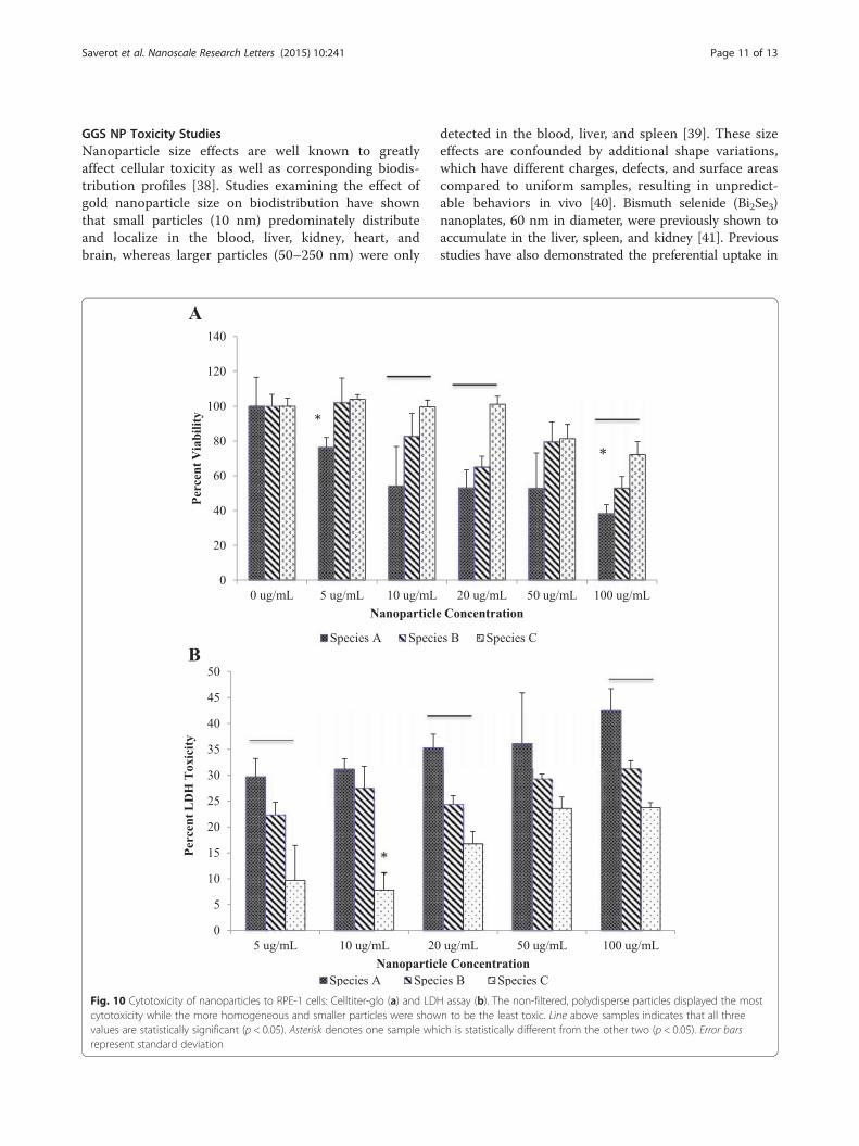
GGS NP Toxicity StudiesNanoparticle size effects are well known to greatlyaffect cellular toxicity as well as corresponding biodis-tribution profiles [38]. Studies examining the effect ofgold nanoparticle size on biodistribution have shownthat small particles (10 nm) predominately distributeand localize in the blood, liver, kidney, heart, andbrain, whereas larger particles (50–250 nm) were only
Fig. 10 Cytotoxicity of nanoparticles to RPE-1 cells: Celltiter-glo (a) and LDcytotoxicity while the more homogeneous and smaller particles were showvalues are statistically significant (p < 0.05). Asterisk denotes one sample whrepresent standard deviation
detected in the blood, liver, and spleen [39]. These sizeeffects are confounded by additional shape variations,which have different charges, defects, and surface areascompared to uniform samples, resulting in unpredict-able behaviors in vivo [40]. Bismuth selenide (Bi2Se3)nanoplates, 60 nm in diameter, were previously shown toaccumulate in the liver, spleen, and kidney [41]. Previousstudies have also demonstrated the preferential uptake in
H assay (b). The non-filtered, polydisperse particles displayed the mostn to be the least toxic. Line above samples indicates that all threeich is statistically different from the other two (p < 0.05). Error bars

Saverot et al. Nanoscale Research Letters (2015) 10:241 Page 12 of 13
macrophages of spherical particles over those with highaspect ratios [42].To determine the impact of sample purification on
cytotoxicity, the aforementioned three different nanopar-ticle populations (species A, B, and C) were evaluated.Samples were prepared at comparable concentrationsranging from 5 to 100 μg/mL. Prior to conducting experi-ments, particle stability in culture media was analyzed.Toxicity effects can be skewed by the effect of nanoparti-cles settling in the media over time. While all sampleswere stable for 24 h, the nanoplate fraction (species B)showed the least stability, as evidenced by the greatertemporal decrease in the maximum peak absorbance(Fig. 9). This effect is expected due to the larger size, andincreased surface area, of the nanoplates, resulting inincreased settling and possible protein adsorption. How-ever, this effect only resulted in 6 % loss in stability. Usingboth cell viability and LDH assays, our analyses showedthat species A resulted in the greatest cytotoxicity,whereas species C resulted in the least toxicity (Fig. 10).While previous studies have stated that smaller particlesare more toxic than larger nanoparticles of the sameshape due to enhanced surface area, shape effects tend todominate [38, 34]. Specifically, nanoplates are reportedlymore toxic than spherical particles due to their propen-sity for surface defects [40]. However, our results suggestthat polydispersity (with the most variation in both sizeand shape) may result in the greatest cytotoxicity. Whilewe demonstrate that variations in both shape and sizeimpact toxicity using simplified 2D assays, these effectsare also likely to be exacerbated when moving into 3Dassays and animal models. Sample polydispersity not onlyaffects cellular uptake and toxicity but likely provides un-desirable variations in biodistribution in vivo [42]. Thesefindings further dictate the need to produce well charac-terized and monodisperse samples prior to advancing toclinical nanomedicine trials.
ConclusionsConventional one-step synthesis of NIR GGS NP wasoriginally proposed as having an icosahedral, sulfide core-gold shell nanostructure, yet fabrication results in a poly-disperse solution of nanoplates, icosahedral nanoparticles,and irregularly shaped asymmetric nanoparticles. Subse-quent size-selective separation and compositional analysisreveal that the elemental composition of these particles isFCC gold and that nanoplates are the predominant NIR-contributing species, not the icosahedral nanoparticles asoriginally hypothesized. Here, we optimized two filtrationmethods, both of which facilitated nanoparticle separ-ation, with microfiltration providing the most reprodu-cible and rapid fractionation. Importantly, the originalpolydisperse samples produced by the conventional one-step reduction of chloroauric acid by sodium thiosulfate
resulted in enhanced cellular toxicity in vitro compared tomore purified samples. While further work is needed toelucidate the toxic properties of nanomaterials universally,this study highlights the need to fully characterize andoptimize nanomaterials before translating into more com-plex and biologically relevant animal and pre-clinicalmodels. This upstream analysis will expedite the develop-ment of highly promising translational nanomaterials forin vivo applications and ultimate FDA approval.
AbbreviationsNIR: near-infrared; TEM: transmission electron microscopy; EDS: energydispersive x-ray spectroscopy; FCC: face center cubic; SPR: surface plasmonresonance; GGS NP: gold-gold sulfide nanoparticle; FDA: Food and DrugAdministration; OD: optical density; SAED: selected area electron diffraction;FFT: fast Fourier transform; RPE-1: hTERT-RPE-1; LDH: lactose dehydrogenase;ATP: adenosine triphosphate; ANOVA: analysis of variance.
Competing InterestsThe authors declare that they have no competing interests.
Authors’ ContributionsSS and LMR carried out the synthesis of gold nanoparticles and opticalmeasurements. SS completed the stability studies. LMR completedsize-selective separation and toxicity studies. SS, LMR, PJK, and LRB preparedthe manuscript. LRB supervised the experimental design, data interpretation,and discussions. PJK provided assistance with the experimental design.DC provided the RPE-1 cells and participated in the interpretation ofcorresponding experimental data and discussions as well as editing of themanuscript. All authors read and approved the final manuscript.
Authors’ InformationSS is a bachelor degree student from the Department of Biological SystemsEngineering, Virginia Polytechnic and State Institute. LMR and LRB are fromthe Department of Biomedical Engineering and Mechanics, VirginiaPolytechnic and State Institute. LMR has graduated with a master’s degree.LRB is an assistant professor. LMR, PJK, and LRB are affiliates of the VirginiaTech Center for Sunstainable Nanotechnology. PJK is from the Departmentof Civil and Environmental Engineering, Virginia Polytechnic and StateInstitute. PJK is a professor. DC is from the Department of Biological Sciencesand Virginia Bioinformatics Institute, Virginia Polytechnic and State Institute.DC is an associate professor.
AcknowledgementsThe authors would like to thank Chris Winkler from the NanoparticleCharacterization and Fabrication Laboratory (NCFL) for the assistance withnanoparticle characterization and imaging. We also thank Kathy Lowe(Virginia Maryland College of Veterinary Medicine) for the assistance withTEM imaging and Krystal Le (Department of Biochemistry) for the help withnanoparticle fabrication. Special thanks to the Virginia Tech Institute forCritical Technology and Applied Science (ICTAS) and the Virginia Tech Centerfor Sustainable Nanotechnology for the generous funding support.
Author details1Department of Biological Systems Engineering, Virginia Tech, Seitz Hall,Blacksburg, VA 24061, USA. 2Department of Biomedical Engineering andMechanics, Virginia Tech, 325 Stanger Street, 310 Kelly Hall (MC 0298),Blacksburg, VA 24061, USA. 3Virginia Tech Center for SustainableNanotechnology, Virginia Tech, Kelly Hall, Blacksburg, VA 24061, USA.4Department of Biological Sciences and Virginia Bioinformatics Institute,Virginia Tech, 1015 Life Science Circle, Blacksburg, VA 24061, USA.5Department of Civil and Environmental Engineering, Virginia Tech, DurhamHall, Blacksburg, VA 24061, USA. 6Department of Mechanical Engineering,Virginia Tech, Kelly Hall, Blacksburg, VA 24061, USA.
Received: 6 April 2015 Accepted: 15 May 2015
Saverot et al. Nanoscale Research Letters (2015) 10:241 Page 13 of 13
References1. Young JK, Figueroa ER, Drezek RA. Tunable nanostructures as
photothermal theranostic agents. Ann Biomed Eng. 2012;40(2):438–59.doi:10.1007/s10439-011-0472-5.
2. Millstone JE, Hurst SJ, Metraux GS, Cutler JI, Mirkin CA. Colloidalgold and silver triangular nanoprisms. Small. 2009;5(6):646–64.doi:10.1002/smll.200801480.
3. Kennedy LC, Bickford LR, Lewinski NA, Coughlin AJ, Hu Y, Day ES, et al. Anew era for cancer treatment: gold-nanoparticle-mediated thermaltherapies. Small. 2011;7(2):169–83. doi:10.1002/smll.201000134.
4. Ren L, Chow GM. Synthesis of nir-sensitive Au-Au2S nanocolloids for drugdelivery. Mater Sci Eng C. 2003;23(1):113–6. doi:10.1016/S0928-4931(02)00247-3.
5. Gobin AM, Watkins EM, Quevedo E, Colvin VL, West JL. Near-infrared-resonant gold/gold sulfide nanoparticles as a photothermal cancertherapeutic agent. Small. 2010;6(6):745–52. doi:10.1002/smll.200901557.
6. Sun X, Zhang G, Patel D, Stephens D, Gobin AM. Targeted cancer therapy byimmunoconjugated gold-gold sulfide nanoparticles using protein G as acofactor. Ann Biomed Eng. 2012;40(10):2131–9. doi:10.1007/s10439-012-0575-7.
7. Day ES, Bickford LR, Slater JH, Riggall NS, Drezek RA, West JL. Antibody-conjugated gold-gold sulfide nanoparticles as multifunctional agents forimaging and therapy of breast cancer. Int J Nanomedicine. 2010;5:445–54.
8. Li Y, Gobin AM, Dryden GW, Kang X, Xiao D, Li SP, et al. Infraredlight-absorbing gold/gold sulfide nanoparticles induce cell death inesophageal adenocarcinoma. Int J Nanomedicine. 2013;8:2153–61.doi:10.2147/ijn.s37140.
9. Zhou HS, Honma II, Komiyama H, Haus JW. Controlled synthesis andquantum-size effect in gold-coated nanoparticles. Phys Rev B CondensMatter. 1994;50(16):12052–6.
10. Averitt RD, Sarkar D, Halas NJ. Plasmon resonance shifts of Au-coated Au2Snanoshells: insight into multicomponent nanoparticle growth. Phys Rev Lett.1997;78(22):4217–20.
11. Zhang G, Jasinski JB, Howell JL, Patel D, Stephens DP, Gobin AM.Tunability and stability of gold nanoparticles obtained from chloroauricacid and sodium thiosulfate reaction. Nanoscale Res Lett. 2012;7(1):337.doi:10.1186/1556-276x-7-337.
12. Schwartzberg AM, Grant CD, van Buuren T, Zhang JZ. Reduction of HAuCl4by Na2S revisited: the case for Au nanoparticle aggregates and againstAu2S/Au core/shell particles†. J Phys Chem C. 2007;111(25):8892–901.doi:10.1021/jp067697g.
13. Norman TJ, Grant CD, Magana D, Zhang JZ, Liu J, Cao D, et al. Near infraredoptical absorption of gold nanoparticle aggregates. J Phys Chem B.2002;106(28):7005–12. doi:10.1021/jp0204197.
14. Diao JJ, Chen H. Near infrared surface plasmon resonance of gold tabularnanostructures in the HAuCl4-Na2S reaction. J Chem Phys.2006;124(11):116103. doi:10.1063/1.2177661.
15. Likhatskii MN, Mikhlin YL. Influence of sulfide ions on the formation andproperties of gold nanoparticles in aqueous solutions. Glas Phys Chem.2007;33(4):422–5. doi:10.1134/S1087659607040189.
16. Morris T, Copeland H, Szulczewski G. Synthesis and characterization of goldsulfide nanoparticles. Langmuir. 2001;18(2):535–9. doi:10.1021/la011186y.
17. Clancy MK. Chapter 23—clinical translation and regulations of theranostics.In: Chen X, Wong S, editors. Cancer Theranostics. Oxford: Academic; 2014.p. 439–56.
18. Young J, Drezek R, editors. Synthesis of Au2S/Au Core/shell nanostructures.Optics in the life sciences. Monterey, California: Optical Society of America;2011. 2011/04/04.
19. Majimel J, Bacinello D, Durand E, Vallée F, Tréguer-Delapierre M. Synthesis ofhybrid gold-gold sulfide colloidal particles. Langmuir. 2008;24(8):4289–94.doi:10.1021/la702829w.
20. Pelaz B, Grazu V, Ibarra A, Magen C, del Pino P, de la Fuente JM. Tailoringthe synthesis and heating ability of gold nanoprisms for bioapplications.Langmuir. 2012;28(24):8965–70. doi:10.1021/la204712u.
21. Patel D. A novel high yield process for gold sulfide nanoparticle synthesis viashifting equilibrium of self-assembly reaction: University of Louisville. 2012.
22. James KT, O’Toole M, Patel D, Zhang G, Gobin AM, Keynton RS. A high yield,controllable process for producing tunable near infrared-absorbing goldnanoplates. RSC Advances. 2015. doi:10.1039/C4RA14889D.
23. Choi HS, Frangioni JV. Nanoparticles for biomedical imaging: fundamentalsof clinical translation. Mol Imaging. 2010;9(6):291–310.
24. Haiss W, Thanh NT, Aveyard J, Fernig DG. Determination of size andconcentration of gold nanoparticles from UV-vis spectra. Anal Chem.2007;79(11):4215–21. doi:10.1021/ac0702084.
25. Wu W, Huang J, Wu L, Sun D, Lin L, Zhou Y, et al. Two-step size- andshape-separation of biosynthesized gold nanoparticles. Sep Purif Technol.2013;106:117–22. http://www.sciencedirect.com/science/article/pii/S1383586613000208.
26. Hirsch LR, Halas NJ, West JL. Whole-blood immunoassay facilitated by goldnanoshell-conjugate antibodies. Methods Mol Biol. 2005;303:101–11.doi:10.1385/1-59259-901-x:101.
27. Laboratories C. Infinity telomerase-immortalized cell line culturingguide. 1999.
28. Gliga AR, Skoglund S, Wallinder IO, Fadeel B, Karlsson HL. Size-dependentcytotoxicity of silver nanoparticles in human lung cells: the role of cellularuptake, agglomeration and Ag release. Part Fibre Toxicol. 2014;11:11.doi:10.1186/1743-8977-11-11.
29. Van der Eycken EV. Aqueous microwave assisted chemistry. Synthesis andcatalysis. RSC green chemistry series. In: Polshettiwar V, Varma RS, editors.Angew Chem Int Ed. 2010;49(52):10039–40. doi:10.1002/anie.201006427.
30. Chen HM, Liu RS, Jang LY, Lee JF, Hu SF. Characterization of core–shelltype and alloy Ag/Au bimetallic clusters by using extended X-rayabsorption fine structure spectroscopy. Chem Phys Lett. 2006;421(1–3):118–23.http://dx.doi.org/10.1016/j.cplett.2006.01.043.
31. Castaño V, Gómez A, Yacaman MJ. Microdiffraction and surfacestructure of small gold particles. Surf Sci Lett. 1984;146(2–3):L587–92.http://dx.doi.org/10.1016/0167-2584(84)90756-4.
32. Marks LD. Experimental studies of small particle structures. Rep Prog Phys.1994;57(6):603.
33. Daeha S, Hyunjoon S. Synthesis of gold nanoparticles in liquid phase. Goldnanoparticles for physics, chemistry and biology. London: Imperial CollegePress; 2011. p. 103–38.
34. Kumar DVR, Kulkarni AA, Prasad BLV. Synthesis of triangular nanoplates: roleof bromide ion and temperature. Colloids Surf A: Physiochem Eng Asp.2013;422:181–90.
35. Mountrichas G, Pispas S, Kamistos E. Effect on temperature on the directsynthesis of gold nanoparticles mediated by poly(dimethylaminoethylmethacrylate) homopolymer. J Phys Chem C. 2014;118:22754–9.
36. Liu H, Yang Q. A two-step temperature-raising process to gold nanoplateswith optical and surface enhanced Raman spectrum properties. Cryst EngComm. 2011;13:2281.
37. Patel DN, James KT, O’Toole MG, Zhang G, Keynton RS, Gobin AM. A highyield, one-pot dialysis-based process for self-assembly of near infraredabsorbing gold nanoparticles. J Colloids Interface Sci. 2015;441:10–6.
38. Pan Y, Neuss S, Leifert A, Fischler M, Wen F, Simon U, et al. Size-dependent cytotoxicity of gold nanoparticles. Small. 2007;3(11):1941–9.doi:10.1002/smll.200700378.
39. Krystek P. A review on approaches to bio distribution about gold and silverengineered nanoparticles by inductively coupled plasma massspectrometry. Microchem J. 2012;105:39–43.
40. George S, Lin S, Ji Z, Thomas CR, Li L, Mecklenburg M, et al. Surface defectson plate-shaped silver nanoparticles contribute to its hazard potential in afish gill cell line and zebrafish embryos. ACS Nano. 2012;6(5):3745–59.doi:10.1021/nn204671v.
41. Zhang X-D, Chen J, Min Y, Park GB, Shen X, Song S-S, et al. MetabolizableBi2Se3 nanoplates: biodistribution, toxicity, and uses for cancer radiationtherapy and imaging. Adv Funct Mater. 2014;24(12):1718–29.
42. Toy R, Peiris PM, Ghaghada KB, Karathanasis E. Shaping cancernanomedicine: the effect of particle shape on the in vivo journey ofnanoparticles. Nanomedicine. 2014;9(1):121–34.