characterization of diversity of fungi forming arbuscular
TRANSCRIPT

Graduate Theses, Dissertations, and Problem Reports
1998
Characterization of diversity of fungi forming arbuscular Characterization of diversity of fungi forming arbuscular
endomycorrhizae in selected plant communities endomycorrhizae in selected plant communities
Sidney Luiz Sturmer West Virginia University
Follow this and additional works at: https://researchrepository.wvu.edu/etd
Recommended Citation Recommended Citation Sturmer, Sidney Luiz, "Characterization of diversity of fungi forming arbuscular endomycorrhizae in selected plant communities" (1998). Graduate Theses, Dissertations, and Problem Reports. 3151. https://researchrepository.wvu.edu/etd/3151
This Dissertation is protected by copyright and/or related rights. It has been brought to you by the The Research Repository @ WVU with permission from the rights-holder(s). You are free to use this Dissertation in any way that is permitted by the copyright and related rights legislation that applies to your use. For other uses you must obtain permission from the rights-holder(s) directly, unless additional rights are indicated by a Creative Commons license in the record and/ or on the work itself. This Dissertation has been accepted for inclusion in WVU Graduate Theses, Dissertations, and Problem Reports collection by an authorized administrator of The Research Repository @ WVU. For more information, please contact [email protected].
CHARACTERIZATION OF DIVERSITY OF FUNGI
FORMING ARBUSCULAR ENDOMYCORRHIZAE
IN SELECTED PLANT COMMUNITIES
Sidney L. Stürmer
Dissertation submitted to the College of Agriculture, Forestry and ConsumerSciences of West Virginia University in partial fulfillment of the
requirements for the degree of
Doctor in Philosophyin
Developmental Biology
Joseph Morton, ChairJonathan Cumming
Keith GarbuttDaniel Panaccione
Alan Sexstone
December 11, 1998Morgantown, West Virginia
Keywords: Arbuscular Mycorrhizal Fungi, Systematics, Taxonomy, Ecology

CHARACTERIZATION OF DIVERSITY OF FUNGI FORMING ARBUSCULAR
ENDOMYCORRHIZAE IN SELECTED PLANT COMMUNITIES
Sidney L. Stürmer
(ABSTRACT)
Knowledge of taxonomic and functional diversity of arbuscular mycorrhizal fungi (Glomales,Zygomycetes) is important in understanding the biology and ecology of such a widespreadmycorrhizal symbiosis. More definitive criteria for grouping and ranking taxa were establishedby reinterpreting morphological characters in selected species of Glomus (Glomaceae) andAcaulospora and Entrophospora (Acaulosporaceae) based on spore ontogenesis. Members ofboth families shared some patterns, such as discrete stages of spore growth and differentiationin three phases, resulting in the formation of three possible character complexes: a spore wall,flexible inner walls, and a pregermination structure. Families differed in presence or absenceof one or more of these complexes. Genera diverged in subcellular organization of thesecomplexes, and species diverged solely with changes in structure and phenotypic differences inthe spore wall. With improved criteria for species delimitation, studies then were conducted toassess taxonomic structure in fungal communities. This process required methods to induce orstimulate sporulation by cryptic fungal species, such as successive propagation cycles of trappot cultures. Trap cultures varied widely in their efficiency to detect non-sporulating species(15-100%), possibly because of differences in the environment, especially temperature,between the native habitat and greenhouse conditions. Glomus species dominated all baitedcommunities, both in number and amount of sporulation. Infectivity of total fungal propaguleswas predicted by spore numbers, but infectivity was not correlated with species richness.Inocula of single isolates from three fungal communities were established from trap cultures toexamine the proportion of effective and non-effective fungi present in each community.Effectiveness assays using soybean and red clover as hosts revealed that at least one fungalisolate from each community was effective in increasing plant growth and phosphorus foliarcontent. Fungal isolates that were highly effective or non-effective produced similar responsesin both hosts, suggesting that effectiveness has a heritable component. A mix of all membersof each fungal community performed as well or better than the most effective isolate in eachcommunity. These results suggest that management of indigenous fungal communities may bea preferred strategy to introduction of commercial inocula in natural or managed ecosystems.

iii
ACKNOWLEDGEMENTS
I would like to thank my advisor Dr. Joe Morton for all his guidance, support,friendship and criticisms that helped me to be a better researcher and person.
I thank all faculty members of the Plant Pathology and Environmental Microbiology,and Dr. Daniel Panaccione, Dr. Alan Sexstone, Dr. Jonathan Cumming and Dr. Keith Garbuttfor their guidance as members of my committee. Also, thanks to Dr. Edwin Townsend for hishelp and advice on the statistical analyses.
I thank West Virginia University and CNPq-Brazil for financial support.
Also, a warm thank to Dr. Steve Bentivenga who helped me in many ways while atWVU and Marlise Franke-Snyder for being genuine friends.
My thanks to Bill Wheeler and Beth Thomas for helping me with my experiments andto Kris Nichols, Kelly Heldreth and Nancy Arnold for being such nice friends all the time.
I also would like to thanks my former advisor, Dr. Margarida de Mendonça, for allencouragement and support she gave me during my initial steps on my career, and alsoeverybody in the mycorrhizal laboratory in Florianópolis, SC.
My very special thanks to all my Brazilian friends I made here: Edson, Marcia,Alexandre, Karina, Luciana, and Neto who made my days here more pleasant, and especiallyMara (mãezona) for her happiness, support and good cooking.
To my family, especially my parents Noir and Maria Stürmer, and my brothers Pabloand Diego Stürmer, for their love and support for all these years and throughout my life(obrigado de coração!!).
And, to Carla Ferreira, my soul-mate and better-half for her immense support on the laststeps of this journey, thank you for your wonderful and unconditional love.

iv
TABLE OF CONTENTS Page
ABSTRACT...........................................................................................................iiACKNOWLEDGEMENTS................................................................................... iiiTABLE OF CONTENTS....................................................................................... ivLIST OF TABLES ................................................................................................ viLIST OF FIGURES.............................................................................................. viiiINTRODUCTION..................................................................................................1
Chapter 1 – Developmental patterns defining morphological charactersin spores of four species in Glomus ......................................................................6
Introduction ............................................................................................................7Material and Methods .............................................................................................7
Experimental isolates .........................................................................................7Experimental design ...........................................................................................8Stages of spore differentiation ............................................................................8Spore germination ..............................................................................................9
Results....................................................................................................................9Differentiation of Glomus etunicatum spores......................................................9Differentiation of Glomus clarum spores ...........................................................10Differentiation of Glomus intraradices spores ...................................................10Differentiation of Glomus claroideum spores ....................................................11Taxonomic analysis...........................................................................................11
Discussion .............................................................................................................12Developmental interpretation of spore structure.................................................12Interpretation of spore developmental stages .....................................................13Phylogenetic interpretation of developmental sequences....................................13
Literature Cited .....................................................................................................14
Chapter 2 – Taxonomic reinterpretation of morphological charactersin Acaulosporaceae based on developmental patterns in twoAcaulospora and one Entrophospora species.......................................................21
Introduction ...........................................................................................................22Material and Methods ............................................................................................22
Experimental isolates ........................................................................................22Experimental design ..........................................................................................22Stages of spore development .............................................................................23
Results...................................................................................................................23Differentiation of Acaulospora laevis spores .....................................................24Differentiation of Acaulospora spinosa spores...................................................24Differentiation of Entrophospora colombiana spores ........................................25Taxonomic analysis...........................................................................................25
Discussion .............................................................................................................26Literature Cited .....................................................................................................27

v
Chapter 3 – Diversity of arbuscular mycorrhizal fungi in selected habitatsusing trap cultures methodology.........................................................................34Introduction ...........................................................................................................35Material and Methods ............................................................................................36
AMF in Five Soils .............................................................................................36Study sites.....................................................................................................36Experimental procedure ................................................................................37Infectivity assay ............................................................................................37Spore extraction ............................................................................................37
AMF in Sand Dunes ..........................................................................................38Study sites.....................................................................................................38Experimental procedure ................................................................................38Infectivity assay ............................................................................................38
Statistical analysis..................................................................................................39Results...................................................................................................................39
Species diversity in five soils ........................................................................39Infectivity in five soils ..................................................................................40Species diversity in sand dunes .....................................................................40Infectivity in dunes .......................................................................................41Regression models of fungal diversity in dunes.............................................41
Discussion .............................................................................................................41Literature Cited .....................................................................................................46
Chapter 4 – Effect of different fungal isolates from the same mycorrhizalcommunity on plant growth and phosphorus uptake in soybean andred clover .............................................................................................................73
Introduction ...........................................................................................................74Material and Methods ............................................................................................75
Experimental isolates ........................................................................................75Growth medium ................................................................................................75Infectivity assay ................................................................................................75Experiment 1.....................................................................................................76Experiment 2.....................................................................................................77Statistical analysis .............................................................................................77
Results...................................................................................................................77Experiment 1.....................................................................................................77Experiment 2.....................................................................................................78
Discussion .............................................................................................................78Literature Cited .....................................................................................................80
CONCLUSIONS...................................................................................................90VITA.....................................................................................................................93

vi
LIST OF TABLES
Chapter 3 Page
1 Environmental and soil chemical properties of the 5 selected habitatswithin the U.S.A. .................................................................................................49
2 Environmental and soil chemical properties of the sand dunes habitatsstudied. ................................................................................................................50
3 Spore abundance of AMF species present in a revegetated site inCalifornia (CA-R), as detected from field soil and over two cyclesof trap cultures. ....................................................................................................51
4 Spore abundance of AMF species present in a prairie site in Kansas(KS), as detected from field soil and over two cycles of trapcultures. ...............................................................................................................52
5 Spore abundance of AMF species present in a corn field in Minnesota(MN), as detected from field soil and over two cycles of trap cultures. .................53
6 Spore abundance of AMF species present in a abandoned mine site inWest Virginia (WV), as detected from field soil and over two cycles of trap cultures. ...................................................................................................54
7 Similarity coefficients for AMF from five habitats within the U.S.A. ...................55
8 Spore abundance of AMF fungal species present in a sand dune site inBrazil, extracted from field soil and two cycles of trap cultures. ...........................56
9 Spore abundance of AMF fungal species present in a sand dune site inU.S.A., extracted from field soil and two cycles of trap cultures...........................57
10 Spore abundance of AMF fungal species present in a sand dune site inJapan, extracted from field soil and two cycles of trap cultures.............................58
11 Spore abundance of AMF fungal species present in a sand dune site inNetherlands, extracted from field soil and two cycles of trap cultures...................59
12 Similarity coefficients for AMF from four sand dune habitats. .............................60
13 Summary of simple and multiple linear regressions of spore numberof the most abundant species in each soil considered singly or incombination to predict levels of colonization in infectivity assays. .......................61
14 Summary of stepwise regression of soil and environmental characteristicsto predict number of AMF species in all nine habitats studied. .............................62
15 Summary of stepwise regression considering two factors togetherinfluencing number of AMF species in all nine soils studied. ...............................63
Chapter 4
1 Fungal species recovered from bait cultures of rhizosphere soilfrom three plant communities...............................................................................83

vii
2 Mycorrhizal root length of 7 weeks old soybean plants. .......................................84
3 Mycorrhizal root length of 8 weeks old red clover plants......................................85

viii
LIST OF FIGURES
Introduction Page
1 Classification of arbuscular mycorrhizal fungi in Glomales. ..................................5
Chapter 1
1 Murographic representation of the linear sequence in differentiation oflayers in the maturing spore wall of Glomus etunicatum, G. clarum, G.intraradices and G. claroideum. ...........................................................................16
2-7 Glomus etunicatum UT315 - stages in the differentiation of sporewall layers in spores mounted in PVLG + Melzer’s reagent (1:1, v/v). .................17
8-13 Glomus clarum FL979A - stages in the differentiation of spore walllayers in spores mounted in PVLG + Melzer’s reagent (1:1, v/v)..........................18
14-19 Glomus intraradices KS906 - stages in the differentiation of sporewall layers in spores mounted in PVLG + Melzer’s reagent (1:1, v/v). .................19
20-25 Glomus claroideum BR147A - stages in the differentiation of sporewall layers in spores mounted in PVLG + Melzer’s reagent (1:1, v/v). .................20
Chapter 2
1 Murographic representation of the sequence of development of layersin spores of (A) Acaulospora laevis and A. spinosa, and (B)Entrophospora colombiana. .................................................................................30
2-7 Acaulospora laevis AU211 - stages in the differentiation of sporewall and inner walls layers in spores mounted in PVLG + Melzer’sreagent (1:1, v/v)..................................................................................................31
8-13 Acaulospora spinosa WV861A - stages in the differentiation of sporewall and inner wall layers in spores mounted in PVLG + Melzer’sreagent (1:1, v/v)..................................................................................................32
14-19 Entrophospora colombiana CL356 - stages in the differentiation ofspore wall and inner wall layers in spores mounted in PVLG + Melzer’sreagent (1:1, v/v)..................................................................................................33
Chapter 3
1 Number of AMF species recovered from the field and after successivepropagation cycles of trap cultures from 5 habitats in the U.S.A...........................63
2 Species rank-log abundance curves for AMF community recovered inthe field and after two cycles of trap cultures for the five habitatsin the U.S.A. ........................................................................................................64
3 Arbuscular mycorrhizal colonizaton after 21-day infectivity assaysfor field soil, first and second trap cycles from 5 habitats in the U.S.A. ................65

ix
4 Cluster analysis resulting from the average method of soil chemicaland environmental properties by the four sand dune habitats studied. ...................66
5 Number of AMF species recovered from the field and after successivecycles of trap cultures from sand dunes habitats in four countries. ........................67
6 Species rank-log abundance curves for AMF community recoveredin the field and after two cycles of trap cultures for all sand duneshabitats. ...............................................................................................................68
7 Mycorrhizal colonization after 21-day infectivity assays for fieldsoil, first and second trap cycles from four sand dunes habitats. ...........................69
8 The relationship between the number of AMF species and the percentageof colonization of 21-day infectivity assays within the same propagationcycle for sand dune habitats studied (r2 < 0.01, P = 0.93)......................................70
9 The relationship between the percentage colonization of 21-dayinfectivity assays and number of spores detected within the samepropagation cycle for sand dunes habitats studied (r2 < 0.52, P < 0.01).................71
Chapter 4
1 Shoot dry weight of soybean plants inoculated with AMF isolatesfrom three different communities. ........................................................................86
2 Phosphorus content of soybean shoots inoculated with AMF isolatesfrom three different communities. ........................................................................87
3 Shoot dry weight of red clover plants inoculated with AMF isolatesalone or in combination........................................................................................88
4 Phosphorus content of red clover shoots inoculated with AMF isolatesalone or in combination........................................................................................89

1
INTRODUCTION
Mycorrhizae are global associations between roots of terrestrial plants and cosmopolitansoil-borne fungi (Smith and Read, 1997). Among seven different types of mycorrhizalassociations, the one formed by arbuscular mycorrhizal fungi in the order Glomales,Zygomycetes (Fig. 1) is the most common and widespread in nature (Brundrett, 1991).
Arbuscular mycorrhizal fungi (AMF) are hypothesized to have played a crucial role inthe evolution of land plants (Pirozynski and Malloch, 1975), as evidenced by mycorrhiza-likefossils in tissues of Rynia and Asteroxylon from the Devonian (Pirozynski, 1981). Structuresdiscovered in root fossils from the Triassic also resemble vesicles and intraradical spores ofextant AMF (Stubblefield et al., 1987), and arbuscules-like structure were observed in plantfossils dating from the Devonian (Taylor et al., 1995). Using the small-subunit rRNA as amolecular clock, the origin of AM-like fungi has been estimated at 353-462 Myr ago,coincident with emergence of land plants (Simon et al., 1993). Morton (1990) hypothesizedthat the arbuscular mycorrhizal association coevolved with plants since that time, explainingthe widespread presence of AMF in most angiosperms and pteridophytes. AMF are absent inroots of genera of some plant families (Trappe and Luoma, 1992; Peat and Fitter, 1993), apattern suggestive of an association that was shared by all ancestral plants groups and then lostsporadically by some taxa in more recent times (Trappe, 1987). The broad distribution range ofAMF in tropical and temperate forests, deserts, sand dunes, grasslands and agroecosystems(Brundrett, 1991; Hayman, 1982) appears to be a consequence of the extended time scale fordispersal and general lack of host specificity (Morton and Bentivenga, 1994).
Organisms of AMF have a bimodal pattern of differentiation (Morton, 1990). Thevegetative thallus consists of arbuscules, intraradical vesicles (shared only by species in thesuborder Glomineae), extraradical auxiliary cells (shared only by species in the suborderGigasporineae), and intraradical and extraradical hyphae (Smith and Read, 1997; Morton andBenny, 1990). Arbuscules are finely branched structures in close contact with the cell plasmamembrane, functioning in exchange of nutrients between host and fungal cells (Smith andRead, 1997). Hyphae are important in nutrient acquisition and as propagules to initiate newroot colonization (Graham et al., 1982; Friese and Allen, 1991). Vesicles are globose structuresarising from swelling of the hyphae and filled with glycogen granules and lipids and areconsidered to be storage structures (Bonfante-Fasolo, 1984; Brundrett, 1991). AMF reproduceasexually by means of spores formed in the soil or sometimes within roots (Morton, 1990).Spores function in dissemination and short-term survival of the organism and they also containmost of the morphological diversity for classification of species and higher taxa (Morton, 1988;Morton, 1990).
Types and organization of subcellular structures of asexual spores have been the basisfor taxonomy of glomalean fungi. Since the first Linnean classification by Gerdemann andTrappe (1974), progress involved standardization of definitions of discrete subcellularstructures (e.g., types of “spore walls”) in a wide range of taxa (Morton, 1988; Walker, 1983)followed by ontogenetic studies to provide more dynamic interpretations of characters and theirstates and to define their levels of taxonomic resolution (Morton and Bentivenga, 1994).
The focus on developmental processes is a well-established means to define charactersbased on their origin, transformational states during differentiation and sequence in which theyare formed (Gould, 1977). Comparative developmental studies in different taxa also provide

2
the basis to distinguish between phylogenetically informative homologous traits versusnoninformative analogous traits (Patterson, 1982). Franke and Morton (1994) were the first tocarry out rigorous comparative ontogenetic analysis between isolates of Scutellospora pellucidaand S. heterogama and determine origin and individuality of subcellular spore characters.Their results were extended to species of the two genera in the family Gigasporaceae,Scutellospora (Morton, 1995) and Gigaspora (Bentivenga and Morton, 1995). From theseontogenetic studies, a developmental model was proposed (Morton et al., 1995), whichidentified specific character complexes resolving species and higher taxa. However, furthertesting of the universality of this model is required by incorporating data from other families ofGlomales, Glomaceae and Acaulosporaceae.
The definition of grouping and ranking criteria among morphological characters isimportant in assessing taxonomic structure of AMF communities present in almost all plantcommunities worldwide. AMF are found in all habitats, and are absent only in highlydisturbed, eroded, or fumigated soils (Brundrett, 1991; Smith and Read, 1997). Speciesrichness even in soils limiting diversity of plant communities can be high, with 6-8 speciescoexisting in the same root system in many different habitats (Abbott and Robson, 1991;Brundrett, 1991; Morton et al., 1995). Johnson et al. (1991), examining AMF communitiesresulting from old field succession, found 25 species of AMF in 15 sites and Bever et al. (1996)found 23 species in 75m2 area of an old-field community near Durham, North Carolina. At aregional level, Koske (1987) detected 23 AMF species associate with dune plants along theAtlantic coast, with an average species richness of 4.9 and richness increasing along anincreasing temperature gradient southward.
Measurement of AMF species richness and abundance in natural habitats has beenlimited by methodology. Most surveys of AMF in the field are based on samples of field-collected spores, which often are present in such low numbers or so parasitized that accurateidentification is difficult. These data, at best, are informative in assessing species distributionand dispersal patterns. They do not provide an accurate measure of local fungal communitystructure, because nonsporulating species may be present and they are not detected in samples.Trap culture methodologies of field soil samples are only beginning to be used to inducesporulation by these cryptic species, and results thus far (Bever et al., 1996; Stutz and Morton,1996) suggest that greater than 50% of the fungi present in a root system are not detected bydirect field sampling protocols.
Once taxonomic structure of a fungal community is determined, there remains aquestion as to the functional contribution of each member to the mycorrhizal symbiosis in ahost plant. AMF can markedly influence plant growth and yield by enhancing uptake ofmineral nutrients, particularly phosphorus, in nutrient-poor soils (Azcon-Aguilar et al., 1982;Smith and Read, 1997). Shoot biomass and phosphorus uptake have been the main traitsanalyzed when investigating mycorrhizal benefits to plants.
Most studies conducted to date have interpreted results on the basis of plant hosts ofinterest and any fungal isolate that can be obtained, regardless of its origin or properties. Foran understanding of community dynamics, all of the fungi in each community should beisolated and studied individually and together. Procedures involve growing the fungi on planthosts in pot culture, since these fungal symbionts are obligate biotrophs (Morton et al., 1995).

3
My research seeks to accomplish the following objectives to integrate form andfunction: (i) reinterpret subcellular morphological characters in spores of species in Glomus(Glomaceae) and Acaulospora and Entrophospora (Acaulosporaceae) by assessing origin andboundary conditions during ontogenesis (Chapters 1 and 2); (ii) characterize taxonomicstructure of AMF communities in nine habitats by isolating and identifying AMF speciesinduced to sporulate as a result of successive propagation cycles of trap pot cultures (Chapter3); and, (iii) assess functional diversity of AMF isolated from three different communities bymeasuring relative mycorrhizal effectiveness on two mycotrophic assay hosts (Chapter 4).
LITERATURE CITED
Abbott, L.K., and A.D. Robson. 1991. Factors influencing the occurrence of vesicular-arbuscular mycorrhizas. Agriculture, Ecosystem and Environment 35:121-150.
Azcon-Aguilar, C., J.M. Barea, R. Azcon, and J. Olivares. 1982. Effectiveness of Rhizobiumand VA mycorrhiza in the introduction of Hedysarum coronarium in a new habitat.Agriculture, Ecosystem and Environment, 7:199-206.
Bentivenga, S. P., and J. B. Morton. 1995. A monograph of the genus Gigasporaincorporating developmental patterns of morphological characters. Mycologia 87:720-732.
Bever, J.D., J. Morton, J. Antonovics, and P.A. Schultz. 1996. Host-dependent sporulation andspecies diversity of arbuscular mycorrhizal fungi in a mown grassland. Journal of Ecology84:71-82.
Bonfante-Fasolo, P. 1984. Anatomy and morphology of VA mycorrhizae. Pp. 5-33. In: VAMycorrhiza. C.Ll. Powell and D.J. Bagyaraj (Eds.). CRC Press, Boca Raton, Florida.
Brundrett, M.C. 1991. Mycorrhizas in natural ecosystems. Advances in Ecological Research21:171-213.
Franke, M., and J. Morton. 1994. Ontogenetic comparisons of arbuscular mycorrhizal fungiScutellospora heterogama and Scutellospora pellucida: revision of taxonomic characterconcepts, species descriptions, and phylogenetic hypotheses. Canadian Journal of Botany72:122-134.
Friese, C.F., and M.F. Allen. 1991. The spread of VA mycorrhizal fungal hyphae in the soil:inoculum types and external hyphae architecture. Mycological Research 92:317-321.
Gerdemann, J.W., and J.M. Trappe. 1974. Endogonaceae in the Pacific Northwest. MycologiaMemoir 5:1-76.
Gould, S.J. 1977. Ontogeny and Phylogeny. Harvard University Press, Massachusetts. 501pp.
Graham, J.H., R.G. Linderman, and J.A. Menge. 1982. Development of external hyphae bydifferent isolates of mycorrhizal Glomus spp. in relation to root colonization and growth oftroyer citrange. New Phytologist 91:183-189.
Hayman, D.S. 1982. Influence of soils and fertility on activity and survival of vesicular-arbuscular mycorrhizal fungi. Phytopathology 72:1119-1125.
Johnson, N.C., D.R. Zak, D. Tilman, and F.L. Pfleger. 1991. Dynamics of vesicular-arbuscular mycorrhizae during old field succession. Oecologia 86:349-358.
Koske, R.E. 1987. Distribution of VA mycorrhizal fungi along a latitudinal temperaturegradient. Mycologia 79:55-68.
Morton, J.B. 1988. Taxonomy of VA mycorrhizal fungi: classification, nomenclature, andidentification. Mycotaxon 32:267-324.

4
Morton, J. B. 1990. Evolutionary relationships among arbuscular mycorrhizal fungi in theEndogonaceae. Mycologia 82:192-207.
Morton, J.B. 1995. Taxonomic and phylogenetic divergence among five Scutellospora speciesbased on comparative developmental sequence. Mycologia 87:127-137.
Morton, J.B., and G. L. Benny. 1990. Revised classification of arbuscular mycorrhizal fungi(Zygomycetes): a new order, Glomales, two new suborders, Glomineae andGigasporineae, and two new families, Acaulosporaceae and Gigasporaceae, with anemendation of Glomaceae. Mycotaxon 37:471-491.
Morton, J.B., and S.P. Bentivenga. 1994. Levels of diversity in endomycorrhizal fungi(Glomales, Zygomycetes) and their role in defining taxonomic and non-taxonomic groups.Plant and Soil 159:47-59.
Morton, J.B., S. P. Bentivenga, and J. D. Bever. 1995. Discovery, measurement, andinterpretation of diversity in symbiotic endomycorrhizal fungi (Glomales, Zygomycetes).Canadian Journal of Botany 73(suppl. 1):S25-S32.
Patterson, C. 1982. Morphological characters and homology. pp. 21-74. In: Problems ofPhylogenetic reconstruction. Eds., K. A. Joysey and A. E. Friday. Academic Press,London.
Peat, H.J., and A.H. Fitter. 1993. The distribution of arbuscular mycorrhizas in the Britishflora. New Phytologist 125:845-854.
Pirozynski, K.A. 1981. Interactions between fungi and plants through the ages. CanadianJournal of Botany 59:1824-1827.
Pirozynski, K.A., and D.W. Malloch. 1975. The origin of land plants: a matter ofmycotrophism. BioSystems 6:153-164.
Simon, L., J. Bousquet, R. C. Lévesque, and M. Lalonde. 1993. Origin and diversification ofendomycorrhizal fungi and coincidence with vascular land plants. Nature 363:67-69.
Smith, S.E., and D.J. Read. 1997. Mycorrhizal Symbiosis. Academic Press, London. 605 pp.Stubblefield, S.P., T.N. Taylor, and J.M. Trappe. 1987. Fossil mycorrhizae: a case for
symbiosis. Science 237:59-60.Stutz, J.C., and J.B. Morton. 1996. Successive pot cultures reveal high species richness of
arbuscular endomycorrhizal fungi in arid ecosystems. Canadian Journal of Botany74:1883-1889.
Taylor, T.N., W. Remy, H. Hass, and H. Kerp. 1995. Fossil arbuscular mycorrhizae from theEarly Devonian. Mycologia 87:560-573.
Trappe, J.M. 1987. Phylogenetic and ecological aspects of mycotrophy in the angiospermsfrom an evolutionary standpoint. Pp. 5-25. In: Ecophysiology of VA Mycorrhizal Plants.G. Safir (Ed.) CRC Press, Boca Raton, Florida.
Trappe, J.M., and D.L. Luoma. 1992. The ties that bind: fungi in ecosystems. Pp. 17-27. In:The Fungal Community – Its Organization and Role in the Ecosystem. G.C. Carroll andD.T. Wicklow (Eds.) Marcel Dekker, Inc., New York.
Walker, C. 1983. Taxonomic concepts in the Endogonaceae: spore wall characteristics inspecies descriptions. Mycotaxon 18:443-455.

GLOMINEAE GIGASPORINEAE
Glomus AcaulosporaEntrophospora Gigaspora Scutellospora
Fig. 1 Classification of arbuscular mycorrhizal fungi in Glomales
5

6
CHAPTER ONE
DEVELOPMENTAL PATTERNS DEFINING
MORPHOLOGICAL CHARACTERS IN SPORES
OF FOUR SPECIES IN GLOMUS
(Reprinted with permission from Mycologia, vol. 89, no. 1,
copyright 1997, The New York Botanical Garden.)

7
INTRODUCTION
Among arbuscular endomycorrhizal fungi in the order Glomales, 56% of the describedspecies are in the genus Glomus (Morton and Benny, 1990), including species formerly in thegenus Sclerocystis (Almeida and Schenck, 1990). Nucleotide sequence comparisons (Simon etal., 1993; Simon, 1996) suggest considerable genetic divergence within Glomus, whereasmorphological divergence appears to be much smaller (Morton, 1990). The disparity may residein interpretations of morphological characters based only on arbitrary differences in appearancerather than on empirical data.
Species in Glomus and other genera of Glomales are separated by morphologicalcharacters residing mostly in a variety of subcellular structures in spores that have been variouslydefined as "layers" (Berch, 1987), "walls" (Walker, 1983) or “zones” (Maia et al., 1993).Simplicity in design of these structures in Glomus confines the range of potential phenotypicvariation and thus increases the difficulty in determining whether similarities are putativehomologs (arising from a common ancestor) or analogs (arising from different ancestors)(Morton, 1990). Ontogenetic comparisons provide a means to assess positional and temporalrelationships among subcellular structures and thereby delimit character origins andtransformational states important in defining homologies (Patterson, 1982). Studies to date havefocused only on one family in Glomales, Gigasporaceae (Bentivenga and Morton, 1995; Frankeand Morton, 1994; Morton, 1995).
Most ontogenetic studies of species in the family Glomaceae (Giovannetti et al., 1991;Meier and Charvat, 1992; Wu and Sylvia, 1993) have focused on the mode of spore formationrather than patterns of differentiation of subcellular spore structures important in resolvingspecies. Morton (1996) first detailed spore differentiation of Glomus caledonium (Nicol. &Gerd.) Trappe & Gerd. from a reference culture and reinterpreted subcellular structuresdevelopmentally.
In this study, I compare spore differentiation sequences of four additional species inGlomus to determine shared and divergent patterns within the genus and with taxa in the familyGigasporaceae. These data are incorporated into a developmental model accommodating all taxain Glomales (Morton et al., 1995). The four Glomus species also are redescribed based ondevelopmental reinterpretation of taxonomically relevant characters.
MATERIAL AND METHODS
Experimental isolates
All isolates were obtained from accessions in the International Culture Collection ofArbuscular and Vesicular-arbuscular Mycorrhizal Fungi (INVAM) at West Virginia University.Accessions of G. etunicatum included BR218 (from S. Stürmer, Santa Catarina, Brazil), CL519(from E. Sieverding, Valle, Colombia), FL670 (from A. E. Dudeck, Florida, U.S.A.), KS884(from B. D. Hetrick, Kansas, U.S.A.), PA137 (from D. Douds, Pennsylvania, U.S.A.), UT135(from T. Wood, Native Plants, Inc., Salt Lake City, Utah, U.S.A.). Accessions of G. clarumconsisted of BR147B (from M-.T. Lin, São Paulo, Brazil), FL979A (from N. C. Schenck,Florida, U.S.A.). Accessions of G. intraradices included FL208 (from S. Nemec, Florida,U.S.A.), KS906 (cultured by B. D. Hetrick, Kansas, U.S.A.), UT126 (from T. Wood, NativePlants, Inc., Salt Lake City, Utah, U.S.A.). Glomus claroideum was represented by BR147A

8
(from M-.T. Lin, São Paulo, Brazil) and SC186 (from H. Skipper, Clemson, South Carolina,U.S.A.).
Bulk inoculum of each isolate was produced according to standardized protocolsdiscussed by Morton et al. (1993). Briefly, whole inoculum of an INVAM accession (a mix ofpotting medium, chopped roots, spores, hyphal fragments) was mixed 1:10 (v/v) with the samepotting medium previously steamed twice at 100oC. The potting medium consisted of a sandyloam soil (Lily series) premixed 1:2 (v/v) with quartzite sand (average grain size = 0.9 mm) andlimed after steaming to pH 6.2. The inoculated mixture was placed in 15-cm diameter plasticpots and seeded heavily (100-120 seeds) with sudangrass [Sorghum sudanense (Piper) Staph].Plants were grown for 4 months in a growth room under fluorescent lights with a photon fluxdensity of 245 µmol m -2 s-1 at pot level and a 14-hour photoperiod; room temperature rangedfrom 19 to 30oC during the culture period. At harvest, plants were dried in situ for 2-3 weeks at22-24oC, and the pot contents stored in plastic zip-loc bags at 4oC until sampled.
Experimental design
Whole inoculum of each isolate was mixed 1:5 (v/v) with the same growth mediumdescribed above and placed in six 4 × 21 cm cone-tainers™ (Stuewe and Sons, Inc., Corvallis,Oregon). Five to seven surface sterilized sudangrass seeds were placed in each cone-tainer andplants were grown under growth room conditions described above. Spores of each isolate wereextracted from one cone-tainer per week from the third to seventh weeks after seedlingemergence.
Another pot technique also was used to sequentially sample spores in all developmentalstages from the same culture. A cone-tainer containing inoculum of each isolate was seeded withsudangrass and grown for 10 weeks. The contents of a cone-tainer then were transferred to anylon mesh sleeve with 100-µm openings. The sleeve was transplanted into the center of a 15-cm diameter pot. Sterile growth medium (described above) was placed around the sleeve andseeded with sudangrass. A soil core was removed from the newly seeded regions of each pot atweekly intervals between four and eight wk after plant emergence.
Spores were extracted by agitating pot samples (30 ml) in a blender with water for 5-10seconds and forcing the contents through two nested sieves with 500- and 38-µm openings usinga water spray. Material retained on the 38-µm sieve was transferred to 50-cm3 tubes containing20% and 60% sucrose layers and centrifuged at 900 x g for 2 minutes. The supernatantcontaining suspended spores was decanted into a 38-µm sieve, washed with tap water for 1-2minutes, and then transferred to a Petri dish. Individual spores were collected using a Pasteurpipette extruded to a fine tip.
Stages of spore differentiation
Under a stereomicroscope, spores were separated into discrete classes based on color andopacity of spores that corresponded to progressively more mature states. Color was quantifiedby comparing spores and a printed color chart (INVAM color chart) illuminated simultaneouslywith a two-branch fiber optic illuminator at 3400 K. Color values consisted of percentage cyan,magenta, yellow, and black (C-M-Y-K). Maturity class I (most juvenile) spores weredistinguished as white with opaque contents. Maturity class II spores (intermediate) wererecognized by color or opacity of spore contents: G. etunicatum spores were tan (0-10-20-0 to 0-10-60-0) with contents opaque; G. clarum spores were white with a reflective spore wall layer
9
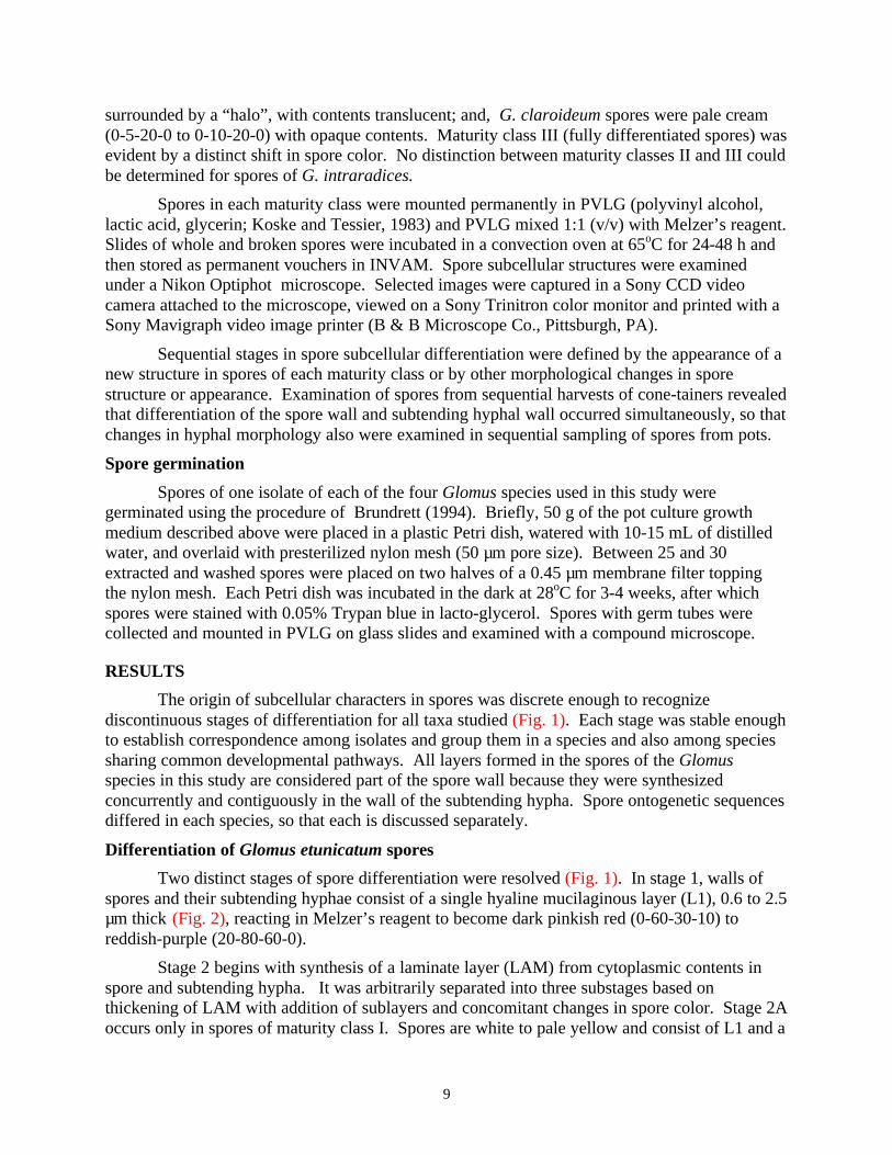
surrounded by a “halo”, with contents translucent; and, G. claroideum spores were pale cream(0-5-20-0 to 0-10-20-0) with opaque contents. Maturity class III (fully differentiated spores) wasevident by a distinct shift in spore color. No distinction between maturity classes II and III couldbe determined for spores of G. intraradices.
Spores in each maturity class were mounted permanently in PVLG (polyvinyl alcohol,lactic acid, glycerin; Koske and Tessier, 1983) and PVLG mixed 1:1 (v/v) with Melzer’s reagent.Slides of whole and broken spores were incubated in a convection oven at 65oC for 24-48 h andthen stored as permanent vouchers in INVAM. Spore subcellular structures were examinedunder a Nikon Optiphot microscope. Selected images were captured in a Sony CCD videocamera attached to the microscope, viewed on a Sony Trinitron color monitor and printed with aSony Mavigraph video image printer (B & B Microscope Co., Pittsburgh, PA).
Sequential stages in spore subcellular differentiation were defined by the appearance of anew structure in spores of each maturity class or by other morphological changes in sporestructure or appearance. Examination of spores from sequential harvests of cone-tainers revealedthat differentiation of the spore wall and subtending hyphal wall occurred simultaneously, so thatchanges in hyphal morphology also were examined in sequential sampling of spores from pots.
Spore germination
Spores of one isolate of each of the four Glomus species used in this study weregerminated using the procedure of Brundrett (1994). Briefly, 50 g of the pot culture growthmedium described above were placed in a plastic Petri dish, watered with 10-15 mL of distilledwater, and overlaid with presterilized nylon mesh (50 µm pore size). Between 25 and 30extracted and washed spores were placed on two halves of a 0.45 µm membrane filter toppingthe nylon mesh. Each Petri dish was incubated in the dark at 28oC for 3-4 weeks, after whichspores were stained with 0.05% Trypan blue in lacto-glycerol. Spores with germ tubes werecollected and mounted in PVLG on glass slides and examined with a compound microscope.
RESULTS
The origin of subcellular characters in spores was discrete enough to recognizediscontinuous stages of differentiation for all taxa studied (Fig. 1). Each stage was stable enoughto establish correspondence among isolates and group them in a species and also among speciessharing common developmental pathways. All layers formed in the spores of the Glomusspecies in this study are considered part of the spore wall because they were synthesizedconcurrently and contiguously in the wall of the subtending hypha. Spore ontogenetic sequencesdiffered in each species, so that each is discussed separately.
Differentiation of Glomus etunicatum spores
Two distinct stages of spore differentiation were resolved (Fig. 1). In stage 1, walls ofspores and their subtending hyphae consist of a single hyaline mucilaginous layer (L1), 0.6 to 2.5µm thick (Fig. 2), reacting in Melzer’s reagent to become dark pinkish red (0-60-30-10) toreddish-purple (20-80-60-0).
Stage 2 begins with synthesis of a laminate layer (LAM) from cytoplasmic contents inspore and subtending hypha. It was arbitrarily separated into three substages based onthickening of LAM with addition of sublayers and concomitant changes in spore color. Stage 2Aoccurs only in spores of maturity class I. Spores are white to pale yellow and consist of L1 and a

10
laminate layer that is 0.3 to 1.8 µm (mean = 0.9 µm) (Fig. 3). In stage 2B (maturity class II),spores become tan (0-20-60-0) as existing sublayers thicken and new sublayers are added (Fig.4). The laminate layer then ranges from 1.2 to 3.1 µm (mean = 2.0 µm) in thickness. The outermucilaginous layer begins to slough or degrade, varying from 0.3 to 1.8 µm thick on individualspores. In stage 2C, LAM terminates differentiation, at which time it is orange-brown (0-10-80-0 to 0-40-100-0) in color and 4.8 to 6.4 µm (mean = 5.4 µm) thick. In most spores, themucilaginous layer has completely sloughed (Fig. 5) at this point. Mature spores range from(60-)80-100(-140) µm in diameter. The innermost sublayer of LAM occasionally separatesslightly from the spore wall (Fig. 6) and resembles a thin flexible layer observed in spores of G.claroideum, but it is rare and not considered stable enough to be taxonomically informative.Germination occurs when a germ tube emerges from the lumen of subtending hypha (Fig. 7).Origin of the germ tube was not observed.
Differentiation of Glomus clarum spores
Three discrete stages were identified in spore differentiation (Fig. 1). In stage 1, thespore wall consists only of a highly plastic and hyaline mucilaginous layer (L1) (Fig. 8) thatproduces folds up to 15.8 µm in height (midrange of 1.2 to 4.7 µm) and stains pinkish-purple(20-80-20-0) in Melzer’s reagent.
Stage 2 is distinguished by synthesis of a rigid hyaline laminate layer (LAM = L2) with agranular consistency when broken (Fig. 9). This stage was partitioned into two substages basedon changes in color of whole spores. Spores in stage 2A are white with opaque contents and(40-)140-180(-280) µm in diameter. L2 increases in thickness from 0.5-0.6 µm at the outset(Fig. 9) to 1.1-5.6 µm (mean = 2.8 µm) at the end of this stage. In stage 2B, spore contentsbecome translucent and whole spores acquire a distinct “halo” in reflected light. L2 continuesdifferentiation, ranging from 2.5-7.5 µm (mean = 4.8 µm) thick (Fig. 10). L1 sloughs to varyingdegrees and appears patchy.
Stage 3 begins with synthesis of a second laminate layer (LAM = L3) that becomes paleyellow (0-0-20-0) to yellow (0-0-60-0) at maturity. This stage also was subdivided in arbitrarysubstages based on visible changes in spore wall components. Stage 3A begins with synthesis ofa pale yellow sublayer of L3 (Fig. 11), ranging from 0.3-0.6 µm (mean = 0.4 µm) thick. Wholespores remain white under reflected light. In stage 3B, both L2 and L3 thicken (Fig. 12) andthen terminate differentiation. L2 is 8.1-15.6 µm (mean = 11.2 µm) thick and L3 is 0.9-3.1 µm(mean = 1.8 µm) thick at maturity. L1 completely sloughs in most spores in this stage. Maturespores are (120-)180-200(-280) µm in diameter, and range in color from hyaline/white to darkyellow (0-0-80-0). Upon germination, the germ tube appears to arise from the innermostsublayer of the subtending hypha near the spore or at break points where branching of thesubtending hypha occurs (Fig. 13).
Differentiation of Glomus intraradices spores
Two discrete stages in spore differentiation were recognized (Fig. 1). In stage 1, thespore wall consists of two phenotypically distinct layers: an outer mucilaginous layer (L1) and aninner semi-flexible hyaline layer (L2) (Fig. 14). L1 initially is very thin (0.3 to 0.6 µm), butgrows in thickness to a maximum of 3.1 µm and stains pinkish red (0-60-30-10) to pale purple(20-60-20-0) in Melzer’s reagent. L2 thickens from less than 0.5 µm to 1.2-1.8 µm (Fig. 15).
Stage 2 begins with synthesis and thickening of a contiguous pale yellow laminate layer(LAM) in the wall of the spore and its subtending hypha. Substages also were defined arbitrarily

11
based on transformational changes in L1, L2 and LAM of the spore wall. In stage 2A, only asingle rigid LAM sublayer, 0.2 to 0.6 µm (mean = 0.4 µm) thick, is present (Fig. 16). Spores arestill in maturity class I and vary from (40-)80-100(-120) µm in diameter. Additional pale yellowsublayers of LAM are added in stage 2B (Fig. 17), increasing thickness of the layer to 2 µm(mean = 1.2 µm). L2 also increases slightly in thickness, from 1.5 to 3.7 µm (mean = 2.5 µm).L1 varies in degree of sloughing and thickness (0.3-3.1 µm) on the same spore. The reaction inMelzer’s reagent of L1 fades to a pale pink (0-30-20-0) color. In stage 2C, both L1 and L2 are inadvanced stages of sloughing, so that only LAM remains as the spore wall of many spores (Fig.18). At maturity, spores are hyaline to greenish yellow (0-0-5-0 to 0-5-80-0) and (60-)80-120(-160) µm in diameter. The germ tube appears to arise from the innermost sublayer of thesubtending hypha near the spore or at breaks in branch hyphae (Fig. 19).
Differentiation of Glomus claroideum spores
Two distinct stages of spore differentiation were resolved (Fig. 1). In stage 1, the sporewall consists of two thin layers: a mucilaginous layer (L1) 0.6-1.8 µm thick that encloses a semi-flexible hyaline layer (L2) 0.6-2.2 µm thick (Fig. 20). L1 stains pink (0-30-20-0) in Melzer’sreagent. Spores are white and (60-)100-120(-140) µm in diameter with opaque contents.
Stage 2 consists of a continuum separated arbitrarily into three substages of synthesis of alaminate layer (LAM = L3). In stage 2A, a pale cream (0-5-20-0) sublayer of LAM issynthesized which ranges from 0.3 to 0.9 µm (mean = 0.5 µm) thick (Fig. 21). Additionalsublayers are formed in stage 2B, so the laminate layer is 0.6-3.1 µm (mean = 1.8 µm) thick (Fig.22). L1 and L2 slough partially or totally in some spores in this stage, although the lattergenerally is more persistent than the former. Spores in this stage are pale cream (0-5-20-0 to 0-10-20-0) in color and of the same size range as those in stages 1 and 2A. Stage 2C is recognizedby the complete growth of the laminate layer (L3), thickening to 3.2-6.2 µm (mean = 4.3 µm)(Fig. 23). L1 and L2 slough completely from most spores and only L3 remains. Spores are adark cream color (0-10-60-0) and vary from (100-)120-140(-180) µm in diameter. In stage 2C, athin (0.3-0.6 µm) flexible layer separates with high frequency in broken spores and thus isconsidered a discrete structure (L4). It often detaches from the spore wall in the region of thesubtending hypha (Fig. 24). The subtending hyphal wall usually thins a short distance from thespore and detaches. Germ tubes, when present, emerge from the lumen of the subtending hypha(Fig. 25). Origin of the germ tube could not be visibly linked to any structural component of thespore or hyphal wall.
Taxonomic analysis
Spore size and color of all G. etunicatum isolates are within the range of that describedby Becker and Gerdemann (1977). Subcellular organization of spores also conformed to detailsof the protologue, except that the red to reddish-purple reaction in Melzer’s reagent of the outerlayer is not reported. The "hyaline outer wall" is reinterpreted developmentally as amucilaginous layer (L1) of the spore wall.
Spores of G. clarum isolates examined here have a slightly narrower size range thanthose measured by Nicolson and Schenck (1979). Subcellular organization of spores is revisedas phenotypically distinct components of the spore wall. The “hyaline outer mucilaginouscoating” is reinterpreted as a distinct layer (L1) differentially reactive in Melzer’s reagent, the“outer wall” is a colorless laminate layer (L2), and the “inner wall” is a separate and yellow

12
laminate layer (L3). Not included in the protologue is the pinkish-purple (20-80-20-0) reactionof L1 in Melzer’s reagent.
Spores of G. intraradices isolates examined in this study have a narrower size range thanthat of isolates measured by Schenck and Smith (1982). These authors describe an “ephemeralouter wall”, which in this study is reinterpreted as an outer mucilaginous layer (L1) and the semi-flexible hyaline layer (L2) of the spore wall. L1 differentially stains in Melzer’s reagent and alsocan differentially slough with spore age. Schenck and Smith (1982) also describe “1-4 laminatewalls” because of the ease with which they separate in broken spores, but developmentalevidence indicates they are sublayers of one layer (LAM) of the spore wall. The number ofsublayers and their degree of separation are highly variable, differing considerably among sporesof one isolate and among different isolates of the species.
Mature spores of G. claroideum isolates in this study had a slightly narrower size rangethan that reported by Schenck and Smith (1982). The subcellular structure in the spore walldiffers appreciably from the protologue in description and interpretation. The outer two layers ofthe spore wall (L1 and L2) were not reported, possibly because only mature spores were studiedin which these layers had sloughed (sometimes creating the appearance of adherent debris). L3and L4 correspond to a "laminated" and a "membranous" wall sensu Walker (1983) respectively,but they are not of independent origin as the original definitions would suggest. The latter is acomponent layer of the spore wall that, upon separation, is only analogous by appearance withflexible inner walls found in spores of species in other genera.
DISCUSSION
Developmental interpretation of spore structure
Developmental patterns elucidated in this study of four Glomus species provide theempirical evidence to reinterpret spore subcellular characters according to their spatial andtemporal origins during differentiation. Berch (1987) was the first to hypothesize that thesestructures were “layers” of a "spore wall" rather than separate “walls” as proposed by Walker(1983). Similarly, Maia et al. (1993) concluded from ultrastructural studies that the spore wall isa single structure subdivided in “zones”. Results of this study and those of Morton (1996)confirm that all of the subcellular structures within spores of the Glomus species studied thus farare component parts of one spore wall. Although each component arises de novo in a linearsequence and possesses a distinctive phenotype, all originate as parts of a hyphal wall and aspore wall that codifferentiate and mature at the same rate.
The term “evanescent wall" (Walker, 1983) has been applied to any sloughing structureson the surface of spores (Morton and Benny, 1990). Comparative evidence in this studyindicates the term encompasses phenotypically distinct layers of the spore wall that havedifferent developmental (and thus phylogenetic) histories. For example, the most juvenile sporesof G. etunicatum have only one outer layer that sloughs with age, whereas those of G.intraradices have two distinct outer layers that together slough with age.
Similarly, the term "membranous wall" (Walker, 1983) defines a structure of similarappearance in species of different genera but which has a different developmental history in eachgenus. In Glomus, as exemplified by G. claroideum, this structure is reinterpreted as a flexiblelayer of the spore wall because its attachment in the region of the subtending hypha (unlessforcibly detached with applied pressure) indicates common origin as part of the hyphal wall. In

13
Scutellospora species, this structure is a separate flexible inner wall consisting of two layers thathave no physical connection to spore or hyphal walls (Franke and Morton, 1994; Morton, 1995).
The term “laminated” wall (Walker, 1983) is reinterpreted developmentally as acomponent layer of the spore wall, consisting of sublayers that are adherent or separate tovarying degrees upon breakage and vary in thickness and number. The laminate layer(s) isabsent in the hyphal and wall of juvenile spores of species examined in this study, but often it isthe only layer(s) present in the wall of mature spores.
Interpretation of spore developmental stages
The various stages in spore differentiation among fungi propagated from geographicallyand ecologically disparate sites are as stable in Glomus as are those observed in Scutellospora(Franke and Morton, 1994; Morton, 1995) and Gigaspora (Bentivenga and Morton, 1995). Theapplication of ontogenetic criteria of temporal origin, position, and structural properties ofsubcellular structures in each stage provide the operational criteria to determine homology(Patterson, 1982) and to group members of a species by common genealogy (Morton et al.,1992).
Comparison of each stage among Glomus species also pinpointed divergence indevelopmental pathways. For example, the most juvenile spores in stage 1 defined two divergentgroups of two species each: (1) G. etunicatum and G. clarum with only one layer in the spore andsubtending hyphal wall; and, (2) G. intraradices and G. claroideum with two layers in spore andhyphal wall. In the first group, G. clarum differed from G. etunicatum in stage 2 in color of thefirst laminate layer and diverged further with synthesis of a second yellow laminate layer. In thesecond group, G. claroideum diverged from G. intraradices in stage 2 in color of the laminatelayer and degree of adherence of sublayers.
Phylogenetic interpretations of developmental sequences
All taxa incorporated into a developmental model of Glomales consistently expressspecies-level differences solely in component layers of the spore wall. This pattern of variationis most evident in species of Gigaspora and Glomus, in which all stages of development involvedifferentiation of a spore wall. Gigaspora has only five species, with variation confined to sporesize and pigmentation or other properties of the laminate layer (Bentivenga and Morton, 1995).Variation in number of layers and the phenotypic properties of each layer in the spore wall isconsiderably greater in Glomus and many of these phenotypes are unique to the genus (Mortonand Benny, 1990; Morton et al., 1995). Of all taxa studied to date, only in Glomus are theselayers formed contiguously in spores and their subtending hyphae. The diversity of thesecomponent layers appears to account for the extensive species-level variation in the genus.
Germination events also appear to provide a constraining influence on variation in layersof the spore wall in species of Gigaspora and not in those of Glomus. In Gigaspora, germ tubeformation is linked directly with an electron-dense sublayer on the inner surface of the laminatelayer of the spore wall (Maia et al., 1993). In contrast, germ tubes in Glomus spores arise mostlyfrom the structure occluding spore contents positioned in the subtending hypha rather thandirectly from the spore wall.
The presence of one or two layers in the most juvenile spore wall (stage 1) indicatesancestral phenotypes of two possibly distinct groups of species with divergent lineages (Fig. 1).Later unique stages also may have similar grouping and ranking criteria if they are shared by two

14
or more species, such as the flexible sublayer of the laminate layer found in G. claroideum and anumber of other Glomus species. Walker (1992) has suggested that this sublayer is evidence ofpolyphyly in Glomus, but its weight in defining distinct lineages has not yet been clearlyestablished.
The morphological evidence for monophyly in Glomales is weak and resides in only onecharacter, arbuscule formation in root cortical cells (Morton, 1990). Molecular evidence also isweak (Simon et al., 1993) in the preponderance of derived taxa sampled and choice of outgroupin the cladistic analysis. Stages of spore development by species in Glomaceae discovered inthis paper and in Gigasporaceae (Bentivenga and Morton, 1995; Franke and Morton, 1994;Morton, 1995) show little evidence of homology based on the criteria of correspondence inorigin of structures or the composition and position of component parts. These patterns, togetherwith distribution of β (1-3) glucans in spore and hyphal cell walls (Gianinazzi-Pearson et al.,1994), strongly support the separation of the two families into two suborders (Morton andBenny, 1990) and suggest Glomales may be polyphyletic. More molecular data setsencompassing a greater number of taxa are needed to further resolve this issue.
LITERATURE CITED
Almeida, R. T., and N. C. Schenck. 1990. A revision of the genus Sclerocystis (Glomaceae,Glomales). Mycologia 82:703-714.
Becker, W. N., and J. W. Gerdemann. 1977. Glomus etunicatus sp. nov. Mycotaxon 6:29-32.Bentivenga, S. P., and J. B. Morton. 1995. A monograph of the genus Gigaspora incorporating
developmental patterns of morphological characters. Mycologia 87:720-732.Berch, S. M. 1987. Endogonaceae: Taxonomy, specificity, fossil record, phylogeny. Frontiers
in Applied Microbioogy 2:161-188.Brundrett, M. 1994. Spores of Glomalean fungi. Pp. 35-41. In: Practical Methods in
mycorrhiza research. Eds., M. Brundrett, L. Melville, and L. Peterson. MycologuePublications, Guelph.
Franke, M., and J. B. Morton. 1994. Ontogenetic comparisons of arbuscular mycorrhizal fungiScutellospora heterogama and Scutellospora pellucida: revision of taxonomic characterconcepts, species descriptions, and phylogenetic hypotheses. Canadian Journal of Botany72:122-134.
Gianinazzi-Pearson, V., M.-C. Lemoine, C. Arnold, A. Gollotte, and J. B. Morton. 1994.Localization of β (1-3) glucans in spore and hyphal walls of fungi in the Glomales.Mycologia 86:478-485.
Giovannetti, M., L. Avio, and L. Salutini. 1991. Morphological, cytochemical, and ontogeneticcharacteristics of a new species of vesicular-arbuscular mycorrhizal fungi. CanadianJournal of Botany 69:161-167.
Koske, R. E., and B. Tessier. 1983. A convenient, permanent slide mounting medium.Mycological Society of America News Letter 34:59.
Maia, L. C., J. W. Kimbrough, and G. Benny. 1993. Ultrastructural studies of the spore wall ofGigaspora albida (Glomales). Mycologia 85:883-889.
Meier, R., and I. Charvat. 1992. Peridial development in Glomus mosseae (Glomaceae).American Journal of Botany 79:928-936.
Morton, J. B. 1990. Evolutionary relationships among arbuscular mycorrhizal fungi in theEndogonaceae. Mycologia 82:192-207.

15
Morton, J.B. 1995. Taxonomic and phylogenetic divergence among five Scutellospora speciesbased on comparative developmental sequence. Mycologia 87:127-137.
Morton, J.B. 1996. Redescription of Glomus caledonium based on correspondence of sporemorphological characters in type specimens and a living reference culture. Mycorrhiza6:161-166.
Morton, J.B., and G. L. Benny. 1990. Revised classification of arbuscular mycorrhizal fungi(Zygomycetes): a new order, Glomales, two new suborders, Glomineae and Gigasporineae,and two new families, Acaulosporaceae and Gigasporaceae, with an emendation ofGlomaceae. Mycotaxon 37:471-491.
Morton, J.B., S. P. Bentivenga, and J. D. Bever. 1995. Discovery, measurement, andinterpretation of diversity in symbiotic endomycorrhizal fungi (Glomales, Zygomycetes).Canadian Journal of Botany 73(suppl. 1):S25-S32.
Morton, J.B., S.P. Bentivenga, and W.W. Wheeler. 1993. Germ plasm in the InternationalCollection of Arbuscular and Vesicular-arbuscular Mycorrhizal Fungi (INVAM) andprocedures for culture development, documentation and storage. Mycotaxon 48:491-528.
Morton, J.B., M. Franke, and G. Cloud. 1992. The nature of fungal species in Glomales andtheir role in endomycorrhizal associations. Pp. 65-73. In: Mycorrhizas in ecosystems. Eds.,D.J. Read, D. H. Lewis, A. H. Fitter and I. J. Alexander. CAB International, UniversityPress, Cambridge.
Nicolson, T. H., and N. C. Schenck. 1979. Endogonaceous mycorrhizal endophytes in Florida.Mycologia 71:178-198.
Patterson, C. 1982. Morphological characters and homology. pp. 21-74. In: Problems ofPhylogenetic reconstruction. Eds., K. A. Joysey and A. E. Friday. Academic Press,London.
Schenck, N. C., and G. S. Smith. 1982. Additional new and unreported species of mycorrhizalfungi (Endogonaceae) from Florida. Mycologia 74:77-92.
Simon, L. 1996. Phylogeny of the Glomales: deciphering the past to understand the present.New Phytologist 133:95-101.
Simon, L., J. Bousquet, R. C. Lévesque, and M. Lalonde. 1993. Origin and diversification ofendomycorrhizal fungi and coincidence with vascular land plants. Nature 363:67-69.
Walker, C. 1983. Taxonomic concepts in the Endogonaceae: spore wall concepts in speciesdescriptions. Mycotaxon 18:443-455.
Walker, C. 1992. Systematics and taxonomy of the arbuscular endomycorrhizal fungi(Glomales) - a possible way forward. Agronomie 12:887-897.
Wu, C. -G., and D. M. Sylvia. 1993. Spore ontogeny of Glomus globiferum. Mycologia85:317-322.

Stage 1
L1
L1 L2
L1 L2
L1
Stage 2 Stage 3
CBA
L1 L2 L2
L1 L2 L3 L3 L3 L4
CBA
L2
L1 L1L2 L2L3 L3 L3
CBA
BA
L1
L1
L2
L2
L1 L2
BA
L1 L2 L2L3 L3
Fig. 1 Murographic representation of the linear sequence in differentiation of layers in thematuring spore wall of andL1 through L4 represent phenotypically discrete layers designated numerically in the orderthey were synthesized. Dashed vertical line alone indicates a layer that has sloughed; dashedvertical lines filling rectangles represent sublayers of a laminate layer (LAM in Figs. 2-25).Filled arrows indicate transitions between discrete and stable stages in spore differentiation;hollow arrows indicate transitions between more arbitrarily defined substages (designatedA-C) within a stage.
Glomus etunicatum, G. clarum, G. intraradices G. claroideum.
16

Figs. 2-7 UT315 - stages in the differentiation of spore wall layers in sporesmounted in PVLG + Melzer’s reagent (1:1, v/v). Numbers in brackets refer to voucher slidesdeposited at INVAM. 2. Most juvenile spore (stage 1), with one mucilaginous layers (L1) present[S2108]. 3. Juvenile spore (stage 2A) with mucilaginous layer (L1) and first sublayer of an orange-brown laminate layer (LAM) arising concurrently in the subtending hyphal wall (arrow) [S2110].4. Juvenile spore (stage 2B) with added sublayers in the laminate layer and in the subtending hypha(arrow) [S2917]. 5. Mature spore (stage 2C) with L1 completely sloughed and only LAM remaining[S2918]. 6. Mature spore (stage 2C) with a rare thin sublayer (arrow) separating slightly from LAM[S2113]. 7. Germ tube (arrow) growing through the lumen of the subtending hypha [S2919]. Scalebars = 5 µm.
Glomus etunicatum
17

Figs. 8-13 FL979A - stages in the differentiation of spore wall layers in spores mountedin PVLG + Melzer’s reagent (1:1, v/v). Numbers in brackets refer to voucher slides deposited at INVAM.8. Most juvenile spore (stage 1) with only one mucilaginous layer (L1) present [S2827]. 9. Juvenilespore (stage 2A) with L1 and the first sublayer of a hyaline laminate layer (LAM=L2) originatingconcurrently in the subtending hyphal wall [S2731]. 10. Juvenile spore (stage 2B) with additionalsublayers in L2 [S2731]. 11. Juvenile spore (stage 3A) with L1, a fully differentiated L2, and the firstyellow sublayer of a second laminate layer (LAM=L3) arising concurrently in the subtending hyphalwall [S2831]. 12. Mature spore (stage 3B) with L1 sloughed and only the laminate layers L2 and L3remaining [S2831]. 13. Germ tube (arrow) growing in the lumen of the subtending hypha of a fullydifferentiated spore [S2832]. Scale bars = 5 µm.
Glomus clarum
18

Figs. 14-19 KS906 - stages in the differentiation of spore wall layers in sporesmounted in PVLG + Melzer’s reagent (1:1, v/v). Numbers in brackets refer to voucher slides depositedat INVAM. 14. Most juvenile spore (stage 1) with an outer mucilaginous layer (L1) and a hyalinesemiflexible layer (L2) present, both originating concurrently in the wall of the subtending hypha[S2136]. 15. Juvenile spore at the end of stage 1 showing an increase in thickness of L1 and L2 of thespore wall [S2136]. 16. Juvenile spore (stage 2A) with a spore wall consisting of L1, L2 and the firstsublayer of a pale yellow laminate layer (LAM) arising concurrently in the subtending hyphal wall(arrow) [S2138]. 17. Juvenile spore (stage 2B) with added sublayers in LAM [S2136]. 18. Maturespore (stage 2C) with remnants of degraded L1 and L2 and separable sublayers of a fully differentiatedLAM [S2695]. 19. Germ tube (arrow) growing through the lumen of the subtending hypha [S2981].Scale bars = 5 µm.
Glomus intraradices
19

Figs. 20-25 BR147A- stages in the differentiation of spore wall layers in sporesmounted in PVLG + Melzer’s reagent (1:1, v/v). Numbers in brackets refer to voucher slides depositedat INVAM. 20. Most juvenile spore (stage 1) with mucilaginous layer (L1) and a hyaline semiflexiblelayer (L2) present, both arising concurrently in the wall of the subtending hypha [S2718], insert showsa magnified view of layers [S2684]. 21. Juvenile spore (stage 2A) with spore wall consisting of L1, L2and the first sublayer of the laminate layer (LAM=L3) that originate concurrently in the subtendinghyphal wall (arrow) [S2718]. 22. Juvenile spore (stage 2B) with added sublayers in L3 [S2719].23. Mature spore (stage 2C) with L1 and L2 intact and a fully differentiated laminate layer [S2720].24. Another mature spore showing a thin layer (L4) (arrow) detaching from the laminate layer (L3)and remnants of L1 and L2 [S2686]. 25. Germ tube (arrow) emerging through the lumen of thesubtending hypha [S2979]. Scale bars = 5 µm.
Glomus claroideum
20

21
CHAPTER TWO
TAXONOMIC REINTERPRETATION OF MORPHOLOGICAL
CHARACTERS IN ACAULOSPORACEAE BASED ON DEVELOPMENTAL
PATTERNS IN TWO ACAULOSPORA
AND ONE ENTROPHOSPORA SPECIES

22
INTRODUCTION
The family Acaulosporaceae diverges morphologically from other families in Glomales(Glomaceae, Gigasporaceae) by synthesis of spores on or within the neck of a transitory terminalspore-like structure on a sporogenous hypha known as a “sporiferous saccule” (Walker et al.,1984; Morton and Benny, 1990). Acaulosporaceae contains two genera, Acaulospora Gerd. &Trappe and Entrophospora Ames & Schneider, that so far contain 33 and four species,respectively.
Two hypotheses have been proposed to explain the evolutionary relationships ofAcaulosporaceae relative to other families in Glomales. Based on morphological patterns ofdivergence, Acaulosporaceae is considered a sister group of Glomaceae (Morton, 1990).Molecular patterns, in contrast, suggest that this family is more closely related to Gigasporaceae(Simon et al., 1993). The occurrence of two taxa with both a glomus-like and acaulospora-likespore morphotypes (Morton et al., 1997) supports the former hypothesis. The conflict betweenmorphological and molecular characters is cause for a study of developmental patterns inAcaulosporaceae to better interpret morphological characters in relation to their origin andtransformational states and to determine if similarities in appearance represent homologous(phylogenetically informative) versus analogous (phylogenetically uninformative) traits(Patterson, 1982).
Mosse (1970a, 1970b, 1970c) was the first to conduct detailed studies of ontogeny ofspores in Acaulospora laevis Gerd. & Trappe. She described the mode of spore formation of thisspecies and the differentiation of spore subcellular structures, but no comparisons were madewith other species. Subsequent work with other fungi in Acaulosporaceae has focused only onevents preceding differentiation of spore subcellular structure (Wu et al., 1995). In this study, Ireexamine spore development in two selected species of Acaulospora and one species inEntrophospora and compare patterns with those published for species in Glomaceae (Morton,1996; Stürmer and Morton, 1997) and Gigasporaceae (Franke and Morton, 1994; Bentivenga andMorton, 1995; Morton, 1995). All three species compared in this study are redescribed based ondevelopmental interpretations of morphological characters.
MATERIAL AND METHODS
Experimental isolates
Isolates used in this study were obtained from the International Culture Collection ofArbuscular and Vesicular-arbuscular Mycorrhizal Fungi (INVAM, West Virginia University).Reference isolates of each species (see Morton, 1996) were chosen: Acaulospora laevis AU211(from C. Gazey, Australia); A. spinosa WV861A (from J. Morton, West Virginia, U.S.A.); and,Entrophospora colombiana CL356 (from E. Sieverding, Colombia). Bulk inoculum for eachisolate was prepared according to standardized protocols for all culture propagation in INVAM(Morton et al., 1993).
Experimental design
Whole inoculum of each isolate was diluted 1:10 with a sterile potting media and placedin seven 4 × 21 cm cone-tainers™ (Stuewe and Sons, Inc., Corvallis, Oregon). Five to tensurface sterilized sudangrass seeds were placed in each cone-tainer and grown in a growth roomunder fluorescent lights with a photon flux density of 245 µmol m-2 s-1 at pot level and a 14-hourphotoperiod. Room temperature ranged from 19 to 30oC during the culture period. Spores were

23
extracted from cone-tainers at weekly intervals between four and eight weeks after seedlingemergence.
A second culture procedure was to transplant 10-week-old mycorrhizal sudangrass from acone-tainer into the center of a 15-cm diameter pot. Sterile potting medium surrounding thecone-tainer was seeded with sudangrass and the plants were grown under conditions specifiedabove. A soil core was removed from the newly seeded area each week between four and eightweeks after seedling emergence.
Spores were extracted by wet-sieving a sample through nested sieves with 500 µm and 38µm openings, followed by sucrose-density gradient centrifugation (20 and 60% sucrose gradient)at 900 x g for 2 minutes. Suspended spores were washed with tap water, transferred to a petridish, and collected manually under a stereomicroscope.
Stages of spore development
Spores of all fungi studied could be separated in distinct maturity classes based on colorand opacity of spore content. Color was estimated by comparing spores and the INVAM colorchart illuminated simultaneously with a two-branch fiber optic illuminator at 3400 K. Color isexpressed as a formula based on the relative proportion of cyan, magenta, yellow and black.Spores were collected into three maturity classes under a stereomicroscope according to thefollowing criteria. Maturity class I (most juvenile) contained spores with opaque contents andpale color: A. laevis and A. spinosa spores were light orange-brown (0-20-60-0); and, E.colombiana spores were cream-colored (0-10-20-0). Maturity class II spores were of similarcolor to that of mature spores but contents still were somewhat opaque. In maturity class III(fully mature), spores had attained full pigmentation and contents were translucent.
After collection, spores were mounted on slides in both PVLG (polyvinyl alcohol, lacticacid, glycerin) and PVLG mixed with Melzer’s reagent (1:1, v/v). They were stored at roomtemperature for a minimum of 7 days while mountants cured and spore contents cleared. Allprepared slides were deposited in the permanent voucher collection of INVAM.
Stages in spore differentiation were distinguished by the synthesis of a new structure inspores of each maturity class or by discrete phenotypic changes in appearance or histochemicalstaining reaction of a structure during its development. Terminology of spore subcellularcharacters follows that of Franke and Morton (1994). Color versions of all figures in this paperare accessible on the World Wide Web URL http://invam.caf.wvu.edu.
RESULTS
Differentiation of spore subcellular characters was discrete enough to recognize, separateand compare stages of development in all species (Fig. 1). Spore development did not beginuntil the sporiferous saccule was fully inflated. Contents of both saccule and spore remaineddense until the spore neared its final size, after which the saccule contents disintegrated and thesaccule collapsed. Spore development proceeded in three separate phases similar to those ofScutellospora species (Franke and Morton, 1994) that differentiated functionally different sporecomponents: a spore wall; flexible inner walls; and, a germination orb. Despite similar stages ofdifferentiation, component structures formed in each stage differed among species, so that eachwill be described separately.
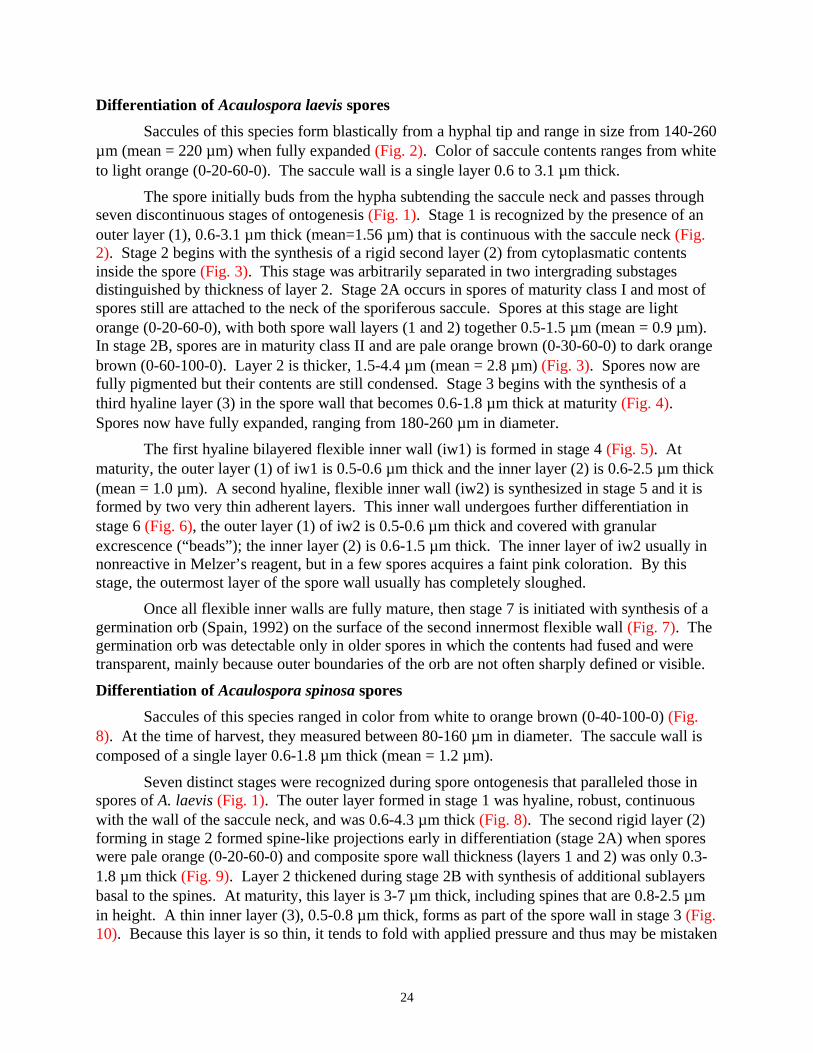
24
Differentiation of Acaulospora laevis spores
Saccules of this species form blastically from a hyphal tip and range in size from 140-260µm (mean = 220 µm) when fully expanded (Fig. 2). Color of saccule contents ranges from whiteto light orange (0-20-60-0). The saccule wall is a single layer 0.6 to 3.1 µm thick.
The spore initially buds from the hypha subtending the saccule neck and passes throughseven discontinuous stages of ontogenesis (Fig. 1). Stage 1 is recognized by the presence of anouter layer (1), 0.6-3.1 µm thick (mean=1.56 µm) that is continuous with the saccule neck (Fig.2). Stage 2 begins with the synthesis of a rigid second layer (2) from cytoplasmatic contentsinside the spore (Fig. 3). This stage was arbitrarily separated in two intergrading substagesdistinguished by thickness of layer 2. Stage 2A occurs in spores of maturity class I and most ofspores still are attached to the neck of the sporiferous saccule. Spores at this stage are lightorange (0-20-60-0), with both spore wall layers (1 and 2) together 0.5-1.5 µm (mean = 0.9 µm).In stage 2B, spores are in maturity class II and are pale orange brown (0-30-60-0) to dark orangebrown (0-60-100-0). Layer 2 is thicker, 1.5-4.4 µm (mean = 2.8 µm) (Fig. 3). Spores now arefully pigmented but their contents are still condensed. Stage 3 begins with the synthesis of athird hyaline layer (3) in the spore wall that becomes 0.6-1.8 µm thick at maturity (Fig. 4).Spores now have fully expanded, ranging from 180-260 µm in diameter.
The first hyaline bilayered flexible inner wall (iw1) is formed in stage 4 (Fig. 5). Atmaturity, the outer layer (1) of iw1 is 0.5-0.6 µm thick and the inner layer (2) is 0.6-2.5 µm thick(mean = 1.0 µm). A second hyaline, flexible inner wall (iw2) is synthesized in stage 5 and it isformed by two very thin adherent layers. This inner wall undergoes further differentiation instage 6 (Fig. 6), the outer layer (1) of iw2 is 0.5-0.6 µm thick and covered with granularexcrescence (“beads”); the inner layer (2) is 0.6-1.5 µm thick. The inner layer of iw2 usually innonreactive in Melzer’s reagent, but in a few spores acquires a faint pink coloration. By thisstage, the outermost layer of the spore wall usually has completely sloughed.
Once all flexible inner walls are fully mature, then stage 7 is initiated with synthesis of agermination orb (Spain, 1992) on the surface of the second innermost flexible wall (Fig. 7). Thegermination orb was detectable only in older spores in which the contents had fused and weretransparent, mainly because outer boundaries of the orb are not often sharply defined or visible.
Differentiation of Acaulospora spinosa spores
Saccules of this species ranged in color from white to orange brown (0-40-100-0) (Fig.8). At the time of harvest, they measured between 80-160 µm in diameter. The saccule wall iscomposed of a single layer 0.6-1.8 µm thick (mean = 1.2 µm).
Seven distinct stages were recognized during spore ontogenesis that paralleled those inspores of A. laevis (Fig. 1). The outer layer formed in stage 1 was hyaline, robust, continuouswith the wall of the saccule neck, and was 0.6-4.3 µm thick (Fig. 8). The second rigid layer (2)forming in stage 2 formed spine-like projections early in differentiation (stage 2A) when sporeswere pale orange (0-20-60-0) and composite spore wall thickness (layers 1 and 2) was only 0.3-1.8 µm thick (Fig. 9). Layer 2 thickened during stage 2B with synthesis of additional sublayersbasal to the spines. At maturity, this layer is 3-7 µm thick, including spines that are 0.8-2.5 µmin height. A thin inner layer (3), 0.5-0.8 µm thick, forms as part of the spore wall in stage 3 (Fig.10). Because this layer is so thin, it tends to fold with applied pressure and thus may be mistaken

25
for a flexible inner wall. With maturation of all three layers of the spore wall, full size andpigmentation have been attained. Spores are 140-180 µm in diameter (mean = 150 µm) andcream (0-10-20-0) to pale orange brown (0-30-60-0) in color.
The first hyaline flexible inner wall (iw1) then forms (stage 4), with an outer layer (1)less than 0.5 µm thick and an inner layer (2) 0.5-1.2 µm thick at maturity (Fig. 11). Once it isfully formed, then a second hyaline, flexible inner wall (iw2) is synthesized (stage 5), in whichboth of its layers undergo visible transformations. Initially, the outer layer (1) of iw2 is smoothand the inner layer (2) does not react in Melzer’s reagent (Fig. 12). In stage 6, the outer layerthen becomes covered with granular excrescences (“beads”) and thickens to 0.5-0.8 µm. Theinner layer becomes more reactive in Melzer’s reagent, staining pink (0-30-20-0) to purplish-pink (0-60-20-0) at maturity and thickens to 0.6-1.5 µm (Fig. 13). Stage 7 was not observed inthe spores examined, but it would consist of synthesis of a germination orb on the surface of iw2.
Differentiation of Entrophospora colombiana spores
Saccules of E. colombiana are formed blastically and expand in size to 160 µm (Fig. 14).They are white to a pale orange-cream (0-30-60-0) in color with a wall consisting of a singlelayer 0.6-1.5 µm thick (mean = 1.3 µm).
Stages of spore differentiation paralleled those of Acaulospora species (Fig. 1) except forthe omission of stage 3 (synthesis of a third layer in the spore wall). As the spore begins toexpand within the neck of the sporiferous saccule (stage 1), the spore wall is 0.6-1.2 µm (mean=0.8 µm) thick and is contiguous with the saccule neck wall (Fig. 14). Delimitation of the sporeas a discrete structure begins in stage 2 with deposition of an additional layer (2) that thickenswith the addition of sublayers and becomes pigmented (Fig. 15). In this stage, spores pass frommaturity class I to class II, expand in diameter to 130-160 µm (range of mature spores), andchange color from pale orange brown (0-40-60-0) to dark orange-brown (0-10-60-0). Outwardlyat the end of stage 2, immature and mature spores are indistinguishable except for presence-absence of attached saccule, because the spore wall has completed differentiation.
The synthesis of two flexible inner walls follows a pattern identical to that ofAcaulospora species (Fig. 1), first with the synthesis of a bilayered wall (iw1 in stage 3, Figs. 16and 17) and then a second bilayered wall (iw2 in stage 4; Fig. 18). However, the inner layer ofiw2 undergoes more of a transformation (stage 5) than the corresponding structure in spores ofeither of the two Acaulospora species studied. The outer layer of iw2 follows the same patternof being initially smooth (Fig. 18) and then synthesizing surface excrescences that give it abeaded appearance (Fig. 19). The inner layer of iw2 thickens and becomes more plastic (oramorphous) concomitant with a change in reaction to Melzer’s reagent from none to light pink(such as found in A. laevis) to purplish-pink (0-60-20-0) (such as found in A. spinosa) to darkred-purple (40-80-60-0) (Fig. 19). The terminal stage (stage 6) was not observed, but conservedpatterns in other species would indicate synthesis of a germination orb on the surface of iw2.
Taxonomic Analysis
Spore size of A. laevis AU211 is within the range described by Gerdemann & Trappe(1974). The authors describe the spore wall as ‘formed of three layers: outer thick and yellow tobrown, inner two colorless and membranous.’ The outer layer they describe is reinterpreted hereas the laminate layer (layer 2) of the spore wall. The outer sloughing layer continuous with thewall of the saccule neck (layer 1) and the inner laminate layer (layer 3) of the spore wall were

26
not described. The other two ‘membranous’ layers are synonymous with the first (iw1) andsecond (iw2) flexible inner walls, each of which is clearly bilayered.
Spores of the A. spinosa isolate WV861A have a narrower size range (140-180 µm) thanthose reported by the holotype of Walker and Trappe (1981), which were 100-298 × 100-335 µmin diameter. Subcellular structure of spores also is interpreted differently. Walker and Trappe(1981) misinterpreted the outer layer (layer 1) of the spore wall as an amorphous materialencrusting the spines of the second (laminate) layer, a condition commonly observed as this layerdegrades on mature spores. The thin inner layer (layer 3) of the spore wall was not described.The ‘two membranous hyaline walls’ are synonymous with the first (iw1) and second (iw2)bilayered flexible inner walls reported in this paper. The light pink to purplish-pink reaction ofthe inner layer of iw2 in Melzer’s reagent was not described in the protologue.
Spores of E. colombiana CL356 were larger (130-160 µm) than those of the holotypedescribed by Schenck et al. (1984), which were 100-115 µm in diameter. Subcellular structureof the spore conforms to the description except that the ‘outer spore wall’ and ‘wall two’ arereinterpreted as layers 1 and 2 of the spore wall, ‘wall three’ is the first inner wall (iw1), and‘wall four’ and ‘wall five’ are layers of the second inner wall (iw2).
DISCUSSION
The developmental sequences observed in spores of the Acaulospora and Entrophosporaspecies examined in this study parallel those in all other genera of Glomales in whichfunctionally distinct subcellular structures differentiate separately, but interdependently, in alinear sequence: spore wall component layers form concomitant with spore expansion from outerto inner surface. Only after the spore wall is formed and expansion has ceased does thesequential differentiation of two bilayered inner flexible walls occur. As a result, sporesappearing to be mature under a dissecting microscope may contain a mixture of any or alldevelopmental stages involving formation of flexible inner walls. A similar pattern is found withScutellospora species (Franke and Morton, 1994; Morton, 1995), the only other genus with trueflexible inner walls. Formation of a germination orb is the terminal event in spore ontogenesis,and occurs only after all other walls are fully differentiated. The germination orb is structurally,positionally, and functionally similar to the germination shield formed in spores of Scutellospora(Gigasporaceae) species, but the orb is evolutionarily analogous because it fails tests of similaritythat establish hypotheses of homology (Patterson, 1982; Wagner, 1989) in that: (i) the sporewithin which it is synthesized is formed uniquely; and, (ii) earlier stages of spore ontogenesisinvolve different structures (spore wall with additional layers, an inner flexible wall with uniqueproperties). The rarity of germination orbs in newly mature spores also is in direct contrast toabundant shield formation in spores of most Scutellospora species. Spain (1992) finds a visiblegermination orb most frequently in older spores in which the contents have undergone someplasmolysis.
Each of the major structures formed during differentiation (spore wall, flexible innerwalls) resolved taxa at different taxonomic levels. Species-level differences resided solely withproperties of the spore wall (color, size, ornamentation patterns, etc.). A similar pattern isevident among fungal species in Gigasporaceae (Bentivenga and Morton, 1995; Morton, 1995)and Glomaceae (Stürmer and Morton, 1997), indicating universality within Glomales. Propertiesof layers of flexible inner walls differed between species examined, but each phenotype is sharedby two or more species in each genus and therefore group species into supraspecific taxa not

27
delimited in the current classification. The conservativeness of different flexible inner wallproperties also grouped species of Scutellospora together (Morton, 1995), suggesting this patternalso is universal within Glomales.
The two flexible inner walls in spores of the species in the present study consisted of twolayers that appeared to differentiate sequentially rather than concurrently. This pattern isevidenced by the gradual change in the inner layer of the second flexible inner wall of E.colombiana in Melzer’s reagent, shifting from non-reactive to dark red-purple at full maturity.Differentiation of bilayered walls in Acaulosporaceae is similar to that in spores ofScutellospora, the other genus in Glomales forming inner walls (Franke and Morton, 1994;Morton, 1995). Ultrastructural observations (Mosse, 1970c) appear to support this pattern.Mosse (1970c) was able to resolve the various layers of the spore wall and to show that the twolayers of at least one flexible inner wall (iw2) formed sequentially rather than concurrently. Inmost ultrastructural studies, however, details of flexible structures like inner walls areconfounded by fixation difficulties (Maia and Kimbrough, 1993).
The phylogenetic relationship between Acaulosporaceae and other families in Glomalesdiffers with the character set used. Morphological data of the whole fungus indicate this familyis more closely related to Glomaceae than Gigasporaceae (Morton, 1990). Sequence data ofgenes for the small-subunit rRNA, in contrast, suggests Acaulosporaceae is a sister group ofGigasporaceae (Simon et al., 1993; Simon, 1996). Ontogenetic evidence in this study providessome clues to help resolve the conflict, but also raises additional questions because theAcaulosporaceae has many unique traits as well as some traits that are similar to those of bothother families.
The sporiferous saccule has obvious glomus-like properties and branches to formglomus-like spores in primitive taxa that also form a Glomus synanamorph, such as Acaulosporagerdemannii (Morton et al., 1997) and Glomus gerdemannii (Dr. J.B. Morton, personalcommunication). The saccule has no apparent function in spore development. Mosse (1970a)described the contents of the saccule as migrating to the spore as the saccule collapsed and thespore formed. However, DAPI-stained nuclei of saccules with attached spores shows that manynuclei do not migrate as spores differentiate (Dr. J.B. Morton, personal communication).
The outer layer of the spore wall is transitory in both Acaulosporaceae and Glomaceae.All subsequent layers of the spore wall are newly formed during spore expansion anddifferentiation. In Glomaceae, layers of the subtending hypha differentiate concurrently withlayers of the spore wall (Stürmer and Morton, 1997). In Acaulosporaceae, additional spore walllayers are formed only within the spore and in the region of attachment to the neck of thesporiferous saccule. In contrast, both layers of the spore and subtending hyphal walls in fungi ofGigasporaceae are present in the most juvenile stages and differentially thicken in the sporeduring later stages of spore wall differentiation (Franke and Morton, 1994; Bentivenga andMorton, 1995; Morton, 1995).
Superficially, the flexible inner walls of Acaulospora and Entrophospora are similar tothose in spores of Scutellospora, but such similarities are interpreted as convergent designconstraints imposed on structures of identical function. Some differences exist, however,between genera in Acaulosporaceae and Scutellospora. Only two flexible inner wallsconsistently are formed in the former, but the number ranges from one to three in the latter(Bentivenga and Morton, 1996). Layers of each inner wall also are more divergent in their

28
properties among Scutellospora species. In spores of all species in Acaulosporaceae comparedto date, layers of the first inner wall (iw1) are thin and of similar thickness and do not react inMelzer’s reagent. The outer layer of the second inner wall (iw2) is uniquely “beaded” (Morton,1990), even though some descriptions would indicate otherwise (e.g., Sieverding and Toro,1987; Wu et al., 1995).
This array of both similar and unique properties among families in Glomales does notprovide the definitive solutions to resolving evolutionary questions in Glomales. Other data setsshould be examined such as life history traits (Biermann and Linderman, 1983), other moleculessuch as sterols (Weete and Gandhi, 1998) or fatty acids (Bentivenga and Morton, 1996), otherstructural components such as cell wall carbohydrates (Gianinazzi et al., 1994) and mostespecially sequences of genes other than those coding for the small rRNA subunit (Simon et al.,1993; Simon, 1996).
LITERATURE CITED
Bentivenga, S.P., and J.B. Morton. 1995. A monograph of the genus Gigaspora, incorporatingdevelopmental patterns of morphological characters. Mycologia 87:719-731.
Bentivenga, S.P., and J.B. Morton. 1996. Congruence of fatty acid methyl ester profiles andmorphological characters of arbuscular mycorrhizal fungi in Gigasporaceae. Proceedings ofthe National Academy of Sciences of USA 93:5659-5662.
Biermann, B., and R.G. Linderman. 1983. Use of vesicular-arbuscular mycorrhizal roots,intraradical vesicles and extraradical vesicles as inoculum. New Phytologist 95:97-105.
Franke, M., and J. Morton. 1994. Ontogenetic comparisons of arbuscular mycorrhizal fungiScutellospora heterogama and Scutellospora pellucida: Revision of taxonomic characterconcepts, species descriptions, and phylogenetic hypotheses. Canadian Journal of Botany72:122-134.
Gerdemann, J.W., and J.M. Trappe. 1974. The Endogonaceae of the Pacific Northwest.Mycologia Memoir 5:1-76.
Gianinazzi-Pearson, V., M-C. Lemoine, C. Arnould, A. Gollote, and J.B. Morton. 1994.Localization of ß 1,3 glucans in spore and hyphal walls of fungi in the Glomales.Mycologia 86:478-485.
Maia, L.C., and J.W. Kimbrough. 1993. Ultrastructural studies of spore walls of Acaulosporamorrowiae and A. scrobiculata. Mycoogical Research 97:1183-1189.
Morton, J.B. 1990. Evolutionary relationships among arbuscular mycorrhizal fungi in theEndogonaceae. Mycologia 82:192-207.
Morton, J.B. 1995. Taxonomic and phylogenetic divergence among five Scutellospora speciesbased on comparative developmental sequences. Mycologia 87:127-137.
Morton, J.B. 1996. Redescription of Glomus caledonium based on correspondence of sporemorphological characters in type specimens and a living reference culture. Mycorrhiza6:161-166.
Morton, J.B., and G.L. Benny. 1990. Revised classification of arbuscular mycorrhizal fungi(Zygomycetes): A new order, Glomales, two new suborders, Glomineae and Gigasporineae,and two new families, Acaulosporaceae and Gigasporaceae, with an emendation ofGlomaceae. Mycotaxon 37:471-491.
Morton, J.B., S.P. Bentivenga, and W.W. Wheeler. 1993. Germ plasm in the InternationalCollection of Arbuscular and Vesicular-arbuscular Mycorrhizal Fungi (INVAM) andprocedures for culture development, documentation and storage. Mycotaxon 48:491-528.
29
Morton, J.B., J.D. Bever, and F.L. Pfleger. 1997. Taxonomy of Acaulospora gerdemannii andGlomus leptotichum, synanamorphs of one anamorphic fungus in Glomales. MycologicalResearch 101:625-631.
Mosse, B. 1970a. Honey-coloured, sessile Endogone spores I. Life history. Archives ofMikrobiology 70: 167-175.
Mosse, B. 1970b. Honey-coloured, sessile Endogone spores: II. Change in fine structure duringspore development. Archives of Mikrobiology 74:129-145.
Mosse, B. 1970c. Honey-coloured, sessile Endogone spores. III. Wall structure. Archives ofMikrobiology 74:146-159.
Patterson, C. 1982. Morphological characters and homology. Pp. 21-74. In: Problems ofphylogenetic reconstruction. Eds., K.A. Joysey, and A.E. Friday. Academic Press, London.
Schenck, N.C., J.L. Spain, E. Sieverding, and R.H. Howeler. 1984. Several new and unreportedvesicular-arbuscular mycorrhizal fungi (Endogonaceae) from Colombia. Mycologia76:685-699.
Sieverding, E., and Toro, S.T. 1987. Acaulospora denticulata sp. nov. and Acaulospora rehmiisp. nov. (Endogonaceae) with ornamented spore walls. Angew. Botanik 61:217-223.
Simon, L. 1996. Phylogeny of the Glomales: deciphering the past to understand the present.New Phytologist 133:95-101.
Simon, L., J. Bousquet, R.C. Levesque, and M. Lalonde. 1993. Origin and diversification ofendomycorrhizal fungi and coincidence with vascular land plants. Nature 363:67-69.
Spain, J. 1992. Patency of shields in water mounted spores of four species in Acaulosporaceae(Glomales). Mycotaxon 43:331-339.
Stürmer, S.L., and J.B. Morton. 1997. Developmental patterns defining morphologicalcharacters in spores of four species in Glomus. Mycologia 89:72-81.
Wagner, G.P. 1989. The origin of morphological characters and the biological basis ofhomology. Evolution 43:1157-1171.
Walker, C., and J.M. Trappe. 1981. Acaulospora spinosa sp. nov. with a key to the species ofAcaulospora. Mycotaxon 12:515-521.
Walker, C., L.E. Reed, and F.E. Sanders. 1984. Acaulospora nicolsonii, a new endogonaceousspecies from Great Britain. Transaction of British mycological Society 82:360-364.
Weete, J.D., and S.R. Gandhi. 1998. Sterols of the phylum Zygomycota: Phylogeneticimplications. Lipids 32:1309-1316.
Wu, C-.G., Y-.S. Liu, and L-.L. Hung. 1995. Spore development of Entrophospora kentinensisin an aeroponic system. Mycologia 87:582-587.

Spore Wall Inner Flexible Walls Germination structure
Phase I Phase II Phase III
Stage 1
Stage 1
Stage 2
Stage 2
Stage 3 Stage 4
Stage 3
Stage 5 Stage 6b
b b
b
Stage 4 Stage 5
Stage 7
Stage 6
A
B
Fig. 1 Murographic representation of the sequence of development of layers in sporesof (A) and , and (B) . Numbersrepresent phenotypically discrete layers designated numerically in the order they weresynthesized in each separate structure. Dashed vertical lines filling rectangles representsublayers of a laminate layer present forming the spore wall. Filled arrows indicatetransition between phases identified by differentiation of a spore wall, inner flexible wallsand a germination structure; hollow arrows indicate transitions between discrete and stablestages during spore development. b = “beaded” layer.
Acaulospora laevis A. spinosa Entrophospora colombiana
30

3
54
6 7
2
31
Figs. 2-7 AU211 - stages in the differentiation of spore wall and inner walls layersin spores mounted in PVLG + Melzer’s reagent (1:1, v/v). 2. Most juvenile spore (stage 1) with sporeattached to the side of the neck of the sporiferous saccule. Spore wall is formed by only one layercontinuous with the hyphal wall of the saccule neck (insert). Bar = 25 µm. 3. Juvenile spore (stage 2)with spore wall (sw) composed of an outer layer (1) and an inner orange-brown laminate layer (2).Bar = 5 µm. 4. Juvenile spore (stage 3) with the spore wall (sw) completely differentiated and formedby an outer layer (1), an orange-brown laminated layer (2) and a colorless laminate layer (3). Bar =10 µm. 5. Stage 4 of spore development characterized by the presence of the first inner wall (iw1).Bar = 10 µm. 6. Spore in stage 6 showing the first (iw1) and the second (iw2) inner wall each formedby two adherent layers (1 and 2). Bar = 5 µm. 7. Mature spore (stage 7) showing the germination orb(arrows) formed between iw1 and iw2. Bar = 10 µm.
Acaulospora laevis

8 9
10
12 13
11
32
Fig. 8-13 WV861A - stages in the differentiation of spore wall and inner wallslayers in spores mounted in PVLG + Melzer’s reagent (1:1, v/v). 8. Most juvenile spore (stage 1) withspore wall formed by only one layer continuous with the neck of the sporiferous saccule. Bar = 25 µm.9. Juvenile spore (stage 2) with spore wall composed by an outer layer (1) and the first sublayers of alaminate layer (2) ornamented with spiny projections. Bar = 10 µm. 10. Juvenile spore (stage 3) withadditional sublayers in layer 2 and a third thin layer (3) formed. Bar = 10 µm. 11. Juvenile spore (stage4) with a fully differentiated spore wall (sw) and the first inner wall (iw1) synthesized and formed bytwo layers (1 and 2). Bar = 10 µm. 12. Mature spore (stage 5) showing the spore wall (sw), the firstbilayered inner wall (iw1) and the second inner wall (iw2) formed by two thin adherent layers. Bar =10 µm. 13. Mature spore (stage 6) showing iw2 completely differentiated. The first layer (1) now is“beaded” and the second (2) reacts in Melzer’s reagent turning pinl to purplish-pink. Bar = 10 µm.
Acaulospora spinosa

14
16
18 19
17
15
33
Figs. 14-19 CL356 - stages in the differentiation of spore wall and innerwalls layers in spores mounted in PVLG + Melzer’s reagent (1:1, v/v). 14. Most juvenile spore (stage 1)with spore wall formed by a layer continuous with the neck of the saccule. Bar = 30 µm. 15. Juvenilespore (stage 2) with spore wall consisting of an outer layer (1) and a pale to dark orange-brown laminatelayer (2). Bar = 5 µm. 16. Juvenile spore (stage 3) with fully developed spore wall and the first innerwall (iw1) formed by only one layer. Bar = 5 µm. 17. Spore with iw1 fully differentiated and formed bytwo layers (1 and 2). Bar = 5 µm. 18. Spore in stage 4 of development with spore wall (sw), first innerwall (iw1) and second inner wall (iw2) each formed by two thin adherent layers. Bar = 5 µm. 19. Spore(stage 5) with iw2 fully differentiated showing the “beaded” outer layer (1) and the innermost layer (2)that turns dark red-purple in Melzer’s reagent. Bar = 5 µm.
Entrophospora colombiana

34
CHAPTER THREE
DIVERSITY OF ARBUSCULAR MYCORRHIZAL FUNGI
IN SELECTED HABITATS USING TRAP
CULTURES METHODOLOGY

35
INTRODUCTION
Arbuscular mycorrhizal fungi (Zygomycetes, Glomales) are ubiquitous soil-borneorganisms associated with plant roots in a variety of ecosystem worldwide. They have beenreported in natural plant communities such as deserts, sand dunes, tropical forests, salt marshesand managed systems such as pastures, orchards and field crops (Brundrett, 1991). Theseorganisms are ancient, as evidenced by their detection in roots of the fossil plant Aglaophytondating from the Devonian period (Taylor et al., 1995). Molecular clock estimates based on thesmall-subunit rRNA sequence also support the origin of arbuscular fungi about 400 millionyears ago (Simon et al., 1993). Therefore, it is not surprising that glomalean fungi are presentin most soils given the long geologic time they have had to co-evolve with plants and todisperse.
The diversity of AMF in many ecosystems has been measured with respect to hostplants, seasonal variation, soil nutritional gradients, crop rotation and plant successional stages(Koske, 1987; Gemma et al., 1989; Anderson et al., 1984; Johnson et al., 1991a). The mostwidespread approach to measure and analyze species diversity of arbuscular mycorrhizal fungi(AMF) is to recover, count and identify spores from the field. However, many problems areassociated with this approach. First, spores recovered from the field usually are in lownumbers and too parasitized for accurate species identification, especially those of Gigasporaand Glomus species. Second, only those fungi sporulating in the rhizosphere of a given plant atthe time of sampling are recovered. It is not uncommon to find nonsporulating (cryptic)species colonizing plants in the field based on trap culture results (Bever et al., 1996; Stutz andMorton, 1996) and PCR products from field-collected roots (Clapp et al., 1995). Reliance onlyon spore counts from the field, therefore, usually underestimates species diversity of AMF inplant communities.
Trap cultures provide a non-molecular approach to baiting cryptic species of AMFpresent in plant communities. In the mycorrhizal literature, few diversity studies haveincorporated this approach. Miller et al. (1985) recovered 14 species of AMF under appleplantations around the United States that were present only in pot cultures with Sorghum andColeus. More recently, 23 species of AMF were detected from a 75 m2 region of a mown fieldwith Sorghum trap cultures or field plants transplanted into trap cultures (Bever et al., 1996).In an arid ecosystem, Stutz and Morton (1996) recovered 15 more species than those detectedin the field after three cycles of trap cultures. These data are limited and indicate a need todetermine the applicability of trap cultures methods to bait AMF in a variety of ecosystems.
Diversity and distribution of AMF species have been explained mainly as a result ofcontemporary ecological processes acting upon the fungal or plant communities. Factors suchas soil pH, soil P, temperature and plant host genotype often have been considered determinantsof the distribution of AMF species at local or regional levels. For example, interpretation of thedistribution of fungal species in a Cedar Creek grassland was attributed to differentsuccessional stages in a old field (Johnson et al., 1991a). Porter et al. (1987) observeddifferential occurrence of a Glomus sp. and Acaulospora laevis in Australia and concluded thatsoil pH was the main factor influencing distribution of these species. Historical processes suchas speciation and dispersal have been largely ignored (Ricklefs and Schuluter, 1993).Interpretation of the possible causal factors determining and maintaining AMF communitiesmay be quite different when historical processes are considered (Morton et al., 1995).

36
Separation of ecological factors from historical factors requires that comparisons aremade among sites with similar habitats from distinct geographical regions (Ricklefs andSchuluter, 1993). Sand dunes represent one example of a habitat that may share similar plantcommunities and soil characteristics in widely separated areas. Koske and Tews (1987)calculated similarity coefficients between mycorrhizal communities of sand dunes in theU.S.A., Italy, Australia and Scotland and found values ranging from 0 (no similarity) up to0.39.
In this study, I measured the taxonomic diversity of arbuscular mycorrhizal fungi indifferent plant communities. Baiting methods were used to recover fungi from fivegeographically separated plant communities in the United States and from four sand dune sitesfrom different countries. Species were recovered by successive trap cultures to inducesporulation of cryptic AMF species. I tested three hypotheses: (i) species diversity of AMFincreases after successive trap culture cycles; (ii) taxa overlap among similar habitats; and, (iii)community structure of AMF will change from a broken stick model in the field to a log seriesmodel after two cycles of trap cultures.
Results provided additional data on the efficacy of trap culture methodologies tomeasure fungal community structure in different ecosystems. Moreover, additional informationon AMF species distribution important for assessing biogeographical data were obtained.
MATERIAL AND METHODS
AMF in Five Soils
Study sites
Species diversity of AMF were measured at five sites within the United Statesrepresenting different habitats. A site in California (CA-D) was from a desert sampled atHarper’s Well near the Anza-Borrego Desert State Park in Imperial County (collected by Dr. J.Stutz, Arizona State University). Soil was collected under the rhizosphere of mesquiteProsopis glandulosa Torr. Another site from California (CA-R) included a revegetated sitewith no management in Shasta County (collected by Dr. Vic Claassen, University ofCalifornia). The plant community at this site was dominated by black oak Quercus kellogiiJapson and poison oak Ceonothus lemonii Parry with an understory of wild oats Bromus mollisL. A site in Kansas (KS) at the Konza Prairie Research Natural Area in Riley County consistedof a natural tallgrass prairie community (collected by Dr. G. Wilson, Kansas State University).Dominant plant species were big bluestem Andropogon gerardii Vitm., prairie cordgrassSpartina pectinata Link and indian grass Sorghastrum nutans (L.) Nash. In Minnesota (MN),an agricultural plot was sampled at the University of Minnesota Southwestern ExperimentStation at Redwood County (collected by Dr. F.L. Pfleger, University of Minnesota). The sitehas been cultivated continuously with corn for over 10 years. The last site was an abandonedmine site at the Coopers Rock State Forest, Monongalia County in West Virginia (WV)(collected by me). Soil was sampled under Andropogon virginicus L. with other plant speciespresent being red maple Acer rubrum Torr & Gray ex K. Koch and black birch Betula lenta L.At each site, three plants present in a 100 m2 area were pulled out from the soil and therhizosphere soil shaken into a plastic bag. This sample therefore consisted of soil from the rootzone and some detached fine roots. Soil chemical properties and environmental conditions foreach site are shown in Table 1.

37
Experimental procedure
Pooled sample from each site was diluted 1:1 (v:v) with a sterile potting mediumconsisting of a sandy loam soil (Lily series) premixed with quartzite sand (1:2, v/v). Thismixture was placed in 15-cm diameter plastic pots and each seeded with 50-60 seeds ofsudangrass, Sorghum sudanense (Piper) Staph. For alkaline soils (CA-D- and KS), the finalsoil pH of the potting medium was 7.2, while for more acid soils, the potting medium pH was6.2. Three pots were set up with soil inocula from each site. Plants were grown in a growthroom under fluorescent lights with a photon flux density of 245 µmol m -2 s-1 at pot level, a 14-hour photoperiod, and an ambient temperature of 21-28oC. After 3 months, watering wassuspended and pot contents were dried in situ for 8-14 days. Shoots were excised and two 50cm3 soil aliquots was sampled from each pot and stored at 4oC until processing. The root balland attached soil for each pot was manually chopped and the contents of all three pooled,mixed and stored at 4oC for at least one month. A second trap culture cycle was started bymixing stored inocula with 300 cm3 of sterile potting media. Host and seeding rates were thesame as in the first trap cycle, and plants were grown for another 3 months-period under similargrowth-room conditions. At harvest, cultures were processed similarly. Three pots containingonly sterile potting media seeded with sudangrass were interspersed between trap cultures ineach cycle to monitor for possible air-borne contamination.
Infectivity Assay
Infectivity of fungi in the field soil and at the end of each trap cycle was measured bythe mean infection percentage (MIP) assay (Moorman and Reeves, 1979). For trap cultures,100 cm3 was sampled after the three replicate pots were pooled. Soil samples were mixed (1:1,v/v) with the same potting media described above and placed in 50 cm3 cone-tainers ™(Stuewe and Sons, Inc., Corvallis, Oregon). Three red clover (Trifolium pratense L.) seedlings,pre-germinated in sterile soil, were transplanted into each cone-tainer and watered with asuspension of Rhizobium trifolii (LiphaTec, Inc., Milwaukee, Wisconsin). Four cone-tainerswere set up for each soil and plants were harvested after 4 weeks. Roots were gently separatedfrom the soil, washed, and stained using the method of Koske and Gemma (1989). Infectivitywas assessed as percentage of mycorrhizal colonization by the grid-line intersect method(Giovannetti and Mosse, 1980).
Spore Extraction
Spores were extracted from 50 cm3 of soil from the field or from each trap pot byblending in two 10-second bursts and passing this material through nested sieves with 500 and45 µm openings. Material retained on the 45 µm sieve was transferred to a 50 cm 3 tubecontaining a 20/60% sucrose gradient and centrifuged at 900 x g for 1 minute. Supernatant waspassed through a 45 µm sieve and the material retained was washed under tap water for at least30 seconds and transferred to a Petri dish. Under the dissecting microscope, spores of eachmorphotype were collected and counted separately. Spores also were mounted on glass slidesin PVLG (Polyvinyl alcohol, lactic acid, glycerol) and PVLG mixed with Melzer’s reagent foridentification and preservation as voucher specimens.

38
AMF in Sand Dunes
Study sites
Diversity of AMF species was measured in sand dune habitats in different geographicalregions. Samples were obtained from four countries on separate continents. In the UnitedStates, sand dune samples were collected from Ammophila breviligulata Fern. at Bass RiverBeach, Massachusetts (collected by Dr. Joseph Morton, West Virginia University). In Brazil, asample was collected from Spartina ciliata Brongn. in Joaquina Beach, Santa Catarina state(collected by Dr. Margarida de Mendonça, Universidade Federal de Santa Catarina). In theNetherlands, samples were obtained from Ammophila arenaria (L.) Link growing in the sanddunes of the Island of Terschelling (collected by Dr. Thomas Kuyper, Wageningen Agric.University). In Japan, samples came from Elymus mollis Trin. growing at Hasaki Beach,Ibaraki Prefecture (collected by Dr. Jun-ichi Abe, University of Tsukuba). At each site,rhizosphere soil was collected from three separate plants in a 100 m2 area. Plants were pulledout from the soil and roots shaken to remove attached soil into a plastic bag, which alsoincluded some detached fine root fragments. All samples were collected between Septemberand October, 1996, dried and shipped to Morgantown, WV. Soil chemical properties andenvironmental conditions of each site are summarized in Table 2.
Experimental procedure
Field samples from sand dunes were mixed 1:1 (v/v) with sterile quartzite sand (averagegrain size = 0.9 mm) and placed in 15-cm diameter plastic pots. Sudangrass was seeded ineach pot at a rate of 50-60 seeds per pot and seeds covered with a 2-cm layer of sterile sand.Three pots were set up for each site and plants were grown in a growth room under conditionsdescribed above. Three additional pots containing only sterile sand were placed randomlyamong other pots to check for air-borne contamination. Plants were fertilized when neededwith a 25-5-20 (N-P-K) solution. After 3 months, shoots were removed and two sand samples(ca. 250 cm3 after pooling) from opposite sides of each pot were collected and stored at 4oCuntil processing. Empty portions of the pots after sampling were filled with sterile sand andeach pot was reseeded for a second propagation cycle. Spores were extracted and processed asdescribed previously. In this experiment, two different approaches relative to experiment 1 wastaken: (i) pot contents were not disturbed between propagation cycles to avoid majorbreakdown of the fungal hyphal network established in the soil; and, (ii) pots wereindividualized in the sense that spore counts and identification and infectivity assay were donein the same pot in both propagation cycles.
Infectivity Assay
Infectivity of fungal propagules (hyphae, roots, spores) was measured in the field soiland after each trap culture cycle by the mean infection percentage (MIP) assay (Moorman andReeves, 1979). Samples from the field and from each trap culture were diluted (1:1, v/v) withsterile quartzite sand and placed in 100-ml plastic cone-tainers. Each cone-tainer was sownwith three surface-sterilized seeds of sudangrass and thinned to one plant after emergence.Each assay consisted of four replicate cone-tainers and plants were grown for 3 weeks undergrowth room conditions as described previously. Harvested roots were stained and fungalcolonization measured as described previously. In order to statistically measure anyrelationship between mycorrhizal infectivity and number of spores and species, pots wereassayed separately.

39
Statistical Analyses
The number of spores produced for each species in the field and after two propagationcycles was plotted in a species rank-log abundance graph to depict fungal community structure.The curves were tested against theoretical curves of the log series and the broken stick modelsaccording to the procedures explained by Magurran (1988). Two goodness-of-fit tests wereused: the log likelihood ratio, or G test (Sokal and Rohlf, 1981) and the Kolmogorov-Smirnovtest (Tokeshi, 1983). The number of species recovered in each habitat was low and precluded amore accurate testing of these models, therefore, for each experiment, number of speciesrecovered in the field and in the second trap culture was pooled from all habitats. Beforeanalysis of variance of the infectivity assay, data on percentage of colonization weretransformed using the arcsine square root transformation and screened for homogeneity ofvariance using Levene’s test. A t-test then was used for comparison of group means. Linearregression was used to determine if number of spores and species number were a goodpredictor of the mycorrhizal infectivity levels obtained on trap cultures. Stepwise regressionwas used to determine which soil or environmental factors was influencing most the number ofAMF species. Hierarchical cluster analysis was performed on the soil and environmentalcharacteristics of sand dunes samples through the application of the average linkage method.Statistical tests were performed using JMP® (SAS, 1995)
RESULTS
Species diversity in five soils
For most soils, the number of sporulating species of AMF did not increase after the firsttrap culture cycle (Fig. 1). In the Kansas soil, however, three additional species sporulated inthe second trap culture. Only one species sporulated in the first trap cycle in the soils from thedesert site in California and the Kansas site. For all other soils, the first cycle of trap culturedetected 33-46% of the total number of species recovered. No spores were detected in thecontrol pots.
In total, 30 AMF species were detected, 13 of which could not be placed into describedtaxa (Tables 3-6). Fungi in the families Glomaceae and Acaulosporaceae were detected in100% and 80% of the soils, respectively. The family Gigasporaceae was represented by onlyone species, Scutellospora calospora, which was detected only from the revegetated site fromCalifornia (Table 3). At the genus level, Glomus was dominant in both number of species ineach trap culture and in abundance of spores. The most common species sporulating in three ofthe five soils were Glomus intraradices, Glomus occultum and Glomus microaggregatum. Forall soils, total number of spores tended to increase from the field to the first trap culture anddeclined thereafter.
The number of species detected in each soil varied according to the habitat (Tables 3-6).The soil from the California desert represents one extreme where only G. microaggregatumwas detected. This species sporulated so prolifically after the first cycle that spore countingwas done by dilution; number of spores reached 50,022 ± 11,622 (mean ± standard error) per30 ml of soil. At the other extreme were soils from Kansas and Minnesota where 10 and 13species were detected, respectively.
Similarity among AMF species for all five soils ranged from no similarity at all up to0.26 (Table 7). Highest similarity was found between the sites in Minnesota and Kansas

40
(C=0.26) and between the two sites in California (C=0.25). The five habitats did not share anycommon plant species and therefore the similarity among habitats considering plant communitywas zero.
The structure of AMF communities in the field and after two propagation cyclesapproached a log series model (Fig. 2). Testing the curves against theoretical models showsthat both curves are significantly different from the broken stick model, but not different fromthe log series model using the G test or the Kolmogorov-Smirnov test.
Infectivity in five soils
Infectivity of mycorrhizal fungi followed the same trend in most soils: highestinfectivity after the first trap culture followed by a decline in the second cycle of culture (Fig.3). The West Virginia soil differed in that the lowest infectivity was observed in the first trapwhile infectivity was higher in the field and in the second trap culture cycle (Fig. 3). Nostatistical analyses were carried out to detect significant differences between means because oflack of replication.
Species diversity in sand dunes
Results from the average linkage method shows that the four sand dunes sites did notform only one cluster (Fig. 4). Sand dunes from U.S.A. and Brazil formed one cluster whiledunes from Japan and Netherlands formed another cluster
Number of AMF species recovered in trap cultures exceeded that in the field and oftenreached a plateau after the first propagation cycle (Fig. 5). The Netherlands soil wasexceptional in that three additional species sporulated during the second propagation cycle. Nospores were detected in the field soil from Japan and only one and two additional species weredetected in traps that were not present in the field soil for U.S.A. and Brazil, respectively (Fig.5). Based on the total number of species detected at each site from all samples, trap culturesdetected 15-30% of AMF species in dunes of U.S.A. and Brazil and 75-100% for dunes inNetherlands and Japan. No spores were detected in control pots.
Eighteen species of AMF were recovered in this study, three of which are putativelynew species. Members of all three families of Glomales were detected in soils from Brazil andU.S.A., while only members of Glomaceae were detected in Netherlands and Japan (Tables 8-11). All species sporulating in trap cultures were in the genus Glomus, except the occurrenceof A. trappei from the Brazil soil (Table 8). When present, spores of Scutellospora andGigaspora species were detected in the field soil and were fewer in number after trapping.Glomus intraradices was the only species detected in all four sand dunes habitats. For all soils,total number of spores tended to increase from the field to the second trap culture cycle.
Coefficients of similarity between AMF communities indicated that dunes of Brazil andJapan shared the greatest number of species (C=0.40) followed by dunes of Japan and theNetherlands (C=0.36) (Table 12). However, coefficients of similarity for the plantcommunities of all four sand dunes habitats were zero.
The structure of AMF community in the field was not significantly different betweenthe broken stick and the log series models (Fig. 6). However, the community structure of thesecond trap culture cycle fit only the log series model (Fig. 6).

41
Infectivity in dunes
Infectivity did not differ significantly between individual pots within each propagationcycle. Therefore, data from the three pots were pooled to compare changes in infectivitybetween propagation cycles. Infectivity measured at the field soil and after each trap culturegeneration followed the same trend among soils: infectivity was low in the field and after thefirst trap culture and increased after the second trap culture (Fig. 7). Infectivity in the field andfirst trap ranged from 0.5% to 5.3%. After the second cycle, trap from Japan had the highestinfectivity (32.6%) while traps from Brazil and U.S.A. had similar levels (Fig. 7).
Regression models of fungal diversity in dunes
Data for the sand dunes soils were pooled to carry out two types of analyses. First, therelation between total sporulation/species richness and infectivity within the same soil orpropagation cycle. In further analysis, only spore number of the most abundant species for thesand dune habitats were examined. Second, the effect of infectivity on sporulation and speciesrichness of the subsequent propagation cycle also was explored. Results of the secondanalyses, however, showed the same trend and therefore are not reported.
Within the same soil or propagation cycle, no significant relationship was observedbetween species richness and infectivity (r2 < 0.01, P = 0.93) (Fig. 8), but total spore numbersaccounted for about 52% of the variability in infectivity (r2 = 0.52, P < 0.01) (Fig. 9).
The number of spores of Glomus clarum and G. pustulatum, separately or combined,was a good predictor of the infectivity levels for the U.S.A. soil (Table 13). The number ofspores of G. intraradices alone or with other fungal species predicted infectivity levels of theJapanese soil. Infectivity for the site in Brazil was predicted only from spore numbers ofAcaulospora scrobiculata (Table 13).
Stepwise regression could detect only mean annual temperature as a good predictor ofthe number of AMF species recovered in each soil (r2=0.54, P=0.02) (Table 14). Furtheranalyses considering all possibel pairs of soil and environmental factors indicated thatphosphorus (P) and potassium (K) paired account for 69% of the variability found in thenumber of species, followed by potassium/organic matter, and mean annualtemperature/organic matter (Table 15).
DISCUSSION
Successive cycles of trap cultures led to the detection of AMF species not sporulating ina wide range of habitats. One or two propagation cycles were needed to detect these additionalspecies, corroborating patterns observed in an old field in North Carolina (Bever et al., 1996)and arid sites of southwestern U.S.A. (Stutz and Morton, 1996). However, the number ofcryptic species detected by trapping varied between habitats. For example, only Glomusmicroaggregatum was detected in the soil from Arizona whereas as many as six species weredetected after two trapping cycles of dunes from the Netherlands and soils from Minnesota.
Disparity between sites clearly depends on the actual number of species present in thesoil. However, sampling methodology also may have been a major limitation that influencedthe number of species detected. Soil inoculum used to establish trap cultures consisted ofrhizosphere soil and excluded a majority of plant roots. This approach could exclude fungalspecies that were growing predominantly within the roots or species whose propagules were

42
infective only when attached to mycorrhizal roots. Moreover, rare species that sporulatedinfrequently or had limited growth within roots may have been excluded.
A much higher number of species is expected to be detected in the trap cultures thanfield soil if plant roots are a major component of the soil inoculum used to start trap cultures.Failing to consider this factor during sampling, therefore, can explain the disparity between thetotal number of AMF species found using trap cultures with that reported in the literature forthe same habitats. Abe et al. (1994), sampling from the same area and host plant in Japan sanddunes, found S. gregaria and Acaulospora sp. sporulating in the field, two species that were notrecovered by trap cultures. Stürmer and Bellei (1994) recovered 13 AMF species fromBrazilian sand dunes, while only 7 species were detected in this present study. Also, only twoadditional species were detected in this study compared to 11 species found in a corn field byJohnson et al. (1991b).
Disparity on the number of species recovered among habitats also may depend on theextent to which culture conditions approximate those of source habitat. The low number ofspecies recovered from the desert site in California was not expected. Stutz and Morton (1996)detected six to seven species after two cycles of trapping soils from a similar habitat in thesame region. In their study, however, trap cultures were grown in a greenhouse at ArizonaState University where light and temperature may more closely approximated site conditionsthan did the greenhouse at West Virginia University.
Light and temperature are dominant variables that impact plant growth, mycorrhizalcolonization and fungal sporulation. Hayman (1974) observed that mycorrhizal colonization inonion roots was reduced and consisted of fewer arbuscules and external hyphal under loweredlight intensity. Sporulation by an isolate of Glomus fasciculatum was greatest at 30oC, whichcoincided with the optimum temperature for the host plant (Ferguson and Menge, 1982). Lightconditions for trap cultures did not appear to be a limiting factor because sporulation by a widerange of INVAM fungal isolates on the same host used in this study consistently is adequate tohigh (Dr. J.B. Morton, personal communication). Temperature is more likely a critical variableif it influences the physiological activity of the propagules present in the original inoculum toinitiate colonization (Sieverding, 1983). Temperature also can influence secondarymycorrhizal colonization so that certain species might not achieve that minimum level ofcolonization necessary to trigger sporulation (Franke and Morton, 1994) and, therefore, remainundetected. In the Californian desert soil, lower temperatures at the growth room relative to thefield soil may have selected only G. microaggregatum to establish colonization. However, thelargest differences between temperature at the field and growth room conditions of this studyoccurred for the Minnesota and Netherlands soil, where the highest number of speciessporulating in trap cultures were detected (six species). This result suggests that highertemperatures in the growth room where traps were maintained could have activated propagulesof certain species that were not sporulating in the field.
Temperature is certainly one of the major environmental factors influencing AMFspecies distribution in the field. Schenck and Schroder (1974) observed that sporulation andmycelia growth of a Gigaspora sp. was optimized at the higher temperatures within the rangeof temperatures tested. The authors correlated the affinity of the fungus for high temperaturewith its presence and abundance in summer rather than spring or winter crops. Koske (1987)observed that some AMF species were more abundant in northern cooler regions than insouthern warmer regions within a latitudinal temperature gradient. He also found that

43
frequency of some species such as Scutellospora weresubiae and Glomus tortuosum wascorrelated with higher temperatures. When each factor was considered alone in the presentstudy, stepwise regression detected the mean annual temperature as the only factor explainingnumber of AMF species in each site, which supports the role of temperature on speciesdiversity.
Soil factors also have been implicated as regulators of AMF species diversity. Forexample, Porter et al. (1987) found a correlation between soil pH and the occurrence of aGlomus sp. and Acaulospora laevis, the latter tending to dominate in more acid soils. Johnsonet al. (1991a) found species richness to be positive correlated with total P in the soil andnegative correlated with soil pH, although the correlation coefficients were not significant. Inthe present study, phosphorus, potassium and organic matter could explain a great portion ofthe variability on AMF species richness. One of the possible reason for this correlation is thedirect effect of these factors on plant growth, increasing the amount of root biomass andproviding the niche necessary for more fungi to colonize and sporulate. Alternatively, it is wellknown that fertilization can limit fungal development within the roots. Thus, fertilization couldlimit colonization by more aggressive fungi so that other slow growing fungal species cancolonize and sporulate as well under field conditions. In the literature, contradictory results arecommonly found when the influence of soil factors upon species diversity is evaluated. One ofthe drawbacks to interpreting these result is that regression analyses may not reflect cause andeffect relationships. Also, these analyses do not consider that species diversity is a function ofhistorical processes that ultimate determine which species may be present in a given habitat,each with possibly their unique life history traits.
Sporulation of cryptic species in trap culture suggests that a minimum threshold level offungal colonization in the roots may be necessary for sporulation to be triggered. Gazey et al.(1992) observed such a threshold for an isolate of Acaulospora laevis, which sporulated onlyafter 5.7 cm of root was colonized. Similarly, Franke and Morton (1994) measured a minimumlevel of mycorrhizal root length necessary to trigger sporulation of Scutellospora pellucida andS. heterogama. This level seemed to vary with host species as well. Containerization of trapcultures favors accumulation of abundant root biomass in a confined space, which likely favorsinfection and colonization by cryptic species. Continuous growth and rapid turnover of plantroots in the field may impact the rate at each fungal species colonize the roots, so that only themore aggressive fungi are able to sporulate. Spatial heterogeneity within the rhizosphereenvironment also may account for lack of recovery of some species during sampling. Speciesnot detected in field sampling in this study may be colonizing and sporulating in other regionsof the plant rhizosphere that were excluded by the sampling regime. Seasonal variation onsporulation patterns by mycorrhizal species has been studied extensively (Gemma et al., 1989)and represents another possible reason for not detecting AMF species in the field. Trappingfield soil collected over seasons (Bever et al., 1996) represents an alternative strategy to recovermore species in a habitat when complete characterization of an AMF community is a goal.
In all habitats studied, Glomus species were the predominant sporulators in trapcultures. With few exceptions, most of the Glomus species were hyaline to light colored, withthin spore walls and small spores (< 150 µm diameter). Species of Gigaspora andScutellospora, when present, were detected sporulating in the field and number of sporesdecreased with successive trap culture cycles. Three hypotheses may explain this pattern.First, the genus Glomus circumscribes 56% of the described species in Glomales, and is widely

44
distributed worldwide (INVAM Biogeography Database, unpublished), such a broad dispersalincreases the probability of Glomus species being found in most plant communities. Second,genera differ greatly in types of infective propagules and the biology of mycorrhizalestablishment. In Glomus, all parts of the fungus (spores, pieces of colonized roots and hyphalfragments in soil) are totipotent and capable of initiating colonization (Biermann andLinderman, 1983; Jasper et al., 1989). In Gigaspora and Scutellospora, conversely, onlyspores from disturbed soils are able to initiate new infections in roots (Biermann andLinderman, 1983; Pearson and Schweiger, 1994). Third, trap culture environment may bemore conducive to colonization and sporulation of Glomus species given their broader range ofinfective propagules and lower demand on the host for carbon during sporulation. Spores ofGigaspora and Scutellospora tend to be larger in diameter than spores of Glomus and thusmore carbon must be allocated by the plant to each spore of the former compared to the latter.As a result of the sampling regime, the level of infective propagules (spores) of Gigaspora andScutellospora may have been diluted or excluded so that extensive root colonization andsporulation could not occur in trap cultures.
Infectivity bioassays measure the inoculum potential of mycorrhizal fungi and is theresult of cumulative interactions between propagule density, host preference, andenvironmental variables conducive to colonization. Two trends were observed in infectivityassays carried out with field soil and soil at the end of each trap culture cycle: infectivity wasgreatest in the first trap culture cycle, decreasing in the second trap cycle of the five soils,whereas infectivity increased from field soils to the second trap cycles of sand dunes samples.Variation in culture regimes between soil and sand dunes experiments is the likely cause. Forsoils, contents of each pot culture were chopped and thoroughly mixed between propagationcycles. Mixing of soils in the first experiment undoubtedly disrupted established hyphalnetworks in each pot. Hyphae are more sensitive to disturbance than other propagules ofmycorrhizal fungi (Jasper et al., 1989, 1991) and, therefore, such disruptions reduced inoculumpotential. Moreover, spores contributed only a small proportion of the total infectivity,corroborating the role of hyphal networks in initiating colonization. In contrast, pot contents ofsand dune cultures were left undisturbed from one propagation cycle to the next.
West Virginia soil was an exception to the general pattern among the five soilsexamined in that inoculum potential increased markedly from the first to the second culturecycle. Colonization in the bioassay plants was dominated by Glomus clarum. This fungusforms extensive thick-walled external mycelium that may be more tolerant to soil disturbance.If this is true, then fungal species composition may exert considerable influence on effects ofsoil disturbance.
The number of spores tended to be much higher in trap cultures established with thefive soils than with sand dunes, which may be a consequence of the diluent soil used toestablish trap cultures. A sandy loam soil:sand mixing was used in the first experiment withsoils while pure quartzite sand was used in sand dune soils of the second experiment. Differentdiluents were used to mimic original soil conditions from each broad habitat. Higher numberof spores in the first experiment thus may be a result of indirect effect of physical and chemicalsoil properties (nutrient levels, CEC, watering holding capacity, etc.) upon the plants ratherthan directly on the fungi. Sand as a diluent is low in nutrients and added nutrients are morelikely to leach due to poor water retention capacity. One trap culture pot from a sand dunehabitat was set up with soil as diluent and the number of spores was much higher than in sand,

45
but the number of species was not affected (data not shown). Based on this result, the use of asoil-based medium for a diluent when setting up trap cultures is recommended, as its nutritionalcondition and buffering properties are conductive to plant and fungal growth.
The community structure, depicted by the species rank-log abundance curves, showedthat AMF community in the second trap culture followed a log series model distribution in bothexperiments. This indicates that niche pre-emption by some more aggressive species takesplace in trap cultures, which in turn leads to dominance of one or two species that sporulateprolifically. A log series model distribution suggests that trap cultures are selective for somefungal species and may limit detection of less aggressive or rare species. Niche pre-emptionalso may occur in the field as suggested by the community structure of field species in soilswithin the U.S.A. which fit only the log series model. In sand dunes, the field community fitboth broken stick and log series model. Lack of statistical separation may partly be due to therelatively small numbers of species (12) available for analysis. With few species allocated toabundance classes detection of differences between observed and expected distributionbecomes difficult (Magurran, 1988). Alternatively, AMF community structure in the field mayfit a broken stick model because they are associated with slow grower plants that are found inisolated patches in the sand dunes. Therefore, niche pre-emption may not occur and lessaggressive fungi also are able to colonize roots and sporulate. Species abundance models asdepicted for AMF in this study must be viewed and interpreted carefully, as these models havebeen developed from study of animals where functional individuals are readily identified(Magurran, 1988). Arbuscular mycorrhizal fungi are modular organisms and their abundance isnot reflected by spore numbers, although spores are the portion of the fungal organism used toidentify and quantify a species. The rank-abundance models, however, can reflect indirectlycarbon allocation by each species of fungus to sporulation (Morton et al., 1995; Stürmer andBellei, 1994).
No relationship was observed between the number of AMF species and infectivity ofpropagules at the end of a propagation cycle. Results suggest that a community with a highspecies diversity is not needed for high inoculum potential in a given soil. Clearly, infectivityis more a function of the activity of total propagules and aggressiveness of species in a fungalcommunity than the number of species. The lack of correlation in a wide variety of soilconditions and habitats indicates this phenomena may be widespread in natural systems.
In the present study, spore propagules accounted for 52 % of the variability ininfectivity assays. In Australia, spore numbers accounted for 37% of the variability in theinfectivity assay from 21 soils from undisturbed and disturbed habitats (Brundrett et al., 1996).Spore counts of the most abundant species accounted for even more variability in totalinfectivity (see Table 13), suggesting that the most prolific sporulators in some soils may beresponsible for most of the initial fungal propagules. For example, the two most abundantspecies in the U.S.A. soil accounted for 79-85% of the infectivity, although this relationshipmust be interpreted cautiously because this significant relation was caused by only one datapoint in the analyses. While the contribution of spores is important, a large proportion ofinfectivity must be caused by other infective propagules such as extraradical mycelium andcolonized root pieces.
In some soils, a high number of spores was not accompanied by high infectivity. Sporesmay not have germinated readily and, therefore, would not take part in early root colonization.Alternatively, high sporulation might reflect an “r” strategy adopted by some species that

46
allocate most of the host-derived carbon to sporulation at the expense of vegetative growth.These species may not be rapid colonizers and, therefore, would not contribute as much to totalinfectivity. Moreover, some species, such as Glomus occultum, produce intraradicalmycorrhizal structures that stain so faintly in trypan blue that they often are undetected and leadto underestimation of infectivity (Morton, 1985).
The similarity index calculated for all sites served as a conservative indicator ofprocesses regulating AMF species diversity in natural systems. Habitats sharing abioticcharacteristics, such as soil chemical properties, would be expected to have similar AMFspecies if ecological processes are important determinants in mycorrhizal community structure(Ricklefs and Schuluter, 1993). Host plant and soil fertility levels have been implicated insome studies as important variables regulating species diversity (Porter et al., 1987; Johnson etal., 1992). Very few examples in the literature can be found where AMF species diversity fromdifferent sites is compared over a wide range of similar habitats. Koske and Tews (1987)calculated a similarity index between AMF species from sand dunes and they obtained valuesin the same range as that reported in this study. Nevertheless, the similarity indices found inthe present study can be considered high given the disparity on soil and environmentalproperties between habitats as well as plant communities. When disparate habitats fromgeographically different areas converge in their species composition, historical processes suchas speciation or dispersal can be implicated as causal factors explaining distribution of aparticular group of organisms (Ricklefs and Schuluter, 1993). For AMF, it is not surprisingthat, given the time since their evolution about 400 million years ago (Taylor et al., 1995),dispersal could account for the widespread distribution of species over a range of naturalecosystems.
Measurements of species diversity in several habitats are informative in assessing thedistribution of AMF species, which provides a foundation for further investigations on thebiogeography of these organisms. The extensive surveys of habitats found in the literatureprovide some information on species distributions, but despite the methodological limitations inthis study, results suggest that AMF diversity often is underestimated. Trap cultures provide animportant tool for detecting species not sporulating in the field or in detecting additionalspecies when samples are collected at only one point in time. Moreover, trap cultures alsorender abundant and healthy spores for establishing single isolate cultures, a prerequisite totesting physiological diversity of a mycorrhizal community by effectivity assays (see Chapter4). This study indicates that: (i) trap cultures of field soils should be incorporated in surveyswhere measurement of AMF taxonomic structure is desired; (ii) sampling procedures mustinsure that plant root biomass is a major component of the inocula used to start trap cultures;and, (iii) two or more plant host may need to be used to assess any signs of differential hostpreference for certain fungal species which may result in a higher number of AMF speciesdetected.
LITERATURE CITED
Abe, J.P., G. Masuhara, and K. Katsuya. 1994. Vesicular-arbuscular mycorrhizal fungi incoastal dune plant communities. I. Spore formation of Glomus spp. predominates under apatch of Elymus mollis. Mycoscience 35:233-238.

47
Anderson, R.C., A.E. Liberta, and L.A. Dickman. 1984. Interaction of vascular plants andvesicular-arbuscular mycorrhizal fungi across a soil moisture-nutrient gradient. Oecologia64:111-117.
Bever, J.D., J. Morton, J. Antonovics, and P.A. Schultz. 1996. Host-dependent sporulation andspecies diversity of arbuscular mycorrhizal fungi in a mown grassland. Journal of Ecology84:71-82.
Biermann, B.J., and R.G. Linderman. 1983. Use of vesicular-arbuscular mycorrhizal roots,intraradical vesicles and extraradical vesicles as inoculum. New Phytologist 95:97-105.
Brundrett, M.C. 1991. Mycorrhizas in natural ecosystems. Advances in Ecological Research21:171-213.
Brundrett, M.C., N. Ashwath, and D.A. Jasper. 1996. Mycorrhizas in the Kakadu region oftropical Australia I. Propagules of mycorrhizal fungi and soil properties in naturalhabitats. Plant and Soil 184:159-171.
Clapp, J.P., J.P.W. Young, J.W. Merryweather, and A.H. Fitter. 1995. Diversity of fungalsymbionts in arbuscular mycorrhizas from a natural community. New Phytologist130:259-265.
Ferguson, J.J., and J.A. Menge. 1982. Factors that affect production of endomycorrhizalinoculum. Proceedings Florida State Horticulture Society 95:37-39.
Franke, M., and J. Morton. 1994. Ontogenetic comparisons of arbuscular mycorrhizal fungiScutellospora heterogama and Scutellospora pellucida: revision of taxonomic characterconcepts, species descriptions, and phylogenetic hypotheses. Canadian Journal of Botany72:122-134.
Gazey, C., L.K. Abbott, and A.D. Robson. 1992. The rate of development of mycorrhizasaffects the onset of sporulation and production of external hyphae by two species ofAcaulospora. Mycological Research 96:643-650.
Gemma, J.N., R.E. Koske, and M. Carreiro. 1989. Seasonal dynamics of selected species ofVA mycorrhizal fungi in a sand dune. Mycological Research 92:317-321.
Giovannetti, M., and B. Mosse. 1980. An evaluation of techniques for measuring vesiculararbuscular mycorrhizal infection in roots. New Phytologist 84:489-500.
Hayman, D.S. 1974. Plant growth responses to vesicular-arbuscular mycorrhiza. VI. Effect oflight and temperature. New Phytologist 73:71-80.
Jasper, D.A., L.K. Abbott, and A.D. Robson. 1989. Soil disturbance reduces the infectivity ofexternal hyphae of vesicular-arbuscular mycorrhizal fungi. New Phytologist 112:93-100.
Jasper, D.A., L.K. Abbott, and A.D. Robson. 1991. The effect of soil disturbance onvesicular-arbuscular mycorrhizal fungi in soils from different vegetation types. NewPhytologist 118:471-476.
Johnson, N.C., D. Tilman, and D. Wedin. 1992. Plant and soil controls on mycorrhizal fungalcommunities. Ecology 73:2034-2042.
Johnson, N.C., D.R. Zak, D. Tilman, and F.L. Pfleger. 1991a. Dynamics of vesicular-arbuscular mycorrhizae during old field succession. Oecologia 86:349-358.
Johnson, N.C., F.L. Pfleger, R.K. Crookston, S.R. Simmons, and P.J. Copeland. 1991b.Vesiscular-arbuscular mycorrhizas respond to corn and soybean cropping history. NewPhytologist 117:657-663.
Koske, R.E. 1987. Distribution of VA mycorrhizal fungi along a latitudinal temperaturegradient. Mycologia 79:55-68.

48
Koske, R.E., and L.L. Tews. 1987. Vesicular-arbuscular mycorrhizal fungi of Wisconsinsandy soils. Mycologia 79:901-905.
Koske,R.E., and J.N. Gemma. 1989. A modified procedure for staining roots to detect V-Amycorrhizas. Mycological Research 92:486-488.
Magurran, A.E. 1988. Ecological Diversity and Its Measurements. Princeton UniversityPress, Princeton, New Jersey. 179 pp.
Miller, D.D., P.A. Domoto, and C. Walker. 1985. Mycorrhizal fungi at eighteen applerootstocks plantings in the United States. New Phytologist 100:379-391.
Moorman, T., and F.B. Reeves. 1979. The role of endomycorrhizae in revegetation practicesin the semi-arid West. II. A bioassay to determine the effect of land disturbance onendomycorrhizal populations. American Journal of Botany 66:14-18.
Morton, J.B. 1985. Underestimation of most probable numbers of vesicular-arbuscularmycorrhizae endophytes because of non-staining mycorrhizae. Soil Biology andBiochemistry 17:383-384.
Morton, J.B., S.P. Bentivenga, and J.D. Bever. 1995. Discovery, measurement, andinterpretation of diversity in arbuscular endomycorrhizal fungi (Glomales, Zygomycetes).Canadian Journal of Botany 73(suppl. 1):S25-S32.
Pearson, J.N., and P. Schweiger. 1994. Scuttelospora calospora (Nicol. and Gerd.) Walkerand Sanders associated with subterranean clover produces non-infective hyphae duringsporulation. New Phytologist 127:697-701.
Porter, W.M., A.D. Robson, and L.K. Abbott. 1987. Factors controlling the distribution ofVAM fungi in relation to soil pH. Journal of Applied Ecology 24:663-672.
Ricklefs, R.E., and D. Schuluter. 1993. Species Diversity: an Introduction to the Problem. Pp.1-10. In: Species Diversity in Ecological Communities – Historical and GeographicalPerspectives. Eds. R.E. Ricklefs and D. Schuluter. The University of Chicago Press,Chicago.
Schenck, N.C., and V.N. Schroder. 1974. Temperature response of Endogone mycorrhiza onsoybean roots. Mycologia 66:600-605.
SAS. 1995. JMP® Statistics and Graphics Guide. SAS Institute Inc., Cary, NC.Sieverding, E. 1983. Influence of soil water regimes on vesicular-arbuscular mycorrhizae II.
Effect of soil temperature and water regime on growth, nutrient uptake, and waterutilization of Eupatorium odoratum L. Z. Acker. Pflanzenbau. 152:56-67.
Simon, L., J. Bousquet, R.C. Levesque, and M. Lalonde. 1993. Origin and diversification ofendomycorrhizal fungi and coincidence with vascular land plants. Nature 363:67-69.
Sokal, R.R., and F.J. Rohlf. 1981. Biometry. W.H. Freeman and Company, New York. 859pp.
Stürmer, S.L., and M.M. Bellei. 1994. Composition and seasonal variation of sporepopulations of arbuscular mycorrhizal fungi in dune soils on the island of Santa Catarina,Brazil. Canadian Journal of Botany 72:359-363.
Stutz, J.C., and J.B. Morton. 1996. Successive pot cultures reveal high species richness ofarbuscular endomycorrhizal fungi in arid ecosystems. Canadian Journal of Botany74:1883-1889.
Taylor, T.N., W. Remy, H. Hass, and H. Kerp. 1995. Fossil arbuscular mycorrhizae from theEarly Devonian. Mycologia 87:560-573.
Tokeshi, M. 1993. Species abundance patterns and community structure. Advances inEcological Research 24:111-186.

49
Table 1 Environmental and soil chemical properties of the 5 selected sites within the U.S.A.CEC = Cation Exchange Capacity, BS = Base Saturation.
Properties California-D California-R Kansas Minnesota WestVirginia
Mean Annual Temp. (oC) 22 17 13 1 9
Mean Annual Rainfall (mm) 65 1595 835 697 1417
pH 8.0 6.2 7.2 5.8 4.1
P (ppm) 33.5 3.5 57 32 4
K (ppm) 477 9.5 203 87 28
Ca (ppm) 8285 1280 5815 2320 70
Mg (ppm) 287 137 139 483 10.5
Organic Matter (%) 0.7 2.3 3.2 3.4 2.4
CEC & B.S. (meq/100g) 45 10 31 18 10

50
Table 2 Environmental and soil chemical properties of the sand dunes habitats studied. CEC =Cation Exchange Capacity, BS = Base Saturation.
Properties Brazil U.S.A. Japan Netherlands
Mean Annual Temp. (oC) 20 12 15 9
Mean Annual Rainfall (mm) 1350 1112 1558 750
pH 7.1 6.0 9.2 8.0
P (ppm) 3.5 13.5 26 12
K (ppm) 9.5 8.5 174 13
Ca (ppm) 100 115 1965 4210
Mg (ppm) 28 12.5 196 74
Organic Matter (%) 0.1 0.1 0.8 0.8
CEC & BS (meq/100g) 1 2 12 22

51
Table 3 Spore abundance of AMF fungal species present in a revegetated site in California(CA-R), as detected from field soil and over 2 cycles of trap cultures. Values represent themean of two replicate samples from field soil and three replicate samples from trap culture pots(mean ± s.e.).
Species Field soil 1st trap 2nd trap
Glomaceae
G. microaggregatum Koske, Gemma & Olexia — 336 ± 49.4 88 ± 8.3
Glomus sp. 1 (like G. spurcum) — 653 ± 78.1 390 ± 32.5
Glomus sp. 2 (like G. intraradices) — 346 ± 69.5 —
Acaulosporaceae
Acaulospora koskeii Blaskowski 22 ± 1.5 6 ± 0.6 1 ± 0.3
Acaulospora sp. 1 (like A. scrobiculata) 60 ± 4.5 88 ± 19.5 8 ± 1.4
Acaulospora sp. 2 35 ± 4.5 8 ± 3.5 5 ± 1.0
Gigasporaceae
Scutellospora calospora Koske & Walker 2 ± — —
Total 119 1440 495

52
Table 4 Spore abundance of AMF fungal species present in a prairie site in Kansas, as detectedfrom field soil and over 2 cycles of trap cultures. Values represent the mean of two replicatesamples from field soil and three replicate samples from trap culture pots (mean ± s.e.).
Species Field soil 1st trap 2nd trap
Glomaceae
Glomus intraradices Schenck & Smith 139 ± 17 - -
G. occultum Walker 12 ± 0.5 15,422 ± 1372 466 ± 105.7
G. geosporum (Nicol. & Gerd.)Gerd. & Trappe 17 ± 1.5 23 ± 2.2 8 ± 1.0
G. mosseae (Nicol. & Gerd.)Gerd. & Trappe 2 ± 0 2 ± 0.9 7 ± 2.4
G. claroideum Schenck & Smith 2 ± 0.5 29 ± 7.0 3 ± 0.9
G. aggregatum Koske - - 30 ± 29.8
G. microaggregatum Koske, Gemma & Olexia - - 42 ± 14.6
Glomus sp. 3 (like G. clarum) - 50 ± 12.8 9 ± 4.8
Glomus sp. 4 - - 74 ± 11.4
Acaulosporaceae
Entrophospora infrequens Hall 3 ± 0 9 ± 1.2 1 ± 0.5
Total 175 15,536 645

53
Table 5 Spore abundance of AMF fungal species present in a corn field in Minnesota, asdetected from field soil and over 2 cycles of trap cultures. Values represent the mean of tworeplicate samples from field soil and three replicate samples from trap culture pots (mean ±s.e.).
Species Field soil 1st trap 2nd trap
Glomaceae
G. mosseae (Nicol. & Gerd.) Gerd. & Trappe 9 ± 0.5 3 ± 1.7 2 ± 0.7
G. fasciculatum Walker & Koske 9 ± 5.5 4 ± 1.6 -
G. brasilianum Spain & Miranda - 14 ± 2.1 2 ± 0.6
G. occultum Walker - 1,003 ± 237 503 ± 127
G. tenue (Greenall) Hall - 5,041 ± 1061 166 ± 35.7
G. intraradices Schenck & Smith - 3 ± 0.6 -
Glomus sp. 5 5 ± 4.0 8 ± 4.9 62 ± 11.2
Glomus sp. 6 (like G. claroideum) 14 ± 5.5 4 ± 0.7 -
Glomus sp. 7 4 ± 3.0 - -
Glomus sp. 8 (like G. leptotichum) - 6 ± 1.7 -
Acaulosporaceae
Acaulospora spinosa Walker & Trappe 3 ± 1.5 6 ± 3.6 3 ± 1.0
A. trappei Ames & Linderman 1 ± 1.0
A. gerdemanni Schenck & Nicolson - 3 ± 2.8
Total 45 6095 738

54
Table 6 Spore abundance of AMF fungal species present in a abandoned mine site in WestVirginia, as detected from field soil and over 2 cycles of trap cultures. Values represent themean of two replicate samples from field soil and three replicate samples from trap culture pots(mean ± s.e.).
Species Field soil 1st trap 2nd trap
Glomaceae
Glomus occultum Walker 258 ± 12.5 109 ± 12.6 806 ± 205.3
Glomus clarum Nicol. & Schenck 50 ± 24.5 697 ± 123.7 406 ± 64.3
Glomus sp. 9 - 159 ± 33.1 -
Acaulosporaceae
Acaulospora dilatata Morton 172 ± 49.5 6 ± 1.1 -
A. lacunosa Morton 86 ± 12.0 - -
Entrophospora sp. 1 - 1077 ± 426.8 706 ± 126.1
Total 566 2048 1918

55
Table 7 Similarity coefficients for AMF from five habitats within the U.S.A. Coefficient wascalculated as C=2w/a+b, where “w” is the total number of species common for both soils, “a”is the number of species in one soil, and “b” is the number of species in the other soil. See textfor abbreviations.
CA-D CA-R KS MN
CA-R 0.25 - - -
KS 0.18 0.23 - -
MN 0 0 0.26 -
WV 0 0 0.12 0.10

56
Table 8 Spore abundance of AMF fungal species present in a sand dune site in Brazil,extracted from field soil and two cycles of trap cultures. Values represent the mean of threereplicates (mean ± s.e.).
Species Field soil 1st trap 2nd trap
Glomaceae
Glomus spurcum Pfeiffer, Walker & Bloss 25 ± 0.8 - 9 ± 1.5
G. pustulatum Koske, Friese, Walker & Dalpé 19 ± 5.3 130 ± 48.2 368 ± 202
G. brasilianum Spain & Miranda 2 ± 0.3 6 ± 3.6 6 ± 1.7
G. intraradices Schenck & Smith - 2 ± 2.3 10 ± 1.0
Acaulosporaceae
Acaulospora scrobiculata Trappe 13 ± 2.4 2 ± 0.3 33 ± 16.8
A. trappei Ames & Linderman - 7 ± 2.0 54 ±21.3
Gigasporaceae
Gigaspora albida Schenck & Smith 28 ± 2.1 9 ± 1.0 21 ±1.7
Total 87 153 501

57
Table 9 Spore abundance of AMF fungal species present in a sand dune site in U.S.A.,extracted from field soil and over two cycles of trap cultures. Values represent the mean ofthree replicates (mean ± s.e.).
Species Field soil 1st trap 2nd trap
Glomaceae
Glomus clarum Nicolson & Schenck 35 ± 5.5 19 ± 8.5 102 ± 67.9
G. intraradices Schenck & Smith - 257 ±36.6 453 ± 160.3
Acaulosporaceae
Acaulospora spinosa Walker & Trappe 1 ± 0.3 13 ± 10.9 7 ± 2.1
A. morrowiae Schenck, Spain, Sieverding & Howeler 1 ± 0 7 ± 5.9 -
Gigasporaceae
Scutellospora erythropa Koske & Walker 18 ± 2.0 3 ± 0.3 1 ± 0.8
Gi. gigantea (Nicol. & Gerd.) Gerd. & Trappe 9 ± 2.0 3 ± 1.4 1 ± 0.6
Total 64 302 564

58
Table 10 Spore abundance of AMF fungal species present in a sand dune site in Japan,extracted from field soil and over two cycles of trap cultures. Values represent the mean ofthree replicates (mean ± s.e.).
Species Field soil 1st trap 2nd trap
Glomaceae
Glomus intraradices Schenck & Smith - 746 ± 303.6 2220 ± 88.3
G. pustulatum Koske, Friese, Walker & Dalpé - 105 ± 19.8 28 ± 22.6
G. tortuosum Schenck & Smith - 7 ± 4.0 14 ± 8.3
Total 0 858 2262

59
Table 11 Spore abundance of AMF fungal species present in a sand dune site in theNetherlands, extracted from field soil and over two cycles of trap cultures. Values representthe mean of three replicates (mean ± s.e.).
Species Field soil 1st trap 2nd trap
Glomaceae
Glomus intraradices Schenck & Smith - 105 ± 71.9 51 ± 22.2
G. pustulatum Koske, Friese, Walker & Dalpé - 5 ± 4.5 30 ± 27.1
Glomus clarum Nicolson & Schenck - - 3 ± 1.3
Glomus sp. 6 (like G. claroideum) 4 ± 2.3 19 ± 12.5 16 ± 9.2
Glomus sp. 10 27 ± 6.0 - -
Glomus sp. 11 - 20 ± 18.3 306 ± 211.2
Glomus sp. 12 (like G. etunicatum) - - 2 ± 0.3
Glomus sp. 13 - - 91 ± 45.1
Total 31 149 499

60
Table 12 Similarity coefficients for AMF from four sand dune habitats. Coefficient wascalculated as C=2w/a+b, where “w” is the total number of species common for both soils, “a”is the number of species in one soil, and “b” is the number of species in the other soil.
Brazil U.S.A. Japan
U.S.A. 0.15 - -
Japan 0.40 0.22 -
Netherlands 0.26 0.28 0.36

61
Table 13 Summary of simple and multiple linear regressions of spore number of the mostabundant species in each soil considered singly or in combination to predict levels ofcolonization in infectivity assays. ns=not significant.
r2 F P
Brazil
G. pustulatum 0.007 0.03 ns
A. scrobiculata 0.67 10.44 0.02
A. trappei 0.12 0.68 ns
Gi. albida 0.31 2.27 ns
All 4 species 0.74 1.4 ns
U.S.A.
G. clarum 0.85 30.63 0.002
G. pustulatum 0.79 18.85 0.007
Both species 0.89 17.47 0.01
Netherlands
G. intraradices 0.06 0.34 ns
Glomus sp. 11 0.06 0.35 ns
Glomus sp. 13 0.35 2.75 ns
All 3 species 0.39 0.57 ns
Japan
G. pustulatum 0.28 2.00 ns
G. intraradices 0.75 15.05 0.01
Both species 0.88 15.73 0.01

62
Table 14 Summary of stepwise regression of soil and environmental characteristics to predictnumber of AMF species in all nine habitats studied. Probability for a factor to enter the modelwas set as 0.75.
r2 P
Temperature 0.54 0.02
Organic matter 0.62 0.29
Potassium (K) 0.72 0.24
Rainfall 0.84 0.15
Phosphorus (P) 0.92 0.15
Magnesium (Mg) 0.97 0.16
Calcium (Ca) 0.98 0.68

63
Table 15 Summary of stepwise regression considering two factors together influencing numberof AMF species in all nine soils studied. Only the three most significant factors are shown. P =phosphorus, K = potassium, MAT = mean annual temperature, and O.M.= organic matter.
r2 F P
P and K 0.69 6.86 0.02
K and O.M. 0.67 6.16 0.03
MAT and O.M. 0.62 5.01 0.05

Fig. 1 Number of AMF species recovered from the field and after successivepropagation cycles of trap cultures from 5 habitats in the United States.
Source of extracted spores
0Field 1st trap cycle 2nd trap cycle
2
4
6
8
10
12
14
*
*
*
CA-DCA-RKSMNWV*
64

Species Rank
1 5
Field2nd trap
10 15 20
10
1
100
1000
Fig. 2 Species rank-log abundance curves for AMF community recovered in the field andafter two cycles of trap cultures for the five habitats within the U.S.A.
65

CA-D CA-R KS MN WV0
10
20
30
40
50
60
Soils
Fig. 3 Arbuscular mycorrhizal colonization after 21-day infectivity assays for fieldsoil, first and second trap cycle from five habitats in the U.S.A. Bars represent meanof four replicates, lines represent standard error. See text for abbreviation of soils.
Field soil1st cycle2nd cycle
66

Fig. 4 Cluster analysis resulting from the average method of soil chemical andenvironmental properties for the sand dune habitats. Numbers indicate distancebetween branch points.
U.S.A.
Brazil
Netherlands
Japan
67
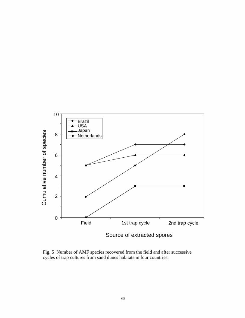
Source of extracted spores
0Field 1st trap cycle 2nd trap cycle
2
4
6
8
10Brazil
JapanUSA
Netherlands
Fig. 5 Number of AMF species recovered from the field and after successivecycles of trap cultures from sand dunes habitats in four countries.
68

Species Rank
11 5
Field2nd trap
10 15 20
10
100
1000
10000
Fig. 6 Species rank-log abundance curves for AMF community recovered in the field andafter two cycles of trap cultures for all sand dunes habitats.
69

0
10
20
30
40
b
b
b
c
c
b
aa
a aa a
Countries
Brazil U.S.A. Japan Netherlands
Fig. 7 Mycorrhizal colonization after 21-day infectivity assays for field soil, first andsecond trap cycle from four sand dunes habitats. Different letters within each soil representstatistically different means. Bars represent mean of five replicates for the field soil andnine replicates for each trap cycle. Lines represent standard error.
Field soil1st cycle2nd cycle
70

Number of species
00
10
20
30
40
2 4 6 8
Fig. 8 The relationship between the number of AMF species and thefor sand dunes soils studied
(r < 0.01, P = 0.93).
percentage colonizationof 21 days infectivity assays within the same propagation cycle
2
71

Number of spores
00
500 2000 25001000 1500 3000
10
20
30
40
Fig. 9 The relationship between thenumber of spores detected within the same propagation cycle for sand dunes soils studied(r < 0.52, P < 0.01).
percentage colonization of 21-day infectivity assays and
2
72

73
CHAPTER FOUR
EFFECT OF DIFFERENT FUNGAL ISOLATES
FROM THE SAME MYCORRHIZAL COMMUNITY
ON PLANT GROWTH AND PHOSPHORUS UPTAKE
IN SOYBEAN AND RED CLOVER

74
INTRODUCTION
Among the benefits arbuscular mycorrhizal fungi (AMF) provide to native plants andcrop species are improved shoot growth and reproductive potential (Smith and Read, 1997).These benefit is attributed primarily to increased uptake and transport of phosphorus by fungalhyphae beyond root depletion zones in soil. The plant host, in turn, provides the sole source ofcarbon for fungal growth and reproduction (Smith and Gianinazzi-Pearson, 1988). The relativeability of AMF isolates to improve plant growth and/or reproduction in a phosphate-deficientsoil is termed symbiotic effectiveness (Abbott and Robson, 1981). Differences in effectivenessamong AMF isolates are affected by a multitude of variables, such as rate of root colonization(Abbott and Robson, 1981), abundance of external hypha in the soil and hyphal distribution(Graham et al., 1982) and hyphal activity and longevity (Sylvia, 1988). Soil properties, such aspH (Skipper and Smith, 1979), temperature (Schenck and Smith, 1982) and P content (Schubertand Hayman, 1986) also impact effectiveness.
In many studies, effectiveness has been interpreted as a species-level property, but largedifferences between isolates of the same species have been identified and measured. Forexample, seven isolates of Glomus macrocarpum differed significantly in their ability topromote growth of pepper (Hass and Krikun, 1985). Similarly, different geographic isolates ofGlomus mosseae varied in improving water relations of Agropyron smithii (Stahl and Smith,1984). Two isolates of G. mosseae from an arid and a mesic environment improved soybeandry matter production relative to the control plants, but this benefit was not measured for anisolate from a semi-arid environment (Bethlenfalvay et al., 1989).
In most studies, effectiveness has been measured in single isolates propagated alone.However, fungi form complex communities within a root system in the field (Brundrett, 1991).Experiments involving more than one isolate on a single host would more likely reflectprocesses occurring naturally. Interspecific competition has been measured as a function ofdifferential sporulation (Daft, 1983; Daft and Hogarth, 1983) and differential entry pointformation in colonizing roots (Wilson, 1984). In spite of these interactions, a mixture of fungalisolates has been shown to provide more growth benefit than isolates colonizing alone (Daft,1983).
In studies involving mixtures of fungi, isolates being compared often originated fromdifferent habitats rather than from the same community. The results, therefore, provide noinformation on the relative proportion of effective and noneffective fungi within a mycorrhizalcommunity. Dissecting a mycorrhizal fungal community and testing the relative effectivenessof each fungal member has important implications. First, the net impact of fungal communitieson their plant hosts determines the stability of the symbiosis over time. Morton (1997)hypothesized that the fungal community must have a net positive effect on plants (the higherunit of selection) for the symbiosis to be maintained on 80% of all plant lineages for over 400million years. A net negative effect on some plant lineages may explain why the symbiosiswas sporadically lost in more recent times (Trappe, 1987). Second, manipulation of fungi tooptimally benefit agricultural or natural ecosystems involves either management of indigenouscommunities or introduction of a fungal inoculum. Each strategy depends on a completeunderstanding of the effectiveness of native or introduced fungal genotypes.
For fungi that are likely to be used as inocula (singly or as a “fungal community”), aparamount question is whether effectiveness on an assay host will hold true for other hosts and

75
the environments in which they are intended to grow. Only one study involving fungal isolatestested on different soils in seven separate greenhouses from Maryland to Florida has addressedthis issue (Sylvia et al., 1993). Results showed that only isolates with extremes in effectiveness(those that were highly effective and those that were ineffective) did not vary significantly withchanges in host or greenhouse environments. Isolates of intermediate effectiveness varied withhost or greenhouse environments.
In the present study, fungal isolates from three different communities were establishedin culture and then used as inocula to assess physiological diversity as expressed by relativeeffectiveness on a common assay host. The following specific hypotheses were tested: i) atleast one isolate from each community will effectively promote plant growth; ii) highlyeffective or ineffective fungi will express similar responses regardless of host environment; iii)non-effective isolates will not be detrimental to plant growth when an effective fungalsymbiont also is present in a mycorrhiza; and, iv) a mixture of AMF isolates from the samecommunity will enhance plant growth and phosphorus nutrition more than each isolatecolonizing alone.
MATERIAL AND METHODS
Experimental isolates
Fungal isolates used in this study were obtained from the International CultureCollection of Arbuscular and Vesicular-arbuscular Mycorrhizal Fungi (INVAM, West VirginiaUniversity). Twelve isolates were obtained from three separate mycorrhizal fungalcommunities designated as JA205, MN414 and VA105 (Table 1). Single species cultures wereestablished from spores retrieved after trapping native fungi from field soil at each of three sitesaccording to methods described in Chapter 3. Inoculum of all isolates were bulked by growingthem with sudangrass in 1.2 kg plastic pots for 3-4 months with a growth substrate definedbelow. Pots containing only sterile substrate also were seeded with sudangrass to produce non-mycorrhizal inocula for effectiveness assays.
Growth medium
The growth medium used on inoculum production pots and on effectiveness assays wasa sandy loam (Lily series) soil mixed (1:2, v/v) with quartzite sand (average grain size = 0.9mm), steam sterilized twice for one hour with a 24 hour cooling period between cycles. Aftersterilization, the substrate was wetted and dried in two cycles to reduce available Mn levels inthe soil elevated by steaming. Calcium carbonate (CaCO3) was added to the soil to attain afinal pH of 6.1-6.3. Soil was amended with 21 and 40 mg/kg-1 KH2PO4 for experiments 1 and2, respectively.
Infectivity assay
Inoculum potential of each isolate was measured by the mean percentage method(Moorman and Reeves, 1979) (Chapter 3) and then adjusted by soil dilution to equalizeinfectivity at the outset of each effectiveness assay. For experiment 1, inoculum of each isolatewas diluted 1:1 with sterile substrate and the mixture placed in 100 cm3 cone-tainers™ (Stueweand Sons, Inc., Corvallis, Oregon). Three seeds of sorghum (Sorghum vulgare L.) were placedin each cone and thinned to one plant after seedling emergence. After three weeks, plant rootswere gently separated from the growth medium by immersing the entire cone contents in water.Roots were placed in tissue capsules and stained according to the method of Koske and Gemma

76
(1989). Roots were spread over a Petri plate with a 0.5 inch grid etched in the bottom andmycorrhizal colonization estimated using the grid-line intersect method (Giovannetti andMosse, 1980).
Inocula of each isolate to be used in experiment 2 was diluted 1:5 (v/v) with sterilesubstrate, placed in 100 cm3 cone-tainers and seeded with red clover (Trifolium pratense L.).Seedlings were thinned to three per cone after emergence and watered with a solutioncontaining a commercial inoculum of Rhizobium trifolii (LiphaTec, Inc., Milwaukee,Wisconsin). Plants were grown for 21 days and roots processed and scored for mycorrhizalcolonization as described above.
Experiment 1
An effectiveness assay was set up with the Ark Centennial variety of soybean (Glycinemax L.) as the assay host, the growth medium described earlier, and 12 isolates of mycorrhizalfungi from three communities. Inoculum of each of the 12 isolates was diluted 1:5 (v/v) withsterile substrate and placed in 750 cm3 Deepots (Stuewe and Sons, Inc., Corvallis, Oregon).Inoculum of control pots consisted of pot contents of 3-month old sudangrass grown in thegrowth medium without AMF. An “artificial community” (AC) was established by mixing150ml of each of the following isolates: Acaulospora mellea CL358, Glomus mosseae FL156,Glomus clarum FL979A and Gigaspora gigantea NC119A. This AC was included as apositive control.
Three seeds were sown in each pot and thinned to one plant after emergence. Seedlingswere watered with a commercial solution of Bradyrhizobium inoculant for soybean (LiphaTec,Inc., Milwaukee, Wisconsin). Plants were grown for 7 weeks in a greenhouse withsupplementary lights (130 µmol m -2 s-1 at pot level) and ambient temperature ranging from15oC (night) to 35oC (day). The treatments were arranged in a complete randomized designwith 14 treatments and 5 replicates per treatment. The exception was Acaulospora melleaMN414A, whose inoculum potential was so low that only 3 replicates could be established.
At harvest, shoots were cut at the soil line and dried in a convection oven at 65oC for 4days. Roots were separated gently from the soil and immersed in water to remove soil particlesand debris. Roots were blot-dried and 0.5 g fresh weight of root was sampled randomly forroot colonization estimates. Remaining roots were dried in a convection oven at 65oC for 4days. Fresh root samples were stained according to the method of Koske and Gemma (1989)and mycorrhizal colonization measured by the grid-line intersect method (Giovannetti andMosse, 1980). Mycorrhizal root length was calculated for the entire pot based on total root drymass.
Shoot phosphorus content was measured using the sealed chamber digestion method(Anderson and Henderson, 1986) with some modifications. Approximately 0.25 g of oven-dried shoot material was placed in a heavy-walled teflon vessel, 3 ml of concentrated HNO3
and 0.5 ml of H2O2 added and the mixture incubated at room temperature for 15 minutes.Vessels were placed in a microwave and programmed for complete tissue digestion. Deionizedwater was added to the final digest to 25 ml and this slurry was filtered through Whatman #42paper. The phosphorus digest was analyzed with a inductively-coupled argon plasma (ICAP).

77
Experiment 2
Effectiveness assays of isolates alone or in combination were conducted using redclover (Trifolium pratense L.) as the host plant. Isolates were selected based on their P uptakebenefit in soybean (see experiment 1). Treatments were: VA105B and VA105D singly ortogether (VA105B + D), and MN414B, MN414C and MN414D singly or in all possiblecombinations (MN414B +C, MN414B+D and MN414C+D). Two additional treatments,designated VA105 community and MN414 community, were included by combining isolatesfrom each community. Pots were arranged in a complete randomized design with 12treatments and 5 replicates per treatment.
Inoculum of each treatment was increased for 3 months with red clover before startingof the effectiveness assay. Each inoculum then was diluted 1:5 (v/v) with the sterile growthmedium and placed in 750 cm3 Deepots. Red clover was sown and thinned to three plants perdeepot after emergence. At this time, seedlings were watered with a suspension of Rhizobiumtrifolii. Plants were cultivated in a growth room (245 µmol m-2 s-1 at pot level, a 14-hourphotoperiod, and ambient temperature ranging from 21-28oC).
At harvest, shoot and roots were processed as described above for soybean, except that0.25 grams of roots were used to measure mycorrhizal colonization. Shoot phosphorus wasanalyzed by the Kjedahl method using concentrated sulfuric acid (H2SO4) and hydrogenperoxide (H2O2) for digestion of plant tissues in a block digester (Lowther, 1980). Afterdigestion, phosphorus was measured by flow injection analysis colorimetry in a LachatQuickchem 8000L.
Statistical analyses
Data were screened for homogeneity of variance according to Levene’s test beforeanalysis of variance. Percentage of phosphorus in the shoots were arcsine square roottransformed before analysis. When homogeneity of variance was not achieved even aftertransformation, data were ranked and analyses were performed on the ranks. All statisticalanalyses were done using JMP® (SAS Inst. Inc., 1995).
RESULTS
Experiment 1
Mycorrhizal colonization was observed in plant roots of all pots except the controls andthose with Gi. gigantea VA105C (Table 2). Gigaspora gigantea lost infectivity during theexperiment and was removed from the statistical analyses. Mycorrhizal root length wasgreatest in plants inoculated with Glomus clarum VA105D and Entrophospora sp. VA105E,obtaining lengths of 2663 and 1164 cm, respectively.
All three isolates of the JA205 community improved shoot dry mass of soybean relativeto control plants (Fig. 1). Shoot dry mass of plants colonized by any isolate from the MN414community did not differ significantly from that of non-mycorrhizal controls. Only Glomusclarum from the VA105 community significantly increased shoot dry mass relative to the non-mycorrhizal control (Fig. 1).
Mycorrhizal colonization had a greater impact on phosphorus uptake than shoot drymass on soybean (Fig. 2). All isolates of the JA205 community, except A. trappei, increased P
78
uptake in shoots (Fig. 2). Two isolates of each MN414 and VA105 communities significantlyimproved percentage of P on soybean shoots relative to control plants (Fig. 2).
In each fungal community, at least two isolates improved either shoot dry matter orfoliar phosphorus content of soybean relative to control plants. Colonization by the “artificialcommunity” significantly improved both shoot dry mass and phosphorus in soybean (Figs. 1and 2).
Experiment 2
Plants in the control treatments showed no evidence of forming mycorrhizae and thehighest levels of colonization were observed for plants with VA105 community, Glomusclarum VA105D and Gigaspora gigantea MN414D and (Table 3). For most fungi,mycorrhizal root length ranged from 36 to 80 cm.
In the VA105 community, red clover colonized by Scutellospora verrucosa and Glomusclarum significantly produced more shoot dry mass and increased P uptake percentage thancontrols (Fig. 3 and 4). Glomus sp. and Gigaspora gigantea from the MN414 community alsoproduced more shoot dry mass and had higher P content in the shoots than control plants.Colonization by S. calospora MN414C significantly improved phosphorus uptake, but notshoot dry weight in red clover (Figs. 3 and 4).
Co-inoculation of two isolates did not always produced greater benefit than singleisolates. Shoot dry matter of plants colonized with S. verrucosa VA105B + Glomus clarumVA105D was significantly different from plants inoculated with S. verrucosa alone, but notfrom plants associated with G. clarum (Fig. 3). Plants colonized with isolates VA105B +VA105D, however, had significantly higher phosphorus content in the shoots relative to plantscolonized by either isolate alone (Fig. 4). Plants associated with Glomus sp. MN414B + S.calospora MN414C significantly produced more biomass and had a higher phosphorus contentthan plants associated with S. calospora alone, but they were not different from plantsinoculated with Glomus sp. alone (Figs. 3 and 4). Both shoot dry matter and phosphoruscontent of plants colonized by Glomus sp. MN414B + Gigaspora gigantea MN414D did notdiffer significantly from these values for plants colonized by either isolate alone. Plantscolonized with S. calospora MN414C + Gi. gigantea MN414D were not different from plantsinoculated with S. calospora alone, but produced significantly less shoot dry matter than plantstreated with Gi. gigantea alone (Fig. 3). Conversely, phosphorus content of plants grown withisolates MN414C + MN414D was not different from those grown with MN414D, but werehigher than those with MN414C alone (Fig. 4).
Colonization with fungi from the VA105 community improved dry matter accumulationmore than colonization with S. verrucosa VA105B alone, but not more than colonization withG. clarum VA105D (Fig. 3). Foliar phosphorus content was significantly higher in plantstreated with VA105 community than in those associated with either VA105B or VA105Dalone. Plants associated with MN414 community were not different in shoot dry matter or Pcontent from plants colonized by either MN414B or MN414D alone and they had higher shootdry matter and phosphorus content than MN414C alone (Figs. 3 and 4).
DISCUSSION
The effectivity assay on soybean tested the hypothesis that at least one isolate in eachmycorrhizal fungal community effectively promotes dry matter production. Plant biomass and

79
phosphorus uptake are common measurements evaluated when mycorrhizal effectiveness istested (Abbott and Robson, 1984). Using dry matter production as one criterion, all isolates ofthe JA205 community and G. clarum from the VA105 community were effective relative to thecontrols. None of the fungi in the MN414 community showed evidence of effectiveness. Incontrast, shoot P content increased with mycorrhizal colonization by at least two isolates fromeach of the three fungal communities. These results suggest that the net benefit of themycorrhizal symbiosis in which all fungi in each community are assumed to colonize roots willbe positive in either one or both of these parameters.
Differential responses in plants inoculated with different fungi in each of the twovariables measured is becoming increasingly recognized. For example, three co-occurringAMF differentially influenced size and clonal growth traits of two Prunella species: G.geosporum and another Glomus species produced a similar total leaf area response, but thenumber of stolon branches were higher in plants colonized by the former when compared to thelatter fungus (Streitwolf-Engel et al., 1997). In soybean, Acaulospora trappei JA205Bpromoted plant growth but not P content whereas Acaulospora mellea MN414A promoted Puptake but had no measurable effect on shoot dry matter production.
Part of the reason for differential host-fungus interactions is likely to be compatibilitybetween plant and fungal genotypes. Soybean typically is grown in soils either with high P orwith high P fertilizer applications. The JA205 fungal community was unusual in that all of itsmembers effectively increased soybean growth and/or P uptake. This community originatedfrom an experimental station where soil phosphorus levels (24 ppm) were relatively higher thanfrom the two other communities (<15 ppm), so isolates may be more adapted to conditionsfavorable to soybean growth and reproduction. If this is true, the results contradict Johnson’s(1993) hypothesis that fertilization selects for less beneficial fungal mutualists over time.However, soybean effectiveness assays measure benefit directly whereas Johnson based herresults on presence and abundance of sporulation as a measure of colonization and potentialbenefit.
Results of biomass and P content responses in both soybean and red clover support thehypothesis that either extreme in effectiveness by a fungal isolate (highly effective/ineffective)is conserved in changing host-soil environments. For example, G. clarum VA105D wasstrongly effective at promoting shoot growth and P uptake in both soybean and clover whereasS. calospora MN414C was equally ineffective for shoot growth measured in the two hostspecies. The comprehensive study conducted by Sylvia et al. (1993) emphasizesconservativeness of only the extremes in effectiveness or ineffectiveness. Intermediateresponses were more variable and could not be predicted from any given assay. It is themagnitude of effectiveness that may explain why a fungal isolate differs between host species(Streitwolf-Engel et al., 1997) or in different soils (Anderson et al., 1994).
The hypothesis that ineffective fungal isolates would not inhibit plant growth when co-colonizing with an effective fungus is supported by the red clover experiment, where selectedisolates were tested alone or together from different fungal communities. An additive effect onfoliar P content was evident when S. verrucosa VA105B and Glomus clarum VA105D formedmycorrhizae together. Contrasting trends were observed with the MN414 fungal isolates,where the effective Glomus sp. MN414B together with the ineffective Scutellospora calosporaMN414C produced similar shoot dry mass to red clover plants colonized by Glomus sp. alone.

80
These data suggest that interactive processes which impact effectiveness responses mayoccur among AMF isolates co-colonizing a root system. Part of these interactions undoubtedlyinvolves extent of mycorrhizal development, which would affect the magnitude of hostresponse. One fungal isolate can form mycorrhizae disproportionate to another by pre-emptingroot space available for colonization (Wilson, 1984). In the red clover experiment, the lesseffective isolate of the MN414 community may be a faster grower than the effective isolate.Differences in the rate of infection unit formation also may be a factor in differentialmycorrhizal development (Wilson and Trinick, 1983), although it should have been madenegligible in the red clover experiment by equalizing infectivity of all isolates. Interactionsbetween mycorrhizal fungi are certain to occur in nature where they form complexcommunities or when isolates are co-inoculated. The nature and intensity of these interactionsdepends as much on intrinsic properties of each fungus as the effects of soil and hostenvironments. These are hard to measure, since the fungus cannot be separated from its hostplant.
The performance of plants associated with all members of each fungal community wasequal or better than that of plants colonized by the most effective isolate in each community,based on shoot biomass, foliar P content, or both. These data, however, are not sufficientlyrobust to conclude that fungal mixtures consistently will benefit plants more than singleisolates. Clearly, dual inoculations sometimes produced additive responses, suggesting thatresults are strongly dependent on the composition of each mixture. Other research suggeststhat combining isolates may eliminate a poor performance by single isolates and give moreconsistent results (Daft and Hogarth, 1983). Testing the effectiveness of isolates separately andtogether is a necessary approach to fully characterize the performance of any inoculum mixturefor application to field settings.
Dissection of a mycorrhizal community by culturing all of its fungal members andtesting each isolate individually or in selected mixtures is a difficult task, explaining theabsence of such an approach to date. Gavito and Varela (1995), for example, set out to cultureand test 14 species in a fungal community associated with corn, but they were successful withonly two species. In this study, only three species were isolated from each fungal community,but it is likely that other less aggressive, nonsporulating fungi also may have been present inthe original community since three to five species is a relatively simple community (Morton etal., 1995). Nonetheless, results indicate that effective fungi are present in all of thecommunities examined thus far, suggesting that management of native fungi may be a morewidely useful strategy in most habitats than the introduction of nonindigenous species incommercial inocula. From a practical viewpoint, net effectiveness of the whole fungalcommunity can be tested from trap culture inoculum (see Chapter 3), which obviates thenecessity of culturing and testing each fungal species separately. If a mixed inoculum testsineffective, then more work can be done with member fungi to determine effectiveness andinfectivity of each isolate.
LITERATURE CITED
Abbott, L.K., and A.D. Robson. 1981. Infectivity and effectiveness of five endomycorrhizalfungi: competition with indigenous fungi in field soils. Australian Journal of AgriculturalResearch 32:621-630.
81
Abbott, L.K., and A.D. Robson. 1984. The effect of VA mycorrhizae on plant growth. Pp.113-130. C. Ll. Powell and D. J. Bagyaraj (Eds.), In: VA Mycorrhiza. CRC, Boca Raton,Florida.
Anderson, D.L., and L.J. Henderson. 1986. Sealed chamber digestion for plant nutrientanalyses. Agronomy Journal 78:937-939.
Anderson, R.C., B.A.D. Hetrick, and G.W.T. Wilson. 1994. Mycorrhizal dependence ofAndropogon gerardii and Schizachyrium scoparium in two prairie soils. The AmericanMidland Naturalist 132:366-376.
Bethlenfalvay, G.J., R.L. Franson, M.S. Brown, and K.L. Mihara. 1989. The Glycine-Glomus-Bradyrhizobium symbiosis. IX. Nutritional, morphological and physiological responses ofnodulated soybean to geographic isolates of the mycorrhizal fungus Glomus mosseae.Physiolpgia Plantarum 76:226-232.
Brundrett, M.C. 1991. Mycorrhizas in natural ecosystems. Advances in Ecological Research21:171-213.
Daft, M.J. 1983. The influence of mixed inocula on endomycorrhizal development. Plant andSoil 71:331-337.
Daft, M.J., and B.G. Hogarth. 1983. Competitive interactions amongst four species of Glomuson maize and onion. Transaction of British Mycological Society 80:339-345.
Gavito, M.E., and L. Varela. 1995. Response of “criollo” maize to single and mixed speciesinocula of arbuscular mycorrhizal fungi. Plant and Soil 176:101-105.
Giovannetti, M., and B. Mosse. 1980. An evaluation of techniques for measuring vesiculararbuscular mycorrhizal infection in roots. New Phytologist 84:489-500.
Graham, J.H., R.G. Linderman, and J.A. Menge. 1982. Development of external hyphae bydifferent isolates of mycorrhizal Glomus spp. in relation to root colonization and growth oftroyer citrange. New Phytologist 91:183-189.
Haas, J.H., and J. Krikun. 1985. Efficacy of endomycorrhizal-fungus isolates and inoculumquantities required for growth response. New Phytologist 100:613-621.
Johnson, N.C. 1993. Can fertilization of soil select less mutualistic mycorrhizae? EcologicalApplications 3:749-757.
Koske, R.E., and J.N. Gemma. 1989. A modified procedure for staining roots to detect VAmycorrhizas. Mycoogical Research 92:486-505.
Lowther, J.R. 1980. Use of a single sulfuric acid hydrogen peroxide digest for the analysis ofPinus radiata needles. Communication Soil Science Plant Anals 11:175-188.
Moorman, T., and F.B. Reeves. 1979. The role of endomycorrhizae in revegetation practicesin the semi-arid West. II. A bioassay to determine the effect of land disturbance onendomycorrhizal populations. American Journal of Botany 66:14-18.
Morton, J.B. 1997. Evolution of fungi in Glomales. J. White (Ed.). In: Evolution ofEndophytism in Plants. (in press)
Morton, J.B., S.P. Bentivenga, and J.D. Bever. 1995. Discovery, measurement, andinterpretation of diversity in arbuscular endomycorrhizal fungi (Glomales, Zygomycetes).Canadian Journal of Botany 73(suppl. 1):S25-S32.
SAS. 1995. JMP® Statistics and Graphics Guide. SAS Institute Inc., Cary, NC.Schenck, N.C., and G.S. Smith. 1982. Responses of six species of vesicular-arbuscular
mycorrhizal fungi and their effects on soybean at four soil temperatures. New Phytologist92:193-201.

82
Schubert, A., and D.S. Hayman. 1986. Plant growth responses to vesicular-arbuscularmycorrhiza. XVI. Effectiveness of different endophytes at different levels of soilphosphate. New Phytologist 103:79-90.
Skipper, H.D., and G.W. Smith. 1979. Influence of soil pH on the soybean-endomycorrhizasymbiosis. Plant and Soil 53:559-563.
Smith, S.E., and V. Gianinazzi-Pearson. 1988. Physiological interactions between symbiontsin vesicular-arbuscular mycorrhizal plants. Annual Review Plant Physiology and PlantMolecular Biology 39:221-244.
Smith, S.E., and D.J. Read. 1997. Mycorrhizal Symbiosis. Academic Press, London, 605 pp.Stahl, P.D., and W.K. Smith. 1984. Effects of different geographic isolates of Glomus on the
water relations of Agropyron smithii. Mycologia 76:261-267.Streitwolf-Engel, R., T. Boller, A. Wiemken, and I. Sanders. 1997. Clonal growth traits of two
Prunella species are determined by co-occurring arbuscular mycorrhizal fungi fromcalcareous grasslands. Journal of Ecology 85:181-191.
Sylvia, D.M. 1988. Activity of external hyphae of vesicular-arbuscular mycorrhizal fungi.Soil Biology and Biochemistry 20:39-43.
Sylvia, D.M., D.O. Wilson, J.H. Graham, J.J. Maddox, P.P. Millner, J.B. Morton, H.D.Skipper, S.F. Wright, and A.G. Jarstfer. 1993. Evaluation of vesicular-arbuscularmycorrhizal fungi in diverse plants and soils. Soil Biology and Biochemistry 25:705-713.
Trappe, J.M. 1987. Phylogenetic and ecological aspects of mycotrophy in the angiospermsfrom an evolutionary standpoint. Pp. 5-25. G.R. Safir (Ed.), In: Ecophysiology of VAMycorrhizal Plants. CRC Press, Boca Raton, Florida.
Wilson, J.M. 1984. Competition for infection between vesicular-arbuscular mycorrhizal fungi.New Phytologist 97:427-435.
Wilson, J.M., and M.J. Trinick. 1983. Infection, development and interaction betweenvesicular-arbuscular mycorrhizal fungi. New Phytologist 93:543-554.

83
Table 1 Fungal species recovered from bait cultures of rhizosphere soil from three plantcommunities.
Plant Community/Site Fungal Species INVAM No.
Scutellospora fulgida Koske & Walker VA105A
S. verrucosa Koske & Walker VA105B
Gigaspora gigantea
(Nicol & Gerd.) Gerd. & Trappe
VA105C
Glomus clarum Nicolson & Schenck VA105D
Ammophila and Solidago specieson a sand dune of AssateagueIsland, VA, U.S.A.pH 6.2-6.8, < 10 ppm P
Entrophospora sp. VA105E
Acaulospora trappei Ames & Linderman JA205B
G. mosseae (Nicol & Gerd.) Gerd. & Trappe JA205C
Triticum aestivum at anexperiment station in Hokkaido,Japan. pH 6.1, 24 ppm P
Glomus sp. JA205E
Acaulospora mellea
Schenck, Spain, Sieverding & Howeler
MN414A
Glomus sp. MN414B
S. calospora Koske & Walker MN414C
Gigaspora gigantea
(Nicol & Gerd.) Gerd. & Trappe
MN414D
Andropogon sp. on a 60 years oldfield at Cedar Creek, MN, U.S.A.pH 5.1, < 15 ppm P
Glomus sp. MN414E

84
Table 2 Mycorrhizal root length of 7-week old soybean plants. Numbers represent mean ±standard error.
Treatments Mycorrhizal Root Length (cm)
A. trappei JA205B 99.7 ± 33.2
G. mosseae JA205C 378.6 ± 49.8
Glomus sp. JA205E 356.9 ± 40.76
A. mellea MN414A 214.5 ± 55.18
Glomus sp. MN414B 591.6 ± 58.8
S. calospora MN414C 71.1 ± 10.0
Gi. gigantea MN414D 786.9 ± 86.7
Glomus sp. MN414E 304.0 ± 83.6
S. verrucosa VA105B 26.6 ± 12.1
G. clarum VA105D 2663.0 ± 103.3
Entrophospora sp. VA105E 1164.2 ± 166.6
Artificial Community 568.8 ± 42.8

85
Table 3 Mycorrhizal root length of 8-week old red clover plants. Numbers represent mean ±standard error.
Treatments Mycorrhizal Root Length (cm)
S. verrucosa VA105B 79.5 ± 22.1
G. clarum VA105D 250.2 ± 34.7
S. verrucosa + G. clarum 154.7 ± 25.2
VA105 community 425.5 ± 187.8
Glomus sp. MN414B 50.8 ± 6.7
S. calospora MN414C 60.3 ± 1.5
Gi. gigantea MN414D 232.7 ± 43.8
Glomus sp. + S. calospora 36.0 ± 4.2
Glomus sp. + Gi. gigantea 57.2 ± 5.4
S. calospora + Gi. gigantea 66.9 ± 27.2
MN414 community 68.8 ± 14.4

0
1
2
0.5
1.5
2.5
Isolates
86
Fig. 1 Shoot dry weight of soybean plants inoculated with AMF isolates from three differentcommunities. Columns represent the means of 5 replicates and bars represent standard errors.

0
0.04
0.08
0.16
0.12
Isolates
de dede d
bcab
bc bc
e e
a
c c
87
Fig. 2 Phosphorus content of soybean shoots inoculated with AMF isolates from three differentcommunities. Columns represent the means of 5 replicates and bars represent standard errors.

Isolates
0
0.4
0.5
0.3
0.2
0.1
a
bcbc
bcbc
b
c
d d
d
a
a
88
Fig. 3 Shoot dry weight of red clover plants inoculated with AMF isolates alone or incombination. Columns represent the means of 5 replicates and bars represent standard errors.

Isolates
0
0.3
0.2
0.1
a
ab ab
bc
dee
f
abcbc bc
cd cd
89
Fig. 4 Phosphorus content of red clover shoots inoculated with AMF isolates alone or incombination. Columns represent the means of 5 replicates and bars represent standard errors.

90
CONCLUSIONS
Research was conducted to measure diversity of arbuscular mycorrhizal fungitaxonomically and physiologically at the levels of individual isolates, species and communities.
I – Comparative Taxonomy
Morphology of subcellular structure in asexual spores was interpreted from ontogeneticcomparisons among species of Glomus in the family Glomaceae and Acaulospora andEntrophospora in the family Acaulosporaceae to better define species and to interpret ranking ofhigher taxa. Conclusions:
1) Comparative spore developmental studies have important implications for the systematic ofthe order Glomales.
a) Spore development in families Glomaceae and Acaulosporaceae divergesconsiderably from that in family Gigasporaceae and suggests that the order Glomalesis polyphyletic. Results of the present study together with biochemical properties andlife history traits provide convincing evidence for maintaining Glomaceae andAcaulosporaceae in order Glomales and placing Gigasporaceae in a new orderGigasporales.
b) Properties of flexible inner walls are shared by two or more species, providinggrouping criteria for a new taxonomic rank not yet recognized in Glomales. Thisrank is positioned between genus and species in the taxonomic hierarchy andtherefore is likely to be designated as subgenus.
c) Spore wall properties are the criteria which group different populations into the samemorphospecies.
2) Patterns of spore ontogenesis shared by all fungal species in Glomaceae and Acaulosporaceaewere:
a) Spore formation and differentiation could be separated into three phases, in whichspore expansion and spore wall differentiation occurred concurrently (phase I),flexible inner walls were differentiated with no further spore expansion (phase II),and a pre-germination structure was formed (phase III).
b) Subcellular parts of a spore differentiated in a linear sequence of separate and discretestages within each phase, as defined by addition and transformation of a structure.Stages were causally linked in that each new stage required completion ofdifferentiation in the previous stage. Each stage developed consistently amonggeographically disjunct fungal isolates of a species, thus providing reliable andconsistent grouping criteria to circumscribe species.
c) Within each stage, structures were differentiated hierarchically. Layers weresynthesized sequentially, followed by differentiation of phenotypic properties of eachlayer.
3) Ontogenetic patterns of spores unique to species of Glomus (Glomaceae) were:
a) The spore wall was the only major character complex formed in each spore, with eachlayer formed consecutively and concurrently with layers of the sporogenous hyphalwall. The number of layers in the spore wall and the phenotypes of each layer varied

91
considerably among species. One or two outer layers of the spore wall usuallydegraded and sloughed from mature spores, so that some taxonomically informativestructures were present only on juvenile spores.
b) Divergence in number of layers (1-2) in spore and hyphal walls of the most juvenilespores suggested distinct taxonomic lineages currently not recognized by currentclassification of Glomales. Divergence in number of phenotypes of spore wall layersdefined species.
c) There is no pregermination structure formed. Instead, the germ tube arises fromregrowth of the inner layer of the subtending hyphal wall after it (and the spore) havecompleted differentiation.
4) Ontogenetic patterns unique to species of Acaulospora and Entrophospora (Acaulosporaceae)were:
a) The spore wall consistently is made up of three layers in Acaulospora and two layersin Entrophospora, with the outer layer sloughing with maturation. Properties of theselayers define species.
b) Two hyaline bilayered flexible inner walls were always formed sequentially after thespore wall has completed differentiation. Properties of these layers are moreconserved and group species into supra-specific taxa currently not recognized in thepresent classification of Glomales.
II – Measurement of Community Structure
With improved interpretations of morphology to define species, the next goal was toassess taxonomic structure of fungal communities in five disparate habitats within the UnitedStates and in four sand dunes from around the world. Current assessment relies on sporulationby fungi colonizing a root system, so that successive propagation of trap cultures was examinedto test recovery and detection of nonsporulating fungal species. Conclusions:
1) Successive trap pot cultures established with native soils from disparate habitats detectedAMF species not sporulating in the field. Recovery efficiency ranged from 15-100% of thesefungi, appearing to vary most when temperature of the original habitat and that in thegreenhouse differed the greatest.
2) Species of Glomus were the predominant sporulators in trap cultures both in number ofspecies and in abundance of sporulation which supports other data indicating that this genus ismost widespread in nature.
3) AMF community structure approached a log series model after two cycles of trappingsuggesting that trap cultures cycles select for more aggressive fungi that sporulate prolificallyand dominate the mycorrhizal community.
4) Total number of spores and mycorrhizal infectivity of inocula from the five sites in the U.S.A.increased in the first propagation cycle and declined thereafter, whereas both variablesincreased through the second propagation cycle in the four sand dune sites. Differences areattributed mostly to the amount of disturbance to pot contents between propagation cycles -greater disturbance caused a loss of infectivity.

92
5) Taxonomic diversity had no influence on activity of the mycorrhizal community as evidencedby the absence of a relationship between AMF species richness and levels of mycorrhizalcolonization in infectivity assays. Total spore numbers, as propagules of AMF, explain about50% of the infectivity detected in the field and trap cultures and indicate that other fungalstructures are important infective propagules.
6) Spore numbers of the most abundant sporulators explained a large portion of the mycorrhizalcolonization observed in infectivity levels of some soils, suggesting that, in some cases, themost prolific spore producers are responsible for initial infection and secondary colonizationof assay hosts.
7) Index of similarity was relatively high between habitats differing in abiotic properties,suggesting that historical processes partly explain distribution of AMF species.
III – Physiological Diversity on Fungal Communities
Assays of effectiveness of component members of a mycorrhizal community inpromoting plant growth or plant phosphorus status complement taxonomic diversity by assessingfunctional relationships. Members of three fungal communities were screened to assesseffectiveness on two mycotrophic assay hosts, soybean and red clover. Conclusions:
1) At least one isolate from each AMF community was effective in increasing plant shootbiomass or foliar P content. Some isolates differentially promoted shoot dry mass while otherisolates differentially enhanced shoot P content.
2) Responses of highly effective or non-effective isolates were consistent in both soybean andred clover. Conservativeness in effectiveness responses suggests that this phenotype may beheritable.
3) Co-occurrence of paired effective and ineffective fungal isolates originating from the samemycorrhizal community resulted in either an additive or neutral effect on shoot biomass and Pcontent relative to either isolate alone. Plants colonized by each pair of isolates alwaysshowed enhanced shoot biomass and foliar P contents relative to nonmycorrhizal controls.
4) In comparing a total fungal community relative to single isolates in each community, bothcommunities tested (VA105 and MN414) performed equal to or better than their mosteffective isolate using either or both shoot biomass or foliar P content as criteria ofeffectiveness.
5) A net benefit was observed in red clover assay plants colonized by two of the fungalcommunities relative to nonmycorrhizal controls. This result suggests that management ofnative fungi may be a better strategy to increase plant productivity in natural or managedecosystems than introduction of exotic fungi produced commercially.

93
Sidney L. Stürmer
EDUCATION
1994-1998 - Ph.D. in Developmental Biology in the Division of Plant and Soil Sciences atWest Virginia University in Morgantown, WVDissertation Title: Characterization of Diversity of Fungi Forming ArbuscularEndomycorrhizae in Selected Plant Communities.
1987-1991 - B.S. in Biological Sciences in the Biology Department at Universidade Federal deSanta Catarina in Florianópolis, SC, Brazil
RESEARCH INTERESTS
Biology, ecology and systematic of symbiotic fungi forming arbuscular endomycorrhizaewith plants.
EXPERIENCE
Ø Research Assistant, Division of Plant and Soil Sciences, West Virginia University. Jan.1994 - Dec. 1995.
Ø Research Assistant, CNPq Scholarship. Project: “Biology and Ecology of vesicular-arbuscular endomycorrhizal fungi in dune regions in the Island of Santa Catarina (SC)”.Jan. - Aug. 1993.
Ø Research Assistant, CNPq/UFSC Scholarship. Project: “Selection and inoculation ofmycorrhizal fungi in fruit seedlings in nurseries of Santa Catarina”. Aug. 1990 - Dec.1992.
Ø Teaching Assistant ‘Applied Microbiology’. Dept. de Microbiologia e Parasitologia,Universidade Federal de Santa Catarina, Brazil. March - Aug. 1989.
PROFESSIONAL SERVICES
Ø Instructor, Workshop Taxonomy of Endomycorrhizal Fungi. 5th REBRAM (BrazilianMeeting about Mycorrhiza), Florianópolis, Brazil. October 1994.
Ø Reviewer, Mycologia, 1996
Ø Reviewer, Plant and Soil, 1997, 1998
Ø Instructor, Workshop Taxonomy of Endomycorrhizal Fungi. 1st ICOM (InternationalConference on Mycorrhizal), Berkeley, CA - USA. August 1996.
RESEARCH PUBLICATIONS
Book Chapters
Morton, J. B.; R. E. Koske; S. L. Stürmer, and S. P. Bentivenga. 1999. Protocols formeasurement of diversity among arbuscular fungi. In: Biological Diversity Handbook Series:Standard Methods for Fungi. Mueller, G., A. Rossman, and G. Bills, Eds. SmithsonianInstitute Press, Washington, DC. (in press).

94
Journal Articles
Stürmer, S.L. and M.M. Bellei. 1994. Composition and seasonal variation of spore populationsof arbuscular mycorrhizal fungi in dune soils on the island of Santa Catarina, Brazil.Canadian Journal of Botany, 72:359-363.
Stürmer, S.L. and J.B. Morton. 1997. Developmental patterns defining morphologicalcharacters in spores of four species in Glomus. Mycologia 89:72-81.
Stürmer, S.L. and J.B. Morton. 1999. Scutellospora rubra, a new arbuscular mycorrhizalspecies from Brazil. Mycological Research (in press).
Stürmer, S.L. and J.B. Morton. Taxonomic reinterpretation of morphological characters inAcaulosporaceae based on developmental patterns in two Acaulospora and oneEntrophospora species. Mycologia (submitted).
Published Abstracts
Stürmer, S.L. and M.M. Bellei. 1991. Ecology of vesicular-arbuscular mycorrhizal fungi indunes in the Island of Santa Catarina. 4th REBRAM, Mendes, Brazil.
Stürmer, S.L. and M.M. Bellei. 1992. Seasonal variation of arbuscular mycorrhizal fungipopulations on dune soils in the Island of Santa Catarina (South Brazil). The InternationalSymposium on Management of Mycorrhizas in Agriculture, Horticulture and Forestry. Perth,Western Australia.
Klauberg F., O.; M.M. Bellei and S.L. Stürmer. 1993. Ecology of endomycorrhizal (AM) fungiin South Brazil. 9th NACOM, Guelph, Canada.
Stürmer, S.L.; M.M. Bellei and O. Klauberg F. 1993. Occurrence of endomycorrhizal fungi insoils and on roots of 3 successional stages in the development of Atlantic Forest in Brazil.9th NACOM, Guelph, Canada.
Stürmer, S.L. and J.B. Morton. 1994. Spore differentiation sequences among isolates of Glomusetunicatum and Glomus intraradices. 5th REBRAM, Florianópolis, Brazil.
Stürmer, S.L. and J.B. Morton. 1998. Developmental patterns in three species ofAcaulosporaceae redefining interpretation of morphological character sets. II InternationalConference on Mycorrhiza (ICOM), Uppsala, Sweden.
Stürmer, S.L. and J.B. Morton. 1998. Patterns of sporulation of AMF in sand dunes systemsafter successive cycles of trap cultures. II International Conference on Mycorrhiza (ICOM),Uppsala, Sweden.
INVITED SPEAKER
Stürmer, S.L. 1998. Analyses of arbuscular mycorrhizal fungi communities – Structure andfunction. II Brazilian Conference of Mycology, Rio de Janeiro, Brazil, April 17-21, 1998.
Stürmer, S.L. 1998. The role of morphology to reinterpret evolutionary patterns and process inAMF. FERTBIO98, Caxambu, Brazil, October 11-16, 1998.