characterization of exopolymeric substances (eps) produced...
TRANSCRIPT
This article was downloaded by: [Biblioteca Universidad Complutense de Madrid], [Antonio Ballester]On: 01 April 2014, At: 01:30Publisher: Taylor & FrancisInforma Ltd Registered in England and Wales Registered Number: 1072954 Registered office: Mortimer House,37-41 Mortimer Street, London W1T 3JH, UK
Biofouling: The Journal of Bioadhesion and BiofilmResearchPublication details, including instructions for authors and subscription information:http://www.tandfonline.com/loi/gbif20
Characterization of exopolymeric substances (EPS)produced by Aeromonas hydrophila under reducingconditionsLaura Castroa, Ruiyong Zhangb, Jesús A. Muñoza, Felisa Gonzáleza, M. Luisa Blázqueza,Wolfgang Sandb & Antonio Ballestera
a Department of Material Science and Metallurgical Engineering, Complutense University ofMadrid, Madrid, Spainb Biofilm Centre, Aquatic Biotechnology, University of Duisburg-Essen, Essen, GermanyPublished online: 27 Mar 2014.
To cite this article: Laura Castro, Ruiyong Zhang, Jesús A. Muñoz, Felisa González, M. Luisa Blázquez, Wolfgang Sand &Antonio Ballester (2014): Characterization of exopolymeric substances (EPS) produced by Aeromonas hydrophila underreducing conditions, Biofouling: The Journal of Bioadhesion and Biofilm Research, DOI: 10.1080/08927014.2014.892586
To link to this article: http://dx.doi.org/10.1080/08927014.2014.892586
PLEASE SCROLL DOWN FOR ARTICLE
Taylor & Francis makes every effort to ensure the accuracy of all the information (the “Content”) containedin the publications on our platform. However, Taylor & Francis, our agents, and our licensors make norepresentations or warranties whatsoever as to the accuracy, completeness, or suitability for any purpose of theContent. Any opinions and views expressed in this publication are the opinions and views of the authors, andare not the views of or endorsed by Taylor & Francis. The accuracy of the Content should not be relied upon andshould be independently verified with primary sources of information. Taylor and Francis shall not be liable forany losses, actions, claims, proceedings, demands, costs, expenses, damages, and other liabilities whatsoeveror howsoever caused arising directly or indirectly in connection with, in relation to or arising out of the use ofthe Content.
This article may be used for research, teaching, and private study purposes. Any substantial or systematicreproduction, redistribution, reselling, loan, sub-licensing, systematic supply, or distribution in anyform to anyone is expressly forbidden. Terms & Conditions of access and use can be found at http://www.tandfonline.com/page/terms-and-conditions
Characterization of exopolymeric substances (EPS) produced by Aeromonas hydrophila underreducing conditions
Laura Castroa, Ruiyong Zhangb, Jesús A. Muñoza, Felisa Gonzáleza, M. Luisa Blázqueza, Wolfgang Sandb and AntonioBallestera*aDepartment of Material Science and Metallurgical Engineering, Complutense University of Madrid, Madrid, Spain; bBiofilm Centre,Aquatic Biotechnology, University of Duisburg-Essen, Essen, Germany
(Received 15 July 2013; accepted 4 February 2014)
The aim of this work was to investigate the production of extracellular polymeric substances (EPS) by Aeromonas hy-drophila grown under anaerobic conditions. EPS composition was studied for planktonic cells, cells attached to carbonfibre supports using a soluble ferric iron source and cells grown with a solid ferric iron mineral (gossan). Conventionalspectrophotometric methods, Fourier transform infrared (FTIR) and confocal laser scanning microscopy (CLSM) wereused to determine the main components in the biofilm extracted from the cultures. The key EPS components were pro-teins, indicating their importance for electron transfer reactions. Carbohydrates were observed mostly on the mineral andcontained terminal mannosyl and/or terminal glucose, fucose and N-acetylgalactosamine residues.
Keywords: Aeromonas hydrophila; extracellular polymeric substances; CLSM; FTIR; SEM
Introduction
Microorganisms grow in natural environments usually inthe form of interfacial films bound to different substrata.Bacterial biofilms are composed of a hydrated matrix ofextracellular polymeric substances (EPS) which form theirimmediate environment. The typical constituents of EPSare polysaccharides, proteins, nucleic acids and lipids(Flemming et al. 2007). These compounds provide themechanical stability for these biofilms (Sutherland 2001),mediate their adhesion to surfaces (Lüttge et al. 2005) andform a cohesive three-dimensional polymer network inter-connecting and immobilizing cells. EPS can be subdividedinto bound/capsular EPS and soluble/colloidal EPS(Sheng et al. 2010). Capsular EPS are tightly bound tocells, while colloidal EPS are weakly bound to cells oreasily lost into the solution. Generally, these two types ofEPS can be separated by centrifugation, with thoseremaining in the supernatant being soluble, colloidal EPSand those forming microbial pellets being bound capsularEPS. EPS of the biofilm matrix appear to have several keyproperties and functions. Polymers represent a structuralcomponent of microbial cell surfaces. EPS may carrycharged or hydrophobic groups resulting in an adsorptivepolymer which can bind various nutrients, metals and con-taminants (Harrison et al. 2007). Some EPS are involvedin informative functions (Whitchurch et al. 2002), whereasothers are able to condition interfaces (Zhong et al. 2007).Moreover, some polymers represent an ideal source ofnutrients (Davey & O’Toole 2000) and some can be
involved in motility (Lu et al. 2005). Although specula-tive, some evidence shows that there is a potential role forredox-active EPS used by certain bacteria for anaerobicrespiration (Neu & Lawrence 2009). Extensive researchundertaken in the past few decades has focused on under-standing the properties of these biopolymers. Several ana-lytical techniques are used for studying EPS components.The use of Fourier transform infrared spectroscopy (FTIR)in the field of microbiology has proved to be a promisingtechnique. FTIR spectroscopy simultaneously measuresthe vibrations of functional groups of different cellcomponents in multi-component mixtures (Schmitt &Flemming 1998; Sheng et al. 2006). Furthermore, therehas been the development and application of techniquesallowing the observation of fully hydrated interfacialmicrobial cultures using confocal laser scanningmicroscopy (CLSM) Neu & Lawrence 1997; Neu et al.2001; Zippel & Neu 2011).
In anaerobic soil and sedimentary environments,microbial reductive dissolution of iron oxides coupled tooxidation of organic matter (electron donor) is thought tobe an important biogeochemical process, resulting in theformation of biogenic ferrous iron (Fe(II)) species.
Gossan ores are natural minerals composed mainly ofiron oxyhydroxides with residual unaltered minerals suchas quartz. In addition to iron oxyhydroxides, various pre-cious metals, such as gold and silver, and various sul-phate and silicate minerals can occur in gossans (Essalhiet al. 2011). Anaerobic iron bioleaching is ubiquitous in
*Corresponding author. Email: [email protected]
© 2014 Taylor & Francis
Biofouling, 2014http://dx.doi.org/10.1080/08927014.2014.892586
Dow
nloa
ded
by [
Bib
liote
ca U
nive
rsid
ad C
ompl
uten
se d
e M
adri
d], [
Ant
onio
Bal
lest
er]
at 0
1:30
01
Apr
il 20
14

subsurface environments, including mining-impactedareas. The extent of iron oxide microbial dissolution insubsurface sediments can vary greatly depending on dif-ferent environmental conditions (Bonneville et al. 2009;Salas et al. 2010) including the specific bacteria involvedin this process, the surface area, particle size and crystal-linity of iron oxides, the availability of electron donors,and the interaction between iron oxides and bacteria.
The immobilization technology of cells and enzymeshas rapidly developed and has many applications. Cellimmobilization is characterized by maintaining a stablehigh density of cells on the support matrix and couldimprove the reductive rate of Fe(III) reduction. It has beenapplied to treat acid mine-drainage or bioleach minerals.Another application of cell immobilization could be bac-terial fuel cells. Some species can colonize the anode anddirectly transfer electrons to the electrode from the bacte-rial EPS that are in contact with it (Torres et al. 2010).
In the present study, the colloidal and capsular EPSfractions obtained from pure cultures of Aeromonas hy-drophila are characterized. The aim was to identify theconstituents of EPS by chemical extraction using EDTAand their spatial interactions with each other and with thesupport. This work was focused on the EPS of the Gram-negative bacterium A. hydrophila, which is known toreduce Fe(III) and keep Fe(II) in solution (García-Balboaet al. 2010). For that purpose, A. hydrophila was culti-vated using soluble ferric citrate as iron source. The EPSwere extracted from planktonic cells and from cells grownon carbon fibres. Moreover, the EPS produced by micro-organisms grown on a gossan ore as an insoluble ironsource were studied. The formation of A. hydrophila bio-films on different substrata under iron reducing conditionswas examined using scanning electron microscopy (SEM)and CLSM. Fluorescently labelled lectins were used tostain the EPS, in order to gain hints about the chemicalcomposition of the polysaccharides in the biofilm.
Materials and methods
Bacterial culture
The A. hydrophila strain for the study was isolated fromthe edge of an open-pit lake surrounding an abandonedmine site named ‘Brunita’ (formerly a source of Pb–Znores) near La Unión (Murcia, Spain).
A modified Postgate C medium was used to growthe bacterium. It contained the following salts (g l−1):KH2PO4, 0.5; NH4Cl, 1.0; Na2SO4, 4.5; CaCl2·6H2O,0.06; MgSO4·7H2O, 0.06; sodium lactate, 6.0; yeastextract, 1.0; and sodium citrate heptahydrate, 0.3. ThepH was adjusted to 7.0 ± 0.2.
Standard anaerobic techniques were used in thisresearch. Sterilized medium (autoclave conditions:121°C, 30 min) was poured into individual glass flasks.The medium was supplemented with ferric citrate
(60 mM in Fe3+) or with the gossan mineral (10 g l−1).Then, the solution was vigorously bubbled with N2:CO2
(80/20, v/v) to strip dissolved oxygen. The flasks werecapped with butyl rubber stoppers and sealed with alu-minium crimps. A 10% inoculum of a Fe(III)-reducingculture was used (8 × 10⁸ cells ml−1). The cultures wereincubated unstirred at 30°C in darkness.
EPS extraction method
The extraction protocol is summarized in Figure 1. Whenthe cultures reached the stationary growth phase, theywere centrifuged for 10 min at 7,500 rpm (9,900 g) and4°C. The supernatant was collected and filtered through0.2 μm pore size filters under sterile conditions to elimi-nate remaining bacteria. The fraction obtained containedthe colloidal EPS. To extract the bound EPS, the pelletwas re-suspended in 10 ml of salt solution and centri-fuged for 10 min at 7,500 rpm (9,900 g) and 4°C. Thesupernatant was named the washed fraction. The newpellet was re-suspended in 10 ml of 20 mM EDTA at pH7 and the suspension was incubated with shaking for 1 hat 4°C. Thereafter, the mixtures of bacterial suspensionsand extracting agents were centrifuged for 10 min at7,500 rpm (9,900 g) and 4°C to remove the remainingcells. This process was repeated three times and the threeresulting supernatants contained the capsular fraction. Toremove the residual salts from the growth medium andthe extraction agent, each EPS fraction was dialysed, firstwith deionized water for 12 h and then with milliQ waterfor 72 h at 4°C. Cellulose membrane dialysis tubing of3,500 Da was used.
The EPS mass was obtained after freeze drying 2 ml ofthe purified EPS solution. The freeze drying was per-formed using a freeze dryer ALPHA 2-4 LSC at 1 mbarand –80°C for 24 h (Christ, Osterode am Harz, Germany).
Characterization of the chemical composition of theEPS
Carbohydrates, proteins, uronic acids and DNA weredetermined photocolorimetrically using a UV–Vis spec-trophotometer (JASCO V-650, Jasco, Tokyo, Japan). Theamount of carbohydrate was determined by the Duboisassay (Dubois et al. 1956) using glucose as standard anda wavelength at 490 nm. The protein content was deter-mined according to the Lowry method (Lowry et al.1951) using bovine serum albumin (BSA) as standardand measuring absorbance at 595 nm. The quantitativedetermination of uronic acids was performed using themethod of Blumenkrantz and Asboe-Hansen (1973). D(+)-glucuronic acid was used as standard and the absor-bance was measured at 520 nm. The extent of EPS con-tamination by cell lysis during extraction was estimatedby analysing the quantity of 2-keto-3-deoxyoctonate
2 L. Castro et al.
Dow
nloa
ded
by [
Bib
liote
ca U
nive
rsid
ad C
ompl
uten
se d
e M
adri
d], [
Ant
onio
Bal
lest
er]
at 0
1:30
01
Apr
il 20
14

(KDO) in samples using KDO as a standard (Karkhaniset al. 1978).
Fourier transformed infrared (FTIR) spectroscopy
FTIR was used to determine the variation in the chemi-cal groups in the EPS due to different growth conditions.Previous to FTIR analysis, the EPS sample and the con-trol were freeze-dried (ALPHA 2-4 LSC, –80°C). Thesesamples were pelleted, mixing the EPS sample with~250 mg of KBr. The spectra were recorded with a Nico-let Magna 750 in the region of 500–4,000 cm−1 at a res-olution of 4 cm−1 (Thermo Scientific, West Palm Beach,FL, USA).
Scanning electron microscopy (SEM)
Cultures were grown under different conditions and fil-tered onto a 0.2 μm pore-size filter. The samples weresuccessively dehydrated with acetone and stored over-night at 4°C in 90% acetone. The samples were dried bycritical-point drying and coated with graphite and gold.Specimens were examined with a JEOL JSM-6330Fmicroscope, FE-SEM at 10 kV to determine the spatial
distribution of cells on the surfaces (JEOL, Tokyo,Japan).
Confocal laser scanning microscopy (CLSM)
The visualization of the fluorescent markers was per-formed using a laser scanning module LSM 510 coupledwith an inverted microscope 100M BP (Zeiss, Jena, Ger-many). The CLSM images were generated in a multi-track mode which allowed the separate recording of thedifferent signals corresponding to the respective laserwavelength. Image analysis was performed with softwareprograms LSM 510 version SP2 (Zeiss), Volocity 3.5(Improvision) and AxioVersion 3.1 (Zeiss, Jena,Germany).
Nucleic acid staining
The spatial distribution of microbial cells within theaggregates was shown with general nucleic acid specificstains: DAPI (diamidino-2-phenylindole), SYTO 9 andSYTO 62. DAPI is a blue fluorescent probe that fluo-resces brightly upon selectively binding to the minorgroove of double stranded DNA. SYTO 9 is a green
Figure 1. General procedure for EPS extraction using EDTA.
Biofouling 3
Dow
nloa
ded
by [
Bib
liote
ca U
nive
rsid
ad C
ompl
uten
se d
e M
adri
d], [
Ant
onio
Bal
lest
er]
at 0
1:30
01
Apr
il 20
14

fluorescent nucleic acid dye to stain live and dead bacte-ria. The cell-permeant SYTO 62, a fluorescent nucleicacid stain, exhibits bright and red fluorescence uponbinding to nucleic acids.
Lectin binding assays
Ten lectins were selected to determine whether the EPScontained fucose, mannose, glucose, galactose, glucosa-mine, N-acetyl galactosamine or other residues. To labelcarbohydrates the following fluoresceine isothiocyanate(FITC) and tetramethylrhodamine isothiocyanate(TRITC) labelled lectins were used in these experiments:Arachis hypogaea (PNA) specific for D(+)galactose,Canavalia ensiformis (Con A) specific for D(+)glucoseand D(+)mannose, Erythrina cristagalli (ECA) specificfor N-acetyl-D-galactosamine and D-galactose, Ulexeuropaeus (UEA I) specific for L(-)fucose, Lens culinaris(LcH) specific for α-mannose and α-glucose, Triticumvulgaris (WGA) specific for N-acetyl-glucosamine, Gly-cine max (SBA) specific for α- and β-N-acetylgalactos-amine and galactopyranosyl residues, Phytolaccaamericana (PWM) specific for N-acetyl-glucosamine,Bandeiraea simplicifolia (BS I) specific for α-D-galacto-syl and N-acetyl-α-D-galactosaminyl residues and Phase-olus vulgaris (PHAE) specific for galactose. Briefly,samples were incubated with 0.05 mg ml−1 of the lectinsfor 40 min at room temperature. Stained samples werethen washed three times with filter-sterilized tap water inorder to remove the non-bound lectins. Direct light expo-sure was avoided as much as possible. In order to pro-long the fluorescence of the dyes, an antifading agent(CitifluorTMAF2, Citifluor, Eschborn, Germany) wasused when mounting the samples.
Results and discussion
EPS production and chemical composition
The EPS extraction yield using EDTA treatment hasbeen considered to be relatively low compared withother chemical extraction methods for activated sludge(Zhang et al. 1999). For example, regular centrifugationwith formaldehyde extraction results in a yield of 10%,whereas EDTA extraction reaches a yield of 2%. How-ever, the EPS extraction method using EDTA allowsdetection of the protein content in the EPS. There is,therefore, a compromise between EPS yield and avail-ability for specific analyses. In addition, no significantcell lysis occurs during EDTA extraction (Zhang et al.1999), which makes this method a good choice for EPSextraction from biofilms. Cell lysis during extraction wasestimated by analysing the quantity of 3-Deoxy-D-manno-oct-2-ulosonic acid (KDO) in the samples. KDOis part of the cell membrane of Gram-negative bacteria
and therefore can be used as a marker for contaminationby membrane compounds. The amounts of KDO in thecell pellets after cell lysis with sodium dodecyl sulphate(SDS) were compared with that in EPS. KDO in theEPS fraction was detected after extraction for 9 h andwas < 5% of that in cell pellets. A low content of KDOindicated that there was no significant cell lysis causingintracellular materials to be released into the solutionduring the extraction.
Both colloidal and capsular EPS from A. hydrophilabiofilms were extracted and analysed. Bacterial cells pro-duced more capsular EPS, when forming biofilms, com-pared to planktonic cells (Figure 2). This evidence wasalso corroborated in previous studies on bacterial biofilmformation (Laspidou & Rittmann 2002; Cao et al. 2011).The increased amount of colloidal EPS when using asoluble iron source, citrate-Fe(III), was accounted for bythe citrate remaining in this fraction after dialysis treat-ment. Citrate was not present when iron was provided asa solid mineral.
The composition of the EPS matrix in biofilms isreported to be very complex, containing proteins, carbo-hydrates, nucleic acids, lipids, amphiphilic moleculesand humic substances (Flemming et al. 2007). In thisstudy, conventional chemical colorimetric analyses wereused to quantify the composition of the purified EPSproduced by A. hydrophila and extracted from sampleswith different growth conditions. The results are listed inTable 1.
The EPS extracted from anaerobic cultures wasmainly composed of proteins. However, the content ofpolysaccharides was very low. This fact may be
Figure 2. Amount of EPS produced per cell of A. hydrophilagrown under different conditions and by planktonic cells, cellsattached to carbon fibre supports using a soluble ferric ironsource and by cells grown with a solid ferric iron mineral(gossan).
4 L. Castro et al.
Dow
nloa
ded
by [
Bib
liote
ca U
nive
rsid
ad C
ompl
uten
se d
e M
adri
d], [
Ant
onio
Bal
lest
er]
at 0
1:30
01
Apr
il 20
14
explained by the transporter profile of A. hydrophilawhich is comparable to those of pseudomonads and vib-rios, with an abundance of amino acid and peptide trans-porters and relatively few sugar uptake systems(Seshadri et al. 2006). A. hydrophila is able to reducemetals and proteins that could act as redox active mole-cules (Neu & Lawrence 2009), facilitating and directingelectron transfer, and may aid in the efflux of heavy met-als or toxic compounds encountered in potentially highlypolluted water.
Polysaccharides were found in the capsular fractionof the biofilm formed on the mineral surface. Likely,they are involved in the formation of the biofilm matrixmediating the mechanical stability of biofilms and deter-mining its architecture.
EPS produced by A. hydrophila contained apolarregions rich in uronic acids. Uronic acids could promotepolysaccharide gel formation, ion exchange, mineral for-mation and the accumulation of toxic metal ions (con-tributing to environmental detoxification). In addition,charged functional groups of uronic acids have the abil-ity to bind metal cations.
FTIR characterization of EPS
FTIR spectra of EPS isolated from A. hydrophila areshown in Figure 3. Figure 3A shows the spectrum ofEPS for cells grown on ferric citrate as a soluble ironsource. Figure 3B displays the spectrum of EPS of cellsgrown on insoluble gossan mineral. The spectra of col-loidal EPS were different in both cases. Colloidal EPS ofcells grown with ferric citrate revealed the presence oftwo very intense bands at 1,621 and 1,380 cm−1. Thefirst band is characteristic of antisymmetric stretchingvibrations and the second absorption corresponds to thesymmetric stretching vibrations of the COO– group incarboxylate anions. These bands are associated with thepresence of citrate in the medium. A. hydrophila useslactate as an energy source instead of citrate and citrateremains in solution. Moreover, the intense broad absor-bance at 3,435 cm−1 is attributed to the O–H stretchingmodes of vibration in the hydroxyl functional group and
N–H stretching vibrations in amides and amines. Thisbroad band at ~ 3,400 cm−1 appeared also in the colloi-dal EPS, when cells were grown on gossan mineral. Inaddition, the absorption peak at 1,631 cm−1 correspondsto the amide I band and the band at 1397 cm−1 isassigned to the C–N stretching vibration. The two bandsat 1,114 and 1,056 cm−1 could be assigned to C–Ostretching vibration modes of alcohols. The spectrum ofcolloidal EPS on mineral revealed the presence of vari-ous functional groups, typically found in proteins andpolysaccharides.
On the other hand, almost negligible differences werenoted for the FTIR spectra of the capsular fraction.There, C–O vibrations of oligo- and polysaccharidestructures and carboxylate groups were mainly detected.The spectra showed a peak around 3,520 cm−1 corre-sponding to the ‘free’ O–H stretching vibration of thecarboxyl group and another at 3,392 cm−1 correspondingto the associated O–H vibration. The absorption peak at2,977 cm−1 is assigned to C–H stretching vibrationmodes in the hydrocarbon chains and at 3,028 cm−1 toC–H stretching vibration in alkenes. FTIR analysis ofthe capsular EPS revealed the presence of three intensebands at 1,630, 1,475 and 1,396 cm−1. The first two arecharacteristic of amide I and II bands, respectively. Theother spectral band at 1,396 cm−1 is assigned to the C–Nstretching vibration of proteins. In consequence, the mostrelevant bands in the capsular EPS of A. hydrophila arerelated to the presence of amino acids. Dissimilatorymetal-reducing bacteria are an important group ofmicrobes that can directly or indirectly catalyse thereduction of redox-reactive metal ions such as Fe andMn. In this way, EPS may contain redox active mole-cules, such as outer membrane cytochromes, capable offacilitating and directing electron transfer (Summerset al. 2010).
The major components in microorganisms have alarge number of sharp and characteristic absorptionbands in the fundamental infrared region, allowing evalu-ation of the composition of EPS without separation intoindividual components. Typical characteristic adsorptionbands were not obtained for quantitative analysis becauseof the overlap of the spectral bands. Nevertheless, quan-titative chemical assays of protein and carbohydrate con-tents were consistent with the different functional groupsdetermined by FTIR measurements (Table 1).
Comparative SEM and CLSM analysis of biofilmstructure
Planktonic and sessile cells of A. hydrophila were visual-ized using SEM (Figure 4). The planktonic and the ses-sile cells showed a marked difference in their generalappearance. When A. hydrophila cells were grown on acarbon substratum, cells attached to the surface by
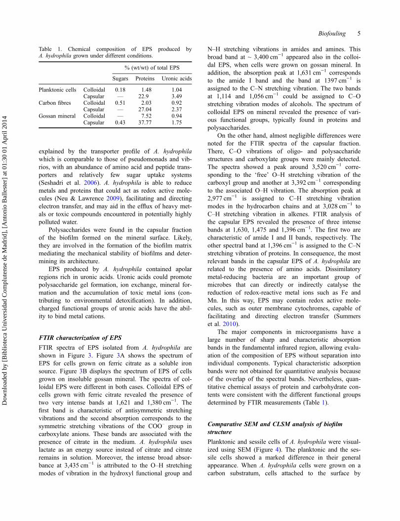
Table 1. Chemical composition of EPS produced byA. hydrophila grown under different conditions.
% (wt/wt) of total EPS
Sugars Proteins Uronic acids
Planktonic cells Colloidal 0.18 1.48 1.04Capsular — 22.9 3.49
Carbon fibres Colloidal 0.51 2.03 0.92Capsular — 27.04 2.37
Gossan mineral Colloidal — 7.52 0.94Capsular 0.43 37.77 1.75
Biofouling 5
Dow
nloa
ded
by [
Bib
liote
ca U
nive
rsid
ad C
ompl
uten
se d
e M
adri
d], [
Ant
onio
Bal
lest
er]
at 0
1:30
01
Apr
il 20
14

‘nanowires’ (Figure 4B). These nanowires appeared notonly between the cells and the surface but also between(connecting) the cells. Several findings suggest that spe-cialized bacterial pili act as electron conductors to metalsas acceptors during respiration (Reguera et al. 2005).Additionally, these nanowires may also play a role in in-terbacterial signalling. Analyses of bacteria such as
Geobacter sulfurreducens and Shewanella oneidensishave revealed this as a possible contact-dependent mech-anism of communication (Reguera et al. 2005; Gorbyet al. 2006). These strains reduce metals including Fe(III) and Mn(IV) apparently via conductance of electronsfrom the cytoplasmic membrane through nanowire pili.Based on the observation that pili tend to be intertwined
(A)
(B)
Figure 3. FTIR spectra of EPS (colloidal fraction in red and capsular fraction in blue) of A. hydrophila grown: (A) with ferric citrateon carbon fibres; and (B) on gossan mineral as sole electron acceptor.
6 L. Castro et al.
Dow
nloa
ded
by [
Bib
liote
ca U
nive
rsid
ad C
ompl
uten
se d
e M
adri
d], [
Ant
onio
Bal
lest
er]
at 0
1:30
01
Apr
il 20
14

Figure 4. SEM and CLSM images of planktonic cells of A. hydrophila (A, D), cells attached to a carbon fibre surface (B, E) andon gossan mineral (C, F). CLSM images of a biofilm region stained with SYTO 9 nucleic acid stain (green) to show the distributionof bacterial cells within the biofilm and with Con A lectin (red) for sugars.
Biofouling 7
Dow
nloa
ded
by [
Bib
liote
ca U
nive
rsid
ad C
ompl
uten
se d
e M
adri
d], [
Ant
onio
Bal
lest
er]
at 0
1:30
01
Apr
il 20
14
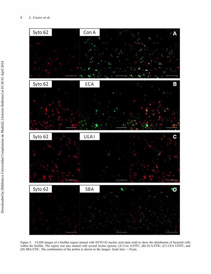
Figure 5. CLSM images of a biofilm region stained with SYTO 62 nucleic acid stain (red) to show the distribution of bacterial cellswithin the biofilm. The region was also stained with several lectins (green): (A) Con A-FITC, (B) ECA-FTIC, (C) UEA I-FITC, and(D) SBA-FTIC. The combination of the probes is shown in the images. Scale bars = 10 μm.
8 L. Castro et al.
Dow
nloa
ded
by [
Bib
liote
ca U
nive
rsid
ad C
ompl
uten
se d
e M
adri
d], [
Ant
onio
Bal
lest
er]
at 0
1:30
01
Apr
il 20
14

between cells, it is possible that these bacteria are liter-ally plugged into each other via nanowire contacts.
SEM observations demonstrated the presence of EPSon solid mineral (Figure 4C). Bacterial biofilm formed adiscontinuous coating on the surface of mineral particlesand in some places EPS formed organic bridges linkingtogether contiguous particles.
Planktonic cells, cells attached to a carbon surface,cells grown on gossan mineral and biofilms were stainedby fluorescently labelled lectins and analysed by CLSM(Figure 4). This technique allows the observation of fullyhydrated samples and provides the original shape andstructures of the cells and EPS. Nucleic acids werestained with SYTO 9 (green) and fluorescently labelledConcavalin A lectin (red) was used to visualize exopoly-saccharides in a biofilm. Nucleic acid/SYTO 9 stainingallowed the observation of the cell distribution in thebiofilm. In agreement with the chemical analysis sugarswere found only in the EPS developed on the gossanmineral.
Staining of the EPS of a biofilm on a gossan mineralsurface using fluorescent lectins
CLSM is the most popular non-destructive technique usedto identify visually the different components of the EPSin combination with fluorescent probes (Karunakaranet al. 2011). CSLM coupled with the fluorescentlylabelled lectins provides a rapid method for easily visual-izing and localizing various parts of exopolysaccharides.Previously lectins have been used to obtain useful infor-mation on the EPS of pure cultures for analysis of sugarsin cell walls (Johnsen et al. 2000; Strathmann et al. 2002;Daubenspeck et al. 2009).
Analysis of biofilm chemistry using CLSM showedthe complex chemical composition of the biofilm(Figure 5). Nucleic acid staining showed a uniformdistribution of cells on the mineral surface.
Lectins have been used as probes in studies of envi-ronmental biofilm systems (Neu et al. 2001; Lawrenceet al. 2007). A panel of FITC and TRITC conjugatedlectins was tested to analyse the chemical composition ofthe polysaccharides of A. hydrophila in the biofilmformed on the gossan mineral. Only four of the 10 lec-tins showed a positive response: Canavalia ensiformis(Con A), Erythrina cristagalli (ECA), Ulex europaeusagglutinin I (UEA I), and soybean agglutinin (SBA).Figure 5 shows the combination of two different stainingtechniques applied sequentially to the same biofilm loca-tion: lectins (Con A, ECA, UEA I and SBA) for sugarresidues and SYTO 62 for nucleic acids. The resultsindicated areas that bound none of the probes (black),the presence of cells (red), lectin binding residues (green)or both (yellow).
Con A, ECA and UEA I stained flocs in the cultureand did not stain the cells (Figure 5A, B and C). Thestaining of the living biofilms by Con A could beexplained as binding of the lectin to the terminal manno-syl and terminal glucosyl residues in the EPS secretedby the microorganisms. Mannose and glucose are foundin a polysaccharide named Psl, which is involved in theadherence to abiotic and biotic surfaces and in the main-tenance of biofilm structure (Ghafoor et al. 2011). Dur-ing attachment, Psl is anchored to the cell surface in ahelical pattern, possibly promoting cell–cell interactions.In addition, other polysaccharides named Pel are rich inglucose and are essential for the formation of biofilmsthat are attached to a surface (Flemming & Wingender2010). UEA I stains fucose, while ECA stains galactoseand N-acetylglucosamine. Extensively studied bacterialfucose-containing EPS include colanic acid, fucogel andclavan (Freitas et al. 2011). Colanic acid is a polysaccha-ride composed of fucose, glucose, galactose andglucuronic acid. Fucogel is composed of galactose,4-O-acetyl-galacturonic acid and fucose and clavan ofglucose, galactose and fucose.
The lectin SBA stained floc structures and also cells(Figure 5D). It gave a positive signal when binding toEPS, indicating the presence of α-linked N-acetylgalac-tosamine residues. The identification of SBA as a lectinthat binds to EPS likely indicated that some capsularproteins of A. hydrophila are glycoproteins containingterminal GalNAc residues as part of their glycan compo-nent. Evidence of protein glycosylation in Pseudomonasaeruginosa (Brimer & Montie 1998), Borrelia burgdor-feri (Ge et al. 1998) and Campylobacter jejuni (Lintonet al. 2002) has been presented.
Conclusions
In this study the composition and distribution of EPSproduced by A. hydrophila have been investigated. Con-ventional chemical colorimetric analyses were used toquantify the contents of the EPS, and FTIR spectroscopyallowed a direct analysis of specific components inmulti-component mixtures without separation. For theEPS the main components were proteins, indicating pos-sibly their importance for electron transfer reactions. Car-bohydrates were found mainly for mineral bound cellslikely performing a structural role.
Samples were observed by SEM and CLSM toobtain the shapes and structure of the EPS, as well as itsspatial distribution. In addition, CLSM allowed theobservation of fully hydrated samples and, combinedwith a lectin-binding analysis, may be a very useful toolto pre-examine EPS-specific glycoconjugates producedby microorganisms that contribute to biogeochemicalprocesses of iron reduction.
Biofouling 9
Dow
nloa
ded
by [
Bib
liote
ca U
nive
rsid
ad C
ompl
uten
se d
e M
adri
d], [
Ant
onio
Bal
lest
er]
at 0
1:30
01
Apr
il 20
14
AcknowledgementsThe authors are grateful for the financial support given by theSpanish Ministry of Economy and Competitiveness to fund thiswork (project MAT2011-24186, for L. Castro’s fellowship BES-2009-021700), and for the CSC funding (R. Zhangs’s fellow-ship). They also thank José E. Fernández from CAI Espectro-scopía (Unidad Infrarrojo-Raman-Correlador) of ComplutenseUniversity of Madrid, Spain.
ReferencesBlumenkrantz N, Asboe-Hansen G. 1973. New method for
quantitative determination of uronic acids. Anal Biochem.54:484–489.
Bonneville S, Behrends T, Van Cappellen P. 2009. Solubilityand dissimilatory reduction kinetics of iron(III) oxyhydrox-ides: a linear free energy relationship. Geochim Cosmo-chim Ac. 73:5273–5282.
Brimer CD, Montie TC. 1998. Cloning and comparison of fliCgenes and identification of glycosylation in the flagellin ofPseudomonas aeruginosa a-type strains. J Bacteriol.180:3209–3217.
Cao B, Shi L, Brown RN, Xiong Y, Fredrickson JK, RomineMF, Marshall MJ, Lipton MS, Beyenal H. 2011. Extracel-lular polymeric substances from Shewanella sp. HRCR-1biofilms: characterization by infrared spectroscopy and pro-teomics. Environ Microbiol. 13:1018–1031.
Daubenspeck JM, Bolland JR, Luo W, Simmons WL, DybvigK. 2009. Identification of exopolysaccharide-deficientmutants of Mycoplasma pulmonis. Mol Microbiol.72:1235–1245.
Davey ME, O’Toole GA. 2000. Microbial biofilms: from ecol-ogy to molecular genetics. Microbiol Mol Biol Rev.64:847–867.
Dubois M, Gilles KA, Hamilton JK, Rebers PA, Smith F.1956. Colorimetric method for determination of sugars andrelated substances. Anal Chem. 28:350–356.
Essalhi M, Sizaret S, Barbanson L, Chen Y, Lagroix F,Demory F, Nieto JM, Saez R, Capitan MA. 2011. A casestudy of the internal structures of gossans and weatheringprocesses in the Iberian Pyrite Belt using magnetic fabricsand paleomagnetic dating. Miner Deposita. 46:981–999.
Flemming H-C, Wingender J. 2010. The biofilm matrix. NatRev Micro. 8:623–633.
Flemming H-C, Neu TR, Wozniak DJ. 2007. The EPS matrix:the “house of biofilm cells”. J Bacteriol. 189:7945–7947.
Freitas F, Alves VD, Torres CAV, Cruz M, Sousa I, Melo MJ,Ramos AM, Reis MAM. 2011. Fucose-containing exopoly-saccharide produced by the newly isolated Enterobacterstrain A47 DSM 23139. Carbohyd Polym. 83:159–165.
García-Balboa C, Bedoya IC, González F, Blázquez ML,Muñoz JA, Ballester A. 2010. Bio-reduction of Fe(III) oresusing three pure strains of Aeromonas hydrophila, Serratiafonticola and Clostridium celerecrescens and a natural con-sortium. Bioresource Technol. 101:7864–7871.
Ge Y, Li C, Corum L, Slaughter CA, Charon NW. 1998. Struc-ture and expression of the FlaA periplasmic flagellar pro-tein of Borrelia burgdorferi. J Bacteriol. 180:2418–2425.
Ghafoor A, Hay ID, Rehm BHA. 2011. Role of exopolysaccha-rides in Pseudomonas aeruginosa biofilm formation andarchitecture. App Env Microbiol. 77:5238–5246.
Gorby YA, Yanina S, McLean JS, Rosso KM, Moyles D,Dohnalkova A, Beveridge TJ, Chang IS, Kim BH, KimKS, et al. 2006. Electrically conductive bacterial nanowires
produced by Shewanella oneidensis strain MR-1 and othermicroorganisms. P Natl Acad Sci. 103:11358–11363.
Harrison JJ, Ceri H, Turner RJ. 2007. Multimetal resistanceand tolerance in microbial biofilms. Nat Rev Microbiol.5:928–938.
Johnsen AR, Hausner M, Schnell A, Wuertz S. 2000. Evaluationof fluorescently labeled lectins for non-invasive localizationof extracellular polymeric substances in Sphingomonas bio-films. App Environ Microbiol. 66:3487–3491.
Karkhanis YD, Zeltner JY, Jackson JJ, Carlo DJ. 1978. A newand improved microassay to determine 2-keto-3-deoxyocto-nate in lipopolysaccharide of Gram-negative bacteria. AnalBiochem. 85:595–601.
Karunakaran E, Mukherjee J, Ramalingam B, Biggs C. 2011. ‘Bio-filmology’: a multidisciplinary review of the study of microbialbiofilms. Appl Microbiol Biotechnol. 90:1869–1881.
Laspidou CS, Rittmann BE. 2002. A unified theory for extra-cellular polymeric substances, soluble microbial products,and active and inert biomass. Water Res. 36:2711–2720.
Lawrence JR, Swerhone GDW, Kuhlicke U, Neu TR. 2007. Insitu evidence for microdomains in the polymer matrix ofbacterial microcolonies. Can J Microbiol. 53:450–458.
Linton D, Allan E, Karlyshev AV, Cronshaw AD, Wren BW.2002. Identification of N-acetylgalactosamine-containingglycoproteins PEB3 and CgpA in Campylobacter jejuni.Mol Microbiol. 43:497–508.
Lowry OH, Rosebrough NJ, Farr AL, Randall RJ. 1951. Pro-tein measurement with the Folin phenol reagent. J BiolChem. 193:265–275.
Lu A, Cho K, Black WP, Duan XY, Lux R, Yang Z, KaplanHB, Zusman DR, Shi W. 2005. Exopolysaccharide biosyn-thesis genes required for social motility in Myxococcusxanthus. Mol Microbiol. 55:206–220.
Lüttge A, Zhang L, Nealson KH. 2005. Mineral surfaces andtheir implications for microbial attachment: results fromMonte Carlo simulations and direct surface observations.Am J Sci. 305:766–790.
Neu TR, Lawrence JR. 1997. Development and structure ofmicrobial biofilms in river water studied by confocal laserscanning microscopy. FEMS Microbiol Ecol. 24:11–25.
Neu TR, Lawrence JR. 2009. Extracellular polymeric sub-stances in microbial biofilms. In: Moran A, Holst O, Bren-nan P, von Itzstein M, editors. Microbial glycobiology:structures, relevance and applications. San Diego (CA):Academic Press, 735–758.
Neu TR, Swerhone GDW, Lawrence JR. 2001. Assessment oflectin-binding analysis for in situ detection of glycoconju-gates in biofilm systems. Microbiol. 147:299–313.
Reguera G, McCarthy KD, Mehta T, Nicoll JS, Tuominen MT,Lovley DR. 2005. Extracellular electron transfer via micro-bial nanowires. Nature. 435:1098–1101.
Salas EC, Berelson WM, Hammond DE, Kampf AR, NealsonKH. 2010. The impact of bacterial strain on the products ofdissimilatory iron reduction. Geochim Cosmochim Ac.74:574–583.
Schmitt J, Flemming H-C. 1998. FTIR-spectroscopy in micro-bial and material analysis. Int Biodeter Biodegr. 41:1–11.
Seshadri R, Joseph SW, Chopra AK, Sha J, Shaw J, Graf J,Haft D, Wu M, Ren Q, Rosovitz MJ, et al. 2006. Genomesequence of Aeromonas hydrophila ATCC 7966T: Jack ofall trades. J Bacteriol. 188:8272–8282.
Sheng G-P, Yu H-Q, Wang C-M. 2006. FTIR-spectral analysisof two photosynthetic H2-producing strains and theirextracellular polymeric substances. Appl MicrobiolBiotechnol. 73:204–210.
10 L. Castro et al.
Dow
nloa
ded
by [
Bib
liote
ca U
nive
rsid
ad C
ompl
uten
se d
e M
adri
d], [
Ant
onio
Bal
lest
er]
at 0
1:30
01
Apr
il 20
14

Sheng G-P, Yu H-Q, Li X-Y. 2010. Extracellular polymericsubstances (EPS) of microbial aggregates in biologicalwastewater treatment systems: a review. Biotechnol Adv.28:882–894.
Strathmann M, Wingender J, Flemming H-C. 2002. Applicationof fluorescently labelled lectins for the visualization and bio-chemical characterization of polysaccharides in biofilms ofPseudomonas aeruginosa. J Microbiol Meth. 50:237–248.
Summers ZM, Fogarty HE, Leang C, Franks AE, MalvankarNS, Lovley DR. 2010. Direct exchange of electrons withinaggregates of an evolved syntrophic coculture of anaerobicbacteria. Science. 330:1413–1415.
Sutherland I. 2001. Biofilm exopolysaccharides: a strong andsticky framework. Microbiology. 147:3–9.
Torres CI, Marcus AK, Lee H-S, Parameswaran P, Krajmalnik-Brown R, Rittmann BE. 2010. A kinetic perspective on
extracellular electron transfer by anode-respiring bacteria.FEMS Microbiol Rev. 34:3–17.
Whitchurch CB, Tolker-Nielsen T, Ragas PC, Mattick JS.2002. Extracellular DNA required for bacterial biofilm for-mation. Science. 295:1487.
Zhang X, Bishop PL, Kinkle BK. 1999. Comparison of extrac-tion methods for quantifying extracellular polymers in bio-films. Water Sci Technol. 39:211–218.
Zhong H, Zeng GM, Yuan XZ, Fu HY, Huang GH, Ren FY.2007. Adsorption of dirhamnolipid on four microorganismsand the effect on cell surface hydrophobicity. Appl Micro-biol Biotechnol. 77:447–455.
Zippel B, Neu TR. 2011. Characterization of glycoconjugatesof extracellular polymeric substances in tufa-associated bio-films by using fluorescence lectin-binding analysis. ApplEnv Microbiol. 77:505–516.
Biofouling 11
Dow
nloa
ded
by [
Bib
liote
ca U
nive
rsid
ad C
ompl
uten
se d
e M
adri
d], [
Ant
onio
Bal
lest
er]
at 0
1:30
01
Apr
il 20
14