characterization of genes involved in the metabolism...
TRANSCRIPT
CHARACTERIZATION OF GENES INVOLVED
IN THE METABOLISM OF ATHEROGENIC
LIPOPROTEINS IN THE MOUSE
Mariette Hoffer
CIP-GEGEVENS KONINKLUKE BIBLIOTHEEK, DEN HAAG
Hoffer, Maria Johanna Veronica
Characterization of genes involved in the metabolism of
atherogenic lipoproteins in the mouse / Maria Johanna Veronica Hoffer. -[S.1. : s.n.]. - 111. Proefschrift Leiden. - Met lit. opg. Met een samenvatting in het Nederlands. ISBN 90-5412-013-4 Trefw. : lipoprotéine metabolisme / moleculaire biologie.
STELLINGEN
Bij het onderzoek naar risicofactoren voor hart- en vaatziekten bieden genetisch gemodificeerde muize modellen een extra mogelijkheid om een "brug te slaan" tussen fibrinolyse en stolling enerzijds en het lipoprotéine metabolisme anderzijds.
Kruising van de Ldlr knockout muis met een Apoe knockout muis zal een muis model opleveren dat van nut kan zijn bij de karakterisatie van de chylomicron remnant receptor.
De deeltjes in de IDL/LDL fracties bij scheiding van muize serum met behulp van superose-6 kolomchromatografie zijn wel qua grootte maar niet qua samenstelling identiek aan de gelijknamige humane deeltjes. (Yokode et al., Science 1990; 1250: 1273-1275 en Ishibashi et al., J. Clin. Invest. 1993; 92: 883-893).
De conclusie van Jackie et al. betreffende de rol van de 'LDL receptor-gerelateerde eiwit' (LRP) bij de klaring van triglyceride-rijke lipoproteïnen in de rat gaat voorbij aan de reeds eerder gepubliceerde resultaten van van Dijk et al.. (Jackie et al., J. Lipid Res. 1993; 34:309-314 en van Dijk et al., Biochem. J. 1991; 279:863-870).
De door Shen en Howlett veronderstelde aminozuur samenstelling van het ECL eiwit in de rat wordt onvoldoende ondersteund door hun experimentele resultaten. (Shen en Howlett, Arch. Biochem. Biophys. 1992; 297:345-353)
Door de ziekte van Alzheimer in verband te brengen met het apolipoproteïne E polymorfisme, lijkt de "blood-brain barrier" doorbroken. (Strittmatter et al., Proc. Natl. Acad. Sei, USA 1993; 90:1977-1981).
7 Zonder een betrouwbare genetische diagnostiek is de herkerming van aangedane familieleden van een FCH patiënt een welhaast onmogelijke zaak.
8 Door het reproduceren van de hoofdstukken 2, 3, 4, en 6 in de reeds gepubliceerde vorm zijn voor de totale oplage van dit proefschrift 13000 pagina's bespaard.
9 Het oude spreekwoord "Wat een boer niet kent dat eet hij niet" zou tegenwoordig beter kunnen luiden " Wat een boer goed kent dat eet hij niet".
10 In zijn huidige vorm lijkt de "Annex" meer een "ex-" dan een "toekomstig" laboratorium.
11 Wie het laatst lacht is langzaam van begrip.
Stellingen bij het proefschrift: Characterization of genes involved in the metabolism of atherogenic lipoproteins in the mouse.
Alphen aan den Rijn, 20 september 1993 Mariette Hoffer
CHARACTERIZATION OF GENES INVOLVED
IN THE METABOLISM OF ATHEROGENIC
LIPOPROTEINS IN THE MOUSE
Proefschrift
ter verkrijging van de graad van Doctor aan de Rijksuniversiteit te Leiden,
op gezag van de Rector Magnificus Dr. L. Leertouwer, hoogleraar in de faculteit der Godgeleerdheid, volgens besluit van het College van Dekanen
te verdedigen op donderdag 25 november 1993 te klokke 14.15 uur
door
Maria Johanna Veronica Hoffer
geboren te Wieringermeer in 1965
PROMOTIECOMMISSIE
Promotor: Prof. Dr. G.J.B, van Ommen
Co-promotoren: Dr. M.H. Hoiker
Dr. L.M. Havekes (IWO-TNO, Gaubius Laboratorium, Leiden)
Referent: Prof. Dr. B. Wieringa (Katholieke Universiteit Nijmegen)
Overige leden: Prof. Dr. Th.J.C. van Berkel Prof. Dr. P. Brakman Prof. Dr. Ir. P.H.M. Lohman
The studies presented in this thesis were performed at the Department of Human Genetics of the Leiden University, Leiden, in collaboration with IWO-TNO, Gaubius Laboratory, Leiden, and was supported by HGO-TNO (# 900-539-052). Financial support by the Netherlands Heart Foundation for the publication of this thesis is gratefully acknowledged. Additional support was obtained from IWO-TNO, Gaubius Laboratory, Leiden. Printed by Mosterd & Van Onderen!

Woor Peter
CONTENTS
Chapter 1 page
General Introduction 9 1. Introduction 10 2. The lipoprotein metabolism 10
2.1 The exogenous lipid transport 11 2.2 The endogenous lipid transport 13 2.3 The reverse cholesterol transport 13
3. Genetic aspects of the lipoprotein metabolism 14 3.1 The APOE-C1 -C2 gene cluster 15 3.2 The LDLR gene 18
4. The mouse as an animal model for studying the lipoprotein 21 metabolism
4.1 Advantages of a mouse model 22 4.2 Genetic aspects of the mouse lipoprotein metabolism 23 4.3 Transgenic mouse models 27
5. Outline of this thesis 30
Chapter 2 41
Evolutionary conservation of the mouse apolipoprotein E-C1-C2 gene cluster: structure and genetic variability in inbred mice. Genomics 1993; 15:62-67
Chapter 3 49
The mouse apolipoprotein CI gene: structure and expression. Genomics: in press
Chapter 4 57
Structure and expression of the mouse apolipoprotein C2 gene.
Genomics 1993; 17:45-51
CONTENTS
page
Chapter 5 65
The apolipoprotein C2 linked gene (Act) gene: detection of a novel gene within the apolipoprotein E-C1-C2 gene cluster in the mouse. Submitted
Chapter 6 77
The mouse low density lipoprotein receptor gene: cDNA sequence and exon-intron structure. Biochem. Biophys. Res. Commun. 1993; 191:880-886
Summary 86
Samenvatting 89
Abbreviations 93
Curriculum Vitae 95

CHAPTERl
GENERAL INTRODUCTION
CHAPTER 1 10
1, INTRODUCTION
Lipoproteins are particles consisting of lipid and proteins, which play an important role in the transport of cholesterol and triglycerides through the body. Variation in the levels and structure of circulating lipoproteins are due to heterogeneity in the environmental and genetic background in man. Mutations in genes involved in the lipoprotein metabolism may influence the processing of lipoprotein particles and their plasma concentrations. Interestingly, the majority of single gene defects display a variable influence on lipoprotein levels due to additional genetic factors. Since variations in lipoprotein levels are strongly associated with the susceptibility to atherosclerosis, the characterization of these hereditary influences will contribute in our knowledge about the genetic components involved in this major human disease.
In contrast to human studies, studies with animal models can be done under controlled environmental conditions and against a homogenous genetic background. For genetic studies the mouse is the animal model of choice. Mouse inbred strains are a source of genetically identical individuals and have shown to be important for the identification of genes controlling the lipoprotein structure. To study the influences of genetic factors in more detail characterization of genes known to be involved in the human lipoprotein metabolism is required. A good interpretation of the results found with animal models strongly depends on our knowledge about the mouse lipoprotein metabolism and its associated genes.
2. THE LIPOPROTEIN METABOLISM
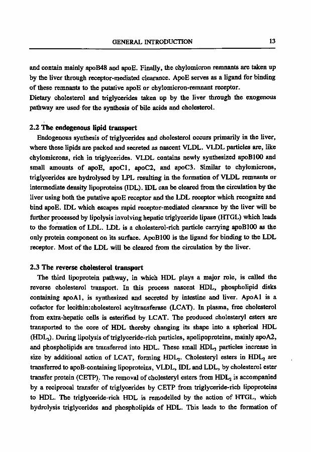
Cholesterol is essential for cell membranes and is used for synthesis of steroid hormones and bile acids, whereas triglycerides are used as a source of energy for muscles and storage in adipose tissues. Cholesterol and triglycerides are transported in the blood within lipoproteins because they are insoluble in an aqueous environment. Lipoproteins are particles consisting of lipids (cholesterol, cholesteryl esters, triglycerides and phospholipids) and proteins, called apolipoproteins. The surface of lipoproteins contains phospholipids, free cholesterol and apolipoproteins while the core of these particles consists mainly of cholesteryl esters and triglycerides. The major lipoprotein classes, chylomicrons, very low density lipoproteins (VLDL), low density lipoproteins (LDL), and high density lipoproteins (HDL), are heterogenous particles
GENERAL INTRODUCTION 11
Table 1. Biophysical and chemical properties
Density (g/ml)
Electrophoretic mobility
Triglycerides (% wt)
Phospholipids (% wt)
Cholesteiyl ester (% wt)
Free cholesterol (% wt)
Protein (% wt)
Major apolipoproteins
Chylomicrons
< 0.96
origin
88
8
3
1
1-2
Al, A4, B48, CI, C3, E
of human plasma lipoproteins ' .
VLDL
0.96-1.006
pre-/3
56
20
15
8
6-10
BlOO, CI, C2, C3, E
LDL
1.019-1.063
ß
13
28
48
10
21
BlOO
HDL
1.063-1.210
a
15
45
30
10
45-55
Al, A2, E
'Gotto et al., 1986
which differ in density, size and electrical charge, and vary in their apolipoprotein composition (Table 1). This heterogeneity leads to differences in their metabolism and mode of cell interaction.
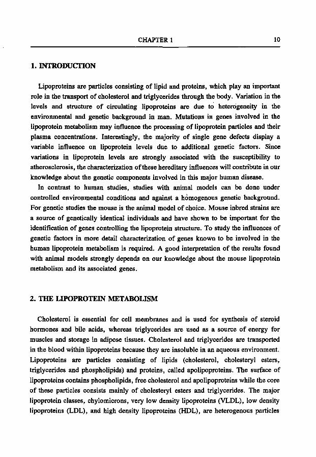
The lipoprotein metabolism can be divided into three different pathways: i) the exogenous lipid transport (Fig. lA), ii) the endogenous lipid transport (Fig. IB), and iii) the reverse cholesterol transport (Fig. IC), (for review, see Mahley et al., 1984; Gotto et al., 1986; Breslow, 1988; Havel and Kane, 1989; Eisenberg, 1990).
2.1 The exogenous lipid transport Dietary cholesterol and triglycerides are absorbed and packed in the intestinal
mucosal cells into large particles called chylomicrons. These very large triglyceride-rich lipoproteins, containing predominantly apolipoprotein (apo) B48, apoAl and apoA4, are secreted into the lymph. After entering the circulation, chylomicrons acquire apoE, apoCl, apoC2 and apoC3 from HDL. These modified chylomicrons are rapidly hydrolysed by endothelial lipoprotein lipase (LPL) with apoC2 serving as a cofactor. During this lipolysis process, excess surface components, consisting of phospholipids and apolipoproteins, are transferred to HDL. The remaining lipoprotein particles are called chylomicron remnants. These particles are enriched in cholesterol
CHAPTER 1 12
A. Exogenous lipid pathway Intestine Liver
B. Endogenous lipid pathway
L iver
Capi l lar ies
C. Reverse cholesterol pathway
Liver
Ex t rahepa t i c Cel ls
Fig. 1 Schematic representation of the lipid transport pathways. CM: chylomicron; CMR: chylomicron remnant. Redrawn from Breslow (1988).
GENERAL INTRODUCTION 13
and contain mainly apoB48 and apoE. Finally, the chylomicron remnants are taken up by the liver through receptor-mediated clearance. ApoE serves as a ligand for binding of these remnants to the putative apoE or chylomicron-remnant receptor. Dietary cholesterol and triglycerides taken up by the liver through the exogenous pathway are used for the synthesis of bile acids and cholesterol.
2.2 The endogenous lipid transport Endogenous synthesis of triglycerides and cholesterol occurs primarily in the liver,
where these lipids are packed and secreted as nascent VLDL. VLDL particles are, like chylomicrons, rich in triglycerides. VLDL contains newly synthesized apoBlOO and small amounts of apoE, apoCl, apoC2, and apoC3. Similar to chylomicrons, triglycerides are hydrolysed by LPL resulting in the formation of VLDL remnants or intermediate density lipoproteins (IDL). IDL can be cleared from the circulation by the liver using both the putative apoE receptor and the LDL receptor which recognize and bind apoE. IDL which escapes rapid receptor-mediated clearance by the liver will be further processed by lipolysis involving hepatic triglyceride lipase (HTGL) which leads to the formation of LDL. LDL is a cholesterol-rich particle carrying apoBlOO as the only protein component on its surface. ApoBlOO is the ligand for binding to the LDL receptor. Most of the LDL will be cleared from the circulation by the liver.
2.3 The reverse cholesterol transport The third lipoprotein pathway, in which HDL plays a major role, is called the
reverse cholesterol transport. In this process nascent HDL, phospholipid disks containing apoAl, is synthesized and secreted by intestine and liver. ApoAl is a cofactor for lecithin:cholesterol acyltransferase (LCAT). In plasma, free cholesterol from extra-hepatic cells is esterified by LCAT. The produced cholesteryl esters are transported to the core of HDL thereby changing its shape into a spherical HDL (HDLj). During lipolysis of triglyceride-rich particles, apolipoproteins, mainly apoA2, and phospholipids are transferred into HDL. These small HDL3 particles increase in size by additional action of LCAT, forming HDLj. Cholesteryl esters in HDLj are transferred to apoB-containing lipoproteins, VLDL, IDL and LDL, by cholesterol ester transfer protein (CETP). The removal of cholesteryl esters from HDLj is accompanied by a reciprocal transfer of triglycerides by CETP from triglyceride-rich lipoproteins to HDL. The triglyceride-rich HDL is remodelled by the action of HTGL, which hydrolysis triglycerides and phospholipids of HDL. This leads to the formation of
CHAPTER 1 14
smaller HDL particles (HDL3) and the dissociation of some apolipoproteins from HDL (Tall, 1992). The redistribution of cholesteryl esters from peripheral tissues to apoB-containing lipoproteins allows their uptake by the liver through LDL receptor-mediated clearance. This pathway, the reverse transport of cholesterol, is involved in the uptake of cholesteryl esters via the LDL receptor pathway in the liver and subsequent removal of excess of cholesterol, partly as bile acid, from the body via the intestine.
3. GENETIC ASPECTS OF THE LIPOPROTEIN METABOLISM
From epidemiological studies it has been estimated that 50% of the variation in plasma levels of lipoproteins in man has a genetic background. Among the genetic factors known to influence the concentration and metabolism of lipoprotein particles are: i) specific receptors involved in binding and intemalisation of lipoproteins, ii) apolipoproteins, and iii) enzymes involved in the modification of lipoproteins.
Disturbances in the lipoprotein metabolism leading to an accumulation of specific lipoproteins in the plasma, are often the result of mutations in these genes (for review, see Havel and Kane, 1989). The most atherogenic lipoproteins are LDL and j3-VLDL. j8-VLDL is derived from chylomicron- or VLDL-remnants. When removal of these lipoproteins is hampered, these particles accumulate in the plasma and become enriched in cholesterol. Elevated plasma levels of LDL and /3-VLDL form a major risk factor for atherosclerosis, which is characterized by foam cell formation in the intima of the arterial wall.
This thesis will focus on the APOE-C 1-C2 gene cluster and the LDLR gene which play an important role in the metabolism of atherogenic lipoproteins. Genetic disorders affecting the expression or function of apoE and the LDL receptor are familial dysbetalipoproteinemia (FD) and familial hypercholesterolemia (FH), respectively. FD is associated with mutations in apoE which lead to an elevated chylomicron- and VLDL-remnant concentration in the plasma (Mahley and Rail, 1989). FH leads to an accumulation of plasma LDL cholesterol while the concentration of other plasma lipoproteins is not affected (Goldstein and Brown, 1989). Mutations in the AP0C2 gene lead to hypertriglyceridemia which is characterized by an accumulation of triglyceride-rich lipoproteins. Sofar no primary qualitative or quantitative abnormalities of human apoCl have been reported.
GENERAL INTRODUCTION 15
APOE APOCl APOCl' APOC2
5 k b I
B
I-
ci 1+ 1 2
CI H 1 • 1 2 3 4
C2 I | - B • I 1 1 2 3 4
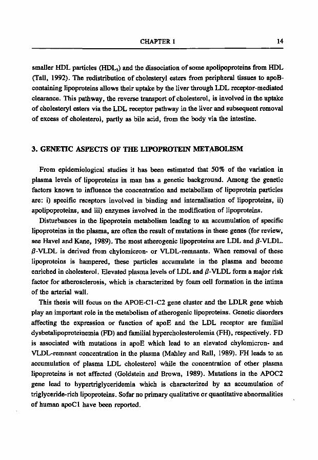
Fig. 2 Structure of the apolipoprotein E-C1-C2 gene cluster and individual genes. (A) The AP0E-C1-C2 gene cluster. Filled boxes indicate genes. (B) The exon-intron structures of the apolipoprotein genes; APOE, APOCl, APOCl' and APOC2.
3.1 The APOE-C1-C2 gene cluster APOE, APOCl and AP0C2 are three of the seven members of the apolipoprotein
gene family, which also includes APOAl, AP0A2, APOA4, and AP0C3 (Li et al., 1988). The proteins expressed by these genes are characterized by extensive regions of amphipatic helix. In humans, APOE is contained together with APOCl, pseudo APOCl, and APOC2 within a 48 kb gene cluster on the long arm of chromosome 19, 19ql2-19ql3.2(Fig. 2A) (Brook etal., 1987; Myklebost and Rogne, 1988; Smit etal., 1988a).
CHAPTER 1 16
The APOE gene; structure and expression The human APOE gene has been characterized extensively. The gene spans a region
of approximately 3.6 kb and consists of four exons (Fig. 2B)(Das et al., 1985; Paik et al., 1985). The mRNA encodes a protein of 317 amino acids including a signal peptide of 18 amino acid residues (Rail et al., 1982; Wallis et al., 1983; McLean et al., 1984). ApoE is a single polypeptide with a molecular weight of 34 kD. There are 3 major isoforms found encoding apoE2, E3 and E4 (Zannis et al., 1981, 1982; Utermann et al., 1982). These isoforms are the result of amino acid changes leading to charge differences. Population studies have shown that this allelic variation causes small but significant changes in plasma LDL levels (Smit et al., 1988b). Most of the FD patients have the apoE2E2 phenotype but only a very small percentage of E2E2 individuals will develop FD. This indicates that additional factors are required for the development of FD (Utermann, 1985).
ApoE is synthesized primarily by the liver but it is also produced by a variety of non-hepatic tissues including brain, kidney and adrenal glands (Wallis et al., 1983; Blue et al., 1983). The tissue specific synthesis of apoE is controlled by an array of elements found in the immediate 5'-flanking region of the APOE gene and throughout the whole APOE-C1-C2 gene cluster (For review, see Taylor et al., 1991; Zannis et al., 1992).
Function of apoE ApoE which is present on chylomicron- and VLDL-remnants, plays an important
role as a ligand for binding of the particles to the LDL receptor and the putative apoE receptor. The protein is organized as two distinct structural domains, each with different functions. The amino-terminal part contains the region of apoE that binds to the LDL receptor and the putative apoE receptor. The three dimensional structure has been determined by crystallography, showing that this domain consists of four helices (Wilson et al., 1991).
The carboxy-terminal part of apoE represents the major lipid-binding region which is composed of a long stretch of a-helices (Weisgraber et al., 1986). In addition, this carboxy-terminal part also contains a heparin binding domain which is thought to play a role in the hydrolysis of triglyceride-rich lipoproteins by anchoring the particle to endothelial heparan-proteoglycans.
GENERAL INTRODUCTION 17
The APOCl gene; structure and expression The human APOCl gene is located 4.3 kb downstream of the APOE gene
(Myklebost and Rogne, 1986). One copy of the APOCl gene, pseudo APOCl, is located 7.5 kb downstream of APOCl (Davison et al., 1986; Lauer et al., 1988). The APOCl gene spans a region of approximately 4.6 kb and is composed of four exons (Fig. 2B). Nucleotide sequence analysis of cloned cDNA (Knott et al., 1984) indicated that apoCl is synthesized with a 26-residue signal peptide that is removed during intracellular processing. Plasma apoCl consists of 57 amino acids residues in a single polypeptide chain with a calculated molecular weight of 6.6 kD (Jackson et al., 1974; Shulman et al., 1975). The APOCl gene is expressed primarily in the liver but expression has also been detected when monocytes differentiate into macrophages (Lauer et al., 1988).
No mRNA products of the pseudo APOCl gene can be detected in any tissue. This is probably the result of a point mutation in exon 3 which introduces a translation stop signal (Lauer et al., 1988). Although the coding region is small (249 bp), human APOCl is 4.6 kb in length due to large introns that contain a total of 9 Alu repeats. Basepair changes in these Alu repeats were used to estimate that the divergence between the APOCl gene and the pseudo APOCl gene took place after the divergence of rodents and primates which occurred 40 million years ago (Raisonnier, 1991).
Function of apoCl ApoCl is a constituent of chylomicrons, VLDL, and HDL (Breslow, 1988) and it
has been shown to activate LCAT in vitro but not as efficiently as apoAl (Soutar et al., 1975). Using synthetic peptides of apoCl, studies indicated that a fragment consisting of residue 17 to 57, contains all of the structural features necessary to activate LCAT and is as active as the intact protein. Residues 32-57 represent one of the major phospholipid-binding regions of apoCl (Soutar et al., 1978). Another function of apoCl is that it inhibits the action of phospholipase A (Poensgen, 1990).
In addition, studies showed that apoE and apoCl have an opposite effect on remnant uptake by the liver (Windier and Havel, 1985). Studies with rat liver membranes showed that the apoE-dependent binding of jS-VLDL to the LDL-receptor related protein is blocked by apoCl (Kowal et al., 1990; Weisgraber et al., 1990). Also studies with the LDL receptor showed that apoCl inhibits effectively the binding of apoE to this receptor (Sehayek and Eisenberg, 1991).
CHAPTER 1 18
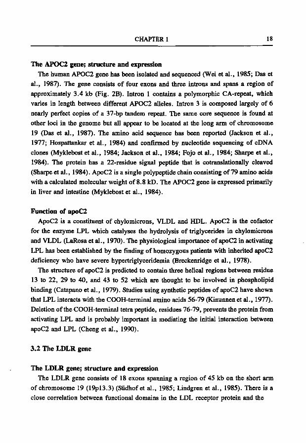
The APOC2 gene; structure and expression The human APOC2 gene has been isolated and sequenced (Wei et al., 1985; Das et
al., 1987). The gene consists of four exons and three introns and spans a region of approximately 3.4 kb (Fig. 2B). Intron 1 contains a polymorphic CA-repeat, which varies in length between different APOC2 alleles. Intron 3 is composed largely of 6 nearly perfect copies of a 37-bp tandem repeat. The same core sequence is found at other loci in the genome but all appear to be located at the long arm of chromosome 19 (Das et al., 1987). The amino acid sequence has been reported (Jackson et al., 1977; Hospattankar et al., 1984) and confirmed by nucleotide sequencing of cDNA clones (Myklebost et al., 1984; Jackson et al., 1984; Fojo et al., 1984; Sharpe et al., 1984). The protein has a 22-residue signal peptide that is cotranslationally cleaved (Sharpe et al., 1984). ApoC2 is a single polypeptide chain consisting of 79 amino acids with a calculated molecular weight of 8.8 kD. The APOC2 gene is expressed primarily in liver and intestine (Myklebost et al., 1984).
Function of apoC2 ApoC2 is a constituent of chylomicrons, VLDL and HDL. ApoC2 is the cofactor
for the enzyme LPL which catalyses the hydrolysis of triglycerides in chylomicrons and VLDL (LaRosa et al., 1970). The physiological importance of apoC2 in activating LPL has been established by the finding of homozygous patients with inherited apoC2 deficiency who have severe hypertriglyceridemia (Breckenridge et al., 1978).
The structure of apoC2 is predicted to contain three helical regions between residue 13 to 22, 29 to 40, and 43 to 52 which are thought to be involved in phospholipid binding (Catapano et al., 1979). Studies using synthetic peptides of apoC2 have shown that LPL interacts with the COOH-terminal amino acids 56-79 (Kinunnen et al., 1977). Deletion of the COOH-terminal tetra peptide, residues 76-79, prevents the protein from activating LPL and is probably important in mediating the initial interaction between apoC2 and LPL (Cheng et al., 1990).
3.2 The LDLR gene
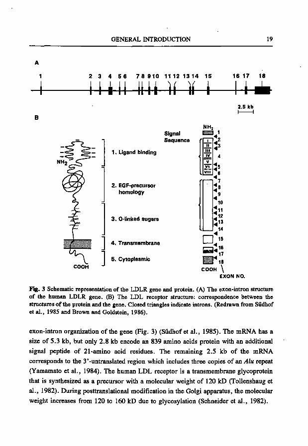
The LDLR gene; structure and expression The LDLR gene consists of 18 exons spanning a region of 45 kb on the short arm
of chromosome 19 (19pl3.3) (Sûdhof et al., 1985; Lindgren et al., 1985). There is a close correlation between functional domains in the LDL receptor protein and the
GENERAL INTRODUCTION 19
2 3 4 5 6 7 8 910 11 12 1314 15 16 17 18
i m i Uli ]i ]i i — u 2.5 leb
COOH
Signal Sequence
1. Ligand binding
2. EGF-precursor homology
1
3. 0-linked sugars
4 . Transmembrane
5. Cytoplasmic
NHj K.:-:-:-a 1
1 11 i l l IV V
Vi V l i
^2 ^3
4
Is 6
7 ^ 8
9
10
^11 ^12 ^13
14
n:» = : " ^ 1 8 COOH \
EXON NO.
Fig. 3 Schematic representation of the LDLR gene and protein. (A) The exon-intron structure of the human LDLR gene. (B) The LDL receptor structure: correspondence between the structures of the protein and the gene. Closed triangles indicate introns. (Redrawn from Sûdhof et al., 1985 and Brown and Goldstein, 1986).
exon-intron organization of the gene (Fig. 3) (Südhof et al., 1985). The mRNA has a
size of 5.3 kb, but only 2.8 kb encode an 839 amino acids protein with an additional
signal peptide of 21-amino acid residues. The remaining 2.5 kb of the mRNA
corresponds to the 3'-untranslated region which includes three copies of an Alu repeat
(Yamamato et al., 1984). The human LDL receptor is a transmembrane glycoprotein
that is synthesized as a precursor with a molecular weight of 120 kD (ToUenshaug et
al., 1982). During posttranslational modification in the Golgi apparatus, the molecular
weight increases from 120 to 160 kD due to glycosylation (Schneider et al., 1982).
CHAPTER 1 20
LDL receptors are present on the surface of essential all cultured mammalian cells. In vivo, however, the most important site of expression is the liver (Brown and Goldstein, 1986). The promoter region of the LDL receptor has been characterized and several important cis-acting regulatory elements have been detected. There are three direct imperfect repeats, each 16 bp in length, and a TATA-like sequence. Each of these elements is essential for full promoter activity. Repeats 1 and 3 contain high-affinity binding sites for the positive transcription factor Spl. Rei>eat 2 does not bind Spl and contains the sterol regulatory element (SREl), that is required for sterol-mediated repression. Most studies point to a complicated mechanism for promoter activity and sterol-mediated regulation (Südhof et al., 1987a, 1987b; Dawson et al., 1988; Smith et al., 1990b). Additional factors such as cellular growth and hormonal control may regulate LDL receptor transcription but the regulatory elements involved in this regulation are still unknown (Auwerx et al., 1989; Wade et al., 1989; Mazzone etal., 1990).
Function of the LDL receptor. The LDL receptor protein consists of five structural domains. From the NH2-
terminus to the COOH-terminus the following domains can be discerned: (i) the ligand binding domain, (ii) the EGF precursor domain, (iii) the O-linked sugar domain, (iv) the transmembrane spanning domain, and (v) the cytoplasmic domain (Yamamoto et al., 1984). The function of these domains have been characterized by studies on numerous naturally occurring mutants and by site directed mutagenesis (For reviews see Hobbs et al., 1990, 1992; Soutar, 1992).
(i) The ligand binding domain consists of 292 amino acids encoded by exons 2-6 and comprises seven copies of a negatively charged cysteine-rich repeat. This domain is involved in the binding of lipoproteins containing apoBlOO and apoE (Innerarity et al., 1984; Yamamoto et al., 1984). Due to the conformation of this domain the negatively charged residues interact with the positively charged clusters on apoE or apoBlOO. Studies on this domain, using site directed mutagenesis, showed that repeats 3-7 are involved in the binding of LDL while repeat 5 is involved in the binding of both LDL and jS-VLDL (Esser et al., 1988; Russell et al., 1989).
(ii) The EGF precursor domain contains approximately 400 amino acids and is encoded by exons 7-14. Three repeated sequences (A, B and C) are found in this region and show homology with repeat sequences in the EGF precursor of the mouse (Russell et al., 1984; Yamamoto et al., 1984; Südhof et al., 1985). This domain is
GENERAL INTRODUCTION 21
required for the release of bound ligands from the receptor in the endosomal compartments, which is essential for recycling of the LDL receptor. Repeat C is essential for binding of LDL and it is suggested that it acts as a spacer region that extends the receptor-binding domain away from the cell surface (Davis et al., 1987).
(iii) The O-linked sugar domain is encoded by exon 15 and comprises a stretch of 58 amino acids, enriched in serine and threonine residues of which the majority is glycosylated. Proteolytic studies have revealed that this region contains the clustered O-linked sugar chains. The ftinction of this domain remains unclear (Davis et al., 1986; Russell et al., 1984).
(iv) The transmembrane spanning domain is encoded by exon 16 and a part of 17 and consists of 22 hydrophobic amino acids. This domain is believed to anchor the receptor in the cell surface by spanning the plasma membrane (Russell et al., 1984).
(v) The last domain of the LDL receptor is the COOH-terminal cytoplasmic domain of 50 amino acid residues and is encoded by parts of exons 17 and 18. This domain plays an important role in clustering in coated pits on the cell surface thereby facilitating endocytosis. A tetrameric sequence Asn-Pro-x-Tyr (NPxY, x stands for any amino acid) in this domain is found to be important for the coated pit internalization. This NPxY sequence has been found in several other surface receptors. It is suggested that the NPxY sequence has a conditional role in ligand-independent internalization of these receptors (Chen et al., 1990).
4. THE MOUSE AS AN ANIMAL MODEL FOR STUDYING THE LIPOPROTEIN METABOLISM
The variability in plasma levels of lipids and lipoproteins is due to heterogeneity in environmental and genetic background. In man, about 50% of this variability is due to multiple genetic factors. Even single gene defects display a variable influence on lipid and apolipoprotein levels because of confounding interactions with environmental and/or genetic factors. This complexity of interacting factors influencing the lipoprotein metabolism makes it difficult to identify these factors individually. As a consequence, studies of plasma lipoproteins or atherosclerosis in man are hampered. However, the use of animal models could circumvent some of these limitations of lipoprotein studies in man by providing a defined genetic background and controlled environmental conditions.
CHAPTER 1 22
4.1 Advantages of a mouse model As in most areas of human biology, studies on human plasma lipoproteins or
atherosclerosis have been enriched and completed by investigating animal models. The use of animal models will increase our knowledge about the interactions between different genetic factors and between genetic and environmental factors involved in the lipoprotein metabolism. However, one has to consider that there are differences between species with respect to quantitative traits, metabolic pathways, and developmental pathways. Animal models do not serve to duplicate precisely genetic variations that occur in humans but rather to gain a better understanding of the biochemical mechanisms and the types of polymorphisms that underlie heritable variations of lipoproteins. In addition, animal models also provide a means for identifying new genes affecting lipid metabolism and for defining in detail the molecular mechanisms involved in lipid transport. Generally used species which serve as models for the study of plasma lipoproteins as well as atherosclerosis include rabbits, hamsters, mice, rats, pigs and non-human primates. (For recent reviews see Armstrong and Heistad, 1990; Reue et al., 1990; Overturf and Loose-Mitchell, 1992).
The selection of a model strongly depends upon the subject of investigation. Animal models with spontaneous mutations affecting various aspects of lipoprotein metabolism have been valuable in the study of genetic and biochemical basis of similar monogenic disorders in humans. Examples are hypercholesterolemia in the Watanabe heritable hyperlipidemic (WHHL) rabbit with a defect in the LDLR gene (Yamamoto et al., 1986) and atherosclerosis in pigs with genetic variation in their apolipoproteins (Rapacz et al., 1986). A problem associated with these animal models is that they are very rare. In addition, some animal species are in favour because their lipoprotein metabolism closely resembles that of humans. These animals are used for studying lipoproteins and the influences of environmental factors such as diet, on the composition of these lipoproteins. A disadvantage of these models is that they mainly represent large animals, like monkeys. These models are expensive to maintain and genetic studies are difficult due to a long generation time and, as in man, heterogeneity in genetic background.
For genetic studies the mammal of choice is the mouse since it offers a variety of advantages when compared with other animal models. There are hundreds of different inbred strains available, each strain representing a unique gene pool in which natural polymorphisms have been fixed by inbreeding. In addition, several sets of recombinant inbred strains have become available as well. Inbred strains of mice form a source of
GENERAL INTRODUCTION 23
infinite numbers of genetically identical individuals. Therefore, the mouse provides an animal model system for examining the heritable variation of lipoproteins since genetic and biochemical analyses are greatly simplified and environmental influences can be controlled. Another major advantage is that the genetic map of the mouse is well defined however, only a small number of genes has been characterized in more detail (Lusis and Sparks, 1989; Hillyard et al., 1993). More recently, special techniques have become available for manipulating the genes of interest by transgenesis and gene targeting. The technology is now available to generate animal models with specific alterations in the genes of interest, independent of spontaneous variations (see below).
For a long time, mice were not used commonly to study lipoprotein metabolism and atherosclerosis, because early attempts to produce atherosclerosis were discouraging due to variable expression of the disease and mortality caused by the experimental diet. The identification of inbred strains of mice, either genetically susceptible or resistant to diet-induced atherosclerosis, resolved some of the problems associated with the use of mice as models for atherosclerosis (Roberts and Thompson, 1976; Morrisett et al.,, 1982; Paigen et al., 1985). The development of appropriate experimental diets have resulted in reproducible atherosclerotic lesions in the mouse (Nishina et al., 1990; Steward-Phillips and Lough, 1991). Due to these characteristics the mouse is widely used as an animal model for studying the interaction between genes and environmental fiictors on the metabolism of lipoproteins and atherosclerosis.
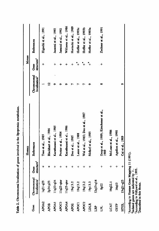
4.2 Genetic aspects of the mouse lipoprotein metabolism The recent development of highly polymorphic genetic markers, simple sequence
repeats, resulted in a large set of genetic markers spanning randomly the whole mouse genome (Comall et al., 1991). The genetic map of the mouse is well defined and it is comprised of a large number of loci/genes including genes involved in the lipoprotein metabolism (Table 2)(Hillyard et al., 1993). Comparison of the linkage maps in man and mouse shows that the loci for the genes involved in the lipoprotein metabolism are contained within homologous linkage groups in mice and man, suggesting a common evolutionary pathway. Mouse and man have essentially the same set of genes encoding enzymes and receptors directing the major lipid transport pathways. The mouse lipoproteins and apolipoproteins appeared to be structurally and functionally similar to the human counterparts (LeBoef et al., 1983; Camus et al., 1983). The homology between human and mouse gene maps will contribute to the mapping of new genes affecting the lipoprotein metabolism in humans. New loci mapped in the mouse could
Tab
le 2
. C
hrom
osom
al l
ocal
izat
ion
of g
enes
inv
olve
d in
aie
Upo
prot
ein
met
abol
ism
.
Hum
an
Mou
se
Gen
e C
hrom
osom
al
Gen
e lo
cali
sati
on'
stru
ctur
e^
Ref
eren
ces
Chr
omos
omal
G
ene
Ref
eren
ces
loca
lisa
tion
' st
ruct
ure^
APO
A2
APO
B
AP
OA
l
APO
C3
APO
A4
APO
E
AP
OC
l
APO
C2
LD
LR
LR
P
LP
L
LC
AT
CE
TP
HT
GL
Iq21
-q23
2p24
-p23
Ilq2
3-q2
4
llq?
,3-q
ter
1 lq
23-q
ter
19ql
3.2
19ql
3.2
19ql
3.2
19pl
3.3
12ql
3-ql
4
8p22
16q2
2.1
16ql
3
15q2
1-q2
3
+
+
+ +
+
+
+
+ + +
+
+
+
Tsa
oet
al.,
19
85
Bla
ckha
rtet
al.,
1986
Kar
atfa
anas
i et
al.
, 19
85
Prot
ter
et a
l.,
1984
Kar
atha
nasi
et
al.,
1986
Das
et
al.,
1985
Lau
eret
al.,
19
88
Wei
et
al.,
1985
; D
as e
t al
., 19
87
Sûdh
of e
tal.
, 19
85
Dee
p et
al.
, 19
89; K
irsc
hene
r et
al.
, 19
89
McL
ean
etal
.,
1986
Age
llon
et a
l.,
1990
Cai
et
al.,
1989
1 12
9 9 9 7 7 7 9
+
+
+
+
+
+•
+•
+•
Hig
uchi
et
al.,
1991
Janu
zzi
et a
l.,
1992
Janu
zzi
et a
l.,
1992
Will
iam
s et
al.
, 19
86
Hor
iuch
i et
al.
, 19
89
Hof
fer
et a
l.,
1993
c
Hof
fer
et a
l.,
1993
b
Hof
fer
et a
l.,
1993
a
Zec
hner
et
al.,
1991
'Acc
ordi
ng t
o H
uman
Gen
e M
q>pi
ng 1
1 is
olat
ed g
enes
are
ind
icat
ed b
y +
. 'A
ccor
ding
to
Hil
lyai
d et
al.
, 19
93.
*Des
crib
ed i
n th
is t
hesi
s.
(199
1).
GENERAL INTRODUCTION 25
also be conserved in humans (see below). Linkage analysis can be used to identify genes for genetic disorders in which no
biochemical defect or gene can be identified. In human, genetic markers are tested for cosegregation with the disease phenotype (monogenetic) in families. The development of highly polymorphic simple sequence repeats has been successful for the identification of loci causing human disease (Wijmenga et al., 1990). However, this approach is difficult to use in human studies involving multifactorial disorders such as atherosclerosis. The multiple segregating loci contributing to the disease are difficult to identify. Therefore, animal models are used for the identification of complex genetic traits.
One approach of identifying these genetic factors is called the quantitative trait locus (QTL) mapping. QTL mapping (for a review, see Warden et al., 1992) requires inbred strains and a highly characterized map of the genome. The development of the highly polymorphic simple sequence repeats made it feasible to use QTL mapping for the identification of multiple loci. For QTL mapping two different inbred strains are crossed to produce F2 backcross progeny. These progeny are individually phenotyped for the trait of interest and genotyped for markers in the genome. Subsequently, statistical analysis are used to test for associations. QTL mapping is a quick and cheap alternative but only dominant traits can be mapped and there is no permanent material. QTL mapping resulted in the mapping of loci contributing to diabetes in the mouse (Todd etal., 1991).
Another approach is to use recombinant inbred (RI) strains (Justice et al., 1992) instead of Fj backcross progeny. RI strains make it possible to analyze both dominant and recessive disease loci. Two inbred strmns which differ for the trait of interest, are crossed and a series of new homozygous recombinant inbred strains is created from their progeny by 20 generations of inbreeding. A disadvantage of this approach is that the production of RI strains is time consuming. Each new recombinant inbred strain consists of a unique mixture of genes in a homozygous state derived from the two parental strains. The set of recombinant inbred str^ns permits rapid linkage analysis because alleles of linked genes tend to be linked in the same combination in the RI strains, whereas unlinked genes are randomized. Another advantage of the use of RI strains is that they provide an unlimited source of material for further studies.
QTL mappping depends on the variability of lipoproteins between inbred strains and therefore characterization of the lipoprotein metabolism is required. Among various strains of inbred mice, there is heterogeneity in the sizes, plasma concentrations and
CHAPTER 1 26
lipid compositions of the lipoproteins (Jiao et al., 1990a, 1990b). It was also shown that there were strain-related differences with respect to mRNA quantities of genes involved in the lipoprotein metabolism (Lusis et al., 1987; Srivastava et al., 1991). These differences in quantitative traits between different inbred strains are used to study the multifactorial genetic influences on the lipoprotein metabolism.
The first approach in identifying genes involved in the variability of lipoproteins in the mouse is called the "candidate gene" approach. For this, polymorphisms in genes, involved in the human lipoprotein metabolism, are tested for cosegregation with specific quantitative traits. This approach has been used to identify genes controlling the structure of mouse lipoproteins. For instance, comparison of HDL sizes and HDL-cholesterol concentrations in plasma of several inbred strains of mice showed dichotomous distributions due to different isoforms of apo A1 and apoA2 (Ben-Zeev et al., 1983; Lusis et al., 1983, 1987). A problem of the "candidate gene" approach is that the "candidate gene" must be identified before one can test its involvement in the variability.
Therefore QTL mapping has been used as an approach to determine genetic factors influencing the variability of lipoproteins. Recombinant inbred strains were used to map the Athl locus which is correlated with susceptibility to atherosclerosis (Paigen et al., 1985). Studying polymorphisms affecting HDL resulted in the identification of new additional loci involved in the heterogeneity of these lipoproteins in the mouse. When mice are fed an atherogenic diet, HDL-cholesterol decreased by 50% in susceptible strains whereas in the resistant strains the HDL-cholesterol levels remained unchanged. The frequency of lesion formation differs considerably between different strains fed the same atherogenic diet and is correlated with HDL-cholesterol levels (Paigen et al., 1985; Ishida et al., 1991). Variations in HDL-cholesterol levels and susceptibility to diet-induced atherosclerosis in mice appear to be determined by the same gene, named Athl (Paigen et al., 1987; LeBoeuf et al., 1990). This new gene, maps to a region near the structural gene for apoA2 on mouse chromosome 1. This gene is distinct from previously known genes in this region. In addition, two other new loci, Ath2 andAth3, were also found to be linked to atherosclerosis and HDL phenotypes (Paigen et al., 1989; Stewart-Phillips et al., 1989). In the above mentioned studies, phenotypes are used to detect the underlying genes which are involved in the variability of lipoproteins.
GENERAL INTRODUCTION 27
4.3 Transgenic mouse models Biochemical studies with mice have shown that there are differences in the relative
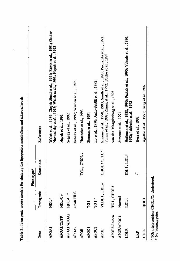
amounts of the density classes of plasma lipoproteins between mice and humans. In mice, HDL is the predominant lipoprotein in the plasma compared to LDL in man. Despite this, when mice are fed a diet comparable with the human diet (atherogenic diet), their lipoprotein profile approaches that of humans. Atherogenic diet consumption results in an increase in total plasma cholesterol which could be attributed to an increase in VLDL/LDL-cholesterol (Paigen et al., 1985). An more elegant approach to influence the lipoprotein metabolism of the mouse is by the use of transgenesis and gene targeting. Transgenesis is a recently developed technology which makes it possible to introduce foreign DNA randomly into the mouse genome (Palmiter and Brinster, 1986; Jaenisch, 1988). These mouse models can be used to study the gene expression and the effects of overexpression of a transgene in the mouse. These animal models cannot be used to study the consequence of a decreased amount or total absence of the expression of a gene. Other technologies, like antisense expression vectors, can only be used to study the absence of expression of a gene (Weintraub, 1990). A more reliable technology is targeted mutagenesis (gene targeting), allowing the introduction of defined changes into any endogenous mouse gene in embryonic stem cells (Capecchi, 1989; Pascoe et al., 1992; Smithies, 1993). Gene targeting can be used to produce animal models of any characterized monogenic human disease or to study the function of new genes by the creation of null alleles (knock-out) for the appropriate gene. The effects of defined mutations can be studied against a homogeneous genetic background and under controlled environmental conditions. Both the genetic background as well as the environmental conditions can be adjusted as much as the researchers wishes. These technologies together, transgenesis and gene targeting, make it possible to study several aspects of gene expression in the mouse. Table 3 shows the transgenic mouse models for the lipoprotein metabolism and atherosclerosis available at present.
Transgenic mice can be used to determine the effects of overexpression of a particular gene on the lipoprotein metabolism. The overexpression of genes can result in a reduction of plasma lipoproteins and protection against diet-induced atherosclerosis. This effect has been found for human APOE, LDLR and APOAl transgenic mouse models (Yokode et al., 1990; Rubin et al., 1991; Shimano et al., 1992). The expression of the LDL receptor in the liver of transgenic mice was used to study whether overexpression would increase the clearance of LDL from the
Tab
le 3
. T
rans
geni
c m
ouse
mod
els
for
stud
ying
the
Upo
prot
ein
met
aboU
sm a
nd a
ther
oscl
eros
is.
Phen
otyp
e'
Gen
e T
rans
geni
c K
nock
-out
R
efer
ence
s
APO
Al
HD
Lt
APO
Al/
CE
TP
APO
A1/
APO
A2
APO
A2
APO
B
APO
Cl
APO
C3
APO
E
APO
E3-
Lei
den
APO
E/A
POC
l
LD
LR
LR
P
CE
TP
HD
L-C
i
HD
L-C
t
smal
l H
DL
TG
t
TG
TT
VL
DL
1, L
DL
i
TG
t, C
HO
Lt
Nor
mal
LD
Li
HD
Ll
TG
i, C
HO
Li
CH
OL
tt,T
Gt
IDL
t, L
DL
t
*
Wal
sh e
t al
., 19
89; C
haje
k-Sh
aul
et a
l.,
1991
; Rub
in e
t al
., 19
91; G
olde
r-N
ovos
elsk
y et
al.
, 19
92; W
alsh
et a
l.,
1993
; Hay
ek e
t al.
, 19
93
Hay
ek e
tal.
, 19
92
Schu
ltz e
tal.
, 19
92
Schu
ltz e
t al
., 19
92; W
arde
n et
al.
, 19
93
Hom
anic
s et
al.
, 19
93
Sim
onet
et
al.,
1991
Ito
et a
l.,
1990
; Aal
to-S
etäl
ä et
al.
, 19
92
Sim
onet
et a
l.,
1990
, 19
93; S
mith
et
al.,
1990
; Pie
drah
itae
t al
., 19
92;
Plum
p et
al.
, 19
92; Z
hang
et a
l.,
1992
; Pop
ko e
t al
., 19
93
van
den
Maa
gden
berg
et
al.,
1993
Sim
onet
et
aL,
1991
Hof
inan
n et
al.
, 19
88,
1990
; Pat
hak
et a
l.,
1990
; Yok
ode
et a
l.,
1990
, 19
92; I
shib
ashi
et
al.,
1993
Her
z et
al.
, 19
92
Age
llon
et a
l.,
1991
; Ji
ang
et a
l. 19
92
' T
G:
trig
lyce
ride
s; C
HO
L/C
: ch
oles
tero
l. *
No
hom
ozyg
otes
.
GENERAL INTRODUCTION 29
plasma. This was the first study with transgenic mice which showed that human and mouse lipoprotein metabolism are compatible, i.e. the human LDL receptor binds and internalizes mouse lipoproteins containing apoB and apoE. The overexpression of human LDL receptors resulted in a rapid clearance of LDL (Hofinann et al., 1988, 1990). In addition, the overexpression of the LDLR gene made it also possible to study the tissue-specifil: sorting of the LDL receptor. This has not been possible before since the number of receptors was below the detection limit in normal mice (Pathak et al., 1990).
In man, the study of mutant genes is often difficult due to additional factors influencing the expression of atherosclerosis. The use of a transgenic animal model offers the possibility to study the mutation in a homogenous environmental and genetic background. The overexpression of a mutant gene, apoE3-Leiden, in a transgenic mouse model resulted in an elevated lipoprotein plasma levels of VLDL and LDL (van den Maagdenberg et al., 1993). This transgenic mouse can serve as a model to elucidate additional factors influencing the plasma lipoproteins and for the etiology of familial dysbetalipoproteinemia.
Studies with transgenic animals can also be informative on tissue-specific regulation of the human gene involved. Transgenic mice can show different levels of expression of the construct involved which dei>ends on the number of constructs which have been integrated and on the position in the genome. The expression of genes is highly variable and unpredictable unless locus control regions (LCR) are included in the constructs used. Studies showed that constructs containing the LCRs had all the regulatory elements involved in the expression of the gene involved (Constantoulakis et al., 1991). An example of an incomplete construct is the study with a transgenic mouse model for apoE containing only the 5' regulatory elements. This construct did not show expression in the liver which is the major site of apoE synthesis. Other studies using different constructs with the human APOE gene showed that the potential liver-specific enhancer element is located between the APOCl and pseudo APOCl genes. These constructs have been very usefiil to study the tissue-specific regulatory elements involved in the APOE gene expression (Smith et al., 1990; Simonet et al., 1990, 1991, 1993).
Gene targeting can be used to study the effect of a deficiency for the gene of interest. Inactivation of a gene (gene knock-out) is the most straigth forward approach to make a defect although the introduction of more subtle mutations are also possible. At present, only Apoe, Apob, Ldlr, and a Lrp knockout mice have been made
CHAPTER 1 30
(Piedrahita et al., 1992; Zhang et al., 1992; Plump et al., 1992; Homanics et al., 1993; Ishibashi et al., 1993; Herz et al., 1992). Apoe-deRcicnt mice showed a phenotype with spontaneous high cholesterol levels and atherosclerotic lesions which give them a unique value. These mice are a reproducible source of atherosclerotic animals which can be used for studies on the progression of the disease and influences of environmental factors without interference with an atherogenic diet.
For studying the lipoprotein metabolism and atherosclerosis, transgenic or gene targeted mice are preferable to mice with diet-induced atherosclerosis. These technologies have been used to produce lines of transgenic mice which carry human genes involved in the lipoprotein metabolism or knock-out mice for specific genes (Table 3). The expression of human genes of interest and the consequence of overexpression of these genes on the lipoprotein metabolism have been studied in these mice. The use of transgenic animals will be very useful in studying multifactorial diseases in future. The lines of transgenic and knock-out mice make it possible to create crosses between different lines. An example is the APOAl/CETP transgenic mouse which has been created from an APOAl and a CETP transgenic mmouse by crossbreeding. Another possibility is to make crosses with other inbred strains. These new niouse models make it possible to study the same transgene with another genetic background. These studies will be very useful for unravelling the genetic factors which are involved in the variability of lipoproteins.
5. OUTLINE OF THE THESIS
The insight in the chylomicron, VLDL, and LDL metabolism, and the relationship with atherosclerosis has increased largely due to human studies. But it also clearly revealed the shortcomings, mainly related to the complex genetic and/or environmental interactions. Natural animal models have proven to be of great value to complement the human studies. Recent developments in the technology of producing specific transgenic mouse models by conventional transgenesis and targeted mutagenesis, will give unforseen new possibilities. However, this also further emphasizes the necessity of detailed insight in the structure and function of the corresponding mouse genes involved.
Accordingly, this thesis has been focused to the study of the Apoe-cl-c2 gene cluster (Chapter 2-5) and the Ldlr gene (Chapter 6). These genes have been chosen because
GENERAL INTRODUCTION 31
they play a central role in the metabolism of atherogenic lipoproteins. Of the four mentioned genes, only the mouse Apoe gene has already been isolated and characterized (Table 2).
We have isolated the mouse Apoe-cl-c2 gene cluster and the exact location of the Apod and Apoc2 genes was determined relatively to the Apoe gene. The genetic variability in this gene cluster between inbred strains of mice has been determined (Chapter 2). The Apocl gene (Chapter 3), the Apoc2 gene (Chapter 4) have been isolated and characterized in more detail on cDNA and genomic level. The expression of these genes in mice was also investigated. The second part of chapter 4 reports the detection of a larger transcript which has not been described sofar and showed evolutionary conservation. Further study on this transcript resulted in the isolation of cDNA clones encoded by a novel gene within the Apoe-cl-c2 gene cluster. This new gene, the apolipoprotein C2 linked (Acl) gene, has been fiirther characterized (Chapter 5). Finally, the characterization of the Ldlr gene on cDNA and genomic level is described in Chapter 6.
REFERENCES
Aalto-Setälä K, Fisher EA, Chen X, Chajek-Shaul T, Hayek T, Zechner R, Walsh A, Ramakrishnan R, Ginsberg HN, and Breslow JL (1992). Mechanism of hypertriglyceridemia in human apolipoprotein (Apo) CHI transgenic mice. J Clin Invest 90: 1889-1900.
Agellon LB, Quinet EM, Gillette TG, Drayna DT, Brown ML, and Tall RA (1990). Organization of the human cholesteryl ester transfer protein gene. Biochem 29: 1372-1376.
Agellon JB, Walsh A, Haydc T, Moulin P, Jiang XC, Shelanski SA, Breslow JL, and Tall AR (1991). Reduced high density lipoprotein cholesterol in human cholesteryl ester transfer protein transgenic mice. J Biol Chem 266: 10796-10801.
Armstrong ML, and Heistad DD (1990). Animal models of atherosclerosis. Atherosclerosis 85: 15-23. Auwerx JH, Chait A, Wolfbauer G, and Deeb SS (1989). Involvement of second messenger in regulation
of the low density lipoprotein receptor gene. Mol Cell Biol 9: 2298-2302. Ben-Zeev O, Lusis AJ, LeBoeuf RC, Nikazy J, and Schotz MC (1983). Evidence for independent genetic
regulation of heart and adipose lipoprotein activity. J Biol Chem 258: 13632-13636. Blackhart BD, Ludwig EM, Pierotti VR, Caiatai L, Onasch MA, Wallis SC, Powell L, Pease R, Knott TJ,
Chu ML, Mahley RW, Scott J, McCarthy BJ, and Levy-Wilson B (1986). Structure of the human apolipoprotein B gene. J Biol Chem 261: 1S364-15367.
Blue ML, Williams DL, Zucker S, Khan A, and Blum CB (1983). Apolipoprotein E synthesis in human kidney, adrenal gland, and liver. Proc Natl Acad Sei USA 80: 283-287.
Breckenridge WC, Little JA, Steiner G, Chow A, and Poapst M (1978). Hypertriglyceridemia associated with deficiency of ^olipoprotein C-II. N Engl J Med 298: 1265-1273.
Breslow JL (1988). Apolipoprotein genetic variation and human disease. Physiol Rev 68: 85 132. Brook JD, Skinner M, Roberts SH, Rettig WJ, Almond JW, and Shaw DJ (1987). Further mapping of
markers around the centromere of human chromosome 19. Genomics 1: 320-328.
CHAPTER 1 32
Brown MS, and Goldstein JL (1986). A receptor-mediated pathway for cholesterol homeostasis. Science 232: 34-47.
Cai S-J, Wong DM, Chen S-H, and Chan L (1989). Structure of the human hepatic triglyceride lipase gene. Biochem 28: 8966-8971.
Camus M-C, Chapman MJ, Forgez P, and Laplaud PM (1983). Distribution and characterization of the serum lipoproteins and apoproteins in the mouse, Mus musculus. J Lipid Res 24: 1210-1228.
Capecchi M (1989). The new mouse genetics altering the genome by gene targeting. Trends Genet 5: 70-76.
Catapano AL, Kinnunen PKJ, Breckenridge WC, Gotto AM Jr, Jackson RL, Little JA, Smith LC, and Sparrow JT (1979). Lipolysis of apoC-II deficient very low density lipoproteins: enhancement of lipoprotein lipase action by synthenic fragments of apoC-II. Biochem Biophys Res Commun 89: 951-957.
Chajek-Shaul T, Hayer T, Walsh A, and Breslow JL (1991). Expression of the human apolipoprotein A-I gene in transgenic mice alters high density lipoprotein (HDL) particle size distribution and diminishes selective uptake of HDL cholesteiyl esters. Proc Natl Acad Sei USA 88: 6731-6735.
Chen W-J, Goldstein JL, and Brown MS (1990). NPXY, a sequence often found in cjrtoplasmatic tails, is required for coated pit-mediated internalization of the low density lipoprotein receptor. J Biol Chem 265:3116-3123.
Cheng Q, Blackett P, Jackson KW, McConathy WJ, and Wang C-S (1990). C-terminal domain of apolipoprotein CII as both activator and competitive inhibitor of lipoprotein lipase. Biochem J 269: 403-407.
Constantoulakis P, Josephson B, Mangahas L, PE4>ayannopoulou T, Enver T, Constantini F, and Stamatoyannopoulos(1991). Locus control region-Ax transgenic mice: a new model for studying the induction of fetal hemoglobin in the adult. Blood 77: 1326-1333.
Cornall RI, Aitman TJ, Hearne CM, and Todd JA (1991). The generation of a library of PCR analyzed microsatellite variants for genetic mapping of the mouse genome. Genomics 10: 874-881.
Das HK, McPherson J, Bruns AP, KarathanasisSK, and Breslow JL (1985). Isolation, characterization and mapping to chromosome 19 of the human apolipoprotein E gene. J Biol Chem 260: 6240-6247.
Das HK, Jackson CL, Miller DA, Leff T, and Breslow JL (1987). The human apolipoprotein C-II gene sequence contains a novel chromosome 19-specific minisatellite in its third intron. J Biol Chem 262: 4787-4793.
Davis CG, Elhammer A, Russell DW, Schneider WJ, Kornfeld S, Brown MS, and Goldstein JL (1986). Deletion of clustered O-linked carbohydrates does not impair function of low density lipoprotein receptor in transfected fibroblasts. J Biol Chem 261: 2828-2838.
Davis CG, Goldstein JL, Südhof TC, Anderson RGW, Russell DW, and Brown MS (1987). Acid-dependent ligand dissociation and recycling of LDL rechter mediated by growth factor homology region. Nature 326:760-765.
Davison PJ, Norton P, Wallis SC, Gill L, Cook M, Williamson R, and Humphries SE (1986). There are two gene sequences for human apolipoprotein CI (APOCl) on chromosome 19, one of which is 4 kb from the gene for APOE. Biochem Biophys Res Commun 136: 876-884.
Dawson PA, Hofmann SL, Van der Westhuyzen DR, Südhof TC, Brown MS, and Goldstein JL (1988). Sterol-dependent repression of low density lipoprotein receptor promoter mediated by 16-base pair sequence adjacent to binding site for transcription factor Sp 1. J Biol Chem 263: 3372-3379.
Deep SS, and Peng R (1989). Structure of the human lipoprotein lipase gene. Biochem 28: 4131-4135. Eisenberg S (1990). Metabolism of apolipoproteins and lipoproteins. Curr Opin Lipidol 1: 205 215. Esser V, Limbird LE, Brown MS, Goldstein JL, and Russell DW (1988). Mutational analysis of the ligand
binding domain of the low density lipoprotein receptor. J Biol Chem 263: 13282-13290. Fojo SS, Law SW, and Brewer HB Jr (1984). Human apolipoprotein C-II: complete nucleic acid sequence
of preapolipoproteinC-II. Proc Natl Acad Sei USA 81: 6354-6357. Golder-Novoselsky E, Forte TM, Nichols AV, and Rubin EM (1992). Apolipoprotein AI expression and
GENERAL INTRODUCTION 33
high density lipoprotein distribution in transgenic mice during development. J Biol Chem 267: 20787-20790.
Goldstein JL, and Brown MS (1989). Familial hypercholesterolemia. In: The metabolic basis of inherited disease (Scriver CR, Beaudet AL, Sly WS, and Valle D, Eds.). 6th ed McGraw-Hill, New York, pp. 1215-1250.
Gotto AM, Pownall HJ, and Havel JR (1986). Introduction to the plasma lipoproteins. Methods Enzymol 128: 3-40.
Havel RJ, and Kane JP (1989). Introduction: Structure and metabolism of plasma lipoproteins. In: The metabolic basis of inherited disease (Scriver CR, Beaudet AL, Sly WS, and Valle D, Eds.). 6th ed McGraw-Hill, New York, pp. 1129-1138.
Hayek T, Chajek-Shaul T, Walsh A, Agellon LB, Moulin P, Tall AR, and Breslow JL (1992). An interaction between the human cholesteryl ester transfer protein (CETP) and apolipoprotein A-I genes in transgenic mice results in a profound CETP-mediated depression of high density lipoprotein cholesterol levels. J Clin Invest 90: 505-510.
Herz J, Clouthier DE, and Hammer RE (1992). LDL receptor-related protein internalizes and degrades uPA-PAI-1 complexes and is essential for embryonic implantation. Cell 71: 411-421.
Higuchi K, Kitagawa K, Naiki H, Hanada K, Hosokawa M, and Takeda T (1991). Polymorphism of apolipoprotein A-II (apoA-II) among inbred strains of mice. Biochem J 279: 427-433.
Hillyard AL, Doolittle DP, Davissen MT, and Roderick TH (1993). Locus map of the mouse withcomparative map points of human on mouse. Mouse Genome 91: 15-39.
Hobbs HH, Russell DW, Brown MS, and Goldstein JL (1990). The LDL receptor locus in familial hypercholesterolemia: mutational analysis of a membrane protein. Annu Rev Genet 24: 133-170.
Hobbs HH, Brown MS, and Goldstein JL (1992). Molecular genetics of the LDL receptor gene in familial hypercholesterolemia. Hum Mutation 1: 445-466.
Hoffer MJV, van Eck MM, Petrij F, van der Zee A, de Wit E, Meijer D, Grosveld G, Havekes LM, Hofker MH, and Frants RR (1993a). The mouse low density lipoprotein receptor gene: cDNA sequence and exon-intron structure. Biochem Biophys Res Commun 191:880-886.
Hoffer MJV, van Eck MM, Havekes LM, Hofker MH, and Frants RR (1993b). Structure and expression of the mouse apolipoprotein C2 gene. Genomics 17:45-51.
Hoffer MJV, van Eck MM, Havekes LM, Hofker MH, and Frants RR (1993c). The mouse apolipoprotein CI gene: structure and expression. Genomics: in press.
Hofmann SL, Russell DW, Brown MS, Goldstein JL, and Hammer RE (1988). Overexpression of low density lipoprotein (LDL) receptor eliminates LDL from plasma in transgenic mice. Science 239: 1277-1281.
Hofmann SL, Eaton DL, Brown MS, McConathy WJ, Goldstein JL, and Hammer RE (1990). Overexpression of human low density lipoprotein r e c i t e r s leads to accelerated catabolism of Lp(a) lipoprotein in transgenic mice. J Clin Invest 85: 1542-1547.
Homanics GE, Smith TJ, Zhang SH, Lee D, Young SG, and Maeda N (1993). Targeted modification of the apolipoprotein B gene results in hypobetalipoproteinemiaand developmental abnormalities in mice. Proc Naü Acad Sei USA 90: 2389-2393.
Horiuchi K, Tajima S, Menju M, and Yamamoto A (1989). Structure and expression of mouse apolipoproteins gene. J Biochem 106: 98-103.
Hospattankar AV, Fairwell T, Ronan R, and Brewer HB Jr (1984). Amino acid sequence of human plasma apolipoprotein C-II from normal and hyperlipoproteinemic subjects. J Biol Chem 259: 318-322.
Innerarity TL, Weisgraber KH, Arnold KS, Rail SC Jr, and Mahley RW (1984). Normalization of receptor binding of apolipoprotein E2: Evidence for modulation of the binding site conformation. J Biol Chem 259: 7261-7267.
Ishida BY, Blanche PJ, Nichols AV, Yashar Y, and Paigen B (1991). Effects of atherogenic diet consumption on lipoproteins in mouse strains C57BL/6 and C3H. J Lipid Res 32: 559-568.
Ishibashi S, Brown MS, Goldstein JL, Gerard RD, Hammer RE, and Herz J (1993). Hypercholesterolemia
CHAPTER 1 34
in low density lipoprotein teceptor knockout mice and its reversal by adenovirus-mediated gene delivery. J Clin Invest 92: 883-893.
Ito Y, Azrolan N, O'Connell A, Walsh A, and Breslow JL (1990). Hypertriglyceridemia as a result of human apo CIII gene expression in transgenic mice. Science 249: 790-793.
Jackson CL, Bruns GAP, and Breslow JL (1984). Isolation and sequence of a human apolipoprotein CII cDNA clone and its use to isolate and map to human chromosome 19 the gene for apolipoprotein CIL Proc Natl Acad Sei USA 81: 2945-2949.
Jackson RL, Sparrow JT, Baker HN, Morrisett J, Taunton OD, and Gotto AM Jr (1974). The primary structure of apolipoprotein-serine. J Biol Chem 249: 5308-5313.
Jackson RL, Baker HN, Gilliam EB, and Gotto AM Jr (1977). Primary structure of very low density apolipoprotein C-II of human plasma. Proc Natl Acad Sei USA 74: 1942-1945.
Jaenisch R (1988). Transgenic animals. Science 240: 1468-74. Januzzi JL, Azrolan N, O'Connell A, Aalto-Setälä K, and Breslow JL (1992). Characterization of the
mouse apolipoprotein Apoa-l/Apoc-3 gene locus: genomic, mRNA, and protein sequences with comparisons to other species. Genomics 14: 1081-1088.
Jiang XC, Agellon LB, Walsh A, Breslow JL, and Tall A (1992). Dietary cholesterol increases transcription of the human cholesteryl ester transfer protein gene in transgenic mice. J Clin Invest 90: 1290-1295.
Jiao S, Cole TG, Kitchens RT, Pfleger B, and Schonfeld G (1990a). Genetic heterogeneity of lipoproteins in inbred strains of mice: analysis by gel-permeation chromatography. Metabolism 39: 155-160.
Jiao S, Cole TG, Kitchens T, Pfleger B, and Schonfeld G (1990b). Genetic heterogeneity of plasma lipoproteins in the mouse: control of low density lipoprotein particle sizes by genetic factors. J Lipid Res 31: 467-477.
Justice MJ, Jenkins NA, and Copeland NG (1992). Recombinant inbred mouse strains: models for disease study. Trends Biotech 10: 120-126.
Karathanasis SK, Zannis VI, and Breslow JL (1985). Isolation and characterization of the human apolipoprotein A-I gene. Proc Natl Acad Sei USA 80: 6147-6151.
Karathanasis SK, Oettgen P, Haddad lA, and Antonarakis SE (1986). Structure, evolution, and polymorphisms of the human apolipoprotein A4 gene (APOA4). Proc Natl Acad Sei USA 83: 8457-8461.
Kinnunen PKJ, Jackson RL, Smith LC, Gotto AM Jr, and Sparrow JT (1977). Activation of lipoprotein lipase by native and synthetic fragments of human plasma apolipoprotein C-II. Proc Natl Acad Sei USA 74:4848-4851.
KirchgessnerTG, Chaut JC, Heinzmann C, EitienneJ, GuflhotS, Svenson K, Ameis D, Pilon C, D'Auriol L, Andalibi A, Schotz MC, Galifert F, and Lusis AJ (1989). Organization of the human lipoprotein lipase gene and evolution of the lipase family. Proc Natl acad Sei USA 86: 9647-9651.
Knott TJ, Robertson ME, Priestley LM, Wallis S, and Scott J (1984). Characterization of mRNAs encoding the precursor for human apolipoprotein CI. Nucl Acids Res 12: 3909-3915.
Kowal RC, Herz J, Weisgraber KH, Mahley RW, Brown MS, and Goldstein JL (1990). Opposing effects of apolipoproteins E and C on lipoprotein binding to low density lipoprotein receptor-related protein. J Biol Chem 265: 10771-10779.
LaRosa JC, Levy RI, Herbert P, Lux S E, and Frederickson DS ( 1970). A specific apoprotein activator for lipoprotein lipase. Biochem Biophys Res Commun 41: 57-62.
Lauer SJ, Walker D, Elshourbagy NA, Reardon CA, Levy-Wilson B, and Taylor JM (1988). Two copies of the human apolipoprotein C-I genearelinkedclosely to the apolipoprotein E gene. J Biol Chem 263: 7277-7286.
LeBoeuf RC, PuppionDL, SchumakerVN, and Lusis AJ (1983). Genetic control of lipid transport in mice. I. Structural properties and polymorphisms of plasma lipoproteins. J Biol Chem 258: 5063-5070.
LeBoeuf RC, Doolittle MH, Montcalm A, Martin DC, Reue K, and Lusis AJ (1990). Phenotypic characterization of the Ath-1 gene controlling high density lipoprotein levels and susceptibility to
GENERAL INTRODUCTION 35
atherosclerosis. J Lipid Res 31: 91-101. Li W-H, Tanimura M, Luo C-C, Datta S, and Chan L (1988). The apolipoprotein multigene femily:
biosynthesis, structure, structure-evolution relationships, and evolution. J Lipid Res 29: 245-271. Lindgren V, Luskey KL, Russell DW, and Francke U (1985). Human genes involved in cholesterol
metabolism: Chromosomal mapping of the loci for the low density lipoprotein receptor and 3-hydroxy-3-methylglutaryl-coenzymeA reductase with cDNA probes. Proc Natl Acad Sei USA 82: 8567-8571.
Lusis AJ, Taylor BA, Wangenstein RW, and LeBoeuf RC (1983). Genetic control of lipid transport in mice. II. Genes controlling structure of high density lipoproteins. J Biol Chem 258: 5071-5078.
Lusis AJ, and LeBoeuf RC (1986). Genetic control of plasma lipid transport: mouse model. Methods in Enzymol 128: 877-894.
Lusis AJ, Taylor BA, Quon D, Zollman S, and LeBoeuf RC (1987). Genetic factors controlling structure and expression of apolipoproteins B and E in mice. J Biol Chem 262: 7594-7604.
Lusis, AJ (1988). Genetic factors affecting blood lipoproteins: the candidate gene tqiproach. J Lipid Res 29: 397-429.
Lusis AJ, and Sparks RS (1989). Chromosomal organization of genes involved in plasma lipoprotein metabolism: human and mouse "fat maps". In: Genetic factors in atherosclerosis: approachesand model systems (Lusis AJ, and Sparkes SR, Eds.). Monogr Hum Genet, Basel Karger, vol 12, pp. 79-94.
Mahley RW, Innerarity TL, Rail SC Jr, and Weisgraber KH (1984). Plasma lipoproteins: apolipoprotein structure and function. J Lipid Res 25: 1277-1294.
Mahley RW, and Rail SC Jr (1989). Type III hyperlipoproteinemia(dysbetalipoproteinemia):The role of apolipoprotein E in normal and abnormal lipoprotein metabolism. In: The metabolic basis of inherited disease (Scriver CR, Beaudet AL, Sly WS, and Valle D, Eds). 6th ed McGraw-Hill, New York, pp. 1195-1213.
Marotti KR, Castle CK, Boyle TP, Lin AH, Murray RW, and Melchior GW (1993). Severe atherosclerosis in transgenic mice expressing simian cholesteryl ester transfer protein. Nature 364: 73-75.
Mazzone T, Basheeruddin K, Ping L, and Schick C (1990). Relation of growth- and sterol related regulatoiy pathways for low density lipoprotein receptor gene expression. J Biol Chem 265: 5145-5149.
McLean JW, Elshourbagy NA, Chang DJ, Mahley RW, and Taylor JM (1984). Human apolipoprotein E mRNA. cDNA cloning and nucleotide sequencing of a new variant. J Biol Chem 259: 6498-6504.
McLean J, Wion K, Drayna D, Fielding C, and Lawn R (1986). Human lecithin-cholestrolacyltransferase gene: complete gene sequence and sites of expression. Nucl Acid Res 14: 9397-9406.
Morrisett JD, Kim H-S, Patsch JR, Datta SK, andTrentin JJ (1982). Genetic susceptibility and resistance to diet-indiced atherosclerosis and hyperlipoproteinemia. Ateriosclerosis 2: 312-324.
Myklebost O, Williamson B, Markham AF, Mycklebost SR, Rogers J, Woods DE, and Humphries SE (1984). The isolation and characterization of cDNA clones for human apolipoprotein CIL J Biol Chem 259: 4401-4404.
Myklebost O, and Rogne S (1986). The gene for human f^olipoprotein CI is located 4.3 kb away from the apolipoprotein E gene on chromosome 19. Hum Genet 73: 286-289.
Myklebost O, and Rogne S (1988). A physical map of the apolipoprotein' gene cluster on human chromosome 19. Hum Genet 78: 241-247.
Nadeau JH and Taylor BA (1984). Lengths of chromosomal segments conserved since divergence of man and mouse. Proc Natl Acad Sei USA 81: 814-818.
Nishina PM, Verstuyft J, and Paigen B (1990). Synthetic low and high fat diets for the study of atherosclerosis in the mouse. J Lipid Res 31: 859-869.
Overturf ML, and Loose-Mitchell DS (1992). In vivo model systems: the choise of the experimental animal models for analysis of lipoproteins and atherosclerosis. Curr Opin Lipidol 3: 179-185.
Paigen B, Morrow A, Brandon C, Mitchell D, and Holmes P (1985). Variation in susceptibility to atherosclerosis among inbred strains of mice. Atherosclerosis 57: 65-73.
Paigen B, Mitchell D, Reue K, Morrow A, Lusis AJ, and Leboeuf RC (1987). Ath-I, a gene determining atherosclerosis susceptibility and hig density lipoprotein levels in mice. Proc Natl Acad Sei USA 84:
CHAPTER 1 36
3763-3767. Paigen B, Nesbitt MN, Mitchell D, Albee D, and LeBoeuf RC (1989). Ath2, a second gene determining
atherosclerosis susceptibility and high density lipoprotein levels in mice. Genetics 122: 163-168. Paik YK, Chang DJ, Reardon CA, Davis GE, Mahley RW, and Taylor JM (1985). Nucleotide sequence
and structure of the human apolipoprotein E gene. Proc Natl Acad Sei USA 82: 3445-3449. Palmiter RD, and Brinster RL (1986). Germ-line transformation of mice. Ann Rev Genet 20: 465-499. Pascoe WS, Kemler R, and Wood SA (1992). Genes and functions: trapping and targetting in embryonic
stem cells. Biochim Biophys Acta 1114: 209-221. Pathak RK, Yokode M, Hammer RE, Hofmann SL, Brown MS, Goldstein JL, and Anderson RGW (1990).
Tissue-specific sorting of the human LDL receptor in polarized epithelia of transgenic mice. J Cell Biol 111:347-359.
Piedrahita JA, Zhang SH, Hagaman JR, Oliver PM, and Maeda N (1992). Generation of mice carrying a mutant apolipoprotein E gene inactivated by gene targetting in embryonic stem cells. Proc Natl Acad Sei USA 89: 4471-4475.
Plump AS, Smith JD, Hayek T, Aalto-Selätä K, Walsh A, Verstuyft JG, Rubin EM, and Breslow JL (1992). Severe hypercholesterolemia and atherosclerosis in apolipoprotein E-deficient mice created by homologous recombination in ES cells. Cell 71: 343-353.
Poensgen J (1990). Apolipoprotein C-I inhibits the hydrolysis by phosphlipase Aj of phospholipids in liposomes and cell membranes. Biochim Biophys Acta 1042: 188-192.
Popko B, Goodrum JF, Bouldin TW, Zhang SH, and Maeda N (1993). Nerve regeneration occurs in the absence of ^olipoprotein E in mice. J Neurochem 60: 1155-1158.
Protter AA, Levy-Wilson B, Miller J, Bencen G, White T, and Seilhamer JJ (1984). Isolation and sequence analysis of the human apolipoprotein CIII gene and the intergenic région between the apo AI and apo CIII genes. DNA 3: 449-456.
Raisonnier A (1991). Duplication of the apolipoprotein C-I gene occurred about forty million years ago. J Mol Evol 32: 211-219.
Rail SC, Weisgraber KH, and Mahley RW (1982). Human apolipiprotein E. The complete amino acid sequence. J Biol Chem 257: 4171-4178.
Rapacz J, Hasler-Rapacz J, Taylor KM, Checovich WJ, and Attle AD ( 1986). Lipoprotein mutations in pigs are associated with elevated plasma cholesterol and atherosclerosis. Science 234: 1573-1577.
Reue KL, Quon DH, O'Donnell KA, Dizikes GJ, Fareed GC, and Lusis AJ (1984). Cloning and regulation of messenger RNA for mouse apolipoprotein E. J Biol Chem 259: 2100-2107.
Reue KL, Warden CH, and Lusis AJ (1990). Animal models for genetic lipoprotein disorders. Curr Opin Lipidol 1: 143-150.
Roberts A, and Thompson JS (1976). Inbred mice and their hybrids as an animal model for atherosclerosis research. Adv Exp Med Biol 67: 313-327.
Rubin EM, KraussRM, Spangler EA, Verstuyft JG, and CliftSM (1991). Inhibition of early atherogenesis in transgenic mice by human apolipoprotein AI. Nature 353: 265-267.
Russell DW, Yamamoto T, Schneider WJ, Luskey KL, Brown MS, and Goldstein JL (1984). Domain map of the LDL receptor: sequence homology with the epidermal growth factor precursor. Cell 37:577-585.
Russell DW, Brown MS, and Goldstein JL (1989). Different combinations of cysteine-rich repeats mediate binding of low density lipoprotein receptor to two different proteins. J Biol Chem 264: 21682-21688.
Saunders AM, and Seldin MF (1990). A molecular linkage map of mouse chromosome 7. Genomics 8: 525-535.
Schneider WJ, Beisiegel U, Goldstein JL, and Brown MS (1982). Purification of the low density lipoprotein receptor, an acidic glycoprotein of 164,00 molecular weigth. J Biol Chem 257: 2664-2673.
Schultz JR, Gong EL, McCall MR, Nichols AV, Clift SM, and Rubin EM (1992). Expression of humans apolipoprotein A-II and its effect on high density lipoproteins in transgenic mice. J Biol Chem 267: 21630-21636.
Sehayek E, and Eisenberg S (1991). Mechanisms of inhibition by apolipoprotein C of apolipoprotein E
GENERAL INTRODUCTION 37
d^endent cellular metabolism of human triglyceride-rich lipoproteins through the low density lipoprotein receptor pathway. J Biol Chem 266: 18259-18267.
Sharpe CR, Sidoli A, Shelley CS, Lucero MA, Shoulders CC, and Baralle FE (1984). Human apolipoproteins AI, All, CII, and CIII, cDNA sequencesand mRNA abundance. Nucl Acids Res 12: 3917-3932.
Shimano H, Yamada N, Katsuki M, Shimada M, Gotoda T, Harade K, Murase T, Fukazawa C, Takaku F, and Yazki Y (1992). Overexpression of apolipoprotein E in transgenic mice: marked reduction in plasma lipoproteins except high density lipoprotein and resistance against diet^induced hypercholesterolemia. Proc Natl Acad Sei USA 89: 1750-1754.
Shulman RS, Herbert PN, Wehrly K, and Frederickson DS (1975). The complete amino acid sequence of C-I (apoLP-Ser), an apolipoprotein from human very low density lipoproteins. J Biol Chem 250: 182-190.
Simonet WS, Bucay N, Lauer SJ, Wirak DO, Stevens ME, Weisgraber KH, Pitas RE, and Taylor JM ( 1990). In the absence of a downstream element, the {^olipoprotein E gene is expressed at high levels in kidney of transgenic mice. J Biol Chem 265: 10809-10812.
Simonet WS, Bucay N, Pitas RE, Lauer SJ, and Taylor JM (1991). Multiple tissue-specific elements control the apolipoprotein E/C-I gene locus in transgenic mice. J Biol Chem 265: 8651-8654.
Simonet WS, Bucay N, Lauer SJ, and Taylor JM (1993). A Far-downstream hepatocyte-specificcontrol region direct expression of the linked human apolipoprotein E and CI genes in transgenic mice. J Biol Chem 268: 8221-8229.
Smit M, van der Kooij-Meijs E, Frants RR, Havdces L, Klasen EC (1988a). Apolipoprotein gene cluster on chromosome 19. Hum Genet 78: 90-93.
Smit M, de Knijff P, Rosseneu M, Bury J, Klasen E, Frants R, and Havekes LM (1988b). Apolipoprotein E polymorphism in the Netherlands and its effect on plasma lipid and apolipoprotein levels. Hum Genet 80: 287-292.
Smith JD, Plump AS, Hayek T, Walsh A, and Breslow JL (1990a). Accumulation of human apolipoprotein E in the plasma of transgenic mice. J Biol Chem 265: 14709-14712.
Smith JR, Osborne TF, Goldstein JL, and Brown MS (1990b). Identification of nucleotides responsible for enhancer activity of sterol regulatoiy elements in low density lipoprotein receptor gene. J Biol Chem 265: 2306-2310.
Smithies O (1993). Animal models of human genetic diseases. Trens Genet 9: 112-116. Soutar AK, Sigler GF, Smith LC, Gotto AM, and Sparrow JT (1978). Lecithin:cholesterolacyltransferase
activation and lipid binding by synthetic fragments of apolipoprotein C-I. Scand J Clin Lab Invest 38: 53-58.
Soutar AK, Garner CW, Baker HN, Sparrow JT, Jackson RL, Gotto AM, and Smith LC (1975). Effect of the human plasma apolipoproteins and phosphatidylcholine acyl donor on the activity of lecithin: cholesterol acyltransferase. Biochem 14: 3057-3064.
Soutar AK (1992). Familial hypercholesterolemia and LDL receptor mutations. J Int Med 231: 633-641. Srivastava RA, Jiao S, Tang J, Pfleger BA, Kitchens RT, and Schonfeld G (1991). In vivo regulation of
low-density lipoprotein receptor and apolipoprotein B gene expressions by dietary fat and cholesterol in inbred strains of mice. Biochim Biophys Acta 1086: 29-43.
Stewart-Phillips JL, Lough J, and Skamene E (1989). ATH-3, a new gene for atherosclerosis in the mouse. Clin Invest Med 12: 121-126.
Stewart-Phillips JL, and Lough J (1991). Pathology of atherosclerosis in cholesterol-fed, suscq>tible mice. Atheroscleroses 90: 211 -218.
Südhof TC, Goldstein JL, Brown MS, and Russell DW ( 1985). The LDL receptor gene: a mosaic of exons shared with different proteins. Science 228: 815-822.
Südhof TC, Van Der Westhuyzen DR, Goldstein JL, Brown MS, and Russell DW (1987a). Three indirect repeats and a TATA-like sequence are required for regulated expression of the human low density lipoprotein receptor gene. J Biol Chem 262: 10773-10779.
C H A P T E R 1 38
Südhof TC, Russell DW, Brown MS, and Goldstein JL (1987b). 42 bp element from LDL receptor gene confers end-product repression by sterols when inserted into viral TK promoter. Cell 48: 1061-1069.
Tall AR (1992). MetabiDlic and genetic control of HDL cholesterol levels. J Int Med 231: 661 668. Taylor JM, Simonet WS, Bucay N, Lauer SJ, and de Silva HV (1991). Expression of the human
apolipoprotein E/apolipoprotein C-I locus in transgenic mice. Curr Opin Lipidol 2: 73-80. Todd JA, Aitman TJ, Comall RJ, Ghosh S, Hall JRS, Hearne CM, Knight AM, Love JM, McAleer MA,
Prins J-B, Rodrigues N, Lathrop M, Pressey A, Delarato NH, Peterson LB, and Wicker LS (1991). Genetic analysis of autoimmune type 1 diabetes mellitus in mice. Nature 351: 542-547.
Tolleshaug H, Goldstein JL, Schneider WJ, and Brown MS (1982). Posttranslational processing of the LDL receptor and its genetic disruption in familial hypercholesterolemia. Cell 30: 715-724.
Tsao Y-K, Wei C-F, Robberson DL, Gotto AM, and Chan L (1985). Isolation and characterization of the human apolipoprotein A-II gene. J Biol Chem 260: 15222-15231.
Utermann G, Steinmetz A, and Weber W (1982). Genetic control of human apolipoprotein E polymorphism: comparison of one- and two-dimensial techniques of isoprotein analysis. Hum Genet 60: 344-351.
Utermann, G (1985). Genetic polymorphism of apolipoprotein E - Impact on plasma lipoprotein metabolism. In: Diabetes, obesity and hyperlipidemias (Crepaldi G, Tiengo A, and Baggio G, Eds.). Amsterdam, NY: Elsevier. 3: 1-28.
van den Maagdenberg AM JM, Hofker MH, Krimpenford PJA, de Bruijn I, van Vlijmen B, van der Boom H, Havekes LM, and Frants RR (1993). Transgenic mice carrying the ^olipoprotein E3-Leiden gene exhibit hyperlipoproteinemia. J Biol Chem 268: 10540-10545.
Wade DP, Knight BL, and Soutar AK (1989). Regulation of low-density-lipoprotein-receptormRNA by insulin in human hepatoma Hep G2 cells. Eur J Biochem 181: 727-731.
Wallis SC, Rogne S, Gill L, Markham A, Edge M, Woods D, Williamson R, and Humphries SE (1983). The isolation of cDNA clones for human apolipoprotein E and the detection of apo E RNA in hepatic and extra-hepatic tissues. EMBO J 2: 2369-2373.
Walsh A, Ito Y, and Breslow J L ( 1989). High levels of human ^olipoprotein A-I in transgenic mice result in increased plasma levels of small high density lipoprotein (HDL) particles comparable to human HDL,. J Biol Chem 264: 6488-6494.
Walsh A, Azrolan N, Wang K, Marcigliano A, O'Connell A, and Breslow JL (1993). Intestinal expression of the human apoA-I gene in transgenic mice is controlled by a DNA region 3 ' to the gene in the promoter of the adjacent convergently transcribed apoC-III gene. J Lipid Res 34: 617-623.
Warden CH, Daluiski A, and Lusis AJ (1992). Identification of new genes contributing to atherosclerosis: the mapping of genes contributing to complex disorders in animal models. In: Molecular genetics of coronary artery disease. Candidate genes and processes in atherosclerosis. (Eds. Lusis AJ, Rotter JI, and Sparkes RS) Monogr Hum Genet, Basel Karger, vol 14, pp 419-441.
Warden CH, Hedrick CC, Qiao J-H, Castellani LW, and Lusis AJ (1993). Atherosclerosis in transgenic mice overexpressing apolipoprotein A-II. Science 261: 469-472.
Wei C-F, Tsao Y-K, Robberson DL, Gotto AM Jr, Brown K, and Chan L (1985). The structure of the human apolipoprotein C-II gene. J Biol Chem 260: 15211-15221.
Weintraub HM (1990). Antisense RNA and DNA. Scientific American 264: 34-40. Weisgraber KH, Rail SC, Mahley RW, Milne RW, Marcel YL, and Sparrow JT (1986). Human
^olipoprotein E. Determination of the heparin binding sites of apolipoprotein E3. J Biol Chem 261 : 2068-2076.
Weisgraber KH, Mahley RW, Kowal RC, Herz J, Goldstein JL, and Brown MS (1990). Apolipoprotein C-I modulates the interaction of apolipoprotein E with 0-migrating very low density lipoproteins (ß-VLDL) and inhibits binding of/3-VLDL to low density lipoprotein receptor-relatedprotein. J Biol Chem 265: 22453-22459.
Wijmenga C, Frants RR, Brouwer OF, Moerer P, Weber JL, and Padberg GW (1990). Location of facioscapulohumeral muscular dystrophy gene on chromosome 4. Lancet 336: 651-653.
GENERAL INTRODUCTION 39
Williams SC, Bruckheimer SM, Lusis AJ, LeBoeuf RC, and Kinniburgh AJ (1986). Mouse apolipoprotein A-IV gene: nucleotide sequence and induction by a high-lipid diet. Mol Cell Biol 6: 3807-3814.
Wilson C, Warden MR, Weisgraber KH, Mahley RW, and Agard DA (1991). Three dimensional structure of the LDL receptor-binding domain of human apolipoprotein E. Science 252: 8447-8450.
Windier E, and Havel RJ (1985). Inhibitory effect of C apolipoprotein from rats and humans on the uptake of triglyceride-rich lipoproteins and their remnants by the perfused rat liver. J Lipid Res 26: 556-565.
Yamamoto T, Davis CG, Brown MS, Schneider WJ, Casey ML, Goldstein JL, and Russell DW (1984). The human LDL receptor: a cysteine-rich protein with multiple Alu sequences in its mRNA. Cell 39: 27-38.
Yamamoto T, Bischop RW, Brown MS, Goldstein JL, and Russell DW (1986). Deletion in cysteine-rich region of LDL receptor impedes transport to cell surface in WHHL rabbit. Science 232: 1230-1237.
Yokode M, Hammer SE, Ishibashi S, Brown MS, and Goldstein JL (1990). Diet-induced hypercholesterolemiain mice: preventionby overexpression of LDL receptors. Science 250:1273-1275.
Yokodo M, Pathak RK, Hammer RE, Brown MS, Goldstein JL, and Anderson RGW (1992). Cytoplasmic sequence required for basolateral targetting of LDL receptors in livers of transgenic mice. J Cell Biol 117:39-46.
Zannis VI, Just PW, and Breslow JL (1981). Human apolipoprotein E isoprotein subclasses are genetically determined. Am J Hum Genet 33: 11-24.
Zannis VI, Breslow JL, Utermann G, Mahley RW, Weisgraber KH, Havel RJ, Goldstein JL, Brown MS, Schonfeld G, Hazzard WR, and Blum C (1982). Proposed nomenclature of apoE isoproteins, apoE genotypes and phenotypes. J Lipid Res 23: 911-914.
2^nis VI, KardassisD, CardotP, Hadzopoulou-CladarasM, ZanniEE, and CladarasC (1992). Molecular biology of the human apolipoprotein genes: gene regulation and structure/function relationship. Curr Opin Lipidol 3: 96-113.
Zechner R, Newman TC, Steiner E, and Breslow JL( 1991). The structure of the mouse lipoprotein lipase gene: a Bl repetitive element is inserted into the 3' untranslated region of the mRNA. Genomics 11 : 62-76.
Zhang SH, Reddick RL, 'Piedrahita JA, Maeda N (1992). Spontaneous hypercholesterolemia and arterial lesions in mice lacking apolipoprotein E. Science 258: 468-471.
CHAPTER 2
EVOLUTIONARY CONSERVATION OF THE MOUSE APOLIPOPROTEIN E-C1-C2 GENE CLUSTER: STRUCTURE
AND GENETIC VARIABILITY IN INBRED MICE.
M.J.V. Hoffer, M.H. Hofker, M.M. van Eck, L.M. Havekes, and R.R. Frants.
Genomics 1993, 15: 62-67
CHAPTER 2 42
Evolutionary Conservation of the IVIouse Apolipoprotein e-c7-c2 Gene Cluster: Structure and Genetic Variability in Inbred Mice
M A R I E T T E J. V . H O F F E R , M A R T E N H . H O F K E R , ' M A R G A M . V A N E C K , L O U I S M . H A V E K E S , * A N D R U N E R . F R A N T S
MGC-Department of Human Genetics, Leiden University, P.O. Box 9503, 2300 RA Leiden. The Netherlands; and 'IWO-TNO. Gaubius Laboratory, P.O. Box 430, 2300 AK Leiden, The Netherlands
Received June 22, 1992, revised September 18, 1992
The human apolipoprotein E (APOE), APOCl , pseudo APOCl (APOCl'), and APOC2 genes a re clustered within 4 8 kb on the long a rm of chromosome 19. A mouse Apoe cDNA probe was used to isolate overlapping cosmid clones from a cosmid l ibrary of the C67BL/ RÜ inbred mouse strain. These clones were investigated for the presence of the Apoel and Apoc2 genes by heterologous hybridization. Our results show that the Apoe -e I - e2 gene cluster is conserved in the mouse. In line with evolutionary data, the mouse lacks the equivalent of APOCl' . These data were confirmed using a mouse Apoe2 cDNA clone, and surprisingly the cDNA clone isolated here was 965 bp in size, which is on average 450 bp longer than other APOC2 cDNAs described so far. Correspondingly, the Apoe2 gene occupies an unusually large genomic region, due to an extended 5' end. Interestingly, a variable numlier of tandem repeat (VNTR) in the thi rd intron of the human APOC2 gene shows a high sequence homology and is located at the identical position in the mouse gene. Despite the high copy numlier of this VNTR (27 or 34 copies) only two variants were found among 11 different inbred strains. With the aid of six restriction fragment length variations in this gene cluster only two different haplotypes could be deduced, indicating tliat the A p o e - c l - e 2 gene cluster is highly conserved in the inbred strains tliat vrere studied, c m a Atuämmt PnM, inc.
INTRODUCTION
Lipoprotein levels in plasma are important determinants of susceptibility to atherosclerosis, A large number of genes control the lipoprotein metabolism, which may lead to complex inheritance patterns in human populations. In many cases a "candidate gene" approach has been applied to reveal the defective genes in families with aberrant lipid parameters (Mahley and Rail, 1989; Goldstein and Brown, 1989). Recently, inbred mouse strains have become increasingly useful for detecting new candidate loci and dissecting polygenetic traits (Garchon et al., 1991; Comall et oL, 1991). Also in the
' To whom cormpondence should be addressed.
field of lipoprotein metabolism new loci playing a role in the etiology of atherosclerosis have been discovered (Paigen et aL, 1987). In addition, the possibility of manipulating the mouse germline allows elucidation of the role of candidate genes more precisely. Transgenic mice have recently been made to carry, for instance, the apolipoprotein E (APOE) and CI genes (Smith et aL, 1990; Simonet et al., 1990,1991).
The successful use of the mouse as an animal model for lipoprotein metabolism strongly depends on sufii-cient knowledge of the relevant mouse genes. The minority of genes known to play a role in lipoprotein metabolism have been evolutionary conserved. Most of these genes have been demonstrated at the protein level or by hybridization with heterologous DNA probes (Lusis, 1988). However, of the mouse apolipoprotein genes only Apoe (Reue et al., 1984; Horiuchi et al., 1989), Apoal (Ertel Miller et al., 1983), Apoa2 (Kunisada et aL, 1986), and Apoa4 (Buchberg and Kinniburgh, 1985) have been cloned so far.
In man, the APOE gene and the evolutionary related APOCl, APOCl', and APOC2 genes form a gene cluster spanning 48 kb (Davison et al., 1986; Donald et al., 1985; Myklebost et aL, 1984a; Smit et aL, 1988). The APOE gene is contained within a linkage group on chromosome 19 (Olaisen et aL, 1982) that is syntenic with a linkage group localized on mouse chromosome 7 (Saunders and Seldin, 1990).
In this study cosmid cloning was applied to demonstrate the conservation of the AP0E-C1-C2 gene cluster in the mouse. Two inbred mouse strains (C57BL/Rij and 129/J) were investigated in detail and revealed two difierent alleles of the Apoe-cl-c2 gene cluster. No other alleles were found among a larger set of mouse inbred strains.
MATERIALS A N D METHODS
Animals Inbred mouse strains A/J, AKR, BALB/c, CBA/Ca, C3H/He, DBA/2, NZB, STR/Oit, SWR, and 129 were obtained from Harlan CPB (Zeist, The Netherlands). A CS7BL/RÜ mouse was denved from REPGO-TNO (Rijswijk, The Netherlands) and 129/J DNA was obtained from Dr. G. GltMveld (Erasmus University, Rotterdam),
0888-7643/93 » M Copynffat G 1993 by Academic Press, Inc, All rights of npmduetion in any form rssarvid.
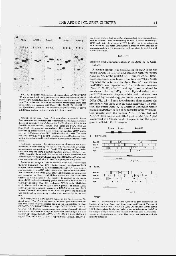
THE AP0E-C1-C2 GENE CLUSTER 43
Ä B Apoa APOCl ÄpBc2 p^ac
6 E H ( C 8 E H K 8 Ê H K B E »
tus, Corp.) and overlaid with 50 «1 of mineral oil. Reaction conditions ApGcZ were as follows: 1 min of denaturing at 94°C. 2 min of annealing at
57°C, and 4 tnin of extension at 72''C for 32 cycles in an automated PCR machine (Bio-med). Amplification products were analyzed by
B £ K electrophoresis in a 2% agarose gel and visualized by staining with ethidium bromide.
;» ^
\iajjâ:.f-
FIG. 1, Southern blot analysis of cosmid clone mAPOE27 DNA (A) and mouse C57BL/Rij genomic DNA (B) hybridized with cDNA probes of the mouse Apoe and the Apoc2 genes and the human APOCl gene. The probes used for each hybridization are indicated above each panel. DNA was digested with BamHI (B), £coRI (E), HmdIII (H) andA'pral (K) as indicated. The molecular weight standards are shown in kilobases and are indicated at the left of each panel.
Isolation of the mouse Apoe-cI-c2 gene duster by cosmid cloning. Two mouse cosmid libraries were constructed by cloning partial Mbol digests of genomic DNA of the strains C57BL/Rij and 129/.J in the BamHI site of the cosmid vectors c2RB (Bates and Swift, 1983) and SuperCosl (Stratagene», respectively. The cosmid libraries were screened by colony hybridization using a mouse Apoe cDNA probe, i.e., the 1.1-kb insert of pmEUClS (Horiuchi et ai. 1989). The probe was labeled with l«-^^P]-dCTP by random priming (Multiprime labeling kit. Amersham) and hybridized (see Southern blot analysis) to the filters.
Restriction mapping. Restriction enzyme digestions were performed as recommended by the supplier (Pharmacia). The DNA fragment sizes were determined on 0.7 and 0.3.5% agarose gels. Restriction sites were mapped using a partial digestion protocol (Hoflcer et ai, 1986). Cosmids clones with the vector c2RB were hybridized with Sa/1/ßamHI and Sall/Nrul fragments of pBR322. SuperCosl cosmid clones were hybridized with T3 and T7 oligonucleotides probes.
Southern blot analysis. Mouse genomic DNA was isolated from the liver (Sambrook et ai , 1989). Restriction enzyme digests of DNA samples were analyzed by electrophoresis through 0.79c agarose gels and blotted to Hybond N* nylon membranes (Amersham) using alkaline transfer (0.4 M NaOH, 1.5 M NaCl). Hybridizations were carried out according to Church and Gilbert (1984) and the filters were washed as recommended by the supplier. In addition to the mouse Apoe cDNA probe the following probes were used: a human APOCl cDNA probe (Knott et ai , 1984), a human AP0C2 probe (Myklebost et a i . 1984b), and a mouse Apoc2 cDNA probe. The mouse <4poc2 cDNA probe was isolated by screening a BALB/c mouse liver cDNA library iClontech) with a human AP0C2 cDNA probe, and its identity was confirmed by sequencing (Hoffer et ai , manuscript in preparation).
PCR amplification of the tandem repeat In the third intron of the Apoc2 gene. The cDNA sequence of the Apoc2 gene was used to design two unique oligonucleotides flanking the minisatellite (5' oligo: CCGATCACCATGGATGAGAA: 3' oligo: GCTTTTGCTGTACAT-GTCCC). One hundred nanograms of total mouse DNA and 100 ng of each primer were used in a bO-fi\ PCR reaction containing 0.2 mM of each dNTP, 50 mM KCl, 10 mM Tris-HCl, pH 8.4, 0.5 mM MgCi,, 0.2 mg/ml BSA, 10% DMSO. 1 unit Taq polymerase (Perkin-Elmer/Ce-
RESULTS
Isolation and Characterization of the Apoe-cl-c2 Gene Cluster
A cosmid library was constructed of DNA from the mouse strain C57BL/Rij and screened with the mouse Apoe cDNA probe pmEUClS (Horiuchi et a i , 1989). Fourteen clones were found to contain the 7.9-kb EcoKl fragment characteristic for Apoe. One of these clones, mAPOE27, was digested with four different enzymes (BamHI, EcoRI, Hindll l , and Kpnl) and analyzed by Southern blotting (Fig. lA). Hybridization with pmEUClS revealed fragments identical in size to those obtained by hybridizing this probe to mouse genomic DNA (Fig. IB). These hybridization data confirm the presence of the Apoe gene in clone mAPOE27. In addition, the entire Apoe-cl 'c2 cluster is contained within cosmid mAPOE27, as can be deduced from the hybridization results with the human APOCl (Fig. lA) and APOC2 (data not shown) cDNA probes. The Apocl gene is confined to a 2.3-kb ßamHI fragment, and the Apoc2 gene to a 9.5-kb EcoRI fragment.
Apoe A p o d Apoc2
A C57BL/RIJ
B . m H , I I I I ! 4.S |2 .3 ! I I 16 I "
"- '" :, iii-i - i L t - i -, , L :
B i 2 g / j
Ba.H, I | | [ h . s H I I I ie ECO m I ' ^ \ » I! 1^3-Mi W-̂ V Hindlll | [ [ î . 5 P g.0 I [ 6.3 I 11
Kpnl [ j f 7.S I t ••' ! I
5 kb
FIG. 2 . Restriction map of the Apoe-cI-c2 gene cluster and the location of the Apoe,/ipoc/, andApr)c2 genes (solid boxes). The map of the gene cluster for the strain C57BL/Rij (A) and that for the strain 129AJ (B) are given for the restriction enzymes indicated at the left of both maps. The positions of the cosmids that were used for characterization are shown below each map. Restriction site variants are indicated by asterisks.
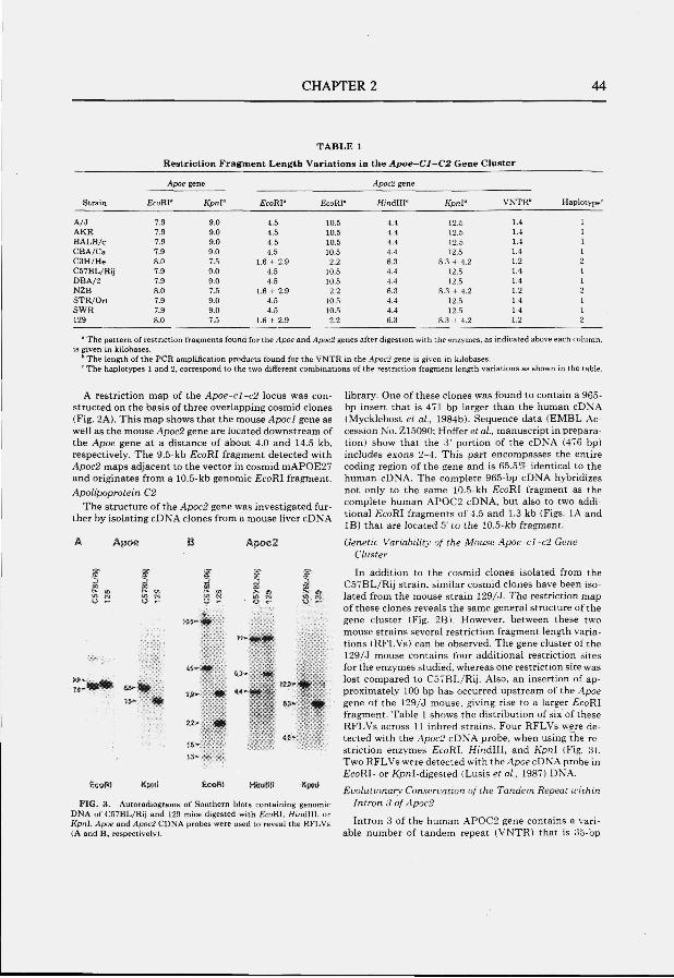
CHAPTER 2 44
TABLE 1
Restriction Fragment Length Variations in the Apoe-~Cl-C2 Gene Cluster
S t r a i n
A / J A K R B A L B / c C B A / C a C 3 H / H e C 5 7 B L / R i j D B A / 2 N Z B S T R / O r t S W R 129
Apoe
E c o W
7.9 7.9 7.9 7.9 8.0 7.9 7.9 8.0 7.9 7.9 8.0
gene
K p n V
9.0 9.0 9.0 9.0 7.5 9.0 9.0 7.5 9.0 9.0 7.5
E c o R I '
4.6 4.5 4.5 4.5
1.6 + 2.9 4.5 4.5
1.6 + 2.9 4.5 4.5
1.6 + 2.9
£ c o R I -
10.5 10.5 10.5 10.5
2.2 10.5 10.5
2.2 10.5 10.5 2.2
Apoc2 gene
H i n d l l l -
4.4 4.4 4.4 4.4 6.3 4.4 4.4 6.,3 4.4 4.4 6.3
K p n l '
12.5 12.5 12.5 12.5
8.3 + 4.2 12.5 12.5
8.3 + 4.2 12.5 12.5
8.3 + 4.2
V N T R '
1.4 1.4 1.4 1.4 1.2 1.4 1.4 1.2 1.4 1.4 1.2
H a p l o t y p e '
2
" The pattern of restriction fragments found for the Apoe and Apoc2 genes after digestion with the enzymes, as indicated above each column, is given in kilobases.
'' The length of the PCR amplification products found for the VNTR in the Apoc2 gene is given in kilobases. " The haplotypes 1 and 2, correspond to the two different combinations of the restriction fragment length variations as shown in the table.
A restriction map of the Apoe-cl-c2 locus was constructed on the basis of three overlapping cosmid clones (Fig. 2A). This map shows that the mouse Apocl gene as well as the mouse Apoc2 gene are located downstream of the Apoe gene at a distance of about 4.0 and 14.5 kb. respectively. The 9.5-kb EcoRI fragment detected with Apoc2 maps adjacent to the vector in cosmid mAPOE27 and originates from a 10.5-kb genomic ^coRI fragment. Apolipoprotein C2
The structure of the Apoc2 gene was investigated further by isolating cDNA clones from a mouse liver cDNA
ApOB E ApoG2
°'lü'''
SsoRf Kpn\
29- m «^-'«ç,
22- m
16»
EcöBä
^i '^ ' -^
^ r
Kptfîl
FIG. 3 . Autoradiograms of Southern blots containing genomic DNA of C57BL/Rij and 129 mice digested with i?coRI. Hinélll, or Kpnl. Apoe and Apoc2 CDNA probes were used to reveal the RFLVs (A and B, respectively).
library. One of these clones was found to contain a 965-bp insert that is 471 bp larger than the human cDNA (Mycklebost et ai , 1984b). Sequence data (EMBL Accession No. Z15090; Hoffer et a i , manuscript in preparation) show that the 3' portion of the cDNA (476 bp) includes exons 2-4. This part encompasses the entire coding region of the gene and is 65.5% identical to the human cDNA. The complete 965-bp cDNA hybridizes not only to the same 10.5-kb EcoRI fragment as the complete human APOC2 cDNA, but also to two additional EcoRI fragments of 4.5 and 1.3 kb (Figs. lA and IB) that are located 5' to the 10.5-kb fragment.
Genetic Variability of the Mouse Apoe-cl~c2 Gene Cluster
In addition to the cosmid clones isolated from the C57BL/Rij strain, similar cosmid clones have been isolated from the mouse strain 129/J. The restriction map of these clones reveals the same general structure of the gene cluster (Fig. 2B). However, between these two mouse strains several restriction fragment length variations (RFLVs) can be observed. The gene cluster of the 129/J mouse contains four additional restriction sites for the enzymes studied, whereas one restriction site was lost compared to C57BL/Rij. Also, an insertion of approximately 100 bp has occurred upstream of the Apoe gene of the 129/J mouse, giving rise to a larger EcoRI fragment. Table 1 shows the distribution of six of these RFLVs across 11 inbred strains. Four RFLVs were detected with the Apoe2 cDNA probe, when using the restriction enzymes EcoRI. KmdIII, and Kpnl (Fig. 3). Two RFLVs were detected with the i4poc cDNA probe in EcoRL or /CpnI-digested (Lusis et a i . 1987) DNA.
Evolutionary Conservation of the Tandem Repeat within Intron 3 of Apoe2
Intron 3 of the human APOC2 gene contains a variable number of tandem repeat (VNTR) that is 35-bp
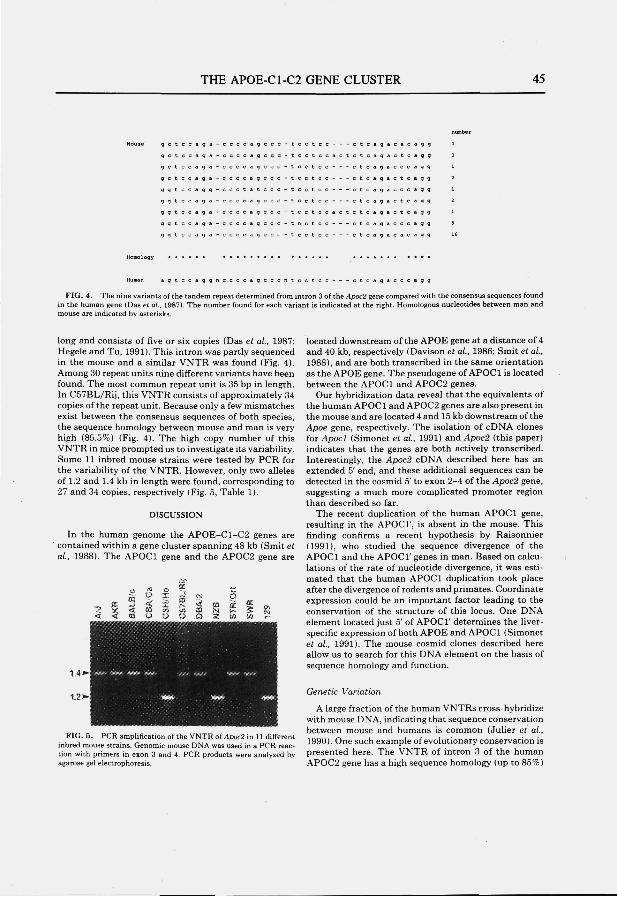
THE APOE-C1-C2 GENE CLUSTER 45
q q t I
q g t <
q g t <
q q t •
q g t i
: a q a -
: a q a -
• a g g -
: a q a •
: a q a •
: a q a -
: a g a <
: a q a .
c a g g
c a g g
c a g g
c a g g
c a g g
c a g g
c a q g
c a g g
c a g g
Human a g t c c a q q n c c c c a g c c c n t c c t c c - - - c t c a q a c c c a g g
FIG. 4. The nine variants of the tandem repeat determined from intron 3 of the Apoc2 gene compared with the consensus sequences found in the human gene (Das et ai , 1987). The number found for each variant is indicated at the right. Homologous nucleotides between man and mouse are indicated by asterisks.
long and consists of five or six copies (Das et a i , 1987; Hegele and Tu, 1991). This intron was partly sequenced in the mouse and a similar VNTR was found (Fig. 4). Among 30 repeat units nine different variants have been found. The most common repeat unit is 35 bp in length. In C57BL/Rij, this VNTR consists of approximately 34 copies of the repeat unit. Because only a few mismatches exist between the consensus sequences of both species, the sequence homology between mouse and man is very high (85.5%) (Fig. 4). The high copy number of this VNTR in mice prompted us to investigate its variability. Some 11 inbred mouse strains were tested by PCR for the variability of the VNTR. However, only two alleles of 1.2 and 1.4 kb in length were found, corresponding to 27 and 34 copies, respectively (Fig. 5, Table 1).
DISCUSSION
In the human genome the APOE-C1-C2 genes are contained within a gene cluster spanning 48 kb (Smit et a i , 1988). The APOCl gene and the APOC2 gene are
FIG. 5. PCR amplification of the VNTR of Apoc2 in 11 different inbred mouse strains. Genomic mouse DNA was used in a PCR reaction with primers in exon 3 and 4. PCR products were analyzed by agarose gel electrophoresis.
located downstream of the APOE gene at a distance of 4 and 40 kb, respectively (Davison et al., 1986; Smit et a i , 1988), and are both transcribed in the same orientation as the APOE gene. The pseudogene of APOCl is located between the APOCl and APOC2 genes.
Our hybridization data reveal that the equivalents of the human APOCl and APOC2 genes are also present in the mouse and are located 4 and 15 kb downstream of the Apoe gene, respectively. The isolation of cDNA clones for Apocl (Simonet et a i , 1991) and Apoc2 (this paper) indicates that the genes are both actively transcribed. Interestingly, the Apoc2 cDNA described here has an extended 5' end, and these additional sequences can be detected in the cosmid 5' to exon 2-4 of the Apoc2 gene, suggesting a much more complicated promoter region than described so far.
The recent duplication of the human APOCl gene, resulting in the APOCl', is absent in the mouse. This finding confirms a recent hypothesis by Raisonnier (1991), who studied the sequence divergence of the APOCl and the APOCl' genes in man. Based on calculations of the rate of nucleotide divergence, it was estimated that the human APOCl duplication took place after the divergence of rodents and primates. Coordinate expression could be an important factor leading to the conservation of the structure of this locus. One DNA element located just 5' of APOCl' determines the liver-specific expression of both APOE and APOC1 ( Simonet et a i , 1991). The mouse cosmid clones described here allow us to search for this DNA element on the basis of sequence homology and function.
Genetic Variation
A large fraction of the human VNTRs cross-hybridize with mouse DNA, indicating that sequence conservation between mouse and humans is common (Julier et a i , 1990). One such example of evolutionary conservation is presented here. The VNTR of intron 3 of the human AP0C2 gene has a high sequence homology {up to 85%)
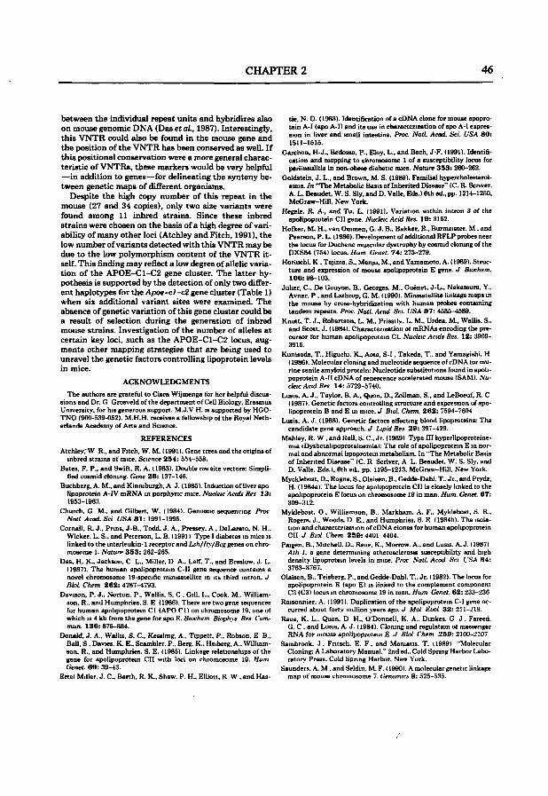
CHAPTER 2 46
between the individual repeat units and hybridizes also on mouse genomic DNA (Das etaL, 1987). Interestingly, this VNTR could also be found in the mouse gene and the position of the VNTR has been conserved as well. If this positional conservation were a more general characteristic of VNTRs, these markers would be very helpful —in addition to genes—for delineating the synteny between genetic maps of different organisms.
Despite the high copy number of this repeat in the mouse (27 and 34 copies), only two size variants were found among 11 inbred strains. Since these inbred strains were chosen on the basis of a high degree of variability of many other loci (Atchley and Fitch, 1991), the low number of variants detected with this VNTR may be due to the low polymorphism content of the VNTR itself. This finding may reflect a low degree of allelic variation of the APOE-C1-C2 gene cluster. The latter hypothesis is supported by the detection of only two different haplotypes for the Apoe-cl -c2 gene cluster (Table 1) when six additional variant sites were examined. The absence of genetic variation of this gene cluster could be a result of selection during the generation of inbred mouse strains. Investigation of the number of alleles at certain key loci, such as the APOE-C1-C2 locus, augments other mapping strategies that are being used to unravel the genetic factors controlling lipoprotein levels in mice.
ACKNOWLEDGMENTS
The author« are grateful to Cisca Wijmenga for her helpful discussions and Dr. G Grosveldof the department of Cell Biology, Erasmus University, for his generous support. M.,J.V H. is supported by HGO-TNO (90O-Ö39-0S2). M.H.H. receives a fellowship ot the Royal Netherlands Academy of Arts and Science.
REFERENCES
Atchleyr W R., and Fitch, W. M. (1991). Gene trees and the origins of inbred strains of mice. Science 254 : ,5.54-558.
Bates, F. P., and Swift, R. A. 11983). Double cos site vectors: Simplified cosmid cloning. Oene 2 6 : 137-146.
Buchberg, A. M., and Kinniburgh, A J. (1985). Induction of liver apolipoprotein A-IV mRNA in Porphyrie mice. Nuctetc 'Acids Res 1 3 : 1953-1963.
Church, G M., and Gilbert, W. (1984). Genomic sequencing Proc Nail Acad. Set USA 8 1 : 1991-199.').
Cornall, R. J., Pnns, J-B.. Todd. J. A., Pressey, A , DeLarato. N. H.. Wicker, L. S., and Peterson, L. B. ( 19911 Tviie 1 diabetes in mice is linked to the interleukin-1 receptor and Lsh/Ity/Bcg genes on chromosome 1. Naturr 3 5 3 : 262-265.
Das, H. K.. Jackson. C L.. Miller, D A.. Letf. T., and Breslow. J. I.. (1987). The human apolipoprotein C-II gene sequence contains a novel chromosome 19-specific minisatellite in its third intron. J Biol. Chem 2 6 2 : 4787-4793.
Davison, P. J., .Norton, P.. Wallis. S. C . Gill. I... Cook. M.. Williamson, R.. and Humphries. S. E ( 1986). There are two gene sequences for human apolipoprotein CI (APO CI) on chromosome 19. one of which IS 4 kb from the gene for apo E. Btuchem Biophys Res Commun. 136: 876-884.
Donald. J. A.. Wallis, S. C , Kessling, A.. Tippett. P., Robson, E B., Ball, S , Davies. K. E., Scambler. P.. Berg. K.. Heiberg, A.. Williamson, R., and Humphries, S. E. ( 1985). Linkage relationships of the gene for apolipoprotein CII with loci on chromosome 19. Hum Genet. 6 9 : 39-43.
Ertel Miller. J. C . Barth, R. K., Shaw. P. H.. Elliott. R W . and Has-
tie, N. O. (1983). Identification of a cDNA clone for mouse apoprotein A-I (apo A-I) and its use in characterization of apo A-I expression in liver and small intestine. Proc. NatL Acad. Set. L̂ SA 8 0 : 1511-1515.
Garehon, H-J., Bedossa, P., Eloy, L., and Bach, J-F. (1991). Identification and mapping to chromosome 1 of a susceptibility locus for periinsulitis in non-obese diabetic mice. Nature 3 5 3 ; 260-262.
Goldstein, J. L., and Brown, M. S. (1989). Familial hypercholesterolemia. In "The Metabolic Basis of Inherited Disease'' (C.R. Scnver, A. L. Beaudet, W. S. Sly, and D. Valle, Eds.) 6th ed., pp. 1214-1250, McGraw-Hill, New York.
Hegele, R. A., and Tu, L. (1991). Variation within intron 3 of the apolipoprotein CII gene. Nucleu: Acid Res. 19: 3162.
Hofker, M. H.. van Ommen, G. J. B., Bakker, E., Burmeister. M . and Pearson, P. L. ( 1986). Development of additional RFLP probes near the locus for Duchene muscular dystrophy by cosmid cloning of the DXS84 (754) locus. Hum Genet. 7 4 : 275-279.
Horiuchi. K , Tajima. S.. Menju, M., and Yamamoto, A. (1989). Structure and expression of mouse apolipoprotein E gene. J Biochem. 106: 98-103.
Julier, C , De Gouyon, B.. Georges, M.. Guénet, J-L., Nakamura. Y.. Avner. P , and Lathrop, G. M. ( 1990). Minisatellite linkage maps in the mouse by cross-hybridization with human probes containing tandem repeats. Proc. NalL Acad Set. USA 8 7 : 4585-4589.
Knott. T. J., Robertson, L. M., Priestly. L. M.. Uniea. M., Wallis. S., and Scott, J. ( 1984). Characterization of mRNAs encoding the precursor for human apolipoprotein CI. Nucleic Acids Res. 12: 3909-3915.
Kunisada, T., Higuchi, K., Aota, S-I, Takeda, T.. and Yamagishi. H ( 1986). Molecular cloning and nucleotide sequence of cDNA tor murine senile amyloid protein: Nucleotide substitutions found in apolipoprotein A-II cDNA of senescence accelerated mouse (SAM). .Vu-cfeic Acid Res 14: 5729-6740.
Lusis. A. J.. Taylor. B. A., Quon. O.. Zollman. S., and LeBoeuf, R. C ( 1987). (Genetic factors controlling structure and expression of apolipoprotein B and E in mice. J BioL Chem. 2 6 2 : 7594-7604
Lusis, A. J. (1988). Genetic factors aifecting blood lipoproteins: The candidate gene approach. J Lipid Res 2 9 : 397-429.
Mahley, R. W . and Rail. S. C . Jr. ( 1989) Type III hyperlipoproteinemia (Dysbetalipoproteinemia): The role of apolipoprotein E in normal and abnormal lipoprotein metabolism. In "The Metabolic Basis of Inherited Disease" (C. R Scriver, A L. Beaudet. W. S. Sly, and D. Valle. Eds.), 6th ed., pp. 1195-1213, McGraw-Hill. New York.
Mycklebost, O.. Rogne, S., Olaisen. B., Gedde-Dahl. T.. Jr., and Ptydz, H. ( 1984a). The locus for apolipoprotein CII is closely linked to the apolipoprotein E locus on chromosome 19 in man. Hum. Genet. 6 7 : 309-312.
Myklebost. O . Williamson, B.. Markham, A. F.. Myklebost. S. R.. Rogers, J.. Woods, D E.. and Humphries, S. E (1984b). The isolation and characterization of cDN A clones for human apolipoprotein CII J Biol Chem 2S9 : 4401-4404.
Paigen. B., Mitchell. D.. Reue. K.. Morrow. A., and Lusis. A. J. ( 1987) Ath-1, a gene determining atherosclerosis susceptibility and high density lipoprotein levels in mice. Proc Natl. Acad. Sci VSA 84: 3763-3767.
Olaisen, B.. Teisberg, P., and Gedde-Dahl. T., Jr. 11982). The locus for apolipoprotein E (apo E) is linked to the complement component C3 (C3) locus in chromosome 19 in man. Hum Genet. 62 : 233-236
Raisonnier. A. (1991). Duplication of the apolipoprotein C-I gene occurred about forty million years ago. J Mol Ei'ol 3 2 : 211-219.
Reue, K. L.. Quon. D H.. O'Donnell, K. A.. Dizikes. G J . Fareed. G. C . and Lusts, A. J. ( 1984). Cloning and regulation ot messenger RNA for mouse apolipoprotein E J Biol Chem 2 5 9 : 2100-2107
.Sambrook. J . Fritsch. E. F , and Maniatis. T. ( 1989) "Molecular Cloning: A Laboratory Manual." 2nd ed.. Cold Spring Harbor Laboratory Press. Cold Spring Harbor. New York.
Saunders. A. M .and Seldin. M. F. (1990). A molecular genetic linkage map of mouse chnimusome 7. Genomics 8: .525-.5.')5.
THE APOE-C1-C2 GENfE CLUSTER 47
Simonet, W. S., Bucay, N., Lauei, S. J., Wirak, D. O., Stevens, M. E., Weisgraber, K. H., Pitaa, R. E., and Taylor, J. M. (1990). In the absence of a downstream element, the apolipoprotein E gene ia expressed at high levels in kidneys of tranagenic mice. «/. BioL Chem. 26S : 10809-10812.
Simonet, W. S., Bucay, N., Pitas, R. E., Lauer, S. J., and Taylor, J. M. (1991). Multiple tiasue-specific elementa control the apolipoprotein E/C-I gene locus in tranagemc mice. J . BioL Chem. 266:6651-8654.
Smit, M., van der Kooy-Meija, E., Frants, R. R., Havakea, L., and Klasen, E. O. (1988). Apolipoprotein gene cluster on chiomoBome 19: Definite localization of the APOC2 gene and the polymoiphic Hpal aite aaaociated with type III hyperlipoproteinemia. Hum. Genet. 7 8 : 90-93.
Smith, J. D., Plump, A S., Hayek, T., Walsh, A., and Brealow, J. L. (1990). Accumulation of human apolipoprotein E in the plasma of transgenic mice. J. BioL Chem. 2 6 5 : 14709-14712.
CHAPTER 3
THE MOUSE APOLIPOPROTEIN CI GENE: STRUCTURE AND EXPRESSION.
M.J.V. Hoffer, M.M. van Eck, L.M. Havekes, M.H. Hofker, and R.R. Frants.
Genomics: in press
CHAPTERS 50
The Mouse Apolipoprotein CI Gene: Structure and Expression M A R I E T T E J. V . H O F F E R , M A R G A M . V A N E C K , L O U I S M . H A V E K E S , * M A R T E N H . H O F K E R , ^ A N D R U N E R . F R A N T S
MGC-Departmertt o f Hirnian Genetics, Leiden University, P.O. Box 9503, 2300 RA Leiden, The Netherlands; and 'IWO-TNO, Gaubius Laboratory, P.O. Box 430, 2300 AK Leiden, The Netherlands
Received May 18, 1993
We liave iaolated and cliaracterlzed cDNA and genomic clones containing the mouse apolipoprotein C I (Apoel) gene. The Apoel gene ia par t of the A p o e - e l -e2 gene d u s t e r and is located 3.4 kb 3 ' of the Apoe gene. The mouse Apoel gene spans a region of approximately 3.3 kb and consists of four exons. The exon-intron s tructure is similar to those of human and baboon genes, although in mouse introns 2 and 3 a re smaller. Significant sequence homology is found bet^reen man and mouse in the promoter and exonic regions (80 and 67%, respectively). Northern blotting and primer extension analsrsis of mouse RNA showed that a major t ranscript 409 bp in size is expressed primarily in fetal and adult Uver. The mouse Apoel cDNA contains an open reading brame encoding a protein of 8 8 amino acids, including a signal peptide of 26 amino acid residues. Comparisons of the deduced amino acid sequence of mouse apoCl with the human, baboon, ra t , and dog sequences showed discrete regions with a high degree of conservation. The delineation of the sequence and structural organization of the mouse Apoel gene is an essential step in enhancing the use of mouse models to study the ftinction of apoCl in the lipoprotein metabolism. eiMSAMdMüePrMi.iM.
mouse model in which overexpression of human apoCl resulted in an increase of the plasma triglyceride levels (Simonet et aL, 1991). Apparently, apoCl plays a role in regulating the catabolism of triglyceride-rich lipoproteins.
Due to the advances in transgenic technology, mice have become increasingly useful for investigation of lipoprotein metabolism. The correct interpretation of results obtained with transgenic mice strongly depends on sufficient knowledge of the genes involved in the mouse lipoprotein metabolism. The human APOCl gene is part of a gene cluster that is located on the long arm of chromosome 19 and includes the genes for apoE, apoCl, pseudo apoCl, and apoC2 (Humphries et aL, 1984; Davison et aL, 1986; Smit et aL, 1988). We have demonstrated a similar gene organization in mouse with the exception that the mouse gene cluster does not contain a pseudo Apocl gene (Hoffer et aL, 1993a). The genomic structure and sequences of the Apoe (Reue et aL, 1984; Horiuchi et aL, 1989) and the Apoc2 genes (Hoffer et aL, 1993b) have been determined previously. This paper describes the characterization of the mouse Apocl gene. We have determined the mouse Apocl cDNA sequence and the exon-intron structure and analyzed the expression of the gene.
INTRODUCTION
Human apolipoprotein CI (apoCl) is a 57-amino-acid peptide that is primarily produced in the liver (Knott et aL, 1984) and is associated with chylomicron, vety-low-density lipoprotein, and high-density lipoprotein (Breslow, 1988). The ftinction of apoCl in the lipoprotein metabolism remains unclear. ApoCl is known to activate lecithinicholesterol acyltransferase (LCAT) in vitro (Soutar et aL, 1975). In addition, apoCl can inhibit the binding of apoE to the low-density lipoprotein receptor-related protein (Weisgraber et aL, 1990). More recent studies showed that this inhibition can also be found for the low-density lipoprotein receptor (Sehayek and Eisenberg, 1991). These studies could be in agreement with the data obtained with a human APOCl transgenic
' To whom correapondence should be addreaaed. Telephone: 31-71-276099. Fai: 31-71-276075.
MATERIALS AND METHODS
Isotation ami sequencing of eDNA ekmes. A Xgtll cDNA library derived from Uver mRNA of an adult male BALB/c mouie (Clontech) woa screened with a Ddel fragment of human APOCl cDNA (Knott et aL, 1984) containingexona 1-4. Inaerta of poaitive clone* were iaolated by PCR amplification using primen (a. 5'-GGTGGCGACGACTCC-TGGAG-3'; b. S'-GGTAATGGTAGCGACCGGCG-31 Bonking the £coRl cloning site. A total of 30 jil of phage lysate and 100 ng of each primer were need in a 60-^1 PCR reaction containing 0.2 tnM of each dNTP, SO mM KCl, 10 toM Tris-HCl (pH 8.4), 2.5 mM MgCl,, 0.2 mg/ml BSA, 10% DMSO, 1 unit Taq polymerase (Perkin-Elmer-Ce-tus) and overlaid with 60 ftl of mineral oil. Reaction conditions were aa foUowa: a denaturing step of 1 min at 94*C, followed by an annealing step of 2 min at 60°C and on extension step of 4 min at 72*C for 32 cycles. PCR products were subcloned into the pT7Blue (R) T-vector (Novagen) and sequenced using the T7 sequencing kit (Pharmacia). Individual clones wen sequenced in both directions using the universal primers or specific synthetic oligonucleotides.
Determination of exon-intron structure. Exons were located by hybridization with a mouse .4poci cONA probe on restriction ensyme
0888-7543/93 SS.00 Copynght O 1993 by Academic Picas. Inc.
All right« of rapnxluctlon in oay fonn naeivad.
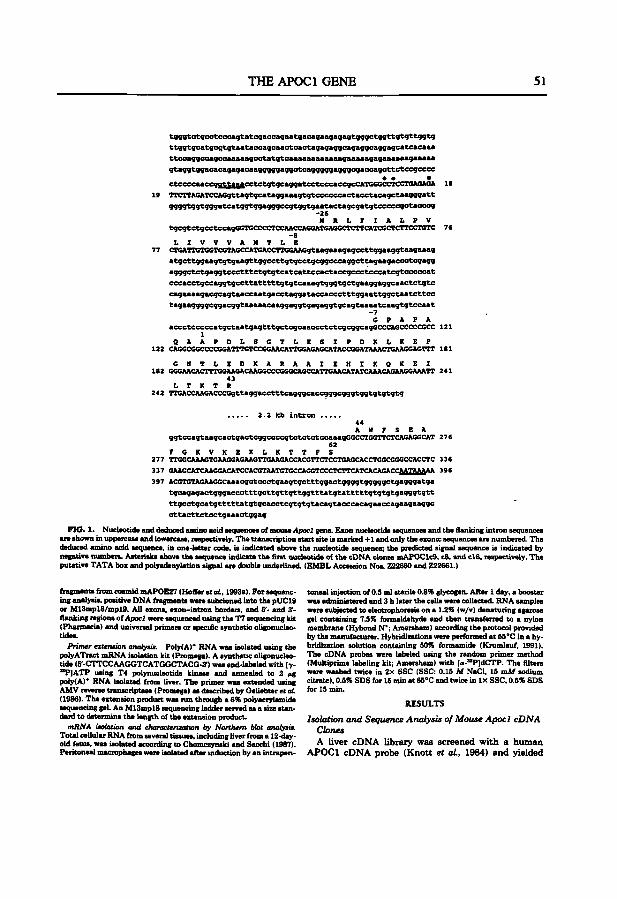
THE APOCl GE^fE 51
tgggtctgcctcccagtategaccagaatgacagaagagagtgggctggttçftgttggtg
gtaggtggaeacagagacaagggggaggetcagggggagggcgaccagcttetecgcccc
ctcceeaaeeoattaaaiMtetatQeaaaateetcecaeecicCATCGGCCTCCTGAgACA 18
19 TTCTTAGATCCAGgttagtgcataggaaagtgtcccceeactacctacagctaogggatt
ggggtqgtgggateatggtggagggccgtqgtgaatactagcgatgtcccccgctacceq -26 M R L F I A L P V
tgcgtetgeetccagGGTGCCCCTCCAACCAGGATGASGCTCTTCATCSCTCTTCCTGTC 76 -e
L I V V V A H T L E 77 CTSATTGTGCTCGTAGCCATGACCTTGSAAGgtaagaaagagecttggaaggtaagaaag
atgettggaagtgtgaagttggcettgtgectgcggcccaggettagaagaeectcgagg
agggututgaggtccctttct<jtgtcatcattccacta€Mgc€»tcccatcgtcccccat
cccacctgccaggtgccttatttttgtgtcaaagtgggtgctgaaggaggcaactctgtc
cagaaaagacgcagtaacïcaatgacctaggataccaccctttggaattggctaatcttcc tagaaggggeggacggtaaaaacaaggaggtgagaggtgcagtaaaatcaagtgtccaat
-7 G P A P A
acccteccccatgctaatgagtttgctcgeaaccctctcgcggcagcCCCAGCCCCCGCC 121 1
Q A A P D L S S T L Z S I P D X L X E F 122 CASGCGGCCCCSGATTTGTCCGGAACATTGGAGAGCATACCGGATAAACTGAASGAGTTT 181
S K T L E O K A R A A I E H I X O X E I 182 GGGAACACTTTGGAAGACAAGGCCCGGGCAGCCATTCAACATATCAAACAGAAGCAAATT 241
43 L T K T R
242 TTGACCAAGACCCGgttaggacctttcagggcaccgggcgggtggtgt:gtgtg
2.2 kb i n t r o n 44 A W F S E A
ggtccagtaagcactgacteggccccgtctctctccaaagGGCCTGGTTCTCAGAGGCAT 276 62
F G K V X E X L K T T F S 277 TTGGCAAAGTGAAGGAGAAGTTGAAGACCACGTTCTCCTSAGCACCTGGCGGGCCACCTC 336
397 A C G T G T A G A A G G C a a a c g c t e c c t g a a g t g c t t t g g a c t g g g g t g g g g g c t g a g g g a t g a
t g c a g a g a c t g g g a c c c t t t g c t t g t t g t t g g t t t a t g t a t t t t t g t g t g t g a q g g t g t t
t t g c c t g c a t g t t t t t a t g t g c a c c t c g t g t g t a c a g t a c c c a c a g a a c c a g a a g a a g g c
c t t a c t t e t c c t g a a a c t g g a g
FIG. 1. Nucleotide and deduced amino acid sequences of mouse Apocl gene. Exon nucleotide sequences and the flanking intron sequences are shown in uppercase and lowercaae, nspectively. The transcription start site is marked .t-l and only the exontc sequences are numbered. The deduced amino acid sequence, in one-letter code, is indicated above the nucleotide sequence; the predicted signal sequence is indicated by negative numbers. Asterisks above the sequence indicate the first nucleotide of the cDNA ckines mAP0Clc9, c8, and cl6, respectively. The putative TATA box and polyadenylation signal are double underlined. (EMBL Accession Nos. Z22660 and Z22661.)
fragmenta from cosmid mAPOE27 (Hoffer et aL, 1993a). For sequenc- toneal injection of 0.5 ml sterile 0.8% glycogen. Alter 1 day, a booster ing analysis, positive DNA fragments war« subcloned into the pUCl9 was administered and 3 h later the cells were eoUecteiL RNA samples or M13mpl8/mpl9. All exons. exon-intron borders, and 6'- and 3'- were subjected to electrophoresis on a 1.2% (w/v) denaturing agarose flanking regions of Apocl were sequenced using the T7 sequencing kit gel containing 7.5% formaldehyde and then transferred to a nylon (Pharmacia) and universal primers or specific syntfaetic oUgonucIeo- membrane (Hybond N*; Amenham) according the protocol provided tides. by the monuf^urer . Hybridizations were performed at 55''C in a hy-
Primer extension anafyeii. Poly(A)* RNA waa iaolated using the bridization solution conuining 50% formamide (Krumlauf, 1991). polyATract mRNA isolation kit (Promega). A synthetic oligonucleo- The cDNA probes wen labeled using the random primer method tide (5'-(nTCCAA<}GTCATGGCrrACG-3') waa end-labeled with [y- (Multiprime labeling kit; Amersham) with [«-"PldCTP. The filters " ^ l A T P using T4 polynucleotide kinase and annealed to 2 (ig ^ • n washed twice in 2X SSC (SSC: 0.15 M NaCl, 15 mM sodium poly(A)* RNA iaolated from liver. The primer was extended using citrate), 0.5% SDS for 15 min at 65°C and twice in I x SSC, 0.5% SDS AMV reverse transcriptase (Promega) as described by Geliebter et oL f"' 15 min. (1986). The extension product was run through a 6% Polyacrylamide RESULTS sequencing gel. An M13mpl8 sequencing ladder served as a size stan- IU.9U1. dord to deurmine the length of the extension product. I so l a t i on a n d S e q u e n c e A n a l y s i s of M o u s e A p o c l c D N A
mRNA isolation and charaeteruatum by Northern blot anaiysis. Clones Total cellular RNA from several tiasues, including liver from a 12-day- r . » , » ,.,. j • u u oldfetua, waa iaolated according to Chomczynski and Sacchi (1987). A l iver c D N A l ib r a ry w a s s c r e e n e d w i t h a h u m a n Peritoneal macrophagea were isolated after induction by an intrapen- A P O C l c D N A p r o b e ( K n o t t e t al., 1984) a n d y ie lded
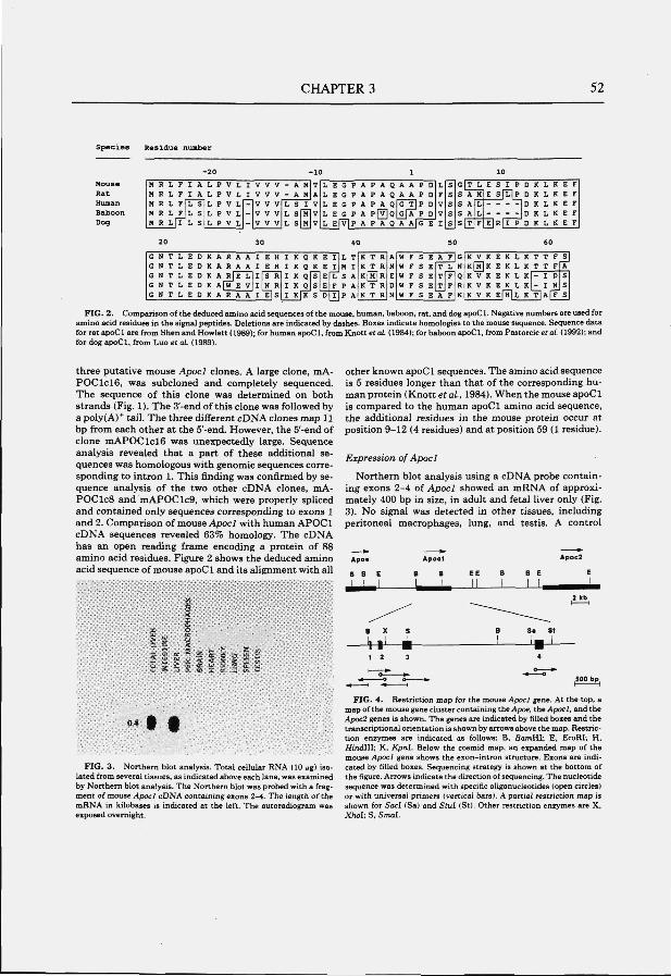
CHAPTER 3 52
Spec ies Residue number
Mouse R a t Human B a b o o n Dog
- 2 0 - 1 0 1
M R L F I A L P V L I V V V M R L F I A L P V L I V V V H R L F L S M R L F J L S M R L [ I L S
L P V L L P V L L P V L
20
-V V V V V V V V V
10
- A M T - A H A L S I V L sfÏÏlv L S M V
L E G P A P A Q A A P D L E G P A P A Q A A P D L E G P A P A Q G T j P D L E G P A p f v l Q G [ A P D
L F V V
L E | V | P A P A Q A A | G E I
40
G N T L E D K A R A A I E H I K Q K E I L T G N T L E D K A R A A I E H I K Q K E I M I G M T L E D G N T L E D G N T L E D
K A RjE L K A [ W E V K A R A A
I s R I H R
l i ls
I K Q S E I K Q S E I K[K S D
L S A F P A T]P A
K T R K T R K[M]R K T R K T R
A K E 0 N
S S s s s
G S s s s
50
W F S E W F S E W F S E W F S E W F S E
A F T L T F T J F A F
G M
Q R K
10
T L A H A A T
L L F
K V KfÏÏ K K K
V V V
E S I P D K L K E s f l l P D K L K
D K L K D K L K
E | R | I P D K L K
E F E F E F E F E F
60
K E K L K T T F K E K L K T T F
S
K E K L K - I D S K E K L K U I NJS K EfÏÏlL K T I A F F S
FIG. 2. Comparison of the deduced amino acid sequences of the mouse, human, baboon, rat. and dog apoCl. Negative numbers are used for amino acid residues in the signal peptides. Deletions are indicated by dashes. Boxes indicate homologies to the mouse sequence. Sequence data for rat apoCl are from Shen and Howlett ( 1989); for human apoCl, from Knott etaL (1984); tor baboon apoCl, from Paatorcic el al (1992); and for dog apoCl, from Luo et at. (1989).
three putative mouse Apocl clones. A large clone, mA-POClc i e , was subcloned and completely sequenced. The sequence of this clone was determined on both strands (Fig. 1). The 3'-end of this clone was followed by a poly(A)* tail. The three different cDNA clones map 11 bp from each other at the 5'-end. However, the 5'-end of clone m.'KPOClcie was unexpectedly large. Sequence analysis revealed that a part of these additional sequences was homologous with genomic sequences corresponding to intron 1. This finding was confirmed by sequence analysis of the two other cDNA clones, mA-POClcS and mAPOClc9, which were properly spUced and contained only sequences corresponding to exons 1 and 2. Comparison of mouse Apocl with human APOCl cDNA sequences revealed 63% homology. The cDNA has an open reading frame encoding a protein of 88 amino acid residues. Figure 2 shows the deduced amino acid sequence of mouse apoCl and its alignment with all
other known apoCl sequences. The amino acid sequence is 5 residues longer than that of the corresponding human protein (Knott et a i , 1984). When the mouse apoCl is compared to the human apoCl amino acid sequence, the additional residues in the mouse protein occur at position 9-12 (4 residues) and at position 59 (1 residue).
Expression of Apocl
Northern blot analysis using a cDNA probe containing exons 2-4 of Apocl showed an mRNA of approximately 400 bp in size, in adult and fetal liver only (Fig. 3). No signal was detected in other tissues, including peritoneal macrophages, lung, and testis. A control
Apo«
B B EE
_ L L -!—U
£1 et *: S tt ^ O
S f « z 2 1 +f^
• •
FIG. 3 . Northern blot analysis. Total cellular RNA (10 ng) isolated from several tissues, as indicated above each lane, was examined by Northern blot analysis. The Northern blot was probed with a fragment of mouse Apocl cDNA containing exons 2-4. The length of the mRNA in kilobases is indicated at the left. The autoradiogram was exposed overnight.
FIG. 4 . Restriction map for the mouse Apocl gene. At the top, a map of the mouse gene cluster containing the Apoe, the Apocl, and the Apoc2 genes is shown. The genes are indicated by filled boxes and the transcriptional orientation is shown by arrows above the map. Restriction enzymes are indicated as follows; B, SamHl; E, EcoRI; H. Hindlll; K, Kpnl. Below the cosmid map, an expanded map of the mouse Apoci gene shows the exon-intron structure. Exons are indicated by filled boxes. Sequencing strategy is shown at the bottom of the figure. Arrows indicate the direction of sequencing. The nucleotide sequence was determined with specific oligonucleotides (open circles) or with universal primers (vertical bars). A partial restriction map is shown for SacI (Sa) and Stul (St). Other restriction enzymes are X, Xhol; S, Smal.
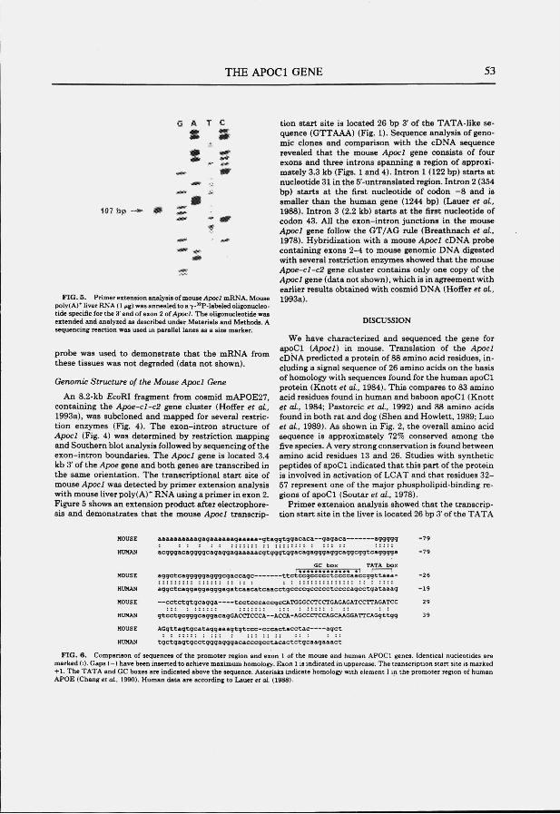
THE APOCl GENE 53
G A T C
S 3
107 bp ^ aSSm'
FIG. 5. Primer extenaion analysis of mouse Apocl mRNA. Mouse poly( A)* liver RNA (1 (ig) was annealed to a 7-"P-labeled oligonucleo-tide specific for the 3'end of exon 2 ot Apocl. The oligonucleotide was extended and analyzed as described under Materials and Methods. A sequencing reaction was used in parallel lanes as a size marker.
probe was used to demonstrate that the mRNA from these tissues was not degraded (data not shown).
Genomic Structure of the Mouse Apocl Gene
An 8.2-kb £coRI fragment from cosmid mAPOE27, containing the Apoe-cl-c2 gene cluster (Hoffer et a i , 1993a), was subcloned and mapped for several restriction enzymes (Fig. 4). The exon-intron structure of Apocl (Fig. 4) was determined by restriction mapping and Southern blot analysis followed by sequencing of the exon-intron boundaries. The Apocl gene is located 3.4 kb 3' of the Apoe gene and both genes are transcribed in the same orientation. The transcriptional start site of mouse Apocl was detected by primer extension anaiysis with mouse liver poly(A)* RNA using a primer in exon 2. Figure 5 shows an extension product after electrophoresis and demonstrates that the mouse Apocl tremscrip-
tion start site is located 26 bp 3' of the TATA-like sequence (GTTAAA) (Fig. 1). Sequence analysis of genomic clones and comparison with the cDNA sequence revealed that the mouse Apocl gene consists of four exons and three introns spanning a region of approximately 3.3 kb (Figs. 1 and 4). Intron 1 (122 bp) starts at nucleotide 31 in the 5'-untranslated region. Intron 2 (354 bp) starts at the first nucleotide of codon —8 and is smaller than the human gene (1244 bp) (Lauer et a i , 1988). Intron 3 (2.2 kb) starts at the first nucleotide of codon 43. All the exon-intron junctions in the mouse Apocl gene follow the GT/AG rule (Breathnach et a i , 1978). Hybridization with a mouse Apocl cDNA probe containing exons 2-4 to mouse genomic DNA digested with several restriction enzymes showed that the mouse Apoe-cl-c2 gene cluster contains only one copy of the Apocl gene (data not shown), which is in agreement with earlier results obtained with cosmid DNA (Hoffer et al., 1993a).
DISCUSSION
We have characterized and sequenced the gene for apoCl (Apocl) in mouse. Translation of the Apocl cDNA predicted a protein of 88 amino acid residues, including a signal sequence of 26 amino acids on the basis of homology with sequences found for the human apoCl protein (Knott et a i , 1984). This compares to 83 amino acid residues found in human and baboon apoCl (Knott et a i , 1984; Pastorcic et a i , 1992) and 88 amino acids found in both rat and dog (Shen and Howlett, 1989; Luo et aL, 1989). As shown in Fig. 2, the overall amino acid sequence is approximately 72% conserved among the five species. A very strong conservation is found between amino acid residues 13 and 26. Studies with synthetic peptides of apoCl indicated that this part of the protein is involved in activation of LCAT and that residues 32-57 represent one of the major phospholipid-binding regions of apoCl (Soutar et a i , 1978).
Primer extension analysis showed that the transcription start site in the liver is located 26 bp 3' of the TATA
HOUSE
MOHAH
aaaaaaaaaagagaaaaaagaaaaa-gtaggtggacaca—gagaca aggggg
acgggacaggggcagaggagaaaaacgtgggtggacagagggaggcaggcggtcagggga
-79
-79
aggctcagggggagggcgaccagc- - t t c t c c g c c c c c t c c c c a a c c g g t t a a a -
HUMAN a g g c t c a g g a g g a g g g a g a t c a a c a t c a a c c t g c c c c g c c c c c t c c c c a g c c t g a t a a a g - 1 9
MOUSE — c c t c t g t g c a g g a tcctcccaccgcCATGGGCCTCCTGAGAGATCCTTAGATCC 29
HUMAN gtcctgcgggcaggacagGACCTCCCA—ACCA-AGCCCTCCAGCAAGGATTCAGgttgg 39
HOUSE A G g t t a g t g c a t a g g a a a g t g t c c c - c c c a c t a c c t a c a g c t
HUMAN tgctgagtgcctgggagggacacccgcctacactctgcaagaaact
FIG. 6. Comparison of sequences of the promoter region and exon 1 of the mouse and human APOCl genes. Identical nucleotides are marked (:). Gaps (—) have been inserted to achieve maximum homology. Exon 1 is indicated in uppercase. The transcription start site is marked +1. The TATA and GC boxes are indicated above the sequence. Asterisks indicate homology with element I in the promoter region of human APOE (Chang et ai, 1990). Human data are according to Lauer et al (198d).
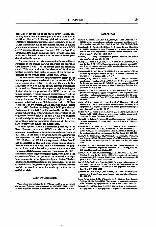
CHAPTERS 54
box. The 5' sequences of the three cDNA clones, containing exons 1-4, are found just 3' o t t h i s start site. In addition, the cDNA library yielded a clone, mA-POClc lS , containing sequences corresponding to intron 1 tha t is probably due to incomplete splicing. A similar phenomenon seems to be the case for the rat APOCl cDNA sequence (Shen and Howlett, 1989) the first 66 bp of which show a high homology (80%) with 3* sequences of intron 1 instead of exon 1 of the mouse Apocl gene, described in this paper.
The exon-intron structure resembles the overall gene structure of the humim APOCl gene with the exception that introns 2 and 3 of the mouse gene are relatively small. This can be due to the absence of A<u-like repeated elementa that make up some of the intron sequences of the human gene (Lauer et aL, 1988).
The nucleotide sequence of the promoter region of the mouse gene was compared to tha t of the human APOCl gene (Lauer et aL, 1988) (Fig. 6), and their nvicleotide sequences show a homology of 80% between nucleotides —114 and —1. However, the region of high homology is limited due to the presence of a SINE repeat in the mouse promoter region located approximately 1(X) bp upstream of the transcription start site. Both genes contain an imperfect TATA box. Interestingly, a 16-bp sequence motif tha t shows 93% homology with a GC box (element I) in the human APOE gene was found (Smith et aL, 1988). Studies involving the APOE gene showed tha t element I binds Sp l and is involved in positive regulation (Chang et aL, 1990). The high conservation of the sequences immediately 5' of the TATA box suggesta functional significance for gene regulation. Further studies of these putative regulatory elements will be necessary to prove any functional significance.
In most species, APOCl is expressed primarily in the Uver. However, in human, APOCl can also be (letected in monocytes differentiating into macrophages (Lauer et aL, 1988). In the mouse, both the Apoe and Apoc2 genes are expressed in peritoneal macrophages (Basu et aL, 1981; Hoffer et aL, 1993b) but expression of Apocl could not be detected in this cell type. Other studies showed limited amounte of Apocl mRNA expression in skin, testis, lung, and submaxillary when a more sensitive RNase protection assay was used (Simonet et aL, 1991). Transgenic mice can be useful for fiirther studies on the expression of the Apocl gene and the interaction of regulatory elementa in the Apoe-cl-c2 gene cluster. The isolation and characterization of the mouse Apocl gene are essential steps for generating an Apocl-deficient mouse model that could be usefiil for studying the fimction of apoCl in uiuo.
ACKNOWLEDGMENTS
The authora acknowledge Dr. K. Willema van Dijk for hia helpful diacuaaiona. This work waa supported by HGO-TNO (900-539-052) and the Royal Netherlanda Academy of Arta and Science (M.H.H.)
REFERENCES
Bam, S. K., Brown, M. S., Ho, Y. K., Havel, R J., and Goldatein, J. L. (1981). Mouae macrophages syntheaiie and aecrete a protein reaem-bling apolipoprotein E. Proc NatL Acad. Sd. USA 7 8 : 7545-7549.
Breathnach, R , Benoiat, C , O'Hare, K., Gannon, F., and Chambon, P. (1978). Ovalbumin gene: Evidence for a leader sequence in mRNA and DNA aequences at the exon-intron boundaries. Proc NatL Acad. ScL USA 7 5 : 4853-4867.
Brealow, J. L. (1988). Apolipoprotein genetic variation and human diieaae. PhysioL Reo. 6 8 : 85-132. . -
Chang, D. J., Paik, Y-K., Leren, T. P., Walker, D. W., Howlett, O. J., and Taylor, J. M. (1990). Characterisation of a human apolipoprotein E gene enhancer element and ita associated protein factors. J. BioL Chem. 2 6 5 : 9496-9504.
Chomczynaki, P., and Sacchi, N. (1987). Single s t ^ method of RNA isolation by acid guanidinium thiocyanate phenol chloroform extraction. Anoi: Biochem. 162: 156-159.
Davison, P. J., Norton, P., Wallis. S. C , GUI. L., Cook, M., Wüliam-aon, R , and Humphries, S. E. (1986). There are two gene sequences for human apolipoprotein CI (APO CI) on chromosome 19, one of which ia 4 kb from the gene for APO E. Biochem. Biophys. Res. Commun. 136: 876-884.
Geliebter, J., ZeS, R A., Melvold, R W., and Nathenson, S. G. (1986). Mitotic recombination m germ cells generated two nuqor histocompatibility complex mutant genes shown to be identical by RNA sequencing analysis: K*^ and K ' " . Proc NatL Acad. ScL USA 8 3 : 3371-3376.
Hoffer, M. 1. V., Hofker, M. H., van Eck, M. M., Havekea, L. M., and Franta, R R (1993a). Evolutionary conaervation of the mouae apo* Upoprotein e.cl.c2 gene cluster. Structure and genetic variabiUty in inbred mice. Genomics 15 : 62-67.
Hofler, M. J. V., van Eck. M. M.. Havekes, L. M., Hofker, M. H., and Franta, R. R (1993b). Structure and expreaaîon of the mouae apoU-popiotein C2 gene. Genomics 17: 46-51.
Horiuchi, K., Tajima, S., Menju, M., and Yamamoto, A. (1989). Structure and expression of mouse apolipoprotein E gene. J. Bioehem. 106 : 98-103.
Humphries, S. E., Berg, K., GUI, L., Cumming, A. M., Robertson, F. W., Stalenhoef, A. F. H., WiUiamaon, R , and Boncaan, A. L. ( 1984). The gene for apolipoprotein Cil ia closely linked to the gene fbr apolipoprotein E on chromosome 19. Clin. Genet. 26 ; 389-396.
Knott. T. J., Robertson, L. M., Prieatly, L. M., Urdea. M.. WalUa, S., and Scott, J. (1984). Characterization of mRNAa encoding the pre-curaor for human apolipoprotein CI. Nucleic Aade Res. 12: 3909-3916.
Krumlauf, R. (1991). Northern blot analysia of gene expression. In "Cïene Tranafer and Expression Protocols" (E. J. Murray, Ed.), pp. 307-360, Humana Preaa, CUiton, NJ.
Lauer, S. J., Walker, D., Elshourbagy, N. A., Reardon, C. A., Levy-WUson, B., and Taylor, J. M. (1988). Two copiée of the human apoUpoprotein C-I gene are linked closely to the apolipoprotein E gene. J. BioL Chem. 2 6 3 : 7277-7286.
Luo, C-C, Li, W-H.. and Chan, L. (1989). Structure and expression of dog apolipoprotein A-1, E, and C-1 mRNAa: Implications for the evolution and functional constraintaof apolipoprotein structure. J . Lipid Res. 3 0 : 1735-1746.
Paatorcic, M., Bimbaiun, S., and Hixaon, J. E. (1992). Baboon apoUpoprotein C-I: cDNA and gene structure and evolution. Genomics 1 3 : 368-374.
Reue, K. L., Quon. D. H., O'Donnell, K. A., Dizikes, G. J., Fareed, G. C , and Lusis, A. J. ( 1984). Clonmg and regulation of messenger RNA for mouae apolipoprotein E. J. BioL Chem. 2 5 9 : 2100-2107.
Sehayek. E.. and Eisenberg, S. (1991). Mechanisms of inhibition by apolipoprotem C of apolipoprotein E-dependent cellular metabo-
THE APOCl GENE 55
lism of human tri^yceride-rich Upoproteina through the low den- Soutar, A. K., Gamer, C. W., Baker, H. N., Sparrow, J. T., Jackson, aity Upoprotem receptor pathway. J. Bid Chem. 266: 18259^ R. L., Gotto, A. M., and Smith, L. C. (1975). ESbct of the human 18267. plasma apolipoproteins and phosphatidylcholine acyl donor on the
Shen, P. Y., and Hewlett, G. J. (1989). Nucleotide sequence for rat activity of lecithin:choles«erol acyltranaftraae. Biochemistry 14: apoUpoprotein C-L Nucleic Acids Res. 17: 6405. 30S7-3064.
Simon«, W.S.,Bucay,N.,Pitaa,RE.,Lauer,S.J.,andTaylor,J.M. *>utar, A. K., Sigler G F.. Smitt, L C . Gotto, A. M., Jr., and (1991). Multiple ti.aue-q»cific elementa control the apoUpoprotein Sparrow J. T ( 1978). Lecithm:choto.terol aqrltranrfen« acöva-E/C-I gen. kicua in tr«.m«iic mic. J. Biol Chem. 286:8661-8664. ' ^ " ^ J ^ ' » ? ^ « ^ » » " ^ ^ ^ f T ^ . f apohpoprotem C-L
5candL «/. C/ln. £«6./nuear. 38 (Suppl.): 63-58. f l ] ^ r ' r ^ , S S " S ± i ! ^ * * - ^ ' ^ • ! ? " ™ ' ^ K ' ' "•• " * Weiagraber, K. H., Mahley, R W, Kow.1, R C, Herz, J., Goldatein, raoMD, R C. (lM8).^hpoprot«n gen. cluater on chromo«>m. _, j ^ ̂ g , , ^ ^ g ^^^^ Apolipoprotrin C-I moduUt«. th. 1». awn. benec 78: sw-»«. interaction of apoUpoprotein E with ^-micrating very low density
Smith, J. D., M.lian, A., Laff, T., and Braslow, J. L. (1988). Exprès- Upoproteina (9-VLDL) and inhibita binding of/3-VLDL to low den-sion of the human apoUpoprotein E gone is regulated by multiple aity lipoprotein tMeptor-related protein. J. BioL Chem 265: positive and negative element.. J. BioL Chem. 263: 8300-8303. 22453-22469.
CHAPTER 4
STRUCTURE AND EXPRESSION OF THE MOUSE APOLIPOPROTEIN C2 GENE.
M.J.V. Hoffer, M.M. van Eck, L.M. Havekes, M.H. Hofker, and R.R. Frants.
Genomics 1993, 17: 45-51
CHAPTER 4 58
Structure and Expression of the Mouse Apolipoprotein C2 Gene M A R I E T T E J . V . H O F F E R , M A R G A M . V A N E C K , L O U I S M . H A V E K E S , * M A R T E N H . H O F K E R , ' A N D R U N E R . F R A N T S
MGC-Department of Human Genetics, Leiden University, P O. Box 9503, 2300 RA Leiden, The Netherlands: and 'IWO-TNO, Gaubius Laboratory, P.O. Box 430, 2300 AK Leiden, The Netherlands
Received December 30, 1992
Three cDN A clones containing the mouse apolipoprotein C2 (Apoc2) gene were isolated from a mouse liver cDNA library. The inserts from two cDNA clones were 500 bp in size while the insert from the third clone was unexpectedly large, 962 bp. All three clones contained a single open reading frame encoding apoC2. The exon-int ron s tructure of the mouse Apoc2 gene was determined by sequence analysis. Northern blotting and primer extension analysis of mouse RNA showed that the major liver transcript is 500 bp in size and is encoded by four exons. Transcripts for Apoc2 were found in fetal liver, adult liver, intestine, and peritoneal macrophages. The largest cDNA clone. mAPOC2c4, contained an additional 440 bp at the 5' end that are evolutionary conserved between man and mouse. These additional sequences are encoded by two exons located 5' to the major liver start site. Although the larger t ranscript could not be detected by Northern blot analysis, products resulting from an upstream transcription initiation site were detected in the liver using RT-PCR analysis. The sizes of the RT-PCR products are consistent w i t h a l t e r n a t i v e s p l i c i n g , c teas Acatimie PFMI. inc.
INTRODUCTION
Apolipoprotein C2 (apoC2) is a plasma lipoprotein associated with chylomicrons, very low density lipoproteins (VLDL) and high density lipoproteins (HDL). ApoC2 is required for the enzymatic activity of lipoprotein lipase (LPL), the enzyme responsible for hydrolysis of lipoprotein triglycerides to free fatty acids and mono-glycerides (LaRosa et al., 1970; Bier and Havel, 1970). Patients with apoC2 deficiency develop severe hypertriglyceridemia, which results in clinical phenotypes similar to LPL deficiency (Breckenridge et a i , 1978). Human apoC2, in its mature form, contains 79 amino acid residues with a molecular weight of 8.9 kDa (Jackson et a i , 1977) and is primarily synthesized in the liver (Marsh, 1976). A small fraction of human plasma apoC2 is proteolytically processed at the amino-terminal end (Fojo et aL, 1986). AP0C2 nucleotide sequences have
' To whom correspondence should be addressed. Telephone: :U.71-276099. Fax: 31-71-276075.
been determined from several species; human, monkey, dog, guinea pig, and rat (Mycklebost et ed., 1984; Whit-ted et aL, 1989; Datta et a i , 1987; Andersson et a i , 1991a,b). The human AP0C2 gene consists of four exons separated by three introns and has been completely sequenced (Wei et a i , 1985; Das et a i , 1987). In man, the AP0C2 gene is part of a gene cluster that is located on chromosome 19 and includes the APOE, APOCl , pseudo APOCl, and APOC2 genes (Smit et a i , 1988).
We have previously described the structure of the mouse Apoe-cl-c2 gene cluster (Hoffer et aL, 1993). An unusually large Apoc2 cDNA clone containing an extended 5' end was isolated. We now describe the isolation of two additional mouse Apoc2 cDNA clones and the characterization of the gene structure. The expression of the mouse Apoc2 gene was investigated by Northern blotting and RT-PCR analysis, and the results suggest the presence of an additional upstream transcription start site.
MATERIAL AND METHODS
Isolation and sequenctnn of cDNA clones. A cDNA library from liver mRNA of an adult male BALB/c mouse (Clontech) was screened with a human AP0C2 cDNA probe (Myklebost et aL, 1984). Inserts were isolated bv PCR amplification using primers (a: 5'-GGTGGC-OACGACTCCTGOAG-.T: b: 5-GGTAATGGTAGCGACCGGCG-3') flanking the EcoRI cloning site of XgtII. PCR analysis was performed as described for RT-PCR (see below) with the exception that 2.5 inj\^ MgCU was used in the IX reaction buffer and the annealing tempera-ture was (}0°C. EcoRI-digested PCR products were subcloned into pUC19 or iM13mpl8/19 and sequenced using the T7 sequencing kit (Pharmacia). Individual clones were sequenced in both directions using the universal or reverse primer or specific synthetic oligonucleotides
Characterization of exon-intron structure Exons were located by hybridization with the mouse/l)3uc2cDNAprobe (Hofferetal. 1993) For sequencing analysis, restriction enzyme fragments were subcloned into pUC19 and additional subclones were obtained using the Erase-a-base system ( Promega ) according the protocol of the manufacturer. All exons, exon-intron borders, and 5' and 3 ' flanking regions were sequenced using the TT sequencing kit ( Pharmacia) using universal or reverse primers and specific synthetic oligonucleotides.
mRXA isolation and characterization by Northern blotting analysts Peritoneal macrophaftes were induced by an intraperitoneal injection ul 0.5 ml ot sterile 0 8 t̂ glycogen. After 1 day, a booster was administered and .) h later the cells were collected. Total cellular
OSSS-754.1/93 S.S.OO CopvriKlit r 1993 by Academic Preas. Inc.
A)] rifthtK lit reproduction in anv form reserved
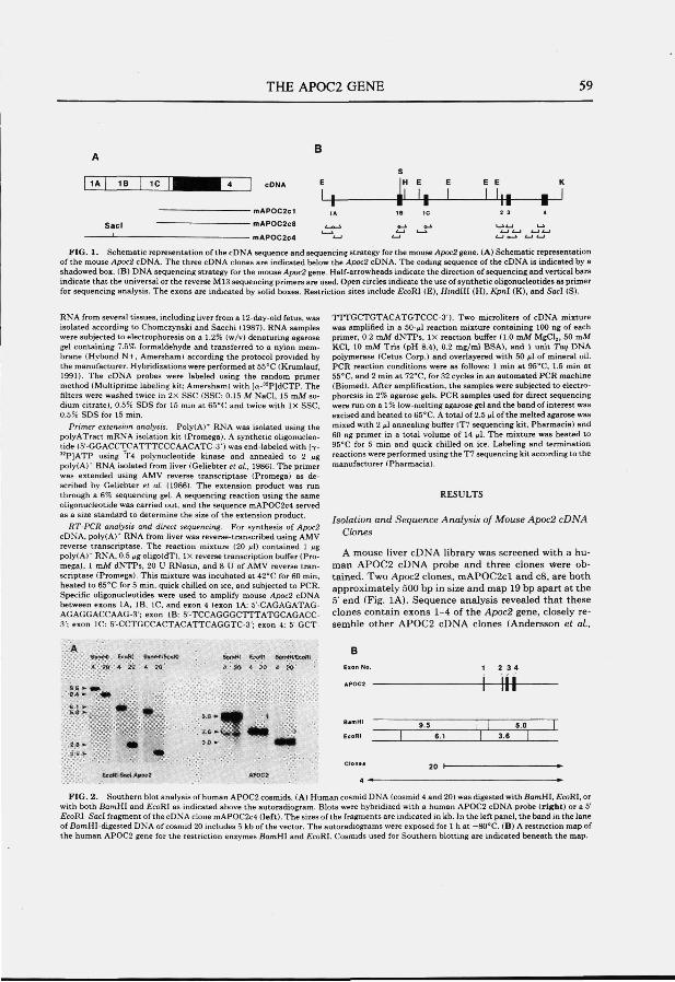
THE APOC2 GENE 59
- mAP0C2c1
- mAPOCZcS
- mAPOC2c4
I !• I E E
J-k
FIG. 1. Schematic representation of the cDNA sequence and sequencing strategy for the mouse Apoc2 gene. (A) Schematic representation of the mouse Apoc2 cDNA. The three cDNA clones are indicated below the Apoc2 cDNA. The coding sequence of the cDNA is indicated by a shadowed box. (B) DNA sequencing strategy for the mouse Apoc2 gene. Half-arrowheads indicate the direction of sequencing and vertical bars indicate that the universal or the reverse Ml3 sequencing primers are used. Open circles indicate the use of synthetic oligonucleotides as primer for sequencing anaiysis. The exons are indicated by solid boxes. Restriction sites include EcoRI (E), Hindlll (H), Kpnl (K), and SacI (8).
RNA from several tissues, including liver from a 12-day-old fetus, was isolated according to Chomczynski and Sacchi (1987). RNA samples were subjected to electrophoresis on a 1.2% (w/v) denaturing agarose gel containing 7.5% formaldehyde and transferred to a nylon membrane (Hybond N+, Amersham) according the protocol provided by the manufacturer. Hybridizations were performed at 55°C (Krumlauf, 1991). The cDNA probes were labeled using the random primer method (Multiprime labeling kit; Amersham) with [a-"P]dCTP. The filters were washed twice in 2x SSC (SSC: 0.15 M NaCl, 15 mM sodium citrate), 0.5% SDS for 15 min at 65°C and twice with IX SSC, 0.5% SDS for 15 min.
Primer extension analysis. Poly(A)* RNA was isolated using the polyATract mRNA isolation kit (Promega). A synthetic oligonucleotide (5'-GGACCTCATTTCCCAACATC-3') was end-labeled with [7-^^PJATP using T4 polynucleotide kinase and annealed to 2 jig poly(A)'' RNA isolated from liver (Geliebter et ai, 1986). The primer was extended using AMV reverse transcriptase (Promega) as described by Geliebter et ai (1986). The extension product was run through a 6% sequencing gel. A sequencing reaction using the same oligonucleotide was carried out, and the sequence mAPOC2c4 served as a size standard to determine the size of the extension product.
RT-PCR analysis and direct sequencing. For synthesis of Apoc2 cDNA, poly(A)'' RNA from liver was reverse-transcribed using AMV reverse transcriptase. The reaction mixture (20 ßl) contained 1 Mg poly(A)* RNA, 0.5 ^g oligo(dT), IX reverse transcription buffer (Promega), 1 mM dNTPs, 20 U RNasin, and 8 U of AMV reverse transcriptase (Promega). This mixture was incubated at 42°C for 60 min, heated to eS^C for 5 min, quick chilled on ice, and subjected to PCR. Specific oligonucleotides were used to amplify mouse Apoc2 cDNA between exons IA, IB, IC, and exon 4 (exon lA: 5'-CAGAGATAG-AGAGGACCAAG-3'; exon IB; 5'-TCCAGG G CTTTATGC AG Ace s'; exon IC: S'-CCTGCCACTACATTCAGGTC-S'; exon 4: 5'-GCT-
TTTGCTGTACATGTCCC-3'). Two microliters of cDNA mixture was amplified in a 50-^1 reaction mixture containing 100 ng of each primer, 0.2 mM dNTPs, IX reaction buffer (1.0 mM MgClj, 50 mM KCl, 10 mM Tris (pH 8.4), 0.2 mg/ml BSA), and 1 unit Taq DNA polymerase (Cetus Corp.) and overlayered with 50 ^1 of mineral oil. PCR reaction conditions were as follows; 1 min at 95"'C, 1.5 min at 55°C. and 2 min at 72°C, for 32 cycles in an automated PCR machine (Biomed). After amplification, the samples were subjected to electrophoresis in 2% agarose gels. PCR samples used for direct sequencing were run on a 1% low-melting agarose geland the band of interest was excised and heated to 65°C. A total of 2.5 ul of the melted agarose was mixed with 2 fil annealing buffer (T7 sequencing kit, Pharmacia) and 60 ng primer in a total volume of 14 fA. The mixture was heated to 95''C for 5 min and quick chilled on ice. Labeling and termination reactions were performed using the T7 sequencing kit according to the manufacturer (Pharmacia).
Isolation and Sequence Analysis of Mouse Apoc2 cDNA Clones
A mouse liver cDNA library was screened with a human APOC2 cDNA probe and three clones were obtained. Two Apoc2 clones, mAP0C2cl and c8, are both approximately 500 bp in size and map 19 bp apart at the 5' end (Fig. lA). Sequence analysis revealed that these clones contain exons 1-4 of the Apoc2 gene, closely resemble other APOC2 cDNA clones (Andersson et a i ,
BssrMt tcoRi fift-nHffccoRi B
-h-ttf
• « f 3 0 ••
BamHI
EeoUI I n
4 -* ^
FIG. 2. Southern blot analysis of human AP0C2 cosmids. (A) Human cosmid DNA (cosmid 4 and 20) was digested with SomHI, £coRI, or with both BamHI and EcoKl as indicated above the autoradiogram. Blots were hybridized with a human AP0C2 cDNA probe (right) or a 5' £coRI-SacI fragment of the cDNA clone mAPOC2c4 (left). The sizes of the fragments are indicated in kb. In the left panel, the band in the lane of BomHl-digested DNA of cosmid 20 includes 5 kb of the vector. The autoradiograms were exposed for 1 h at -80°C. (B) A restriction map of the human APOC2 gene for the restriction enzymes BamHI and £coRI. Cosmids used for Southern blotting are indicated beneath the map.
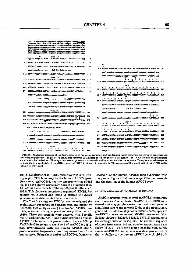
CHAPTER 4 60
173
223
273
318
qaattccaqqccaacctqqaetacttQaaaceetatctcaaaatataa^^a gaaggaacccctcacatBcaccaccaatctctcaagtatsgattacggac tcaa 11ctcaaaggaagaa actgtatccaggaaa actctgggggggtgtg tggagtcagtgttggggettcgtgtcttgtacaaggccagggcggcacac tgtttaggaaaggaggcaggcs agggcagaca tggcctctgactgtcact gtcactggccccatcgctttgaagtcctgccaaggagtgtggggaggtca caaggggagggctctttccagtgctgcetgtcccttccccggtgaaagag
V axon lA 1 tggagtacagCAGGAGGGACAGAGATACAGAGGACCAAGTCCCCAGAAAT
91 TGCTGTTCTTGGTCGCTTTGTAGgtaaggagttggggggetgctggaggt
ggggttctctgag 2.5 kb intron
114 tagtatctggtttgtcaectgagatgtcccctttatacagGGGCCCTGGA
axon 18
124 GCTGTCCAGGGCTTTATGCA6ACCTACTATCAAGATCACCTGAAAGACCT
174 GGGTCCCOGAACTCAGGCCTGGCTGCAGAGCTCCAGAGACCACCTCCTAA
224 ACAAGACCCACAGCCTGTGTCCCAGACTGCTCTGCAAAGACCGGACTCAG
274 GGTTAAAGTGCCACGTACCTCCCTCCCCTGGCACCCACAATAAAGgtgtt
tgttcttgatctgtgtcaccggacgcatagcttc
1.5 kb intron gaattcttgatcctcctg
cctcagccttctgaaagagcttgaggttacagctctgcccagtttatggg
tgatttctcaaagtggggctcccctgtggtttgacctgacaggaaatgtc exon IC
319 actggccattacccttccagGCCTGCCACCCCACCCCCCTGTGGCTTTGT 348
349 TGGCCAGGACTTTGGCCTAGCACAGAGCAGCTGCCTGATATGGAAGCAGG 398 • T
399 GGGCCCTCCAGAGCXAZA&&GCCTGCCAACCCACTGGAGTACTGGACTGA 448
449 GCCAGGATAGTCCCTTCCTGCCACTACATTCAGgtaagaggtggaccagg 482
ggttggggcagaatataaggggggcaagg
3.5 kb intron gaattcgttaaccaggagag
acctcatctgaattaacaggtttcaaatcttggttgggcaggaataggga
cagaataggtcccacccacgcctggcgaacaggaacgcagttccagtgat
483 ccctctgtctaacacaaccctgtccttaatgctccagGTCCCAGACAGCA 495
-22 exon 2 H G S R F F L A L F L V I L H L G
496 TGGGGTCTCGGTTCTTCCTGCCTCTATTCCTGGTCATCCTGATGTTCCGA 545
546 AATGgtaagtgggtcgggtggcaagatggcgggaggagtgagtaaggaag 549
gggtggggtgaccctcaagcacctccctacattcttcaaccattccttcg
tcttcctcacctgcctcgactcacccaaagtccccttagcacctctaatc -4 -I 1 E V Q G V Q E D D S
550 taattatctgcttCCCtcagAGCTCCAGGGGAACCAGGAACATGACTCGG 579
580 GCAGCCTGGCTCTGCTGGGCACGGTCCAGGGCTCCCTCTTAAGTTACTGG 629
K D L Y Q T Y P I 630 ACCTCTGCCAAGGAGGTTGCCAAAGACCTGTACCAGAAGACATACCCGAT 679
47 S H D E K L R
680 CAGCATGGATGAGAAACTCAGgtgtgtccctcccagtggggacagatccc 700
ggcatatgcacaccctcgaccc aanqgtccaoaecctatcçptecteeet
eaaacceaaaaq 1.4 kb intron
QQQCtecaQacgceaqecctcctecctcaaacteaaaQCteaaectetan 48 D H Y
701 tgcttccttCcaacccctccctgtgctttcttctCtccagGGACATCTAC 710
711 AGCAAAAGCTCGGCGGCCATGAGCACTTACGCAGGCATTTTTACAGACCA 760 75
L L T L L R G E 761 CCTTCTTACTCTCCTGAGGGGAGAGTAGCTGCCAGCAAGCCCCAAGTTA6 810
861 AGACCAGGATGGTCCTACACCACCCTGTCCCAGCTCTCCCGCCTTCCTGC 910
911 TCCCAACTAGAGCCCAGAGTCTTTCAGAGAAAGJl^Xf^TGAGTCCAGGT 960
961 TCactggcgtcgtgtggcttcttttcagaggttcccaggncaagggcatg 962
caagctt
FIG. 3 . Nucleotide sequence of the Apoc2 gene. Exon nucleotide sequences and the flanking intron sequences are shown in uppercase and lowercase, respectively. The predicted amino acid sequence is indicated above the nucleotide sequence. The TATA box and polyadenylation signal are double underlined. The major liver transcription start site is indicated by an arrow above the sequence Triangles above the sequence indicate the first nucleotide of the cDNA clones mAPOC2c4, c8, and cl , respectively The sequence of the repeat unit of the VNTR within intrun '.i is underlined.
1991b; Myklebost et a i , 1984), and show within the coding region 71% homology to the human APOC2 gene. One clone, mAPOC2c4, had the unexpected size of 962 bp. We have shown previously, that the 5' portion (Fig. lA) of this clone maps 5'of the Apoc2 gene (Hoffer eta/., 1993). This clone was completely sequenced (EMBL Accession No. Z15090) and found to contain the Apoc2 gene and an additional 440 bp at the 5' end.
The 5' end of clone mAPOC2c4 was investigated for evolutionary conservation between man and mouse by Southern blot analysis using two human APOC2 cosmids, obtained during a previous study (Smit et a i . 1988). These two cosmids were digested with fiamHI, EcoRI, and BamHI/EcoRl and hybridized with a human AP0C2 probe or with a probe derived from a 200-bp EcoRI-SacI fragment of the 5' end of mAPOC2c4 (Fig. 2A). Hybridization with the human AP0C2 cDNA probe revealed fragments containing exons 1-4 of the human gene. Using the 5' end of mAPOC2c4, fragments
located 5' of the human APOC2 gene hybridized with this probe. Figure 2B shows a map of the two cosmids and the position of the human APOC2 gene.
Genomic Structure of the Mouse Apoc2 Gene
EcoRI fragments from cosmid mAPOE27 containing the Apoe-cl~c2 gene cluster (Hoffer et a i , 1993) were cloned and mapped for several restriction enzymes. A significant part of the genomic DNA of the mouse Apoc2 gene and the additional genomic regions homologous to mAPOC2c4 were sequenced (EMBL Accession Nos. Z22213, Z22214, Z22215, Z22216, Z22217) according to the strategy outlined in Fig. IB . The genomic sequence o{Apoc2 from exons 2-4 and a region around exon 1 are shown (Fig. 3). This gene region encodes both cDNA clones mAPOC2cl and c8 and reveals a gene structure that is similar to the human AP0C2 gene. A 195-bp 5'
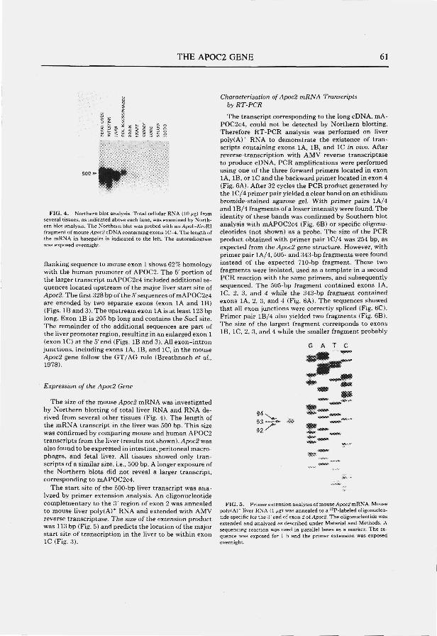
THE AP0C2 GENE 61
r FIG. 4. Northern blot analysis. Total cellular RNA (10 ^g) from
several tissues, as indicated above each lane, was examined by Northern blot analysis. The Northern blot was probed with an Apal-EcoRl frajiment of mouse Apoc2cDN A containing exons lC-4.The lengthof the mRNA in basepairs is indicated to the left. The autoradiogram was exposed overnight.
flanking sequence to mouse exon 1 shows 62% homology with the human promoter of AP0C2. The 5' portion of the larger transcript mAPOC2c4 included additional sequences located upstream of the major liver start site of Apoc2. The first 328 bp of the 5' sequences of mAPOC2c4 are encoded by two separate exons (exon lA and IB) (Figs. IB and 3). The upstream exon lA is at least 123 bp long. Exon IB is 205 bp long and contains the S a d site. The remainder of the additional sequences are part of the liver promoter region, resulting in an enlarged exon 1 (exon IC) at the 5' end (Figs. IB and 3). All exon-intron junctions, including exons lA, IB, and IC, in the mouse Apoc2 gene follow the GT/AG rule (Breathnach et a i , 1978).
Expression of the Apoc2 Gene
The size of the mouse Apoc2 mRNA was investigated by Northern blotting of total liver RNA and RNA derived from several other tissues (Fig. 4). The length of the mRNA transcript in the liver was 500 bp. This size was confirmed by comparing mouse and human APOC2 transcripts from the liver ( results not shown ). Apoc2 was also found to be expressed in intestine, peritoneal macrophages, and fetal Uver. AU tissues showed only transcripts of a similar size, i.e., 500 bp. A longer exposure of the Northern blots did not reveal a larger transcript, corresponding to mAPOC2c4.
The start site of the 500-bp liver transcript was analyzed by primer extension analysis. An oligonucleotide complementary to the 3' region of exon 2 was annealed to mouse liver poly(A)^ RNA and extended with AMV reverse transcriptase. The size of the extension product was 113 bp (Fig. 5) and predicts the location of the major start site of transcription in the liver to be within exon IC (Fig. 3).
Characterization of Apoc2 mRNA Transcripts by RT-PCR
The transcript corresponding to the long cDNA, mA-POC2c4, could not be detected by Northern blotting. Therefore RT-PCR analysis was performed on liver poly(A)^ RNA to demonstrate the existence of transcripts containing exons lA, IB, and IC in vivo. After reverse-transcription with AMV reverse transcriptase to produce cDNA, PCR amplifications were performed using one of the three forward primers located in exon IA, IB, or IC and the backward primer located in exon 4 (Fig. 6A). After 32 cycles the PCR product generated by the lC/4 primer pair yielded a clear band on an ethidium bromide-stained agarose gel. With primer pairs lA/4 and l B / 4 fragments of a lesser intensity were found. The identity of these bands was confirmed by Southern blot analysis with mAPOC2c4 (Fig. 6B) or specific oligonucleotides (not shown) as a probe. The size of the PCR product obtained with primer pair lC/4 was 254 bp, as expected from the Apoc2 gene structure. However, with primer pair lA/4, 505- and 343-bp fragments were found instead of the expected 710-bp fragment. These two fragments were isolated, used as a template in a second PCR reaction with the same primers, and subsequently sequenced. The 505-bp fragment contained exons IA, IC, 2, 3, and 4 while the 343-bp fragment contained exons IA, 2, 3, and 4 (Fig. 6A). The sequences showed that all exon junctions were correctly spliced (Fig. 6C). Primer pair lB/4 also yielded two fragments (Fig. 6B). The size of the largest fragment corresponds to exons IB, IC, 2, 3, and 4 while the smaller fragment probably
G Â T C
FIG. 5. Primer extension analysis of mouse Apoc2 mRNA, Mouse poly(A)" liver RNA (1 ßg) was annealed to a ^^P-labeled oligonucleotide specific for the 3'end of exon 2 oiApoc2. The oligonucleotide was extended and analyzed as described under Material and Methods. A sequencing reaction was used in parallel lanes as a marker. The sequence was exposed for 1 h and the primer extension was exposed overnight.
CHAPTER 4 62
Full cDNA
Primsrs
RT-PCR analysl«
| 1 A | IB
QZ 1 1B
c
| , C | 2 |
1 1«= 1 = 1
|,.|.| 1B 2
Ihl
3
3
3
3
3
3
1 * 1
4 1
^ 1
' 1
505
343*
590
428
254
S05 343
B30 428 'A
6 A T C
E S ™, «a«
iSLj
2 « b
«a «s»
w *= î
!3
c o c ^ 1 r^
<3 î
' O
T ^ T >
FIG. 6, RT-PCR analysis and sequencing of rare Apoc2 transcripts. (A) A schematic representation of the Apoc2 cDNA, the position of the primers used for the PCR analysis, and the alternative splicing products found by RT-PCR. Primers are indicated below the cDNA as arrows. The PCR products found with the RT-PCR are indicated below the cDNA. The size of the fragments is given to the right of each product. The fragment sizes indicated by an asterisk are confirmed by sequencing, (B) RT-PCR analysis with primer pairs as indicated above each lane. After PCR analysis and electrophoresis on an agarose gel. Southern blotting was performed with an Apoc2 cDNA probe. The length of the fragments is indicated to the left of each lane. (C) Sequence of the fragments found with primer pair lA/4. The alternative splicing boundaries are indicated to the right of each sequence. The left panel shows the 505-bp fragment and the right panel shows the 343-bp fragment.
lacks exon IC (Fig. 6A). RNA from the other tissues expressing the 500-bp transcript did not show products with the lA/4 and lB/4 primer pairs, except for fetal liver, which gave results identical to those of liver.
DISCUSSION
Apoc2 Gene Structure
In the mouse, the Apoc2 gene is contained within the Apoe-cl-c2 gene cluster spanning 30 kb (Hoff"er et a i , 1993). In the present study, we have isolated and characterized cDNA and genomic clones of the mouse Apoc2 gene. The mouse cDNA encodes a protein of 97 amino acids residues including a signal peptide of 22 amino acid residues. In contrast to the human apoC2 protein (Jackson et a i , 1977), the deduced amino acid sequence of mouse apoC2 is, like in the rat (Andersson et a i , 1991b), one residue shorter at the carboxyl terminus and lacks the conserved site for proteolytic cleavage of the propro-tein. Interestingly, one of our cDNA clones, mAPOC2c4, contains a 962-bp insert due to an extended 5' region
compared to the other two cDNAs. However, the mA-POC2c4 clone lacks additional open reading frames. This result indicates that this long transcript still encodes the same protein. The cDNA clones mAP0C2cl and c8 are transcribed from four exons. Exon 2 includes a part of the 5' untranslated region and the signal peptide. Exons 3 and 4 include 79 amino acids of the protein coding sequences, and exon 4 includes the 3' untranslated region, which is similar to the structure described for the human gene (Das et a i , 1987; Wei et ai , 1985). Primer extension analysis showed that the major liver start site of the 500-bp transcript is located in exon IC. This start site coincides with a region of homology to the human APOC2 gene of 62% over 195 bp (Das et a i . 1987; Wei et a i , 1985). A classical TATA box resides 22 bp upstream of this site. In addition, the 5'ends of the mA-POC2cl and c8 clones are located just 3 ' of the liver transcription start site.
Expression of mouse Apoc2 was found in the liver and intestine as demonstrated in other species (Myklebost et a i , 1984; Andersson et a i . 1991b). In addition, we show that Apoc2 is also expressed in peritoneal macrophages
THE AP0C2 GENE 63
like the other two genes, the mouse Apoe and the human APOCl, of this gene cluster (Basu et a i , 1981; Lauer et al., 1988). High level expression of Apoc2 was also detected in fetal liver mRNA.
Additional Sequences at the 5 ' End
Sequence analysis of mAPOC2c4 and genomic clones shows the presence oftwo additional exons 5' to the major liver start site. The exon-intron splice junctions of both exons follow the GT/AG rule (Breathnach et a i , 1978). Previously, a similar 5' extended region was found in the cynomolgus monkey (Whitted et al., 1989). Comparison of these 5' additional sequences between monkey and mouse showed only 3L 71% homology for exon IA. Southern blot analysis confirmed evolutionary conservation of the region 5' of the major liver start site between man and mouse, indicating that these sequences may be of functional importance. However, the expression level of the large transcript in the tissues used for Northern blotting was below the detection level in all tissues investigated. Using the more sensitive RT-PCR analysis, transcripts were detected originating from a region 5' of the major liver start site. Although the full-length transcript corresponding to mAPOC2c4 could not be demonstrated, other transcripts were found containing exon IA but lacking exon IB or exons IB and IC, indicatmg alternative splicing.
One explanation for the appearance of these larger transcripts could be the presence of upstream promoter sequences. The 5' start site could be determined by a TATA box sequence located 315 bp 5' of exon IA. A similar situation was found for another protein produced mainly in the liver, the human al-anti trypsin gene (Per-lino et a i , 1987). Two different promoters, located 2 kb from each other, are active in macrophages or hepatocytes, resulting in two different transcripts. Since the liver contains Kupffer cells, endothelial cells, and fat cells, in addition to hepatocytes, we cannot exclude the fact that the 5' transcripts originate from one of the latter less-abundant cell types. Also, the longer transcript may be present a t a higher level during fetal gene expression, however, at an earlier stage than the fetal livers we have examined.
Another explanation for the low level transcripts can be offered by a recent finding of the rat ECL gene. The ECL gene may represent an additional gene within the APOE-C1-C2 gene cluster of the rat (Shen and Howlett, 1992). Comparison of the sequences of exons 1A and IB of the Apoc2 gene with the sequence of the rat ECL gene showed homologies of 87 and 95%, respectively. The orientation of the open reading frame of ECL implies that the transcription would be in the opposite direction with respect to Apoc2. Transcription of this gene may cause an "open chromatin" structure, clearing access of transcription factors to a cryptic Apoc2 promoter region.
The mouse Apoc2 gene has been characterized at the cDNA and genomic levels. Here we show that the major
500-bp Apoc2 transcript in the liver is encoded by four exons. A rare transcript with additional sequences at the 5' end has also been detected tha t is conserved among several species. The isolation of a larger cDNA clone and the detection of alternative splicing products of a larger transcript by RT-PCR indicate that transcription originates from a region 5' of the major liver start site in the mouse Apoc2 gene. Since all PCR products and the cDNA clone containing additional 5' sequences were spliced properly, it is unlikely that they result from artifacts. Further studies are needed to unravel the origin and possible biological function of the apparently untranslated 5' end of these larger Apoc2 transcripts.
ACKNOWLEDGMENTS
The authors acknowledge Dr. K. Willems van Dijk for his helpful discussions and Dr. M Kielman for providing fetal liver mRNA. This work was supported by HGO-TNO (900-5,39-052) and the Royal Netherlands Academy of Arts and Science (M.H.H.).
REFERENCES
Andersson, Y., Thelander. L , and Bengtsson-Olivecrona. G. (1991a). Demonstration of apolipoprotein CII in gumea pigs J. Biol Chem 2 6 6 : 4074-4080.
Andersson, Y., Thelander, L., and Bengtsson-Olivecrona, G. (1991b). Rat apolipoprotein C-II lacks the conserved site lor proteolytic cleavage ot the pro-form. J Lipid Res 32 : 180.5-1S09.
Basu. S K , Brown, M. S., Ho, Y. K., Havel, R. .J , and Goldstein. J. L. {19S1 ). Mouse macrophages synthesize and secrete a protein resembling apolipoprotein E Proc NatL Acad. Sci USA 78 : 754.5-7549.
Bier, D M.. and Havel, R. J. ( 1970). Activation oi lipoprotein lipase by lipoprotein tractions of human serum. J. Lipid Res 1 1 : 565-570.
Breathnach, R.. Benoist, C, O'Hare, K.. Gannon, F., and Chambon. P (1978). Ovalbumin gene: Evidence ior a leader sequence in mRNA and DNA sequences at the exon-intron boundaries. Proc. Natl Acad Sci. USA 7 5 : 485.1-4857
Breckenridge, W. C Little, .J. A., Steiner, G., Chow. A., and Poapst. M. ( 1978) Hypertriglyceridemia associated with dehciency of apolipoprotein C-II. N Engl J Med 2 9 8 : 1265-1273.
Chomczynski, P., and Sacchi. N. (19871. Single step method oi RNA isolation by acid guanidinium thiocyanate phenol chloroform extraction. Anal Biochem. 162: 156-1.59.
Das, H. K., .Jackson, C. L., Miller. D. A., Leif, T., and Breslow. J. L. ( 1987) The human apolipoprotein C-II gene sequence contains a novel chromosome 19-specific minisatellite in its third intron. J Biol. Chem 2 6 2 : 4787-4793.
Datta, S.. Li, W-H.. Ghosh, I., Luo, C-C, and Chan, L. (1987). Structure and expression of dog apolipoprotein C-II and C-III mRNAs. J. Biol. Chem 2 6 2 : 10.588-10593.
Fojo, S. S., Team. L., Fairwell. T., Ronan. R., Bishop. C , Meng, M. S , Hoeg, J. M., Sprecher. D. L., and Brewer, H. B., .Jr. (1986). Human preproapolipoprotein C-II J. BioL Chem. 2 6 1 : 9591-9594.
Geliebter, J., Zeif, R. A., Melvold, R. W., and Nathenson. S. G. (1986). Mitotic recombination in germ cells generated two major histocompatibility complex mutant genes shown to be identical by RNA sequencing analysis: K*™" and K'™'. Proc NatL Acad Sci. USA 8 3 : 3371-3375.
Holfer, M. J V.. Hoiker, M. H . van Eck, M. M . Havekea. L M., and Frants, R. R. ( 1993) Evolutionary conservation of the mouse apohpoprotem e-cl-c:i gene cluster: Structure and genetic variability in inbred mice. Genomics 15: 62-67.
.Jackson, R .J, Baker, H. N., Gilliam, E. B., and Gotto, A. M., Jr. (1977). Primary structure of very low density apolipoprotein C-II of human plasma Proc. Natl Acad Sci. USA 74: 1942-1944.
Krumlauf, R. (1991). Northern blot analysis of gene expression. In
CHAPTER 4 64
"Gene Transfer and Expression Protocols" (E. J. Murray, Ed.), pp. 307-360. Humana Press, Clifton, NJ.
LaRosa, J. C , Levy, R. L, Herbert, P., Lux, S. E., and Frederickson, D. S. (1970). A specific apoprotein activator for lipoprotein lipase. Biochem. Bu^hys. Res. Commun. 4 1 : 57-62.
Lauer, S. J., Walker, D., Elshourbagy, N. A.. Reardon, C. A., Levy-Wilson, B., and Taylor, J. M. (1988). Two copies of the human apolipoprotem C-I gene are linked closely to the apolipoprotein E gene. J. BioL Chem. 2 6 3 : 7277-7286.
Marsh, J. B. (1976). Apoprotems of the lipoproteins in a nonreciicu-lating perfusate of rat liver. J. Lipid Rea 17: 86-90.
Myklebost. O., Williamson, B., Markham, A. F., Myklebost, S. R., Rogers, J., Woods, D. £., and Humphries, S. E. (1984). The isolation and characterization of cDNA clones tor human f^wllpoprotein CII. J. Biol Chem 2 6 9 : 4401-4404.
Perlino. E., Cortese, R., and Ciliberto, G. (1987). The human a,-anti-
tiypsin gene is transcribed from two diflferent promoters in macrophages and hepatocytes. EMBO J 6 : 2767-2771.
Shen, P., and Howlett, G. J. (1992). Two coding regions closely linked to the rat apolipoprotein E gene: Nucleotide sequences of rat apolipoprotein C-1 and ECL cDNA. Arch. Biochem. Biophys. 2 9 7 : 345-353.
Smit, M., van der Kooy-Meys, E., Frants, R. R., Havekes, L. M., and Klasen, E. C. (1988). Apolipoprotem gene cluster on chromosome 19. Hum. Genet 78 : 90-93,
Wei, C-F., Tsao, Y-K., Robberson, D. L., Gotto, A. M., Jr., Brown. K.. and Chan, L. (1985). The structure of the human apolipoprotein C-II gene. J. Biol. Chem 2 6 0 : 15211-15221.
Whitted, B. E., Castle, C. K., Polîtes, H. G., Melchior, G. W., and Marotti, K. R. (1989). Purification, cloning and nucleotide sequence determination of cynomolgus monkey apolipoprotein C-II: Comparison to the human sequence. MoL CelL Biochem. 9 0 : 69-79.
CHAPTER 5
THE APOLIPOPROTEIN C2 LINKED GENE {Act) GENE: DETECTION OF A NOVEL GENE WITHIN THE
APOLIPOPROTEIN E-C1-C2 GENE CLUSTER IN THE MOUSE.
M.J.V. Hoffer, M.M. van Eck, L.M. Havekes, M.H. Hoflcer, and R.R. Frants.
Submitted
CHAPTER 5 66
THE APOLIPOPROTEIN C2 LINKED (Act) GENE: A NOVEL GENE
WITHIN THE MOUSE APOLIPOPROTEIN E-C1-C2 GENE CLUSTER
Mariette J.V. Hoffer, Marga M. van Eck, Louis M. Havekes*, Rune R. Frants and Marten H. Hofker
MGC-Department of Human Genetics, Leiden University, P.O.Box 9503, 2300 RA Leiden, The Netherlands.IVVO-TNO, Gaubius Laboratory, P.O.Box 430, 2300 AK Leiden, The Netherlands.
SUMMARY
The apolipoprotein E, Cl and C2 genes are contained within a gene cluster in man. Previously, we have shown that this gene cluster has been evolutionarily conserved in mouse. During the characterization of the mouse Apocl gene, evolutionarily conserved and transcribed sequences were found S' of the Apocl gene. In this study, we show that these 5' additional sequences represent a novel gene within the gene cluster, designated the apolipoprotein C2 linked gene {Acl). The Acl gene is located 2 kb S' to the Apocl gene and has the same transcriptional orientation as all the other genes within the Apoe-cl-cl gene cluster. We have sequenced the entire mouse Acl gene on cDNA and genomic level. The gene consists of 3 exons, spanning a region of approximately 3.6 kb. The Acl gene is highly expressed in the liver with a transcript of 473 bp in size and encodes a putative protein of 124 amino acid residues.
INTRODUCTION
Apolipoproteins are structural constituents of lipoprotein particles and exhibit specialized functions in lipoprotein metabolism, serving as enzyme cofactors and as ligands for receptor-mediated uptake of lipoprotein particles (Breslow, 1988). Seven of these well-characterized apolipoproteins (APO), APOAl, A2, A4, Cl, C2, C3, and E, are members of a multigene gene family because of structural similarities (for a review, see Li et al., 1988). All these genes consist of 4 exons with the exception of APO A4 which consists of only 3 exons. In addition, the coding regions are composed
THE ACL GENE 67
of tandem repeats of 11 codons, indicating that these genes have evolved through duplications of a primordial gene (Luo et al., 1986; Li et al., 1988).
The apolipoprotein genes have become dispersed in mammalian genomes. In man six of these genes are found in two gene clusters, the APOA1-C3-A4 gene cluster on chromosome 11 (Bruns et al., 1984; Karathanasis, 1985) and the APOE-C1-C2 gene cluster on chromosome 19 (Lusis et al., 1986). The APOA2 gene is found as single gene on chromosome 1 (Lackner et al., 1984). These loci are contained in syntenic linkage groups in the mouse, which implies that all these genes have followed the same evolutionary pathway in humans and rodents (Lusis and Sparkes, 1989).
Recently, we have characterized the Apoe-cl-cl gene cluster in the mouse (Hoffer et al., 1993a) and demonstrated a similar gene organization as in man with the exception that the mouse gene cluster does not contain a pseudo Apocl gene. Characterization of the genomic structure of the individual mouse genes showed that the mouse Apoe (Horiuchi et al., 1989) and Apocl (Hoffer et al., 1993c) genes are similar to the corresponding human genes. Also the structure of the mouse Apocl gene is comparable with its human counterpart. Interestingly, in addition to the normal sized cDNA clones we also found a relatively large Apocl cDNA clone containing additional 5' sequences (Hoffer et al., 1993b). However, the corresponding transcript could not be detected by Northern blot analysis suggesting that it represented a low abundant mRNA species. In the present study we show that these additional 5' sequences of the large Apocl cDNA clone are part of a novel gene within the Apoe-cl-cl gene cluster. This novel gene, designated the apolipoprotein C2 linked {Act) gene, has been characterized further and the tissue expression has been determined.
MATERIALS AND METHODS
Isolation and sequencing ofcDNA and genomic clones. An Eco91-Sac\ restriction fragment of the 5'-end of the long Apoc2 cDNA clone mAPOC2c4
was subcloned and used as a probe to screen a liver cDNA library (BALB/c) in Xgtll (Clontech). Inserts of positive clones were obtained by PCR using primers derived from the the nucleotide sequence around the cloning EcoRI site of lambda GTll (Hoffer et al., 1993b) and either sequenced directly (Circumvent Thermal Cycle dideoxy DNA sequencing kit, Biolabs) or subcloned into the pT7Blue (R) T-vector (Novagen), followed by sequencing using the T7 sequencing kit (Pharmacia). Individual clones were sequenced in both directions using universal primers or specific oligonucleotides. A 4.5 kb EcoKl fragment derived from the genomic cosmid clone, mAPOE27 (Hoffer et al., 1993a) was subcloned into pUC19 for sequencing. Sequencing
CHAPTER 5 68
reactions were performed using the T7 sequencing kit (Pharmacia) and specific sjrnthetic oligonucleotide primers (Fig. 2).
Northern blot analysis. Total cellular RNA from several mouse tissues, including liver from a 12-day-old fetus, was
isolated according to Chomczynski and Sacchi (1987). Peritoneal macrophages were isolated after induction by intraperitoneal injection of 0.5 ml sterile 0.8% glycogen. After one day a booster was administered and 3 hours later the cells were collected. RNA samples were subjected to electrophoresis on a 1.2% (w/v) denaturing agarose gel containing 7.5%
, formaldehyde and transferred to a nylon membrane (Hybond N**̂, Amersham) according to the . protocol provided by the manufacturer. Hybridizations were performed at 55 °C in a solution containing50% formamide (Krumlauf, 1991). The cDNA probes were labelled using the random primer method (Multiprime labelling kit, Amersham) with [a-'^P]-dCTP. The filters were washed twice in 2 x SSC (SSC: 0.15 M NaCl, 15 mM sodium citrate), 0.1% SDS for 15 min at 65°C and twice with 1 x SSC, 0.1% SDS for 15 min.
Computer analysis. Sequences from the Genbank or from the EMBL DNA data banks were compared using the
"fasta" algorithm of the Genetics Computer Group Sequence Analysis Software Package Version 7.2 (Madison, Wisconsin, USA).
RESULTS
Isolation ofcDNA clones.
During the characterization of the mouse Apocl gene, an unusually large cDNA
clone, mAPOC2c4, was isolated (Hoffer et al., 1993b). This clone contained additional
5 ' sequences compared with the most abundant, normal sized Apocl liver transcript.
These additional sequences are encoded by two exons located 5 ' of the major Apocl
liver start site and are evolutionary conserved (Hoffer et al., 1993b) indicating that
these sequences could be of fimctional importance. Further studies were performed to
unravel the origin of these additional 5 ' sequences.
Therefore, a liver cDNA library, primed with random primers and oligo(dT), was
screened with a 0.2 kb Eco^-Sacl fragment as probe, containing only a part of the
additional 5 ' sequences of the large Apocl cDNA clone mAPOC2c4. The screening
revealed one clone, mACLcl, with an insert of 431 bp (Fig. 1). Sequence analysis
revealed a polyA tail, indicating that this clone was complete at its 3 ' end. This clone
contains also sequences not corresponding with the 5 ' additional sequences of the large
Apocl cDNA clone. At the 5 ' end the sequence was incomplete compared with the
sequences of the EcoSl-Sacl fragment, used as a probe for the cDNA library
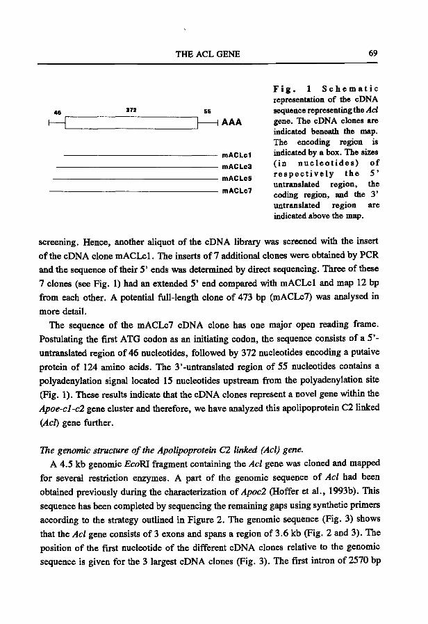
THE ACL GENE 69
46 372
H AAA
mACLcl
mACLc3
niACLcS
mACLc7
F i g . 1 S c h e m a t i c representation of the cDNA sequence representing the Acl gene. The cDNA clones are indicated beneath the map. The encoding region is indicated by a box. The sizes (in n u c l e o t i d e s ) of r e s p e c t i v e l y the 5 ' untranslated region, the coding region, and the 3' untranslated region are indicated above the map.
screening. Hence, another aliquot of the cDNA library was screened with the insert
of the cDNA clone mACLcl. The inserts of 7 additional clones were obtained by PCR
and the sequence of their 5 ' ends was determined by direct sequencing. Three of these
7 clones (see Fig. 1) had an extended 5 ' end compared with mACLcl and map 12 bp
from each other. A potential fiiU-length clone of 473 bp (mACLcT) was analysed in
more detail.
The sequence of the mACLc7 cDNA clone has one major open reading frame.
Postulating the first ATG codon as an initiating codon, the sequence consists of a 5 '-
untranslated region of 46 nucleotides, followed by 372 nucleotides encoding a putaive
protein of 124 amino acids. The 3'-untranslated region of 55 nucleotides contains a
polyadenylation signal located 15 nucleotides upstream from the polyadenylation site
(Fig. 1). These results indicate that the cDNA clones represent a novel gene within the
Apoe-cl-cl gene cluster and therefore, we have analyzed this apolipoprotein C2 linked
{Act) gene further.
The genomic structure of the Apolipoprotein Cl linked (Acl) gene.
A 4.5 kb genomic EcoRI firagment containing the Acl gene was cloned and mapped
for several restriction enzymes. A part of the genomic sequence of Acl had been
obtained previously during the characterization of Apocl (Hoffer et al., 1993b). This
sequence has been completed by sequencing the remaining gaps using synthetic primers
according to the strategy outlined in Figure 2. The genomic sequence (Fig. 3) shows
that the Acl gene consists of 3 exons and spans a region of 3.6 kb (Fig. 2 and 3). The
position of the first nucleotide of the different cDNA clones relative to the genomic
sequence is given for the 3 largest cDNA clones (Fig. 3). The first intron of 2570 bp
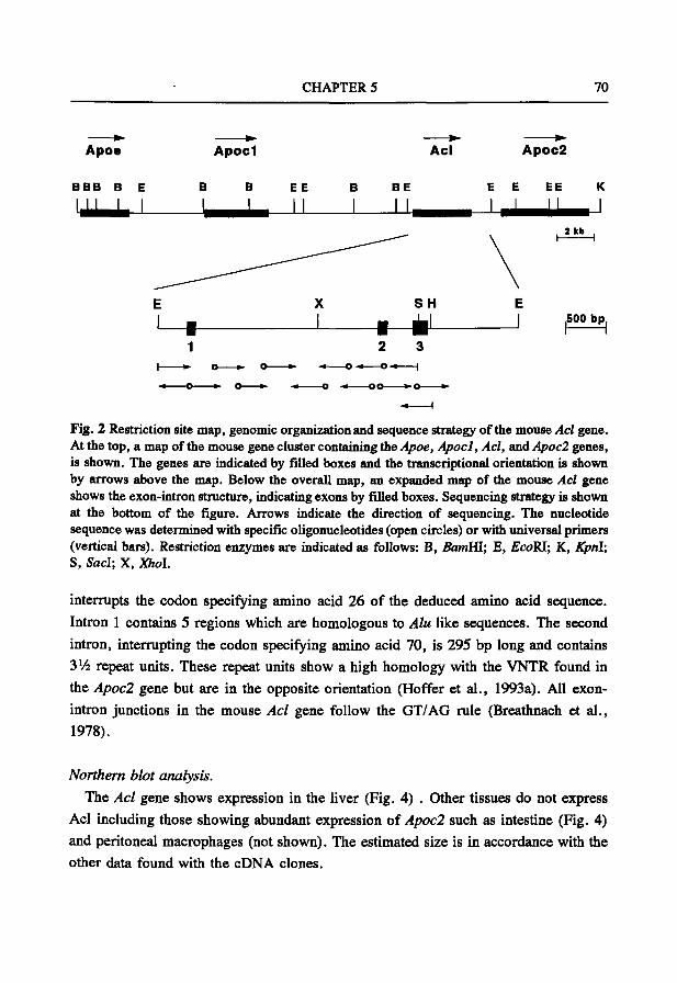
CHAPTER 5 70
Apoe A p o c l Ac l Apoc2
S H ,500 bp,
2 3 -« o • • — 0 - « 1
• • o » o » .« o
-t
Flg. 2 Restriction site map, genomic organization and sequence strategy of the mouse Acl gene. At the top, a map of the mouse gene cluster containing the Apoe, Apocl, Acl, and Apoc2 genes, is shown. The genes are indicated by filled boxes and the transcriptional orientation is shown by arrows above the map. Below the overall map, an expanded map of the mouse Acl gene shows the exon-intron structure, indicating exons by filled boxes. Sequencing strategy is shown at the bottom of the figure. Arrows indicate the direction of sequencing. The nucleotide sequence was determined with specific oligonucleotides (open circles) or with universal primers (vertical bars). Restriction enzymes are indicated as follows: B, BamHI; E, EcoRI; K, KprA; S, Sad; X, Xhol.
interrupts the codon specifying amino acid 26 of the deduced amino acid sequence.
Intron 1 contains 5 regions which are homologous to Alu like sequences. The second
intron, interrupting the codon specifying amino acid 70, is 295 bp long and contains
3Vi repeat units. These repeat units show a high homology with the VNTR found in
the Apocl gene but are in the opposite orientation (Hoffer et al., 1993a). All exon-
intron junctions in the mouse Acl gene follow the GT/AG rule (Breathnach et al.,
1978).
Northern blot analysis.
The Acl gene shows expression in the liver (Fig. 4) . Other tissues do not express
Acl including those showing abundant expression of Apocl such as intestine (Fig. 4)
and peritoneal macrophages (not shown). The estimated size is in accordance with the
other data found with the cDNA clones.
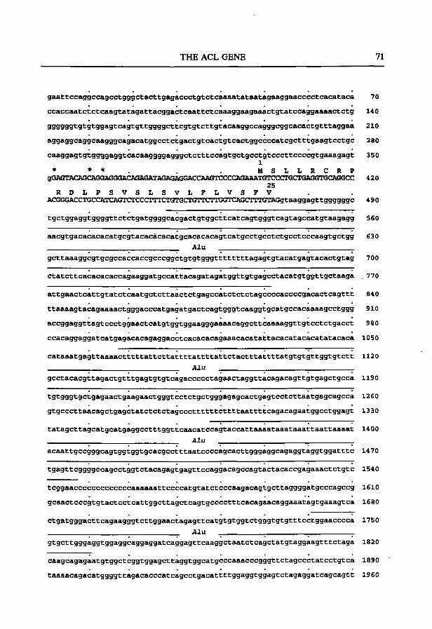
THE ACL GENE 71
gaattccaggccagcctgggctacttgagaccctgtctcaaéULtataatagaaggaacccctcacataca 70
ccacceiatctctcaagtatagattacggactcaattctcaêiaggaagaaactgtatccaggaaaactctg 140
999399tgtgtggagtcagtgttggggcttcgtgtcttgtacaaggccagggcggcacactgtttaggaa 210
aggaggcaggcaagggcagacatggcctctgactgtcactgtcactggccccatcgctttgaagtcctgc 280
ceiaggagtgtggggaggtcaceiaggggagggctctttccagtgctgcctgtcccttccccgtgaaagagt 350
1 * * * M S L L R C R P
gQAGTACAGCAGGAGGGACAGAGATAGAGAGGACCAAGTCCCCASAAATGTCCCTGCTGAGGTGCAGGCC 420 25
R D L P S V S L S V L F L V S F V ACGGGACCTGCCATCAGTCTCCCITTCTGTGCTGTTCrTGGTCAGCTTTGTAGgtaaggagttggggggc 490
tgctggaggtggggttctctgatggggcacgactgtggcttcatcagtgggtcagtagccatgtaagagg 560
aacgtgacacacacatgcgtacacâcacatgcacacacagtcatgcctgcctctgcctcccaagtgctgg 630
Alu
gcttaaaggcgtgcgccaccaccgcccggctgtgtgggttttttttagagtgtacatgagtacactgtag 700
ctatcttcacacacaccagaaggatgccattacagatagatggttgtgagcctacatgtggttgctaaga . 770
attgaactcattgtatctcaatgctcttaactctgagccatctctctagccccaccccgacactcagttt 840
ttaaaagtacagaaaactgggacccatgagatgactcagtgggtcaaggtgcatgccacaaaagcctggg 910
accggaggttagtccctggaactcatgtggtggaagggaaaacaggcttcaaaaggttgtcctctgacct 980
ccacaggaggatcatgagacacagaggacctcacacacagaaacacatattacacatacacatatacaca 1050
cataaatgagttaaaactttttattcttattttattttattctactttattttatgtgtgttggtgtctt 1120
Alu
gcctacacgttagactgtttgagtgtgtcagaccccctagaactaggttacagacagttgtgagctgcca 1190
tgtgggtgctgagaactgaagaactgggtcctctgctgggagagcactgagtcctcttaatgagcagcca 1260
gtgcccttaacagctgagctatctctctagcccttttttcttttaattttcagacagaatggcctggagt 1330
tatagcttagcatgcatgaggcctttggttcaacatccagtaccattaaaataaataaattaattaaaat 1400
Alu
acaattgccgggcagtggtggtgcacgcctttaatccccagcacttgggaggcagaggtaggtggatttc 1470
tgagttcggggccagcctggtctacagagtgagttccaggacagccagtactacaccgagaaactctgtc 1540
tcggaacccccccccccccaaaaaattccccatgtatctcccaagacagtgcttaggggatgcccagccg 1610
gcaactcccgtgtactcctcattggcttagctcagtgcccctttcacagaacaggaaatagtgaaagtca 1680
ctgatgggacttcagaagggtcttggaactagagttcatgtgtggtctgggtgtgtttcctggaacccca 1750
Alu
gtgcttgggaggtggaggcaggaggatcaggagttcaaggctaatctcagctatgtaggaagtttctaga 1820
caagcagagaatgtggctcggtggagcttaggtggcatgcccaaacccgggttctagccctatcctgtca 1890
taaaacagacatggggttagacacccatcagcctgacattttggaggtggagtctagaggatcagcagtt 1960
CHAPTER 5 72
taa tggcagaggtcatcct tgacactcgaggccatc tggaacta taagatggccatgtcatcaaaataac 2030
aacaaccutcaataataacaataacëiataggctagagggatgtctcagtggtagccccatagttaagggca 2100
tgcactgctct tacagaggaccagagt tcagt tcccagcctcacagctgcctgtaactccagctgcaggg 2170
ac tc tgacacct t tgc tc t tcacaggcacaggcagtacact tg t t ta tg tgcccgcagacaggaccatac 2240 Alu
acacaggataaaaaataaaatagtgctggagagatggctcagtggttaagagcactggctgctcttccag 2310
aggtcctgagttcaattcccagcaaccacatggtgcctcacaaccatctgtggtgggcctgtctaatgga 2380
tctatgccctcttctggtgtgtctgaagatagctacagtgtactcacatacatgaaataagtaaataagt 2450
aaataaattcaaaaaaatgtttaaaaaaaaaaaacaaacaacacaagcatgctcatgtatacttgtgtgc 2520
caggtagtatctgggcgtgagtctctacctagttgtgtgggtgctggagcaggaggaatgtgtgtgtgtg 2590
tgtgtgtgtgtgtgtgtgtgtgtgtgtgtgtcccagagggcagagggttaggtggttagaggagagaggc 2660
accaggtgggaagtggagttgcacactgagcaagaaaatcccagtagacagagaaacaagggagggacca 2730
gcagtgaggaagagacagatcaaatggtggagagacggttgtggtgtcagaggcctaataggccaggatg 2800
tgcaaagtagtgcaagagactgggtactttcttcttaactatgcccctgtgttgtataatacccttcaaa 2870
aaggacatcatggtgccctcttctacaggatgggaaactgaatctaggccaggtgaaatcaggtgtggcc 2940
tggaataaggggtgatgtgggtgtctaggtacaaggatgagcccacctgtccccaagagaggaatggggt 3010 26 A S H S T E S L S P T P
cctgagatctcaagcccctctgtggtttccccagCATCCATGTCTACAGAAAGCCTGAGCCCCACGCCTG 3080 G P E S S R W S L V R A R V L E M V E P L V T R GCCCTGAGAGCAGCCGCTGGAGCCTGGTAAGGGCCAGGGTGTTGGAGATGGTGGftGCCACTGGTGACCAG 3150
70 T R D R W Q W F W .
AACCAGGGACAGATGGCAGTGGTTCTGgtgagacaatcctagtgcttggctgggagggaacaccagagtt 3220
• « • - «
agagggggaaagagagcctgtggctagatcctgggtctgagggaggagggctggggcctggagccctgag 3290
»—* »-« tctgaaggaggatggctggggcctgaactcttgggtctgagggaggagagctggggcctggacccctggg 3360
cctaaaggaggagtacaaagactctgaatcttgagtctaagaagaaggtcataagtcgggattgaacccc 3430 71 G P G A V Q G F M
tgtagtatctggtttgtcacctgagatgtcccctttatacagGGGCCCTGGAGCrrGTCCAGGGCTTTATG 3500 Q T Y Y E D H L K D L G P R T Q A W L Q S S R CAGACCTACTATGAAGATCÄCCTGAAAGACCTGGGTCCCCGAACTCAGGCCTGGCTGCAGAGCTCCAGAG 3570
N 124 D H L L N K T H S L C P R L L C K D R T Q G ACCACCTCCTAAACAAGACCCACAGCCTGTGTCCCÄGACTGCTCTGCAÄAGACCGGACTCAGGGTTAAAG 3640
TGCCACGTACCTCCCTCCCCTGGCACCCACAATAAAGGTGTrTGTTCTTGatCtqtqtcaccqqacqcat 3710
agcttctcttgaggccagaggtcaaccttccgtcatatctaggagttaataagtggaaaagttgtggcct 3780
ggctggggaggacacaaggaaagctttaaaggaggggaccaaagtgataaacctccccctgccccacagg 3850
ttcctggcttgatgagaacaggagacttcca 3920
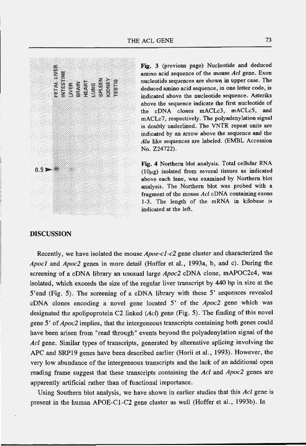
THE ACL GENE 73
CE Fig. 3 (previous page) Nucleotide and deduced > g amino acid sequence of the mouse Acl gene. Exon " ^ i f f z f e c s S w S nucleotide sequences are shown in upper case. The S t f ^ < < « z ä l « deduced amino acid sequence, in one letter code, is y , £ ^ a i j « ü : h ~ indicated above the nucleotide sequence. Asteriks
above the sequence indicate the first nucleotide of the cDNA clones mACLc3, mACLc5, and mACLc7, respectively. The polyadenylation signal is doubly underlined. The VNTR repeat units are indicated by an arrow above the sequence and the Alu like sequences are labeled. (EMBL Accession No. Z24722).
Fig. 4 Northern blot analysis. Total cellular RNA 0-5 *^ * *' (10/ig) isolated from several tissues as indicated
above each lane, was examined by Northern blot analysis. The Northern blot was probed with a fragment of the mouse Acl cDNA containing exons 1-3. The length of the mRNA in kilobase is indicated at the left.
DISCUSSION
Recently, we have isolated the mouse Apoe-cl-cl gene cluster and characterized the
Apocl and Apocl genes in more detail (Hoffer et al., 1993a, b, and c). During the
screening of a cDNA library an unusual large Apocl cDNA clone, mAPOC2c4, was
isolated, which exceeds the size of the regular liver transcript by 440 bp in size at the
5'end (Fig. 5). The screening of a cDNA library with these 5 ' sequences revealed
cDNA clones encoding a novel gene located 5 ' of the Apocl gene which was
designated the apolipoprotein C2 linked {Act) gene (Fig. 5). The finding of this novel
gene 5 ' o i Apocl implies, that the intergeneous transcripts containing both genes could
have been arisen from "read through" events beyond the polyadenylation signal of the
Acl gene. Similar types of transcripts, generated by alternative splicing involving the
APC and SRP19 genes have been described earlier (Horii et al., 1993). However, the
very low abundance of the intergeneous transcripts and the lack of an additional open
reading frame suggest that these transcripts containing the Acl and Apocl genes are
apparently artificial rather than of functional importance.
Using Southern blot analysis, we have shown in earlier studies that this Acl gene is
present in the human APOE-C 1-C2 gene cluster as well (Hoffer et al., 1993b). In
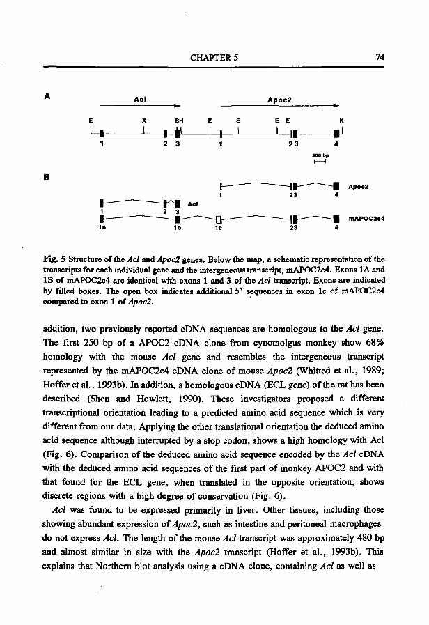
CHAPTER 5 74
Acl
SH
++
Apoc2
E E
2 3 23
500 bp
B Apoc2
mAPOC2c4
Fig. 5 Structure of the Acl and Apoc2 genes. Below the map, a schematic representation of the transcripts for each individual gene and the intergeneous transcript, mAPOC2c4. Exons 1A and IB of niAPOC2c4 are. identical with exons 1 and 3 of the Acl transcript. Exons are indicated by filled boxes. The open box indicates additional 5' sequences in exon Ic of mAPOC2c4 compared to exon 1 of Apoc2.
addition, two previously reported cDNA sequences are homologous to the Acl gene. The first 250 bp of a AP0C2 cDNA clone from cynomolgus monkey show 68% homology with the mouse Acl gene and resembles the intergeneous transcript represented by the mAPOC2c4 cDNA clone of mouse Apocl (Whitted et al., 1989; Hoffer et al., 1993b). In addition, a homologous cDNA (ECL gene) of the rat has been described (Shen and Howlett, 1990). These investigators proposed a different transcriptional orientation leading to a predicted amino acid sequence which is very different from our data. Applying the other translational orientation the deduced amino acid sequence although interrupted by a stop codon, shows a high homology with Acl (Fig. 6). Comparison of the deduced amino acid sequence encoded by the Acl cDNA with the deduced amino acid sequences of the first part of monkey AP0C2 and with that found for the ECL gene, when translated in the opposite orientation, shows discrete regions with a high degree of conservation (Fig. 6).
Acl was found to be expressed primarily in liver. Other tissues, including those showing abundant expression of Apocl, such as intestine and peritoneal macrophages do not express Acl. The length of the mouse Acl transcript was approximately 480 bp and almost similar in size with the Apocl transcript (Hoffer et al., 1993b). This explains that Northern blot analysis using a cDNA clone, containing Acl as well as
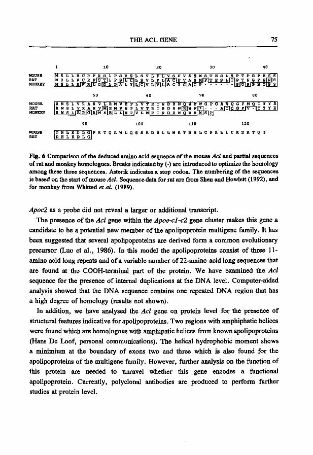
THE ACL GENE 75
HOUSE RAT MONKEY
MOUSE RAT MONKEY
50 SO 70
R W S L V R A R V L E M V E P I i V T R T R D R W O W F W G P G A V Q G F M R W S L V R A R V J M I E M V E P I. V T R T R D R W [ R ] W F F * ] - - - A r f l Q G FfV R W s L lÂlRfGlRfM K | E | L L | E P | V h NIR T R D R W Q W F W T S I P I
T Y Y E T Y Y E
90 120
MOUSE D H L K D L G P R T Q A W I . g S S R D H L I i M K T H S L C P R L L C K D R T Q G RAT D H L K D II G
Fig. 6 Comparison of the deduced amino acid sequence of the mouse Acl and partial sequences of rat and monkey homologues. Breaks indicated by (-) are introduced to optimize the homology among these three sequences. Asterik indicates a stop codon. The numbering of the sequences is based on the start of mouse Acl. Sequence data for rat are firom Shen and Howlett (1992), and for monkey from Whitted et al. (1989).
Apocl as a probe did not reveal a larger or additional transcript. The presence of the Acl gene within the Apoe-cl-cl gene cluster makes this gene a
candidate to be a potential new member of the apolipoprotein multigene family. It has been suggested that several apolipoproteins are derived form a common evolutionary precursor (Luo et al., 1986). In this model the apolipoproteins consist of three 11-amino acid long repeats and of a variable number of 22-amino-acid long sequences that are found at the COOH-terminal part of the protein. We have examined the Acl sequence for the presence of internal duplications at the DNA level. Computer-aided analysis showed that the DNA sequence contains one repeated DNA region that has a high degree of homology (results not shown).
In addition, we have analysed the Acl gene on protein level for the presence of structural features indicative for apolipoproteins. Two regions with amphiphatic helices were found which are homologous with amphipatic helices from known apolipoproteins (Hans De Loof, personal communications). The helical hydrophobic moment shows a minimium at the boundary of exons two and three which is also found for the apolipoproteins of the multigene family. However, further analysis on the fiinction of this protein are needed to unravel whether this gene encodes a fiinctional apolipoprotein. Currently, polyclonal antibodies are produced to perform further studies at protein level.
CHAPTER 5 76
ACKNOWLEDGEMENTS
The authors acknowledge Dr. H. De Loof, Dr. P. de Knijff and Dr. R. Fodde for their helpful discussions. This work was supported by HGO-TNO (900-539-052) and'the Royal Netherlands Academy of Arts and Science (M.H.H.).
REFERENCES
Breathnach R, Benoist C, O'Hare K, Gannon F, and Chambon P (1978). Ovalbumin gene: evidence for a leader sequence in mRNA and DNA sequences at the exon-intron boundaries. Ptoc Natl Acad Sci USA 75: 4853-4857.
Breslow JL (1988). Apolipoprotein genetic variation and human disease. Physiol Rev 68:85-132. Bruns GAP, KarathanasisSK, and Breslow JL (1984). Human apolipoprotein A-I-C-III complex is located
on chromosome 11. Arteriosclerosis 4: 97-102. Chomczynski P, and Sacchi N (1987). Single step method of RNA isolation by acid guanidinium
thiocyanate phenol chloroform extraction. Anal Biochem 162: 156-159. Das HK, Jackson CL, Miller DA, Leff T, and Breslow JL (1987). The human apolipoprotein C-II gene
sequence contains a novel chromosome 19-specific minisatellite in its third intron. J Biol Chem 262: 4787-4793.
Hoffer MJV, Hofker MH, van Eck MM, Havekes LM, and Frants RR (1992). Evolutionary conservation of the mouse apolipoprotein e-cl-c2 gene cluster: structure and genetic variability in inbred mice. Genomics IS: 62-67
Hoffer MJV, van Eck MM, Havekes LM, Hofker MH, and Frants RR (1993b). Structure and expression of the mouse apolipoprotein C2 gene. Genomics 17: 45-51.
Hoffer MJV, van Eck MM, Havekes LM, Hofker MH, and Frants RR (1993c). The mouse apolipoprotein Cl gene: structure and expression. Genomics: in press.
Horii A, Nakatsuru S, Ichii S, Nagase H, and Nakamura Y (1993). Multiple forms of the APC gene transcripts and their tissue-specific expression. Hum Mol Genet 2: 283-287.
Horiuchi K, Tajima S, Menju M, and Yamamoto A (1989). Structure and expression of mouse apolipoprotein E gene. J Biochem 106: 98-103.
Karathanasis SK (1985). Apolipoprotein multigene family: tandem organization of human apolipoprotein AI, c m and AIV genes. Proc Natl Acad Sci USA 82: 6374-6378.
Krumlauf R (1991). Northern blot analysis of gene expression. In: Gene transfer and expression protocols (Murray EJ ed.). Humana Press, Clifton, New Yersey p: 307-360.
Lackner KJ, Law SW, Brewer HB, Sakaguchi AY, and Naylor SL (1984). The human apolipoprotein A-II gene is located on chromosome 1. Biochem Biophys Res Comm 122: 877-883.
Li W-H, Tanimura M, Luo C-C, Datta S, and Chan L (1988). The apolipoprotein multigene family: biosynthesis, structure-function relationships, and evolution. J Lipid Res 29: 245-271.
LuoCC, Li WH, Moore MN, and Chan L (1986). Structure and evolution of the apolipoprotein multigene family. J Mol Biol 187: 325-340.
Lusis AJ, Heinzmann C, Sparks RS, Scott J, Knott TJ, Geller R, Sparkes MC, and Mohandas T (1985). Regional mapping of human chromosome 19: organization of genes for plasma lipid transport (APOC 1, -C2, and -E and LDLR) and the genes C3, PEPD and GPI Proc. Natl Acad Sci USA 83: 3929-3933.
Lusis AJ, and Sparks RC (1989). Chromosomal organization of genes involved in plasma lipoprotein metabolism: human and mouse "fat maps". In: Genetic factors in atherosclerosis: approachesand model systems (Lusis AJ, and Sparkes SR, Eds.). Monogr Hum Genet, Basel Karger, vol 12, pp. 79-94.
Shen P, and Howlett GJ (1992). Two coding regions closely linked to the rat apolipoprotein E gene: nucleotide sequences of rat apolipoprotein C-1 and ECL cDNA. Arch Biochem Biophys 297: 345-353.
Whitted BE, Castle CK, Polites HG, Melchior GW, and Marotti KR (1989). Purification, cloning and nucleotide sequence determination of cynomolgus monkey apolipoprotein C-II: comparison to the human sequence. Mol Cell Biochem 90: 69-79.
CHAPTER 6
THE MOUSE LOW DENSITY LIPOPROTEIN RECEPTOR GENE: cDNA SEQUENCE AND EXON-INTRON
STRUCTURE.
M.J.V. Hoffer, M.M. van Eck, F. Petrij, A. van der Zee, E. de Wit, D. Meijer, G.Grosveld, L.M. Havekes, M.H. Hofker, and
R.R. Frants.
Biochem. Biophys. Res. Commun. 1993, 191: 880-886
C H A P T E R 6 78
THE MOUSE LOW DENSITY LIPOFROTEIN RECEPTOR GENE: cDNA SEQUENCE AND EXON-INTRON STRUCTURE 1
Mariette J.V. Hoffer, Marga M. van Eck, Fred Petrij, André van der Zee, Elly de Wit*, Dies Meijer', Gerard Grosveld', Louis M. Havekes', Marten H. Hofker^
and Rune R. Frants
MGC-Department of Human Genetics, Leiden University, P.O.Box 9503, 2300 RA Leiden, The Netherlands
IVVO-TNO, Gaubius Laboratory, P.O.Box 430, 2300 AK Leiden, The Netherlands
*Department of Cell Biology, Erasmus University, P.O.Box 1738, 3000 DR Rotterdam, The Netherlands
Received January 19, 1993
SUMMARY: The low density lipoprotein (LDL) receptor plays a central role in the cholesterol metabolism. The cDNA sequence of the mouse low density lipoprotein receptor {Ldli) gene has been determined and shows 76% homology with the human gene. The exon-intron structure has been determined for the 129/J mouse strain. The gene is composed of 18 exons and spans a region of 28 kb. In addition, the promoter regions of the mouse and human genes are homologous. Northern blot analysis revealed an mRNA of approximately 5 kb. The cloning of the Ldlr gene will enhance the usefulness of the mouse for the study of cholesterol metabolism and, in particular, for carrying out gene targeting experiments. o 1993 Academic Press, Inc.
The low density lipoprotein (LDL) receptor mediates the clearance of lipoproteins containing
apolipoprotein E (apoE) and apoB-100 from plasma. This receptor-mediated pathway supplies
cholesterol to cells in the liver and other organs (1). Mutations in the low density lipoprotein
receptor (LDLR) gene are associated with familial hypercholesterolemia characterized by
elevated LDL cholesterol levels in the plasma, which lead to an increased risk for
atherosclerosis (2). Several animal models like rabbit and monkey (3, 4) have been used to
study the role of the LDL receptor in the cholesterol metabolism. Mice have proven to be
an important resource for the development of experimental animal models (5). In particular,"
^Sequence data from this article have been deposited with the EMBL/GenBank/DDBJ Data Libraries under Accession Nos. Z19520 and Z19S21.
^ To whom correspondence should be addressed.
Abbreviations; LDLR: Low Density Lipoprotein Receptor; LDL: Low Density Lipoprotein; APO: Apolipoprotein; RT-PCR: Reverse Transcriptase Polymerase Chain Reaction.
0006-291X/93 $4.00 Copyright © 1993 by Academic Press, Inc. All rights of reproduction in any form reserved. °oO
THE LDLR GENE 79
a powerful approach for the generation of mouse models takes advantage of homologous
recombination in pluripotent embryonic stem cells (6) enabling the generation of animals with
specific genetic changes.
The use of mice as an animal model for studies on the cholesterol metabolism requires
detailed knowledge of the underlying genes. The sequence of the LDLR gene has been
reported for several species: human, bovine, rabbit, rat, Xenopus and hamster (7-12). At the
genomic level, only the human and hamster LDLR genes have been characterized (12, 13).
In the current study we present the coding sequence of the mouse Ldlr gene, as well as the
exon-intron structure.
MATERIAL AND METHODS
Isolation and sequencing of cDNA clones: A cDNA library derived from liver mRNA of an adult male BALB/c mouse (Clonetech) was screened witti a probe derived from the rat LDLR cDNA clone pLDL-1 (10) containing a 2 kb EcoSX insert with exons 5-18, and a probe derived from the human LDLR cDNA clone pLDLR-3 (ATCC, # 57005) containing a 1 kb PsA insert with exons 2-8. Phage inserts were isolated by PCR amplification (14) using two PCR primers flanking the EcoSl cloning site of Xgtll (5'-GGTGGCGACGACTCCTGGAG-3'; 5'-CGTAATGGTAGCGACCGGCG-3'). After amplification, the PCR products were cloned into the M13mpl8/19 vector and sequenced using the T7 sequencing kit (Pharmacia). For RT-PCR analysis, first strand cDNA was synthesized using a primer in exon 12 (5'-CTCAAAGACGGCCAAGGAGA-3'), and AMV reverse transcriptase according to the protocol of the manufacturer (Promega), followed by PCR using primers in exon 1 (5'-TCCTGGCTGCTGCTGGAGTT-3') and in exon 5 (5'-CGAGCTCGTCGCTCATGTCC-3'). The primers in exons I and 12 were based on the rat and human LDLR sequence, respectively (7, 10). Determination of the exon-intron structure: A library was constructed by cloning partial Mbol digests of genomic DNA from the mouse inbred strain 129/J into the BamSl site of the phage EMBL3. This library was screened using a human LDLR cDNA probe (see above), and a mouse cDNA pnÂe containing exons 15-18 (i.e. the Sali-EcöBl fragment of mLDLRc8). Phage 1-1 was derived from the Swiss mouse strain and was provided by Dr. A.J. Lusis (University of California, Los Angeles, CA). Individual exons were mapped by restriction enzyme analysis using multiple digests and Southern blot analysis (IS) using probes for the individual exons. These probes were obtained from the human LDLR gene by PCR (16). Some intron sizes were established by PCR analysis using a primer in exon 18 ( 5 ' - T G C C A C A T C A T C C T C C A G G C - 3 ' ) , and in exon 16 ( 5 ' -ACATGGCTGGCAGAGGGAAT-3') or 17 (5'-AGGAACTGGCGGCTGAGGAA-3'). The primers in exons 17 and 18 were based on the rat cDNA sequence (10). Northern blotting and ligand blotting: Poly (A)'̂ RNA was isolated from liver using the polyATract mRNA isolation kit (Promega) and Northern blot analysis was performed according to standard protocols (17) using mLDLRc8 as a probe. Membrane associated proteins were isolated from a mouse liver, and used for ligand blotting as described by Lombardi et al. (18) using human '"I-LDL.
RESULTS AND DISCUSSION
Isolation of the cDNA encoding the mouse LDL receptor. A mouse liver cDNA library
was screened with a rat LDLR cDNA probe. Clone mLDLRc8 was obtained which is l.S
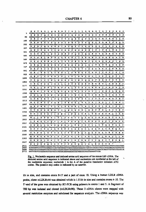
CHAPTER 6 80
H S T A O L M R R W V I A L L L A A A G V A V E D S G S 1 ATGAGCACCGCGGATCTGATGCGTCGCTGGGTCATCGCCCTGCTCCTTGCTGCTGCCGGAGTTGCAGTAGAAGACTCAGGCAGC
R N E F Q C R D G K C I A S K W V C D G S P E C P D G S 85 AGGAACGAGTTCCAGTGTAGAGACGGAAAATGCATCGCTAGCAAGTGGGTGTGCGATGGCAGCCCCGAGTGCCCGGATGGCTCC
D E S P K T C H S V T C g S N Q F S C G G R V S R C I P 169 GATGAGTCCCCAAAGACATGCATGTCTGTCACCTGTCAGTCCAATCAATTCAGCTGTGGAGGCCGTGTCAGCCGATCCATTCCT
D S W R C D G Q V D C E N D S D E Q G C P P K T C S Q D 2S3 GACTCCTGGAGATGTGATGGACAGGTAGACTGTGAAAATGACTCAGACGAACAAGGCTGTCCCCCCAAGACGTGCTCCCAGGAT
O F R C Q D G X C I S P Q F V C D G D R D C L D G S D E 337 GACTTCCGATGCCAGGATGGCAAGTGCATCTCCCCGCAGTTTGTGTGTGATGGAGACCGAGATTGCCTAGATGCCTCTGATGAG
A H C P A T T C C F A H F R C K S S I C I P S L W A C D 421 GCCCACTGCCCAGCCACCACTTGTGGCCCCGCCCACTTCCGCTGCAAATCATCCATATGCATCCCCAGTCTTTGGGCCTCCGAC
G D V D C V D G S H E W P Q N C Q A E D T A S X 6 V S S 505 GGGGATGTCGACTGTGTTGACGGCTCCCATGAGTGGCCACAGAACTGCCAGGCCGAAGACACGGCCTCCAAAGGCGTTAaCAGC
P C S S L E F H C G S S E C I H R S W V C D G E A D C K 589 CCCTCCTCCTCCCTGGAGTTCCACTGTGGTAGCAGTGAGTGTATCCATCGCAGCTGGGTCTGTGACGGCGAGGCAGACTGCAAG
D K S D E E H C A V A T C R P D E F Q C A D G S C I H G 673 GACAACTCAGATGAGGAGCACTGCGCGGTGGCCACCTGCCGACCTGATGAATTCCAGTGTGCAGATGGCTCCTCCATTCACGGT
S R Q C D R E H D C K D M S D E L G C V N V T Q C D G P 757 AGCCGCCAGTGTGACCGTGAACATGACTGCAAGGACATGAGCGACGAGCTCGGCTGCGTCAATGTGACACAGTGTGATGGCCCC
M K F K C H S G E C I S L D K V C D S A R D C Q D W S D 841 AACAACTTCAAGTGTCACAGTGGGGAGTGCATCAGCTTGGACAAGGTGTGCGACTCCGCCCGCGACTGCCAGGACTGGTCGGAT
E P t K E C K T N E C L D N N G G C S H I C X D L K t G 925 GAGCCCATCAAGGAGTGCAAGACCAACGAGTGTTTGGACAACAATGGTGGCTGTTCCCACATCTGCAAGGACCTCAAGATTGGC
S E C L C P S G F R L V D L H R C E D I D E C Q E P O T 1009 TCTGAGTGCCTGTGTCCCAGCGGCTTCCGGTTGGTGGACCTCCACAGGTGTGAAGATATTGACGAGTGTCAGGAGCCAGACACC
C S Q L C V N L E G S Ï K C E C Q A G F H M D P H T R V 1093 TGCAGCCAGCTCTGTGTGAACCTGGAAGGCAGCTACAAGTGTGAGTGCCAGGCCGGCTTCCACATGGACCCACACACCAGGGTC
C K A V G S I G Y L L F T N R H E V R K H T L D R S E Y 1177 TGCAAGGCTGTGGGCTCCATAGGCTATCTGCTCTTCACCAACCGCCACGAGGTCCGGAAGATGACCCTGGACCGCAGCGAGTAC
T S L L P N L K N V V A L D T E V T N M R I Ï W S D L S 1261 ACCAGTCTGCTCCCCAACCTGAAGAATGTGGTGGCTCTCGACACGGAGGTGACCAACAATAGAATCTACTGGTCCGACCTGTCC
Q K K I Y S A L H D Q A P N L S Y D T I I S E D L H A F 1345 CAAAAAAAGATCTACAGCGCCCTGATGGACCAGGCCCCTAACTTGTCCTACGACACCATCATCAGTGAGGACCTGCATGCCCCT
D G L A V D W I H R N I Y W T D S V P G S V S V A D T K 1429 GACGGGCTGGCGGTAGACTGGATCCACCGCAACATCTACTGGACAGATTCAGTCCCAGGCAGCGTATCTGTGGCTGACACCAAG
G V K R R T L F Q E A G S R P R A I V V D P V H O F M Y 1513 GGCGTAAAGAGGAGGACACTGTTCCAAGAGGCAGGGTCCAGACCCAGAGCCATCGTAGTGGACCCTGTGCATGGCTTCATGTAC
W T D W G T P A K I K K G G L N G V . D I H S L V T E N I 1597 TGGACAGATTGGGGAACACCCGCCAAGATCAAGAAAGCGGGTTTGAATGGTGTGGACATCCACTCACTGGTGACCGAAAACATC
g W P N G I T L D L S S G R L Y W V O S K L H S I S S I 1681 CAGTOOCCAAATGGCATCACACTAGATCTTTCCAGTGGCCGTCTCTATTGGOTTGATTCCAAACTCCACTCTATCTCCAGCATC
D V N C G N R K T I L E O E H R L A H P F S L A I Y E D 1765 GATCTCAATGGGGGCAATCGGAAAACCATTTTGGAGGATGAGAACCGGCTGGCCCACCCCTTCTCCTTGGCCATCTATGAGGAC
K V Y W T D V I N E A I F S A N R L T G S D V N L V A E 1849 AAAGTOTATTOGACAGATGTCATAAACCAAGCCATTTTCAGTGCCAATCGACTCACGGOTTCAGATGTGAATTTGOTGGCTGAA
N L L S P E D I V L F H K V T Q P R G V N W C E T T A L 1933 AACCTCTTGTCCCCGGAGGACATTGTCCTGTTCCACAAGGTCACACAGCCTAGAGGGGTGAACTGGTGTGAGACAACAGCCCTC
L P N G O C Q Y L C L P A P Q I G P H S P K F T C A C P 2017 CTCCCCAATGGTGGTTGCCAGTACCTGTGCCTGCCCGCCCCACAGATCGGTCCCCACTCGCCCAAATTCACCTGCGCCTGCCCT
D C H L L A K D M R S C L T E V D T V L T T Q G T S A V 2101 GATGGCATGCTGCTGGCCAAGGACATGCGGAGCTGCCTCACAGAAGTCGACACTGTACTGACCACCCAGGGGACATCCGCCGTC
R P V V T A S A T R P P K H S E D L S A P S T P R Q P V 2185 CGGCCTGTGGTCACCGCATCAGCTACCAGGCCACCGAAOCACAGTGAGGATCTCTCAGCTCCCAGTACTCCTAGGCAGCCTGTG
D T P G L S T V A S V T V S H Q V Q G D H A G R G N E E 2269 GACACCCCAGGGCTCAGCACAGTGGCGTCAGTGACAGTGTCCCACCAAGTCCAGGGTGACATGGCTGGCAGAGGGAATGAGGAG
O P H G M R F L S I P F P I A L V A L L V L G A V L L W 2353 CAGCCACATGCTATGAGGTTCCTGTCCATCTTCTTCCCTATTGCACTGGTTGCCCTCCTTGTCCTTGGGGCCGTCCTGCTGTGG
R M W R L K N I T I N S I N F D N P V Y Q K T T E D E L 2437 AGGAACTGGCGGCTGAAGAACATCACAATCAACAGCATAAACTTTGACAACCCAGTCTACCAGAAGACCACAGAGGACGAGCTC
H I C R S Q D G Y T Y P S R Q H V S L E D D V A * 2521 CACATTTGCCGAAGCCAGGATGGCTATACCTACCCCTCAAGACAGATGGTCAGCCTGGAGGACGATGTGGCATGAGCAGCCGGG 2605 AGAGCCGTCTCTTTCCGGGATCCATTGCCAAGCTTAGGCAGAAAAGACACTCTCTCCAGACCTCCCCATCCAGCACTGGTCCTG 2689 CCACCTCCCTGGGNTCTGTGTTGCTCAAAGCAAGATAGAGCAAAGCTGGGCTGGGGGGCCAAGCTCAGCTTCCTGTCTGCCCCA 2773
Fig. 1. Nucleotide sequence and deduced amino acid sequence of the mouse Ldlr cONA. The deduced amino acid sequence is indicated above and nucleotides are numbered at the left of the nucleotide sequence; nucleotide 1 is the A of the putative translation initiation ATG codon. The putative stop codon is indicated by an asterisk.
kb in size, and contains exons 8-17 and a part of exon 18. Using a human LDLR cDNA
probe, clone mLDLRc44 was obtained which is 1.0 kb in size and contains exons 4-10. The
S'-end of the gene was obtained by RT-PCR using primers in exons 1 and S. A fragment of
700 bp was isolated and cloned (mLDLRc90). These 3 cDNA clones were m^ped with
several restriction enzymes and subcloned for sequence analysis. The cDNA sequence was
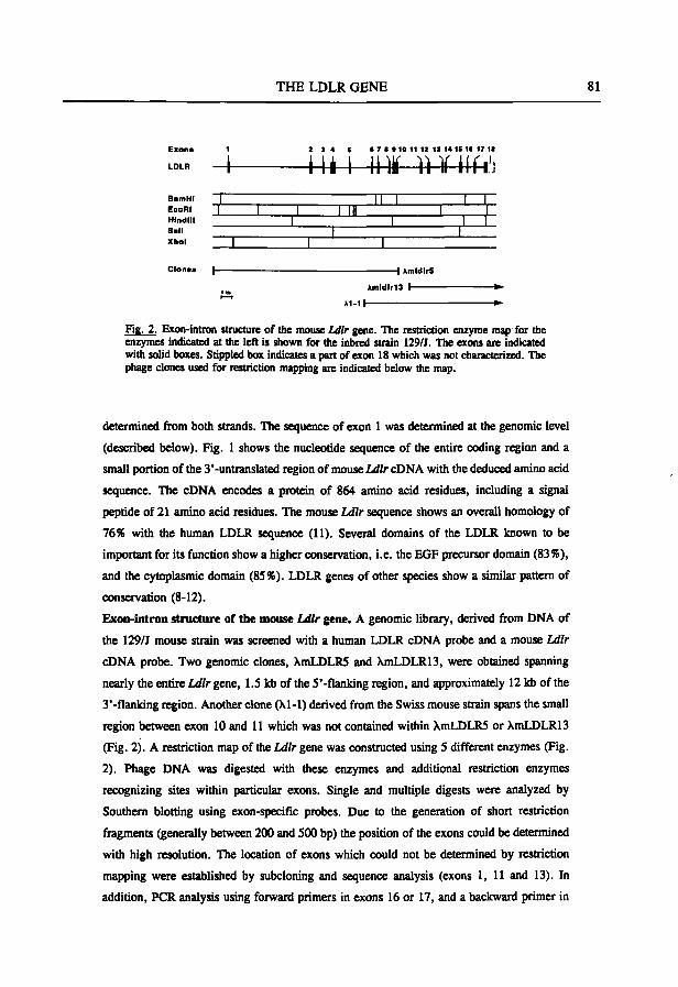
THE LDLR GENE 81
E>an> 1 2 3 4 S • 7 • • 10 11 12 11 t4 IS I I 17 11
LDLR
BamHr EcoRI Hindlll Sail Xhol
-A I4i-^ 4 * - l l ï i l f i ^ 1 II 1 I I 1 1 1 1 III 1 1
1 ' '" 1 I I 1 1
1 1 ' 1
Clones |—
Àmldlr13 h-
»1-1 I
Fig. 2. Exon-intron structure of the mouse Ldlr gene. The restriction enzyme map for the enzymes indicated at the left is shown for the inbred strain 129/J. The exons are indicated with solid boxes. Stippled box indicates a part of exon 18 which was not characterized. The phage clones used for restriction mapping are indicated below the map.
determined from both strands. The sequence of exon 1 was determined at the genomic level
(described below). Fig. 1 shows the nucleotide sequence of the entire coding region and a
small portion of the 3'-untranslated region of mouse Ldlr cDNA with the deduced amino acid
sequence. The cDNA encodes a protein of 864 amino acid residues, including a signal
peptide of 21 amino acid residues. The mouse Ldlr sequence shows an overall homology of
76% with the human LDLR sequence (11). Several domains of the LDLR known to be
important for its function show a higher conservation, i.e. the EGF precursor domain (83%),
and the cytoplasmic domain (85%). LDLR genes of other species show a similar pattern of
conservation (8-12).
Exon-intron structure of the mouse Ldlr gene. A genomic library, derived from DNA of
the 129/J mouse strain was screened with a human LDLR cDNA probe and a mouse Ldlr
cDNA probe. Two genomic clones, XmLDLRS and XmLDLR13, were obtained spanning
nearly the entire Ldlr gene, 1.5 kb of the S'-flanking region, and approximately 12 kb of the
3'-flanking region. Another clone (Xl-1) derived from the Swiss mouse strain spans the small
region between exon 10 and 11 which was not contained within XmLDLRS or XmLDLR13
(Fig. 2). A restriction map of the Ldlr gene was constructed using S different enzymes (Fig.
2). Phage DNA was digested with these enzymes and additional restriction enzymes
recognizing sites within particular exons. Single and multiple digests were analyzed by
Southern blotting using exon-specific probes. Due to the generation of short restriction
fragments (generally between 200 and 500 bp) the position of the exons could be determined
with high resolution. The location of exons which could not be determined by restriction
mapping were established by subcloning and sequence analysis (exons 1, 11 and 13). In
addition, PCR analysis using forward primers in exons 16 or 17, and a backward primer in
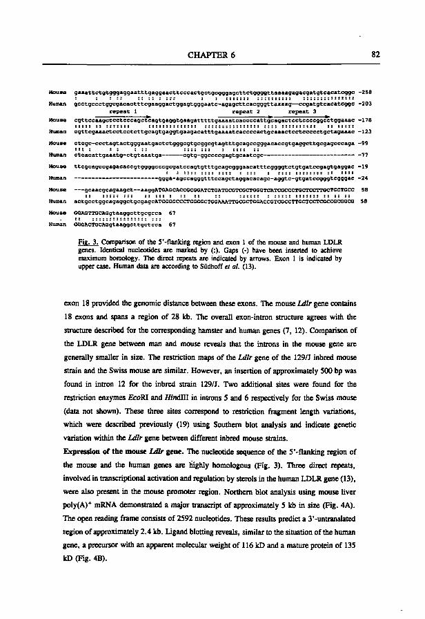
CHAPTER 6 82
Mouse gaaattctgtgggaggaatttgaggaacttcccactgctgcgggagcttctggggttaaaagagacgatgtcacatcggc -258
Human gcctgccctggcgacactttcgaaggactggagtgggaatc-agagcttcacgggttaaaag—ccgatgtcacatcggc -203
repeat 1 repeat 2 repeat 3
House cgttccaagctcctcccagctcagtgaggtgaagatttttgaaaatcaccccattgcagactcctccccggcctggaaac -178 : : : : : : : ; : : : t : : ; : ; : : ; : ; ; : ; : : ; : ; : ; ; ; ; : ; ; : : : : : : : : : ; ; : : : : : : : : : : : : : : : :
Human cgttcgaaactcctcctcttgcagtgaggtgaagacatttgaaaatcaccccactgcaaactcctccccctgctagaaac -123
House ctcgc-ccctagtactgggaatgactctgggcgtgcggcgtagtttgcagccgggacaccgtgaggcttgcgagcccaga -99 : : : : : ; : : : :::;;:: : : : : : : ;
Human ctcacattgaaatg-ctgtaaatga cgtg-ggccccgagtgcaatcgc -77
House ttcgcagccgagacaccgtggggcccgcgatccagtgtttgcagcgggaacatttcggggtctgtgatccgagtgaggac -19
Human ggga-agccagggtttccagctaggacacagc-aggtc-gtgatccgggtcgggac -24
House gcaacgcagaagct—aaggATGAQCACCGCGGATCTGATGCGTCGCTGGCTCATCGCCCTGCTCCTTGCTGCTGCC 58
Human actgCCtggcagaggctgcgagcATGGGGCCCTGGGGCTGGAAATTGCGCTGQACCGTCGCCTTGCTCCTCGCCGCGGCG 58
Mouse GGAGTTGCAGgtaaggcttgcgcca 67
Human GGGACTGCAGgtaaggcttgctcca 67
Fig. 3. Comparison of the S'-flanking region and exon 1 of the mouse and human LDLR genes. Identical nucleotides are marked by (:). Gaps (-) have been inserted to achieve maximum homology. The direct repeats are indicated by arrows. Exon 1 is indicated by upper case. Human data are according to Südhoff ei al. (13).
exon 18 provided the genomic distance between these exons. The mouse Ldlr gene contains
18 exons and spans a region of 28 kb. The overall exon-intron structure agrees with the
structure described for the corresponding hamster and human genes (7, 12). Comparison of
the LDLR gene between man and mouse reveals that the introns in the mouse gene are
generally smaller in size. The restriction maps of the Ldlr gene of the 129/J inbred mouse
strain and the Swiss mouse are similar. However, an insertion of approximately 500 bp was
found in intron 12 for the inbred strain 129/J. Two additional sites were found for the
restriction enzymes EcoRI and Mndlll in introns 5 and 6 respectively for tiie Swiss mouse
(data not shown). These three sites correspond to restriction ftagment lengtii variations,
which were described previously (19) using Southern blot analysis and indicate genetic
variation within die Ldlr gene between different inbred mouse strains.
Expression of the mouse Ldlr gene. The nucleotide sequence of the S'-flanking region of
the mouse and the human genes are highly homologous (Fig. 3). Three direct repeats,
involved in transcriptional activation and regulation by sterols in the human LDLR gene (13),
were also present in the mouse promoter region. Northern blot analysis using mouse liver
poly(A)* mRNA demonstrated a major transcript of approximately S kb in size (Fig. 4A).
The open reading frame consists of 2592 nucleotides. These results predict a 3'-untranslated
region of approximately 2.4 kb. Ligand blotting reveals, similar to the sitiiation of the human
gene, a precursor with an apparent molecular weight of 116 kD and a mature protein of 135
kD (Fig. 4B).
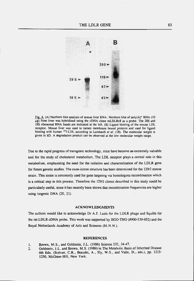
THE LDLR GENE 83
A B
28 s >̂
2 0 § i
116)
" " 't I t S »» 45»»
Fig. 4. (A) Northern blot analysis of mouse liver RNA. Northern blot of poly(A)* RNA (10 /lg) from Uver was hybridized using the cDNA clone mLDLRcS as a probe. The 28S and 18S ribosomal RNA bands are indicated at the left. (B) Ligand blotting of the mouse LDL receptor. Mouse liver was used to isolate membrane bound proteins and used for ligand blotting with human '"I-LDL according to Lombardi et al. (18). The molecular weight is given in kD. A degradation product can be observed at the low molecular weight range.
Due to the rapid progress of transgenic technology, mice have become an extremely valuable
tool for the study of cholesterol metabolism. The LDL receptor plays a central role in this
metabolism, emphasising the need for the isolation and characterization of the LDLR gene
for future genetic studies. The exon-intron structure has been determined for the 129/J mouse
strain. This strain is commonly used for gene targeting via homologous recombination which
is a critical step in this process. Therefore the 129/J clones described in this study could be
particularly useful, since it has recently been shown that recombination frequencies are higher
using isogenic DNA (20, 21).
ACKNOWLEDGMENTS
The authors would like to acknowledge Dr A.J. Lusis for the LDLR phage and Squibb for
the rat LDLR cDNA probe. This work was supported by HGO-TNO (#900-539-052) and the
Royal Netherlands Academy of Arts and Sciences (M.H.H.).
REFERENCES
1. Brown, M.S., and Goldstein, J.L. (1986) Science 232, 34-47. 2. Goldstein, J.L. and Brown, M.S. (1989) in The Metabolic Basis of Inherited Disease
6th Edn. (Scriver, C.R., Beaudet, A., Sly, W.S., and Valle, D., eds.), pp. 1215-1250, McGraw-Hill, New York.
CHAPTER 6 84
3. Watanabe, Y. (1980) Atherosclerosis 36, 261-268. 4. Scanu, A.M., Khalil, A., Neven, L., Tidore, M., Dawson, G., Pfaffinger, D.,
Jackson, E., Carey, K.D., McGill, H.C, and Fless, G.M. (1988) J. Upid. Res. 29, 1671-1681.
5. Hofmann, S.L., Russell, D.W., Brown, M.S., Goldstein, J.L., and Hammer, R.E. (1988) Science 239, 1277-1281.
6. Capecchi, M.R. (1989) Trends Genet. 5, 70-76. 7. Yamamoto, T., Davis, CG., Brown, M.S., Schneider, W.J., Casey, M.L.,
Goldstein, J.L., and Russell, D.W. (1984) Cell 39, 27-38. 8. Russell, D.W., Yamamoto, T., Schneider, W.J., Slaugtiier, C.J., Brown, M.S., and
Goldstein, J.L. (1983) Proc. Natt. Acad. Sci. USA 80, 7501-7505. 9. Yamamoto, T., Bishop, R.W., Brown, M.S., Goldstein, J.L., and Russell, D.W.
(1986) Science 232, 1231-1237.. 10. Lee, L.Y., Mohler, W.A., Schäfer, B.L., Freudenberger, J.S., Byrne-Connolly, N.,
Eager, K.B., Mosley, S.T., Leigthon, J.K., Thrift, R.N., Davis, R.A., and Tanaka, R.D. (1989) Nucleic Acids Res. 17, 1259-1260.
11. Metiia, K.D., Chen, W-J., Goldstein, J.L., and Brown, M.S. (1991) J. Biol. Chem. 266, I0406-I0414.
12. Bishop, R.W. (1992) J. Lipid Res. 33, 549-557. 13. Südhoff, T.C., Goldstein, J.L., Brown, M.S., and Russell, D.W. (1985) Science 228,
815-822. 14. Hoffer, M.J.V., Hofker, M.H., van Eck, M.M., Havekes, L.M., and Frants, R.R.
(1993). Genomics, in press. 15. Sambrook, J., Fritsch, E.F., and Maniatis, T. (1989) Molecular Cloning: a
Laboratory Manual, rev. ed., vols 1-3, Cold Spring Harbor Laboratory, Cold Spring Harbor, NY.
16. Uitersdorf, E., Tobin, E.J., Davignon, J., and Hobbs, H.H. (1990) J. Clin. Invest. 85, 1014-1023.
17. Krumlauf, R. (1991) in Gene Transfer and Expression protocols (Murray, E.J., ed.), pp. 307-360, Humana Press, Clifton, New Jersey.
18. Lombardi, P., de Witt, E., Frants, R.R., and Havekes, L.M. (1990) Biochim. Biophys. Acta 1044, 127-132.
19. Srivastava, R.A.K., Jiao, S., Tang, J., Pfleger, B.A., Kitchens, R.T., and Schonfeld, G. (1991) Biochim. Biophys. AcU 1086, 29-43.
20. te Riele, H., Robanus Maandag, E., and Berns, A. (1992) Proc. Nati. Acad. Sci. USA 89, 5128-5132.
21. van Deursen, J. and Wieringa, B. (1992) Nucleic Acids Res. 20, 3815-3820.

SUMMARY & SAMENVATTING
SUMMARY 86
SUMMARY
Heterogeneity in environmental and genetic background in man leads to a variability in plasma levels of lipids and lipoproteins. Since variations in lipoprotein levels are strongly associated with susceptibility to atherosclerosis, the characterization of the hereditary influences provides an opportunity to a better understanding of the genetic components involved in this major human disease. The mouse provides an animal model for examining the heritable variation of lipoproteins. Many inbred strains are available and form a source of infinite numbers of genetically identical individuals. Together with a well defined genetic map, the use of mouse inbred strains will contribute to the identification of genes controlling the variability of lipoprotein levels. In addition, new technologies, like transgenesis and gene targeting, have been developed for manipulating genes of interest. However, despite the increased popularity of mouse models at present, still very little is known about the genes involved in lipoprotein metabolism in the mouse. The aim of this study was to increase our knowledge about these relevant mouse genes. In this thesis, we have focused on the Apoe-cl-cl gene cluster and the Ldlr gene which both play an important role in the metabolism of atherogenic lipoproteins.
In order to characterize the Apoe-cl-cl gene cluster, overlapping cosmid clones were obtained (Chapter 2). We have demonstrated that the Apoe-cl-cl gene cluster is evolutionarily conserved in the mouse on the basis of Southern blot analysis. In line with the hypothesis that the duplication of the APOCl gene took place after the divergence of rodents and primates, the mouse lacks the equivalent of a human pseudo APOCl gene. We have also determined the genetic variation within this gene cluster. A variable number of tandem repeats (VNTR), in the third intron of human APOC2 was found to be located at an identical position in the mouse gene. Despite a high copy number, this VNTR displayed only two variants among several inbred mouse strains. With the aid of six restriction fragment length variants (RFLVs) only two haplotypes could be deduced among the 11 different inbred strains tested. The absence of genetic variation in this gene cluster could be a result of selection during the generation of inbred mouse strains.
The Apocl gene (Chapter 3) and the Apocl gene (Chapter 4) were characterized in more detail. The Apocl gene, located 3.4 kb 3' of the Apoe gene, was found to span a region of approximately 3.3 kb and consists of 4 exons. The exon-intron structure
SUMMARY 87
resembled the overall gene structure of the human APOCl gene with the exception of introns 2 and 3 which were considerably smaller. This is probably due to the lower number of Alu-Uks repeated elements in the mouse gene. The transcription start site has been determined using primer extension analysis. The mouse Apocl promoter region shows 80% homology with the human counterpart. Interestingly, a 15 bp sequence motif showed 90% homology with a regulatory element in the human APOE gene. The Apocl gene is primarily expressed in the liver by a transcript of 0.4 kb in size encoding a protein of 88 amino acids. Comparisons of this amino acid sequence with apoCl sequences of other species revealed discrete regions with a high degree of conservation.
The mouse Apocl mxne consists of four exons spanning a region of approximately 6 kb (Chapter 4). Transcripts of 0.5 kb in size were found in fetal liver, adult liver, intestine and peritoneal macrophages. The Apocl transcript encodes a protein of 97 amino acids which, like in the rat apoC2, is one residue shorter.
Interestingly, in addition to the normal sized cDNA clones, an unusual large cDNA clone was found containing additional sequences at the 5'-end. The additional 5' sequences were encoded by two exons located 5' of the liver start site of Apocl. Evolutionary conservation between man and mouse of the 5' additional sequences was confirmed by Southern blot analysis. The expression level of this large transcript in the tissues used for Northern blot analysis was below the detection level. Therefore, the more sensitive reverse transcriptase (RT) PCR analysis was used and transcripts were found indicating alternative splicing.
Chapter 5 describes the isolation of cDNA clones encoding a novel gene within the Apoe-cl-cl gene cluster of the mouse using a probe containing only the 5' additional sequences of the large Apocl cDNA clone. This gene, designated apolipoprotein C2 linked {Act) gene was found to span a region of 3.6 kb and consists of three exons located 5' of the Apocl gene. The Acl gene is highly expressed in the liver with a transcript of approximately 0,5 kb in size and encodes a putative protein of 124 amino acid residues.
The finding of a novel gene 5' of the Apocl gene implies that the transcripts containing both genes must have been arisen from "read through" events beyond the polyadenylation signal of the 5' Acl gene. Two previously reported cDNA sequences from other species are homologous to this novel gene. From cynomolgus monkey, the first 250 bp of an APOC2 cDNA clone show a high homology with the mouse Acl gene and resembles the fusion transcript represented by the mAPOC2c4 cDNA clone
SUMMARY 88
of mouse Apocl. In addition, homology was found with a cDNA clone which could represent an additional gene within the APOE-C 1-C2 gene cluster of the rat. Comparisons of the Acl gene with the rat ECL sequences showed a high homology. However, the orientation of the open reading frame implied that the transcription would be in the opposite direction in the rat.
The function of this novel gene within the Apoe-cl-cl gene cluster remains still unclear. Searches in data banks did not reveal recognizable motifs sofar. Further studies with the Acl protein indicated that Acl shows resemblance with other apolipoproteins. However, fiirther studies are needed to determine whether its function also resembles that of apolipoproteins.
The final part of this thesis focuses on the Ldlr gene, which also plays an important role in the metabolism of atherogenic lipoproteins. The cDNA sequence containing the coding region and a small part of the 3' untranslated region encodes a protein of 864 amino acid residues, including a signal peptide of 21 amino acid residues. Comparison of the mouse Ldlr sequences with the human LDLR gene showed a high conservation, especially within the domains which are important for its fiinction. In addition, Northern blot analysis and ligand blotting showed that the mouse Ldlr mRNA and protein are similar to the human counterpart.
The exon-intron structure of the Ldlr gene was determined using Southern blot analysis to map the individually exons. The mouse Ldlr gene consists of 18 exons, spanning a region of 28 kb. Comparison of the LDLR gene between man and mouse revealed that the introns in the mouse gene were generally smaller in size. The regulatory elements in the promoter region of the human LDLR gene were also present in the mouse gene and are highly homologous.
The delineation of the sequence and structural organization of the genes described in this thesis will enable us to improve the usefiilness of mouse models for studying the genetic aspects of lipoprotein metabolism. The results found for the mouse Apocl, Apocl, and Ldlr genes indicate a strong conservation of these genes on DNA and protein level. This resemblance between human and mouse genes makes the mouse a useful animal model. Furthermore, the delineation of the genomic structure of the genes described in this thesis will enable us to carry out gene targeting experiments. These experiments will be very useful for unraveling the physiological function of the corresponding proteins. Also, information about the evolutionary conservation of these genes may provide information about regulatory sequences within the genes or functional domains of the proteins.
SAMENVATTING 89
SAMENVATTING
Bij de mens leidt heterogeniteit in omgevingsfactoren en genetische achtergrond tot variatie in plasma lipiden en lipoproteïnen concentraties. Variatie in lipoproteïnen concentraties is sterk geassocieerd met de aanleg voor athérosclérose. Karakterisatie van de erfelijke factoren die deze variatie beïnvloeden geeft de mogelijkheid tot een beter inzicht in de genetische componenten betrokken bij hart- en vaatziekten. Voor het bestuderen van deze genetische componenten is de muis een geschikt proefdier model. Een groot aantal inteelt stammen zijn beschikbaar en vormen een bron van onuitputtelijke aantallen van genetisch identieke individuen. De genetische kaart is uitgebreid beschreven en zorgt er voor dat de muis een aantrekkelijk proefdier model is voor het identificeren van genen betrokken bij de variatie in lipoproteïnen. Daarnaast zijn er nieuwe technieken beschikbaar voor het manipuleren van kandidaat genen in de muis. Voldoende kennis van de betrokken genen in de muis is belangrijk voor de juiste interpretatie van resultaten verkregen met deze studies. Ondanks een toename in de populariteit van de muis als proefdier model is er nog maar weinig bekend over de muizen genen die belangrijk zijn voor het lipoproteïnen metabolisme. Dit proefschrift beschrijft een aantal relevante muizen genen om dit hiaat in onze kennis te verkleinen. Deze studie beperkt zich tot het Apoe-cl-cl gencluster en het Ldlr gen die beide een belangrijke rol spelen in het metabolisme van atherogene lipoproteïnen.
Overlappende cosmid kloons werden geïsoleerd om het Apoe-cl-cl gencluster te karakteriseren (Hoofdstuk 2). Met behulp van Southern blot analyse hebben we aangetoond dat het Apoe-cl-cl gencluster evolutionair geconserveerd is in de muis. De muis mist het equivalent van het humane pseudo APOCl gen wat in overeenstemming is met de hypothese dat de duplicatie van het APOCl gen plaats vond na de afsplitsing van knaagdieren en primaten. Daarnaast hebben we de genetische variatie van dit gencluster bepaald. Een variabel aantal tandem repeats (VNTR), in het derde intron van het humane APOC2 gen, werd op dezelfde positie gelokaliseerd in het muizen gen. Ondanks het hoge kopie aantal werden er maar twee varianten gevonden bij verschillende inteelt stammen. Naast de VNTR werd ook voor zes restrictie fragment lengte polymorfismen (RFLPs) de genetisch variatie bepaald. Maar twee verschillende haplotypen werden gevonden bij de 11 geteste inteelt stammen. De afwezigheid van genetisch variatie in dit gencluster kan veroorzaakt zijn door selectie gedurende de generatie van deze inteelt stiimmen.
SAMENVATTING 90
Hoofdstuk 3 en 4 beschrijven de karakterisatie van respectievelijk het Apocl gen en het Apocl gen. Het Apocl gen, 3,4 kb 3' van het Apoe gen gelegen, is 3,3 kb groot en bestaat uit 4 exonen. De exon-intron structuur vertoond grote overeenkomsten met het humane gen met uitzondering van intron 2 en 3 die beiden aanzienlijk kleiner zijn. Dit wordt waarschijnlijk veroorzaakt door een kleiner aantal Alu-repeaXs in het muizen gen. Het transcriptie startplaats is bepaald met behulp van primer extentie analyse. De promotor gebied van muizen Apocl vertoond 80% homologie met het overeenkomstige humane gen. Interessant was dat een 15 bp motief 90% homologie vertoonde met een regulatie element in het humane APOE gen. Het Apocl gen komt voornamelijk in de lever tot expressie en het transcript van 0,4 kb codeert voor een eiwit van 88 aminozuren. Vergelijking van de muizen Apocl aminozuur sequentie met die van andere soorten toonde een sterke conservering in bepaalde stukken.
Het muizen Apocl gen bestaat uit 4 exonen en is ongeveer 6 kb groot (Hoofdstuk 4). Een transcript van 0,5 kb in grootte werd aangetoond in foetaal lever, adulte lever, dunne darm en peritoneale macrofagen. Het Apocl transcript codeert voor een eiwit van 97 aminozuren dat, net als in de rat, één residu korter is en de geconserveerde plaats voor Proteolyse van het pro-eiwit mist vergeleken met het humane APOC2 gen.
Naast de cDNA kloons van normale grootte werd een ongewoon grote cDNA kloon, mAPOC2c4, met aan de 5' kant additionele sequenties geïsoleerd. Met behulp van Southern blot analyses werd evolutionaire conservering tussen mens en muis aangetoond. Expressie van dit grote transcript kon niet met Northern blot analyse worden aangetoond. Met behulp van de gevoeligere RT-PCR techniek werden transcripten gevonden die ontstaan waren door alternatieve splicing.
Hoofdstuk 5 beschrijft de isolatie van cDNA klonen, met behulp van een probe die alleen de 5' additionele sequenties bevat van het mAPOC2c4 cDNA kloon, die coderen voor een nieuw gen in het Apoe-cl-cl gencluster. Dit nieuwe gen, het apolipoproteïne C2 gecorreleerde {Act) gen genaamd, is ongeveer 3,6 kb lang en bestaat uit 3 exonen die 5' van het Apocl gen liggen. Het Acl gen komt sterk tot expressie in de lever en het transcript dat 0,5 kb lang is codeert voor een eiwit van 124 aminozuren.
De aanwezigheid van een nieuw gen 5' van het Apocl gen impliceert dat het transcript dat beide genen bevat ontstaan is door het overslaan van het polyadenylerings signaal van het Acl gen. Twee eerder gerapporteerde cDNA sequenties zijn homoloog aan dit gen. De eerst 250 bp van een rhesus aap AP0C2 cDNA sequentie, is sterk homoloog met het muizen Acl gen en vertoond een grote gelijkenis met het fiisie transcript, da Apocl cDNA kloon mAPOC2c4. Daarnaast werd
SAMENVATTING 91
er homologie gevonden met een cDNA sequentie waarvan verondersteld werd dat het een nieuw gen vertegenwoordigt in het APOE-C l-c2 gencluster van de rat. De rat ECL sequentie vertoond een sterke homologie met het Acl gen op één uitzondering na, de oriëntatie van het open reading frame is tegenovergesteld aan dat van het Acl gen. De functie van dit nieuwe gen in het Apoe-cl-cl gencluster is tot nu toe onbekend en verdere studies zijn nodig om de ftinctie van dit gen uit te zoeken.
Het laatste deel van het proefschrift beschrijft het Ldlr gen dat een belangrijke rol speelt in het metabolisme van atherogene lipoproteïnen. De cDNA sequentie dat het coderende deel en een klein deel van het 3' onvertaalde gebied beyat, codeert voor een eiwit van 864 aminozuren inclusief een signaal peptide van 21 aminozuren. Vergelijking van de muizen Ldlr sequentie met het humane gen laat een sterke conservering zien met name in de domeinen die belangrijk zijn voor de ftinctie van het eiwit. Verder vertonen het mRNA en het eiwit grote overeenkomsten met de humane equivalenten.
De exon-intron structuur van het Ldlr gen werd bepaald door middel van Southern blot analyse om ieder individueel exon in kaart te brengen. Het muizen Ldlr gen bestaat uit 18 exonen en is 28 kb lang. In het muizen gen zijn de intronen relatief kleiner vergeleken met die in het humane gen. De regulatie elementen in het promotor gebied van het humane gen werden ook bij de muis gevonden en waren sterk homoloog.
De resultaten beschreven in dit proefschrift bevorderen de toepassing van de muis als proefdiermodel voor het bestuderen van de genetische aspecten van het lipoproteïnen metabolisme. De resultaten gevonden voor de muizen Apocl, Apocl en Ldlr genen laten een sterke conservering zien van deze genen op DNA en eiwit niveau. Door deze gelijkenis tussen humane en muizen genen is de muis een geschikt proefdier model. De karakterisatie van de genomische structuur van de genen beschreven in dit proefschrift maakt het mogelijk om "gene targeting" experimenten op te zetten. Deze experimenten zullen een belangrijke bijdrage gaan leveren aan de bepaling van de functies van de bijbehorende eiwitten. Ook kan informatie over de evolutionaire conservering van deze genen aanwijzingen geven over regulatoire sequenties in deze genen of over de functionele domeinen van de bijbehorende eiwitten.
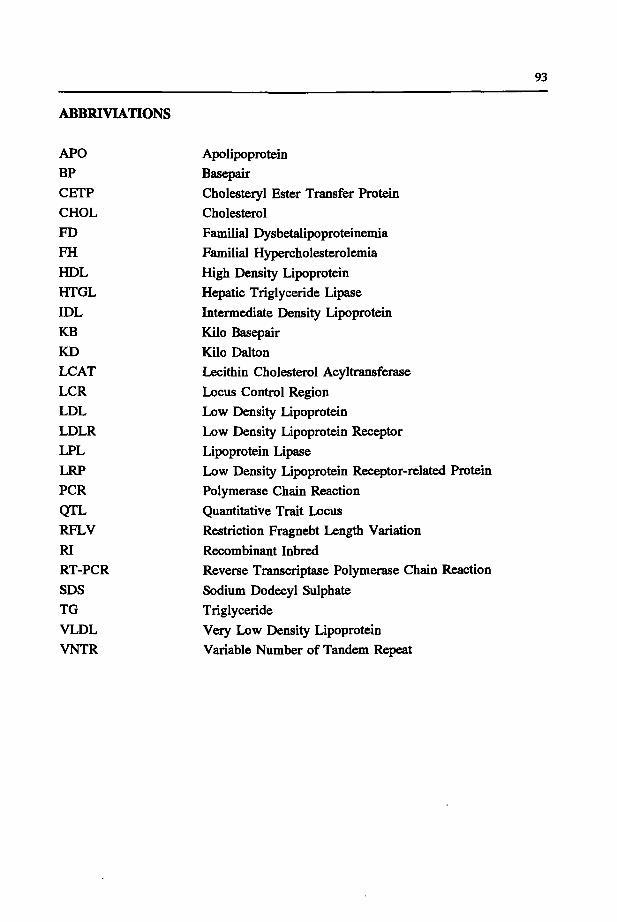
93
ABBRIVIATIONS
APO BP CETP CHOL FD FH HDL HTGL IDL KB KD LCAT LCR LDL LDLR LPL LRP PCR QTL RFLV RI RT-PCR SDS TG VLDL VNTR
Apolipoprotein Basepair Cholesteryl Ester Transfer Protein Cholesterol Familial Dysbetalipoproteinemia Familial Hypercholesterolemia High Density Lipoprotein Hepatic Triglyceride Lipase
Intermediate Density Lipoprotein Kilo Basepair Kilo Dalton Lecithin Cholesterol Acyltransferase Locus Control Region Low Density Lipoprotein Low Density Lipoprotein Receptor Lipoprotein Lipase Low Density Lipoprotein Receptor-related Protein Polymerase Chain Reaction
Quantitative Trait Locus Restriction Fragnebt Length Variation
Recombinant Inbred Reverse Transcriptase Polymerase Chain Reaction
Sodium Dodecyl Sulphate Triglyceride Very Low Density Lipoprotein Variable Number of Tandem Repeat
95
CURRICULUM VITAE
Mariette Hoffer werd 2 juni 1965 geboren te Wieringermeer. In 1983 behaalde zij het Atheneum diploma aan de rijksscholengemeenschap Wiringherlant te Wieringerwerf. In 1983 begon zij aan haar studie biologie aan de Vrije Universiteit te Amsterdam. Het doctoraal examen medische biologie dat bestond uit de hoofdvakken Anthropogenetica (Prof. Dr. A.W. Eriksson) en Immunologie (Dr. G. Kraal en Dr. R. Scheper) werd afgelegd in 1988.
Vanaf september ,1988 was zij als assistent in opleiding verbonden aan de afdeling Anthropogenetica (Prof. Dr. G.J.B, van Ommen) van de Rijksuniversiteit te Leiden. Dit proefschrift beschrijft de resultaten van onderzoek onder leiding van Dr. R.R. Frants en Dr,. Ir. L.M. Havekes (IWO-TNO, Leiden). Sinds augustus 1993 is zij werkzaam op de afdeling Anthropogenetica van de Rijksuniversiteit te Leiden.