characterization of wall teichoic acids in two...
TRANSCRIPT

CHARACTERIZATION OF WALL TZICHOIC ACIDSIN TWO MORPHOLOGICAL FORMS OFArtkpobactev arvstallovoietes
By
JOHN HARDIN HELLMJTH
A DISSERTATION PRESENTED TO THE GRADUATE COUNCIL OF THEUNIVERSITY OF FLORIDA IN PARTIAL
FULFILLMENT OF THE REQUIREMENTS FOR THE DEGREE OFDOCTOR OF PHILOSOPHY
UNIVERSITY OF FLORIDA
1978

ACKNOWLEDGEMENTS
The author wishes to thank sincerely Dr. Edward Previc for his
encouragement, suggestions, and criticisms during the preparation of
this dissertation.
He also wishes to thank the other members of his supervisory
committee, Dr. Arnold Bleiweis, Dr. David Hubbell and Dr. Lonnie
Ingram for the help they contributed in the preparation of this
manuscript.
The author would like to express his gratitude to the other
members of the Department of Microbiology and Cell Science for
generously supplying equipment and materials during his work.
Special thanks to Steven Hurst for technical assistance with
amino acid analyses.
Appreciation is extended by the author to his parents for the
encouragement they gave him to pursue his education.
Finally, the author is particularly indebted to his wife, Martha,
for patience, encouragement, and assistance during his graduate work.

TABLE OF CONTENTS
Page
ACKNOWLEDGEMENTS ii
LIST OF TABLES v
LIST OF FIGURES vi
ABSTRACT vii
INTRODUCTION 1
Taxonomy 1
Control of Morphogenesis 3
Justification for This Study 5
Teichoic Acid Background 6
MATERIALS AND METHODS 9
Organism 9
Media 9
Growth 10Cell Wall Isolation 10Purification of Cell Wall 11Teichoic Acid Isolation 12Analysis of Purified Walls 12Paper Chromatography 13Analytical Procedures 14Amino Acid and Amino Sugar Analysis 16Teichoic Acid Purification 17Gel Filtration 17Chain Length Determination 18Determination of Molar Ratios of Glycerol and
Phosphate in Purified Teichoic Acids 19Characterization of Alkaline Hydrolysate 19
RESULTS 22Growth of ArtkrobaoteT crystallopoietes ATCC 15481 .... 22Amino Acid and Amino Sugar Analysis of
Purified Wall Hydrolysates 22TCA Extraction of Purified Walls 25
Recoveries of Purified Wall and TCA ExtractableMaterial (Crude Teichoic Acid) 29
Characterization of Acid-Hydrolyzed TCA Extractsby Paper Chromatography 29

Page
Amino Acid and Amino Sugar Analysis ofCrude Teichoic Acid 35
Crude Teichoic Acid Purification on DEAE-Sephadex .... 35Purified Teichoic Acid Characterization on
Sephadex G-100 39Chain Lenght Determination 39Molar Ratios of Glycerol and Phosphate in
Purified Teichoic Acids 43Characterization of Alkaline Hydrolysates 43
DISCUSSION 52Peptidoglycan Composition 52TCA Extraction 57Characterization of Acid Hydrolysates 59Amino Acid and Amino Sugar Analyses of Teichoic Acid ... 61DEAE Chromatography 61Acid Hydrolyses of Purified Teichoic Acids 62Gel Filtration 63Determination of Chain Lenght 63Glycerol-Phosphate Ratios 65Alkaline Hydrolysis 66Summary of Teichoic Acid Structure 68Possible Roles for Teichoic Acids in Arthvobastev .... 68
LITERATURE CITED 74
BIOGRAPHICAL SKETCH 81

LIST OF TABLES
Table Page
1 Carbohydrate constituents of representativeteichoic acids 8
2 Molar ratios of amino acids and amino sugars inwalls digested with various enzymes 26
3 Paper chromatography of acid-hydrolyzed TCAextracts. I. Phosphoric acid esters 31
4 Paper chromatography of acid-hydrolyzed TCAextracts. II. cs-Glycols 32
5 Paper chromatography of acid-hydrolyzed TCAextracts. III. Sugars (external standard) 33
6 Paper chromatography of acid-hydrolyzed TCAextracts. IV. Sugars (internal standard) 34
Molar ratios of labile and total phosphate inalkaline phosphatase-digested, purifiedteichoic acids 42
8 Molar ratios of glycerol to inorganic phosphate .... 44

LIST OF FIGURES
Figure Page
1 Growth of Arthrobaater crystallopoietes 24
2 TCA extraction of purified walls 28
3 DEAE-Sephadex chromatography of crude teichoicacid 38
4 Sephadex G-100 chromatography of purifiedteichoic acid 41
5 Alkali-hydrolyzed teichoic acid elution on DEAE-Sephadex 47
6 Sephadex G-50 chromatography of neutral and cationiccomponents of alkali-hydrolyzed GSTA 49
7 Ot-Sephadex chromatography of neutral and cationiccomponents of alkali-hydrolyzed GSTA 51

Abstract of Dissertation Presented to the Graduate Councilof the University of Florida in Partial Fulfillment of the
Requirements for the Degree of Doctor of Philosophy
CHARACTERIZATION OF WALL TEICHOIC ACIDSIN TWO MORPHOLOGICAL FORMS OF
Art hrobacter orvstallovaietes
3y
John Hardin Hellmuth
December 1978
Chairman: Dr. Edward P. PrevicMajor Department: Microbiology and Cell Science
The cell wall teichoic acid isolated from two morphological forms
of Art Ivobacier crystallopoistes is characterized. Cell walls purified
from spherical (GS) cells contained 13.2 ug of phosphorus per mg of cell
wall, while those from rod-shaped (LS) cells contained 21.2 ug phosphorus
per rag of cell wall. Trichloroacetic acid extracts of purified walls of
both forms were found to contain poly (glycerol phosphate) with hexosamine
glycosidically attached. In GS teichoic acid there was 2S% as much
hexosamine as glycerol phosphate and in LS teichoic acid there was
21% as much. The hexosamine included at least 50% N-acetylated glucos-
amine and gaiactosamine in about a 5-to-l ratio. Evidence is presented
which suggests that the hexosamine may exist as trisaccharide side
chains. Chain lengths were estimated by the racic of total phosphate
to alkaline phcsphatase-labile phosphate. By this raechcd, teichoic
acids from GS-grown cells had an average length of 33 glycerol phosphate

units and those from LS-grown cells had an average length of 70
units. The possible significance ot these findings as they relate
to morphogenesis in A.rt hpobaotev is discussed.

INTRODUCTION
Taxonomy
The genus Arthrobaater is characterized by cells which can undergo
nutritionally-controlled sphere-rod morphogenesis (43). The species
Arthrobacve? ovysialloyoietes was first described by Ensign and
Rittenberg (30) who isolated the organism by enrichment cultures con-
taining 2-pyridone. The brilliant green crystalline pigment produced
by this species growing on solid medium containing 2-pyridone was
later identified by Kuhn et al. (57) to be a hydrate of the mono-
potassium salt of 4,5,4' ,5'-tetrahydroxy-3,3'-diazadiphenoquinone-
(2,2'). Recently, Kolenbrander and Weinberger (52) found that A.
arystallopoietes lost the ability to produce pigment from 2-pyridone
at a high spontaneous frequency of 0.26% loss per generation. This
high spontaneous loss has also been observed in the strain of A.
evystallovo'i.etes used in this dissertation research. Kolenbrander and
Weinberger (52) present good evidence that the loss of a plasmid is
correlated with loss of ability to produce pigment. In the present
study this presented a problem since the plasmid-less strain seemed
to grow faster than the parent strain in glucose-salts media. It was
therefore necessary to minimize the number of accumulated mutants by
starting each culture for harvest from a single pigment producing
colony.

According to Bergey's Manual (48), the name Arthrobaotev ovystal-
lopoietes is a probable subjective synonym of Arthrobaoter globiformis.
The Manual also states that the salient feature distinguishing A.
crystallopoietes from .4. globiformis is the ability to utilize
2-pyridone as a sole carbon and energy source and to produce a crystal-
line pigment from it. It thus seems likely that A. cvystallopoietes
which has lost the plasmid has almost exactly the same phenotype as
A. globiformis. Since the presence or absence of the plasmid may
affect the wall composition by either direct effects (e.g., plasmid
genes might modify wall synthesis) or indirect effects (e.g., plasmid
presence might affect growth rate which might in turn affect wall
synthesis) , comparisons between the walls of these bacteria can not
be made on the assumption that A., cvystallovoietes and A. globiformis
are the same bacteria.
The nutritional control of morphology in A. cvystallopoietes was
first demonstrated by Ensign and Wolfe (31). They showed that expo-
nential growth of spheres could be obtained in a defined medium con-
taining glucose. Exponential growth of rods could be obtained by
adding certain morphogenesis-inducing compounds to the defined glucose
medium. This idea forms the basis for the methods of obtaining spheres
and rods in the following work. The only difference is that the rod-
inducing medium contains lactate but no glucose. Since there is a
diauxic suppression of glucose cataboiism and anabolism in the presence
of rod-inducing compounds (54), this difference is minimized.

Control of Morphogenesis
Although the morphology of Arthrobaeter can be extrinsically con-
trolled by nutritional means, this control is only indirectly related
to the unknown intrinsic control by the bacteria. The intrinsic
control is more directly related to growth rate which can be controlled
independently from the type of nutrition. Luscombe and Gray (60) have
done this by growing a strain of Arthrobaater under carbon-limiting
conditions in a chemostat. They found that, at 25°C, rods were only
produced at dilution rates above 0.25 per hour. At rates lower than
this, the cells were always spherical. This raises a problem because
it means that whenever rod and sphere-shaped arthrobacters are compared,
morphology is not the only difference; growth rate also changes and
hence could affect many variables. This is a fundamental problem
which makes it difficult to establish correlations as causal rela-
tionships and it applies to this study. Hamilton et al. (39) have
sidestepped this problem by isolating a spherical morphological
mutant of A. crystallopoietes which is unable to undergo sphere-rod
morphogenesis but increases its growth rate in rod-inducing media.
The genetic nature of this lesion has not yet been reported.
The intrinsic control of morphogenesis probably consists of a
chain of events, some of which are genetic, some enzymatic, and some
structural. Certain changes at any level of this chain can probably
manifest themselves at the morphological level. Therefore, any one
event should not be said to control morphogenesis. Nevertheless,
several authors (39,47,53,61,68,69,73) claim that their effect which

correlates with morphology is probably the factor which controls
morphogenesis in .4. arystallopoietes .
It has been shown that A. arystallopoietes contains two RNA poly-
merases both of which have considerable and almost equal synthetic
capabilities in in vitro studies (47). This correlates with another
finding by the same lab (61) that morphogenesis involves differential
transcription of the DNA with some transcripts being present all the
time and others being present only at certain times during the morpho-
genetic cycle. Furthermore, St. John and Ensign (73) by using RNA
and DNA synthesis inhibitors were able to show that morphogenesis can
occur in the absence of DNA replication but RNA synthesis is required
for morphogenesis to occur.
Hamilton et al. (39) suggest that the level of cyclic adenosine
3' ,5 '-monophosphate (c-AMP) may be important in regulating morpho-
genesis. The levels of c-AMP were shown to rise steeply at times just
prior to the initiation of shape changes (either sphere-to-rod or rod-
to-sphere). This suggested that elevated c-AMP levels may act as the
"trigger" which induces the cells to change shape. Their morphological
mutant (mentioned above) did not show these changes.
Krulwich et al. (53, 55) have shown that the glycans in the pepti-
doglycan of spheres are generally shorter than those of rods. This
change was also correlated with a change in the activity of a wall-
bound N-acetylmuramidase. They implied that morphology, therefore,
depends on glycan chain length.
Previc (68) has suggested that morphology in most bacteria may
be determined by the presence or absence of extra crossbridges invol-
ving free carboxly groups of diaminopimelic acid (Dpm) and other

tetrafunctional amino acid groupings (e.g., lysylaspartyl crossbridges
in some Lactobacillus species). In a mutant of A. cv^jstallovoietes
Previc and Lowell (69) have shown that spherical mutants contain lysine
in the penultimate position of tetrapeptides while rod-shaped mutants
contain Dpm. Transitional stages during sphere-to-rod morphogenesis
show a gradually increasing amount of Dpm present. Thus, morphology
in this strain may be dictated by the presence or absence of the extra
carboxyl group of Dpm.
Several authors seem to agree that a rod shaped morphology requires
a more rigid peptidoglycan than a spherical morphology (37,55,68).
Ward and Claus (84) have found that for .4. cvystallovoistes the rod
peptidoglycan layer is thinner than the sphere peptidoglycan. If the
rod peptidoglycan is more rigid than the sphere peptidoglycan, then
this difference must be due to changes in the peptidoglycan structure
rather than simply a thickening of the wall.
It should now be apparent that there are changes during morpho-
genesis at each step in the chain of events which regulates morphology.
What is still not apparent is how all these changes fit together to
produce morphogenesis. Of course, some of the changes may not be
related to morphogenesis at all but only reflect altered growth rates.
So, the question of how morphogenesis occurs is still unanswered.
Justification for This Study
The original intent of this author was to determine how peptido-
glycan might help to maintain the different shapes of A. arystalloyoietes.
To accomplish this the composition of the wall was examined for changes

which might correlate with morphology. During the beginning of the
quantitative analyses of the components of the wall, there arose
certain evidence that a wall teichoic acid might be present. The
determination of its qualitative and quantitative structure has become
the primary topic of this dissertation. Although this seems far re-
moved from the question of morphogenesis, there may be an intimate
association between the two. These possibilities are examined in the
Discussion.
Teichoic Acid Background
Teichoic acids are molecules which occur in nearly all gram-positive
bacteria (3). These molecules are grouped into two categories de-
pending on their cellular location and their structure; lipoteichoic
acids are found associated with the cell membrane and wall teichoic acids
are associated with the peptidoglycan (4)
.
Lipoteichoic acids are found in most gram-positive bacteria. These
molecules are all of the same structural type, i.e., they consist of
a linear backbone of poly (glycerol phosphate) which is linked by
phosphodiester bonds involving C-l and C-3 of adjacent glycerol
phosphates (3). Diversity of lipoteichoic acid structure is introduced
by various carbohydrate side groups (see Table 1) which are attached
at the C-2 hydroxyl group. D-Alanine is usually found as an ester
linked either to the C-2 hydroxyl of glycerol phosphate or to glycosyl
hydroxyl groups. It is though that all lipoteichoic acids are
covalently attached to glycol.ipid in the cell membrane (51)
.

Wall teichoic acids are more structurally diverse than lipo-
teichoic acids. The classical wall teichoic acids are polymers of
glycerol or ribitol phosphate. Both types can have various carbo-
hydrate side groups (see Table 1) and/or D-alanine. These polymers
are thought to be covalently attached to the peptidoglycan in the cell
wall. Other acidic polymers have been found in the walls of gram-
positive bacteria which resemble the classical teichoic acids. Polymers
of glycerol phosphate and one or more sugars have been found in a
number of cases and polymers of sugar phosphates are also known.
Both types of polymers are similar to teichoic acids in that they
convey a net negative charge to the outer surface of the cell and,
therefore, may have functions similar to the teichoic acids (51).

r-x

MATERIALS AND METHODS
Organism
Arthrobactar arystallopoietes ATCC 15481 was obtained from the
American Type Culture Collection. In order to maintain the presence
of the plasmid which bears the gene(s) responsible for green crystal
production from 2-pyridone, stock cultures were frequently streaked
on 2-pyridone-containing plates (30) and pigmented colonies were
picked for further subculturing. All inocula for cultures to be
harvested were also checked in this manner.
Media
Defined media contained, per liter: 1.73 g K2HP04 , 2.33 g KH2P04 ,
1.00 g (NH 4 ) 2S04, 5.0 g D-glucose (GS) or sodium lactate (LS)
,
0.5 g MgS04 , and 10.0 ml of a trace salts solution. The trace salts
solution contained, per liter: 1.5 g nitriloacetic acid, 0.5 g
MnS04 , 1.0 g NaCl; FeCl 2 , CaCl 2 , CoCl 2 , and ZnS0 4 , 0.1 g each, and
CuS0 4 , A1K(S04 ) 2 , H3BO3, and (NH4 ) 6Mo 7 2 4, 0.01 g each. The trace
salts solution was adjusted to pH 7.0 with 1.76 g NaOH. The glucose,
MgSO, and the trace salts solutions were each autoclaved separately (31)
A complex pigmentation medium containg 0.2% 2-pyridone, 0.05%
yeast extract, and inorganic salts was prepared according to Ensign and
Rittenberg (30).

10
Growth
All cultures were agitated at 30°C in a New Brunswick Gyrotory
shaker-incubator. Optical density (O.D.) was followed at 450 nm
on a Beckman DU-2 spectrophotometer with a 1.0 cm light path. Optical
densities were maintained between 0.05 and 1.0 O.D. units by periodic
transfers to fresh media.
Cell Wall Isolation
Exponential cultures were passed through coiled copper tubing which
was submersed in an ice-water bath. The cooled effluent was directed
into a De Laval Gyro Tester Laboratory Centrifuge with the bowl
prechilled to 4°C. The time for culture liquid to pass through the
cooling coils and out the centrifuge spout was measured using enough
nigrosin dye added to a deionized water run to serve as a visual
marker. The temperature and O.D. at 450 nm of the final effluent
were also measured during bacterial harvests. From these measurements
it was concluded that cultures were cooled from 30°C to 8-10°C in
approximately one minute and that 95% or more of the culture's bacterial
mass was removed from the effluent as determined by O.D. measurement.
All subsequent handling of bacterial samples was at no higher than
4°C unless otherwise specified. Whole cell pellets were washed twice
by resuspension in deionized water and centrifugation at 13,000 x g
for 30 min in a Servall GSA rotor. Bacteria were suspended in
deionized water at a concentration no greater than 50 mg dry wt/ml and
broken in a Braun homogenizer at 4,000 rpm for 4 minutes using 0.11 mm

II
diameter glass beads. Tributyl phosphate (0.5% v/v) was used to reduce
foaming. Glass beads were removed first by filtration on fritted glass
filters and then by centrifugation at 160 x g for 30 min. Crude cell
walls were then pelleted from the supernatant at 27,000 x g for one hour.
Purification of Cell Wall
The method of Braun and Sieglin (14) for wall purification was
modified by an additional sodium dodecyl sulfate (SDS) extraction
before treatment with pronase (69) . Crude wall pellets were suspended
in 0.1 M ethylenediaminetetraacetic acid (EDTA), pH 7.4 (at least
20 ml/g of whole cell dry wt), and then pelleted at 27,000 x g, washed
and resuspended in deionized water. Suspensions were added dropwise
to stirred, boiling 4% SDS (at least 15 ml/g of whole cell dry wt)
.
These mixtures were allowed to cool to room temperature with stirring
overnight. These mixtures were then centrifuged at 27,000 x g for
one hour. Pellets were resuspended in deionized water and reextracted
with boiling kl SDS. These pellets were washed with deionized
water three times and resuspended in 0.05 M Tris, pH 7.4 (at least
5 ml/g of whole cell dry wt) . Pronase was added to a final concen-
tration of 100 jig/ml and the mixtures were digested at 37°C for 16
hours. The walls were centrifuged for one hour at 37,000 x g. The
resultant pellets were reextracted by boiling SDS as above and
rewashed three times with deionized water. The final pellets were
lyophilized for dry weight determinations.

12
Teichoic Acid Isolation
To release teichoic acid (G), purified wallswere extracted with
10% (w/v) tricloroacetic acid (TCA) at 4°C for 24 hours. Walls were
pelleted from this mixture at 27,000 x g for one hour and the pellets
were reextracted with fresh 10% TCA at 4°C for 24 hours. Again fresh
cold TCA was exchanged for supernatant TCA and the extraction was
continued for 38 hours. Crude teichoic acids were collected either
by precipitation from the TCA with absolute ethanol (5 volumes) at
-20°C and subsequent centrifugation at 13,000 x g for 30 minutes, or
by removing the TCA by ether extraction to pH 4 and subsequent lyo-
philization.
Analysis of Purified Walls
Purified walls (0.4 mg/ml) were hydrolyzed in 4 N HC1 at 105°C for
11 hours. The excess acid was then removed by evaporation, followed
by three cycles of deionized water additions and reevaporations. The
residues were dissolved in 0.01 N HC1 (1 mg wall/ml acid) and then
filtered through 0.45 um Millipore filters. Unsolubilized material
was present in negligible amounts. The filtrates were analysed for
amino acids and amino sugars on a JE0LC0 Automated Amino Acid
Analyzer (76)
.

13
Paper Chromatography
Whatman papers no. 1, no. 4, and no. 3 MM were soaked for at least
30 minutes in a solution which was 0.1 N in acetic acid and 0.1 M
in EDTA. This solution was then washed out by suspending the papers
in deionized water and then ramoving excess water by decantation. At
least ten sequential water washes of increasing duration were performed,
with a final wash of at least one hour. Washed papers were allowed
to dry in horizontal stacks at 30°C. Papers to be eluted parallel
to the grain of the paper were cut to 23 cm x 57 cm with the grain
running parallel to the long axis of the paper. Papers to be eluted
perpendicular to the grain were cut to 23 cm x 46 cm with the grain
running perpendicular to the long axis. Samples were spotted 7 cm
from the top of the papers by repeated application of approximately
0.25 ul each time, then drying by hot air. The papers were mountain-
folded along a line at 6 cm and valley-folded along a line at 3 cm
from the top of the papers. This folding allowed the papers to be hung
from a glass trough in a pyrex tank (30.5 cm x 30.5 cm x 61 cm). The
papers were allowed to equilibrate with the vapor phase of the eluting
solvent in a glass chromatography tank for at least 2 hours before
the eluting solvent was added for the beginning of elution. All
chromatograms were eluted at ambient temperature, typically ranging
from 22°-27°C. The solvent systems for elution used were: (A) n-propanol-
ammonia (28-30%)-water (6:3:1) (40) and (3) ethyl acetate-pyridine-water
(10:4:3) (36).

Analytical Procedures
Phosphate Assays
Total phosphate was assayed by the ascorbic acid-molybdate method
developed by Chen et al. (23). The ashing method of Lowry et al. (58)
was used for convenience in handling large numbers of samples.
Inorganic phosphate was measured by the Chen method as modified
by Ames (1). KH2PO4 (anhydrous analytical reagent, Mallinckrodt)
was used as a standard in both total and inorganic phosphate methods.
Chromic acid-washed tubes were used for all phosphate assays.
Amino Sugar Assays
N-Acetylamino sugars were measured by the borate-p-dimethyl-amino-
benzaldehyde (DMAB) method of Reissig et al. (70)
.
N-Acetylglucosamine (Sigma) was used as the standard for this assay.
Amino sugars were assayed for N-acetylation with acetic anhydride by
a method similar to the N-acetylamino sugar assay as described by
Ghuysen et al. (35). This method primarily detects amino sugars
which are free in the C-l position but it also detects amino sugars
which are covalently bound at that position. The extinction coefficient
for the latter reaction, however, is more than thirty fold smaller than
that for the former reaction. D-Glucosamine-HCl (K+K Laboratories, Inc.)
was used as a standard for this assay.
Total amino sugars could be detected if an acid hydrolysis (2 N HC1,
100 C C, 3 hours) followed by neutralization preceded the N-acetylation
step of the amino sugar assay. The same standard was used as for the
amino sugar assay.

15
The minimum amount of N-acetylation of amino sugar was estimated
by a method suggested fay Ellwood et al . (29). This method involves
a mild acid hydrolysis (0.1 N H 2S04 , 100°C, 30 min in a sealed
ampoule) which breaks the glycosidic linkage while leaving most of
the N-acetyl groups intact. After hydrolysis, a slight excess (10%)
of the amount of 3aC0 3 necessary for neutralization was added. The
resulting i 3aS0 4-BaC0 3 mixture was removed by low speed centrifugation.
N-Acetylhexosamines and total amino sugars were then determined for
the neutralized samples.
Carbohydrate Assay
Simple sugars, oligosaccharides, and polysaccharides with either
free or potentially free reducing groups were determined by the phenol-
H2S0
4 method of Dubois et al. (28). D-Glucose (analytical reagent,
Mallinckrodt) was used as a standard.
Protein Assay
Protein was measured by the Lowry method (59) which involves
first a reaction of protein with Cu+2 under alkaline conditions and
then reduction of a phosphomolybdate-phosphotungstate reagent by the
copper-treated protein. 3ovine serum albumin (Sigma) was used as
a standard.

16
Enzymatic Determination of Glycerol
Free glycerol was determined by using the Glycerol Stat-Pack
(Calbiochem). This assay employs the following reaction sequence:
(1) glycerol + adenosine triphosphate (ATP) glycerol kinase
a-glycerophosphate + adenosine diphosphate (ADP)
(2) ADP + phosphoenolpyruvate Pyruvate kinase ^ pyruvate + ATP
(3) pyruvate + nicotinamide adenine dinucleotide (reduced) (NADH)
lactate dehydrogenasei „„*„.„ . , . , ,
- 2 _^ lactate + nicotinamide adenine
dinucleotide (oxidized)
The disappearance of NADH was followed spectrophotometrically at a
wavelength of 340 nm using the same instrument used for O.D. readings.
Glycerol (analytical reagent, Mallinckrodt) was used as a standard.
Amino Acid and Amino Sugar Analysis
Crude teichoic acid samples (GS, 5.0 mg; LS, 5.6 mg) were each
hydrolysed in 1.0 ml of 2N HC1 at 100°C for 3 hours. The hydrolysates
were vacuum dried over NaOH pellets and then redissolved in 1.0 ml
of deionized water. Portions of these (200 yl each) were diluted in
0.01 N HC1 to 2.2 ml (final concentrations: GS , 0.45 mg/ml; LS,
0.51 mg/ml). These samples were subjected to amino acid and amino
sugar analysis in the same way as described for purified wall hydro-
lysates.
Teichoic Acid Purification
Crude teichoic acids were purified on approximately 9 g of DEAE-
Sephadex A-50. One cm of Sephadex G-25 course gel was used at the
bottom of the column as bed protection. The DEAE-Sephadex was swollen
and loaded in 0.1 M NaCl. The bed diameter was 2.5 cm and the bed
length varied depending on ionic strength (35.5 cm at 0.1 M NaCl and
24.0 cm at 1.0 M NaCl). The bed was then washed with 500 ml of
0.1 M NaCl.
Teichoic acids were eluted by an increasing gradient of NaCl.
For the LS preparation, 200 ml of a 0.1 M - 0.5 M gradient was
followed by a 200 ml gradient from 0.5 M to 1.0 M NaCl. A total of
160 fractions of approximately 2.1 ml each was collected. For the GS
preparation, 500 ml of a 0.1 M - 1.0 M NaCl gradient was used of elu-
tion. A total of 95 fractions of approximately 5 . 3 ml each was collect-
ed. The fraction numbers for the GS elution profile in the Results
section have been normalized with the LS profile using equivalent salt
concentrations
.
Gel Filtration
Gel filtration was carried out on Sephadex G-100. Purified teichoic
acids (GS, 42 mg; LS , 6 mg) were applied to a column bed of dimentions:
2.5 cm x 31 cm. Total bed volume was 160 ml. The void volume was
determined to be 54 ml by using Blue Dextran 2000 (Pharmacia, Average
molecular weight, 2 x 10⁶ ) . A Blue Dextran sample (1 ml of a 0.2%
solution) was applied to a column and the effluent was collected in
approximately 3 ml fractions. These fractions were monitered for
O.D. at 260 nm as a measure of Blue Dextran. The number of the
fraction with the highest O.D. was multiplied by the fraction volume
to obtain the void volume. Eluant was deionized water. A constant
pressure head of 31 cm was maintained during loading and running.
A Gilson automatic fraction collector was adjusted to collect 95
drop (approximately 3 ml) fractions.
Results were graphed in terms of pmoles of total phosphate vs.
K. K is defined as: Ve - VQK =
V t - V c
where: V = void volume
V t= total bed volume
Chain Length Determination
In two separate determinations, about 1 mg and 0.5 mg of each
type (GS and LS) of purified teichoic acid was diluted from concen-
trated solutions to 125 yl with deionized water. Then, 125 ul of
0.04 M (NH4 ) 2C03 and 10 pi (0.6 mg or 7.2 units) of Escherichia ooli
alkaline phosphatase (60 mg/ml, 12 units/mg, Worthington Biochemical
Corporation, Code: BAPSF - bacterial alkaline phosphatase salt
fraction) were added. One unit is defined as that activity liberating
one umoie of p-nitrophenol per minute at pH 8.0 and 25°C. The mixture
was incubated in a slowly shaking 37°C water bath for 18 hr. The
samples were then analyzed for inorganic and total phosphate content.
The ratio of total to inorganic phosphate content was taken as an
approximation of chain length.

19
Determination of Molar Ratios of Glycerol and Phosphate
in Purified Teichoic Acids
Purified teichoic acid samples were hydrolyzed in 2 N HC1 at 100°C
for 4 hours. Acid was removed hy vacuum evaporation or lyophilization.
The samples were then digested with alkaline phosphatase by the same
general method described under chain length determination. The
digested samples were then analysed for free glycerol, inorganic and
total phosphorus. Because of phosphorus present in the enzyme
preparation, it was also necessary to run enzyme blanks with no
added sample and to subtract out the blank values from the sample
values. This method was tested on a known concentration of ct-glycero-
monophosphate which produced a molar ratio of glycerol to phosphate
of 0.997.
Characterization of Alkaline Hydrolysate
A purified teichoic acid sample (GS, 32.6 umoles total phosphate)
was hydrolyzed in 1.45 ml of 1 M NaOH at 100°C for 3 hours. The hydro-
lysate was neutralized with 300 ul of 4 N HC1. The final pH was
close to pH 7.0 as judged by pH paper. The neutralized sample was
diluted with deionized water to 14.5 ml to attain a salt concentration
of 0.1 M NaCl.
The diluted sample was loaded on the same DEAE-Sephadex A-50
column used for the teichoic acid purufication. In preparation for
this run, the column had been previously washed with 500 ml of 1 M
NaCl. The washed column was slowly reswollen with a 1.0 M - 0.1 M

mr
NaCl gradient of 500 ml. Finally, the column was washed with another
500 ml of 0.1 M NaCl for final equilibration. Once loaded, the sample
was washed with 500 ml of 0.1 M NaCl. The wash effluent was collected
and lyophilized. The adherent portion of the sample was eluted
from the column with 500 ml of a 0.1 M - 1.0 M NaCl gradient.
Fractions (100, approximately 4 ml each) were collected. These
fractions were assayed for total phosphate and total amino sugars.
The lyophilized wash sample was then filtered on a Sephadex
G-50 column (diameter 1.6 cm, height 62 cm, 124 ml bed volume, 56 ml
void volume) with deionized water in order to resolve low-molecular
weight, non-anionic fragments. Fractions (65 of 3 ml each) were
collected and analyzed for total and inorganic phosphate and total
amino sugars. All phosphate-containing fractions were then pooled
and lyophilized. This material (approximately 2.8 g) was assumed
to be mostly NaCl and was diluted with deionized water to 0.1 M
NaCl on that basis.
This diluted sample was applied to a CM-Sephadex C-50 column
(approximately 1.9 g of gel; column with following dimensions:
diameter, 1.5 cm, length, 30 cm) to further resolve cationic molecules.
The column bed was then washed with 50 ml of 0.1 M NaCl. After
washing, the remaining sample was eluted with 250 ml of a 0.1 M -
1.0 M NaCl gradient, and 46 fractions (5.4 ml each) were collected.
These fractions were analyzed for total phosphorus and total amino
sugars. The two peaks (two fractions/peak) containing significant
amounts of hexosamine were each combined and lyophilized. These
lyophilized samples were triturated in the presence of absolute
ethanol (10) . The ethanol fractions were then evaporated to dryness
and the residues were triturated again with absolute ethanol (1 ml)
.
The ethanol-soluble portions were again evaporated to dryness. The
residues were each dissolved in 450 ul of deionized water and then
analyzed for free glycerol.
RESULTS
Growth of Avilwobaatev ovystaltouo'istes
ATCC 15481
Typical growth curves are shown in Figure 1. Generation times
were: GS, 10.8 hr ± 0.5 hr; LS , 3.5 hr + 0.2 hr.
Amino Acid and Amino Sugar Analysis of Purified Wall Hydrolysates
It was observed that cell walls purified according to the protocol
in Materials and Methods usually contained certain non-peptidoglycan
amino acids. The following experiment was an attempt to remove these
amino acids using proteolytic enzymes.
Cell walls were isolated from lyophilized cells (GS or LS, 300
mg each) and were purified as described in Material and Methods
with one modification. Just before the pronase digestion, each
sample was divided into four equivalent portions. One portion of
each was not treated with any enzyme and served as the control;
the second portions were treated with pronase (500 ug/ml) ; the
third portions were treated with trypsin (100 ug/ml); and the fourth
portions were treated with ot-chymotrypsin (100 ug/ml) . All digestions
were carried out in 0.05 M Tris, pH 7.4 at 37°C for 16 hours. Following
the enzymatic digestions, each sample was treated as described in
Materials and Methods for the rest of the wall purification and
22
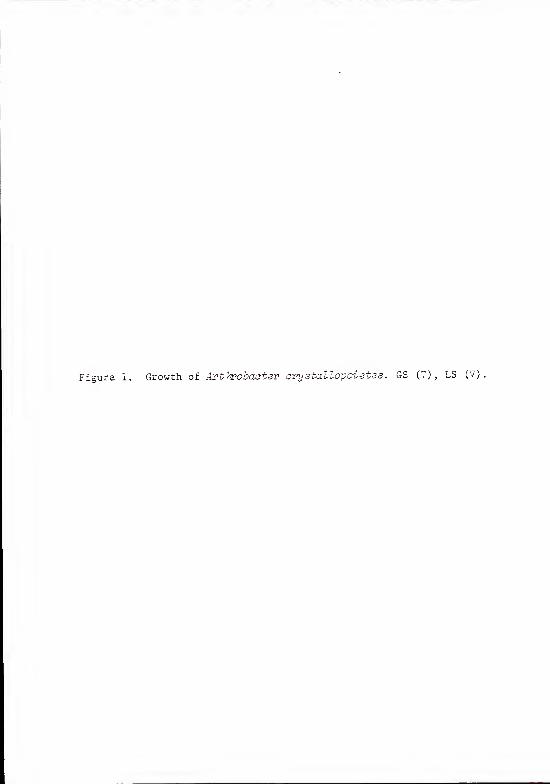
Figure 1. Growth of Art hrobaoter crystallopoietes. GS (7), LS (V).

24
time, hours

amino acid analysis. Peak areas on chromatograms were computed by
a JOELCO integrater. Relative concentrations were then computed
using known areas of standard amino acids and sugars. These relative
concentrations were then divided by the relative concentrations for
the glutamic acid peak on each chromatogram. The values for these
ratios are given in Table 2. These analyses indicate that the normal
peptidoglycan amino acids are present in the expected ratios. They
also show that trypsin is the most effective enzyme in removing
the trace amino acids.
Chemical analysis of purified cell wall hydrolysates also shows
the presence of significant amounts of phosphorus (GS, 18.2 tig P/mg
wall; LS, 21.2 ug P/mg wall). Since Krulwich and Ensign (55) had
reported phosphorus in their cell wall preparations of .4. ovystallo-
ipoietes and had found that all of it could be removed by hot TCA
extraction, a similar experiment was carried out with the cell walls
prepared in this laboratory.
TCA Extraction of Purified Walls
Purified cell walls (GS, 19 mg; LS , 18 rag) were suspended in
10 ml of cold 10% TCA and were stirred with a magnetic stirring bar
at 4 C. At various times, 1.0 ml portions were removed and extracted
three times with petroleum ether (B.P. 30°C) to remove the TCA. The
sample was then centrifuged at 27,000 x g for a half hour.
Total phosphate was determined for both the soluble and insoluble
fractions as shown in Figure 2. Phosphate was extracted by TCA with

^^
TABLE 2
Molar ratios of amino acids and amino sugars in walls digestedawith various enzymes.

CO

||dm Bui/6rt 'j

time as indicated by the increase in TCA-soluble phosphate. This
increase was paralleled by an equivalent decrease in wall associated
phosphate as indicated by the TCA-insoluble curves.
Recoveries of Purified Wall and TCA Extractable Material
(Crude Teichoic Acid )
Walls purified according to the scheme outlined in Material and
Methods yielded 12-14% wall dry weight to cell dry weight for GS
cells and 13-15% for LS cells. In each individual case the LS
cells yielded slightly more wall (6-10% more) on a percentage basis
than the GS cells.
Recoveries of TCA-extractable material varied depending on
whether ethanol precipitation or ether extraction was used to
remove TCA from the samples. When ethanol precipitation was used,
TCA extractable material recoveries (as crude TA dry weight
percentage of purified wall dry weight) were: GS, 21%; LS , 18%.
When ether extraction was used the recoveries were much higher:
GS, 38% and LS, 28%.
Characterization of Acid-Hydrolyzed TCA Extracts by Paper
Chromatography
To determine the nature of this phosphorus containing compound,
acid hydrolyses were performed. The conditions used have been re-
ported to degrade teichoic acids of either polyglycerophosphate or
polyribitol phosphate and substituted with either amino acids or
carbohydrate (3)
.

TCA extracts from two separate preparations were hydrolyzed in
2 N HC1 at 100°C for 3 hours. The residues were dried in vacuo
over NaOH and then redissolved in deionized water. Approximately
25 ug of each sample was spotted on each of two washed Whatman #4
papers along with standards. The papers were then run with solvent
system A perpendicular to the grain. One paper was run ascending
for 32 hours. The other paper was run descending for 5 hours. Both
papers were air dried. Phosphoric acid esters were detected by the
acid-molybdate spray (40). The results of these chromatograms are
given in Table 3. The unknown spots had mobilities characteristic
of 1,2-diphosphoglycerol.
Approximately 50 ug of hydrolyzed TCA extract were run under
descending conditions in solvent system A but, in this case,
ct-glycols were detected with periodate-Schif f spray reagent (9) .
The results are given in Table 4. 3y detecting a-glycols the acid
hydrolysate was shown to contain glycerol and not ribitol.
The acid hydrolysates of teichoic acid were also examined for sugar
content. About 50 yg of each sample was spotted on washed Whatman //l
paper along with appropriate standards. The chromatogram was run
with an ascending front of solvent system B for 18 hours. After
air drying, sugars were detected by the silver nitrate-NaOH spray
(19,82). The results are shown in Table 5. The unknowns in this
case show mobilities characteristic of glucose and probably gluco-
samine.
To confirm the identity of glucose in the unknowns, glucose was
added to each unknown as an internal standard on similar chromatograms.
The results are shown in Table 6. These results tend to confirm the
presence of glucose in acid-hydrolyzed crude teichoic acids.

TABLE 3
Paper chromatography of acid- hydrolyzed TCA extracts.2
I. Phosphoric acid esters.
31
Standards Ascending Descending
1,2 diphosphoglycerol(50 nmoles)
a-glycerophosphate(100 nmoles)
S-glycerophosphate(100 nmoles)
hydrolyzed cardiolipin"(50 nmoles)
Samples
GSTA (25 yg)
LSTA (25 yg)
0.14L
0.09C
0.24
0.31
0.130.09
0.140.10
0.280.16
C
0.39
0.39
0.350.12
0.11
0.14
(a) Elution was with solvent system A and spots were detected with theacid-molybdate spray reagent for phosphoric acid esters.
(b) faint blue spot
(c) dark blue spot
(d) Hydrolysed cardiolipin was used for a 1,3-diphosphoglycerol standard.
(e) not run

3T
TABLE 4
Paper chromatography of acid-hydrolyzed TCA extracts.3
II. a-Glvcols
Standards c
a-glycerophosphate 0.42(600 nmoles)
hydrolyzed ribitol 0,55(65 nmoles)
glycerol 0.75(1 umole)
Samples
GSTA (50 yg) .73b
LSTA (50 yg) 0>74b
(a) Elution was descending with solvent system A and spots weredetected using the periodate-Schif f spray reagent for ct-glycols.
(b) Also had yellow spots at the origin which were the only spotsto develop color slowly.

33
TABLE 5
aPaper chromatography of acid-hydrolyzed TCA extractsIII. Sugars (external standards).
Standards R ,
b
glucose
Glucose 1.00(5 ug)
Galactose 0.88(5 ug)
Glucosamine 0.39(5 ug)
Galactosamine 0.30(5 ug)
Samples
GSTA (50 ug) 1.01- 0.53
C
LSTA (50 ug) 1.10- 0.53
C
(a) Elution was ascending with solvent system B and spots were detectedby the silver nitrate-NaOH spray reagent for sugars.
(b) R , = cm sample spot migrated/cm D-glucose migrated.glucose
(d) Tailing which included darker areas at R . =0.42 and 0.32.glucose
These darker areas were also reactive with a DMAB spray reagent (11)

TABLE 6
Paper chromatography of acid-hydrolyzed TCA extracts/IV. Sugars (internal standards)
Standard rb
glucose
Glucose 1.00
Samples
GSTA + glucose 0.94- 0.42
d
LSTA + glucose 0.98- 0.43
d
(a) Elution was ascending with solvent system B and spots were detectedby the silver nitrate-NaOH spray reagent for sugars.
( b ) Glucose= Cm samP le s Pot migrated/cm D-glucose migrated
(c) To 50 ug of each sample spot, 5 ug of D-glucose was added as aninternal standard.
(d) Tailing which included darker areas similar to those in Table 5.

35
Amino Acid and Amino Sugar Analysis
of Crude Teichoic Acid
When crude teichoic acid samples were subjected to amino acid
and amino sugar analysis, the only major detectable peaks corresponded
to glucosamine and galactosamine. In both types of teichoic acid
samples (GS and LS) , the galactosamine was a minor but not negli-
gible amount of the total hexosamine (GS , 18%; LS, 16%). There
were traces of alanine in both samples, but there was too little
to be quantified by the present method.
These data raised the question of whether the carbohydrate found
in acid hydrolysates was covalently attached to the presumed
teichoic acid or not. Since the carbohydrate material consisted
of neutral or cationic sugars, it was decided that the teichoic acid
could be purified by DEAE chromatography. By allowing the anionic
teichoic acid to stick to the DEAE groups, the carbohydrate, if
not covalently linked, could be washed through the column. Then the
teichoic acid could be recovered by eluting with a NaCl gradient.
Crude Teichoic Acid Purification on DEAE-Sephadex
Crude teichoic acids (GS, 142 mg; LS, 100 mg) were loaded on
DEAE-Sephadex and washed with 0.1 M NaCl. A gradient of NaCl was
used for elution (for specific details see Materials and Methods)
.
The washings and eluate were examined for the presence of phosphorus,
reducing groups, and N-acetylhexosamine. The washings showed
negligible amounts of either acid-molybdate or borate-DMAB reactive

36
material. There was, however, some phenol-l^SO^ reactive material
present. This amounted to about 5 ymoles of reducing groups for
both GS and LS. The eluate profiles are shown in Figure 3. No
reducing groups were found in these fractions. The major portion
of the phosphate-hexosamine peaks eluted at NaCl concentrations of
0.73 M - 0.82 M for the GS preparations and 0.85 M - 0.91 M for the
LS preparation. Both preparations had a smaller amount of material
which eluted at much lower NaCl concentrations. Fractions 66-80
for LSTA and 43-60 for GSTA were pooled, dialyzed, and lyophilized.
Dry weight recoveries were: GS-45.1 mg (32% of total material
loaded on column) and LS-6.6 mg (7% of total material). Total
phosphate recoveries were GS-70% (109 ymoles recovered from 156 ymoles
applied to column) and LS-77% (109 ymoles recovered from 140 ymoles
applied)
.
These purified teichoic acids were analyzed for total amino sugars,
This analysis showed that there were 28% as many amino sugar residues
as phosphate residues for the GS teichoic acid and 21% as many for
the LS teichoic acid. By using a mild acid hydrolysis, it was
possible to release the amino sugars with some of the N-acetyl
groups originally present still intact. This approach yielded
47% (GS) and 54% (LS) of the total amino sugar as N-acetyl hexosamine.
To further characterize the teichoic acids, they were analyzed
by gel filtration.

Figure 3. DEAE-Sephadex Chromatography of Crude Teichoic Acid
Crude teichoic acids were applied to 9 g of DEAE Sephadex.The column was washed with 500 ml of 0.1 M NaCl. A saltgradient from 0.1 M to 1.0 M NaCl was used for elution.Fractions were assayed for total phosphate and N-acetyl-hexosamine.

38
5

jy
Purified Teichoic Acid Characterization
on Sephadex G-100
Purified teichoic acids (GS-42 mg, LS-6 mg) were run on Sephadex
G-100 Fine in deionized water. The results of these runs are shown
in Figure 4. K values for peak fractions were: 0.20 for LSTA and
0.71 and 0.69 for two separate isolations of GSTA (0.69 profile
not shown)
.
These results suggested that the molecular weights of the teichoic
acids might be relatively high and that the LS teichoic acid was
substantially larger than the GS teichoic acid. To test this hypo-
thesis, the average chain length was estimated.
Chain Length Determination
Purified teichoic acid was digested with bacterial alkaline
phosphatase at pH 9.5 for 18 hours at 37°C to release terminal phos-
phate groups. The ratios of total phosphorus to labile phosphorus
from two determinations are given in Table 7. Labile phosphorus
was also measured for untreated purified teichoic acid and was
not detectable.
These results confirmed the prediction made by gel filtration
that LS teichoic acid at about 70 glycerol phosphate units is, on
the average, larger than GS teichoic acid at 38 units.
With these basic characterizations accomplished, it was necessary
to confirm the polyglycerol phosphate backbone structure by showing
that a one-to-one ratio of glycerol and phosphate existed.

Figure 4. Sephadex G-100 proctography of Purified Teichoic Acid.
Purified teichoic acids were filtered« 160 ^of
Sephadex G-100. Deionxzed water was used
Total phosphate was measured for each ^ad
plotted versus K values computed tor the P
fractions.

41

TABLE 7
Molar ratios of labile and total phosphate inalkaline phosphatase digested, purified teichoic
acids. 3
Sample Experiment number Total: labile phosphate
Purified GSTA 1 38
2 38
Purified LSTA 1 66
2 74
(a) Teichoic acids were digested with bacterial alkaline phosphatase.After digestion inorganic and total phosphate were determined.
(b) Values were determined by: Molar concentration of total phos-phate/molar concentration of inorganic phosphate in digestionmixture.

Molar Ratios of Glycerol and Phosphate in
Purified Teichoic Acids
Purified teichoic acids were acid hydrolyzed to break down the
glycerol phosphate backbone into glycerol mono- and di-phosphates,
free glycerol, inorganic phosphate, and free amino sugars. The
mixture was then treated with alkaline phosphatase. This mixture was
assayed for free glycerol, inorganic and total phosphate. The
molar ratios of glycerol to inorganic phosphate for two determinations
are shown in Table 8. Analysis of total phosphate in these digested
samples showed that alkaline phosphatase released 99% for GS and
87% for LS of the total bound phosphate.
With this preliminary confirmation of the poly (glycerol phosphate)
nature of the backbone it remained to be proven, that the amino sugars
present were, in fact, covalently attached to the polyglycerol
phosphate. This was accomplished by alkaline hydrolysis which
breaks phosphodiester linkages but not glycosidic linkages. The
general procedure used was similar to that of Van de Rijn and Bleiweis
(83).
Characterizations of Alkaline Hydrolysates
Purified teichoic acid (GSTA only) was base hydrolyzed under
conditions such as to break phosphodiester linkages without destroying
glycosidic bonds. After neutralization, this hydrolysate was loaded
onto a DEAE column to separate anionic compounds from cationic
and neutral compounds. After the latter compounds were washed from

<PT
TABLE 8
aMolar ratios of glycerol to inorganic phosphate.
Sample Molar ratio (glycerol/Pi) b
c dLyophilized Dessicated
GS 0.86 0.86
LS 0.81 0.85
(a) After acid hydrolysis and acid removal, teichoic acids were digestedwith alkaline phosphatase and free glycerol and inorganic phosphatewere then determined.
(b) determined by dividing the molar concentration of glycerol in thedigest mix by the molar concentration of inorganic phosphate.
(c) Determination where acid was removed by repeated (3 times) lyophili-zation and resuspension in small amounts of deionized water.
(d) determination where acid was removed by vacuum evaportion.
45
the column, the anionic compounds were eluted using a linear salt
gradient. The profile of this elution is shown in Figure 5. The
total phosphate represented in this profile was 58% (19 umoles)
of the total phosphate (32.6 umoles) that was loaded on the column.
The total amino sugar recovery from the elution was 87% (7.9 umoles)
of the total amino sugar (9.1 umoles) that was loaded on the column.
The ratio of total amino sugar to phosphate in the single peak of
the total amino sugar was about 3.2 for ten fractions with the most
amino sugar.
The wash fraction contained the other 42% of the phosphate and
12% of the amino sugar. This fraction was further characterized
by running on a Sephadex G-50 column. This profile is shown in
Figure 6. The phosphate containing fractions (#36 - #61) were
pooled and lyophilized. More than 99% of the phosphate and the amino
sugar was recovered from the gel filtration column. The lyophilized
sample was diluted to 0.1 M NaCl and applied to a CM-Sephadex column
in order to separate cationic molecules from neutral ones. After
washing, the sample was eluted with a salt gradient and the profile
is shown in Figure 7. Phosphate was also measured but none was
detected (<10 nmoles/ml) . Amino sugar was, however, 100% recovered.
The two amino sugar peaks, the pooled wash, and a fraction midway
between the two amino sugar peaks were analyzed for glycerol. The
pooled x^ash and the midway fraction contained no detectable glycerol.
The smaller of the amino sugar peaks (fractions #4 and #5) contained
one glycerol for every 2.9 amino sugar residues while the larger
peak (fractions #41 and #42) contained one glycerol for every 6.5
amino sugar residues.

Figure 5. Alkali-Hydrolyzed Teichoic Acid Elution on DEAE-Sephadex,
Alkali-hydrolyzed GSTA was applied to 9 g of DEAE-Sephadex,Nonadherent compounds were removed with a 500 ml wash of0.1 M NaCl. Adherent compounds were eluted with a linear0,1 M to 1,0 M NaCl gradient. Fractions were assayed fortotal phosphate and total amino sugars.

47
20 40 60fraction no.
80
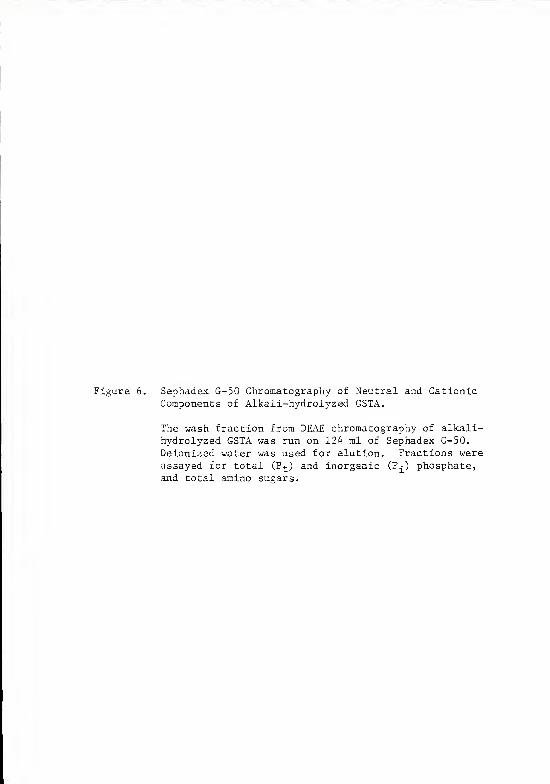
Figure 6. Sephadex G-50 Chromatography of Neutral and CationicComponents of Alkali-hydrolyzed GSTA.
The wash fraction from DEAE chromatography of alkali-hydrolyzed GSTA was run on 124 ml of Sephadex G-50.
Deionized water was used for elution. Fractions wereassayed for total (P t ) and inorganic (Pj) phosphate,and total amino sugars.

49
20 40fraction no.

3 "H W
<!

51
CO CN —|LU/ saioiuu^oi'JDBns ouiiuo 10404

DISCUSSION
Peptidoglycan Composition
Amino acid and amino sugar analyses of purified wall hydrolysates
first suggested the presence of wall associated polymers other than
peptidoglycan in the wall of Artkrobacter arystallopoieies . The
boiling SDS extraction used to purify walls is a very rigorous method
which should remove all noncovalently associated cellular materials.
Despite this, small amounts of the amino acids aspartic acid, threonine,
glycine, and valine were detected (Table 2). These amino acids are
not usually found in the pentapeptide of peptidoglycan. Also, there
was more glucosamine than could be accounted for by peptidoglycan
structure and there was galactosamine present which has not been re-
ported to occur as part of the peptidoglycan.
Krulwich et al . (55) have also reported on the amino acid and
amino sugar composition of walls purified from A. avystallovoietes 15481.
They also found small amounts of aspartic acid (0.15 mole/mole relative
to glutamic acid in spheres, none found in rods) and glycine (0.14
mole/mole in spheres, 0.04-0.05 mole/mole in rods). They did not find
valine or threonine but did find serine (0.13 mole/mole in spheres, none
in rods) . It is conceivable that serine and threonine might be
confused in some amino acid analyser programs since these amino acids
are similar in structure. On the program used for my experiments,
serine and threonine did elute next to each other but they were
52

53
distinctly different peaks on standard runs. Krulwich et al. (56)
used Ckzlaropsis B enzyme (N,0-diacetylmuramidase) (41), lysostaphin
(a mixture containing endo-N-acetylglucosaminidase and a peptidase
which releases N-terminal alanine and glycine from cell walls of
Staphylococcus aureus strain Copenhagen, hydrolyzes polyglycine cross-
bridges, and, to a lesser extent, N-acetylmuramyl-L-alanine linkages)
(15) , and AL-1 (a peptidase which breaks peptide crossbridges and
N-acetylmuramyl-L-alanine linkages) (79) in separate experiments to
obtain soluble fragemnts which were separated by ECTEOLA cellulose
and gel filtration. N-terminal and C- terminal amino acids were then
determined for various fragments. By these analyses, they came to
the conclusion that aspartic acid and serine were not connected with
either peptidoglycan or teichoic acid. They suggested that these
amino acids were either contaminants or part of a small additional
peptide in the wall. According to their results, the glycine present
was a part of some of the crossbridges in spherical populations. The
glycine present in rod populations was not, however, part of the
peptidoglycan and presumably could also be a contaminant or part of
an additional peptide. Tipper et al. (79) working in the same lab
as Krulwich et al. also have reported on the cell wall composition of
spherical .4. crystallopoietes . The composition they obtained is
very similar to that of Krulwich et al. except that Tipper et al. re-
ported about 0.03 umole/mg of wall of aspartic acid, serine, threonine,
leucine, histidine, and methionine. Since their walls contained 0.42
umoles of total glutamic acid/mg of wall, 0.03 umole/mg of wall would
be 0.07 moles/mole of glutamic acid. The nature of these trace

54
amino acids and the ones which I have found is unclear but their
presence calls for more research dealing with this problem.
Krulwich et al. (55) found additional glucosamine (spheres, 2.09
mole/mole of glutamic acid; rods, 1.74-2.01 mole/mole) ,galactosamine
(spheres, 0.31 mole/mole; rods, 0. 25-0. 30 mole/mole) .unidentified
phenol-sulfuric acid-reactive material (spheres, 1.95 mole/mole;
rods 1.04-1.37 mole/mole) , and phosphate (spheres, 2.02 mole/mole; rods,
1.88-2.12 mole/mole). Tipper et al. (41) also reported similar
results for glucosamine and galactosamine.
The walls analyzed in our laboratory also showed extra glucosamine
(Table 1) , although the amount was generally less than that found
by Krulwich et al. (55) or Tipper et al. (70). Quantitative differences
in accessory polymers between the present data and past data are to
be expected since, (1) the media used differ and, (2) the strains
of bacteria used could be different because of extensive subculturing.
Galactosamine was also found by our lab but it was not quantitated in
this experiment since at the time the identity of that peak was
unknown. Phenol-sulfuric acid-reactive material was also found in
TCA extractable material but it was found that this material could
be separated from teichoic acid by chromatography on DEAE-Sephadex.
It seems likely that this material may contain the glucose which
showed up in paper chromatography of acid hydrolysates of TCA extractable
material.
Krulwich et al. (55) were able to extract all the phosphate
present in walls with 100% TCA at 60°C in less than one hour for
both sphere and rod wall preparations. They concluded that this
phosphate must be contained in teichoic acid since teichoic acids
are extractable under those conditions. There are, however, other

55
accessory polymers which have been discovered since the paper of Krulwich
et al. (55) and which are extractable under conditions milder than
those of Krulwich et al. (5,8,51,65). Tipper et al. (79) also found a
phosphate polymer which co-eluted with some of the polysaccharide solu-
bilized by Myxobaater AL-1 enzyme. Nevertheless, Krulwich et al. have
turned out to be correct in their assumption even though today their
data would not be considered sufficient to establish the presence of a
teichoic acid as opposed to a sugar 1-phosphate polymer or a phosphor-
ylated polysaccharide. Krulwich probably realized this later because in
a paper a year after the one described above, Krulwich and Ensign (53)
refer to the same moiety only as a "phosphate-containing polymer of the
cell walls".
Krulwich et al. (55) were correct about the lack of amino acid sub-
stitution of the teichoic acid. They found amino acids in their TCA
extract but they were in the same ratios as were found in the peptido-
glycan. They also incubated their purified cell wall in two alkaline
buffers (pH 9.2 and pH 10.5) and found that no amino acids were released.
This indicates that no amino acids are ester-linked to either polyols or
to polysaccharides. In this work amino acid and amino sugar analyses of
crude teichoic acids showed almost no amino acids present at all. The
trace of alanine present could account for no more than one residue for
every 100 glycerol phosphate residues in either GS or LS teichoic acid.
It is more likely that this alanine arose from peptidoglycan contamina-
tion.
Experimental data (see Table 2) suggest that all four non-penta-
peptide amino acids can be removed if the right protease is used.
This confirms the hypothesis that these amino acids are not part of

56
the pentapeptide. This leaves the possibilities that these amino acids
are contaminants, part of normal crossbridges, or part of a peptide
attached to peptidoglycan.
Trypsin is known to be a specific endopeptidase which breaks peptide
bonds in which the carbonyl group is donated by lysine or arginine.
Most peptidoglycan basic structures are notoriously resistant to
trypsin (74), even those which contain penultimate L-lysine in the
tetrapeptide (64). The reason for this resistance is not clear but
certainly those L-lysine residues which are involved in crossbridges
would not be susceptible due to a lack of positive charge on the
E-amino group which is a necessary condition for trypsin digestion
(49).
My data indicate that trypsin has very little if any effect
on the level of alanine in purified cell walls. The alanine ratios
remain fairly constant regardless of which proteolytic enzyme is
used. The alanine ratios reported here agree fairly well with those
of Krulwich et al . (55). The alanine in excess of the two moles in
the tetrapeptide probably is due to the L-alanine crossbridges
proposed by Krulwich et al . (55). By C-terminal analysis, they
estimated that from 60 to 70% of the peptides are crosslinked, which
also agrees with the amount of excess alanine reported here.
If the effectiveness of the different proteolytic enzymes is
compared, trypsin seems to be the most effective in removing the
trace amino acids. The fact that trypsin removed most of these
amino acids while a-chymotrypsin and pronase did not, may indicate
that cleavage of a specific linkage to peptidoglycan is necessary
to remove a small peptide. Also, it had earlier been noticed that
if the concentration of pronase-digested wall to be analyzed was

57
increased tenfold, a broad spectrum of non-pep tidoglycan amino acids
appeared. This would seem to indicate that the trace amino acid re-
present only the most abundant amino acids present and, therefore,
are the only ones normally detected since the levels of these non-
peptidoglycan amino acids were so low. Whether or not trypsin can
remove or lower the amounts of these other amino acids is unknown.
The possibility that the trace amino acids are no more than
contaminants from membrane protein can not be discounted. This
problem warrants further research. It is possible that by using a
N-acetylmuramidse to solubilize the peptidoglycan, these amino acids
could be characterized as belonging either to a high or low molecular
weight fraction on gel filtration.
Perhaps the most important fact in this experiment is that extra
glucosamine and galactosamine were present. Combined with the presence
of significant amounts of phosphate, this suggested the possibility
that a teichoic acid was present. Since Krulwich et al. (55) had
found that phosphate could be TCA extracted, a similar kinetic
experiment was carried out but using the milder temperature (4°C as
compared to 60°C that Krulwich et al. (55) used) normally used to
extract teichoic acid with the least degradation (8,51).
TCA Extraction
As was shown in the Results section, the initial rate of extraction
of phosphate was relatively high and was almost the same for GS
and LS. By only 8 hours, about 73% of the total extractable phospahte
had already been solubilized. These initially high rates of extraction

5ET
then dropped dramatically so that another 67 hours were required to
remove the remaining 27% of the extractable phosphate. Thus, the
average rate of extraction for the first 8 hours was about 9% of the
extractable phosphate per hour whereas the average rate after 8 hours
was about 0.4% per hour. This amounted to more than a 22-fold differ-
ence. The explanation for this difference in rates is not known but
it does seem to indicate that there may be two different types of
reactions occurring. After 75 hours of extraction, 81% of the GS and
53% of the LS total phosphate was solubilized. If the slow rate of
extraction remained constant, it would take 5 days for GS and 8 days
for LS phosphate to be completely extracted. Ghuysen et al. (34)
reported that it took 3 weeks to extract more than 95% of the teichoic
acid of Staphylococcus aureus. In the light of that study, it is
likely that the rate of extraction does not remain constant but
decreases further still with time.
The actual dry weight of teichoic acid recovered after 96 hours of
extraction varied depending on the method used for recovery of
teichoic acid from TCA supernatants. If the weights recovered are
corrected for the percentage recovery, then the actual weight of
teichoic acid in the native wall (GS or LS) may be as high as about
43% (crude TA dry weight percentage of purified wall dry weight) . A
small proportion of this crude teichoic acid is contaminating poly-
saccharide, so the actual figure is probably closer to around 40%.
The amounts found in certain other gram positive bacteria range from
20% to 50% of the dry weight of the wall (8)
.
59
Characterization of Acid Hydrolysates
To determine whether or not this phosphate-containing polymer was
a teichoic acid, acid hydrolyses were carried out under conditions suf-
ficiently rigorous to degrade a glycerol or ribitol teichoic acid to
its monomers (3)
.
Table 3 shows the phosphate-containing monomers of acid-hydrolyzed
crude teichoic acid. The ascending technique seems to give the least
ambiguous results. The hydrolyzed teichoic acid spots matched fairly
well with the mobility of 1,2 diphosphoglycerol. The fact that there
were two spots in both the standards and the hydrolysates is probably
due to some inorganic phosphate which should migrate a little farther
than 1,2 diphosphoglycerol (50) and exists as a minor contamination in
the 1,2 diphosphoglycerol standard. Hydrolyzed cardiolipin also showed
a pattern of light and dark spots because this compound upon hydrolysis
should give rise to 1,3 diphosphoglycerol (Rf, 0.12) and glycerol mono-
phosphates (Rj, 0.35). These mobilities agree well with those found in
the literature (7,29,50). The hydrolyzed teichoic acid spots on the
descending run make it more difficult to choose between the 1,2 and the
1,3 compound. Indeed, the hydrolyzed teichoic acid samples may be a
mixture of both which runs together on paper chromatography. This is
actually what one would expect since a certain amount of ester migra-
tion can occur under acidic conditions (3) . It is noteworthy that no
glycerol monophosphates were detected but the reason for this is not
clear.
In Table 4, it seems quite clear that the mobility of the hydro-
lyzed teichoic acid spots matched that of glycerol. The true picture,

6tr
however, is somewhat more cloudy than that. Anhydroribitol is re-
ported to have the same mobility as glycerol under the conditions of
chromatography used in this experiment. Apparently, the conditions
of hydrolysis used for both the samples and the standard (2 N HC1,
100°C, 3 hours) were not sufficient to convert a detectable amount
of ribitol to anhydroribitol. Anhydroribitol is reported to give
a slow reaction with periodate-Schif f reagent (7) and so the fast
reaction given by the hydrolyzed teichoic acid samples confirms that
glycerol, not anhydroribitol, was present. The yellow spots which
did not migrate were probably sugars which are characterized in
Table 4.
Both GS and LS crude teichoic acids appear to have the same sugars
present as evidenced by the banding patterns obtained in the ethyl
acetate-pyridine-water solvent system. Both preparations had a
silver nitrate reactive spot which migrated the same distance as
glucose. Both preparations also had darker areas in the long tails
which extended from the origin to about halfway to the solvent front.
These darker areas corresponded well to glucosamine and galactosamine
standards. Later analyses confirmed the covalent linkage of these
latter sugars to teichoic acid. The glucose is not connected with
teichoic acid but may be part of the wall nevertheless.
Since the spots that were presumably glucose did not exactly
correspond in mobility to glucose in every run, glucose was added as
an internal standard to the GS and LS acid-hydrolyzed preparations
(Table 6) . In this chromatograph standard glucose and sample glucose

61
migrated as a single spot. These spots moved slower than the glucose
standard alone. The probable cause for this is the presence of
small amounts of salt in the sample.
Amino Acid and Amino Sugar Analyses of Teichoic Acid
Amino acid and amino sugar analyses confirmed the identity of the
glucosamine and galac to samine indicated by paper chromatography.
These analyses also indicated that there was about five times as
much glucosamine present as galactosamine.
The traces of alanine present would contribute less than one
residue for every 100 glycerol phosphate residues. For this reason,
it is felt that this alanine probably arose from contamination by
peptidoglycan.
DEAE Chromatography
To purify the crude teichoic acids, ion exchange chromatography
was used. The teichoic acid preparations were applied to the DEAE-
Sephadex under conditions which allowed the anionic teichoic acids
to stick to the resin. Water washes removed a similar amount of
phenol-suifuric acid reactive material from both preparations. This
material is presumed to contain the glucose present in crude teichoic
acid samples as demonstrated by paper chromatography. Lyophilization
of this material rendered it water-insoluble and so no further analyses
of this material were carried out. No phenol-H2S04 reactive material

^T
was found in the salt-eluted fractions, so glucose is presumed not
to be part of the teichoic acid structure.
The profiles of Figure 3 show that GSTA and LSTA are different
in their affinity for DEAE groups. The peaks of phosphate eluted
at salt concentrations which were about 0.1 M different. This suggests
that there may be a difference in net charge of the molecules being
separated. These profiles also show that the amino sugar curve
closely matched that of phosphate. If the ratios between phosphate
and amino sugar are calculated for each fraction in the main peaks,
these ratios are relatively constant. They are so constant that in
more than 95% of the cases, the differences can be attributed to a
change from the mean ratio of an amount that can be accounted for
by a change of one or two residues. Thus suggests that these two
moieties were associated with each other and not simply eluting close
to one another.
Dry weight recoveries were quite low as compared to phosphate re-
coveries. This is probably due more to inaccuracies in dry weight
measurement (resulting from incomplete water removal from samples)
than to a removal of contamination.
Acid Hydrolyses of Purified Teichoic Acids
The amount of amino sugar in the two preparations was different
but not radically so. The mild acid hydrolysis only demonstrated
that about one half of the amino sugars were N-acetylated, but this
is strictly a minimum figure. Even under these mild conditions some
hydrolysis of the N-acetyl groups probably occurs. It is therefore

probable that most amino amino sugar was N-acetylated. DMAB reagent
showed a limited reactivity to amino sugar associated with teichoic
acid because the N-acetyl amino sugar is bound at the C-l position.
N-Acetylation with acetic anhydride did not increase this reactivity.
This would seem to indicate that all the amino sugars are already
N-acetylated. When the teichoic acid was acid hydrolysed a large in-
crease in reactivity to DMAB occurred due to exposure of C-l.
Gel Filtration
Teichoic acids were also characterized by gel filtration. It
was found necessary to use Sephadex G-100, a gel with a molecular
weight exclusion limit much higher than any known teichoic acid
molecular weight. This is probably due to the high charge and ex-
tended shape common to teichoic acids (25,27). The profiles in
Figure 4 again show that there was a considerable difference between
GSTA and LSTA. The most obvious cause for this is a difference in
molecular weight but if there were a large difference in charge or
shape of the molecule, it could affect the retention of the teichoic
acid molecules.
Determination of Chain Length
To further characterize the teichoic acids and to determine if
molecular weight was the determining factor in the gel filtration
profile, chain lengths were determined. Average chain length can
be determined by releasing the terminal phosphomonoester group

64
with alkaline phosphatase and measuring the inorganic phosphate released
and comparing that to the total phosphate content. This gives the
average number of glycerol phosphate residues per chain if there
is a one-to-one correspondence of glycerol to phosphate. These deter-
minations showed that GSTA at 38 glycerol phosphate units was only
about half as long as LSTA at an average of 70 units. These numbers
are representative of purified teichoic acid preparations only and
may not reflect accurately the native chain lengths as found in the
wall (8) . TCA is known to break phosphodiester bonds (77) . Phos-
phodiester bonds not only link the glycerol phosphate monomers, they
are also responsible for linking the teichoic acid to peptidoglycan
in the only well-characterized cases to date (22,38,77). It is
not surprising then that a certain amount of degradation of chain
length would occur upon TCA extraction of teichoic acids. Indeed,
this has been observed in several cases (8,34,51). The kinetic data
seem to indicate that there may be two mechanisms of extraction.
This may be reflective of the phosphodiester links which bond the
backbone units and those which may link the backbone to peptidoglycan.
Even so, the difference between GSTA and LSTA would be hard to explain
by differing labilities to acid extraction when their composition
is so similar. Thus, even if the chain lengths do not accurately
reflect those in the native wall, there still should be a significant
difference between the lengths of GSTA and LSTA in the wall.
It is very probable that the differing chain lengths of GSTA
and LSTA account for some of the differences observed in ion-exchange
chromatography. DEAE chromatography has been shown to separate
oligonucleotides according to net negative charge which is a function

65
of chain length under conditions minimizing charge and secondary
binding forces of the purine and pyrimidine bases (72,81,85). It
is reasonable to assume then that LSTA with an average of 70 negative
charges per molecule is going to be eluted at a higher ionic strength
than is GSTA which only has as average of 38 negative charges per
molecule. This is, in fact, what is observed (Figure 3). This
reasoning can be extended to the material which elutes at lower
ionic strengths which would be of lower chain length than the bulk
of the material. This material probably resulted from degradation by
TCA hydrolysis of backbone phosphodiester bonds.
The difference in chain length also is reflected in the gel
filtration profile but it should be noted that the difference in
charge per molecule may also play a role in the behavior of teichoic
acids on Sephadex G-100. Pharmacia states that Sephadex G-100 contains
small numbers of carboxyl groups and, therefore, at low ionic strengths,
negatively charged moleclues may be excluded from the gel to a
greater extent than an uncharged molecule of the same molecular weight
(67).
Glycerol-Phosphate Ratios
Many of the above conclusions can only be drawn by assuming that
there is a poly (glycerol phosphate) backbone. To confirm the presence
of such a backbone, it is necessary to show a one-to-one ratio
between glycerol and phosphate. As Table 8 shows, this is the case
for both GSTA and LSTA.

66
Alkaline Hydrolysis
The final characterization of teichoic acid was by alkaline hydro-
lysis. The primary goal of this experiment was to establish whether
or not the amino sugars were covalently attached to teichoic acid.
Alkaline hydrolysis leaves glycosidic linkages intact while cleaving
phosphodiester linkages where free hydroxyl groups are available on
neighboring carbons allowing for a cyclic phosphate intermediate to
form (50).
Figure 5 shows that a substantial amount of amino sugar elutes
from DEAE-Sephadex as a single peak. Since only anionic molecules
should be bound to this resin, this suggests tha the amino sugar is
still bound to glycerol phosphate. This peak accounts for 87% of
the amino sugar in the alkali-hydrolyzed GSTA. Thus, the majority
of amino sugar is covalently attached to teichoic acid and it sug-
gests that the poly (glycerol phosphate) backbone is 2,3 linked rather
than 1,3 linked. In a 1,3 teichoic acid if a glycosidic bond ties
up C-2 of a glycerol phosphate residue, then there is no free hydroxyl
on that residue for the necessary cyclic phosphate to form. For this
reason, one expects only glycosylglycerol for substituted residues.
In contrast, glycosylglyceromonophophates can only result from a 2,3-
linked teichoic acid structure. It has never been found in membrane
teichoic acids and only in a few cases in wall teichoic acids (32,63).
The amount of amino sugar exceeds the amount of phosphate in the amino
sugar peak by a factor of about 3.2. This suggests that there may
be an average of three amino sugars linked to each other by

67
glycosidic bonds and this trimer is attached to a single glycerol
phosphate residue.
The other 12% of the amino sugar in the alkaline-hydrolyzed
GSTA was characterized as being low molecular weight because it was
almost completely included in a Sephadex G-50 gel (Figure 6) . This
material was then eluted on a cation exchange resin. All the
remaining phosphate was washed through the column with 0.1 M NaCl.
Two peaks of amino sugar were then eluted using a linear NaCl gradient.
Together these accounted for all of the amino sugar from the DEAE
wash fraction. No phosphate was detected in either fraction. 3oth
peaks did contain glycerol. The smaller peak contained about three
amino sugar residues for each glycerol residue while the larger
peak contained 6.5 amino sugars per glycerol. It would seem, there-
fore, that CM-Sephadex acts in manner analogous to DEAE-Sephadex in
that it can separate positively charged molecules with similar charge
densities by chain length. These data also suggest that amino sugar
is covalently attached to teichoic acid and may exist as trimers or
hexamers attached to single glycerol residues. The significance of
the larger amount of the hexamer in this relatively small fraction
of amino sugar is not known. Although these results were obtained
only for GSTA, it seems reasonable that LSTA follows a similar but
not identical pattern.

68
Summary of Teichoic Acid Structure
Arthrobacver arystallopoietes contains a glycerol wall teichoic
acid in both sphere and rod morphologies in approximately equal
amounts. This teichoic acid is 38 glycerol phosphate units long
in spheres and about 70 units long in rods. N-Acetylhexosamine
(N-acetylglucosamine, about 83% and N-acetylgalactosamine, about 17%)
is attached by a glycosidic linkage from CI of the hexosamine to CI
of glycerol. On the average, there are 11 hexosamines per chain in
sphere teichoic acid while there are about 15 hexosamines per chain
in the rod teichoic acid. These hexosamines may exist in polymerized
side chains which have three sugars on the average. Thus, only about
11% (GS) or 7% (LS) of the glycerol phosphate units are substituted.
The poly (glycerol phosphate) backbone is 2,3 linked for GSTA and
probably for LSTA also. This is the first teichoic acid that has
been characterized to this extent in the genus Arthrobaatev
.
Possible Roles for Teichoic Acids in Avthrobaater
One role of teichoic acids that is well established is binding of
cations, particularly divalent cations ( 6,10, 26,42 , 46) . Evidence
has been presented that wall and membrane teichoic acids provide a
controlled reservoir of bound Mg+2 for membrane bound enzymes (46)
which require it for maximal activity (2,21,66). It has been suggested
that the wall teichoic acid scavenges Mg+i- from the environment and
transfers it to lipoteichoic acid which interacts with the membrane

69
enzymes. Baddiley et al. (10) have shown that alanylated teichoic
acid interacts less strongly with Mg+2 and, therefore, the degree of
D-alanine substitution may act as a controlling influence on Mg+2
binding.
It seems reasonable that the teichoic acid in A. aystallopo-ietes
could serve the same function since its structure is similar to
some of those used in the above mentioned studies. If this were
true, then it could be that the longer chains found in rod shaped
bacteria might more effectively scavenge Mg+2
than the short chains
of the spheres. This would be consistent with the higher growth
rate of rods which would presumably necessitate a greater flux of
Mg to the membrane.
Another function of teichoic acids is suggested by the studies
of Burge et al. (20) and Milward and Reavely (62). By examination of
peptidoglycans containing teichoic acid and those which had teichoic
acid artificially removed, these authors found that extraction of
teichoic acid affects the flexibility of the peptidoglycan. It was
suggested (20) that since teichoic acid can pass through the pores
in the outer layers of peptidoglycan it would resist shear between
adjacent peptidoglycan sheets and still allow for expansion or
contraction of the wall.
The most interesting function as far as this work is concerned
is suggested by several studies in which teichoic acid changes
effect autolytic enzymes.
Holtje and Tomasz (45,80) have described an N-acetylmuramyl-L-alanine
amidase of Pneicmocooaus which required choline residues in the wall
teichoic acid for maximal binding. If ethanolamine residues were

70
substituted, the cell walls were resistant to the autolysin. Besides
lowered binding of the autolysin to the wall, this effect was due to
the inability of the altered teichoic acid to convert the autolysin to
its active form. Herbold and Glaser (43) have described a similar N-
acetylmuramyl-L-alanine amidase in Bacillus subtilis . Again cell walls
lacking teichoic acid had a much lower affinity for the enzyme than
those with normal levels of teichoic acid. In a different strain of
Bacillus subtilis, Brown et al. (16,18) had such trouble purifying auto-
lysin from teichoic acid that they suggested that the two molecules
might be covalently attached to each other. They speculated that the
close association between autolysin and the teichoic acid might serve
to localize the autolysin for maximum utilization of substrate.
In a temperature conditional morphological mutant of Bacillus
subtilis, Boylan et al. (12,13,24) found that at the non-permissive
temperature teichoic acid was deficient. These mutants grew as normal
rods at 30°C but changed to irregular spheres at 45°C. The walls of the
spheres were also deficient in N-acyl muramyl-L-alanine amidase. Rogers
et al. have found similar results in seven rod A mutants of Bacillus
subtilis (17,71) and Bacillus liche.nifovm.is (33).
Other authors (44,75) have also suggested that autolysins may
play a significant role in the determination of morphology in all
bacteria.
In A. crystallcpoietes it has been shown that the activity of
N-acetylmuramidase correlates with morphogenesis (53) . The high
activity of the autolysin in spheres results in the relatively short
glycan backbones found in the peptidoglycan. In the same way, the
low activity of this enzyme in rod walls results in relatively

71
long glycans. The length of the glycan chains probably influences
the rigidity of the peptidoglycan and, therefore, the morphology
of the organism (55)
.
The present work has shown that there is almost a two-fold differ-
ence in the length of teichoic acids isolated from spherical and
rod-shaped A. erystallopoietes. In light of the correlations
between morphology, autolysins, and teichoic acids discussed above,
it does not seem unreasonable that this difference may play a role
in morphogenesis.
Phosphate analyses of purified wall show that there is slightly
more phosphate per mg of rod wall than in sphere wall (rods, 21 ug/ml
wall; spheres, 18 ug/mg wall). Since the rod teichoic acid has
almost twice the phosphate per chain as the sphere teichoic acid
because of its length, this means that there are about one half as
many rod teichoic acid chains as there are sphere teichoic acid
chains. If autolysin were binding to the teichoic acid chains, the
rod wall would present about one half as many binding sites proximal
to the wall as would the sphere wall.
A question that arises from this model is why the autolysin does
not cleave the N-acetylmuramyl-N-acetylglucosaminyl residue in peptido-
glycan to which it is attached. This would be disadvantageous to the
cell because both teichoic acid and autolysin would be lost from the
cell which represents a considerable energy investment wasted. One
possible solution to this problem could lie in the specificity of the
autolysin. The autolysin in A. avystallopo-ietes thought to be
responsible for control of glycan length is an N-acetylmuramidase.
It is possible that this enzyme can not hydrolyze at the reducing

72
end of an N-acetylmuramic acid which has the C-6 hydroxyl tied up in
covalent linkage. Lysozyme is known to be inhibited under these
conditions and teichoic acids are known to inhibit lysozyme in certain
cases (78). The only well characterized linkages of teichoic acid
to peptidoglycan are to the C-6 hydroxyl of N-acetylmuramic acid.
In both our laboratory and that of Krulwich et al. (55), lysozyme
was found to be poorly lytic against purified cell walls of A. cvystallo-
poietes. Chalaropsis B enzyme, on the other hand, completely solubilizes
these walls. Chalaropsis B enzyme is an N,0-diacetylmuramidase
which is similar to lysozyme except that it will cleave at the reducing
ends of N-acetylmuramic acids which are substituted in the C-6 position.
So, apparently something is attached at this position in .4. cvystallo-
voietes. It is possible that this might be teichoic acid but it
could also be polysaccharide. If it were teichoic acid, it may be
the reason why autolysin does not remove teichoic acid. Krulwich
and Ensign (53) did find that the autolysin preferentially solubilizes
peptidoglycan which is low in phosphate content. Another reason for
this result might be steric hindrance of the enzyme by the teichoic
acid.
It may be that teichoic acid does not bind the autolysin at all
but only serves in substrate recognition by the enzyme as suggested
by Higgins and Shockman (44) . In Streptococcus faecalis, the N-acetyl-
muramidase binds to TCA extracted walls but these walls are hydrolyzed
slower by autolysin than untreated walls. The TCA extracted walls are,
however, a better substrate for lysozyme.

73
It is also possible that the divalent cation binding property of
teichoic acids is important in regulating the autolysin. Clearly,
there is much more to be found out about the roles of teichoic acids
in .4. cvystallovoietes. Hopefully, further research will shed light
on these roles.

LITERATURE CITED
1. Ames, B.N. 1966, Assay of inorganic phosphate, total phosphate andphosphatases. Meth. in Enzymol. 8: 115-118.
2. Anderson, R.G., H. Hussey, and J, Baddiley. 1973. The mechanism ofwall synthesis in bacteria. Biochem. J. 227:11-25.
3. Archibald, A.R. and J, Baddiley. 1966. The teichoic acid. Adv.Carbohydrate Chem. 21:323-375.
4. Archibald, A.R., J. Baddiley, and N.L, Blumson. 1968. The teichoicacids, Adv. Enzymol. 5/7:223-253.
5. Archibald, A.R., J. Baddiley, D. Button, S. Heptinstall, and G.H.Stafford. 1968. Occurrence of polymers containing N-acetylglucos-amine 1-phosphate in bacterial walls. Nature (London) 22 9:855-856.
6. Archibald, A.R., J. Baddiley, and S. Heptinstall. 1973. The alanineester content and magnesium binding capacity of walls of Staphylococcusaureus H grown at different pH values. Biochim. et Biophys." Acta213:629-634.
7. Armstrong, J.J., J. Baddiley, J.G. Buchanan, B. Carss, and G.R.Greenberg. 1958. Isolation and structure of ribitol phosphate de-rivatives (teichoic acids) from bacterial cell walls. J. Chem. Soc.(London), p. 4344-4354.
8. Baddiley, J. 1972. Teichoic acids in cell walls and membranes ofbacteria. Essays in Biochem. 3: 35-77,
9. 3addiley, J., J.G. Buchanan, R.E. Handschumacher , and J.F. Prescott.1956. Chemical studies in the biosynthesis of purine nucleotides.Part I. The preparation of N-glycylglycosylamines. J. Chem. Soc.pp. 2818-2823.
10. Baddiley, J., I.e. Hancock, and P.M. A. Sherwood. 1973. X-ray photo-electron studies of magnesium ions bound to the cell walls of gram-positive bacteria. Nature (London) 243: 43-45.
11. Belcher, R. , A.J. Nutten, and CM. Sambrook. 1956. The determinationof glucosamine. Analyst 73:201-208.
12. Boylan, R.J. and N.H. Mendelson. 1969, Initial characterization ofa temperature-sensitive rod" mutant of Badlluo subvilis. J.3acteriol. 2 55:1316-1321.,
74
75
13. Boylan, R.J., N.H. Mendelson, D. Brooks, and F.E. Young. 1972.Regulation of ^the bacterial cell wall: Analysis of a mutant ofBacillus subtilis defective in biosynthesis of teichoic acid. J.Bacteriol. 110:281-290.
14. Braun, V. and U. Sieglin. 1970. The covalent murein-lipoproteinstructure of the Escherichia coli cell wall; the attachment siteof the lipoprotein on the murein. Eur. J. Biochem. 13:336-346.
15. Browder, H.P., W.A. Zygmunt, J.R. Young, and P. A. Tavormina. 1965.Lysostaphin: Enzymatic mode of action. Biochem. Biophys. Res.Comm. 75:383-389.
16. Brown, W.C., D.K. Jraser, and F.E. Young. 1970. Problems in puri-fication of a Bacillus subtilis autolytic enzyme caused by associa-tion with teichoic acid. Biochim. Biophys. Acta 135:308-315.
17. Brown, W.C., C.R. Wilson, S. Lukehart, F.E. Young, and M.A. Shiflett.1976. Analysis of autolysins in temperature-sensitive morphologicalmutants of Bacillus subtilis. J. Bacteriol. 125:166-173.
18.
19.
Brown, W.C. and F.E. Young. 1970. Dynamic interactions between cellwall polymers, extracellular proteases and autolytic enzymes.Biochem. Biophys. Res. Comm. 35:564-568.
Brundish, D.E., N. Shaw, and J. Baddiley. 1965. The occurrence ofglycolipids in gram-positive bacteria. Biochem. J. 35 :21c-22c.
20. 3urge, R.E., A.G. Fowler, and D.A. Reaveley. 1977. Structure ofthe peptidoglycan of bacterial cell walls. I. J. Mol. Biol.Ii7:927-953.
21. Burger, M.M. and L. Glaser. 1964. The synthesis of teichoic acids.I. Polyglycerophosphate. J. Biol. Chem. 233:3168-3177.
22. Button, D., A.R. Archibald, and J. Baddiley. 1966. The linkage be-tween teichoic acid and glycosaminopeptide in the walls of a strainof Staphylococcus lactis. Biochem. J. 99 :llc-14c.
23. Chen, P.S., Jr., T.Y. Toribara, and H. Warner. 1956. Microdeter-mination of phosphorus. Anal. Chem. 25:1756-1758.
24. Cole, R.M., T.J. Popkin, R.J. Boylan, and N.H. Mendelson. 1970.Ultrastructure of a temperature-sensitive rod mutant of Bacillussubtilis. J. Bacteriol. 753:793-810.
25. Critchley, P., A.R. Archibald, and J. 3addiley. 1962. The intra-cellular teichoic acid from Lactobacillus arabinosus 17-5.Biochem. J. 55:420-430.
26. Cutinelli, C. and F. Galdiero. 1967. Ion-binding propertiesof the cell wall of Staphylococcus aureus. J. 3acteriol. 33:2022-2023.

76
28
27. Doyle, R.J. , M.L. McDannel, U.N. Streips, D.C. Birdsell, and F.E.Young. 1974. Polyelectrolyte nature of bacterial teichoic acids.J. Bacterid. 125:606-615.
Dubois, K.A., K.A. Gilles, J.K. Hamilton, P. A. Rebers, and F. Smith.1956. Colorimetric method for determination of sugars and relatedsubstances. Anal. Chem. 23:350-356.
29. Ellwood, D.C, M.V.Kelemen, and J. Baddiley. 1963. The glycerolteichoic acid from the walls of Staphylococcus albus N.T.C.C. 7944.Biochem. J. 3(3:213-225.
30. Ensign, J.C. and S.C. Rittenberg. 1963. A crystalline pigmentproduced from 2-hydroxypyridine by Arthrcbacter crystallopoietesn. sp. Arch. Mikrobiol. 47:137-153.
31. Ensign, J.C. and R.S. Wolfe. 1964. Nutritional control of morpho-genesis in Arthrobactsr crystallopoietes . J. Bacterid. 37:924-932.
32. Forrester, I.T. and A.J. Wicken. 1966. The chemical compositionof cell walls of some thermophilic bacilli. J. Gen. Microbiol.42:147-154.
33. Forsberg, C.W. , P.B. Wyrick, J.B. Ward, and H.J. Rogers. 1973.Effect ^of phosphate limitation on the morphology and wall compositionof Bacillus licheniformis and its phosphoglumutase-def icient mutants.J. Bacteriol. 213:969-984.
34. Ghuysen, J.M. , D.J. Tipper, and J.L. Strominger. 1965. Structureof the cell wall of Staphylococcus aureus, strain Copenhagen. IV.The teichoic acid-glycopeptide complex. Biochemistry 4:474-485.
35. Ghuysen, J.M. , D.J. Tipper, and J.L. Strominger. 1966. Enzymesthat degrade bacterial cell walls. Meth. in Enzymol. 3:115-118.
36. Goldstein, I.J. and W.J. Whelan. 1962. Structural studies ofdextrans. Part I. A dextran containing a-1 ,3-glcosidic linkages.J. Chem. Soc. (London), p. 170-175.
37. Goodwin, S.D. and J.G. Shedlarski. Jr. 1975. Purification of cellwall peptidoglycan of the dimorphic bacterium Caulobactev crescentus.Arch. Biochem. 3iophys. 170:23-36.
38. Grant, W.D. and A.J. Wicken. 1968. Muramic acid phosphate and thelinkage of teichoic acid to peptidoglycan in Bacillus stearothermo-philus cell walls. Biochem. Biophys. Res. Comm. -32:122-128.
39. Hamilton, R.W. , E.C. Achberger, and P.E. Kolenbrander . 1977. Con-trol of morphogenesis in Arthrcbaater crystallopoietes: Effect ofcyclic adenosine 3
'
,5 '-monophosphate. J. Bacteriol. 225:874-879.

77
40. Hanes, C.S. and F.A. Isherwood. 1949. Separation of the phosphoricesters on the filter paper chromatogram. Nature (London) J o^: 1107-1112.
43.
44,
41. Hash, J.H. and M.V. Rothlauf. 1967. The N,0-diacetylmuramidase ofChalaropsis species. I.: Purification and crystallization. J. Biol.Chem. 242:5586-5590.
42. Heptinstall, S. , A.R. Archibald, and J. Baddiley. 1970. Teichoicacids and membrane function in bacteria. Nature (London) 225:519-521.
Herbold, D.R. and L. Glaser. 1975. Bacillus subtilis N-acetylmur-amic acid L-alanine amidase. J. Biol. Chem. 25(9:1676-1682.
Higgins, M.L. and G.D. Shockman. 1971. Procaryotic cell divisionwith respect to wall and membranes. In Critical Reviews in Micro-biology. Chemical Rubber Co. p. 29-72.
45. Holtje, J.-V. and A. Tomasz. 1975. Specific recognition of cholineresidues in the cell wall teichoic acid by the N-acetylmuramyl-L-alanine amidase of Pneumococcal . J. Biol. Chem. 255:6072-6076.
46. Hughes, A.H., I.C. Hancock, and J. Baddiley. 1973. The function ofteichoic acids in cation control in bacterial membranes. Biochem.J. 132:83-93.
47. Jacobsen, R.A. and L.F. Mazzuckelli. 1973. Evidence for two RNApolymerases in Arthrobacter , a morphogenetic bacterium. Biochem.Biophys. Res. Coram. 3:867-873.
48. Keddie, R.M. 1974. Genus II. Arthrobacter Conn and Dimmick 1947,300. In Sergey's Manual of Determinative Bacteriology, 8th edn.p. 618-625. Edited by R.E. Buchanan and N.E. Gibbons. Baltimore:Williams & Wilkins.
49. Keil, B. 1971. Trypsin. In The enzymes, 3rd edn. p. 249-275.
50. Kelemen, M.V. and J. 3addiley. 1961. Structure of the intracellularglycerol teichoic acid from Lactobacillus casei A.T.C.C. 7469. Bio-chem. J. 30: 246-254.
51. Knox, K.W. and A.J. Wicken. 1973. Immunological properties ofteichoic acids. Bacteriol. Rev. 37:215-257.
52. Kolenbrander, P.E. and M. Weinberger. 1977. 2-Hydroxypyridinemetabolism and pigment formation in three Arthrobacter species. J.Bacteriol. 132:51-59.
53. Krulwich, T.A. and J.C. Ensign. 1968. Activity of an autolyticM-acetylmuramidase during sphere-rod morphogenesis in Arthrobactercrystallopoietes. J. Bacteriol. 96:857-859.

78
54. Krulwich, T.A. and J.C. Ensign. 1969. Alteration of glucose meta-bolism of Arthrobaoter crystallopoietes by compounds which inducesphere to rod morphogenesis. J. Bacteriol. 37:526-534.
55. Krulwich, T.A., J.C. Ensign, D.J. Tipper, and J.L. Strominger. 1967.Sphere-rod morphogenesis in Arthrobaoter crystallopoietes. I. Cellwall composition and polysaccharides of the peptidoglycan. J. Bac-teriol. 5^:734-740.
56. Krulwich, T.A., J.C. Ensign, D.J. Tipper, and J.L. Strominger.1967. Sphere-rod morphogenesis in Arthrobaoter crystallopoietes.II. Peptides of cell wall peptidoglycan. J. Bacteriol. 34:741-750.
57. Kuhn, R. , M.P. Starr, D.A. Kugn, H. Bauer, and H.-J. Knackmuse.1965. Indigoidine and other bacterial pigments related to 3,3'-bipyridyl. Arch. Mikrobiol. 52:71-84.
58. Lowry, O.H. , N.R. Roberts, K.Y. Leiner, M.-L. Wu, and A.L. Farr.1954. The quantitative histochemistry of brain. I Chemical methods.J. Biol. Chem. 207:1-17.
59. Lowry, O.H., N.J. Rosebrough, A.L. Farr, and R.J. Randall. 1951.Protein measurement with the Folin phenol reagent. J. Biol. Chem.193:265-275.
60. Luscombe, B.M. and T.R.G. Gray. 1971. Effect of varying growthrate on the morphology of Arthrobaoter. J. Gen. Micro. 53:433-434.
61. Massey, L.K. , J.B. Clark, and R.A. Jacobsen. 1973. Changes in RNAtranscription during morphogenesis of Arthrobaoter crystallopoietes.J. Gen. Microbiol. 77:51-60.
62. Millward, G.R. and D.A. Reaveley. 1974. Electron microscopeobservations on the cell walls of some gram-positive bacteria.J. Ultrastruct. Res. 4o:309-326.
63. Naumova, 1.3. and M.Z. Xaretskaya. 1964. Some properties ofglyceroteichoic acids from Streptomyces rimosus T-118 and Strevtomyoesantibiotious 39. Dokl. Akad. Nauk. SSSR. 257:207-210.
64. Park, J.T. and R. Hancock. 1960. A fractionation procedure forstudies of the synthesis of cell-wall mucopeptide and of otherpolymers in cells of Staphylococcus aureus. J. Gen. Microbiol.52:249-258.
65. Partridge, M.D., A.L. Davison, and J. Baddiley. 1971. A polymer ofglucose and N-acetylglucosamine 1-phosphate in the wall ofMicrococcus sp. Al. Biochem. J. 222:695-700.
66. Patterson, P.H. and W.J. Lennarz. 1971. Studies on the membranesof bacilli. I. Phospholipid biosynthesis. J. Biol. Chem. 246:1062-1072.
79
67. Pharmacia booklet "Sephadex-gel filtration in theory and practice."1970. p. 11.
68. Previc, E.P. 1970. Biochemical determination of bacterial morpho-logy and the geometry of cell division. J. Theor. Biol. 27:471-479.
69. Previc, E.P. and N. Lowell. 1975. Peptidoglycan compositions of anew strain of Arthrobacter crystallovoietes during sphere-rod morpho-genesis. Biochem. et Biophys. Acta 422:377-385.
70. Reissig, J.L., J.L. Strominger, and L.F. Leloir. 1955. A modifiedcolorimetric method for the estimation of X-acetylamino sugars.J. Bio. Chem. 227:959-966.
71. Rogers, H.J. and C. Taylor. 1978. Autolysins and shape change inRod A mutants of Bacillus subtilis. J. Bacteriol. 255:1032-1042.
72. Rushizky, G.W. , E.M. 3artos, and H.A. Sober. 1964. Chromatographyof mixed oligonucleotides on DEAE-Sephadex. Biochemistry 5:626-629'.
73. St. John, A.C. and J.C. Ensign. 1976. Macromolecular synthesis andcell division during morphogenesis of Arthrobacter crystallovoietes
.
Arch. Microbiol. 222:51-58.
74. Saiton, M.R.J. 1953. Cell structure and the enzymic lysis ofbacteria. J. Gen. Microbiol. 5:512-523.
75. Schleifer, K.H., W.P. Hammes, and 0, Kandler. 1976, Effect ofendogenous and exogenous factors on the primary structures ofbacterial peptidoglycans. Adv. Microbial Physiol. 25:245-292.
76. Spackman, D.H., W.H. Stein, and S. Moore. 1958. Automatic recordingapparatus for use in the chromatography of amino acids. AnalChem. 55:1190-1206.
77. Strominger, J.L. and J, -M. Ghuysen. 1963. On the linkage betweenteichoic acid and the glycopeptide in the cell wall of Staphylococcusaureus. Biochem. Biophys. Res. Comm. 22:418-424.
73. Strominger, J.L. and J. -M. Ghuysen. 1967. Mechanisms of enzymaticbacteriolysis. Science 156: 213-221.
79. Tipper, D.J., J.L. Strominger, and J.C. Ensign. 1967. Structureof the cell wall of Staphylococcus aureus strain Copenhagen. VII.Mode of action of the bacteriolytic peptidase from Myxobacter andthe isolation of intact cell wall polysaccharides. Biochemistryo:906-920.
SO, Tomasz, A. 1968. Biological consequences of the replacement ofcholine by ethanolamine in the cell wall of Pneumococeus : Chainformation, loss of transf ormability, and loss of autolysins.Proc. Natl. Acad. Sci, U.S.A. 5.9:86-93.

80
81. Tomlinson, R.V. and G.M. Tener. 1963. The effect of urea,formamide, and glycols on the secondary binding forces in theion-exchange chromatography of polynucleotides on DEAE-Cellulose.Biochemistry 2:697-702.
12. Trevelyan, W.E., D.P. Procter, and J.S. Harrison. 1950. Detectionof sugars on paper chromatograms. Nature (London) 186; 444-445.
83. Van De Rijn, I. and A.S. Bleiweis. 1973. Antigens of Streptococcusmutans. I. Characterization of a serotype-specif ic determinantfrom Streptococcus '-nutans. Infect. Immunity 7:795-804.
84. Ward, CM., Jr. and G.W. Claus. 1973. Gram characteristics andwall ultrastructure of Arthrobacter crystallopoietes duringcoccus-rod morphogenesis. J. Bacteriol. 214:378-389.
85. Yogo, Y. and E. Wimmer . 1973. Poly(A) and poly (U) in poliovirusdouble stranded RNA. Nature New Biol. 242:171-174.

BIOGRAPHICAL SKETCH
John H. Hellmuth was bom September 3, 1952, in Jacksonville,
Florida. There he attended The Bolles School and graduated in
May, 1970. In December, 1973, he received the degree of Bachelor
of Science with honors, with a major in biological sciences with
an emphasis in Microbiology from North Carolina State. From January,
1974, until the present time, he has pursued his work toward
the degree of Doctor of Philosophy in the Department of Microbiology
and Cell Science, University of Florida.
John H. Hellmuth was married August 25, 1973, to the former
Martha Jean Payne of Jacksonville, Florida, and is the father of
a daughter, Kelly Leigh.

I certify that I have read this study and that in my opinion itconforms to acceptable standards of scholarly presentation and isfully adequate, in scope and quality, as a dissertation for thedegree of Doctor of Philosophy.
JAtjjirEdward P. Previc, ChairmanAssociate Professor of Microbiologyand Cell Science
I certify that I have read this study and that in my opinion itconforms to acceptable standards of scholarly presentation and isfully adequate, in scope and quality, as a dissertation for thedegree of Doctor of Philosophy.
//'V /-'<r
/.,tustivC-'
Arnold S. BleiweisProfessor of Microbiology andCell Science
I certify that I have read this study and that in my opinion itconforms to acceptable standards of scholarly presentation and isfully adequate, in scope and quality, as a dissertation for thedegree of Doctor of Philosophy.
David H. Hiibbell
Professor of Soil Science
**•-**-

I certify that I have read this study and that in my opinion itconforms to acceptable standards of scholarly presentation and isfully adequate, in scope and quality, as a dissertation for thedegree of Doctor of Philosophy.
Lonnie 0. Ingram $ " ~"
Associate Professor of Microbiologyand Cell Science
This dissertation was submitted to the Graduate Faculty of theDepartment of Microbiology and Cell Science in the College of LiberalArts and Sciences and to the Graduate Council, and was acceptedas partial fulfillment of the requirements for the degree ofDoctor of Philosophy.
December 1978
9ean, Graduate School

UNIVERSITY OF FLORIDA
3 1262 08553 2751