characterizationofthehumansigma-1receptorchaperone ... · bip interactions—bip titrations were...
TRANSCRIPT

Characterization of the Human Sigma-1 Receptor ChaperoneDomain Structure and Binding Immunoglobulin Protein (BiP)Interactions*
Received for publication, January 4, 2013, and in revised form, May 31, 2013 Published, JBC Papers in Press, June 12, 2013, DOI 10.1074/jbc.M113.450379
Jose Luis Ortega-Roldan1, Felipe Ossa, and Jason R. Schnell2
From the Department of Biochemistry, University of Oxford, Oxford OX1 3QU, United Kingdom
Background: Sigma-1 receptor is a ligand-regulated membrane protein chaperone involved in BiP regulation and the ERstress response.Results: The chaperone domain of human sigma-1 receptor is mostly helical with short extended regions.Conclusion: Regions of the sigma-1 receptor chaperone domain implicated in ligand and cholesterol binding can bemapped toseparate helices.Significance: A structural framework for delineating sigma-1 receptor BiP and ligand interactions is presented.
The sigma-1 receptor (S1R) is a ligand-regulated membraneprotein chaperone involved in the ER stress response. S1R activ-ity is implicated indiseases of the central nervous system includ-ing amnesia, schizophrenia, depression, Alzheimer disease, andaddiction. S1R has been shown previously to regulate theHsp70binding immunoglobulin protein (BiP) and the inositol triphos-phate receptor calcium channel through a C-terminal domain.Wehave developedmethods for bacterial expression and recon-stitution of the chaperone domain of human S1R into detergentmicelles that enable its study by solution NMR spectroscopy.The chaperone domain is found to contain a helix at the N ter-minus followed by a largely dynamic region and a structured,helical C-terminal region that encompasses amembrane associ-ated domain containing four helices. The helical region at resi-dues �198–206 is strongly amphipathic and proposed toanchor the chaperone domain to micelles and membranes.Three of the helices in the C-terminal region closely correspondto previously identified cholesterol and drug recognition sites.In addition, it is shown that the chaperone domain interactswith full-length BiP or the isolated nucleotide binding domainofBiP, but not the substrate bindingdomain, suggesting that thenucleotide binding domain is sufficient for S1R interactions.
The sigma-1 receptor (S1R)3 is a ligand-regulatedmembraneprotein chaperone involved in the ER stress response andinterorganelle communication (1–3). S1R is localized to mito-chondria-associated ER membranes (4, 5), which are sites for
regulation of mitochondrial bioenergetics via ER calciumrelease (6). S1R is expressed primarily in cerebral cortex, hip-pocampus, and cerebellum Purkinje cells (7, 8), and has beenproposed as a target for treatment of central nervous systemdiseases, including amnesia, pain, schizophrenia, clinicaldepression, Alzheimer disease, stroke, and addiction (9, 10).S1R activity is modulated by N,N-dimethyltryptamine (11),progesterone (12), and sphingosine (13). In addition, S1R isregulated by a large number of exogenous small molecules,including opiates, antipsychotics, antidepressants, antihista-mines, phencyclidine-like compounds, �-adrenergic receptorligands, and cocaine (reviewed in Ref. 10).Activated S1R dissociates ankyrin from the inositol triphos-
phate receptor (3, 14), which results in calcium release at themitochondria-associated ERmembrane that is efficiently takenup bymitochondria to increase energy production. S1R appearsalso to have other roles in the stress response. ER calciumdeple-tion or agonist binding dissociates S1R from the Hsp70 proteinBiP, resulting in activation of protein chaperoning activity inboth BiP and S1R (1). Chaperone activity and BiP binding (1)and the interaction of S1R with inositol triphosphate receptor(14) have been localized to the C-terminal domain of S1R, andtruncation of the C terminus leads to dysfunction inmitochon-drial stress response (15).The chaperone domain is C-terminal to two putative
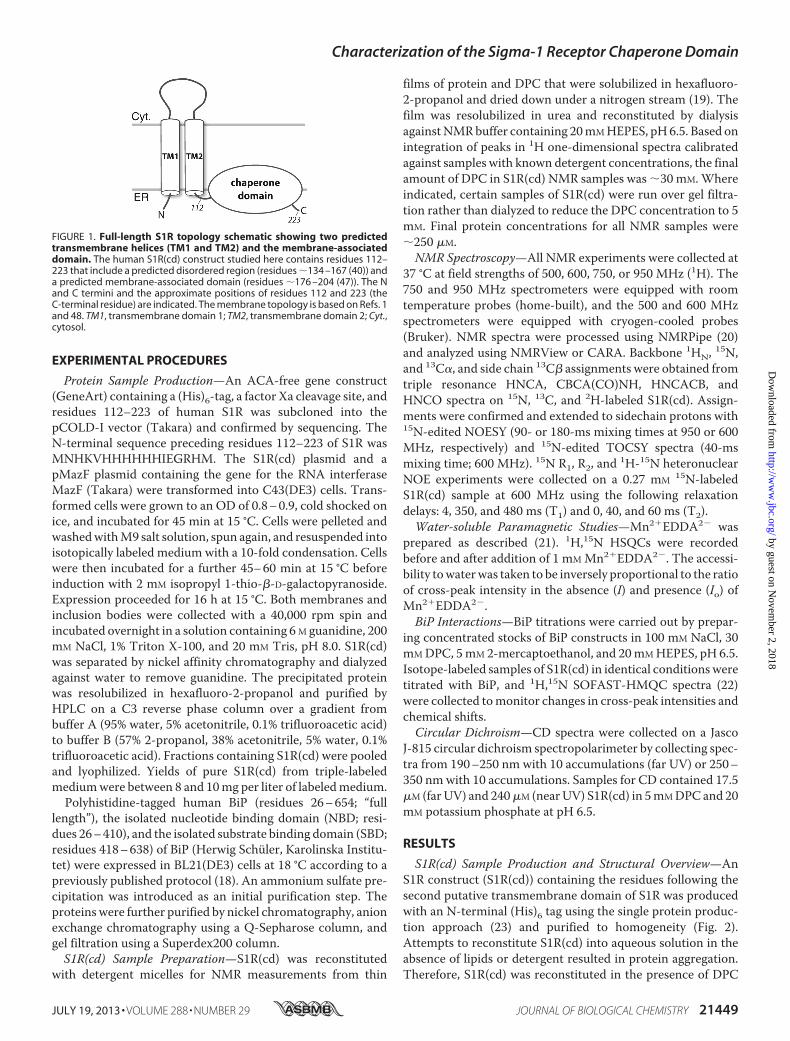
transmembrane domains (residues 11–29 and 80–100) butcontains a predicted membrane associated region (residues�176–204) containing two cholesterol recognition motifs(CRM) (Fig. 1) (16, 17). We have studied the S1R chaperonedomain (residues 112–223; S1R(cd)) reconstituted intodetergent micelles by solution NMR. S1R(cd) solubilized indodecylphosphocholine (DPC) adopts a conformation com-petent to bind BiP in a calcium-dependent manner. Analysisof the structure and dynamics indicates that S1R(cd) con-tains five helices and at least two short extended regions.Three of the helices in the C-terminal membrane-associateddomain correspond closely to regions previously implicatedin cholesterol and drug interactions.
* This work was supported by Medical Research Council Grant K018590 andthe University of Oxford Department of Biochemistry.Author’s Choice—Final version full access.
1 Supported by a FEBS postdoctoral fellowship from the Federation of Euro-pean Biochemical Societies.
2 To whom correspondence should be addressed: Dept. of Biochemistry, Uni-versity of Oxford, South Parks Rd., Oxford OX1 3QU, UK. Tel.: 44-1865-613350; Fax: 44-1865-613201; E-mail: [email protected].
3 The abbreviations used are: S1R, sigma-1 receptor; S1R(cd), sigma-1 recep-tor chaperone domain; ER, endoplasmic reticulum; BiP, binding immuno-globulin protein; DPC, dodecylphosphocholine; NBD, nucleotide bindingdomain; SBD, substrate binding domain; SBDL, steroid binding-likedomain; CRM, cholesterol recognition motif.
THE JOURNAL OF BIOLOGICAL CHEMISTRY VOL. 288, NO. 29, pp. 21448 –21457, July 19, 2013Author’s Choice © 2013 by The American Society for Biochemistry and Molecular Biology, Inc. Published in the U.S.A.
21448 JOURNAL OF BIOLOGICAL CHEMISTRY VOLUME 288 • NUMBER 29 • JULY 19, 2013
by guest on Novem
ber 2, 2018http://w
ww
.jbc.org/D
ownloaded from
EXPERIMENTAL PROCEDURES
Protein Sample Production—An ACA-free gene construct(GeneArt) containing a (His)6-tag, a factor Xa cleavage site, andresidues 112–223 of human S1R was subcloned into thepCOLD-I vector (Takara) and confirmed by sequencing. TheN-terminal sequence preceding residues 112–223 of S1R wasMNHKVHHHHHHIEGRHM. The S1R(cd) plasmid and apMazF plasmid containing the gene for the RNA interferaseMazF (Takara) were transformed into C43(DE3) cells. Trans-formed cells were grown to an OD of 0.8–0.9, cold shocked onice, and incubated for 45 min at 15 °C. Cells were pelleted andwashedwithM9 salt solution, spun again, and resuspended intoisotopically labeled medium with a 10-fold condensation. Cellswere then incubated for a further 45–60 min at 15 °C beforeinduction with 2 mM isopropyl 1-thio-�-D-galactopyranoside.Expression proceeded for 16 h at 15 °C. Both membranes andinclusion bodies were collected with a 40,000 rpm spin andincubated overnight in a solution containing 6M guanidine, 200mM NaCl, 1% Triton X-100, and 20 mM Tris, pH 8.0. S1R(cd)was separated by nickel affinity chromatography and dialyzedagainst water to remove guanidine. The precipitated proteinwas resolubilized in hexafluoro-2-propanol and purified byHPLC on a C3 reverse phase column over a gradient frombuffer A (95% water, 5% acetonitrile, 0.1% trifluoroacetic acid)to buffer B (57% 2-propanol, 38% acetonitrile, 5% water, 0.1%trifluoroacetic acid). Fractions containing S1R(cd) were pooledand lyophilized. Yields of pure S1R(cd) from triple-labeledmediumwere between 8 and 10mg per liter of labeledmedium.Polyhistidine-tagged human BiP (residues 26–654; “full
length”), the isolated nucleotide binding domain (NBD; resi-dues 26–410), and the isolated substrate binding domain (SBD;residues 418–638) of BiP (Herwig Schüler, Karolinska Institu-tet) were expressed in BL21(DE3) cells at 18 °C according to apreviously published protocol (18). An ammonium sulfate pre-cipitation was introduced as an initial purification step. Theproteinswere further purified by nickel chromatography, anionexchange chromatography using a Q-Sepharose column, andgel filtration using a Superdex200 column.S1R(cd) Sample Preparation—S1R(cd) was reconstituted
with detergent micelles for NMR measurements from thin
films of protein and DPC that were solubilized in hexafluoro-2-propanol and dried down under a nitrogen stream (19). Thefilm was resolubilized in urea and reconstituted by dialysisagainstNMRbuffer containing 20mMHEPES, pH6.5. Based onintegration of peaks in 1H one-dimensional spectra calibratedagainst samples with known detergent concentrations, the finalamount of DPC in S1R(cd) NMR samples was �30 mM.Whereindicated, certain samples of S1R(cd) were run over gel filtra-tion rather than dialyzed to reduce the DPC concentration to 5mM. Final protein concentrations for all NMR samples were�250 �M.NMR Spectroscopy—All NMR experiments were collected at
37 °C at field strengths of 500, 600, 750, or 950 MHz (1H). The750 and 950 MHz spectrometers were equipped with roomtemperature probes (home-built), and the 500 and 600 MHzspectrometers were equipped with cryogen-cooled probes(Bruker). NMR spectra were processed using NMRPipe (20)and analyzed using NMRView or CARA. Backbone 1HN, 15N,and 13C�, and side chain 13C� assignments were obtained fromtriple resonance HNCA, CBCA(CO)NH, HNCACB, andHNCO spectra on 15N, 13C, and 2H-labeled S1R(cd). Assign-ments were confirmed and extended to sidechain protons with15N-edited NOESY (90- or 180-ms mixing times at 950 or 600MHz, respectively) and 15N-edited TOCSY spectra (40-msmixing time; 600 MHz). 15N R1, R2, and 1H-15N heteronuclearNOE experiments were collected on a 0.27 mM 15N-labeledS1R(cd) sample at 600 MHz using the following relaxationdelays: 4, 350, and 480 ms (T1) and 0, 40, and 60 ms (T2).Water-soluble Paramagnetic Studies—Mn2�EDDA2� was
prepared as described (21). 1H,15N HSQCs were recordedbefore and after addition of 1 mM Mn2�EDDA2�. The accessi-bility towaterwas taken to be inversely proportional to the ratioof cross-peak intensity in the absence (I) and presence (Io) ofMn2�EDDA2�.BiP Interactions—BiP titrations were carried out by prepar-
ing concentrated stocks of BiP constructs in 100 mM NaCl, 30mMDPC, 5mM 2-mercaptoethanol, and 20mMHEPES, pH 6.5.Isotope-labeled samples of S1R(cd) in identical conditionsweretitrated with BiP, and 1H,15N SOFAST-HMQC spectra (22)were collected tomonitor changes in cross-peak intensities andchemical shifts.Circular Dichroism—CD spectra were collected on a Jasco
J-815 circular dichroism spectropolarimeter by collecting spec-tra from 190–250 nm with 10 accumulations (far UV) or 250–350 nmwith 10 accumulations. Samples for CD contained 17.5�M (farUV) and 240�M (nearUV) S1R(cd) in 5mMDPCand 20mM potassium phosphate at pH 6.5.
RESULTS
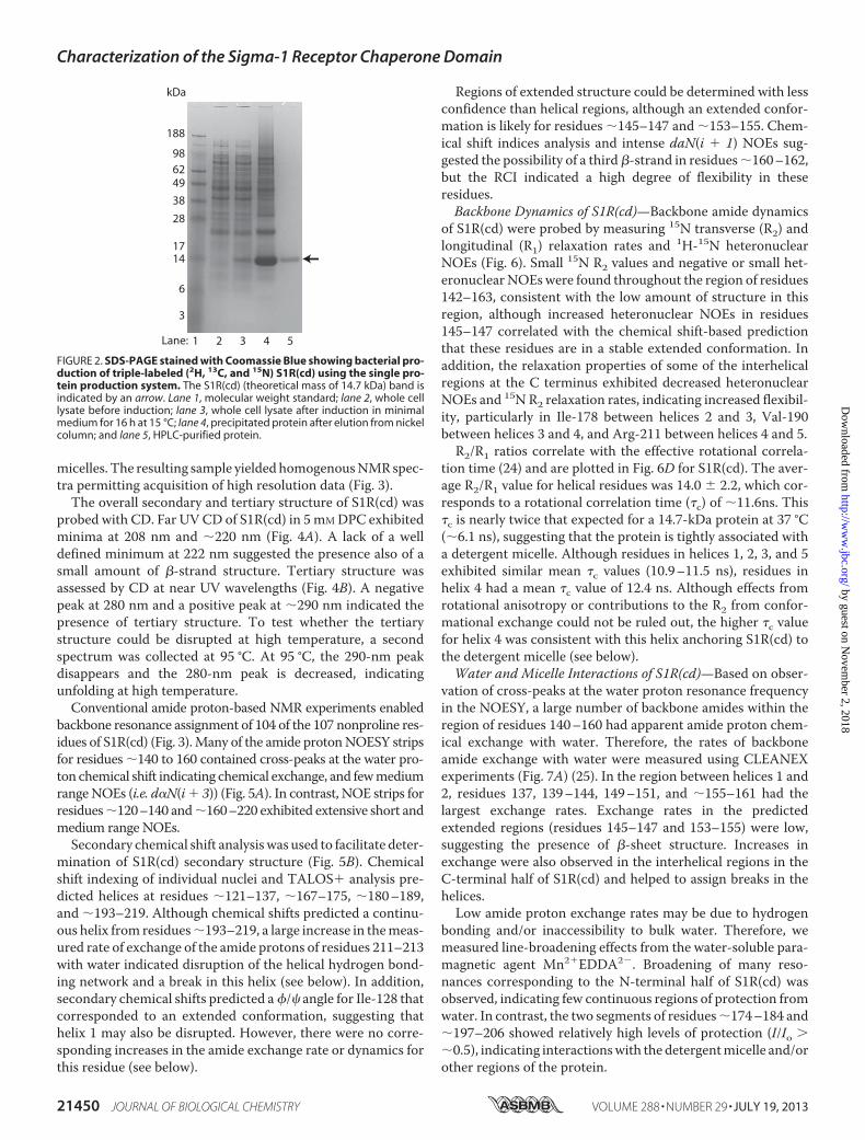
S1R(cd) Sample Production and Structural Overview—AnS1R construct (S1R(cd)) containing the residues following thesecond putative transmembrane domain of S1R was producedwith an N-terminal (His)6 tag using the single protein produc-tion approach (23) and purified to homogeneity (Fig. 2).Attempts to reconstitute S1R(cd) into aqueous solution in theabsence of lipids or detergent resulted in protein aggregation.Therefore, S1R(cd) was reconstituted in the presence of DPC
FIGURE 1. Full-length S1R topology schematic showing two predictedtransmembrane helices (TM1 and TM2) and the membrane-associateddomain. The human S1R(cd) construct studied here contains residues 112–223 that include a predicted disordered region (residues �134 –167 (40)) anda predicted membrane-associated domain (residues �176 –204 (47)). The Nand C termini and the approximate positions of residues 112 and 223 (theC-terminal residue) are indicated. The membrane topology is based on Refs. 1and 48. TM1, transmembrane domain 1; TM2, transmembrane domain 2; Cyt.,cytosol.
Characterization of the Sigma-1 Receptor Chaperone Domain
JULY 19, 2013 • VOLUME 288 • NUMBER 29 JOURNAL OF BIOLOGICAL CHEMISTRY 21449
by guest on Novem
ber 2, 2018http://w
ww
.jbc.org/D
ownloaded from
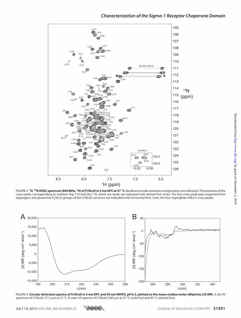
micelles. The resulting sample yieldedhomogenousNMRspec-tra permitting acquisition of high resolution data (Fig. 3).The overall secondary and tertiary structure of S1R(cd) was
probed with CD. Far UVCD of S1R(cd) in 5mMDPC exhibitedminima at 208 nm and �220 nm (Fig. 4A). A lack of a welldefined minimum at 222 nm suggested the presence also of asmall amount of �-strand structure. Tertiary structure wasassessed by CD at near UV wavelengths (Fig. 4B). A negativepeak at 280 nm and a positive peak at �290 nm indicated thepresence of tertiary structure. To test whether the tertiarystructure could be disrupted at high temperature, a secondspectrum was collected at 95 °C. At 95 °C, the 290-nm peakdisappears and the 280-nm peak is decreased, indicatingunfolding at high temperature.Conventional amide proton-based NMR experiments enabled
backbone resonance assignment of 104 of the 107 nonproline res-idues of S1R(cd) (Fig. 3).Many of the amide protonNOESY stripsfor residues �140 to 160 contained cross-peaks at the water pro-ton chemical shift indicating chemical exchange, and fewmediumrangeNOEs (i.e. d�N(i� 3)) (Fig. 5A). In contrast, NOE strips forresidues�120–140 and�160–220 exhibited extensive short andmedium range NOEs.Secondary chemical shift analysiswas used to facilitate deter-
mination of S1R(cd) secondary structure (Fig. 5B). Chemicalshift indexing of individual nuclei and TALOS� analysis pre-dicted helices at residues �121–137, �167–175, �180–189,and �193–219. Although chemical shifts predicted a continu-ous helix from residues�193–219, a large increase in themeas-ured rate of exchange of the amide protons of residues 211–213with water indicated disruption of the helical hydrogen bond-ing network and a break in this helix (see below). In addition,secondary chemical shifts predicted a�/� angle for Ile-128 thatcorresponded to an extended conformation, suggesting thathelix 1 may also be disrupted. However, there were no corre-sponding increases in the amide exchange rate or dynamics forthis residue (see below).
Regions of extended structure could be determined with lessconfidence than helical regions, although an extended confor-mation is likely for residues �145–147 and �153–155. Chem-ical shift indices analysis and intense daN(i � 1) NOEs sug-gested the possibility of a third�-strand in residues�160–162,but the RCI indicated a high degree of flexibility in theseresidues.Backbone Dynamics of S1R(cd)—Backbone amide dynamics
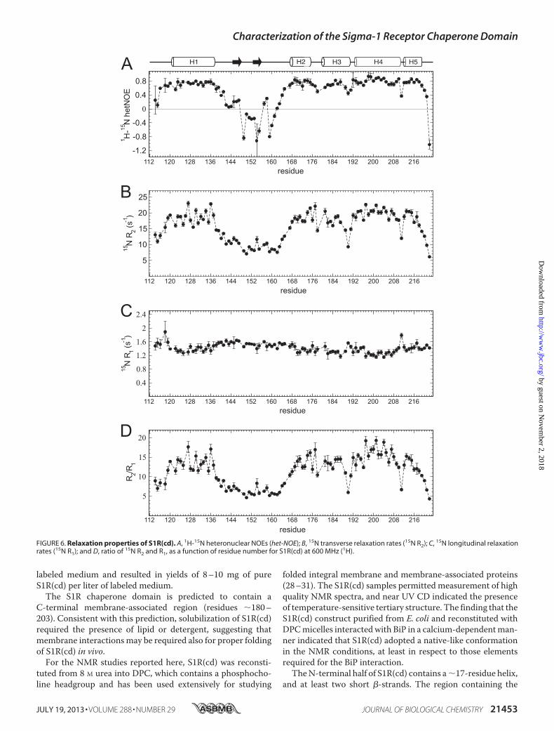
of S1R(cd) were probed by measuring 15N transverse (R2) andlongitudinal (R1) relaxation rates and 1H-15N heteronuclearNOEs (Fig. 6). Small 15N R2 values and negative or small het-eronuclearNOEswere found throughout the region of residues142–163, consistent with the low amount of structure in thisregion, although increased heteronuclear NOEs in residues145–147 correlated with the chemical shift-based predictionthat these residues are in a stable extended conformation. Inaddition, the relaxation properties of some of the interhelicalregions at the C terminus exhibited decreased heteronuclearNOEs and 15N R2 relaxation rates, indicating increased flexibil-ity, particularly in Ile-178 between helices 2 and 3, Val-190between helices 3 and 4, and Arg-211 between helices 4 and 5.R2/R1 ratios correlate with the effective rotational correla-
tion time (24) and are plotted in Fig. 6D for S1R(cd). The aver-age R2/R1 value for helical residues was 14.0 � 2.2, which cor-responds to a rotational correlation time (�c) of �11.6ns. This�c is nearly twice that expected for a 14.7-kDa protein at 37 °C(�6.1 ns), suggesting that the protein is tightly associated witha detergent micelle. Although residues in helices 1, 2, 3, and 5exhibited similar mean �c values (10.9–11.5 ns), residues inhelix 4 had a mean �c value of 12.4 ns. Although effects fromrotational anisotropy or contributions to the R2 from confor-mational exchange could not be ruled out, the higher �c valuefor helix 4 was consistent with this helix anchoring S1R(cd) tothe detergent micelle (see below).Water and Micelle Interactions of S1R(cd)—Based on obser-
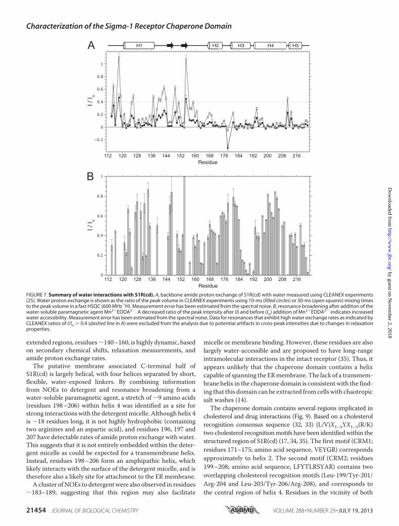
vation of cross-peaks at the water proton resonance frequencyin the NOESY, a large number of backbone amides within theregion of residues 140–160 had apparent amide proton chem-ical exchange with water. Therefore, the rates of backboneamide exchange with water were measured using CLEANEXexperiments (Fig. 7A) (25). In the region between helices 1 and2, residues 137, 139–144, 149–151, and �155–161 had thelargest exchange rates. Exchange rates in the predictedextended regions (residues 145–147 and 153–155) were low,suggesting the presence of �-sheet structure. Increases inexchange were also observed in the interhelical regions in theC-terminal half of S1R(cd) and helped to assign breaks in thehelices.Low amide proton exchange rates may be due to hydrogen
bonding and/or inaccessibility to bulk water. Therefore, wemeasured line-broadening effects from the water-soluble para-magnetic agent Mn2�EDDA2�. Broadening of many reso-nances corresponding to the N-terminal half of S1R(cd) wasobserved, indicating few continuous regions of protection fromwater. In contrast, the two segments of residues�174–184 and�197–206 showed relatively high levels of protection (I/Io ��0.5), indicating interactionswith the detergentmicelle and/orother regions of the protein.
62493828
14
kDa
6
3
98
188
321 54
17
Lane:
FIGURE 2. SDS-PAGE stained with Coomassie Blue showing bacterial pro-duction of triple-labeled (2H, 13C, and 15N) S1R(cd) using the single pro-tein production system. The S1R(cd) (theoretical mass of 14.7 kDa) band isindicated by an arrow. Lane 1, molecular weight standard; lane 2, whole celllysate before induction; lane 3, whole cell lysate after induction in minimalmedium for 16 h at 15 °C; lane 4, precipitated protein after elution from nickelcolumn; and lane 5, HPLC-purified protein.
Characterization of the Sigma-1 Receptor Chaperone Domain
21450 JOURNAL OF BIOLOGICAL CHEMISTRY VOLUME 288 • NUMBER 29 • JULY 19, 2013
by guest on Novem
ber 2, 2018http://w
ww
.jbc.org/D
ownloaded from
8.5 8.0 7.5 7.0 6.5
126
125
124
123
122
121
120
119
118
117
116
115
114
113
112
111
110
109
108
107
106
105
L214
F219T140
T160
G165
G220
G157
G115
G209
G174G176
M170
V171
L186
T141F184
S143
N167
T216
T189
L203
L197
Q194
F200 D195F196I178
V177
V162
Q221
T202
A159
E163
K142
D222
E144W164
A183
A185W169
A161
R211
L199
V153
A187
V152
E123
S205
T132L218
T181
T151
Y173
E172R175
A207
Y201
V190
W136L182
Y217
T198
R119
Gln/Asn H2N(ε2)
G149
G155
H154
R114E158
I129
G139
E150V145
A122
R208
G118
R204
W136
W169W121
W164
Trp HN(ε1)
T215
S192
G131
T193
S130
W121
D188S125
T168
T127
E138
L210
F191
I128
D126
T109E213
H116
I124
T212
Y206Y147
F146
Q135
R137
H134
F133
Y120
129.5
130.5
10.25 10.00
1H (ppm)
15N (ppm)
FIGURE 3. 1H,15N HSQC spectrum (600 MHz, 1H) of S1R(cd) in 5 mM DPC at 37 °C. Backbone amide resonance assignments are indicated. The positions of thecross-peaks corresponding to residues Arg-114 and Gly-118, which are weak, are indicated with dashed line circles. The five cross-peak pairs expected fromasparagine and glutamine H2N(�2) groups of the S1R(cd) construct are indicated with horizontal lines. Inset, the four tryptophan HN(�1) cross-peaks.
-5,000
0
-10,000
-15,000
5,000
240230220210200190 250
[θ] M
R (d
eg c
m2 d
mol
-1)
-200
50
0
-50
-100
-150
340320300280260λ (nm)
[θ] M
R (d
eg c
m2 d
mol
-1)
20,000
15,000
10,000
A B
λ (nm)FIGURE 4. Circular dichroism spectra of S1R(cd) in 5 mM DPC and 20 mM HEPES, pH 6.5, plotted as the mean residue molar ellipticity ([�] MR). A, far UVspectrum of S1R(cd) (17.5 �M) at 37 °C. B, near UV spectra of S1R(cd) (240 �M) at 37 °C (solid line) and 95 °C (dashed line).
Characterization of the Sigma-1 Receptor Chaperone Domain
JULY 19, 2013 • VOLUME 288 • NUMBER 29 JOURNAL OF BIOLOGICAL CHEMISTRY 21451
by guest on Novem
ber 2, 2018http://w
ww
.jbc.org/D
ownloaded from
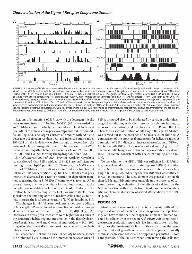
Regions of interactions of S1R(cd) with the detergentmicellewere assessed from an 15N-edited NOESY (90 ms) recorded onan 15N-labeled and partially deuterated sample at high field(950 MHz) to resolve cross-peak overlaps and reduce spin dif-fusion (Fig. 5A). The largest clusters of residues with NOEs todetergent occurred in residues 183–189 in helix 3 and residues197–204 in helix 4. Helix 4 was also strongly protected from thewater-soluble paramagnetic agent. The region �198–206forms an amphipathic helix, with residues Leu-199, Phe-200,Leu-203, and Tyr-206 forming the hydrophobic face (26).S1R(cd) Interactions with BiP—Previous work by Hayashi et
al. (1) showed that S1R residues 116–223 are sufficient forbinding to the Hsp70 protein BiP. Therefore, the NMR spec-trum of 15N-labeled S1R(cd) was monitored as a function ofunlabeled BiP concentration (Fig. 8). The S1R(cd) cross-peakintensities decreased in a BiP concentration-dependent man-ner, suggesting that a BiP�S1R(cd) complex was formed. Afterseveral hours, a white precipitate formed, indicating that thecomplex was unstable in solution. By contrast, BiP alone in thetitration buffer (containing 30mMDPC)wasmore stable. It wasspeculated that binding of the S1R(cd)�micelle complex to BiPmay increase the local concentration of DPC to destabilize BiP.The changes in 1H-15N cross-peak intensities upon addition
of full-length BiP were plotted as a function of residue numberat molar ratios of 0.5 and 1 ([BiP]/[S1R(cd)]) (Fig. 8A).Decreases in cross-peak intensities were higher for residues inthe structured helical regions and smaller in the flexibly disor-dered regions at the N and C termini and residues �140–160,suggesting that these disordered residues retained some flexi-bility in the complex.BiP chaperone (27) and ATPase (1) activity has been shown
to be inhibited by calcium, and the interaction between BiP and
S1R is proposed also to be modulated by calcium under physi-ological conditions, with the presence of calcium leading toincreased association and inactivation of S1R and BiP (1).Therefore, a second titration of full-length BiP against S1R(cd)was carried out in the presence of 2.5 mM calcium chloride. Acomparison of the cross-peak intensities for helical residues asa function of BiP indicates an increased association of S1R(cd)for full-length BiP in the presence of calcium (Fig. 8B). Nochemical shift changes were observed upon addition of calciumto S1R(cd) alone, consistent with a calcium binding site on BiP(18).To test whether the NBD of BiP was sufficient for S1R bind-
ing, the isolated domain was titrated against S1R(cd). Additionof the NBD resulted in similar changes in intensities as full-length BiP (Fig. 8B), indicating that the BiP NBD was sufficientfor S1R interactions. TheNBDdomain was generally less stablethan full-length BiP and more unstable in the presence of cal-cium, preventing evaluation of the effects of calcium on theNBD interactionwith S1R(cd). In contrast, no changes in inten-sities or chemical shifts were observed after addition of the SBD(Fig. 8D).
DISCUSSION
Most membrane-associated proteins remain difficult toexpress at sufficient levels to enable inexpensive isotope label-ing. We have found that the chaperone domain of human S1Rcould be efficiently expressed in Escherichia coli using the sin-gle protein production approach (23). In single protein produc-tion, the cells remainmetabolically active and express the targetprotein, but cell growth is halted, which appears to greatlydiminish expression toxicity. The approach permitted 10-foldcondensation of the cultures when transferring the cells into
RCI
TALOS+
CSI (13
Cβ )
CSI (13
C’)
CSI (13
Cα )
CSI (1Hα )
H1 H3H2 H4 H5H4H2
120 220210200190180170160150140130120 2202102001901801701601501401300
1
2
3
4
1
0.5
0
0.5
1-1
0
1-1
0
1-1
0
1-1
0
1
Residue Residue
H1 H3H2 H4 H5H4H2A B
H
EdN DPC (CH3 )
dN H2O
dαN (i+3)
dαN (i+2)
dαN (i+1)
05
10152025
0
0.2
0.4
0.6
0
0.2
0.4
0
0.5
1.0
1.50
0.2
0.4
0.6
0.8
1.0
dNN (i+1)
0
0.1
0.2
FIGURE 5. A, summary of NOE cross-peaks to backbone amide protons. Amide proton to amide proton NOEs (dNN(i�1)), and amide proton to �-proton NOEs(d�N(i�1), d�N(i�2), and d�N(i�3)), as well as cross-peaks at the position of the water proton (dN H2O), were measured in a three-dimensional 15N-editedNOESY with 180-ms mixing time at 600 MHz (1H) on an 15N-labeled S1R(cd) in 5 mM DPC. Amide proton to DPC methyl proton NOEs (dN DPC (CH3)) wereobtained from a three-dimensional 15N-edited NOESY with 90 ms of mixing time at 950 MHz (1H) on an 15N- and 2H-labeled (�70%) S1R(cd) in 30 mM DPC. Thex axis (arbitrary units) is the cross-peak intensity normalized to that of the diagonal. No attempt was made to deconvolute overlapped cross-peaks. B, thechemical shift indices (CSI) of 1H�, 13C�, 13C�, and 13C� are shown in the top four panels. In panels five and six are shown the secondary structure and random coilindex derived from chemical shift analysis using TALOS� (49) and the method of Berjanskii et al. (50), respectively. For the TALOS� plot, values above or belowthe line indicated that the calculated �/� values correspond to helical (H) or extended conformation (E), respectively. Shown schematically at the top are theS1R(cd) residues determined to be helical (cylinders) or extended (arrow) (see main text for full description of secondary structure assignment).
Characterization of the Sigma-1 Receptor Chaperone Domain
21452 JOURNAL OF BIOLOGICAL CHEMISTRY VOLUME 288 • NUMBER 29 • JULY 19, 2013
by guest on Novem
ber 2, 2018http://w
ww
.jbc.org/D
ownloaded from
labeled medium and resulted in yields of 8–10 mg of pureS1R(cd) per liter of labeled medium.The S1R chaperone domain is predicted to contain a
C-terminal membrane-associated region (residues �180–203). Consistent with this prediction, solubilization of S1R(cd)required the presence of lipid or detergent, suggesting thatmembrane interactionsmay be required also for proper foldingof S1R(cd) in vivo.For the NMR studies reported here, S1R(cd) was reconsti-
tuted from 8 M urea into DPC, which contains a phosphocho-line headgroup and has been used extensively for studying
folded integral membrane and membrane-associated proteins(28–31). The S1R(cd) samples permitted measurement of highquality NMR spectra, and near UV CD indicated the presenceof temperature-sensitive tertiary structure. The finding that theS1R(cd) construct purified from E. coli and reconstituted withDPCmicelles interactedwith BiP in a calcium-dependentman-ner indicated that S1R(cd) adopted a native-like conformationin the NMR conditions, at least in respect to those elementsrequired for the BiP interaction.TheN-terminal half of S1R(cd) contains a�17-residue helix,
and at least two short �-strands. The region containing the
216208200192112 120 128 136 144 152 160 168 176 184
0.4
0.8
1.2
1.6
2
2.4
216208200192112 120 128 136 144 152 160 168 176 184
0
216208200192112 120 128 136 144 152 160 168 176 184
5
10
15
20
216208200192112 120 128 136 144 152 160 168 176 184
A
B
C
D
H1 H3H2 H4 H5H4H2
FIGURE 6. Relaxation properties of S1R(cd). A, 1H-15N heteronuclear NOEs (het-NOE); B, 15N transverse relaxation rates (15N R2); C, 15N longitudinal relaxationrates (15N R1); and D, ratio of 15N R2 and R1, as a function of residue number for S1R(cd) at 600 MHz (1H).
Characterization of the Sigma-1 Receptor Chaperone Domain
JULY 19, 2013 • VOLUME 288 • NUMBER 29 JOURNAL OF BIOLOGICAL CHEMISTRY 21453
by guest on Novem
ber 2, 2018http://w
ww
.jbc.org/D
ownloaded from
extended regions, residues�140–160, is highly dynamic, basedon secondary chemical shifts, relaxation measurements, andamide proton exchange rates.The putative membrane associated C-terminal half of
S1R(cd) is largely helical, with four helices separated by short,flexible, water-exposed linkers. By combining informationfrom NOEs to detergent and resonance broadening from awater-soluble paramagnetic agent, a stretch of �9 amino acids(residues 198–206) within helix 4 was identified as a site forstrong interactionswith the detergentmicelle. Although helix 4is �18 residues long, it is not highly hydrophobic (containingtwo arginines and an aspartic acid), and residues 196, 197 and207 have detectable rates of amide proton exchangewith water.This suggests that it is not entirely embedded within the deter-gent micelle as could be expected for a transmembrane helix.Instead, residues 198–206 form an amphipathic helix, whichlikely interacts with the surface of the detergent micelle, and istherefore also a likely site for attachment to the ER membrane.A cluster ofNOEs to detergentwere also observed in residues
�183–189, suggesting that this region may also facilitate
micelle or membrane binding. However, these residues are alsolargely water-accessible and are proposed to have long-rangeintramolecular interactions in the intact receptor (35). Thus, itappears unlikely that the chaperone domain contains a helixcapable of spanning the ERmembrane. The lack of a transmem-brane helix in the chaperone domain is consistentwith the find-ing that this domain can be extracted fromcells with chaotropicsalt washes (14).The chaperone domain contains several regions implicated in
cholesterol and drug interactions (Fig. 9). Based on a cholesterolrecognition consensus sequence (32, 33) (L/V)X1–5YX1–5(R/K)two cholesterol recognitionmotifs have been identifiedwithin thestructured region of S1R(cd) (17, 34, 35). The first motif (CRM1;residues 171–175; amino acid sequence, VEYGR) correspondsapproximately to helix 2. The second motif (CRM2; residues199–208; amino acid sequence, LFYTLRSYAR) contains twooverlapping cholesterol recognition motifs (Leu-199/Tyr-201/Arg-204 and Leu-203/Tyr-206/Arg-208), and corresponds tothe central region of helix 4. Residues in the vicinity of both
216208200192112 120 128 136 144 152 160 168 176 184Residue
I / I o
216208200192112 120 128 136 144 152 160 168 176 184Residue
I / I o
A
B
H1 H3H2 H4 H5H4H2
FIGURE 7. Summary of water interactions with S1R(cd). A, backbone amide proton exchange of S1R(cd) with water measured using CLEANEX experiments(25). Water proton exchange is shown as the ratio of the peak volume in CLEANEX experiments using 10-ms (filled circles) or 30-ms (open squares) mixing timesto the peak volume in a fast HSQC (600 MHz 1H). Measurement error has been estimated from the spectral noise. B, resonance broadening after addition of thewater-soluble paramagnetic agent Mn2�EDDA2�. A decreased ratio of the peak intensity after (I) and before (Io) addition of Mn2�EDDA2� indicates increasedwater accessibility. Measurement error has been estimated from the spectral noise. Data for resonances that exhibit high water exchange rates as indicated byCLEANEX ratios of I/Io � 0.4 (dashed line in A) were excluded from the analysis due to potential artifacts in cross-peak intensities due to changes in relaxationproperties.
Characterization of the Sigma-1 Receptor Chaperone Domain
21454 JOURNAL OF BIOLOGICAL CHEMISTRY VOLUME 288 • NUMBER 29 • JULY 19, 2013
by guest on Novem
ber 2, 2018http://w
ww
.jbc.org/D
ownloaded from

120 220210200190180170160150140130Residue
1.0
0.0
0.2
0.4
0.6
0.8
A B
Molar Ratio0
0.2
0.4
0.6
0.8
1.0
1 430 2
BiP NBD, no Ca2+
BiP full length, no Ca2+
BiP full length, 2.5 mM Ca2+
I / I o
I / I o
H1 H3H2 H4 H5H4H2
8.5 8.0 7.51H (ppm)
126
124
122
120
118
116
114
112
110
108
106
15N (ppm)
D S1R(cd)S1R(cd) + SBD (1:1)
8.5 8.0 7.51H (ppm)
126
124
122
120
118
116
114
112
110
108
106
15N (ppm)
C S1R(cd)S1R(cd) + BiP (1:1)
L214
F219T140
T160
G165
G220
G157
G115
G209
G174G176
M170
V171
L186
T141F184
S143
N167
T216
T189
L203
L197
Q194
F200 D195I178
V177
V162
Q221
T202
A159
E163
K142
D222
E144 W164
A183
A185W169
A161
R211
L199
V153
A187
V152
E123
S205
T132L218
T181
T151
Y173
E172R175
A207
Y201
W136L182
Y217
T198
R119
Gln/Asn NH2
G149
G155
H154
R114E158
I129
G139
E150
A122
R208
G118
R204
T215
S192
G131
T193
S130
W121
D188
S125
T168
T127
E138
L210
F191
I128
D126
T109E213
H116
Y206Y147
F146
Q135
R137
H134
Y120
FIGURE 8. S1R(cd) interactions with BiP. A, backbone amide cross-peak intensities are plotted as a function of residue number after addition of 0.5 (black bars)and 1.0 (red bars) molar equivalents of full-length BiP (no calcium). The intensities are normalized to the cross-peak intensities in the absence of BiP. B, thenormalized average backbone amide cross-peak intensity of S1R(cd) as a function of the molar ratio of BiP NBD (no calcium) or full-length BiP to S1R(cd) in theabsence or presence of 2.5 mM calcium chloride. The intensity average is for only the helical regions. C, spectral overlays of 15N-labeled S1R(cd) with (red) orwithout (black) full-length BiP (1:1) in the presence of 2.5 mM calcium. D, spectral overlays of 15N-labeled S1R(cd) with (red) and without (black) addition of theBiP SBD (1:1) in the presence of 2.5 mM calcium.
216208200192112 120 128 136 144 152 160 168 176 184
CRM1 CRM2SBDLII
GSRGHSGRYWAEISDTIISGTFHQWREGTTKSEVFYPGETVVHGPGEATAVEWGPNTWMVEYGRGVIPSTLAFALADTVFSTQDFLTLFYTLRSYARGLRLELTTYLFGQDP
* *H1 H3H2 H4 H5H4H2
↓
Potential membraneattachment region
↓
FIGURE 9. The helical (cylinders) and extended (arrow) residues of S1R(cd) determined from a combination of chemical shifts, amide-water protonexchange, 15N relaxation rates, and NOEs. Residues and regions previously implicated in cholesterol binding are indicated: cholesterol binding motifs(CRM1 and CRM2), cocaine binding (SBDLII; residues Asp-188 and Val-190 are indicated by2), and haloperidol binding (residues Asp-126 and Glu-172 areindicated by an asterisk). Residues 198 –206, which are proposed to have the strongest interactions with the ER membrane, are indicated. The amino acidsequence and corresponding residue numbers are shown at the bottom.
Characterization of the Sigma-1 Receptor Chaperone Domain
JULY 19, 2013 • VOLUME 288 • NUMBER 29 JOURNAL OF BIOLOGICAL CHEMISTRY 21455
by guest on Novem
ber 2, 2018http://w
ww
.jbc.org/D
ownloaded from
CRM1 and CRM2 exhibited heightened protection from thewater-soluble paramagnetic agent, although a larger number ofNOEs to DPC were detected in the CRM2, suggesting a moreintimate interaction with the detergent micelle at this site.Although the chaperone domain does not bind drugs in the
absence of the N-terminal transmembrane domain (1, 35), thesecondary structure determined here may reflect the structuralpropensities of the intact receptor. Studies using a chemicallyreactive affinity probe have provided information on the bind-ing site of cocaine in guinea pig S1R, which is 98% similar tohuman S1R (35–37). Those studies defined a steroid binding-like domain (SBDLII) at residues 176–194 and located residuesAsp-188 andVal-190 close to the cocaine interaction site. SBD-LII together with a steroid binding-like domain (denotedSBDLI; residues 91–109) in the second putative transmem-brane helix stabilizes cocaine binding (35). SBDLII correspondsclosely to helix 3 (residues 180–189) and the adjacent residuesconnecting helix 3 to helices 2 and 4 (Fig. 9). Asp-188 and Val-190, which are proposed to be close to the cocaine binding site,are at the C-terminal end of helix 3. Both Asp-188 and Val-190are solvent exposed in S1R(cd), and Val-190 is one of the mostflexible residues within the C-terminal helical region ofS1R(cd). It is unknown whether the flexibility observed here ispreserved in full-length S1R, but such flexibility may facilitatebinding-induced conformational changes necessary to mediateS1R downstream interactions.Mutational studies of S1R have also implicated Asp-126 and
Glu-172 in haloperidol binding (8). Asp-126 and Glu-172 arefound in helices 1 and 2 of S1R(cd), respectively, suggesting thatthese helices may interact in the haloperidol bound conforma-tion of the receptor.BiP is an ER resident chaperone that regulates the unfolded
protein response in addition to assisting protein folding(reviewed in Ref. 38). S1R is proposed to sequester BiP in theabsence of ER stress. Upon depletion of ER calcium or S1Rligand binding the S1R�BiP complex dissociates leading tochaperone activity and downstream signaling of S1R, includinginositol triphosphate receptor-mediated calcium release (1, 3).We have shown here that S1R(cd) under NMR conditionsinteracts with BiP in a calcium-dependent manner and that theNBD of BiP is sufficient for these interactions. The finding thatS1R(cd) interacts with the NBD of BiP is consistent with theknown regulatory interactions with other Hsp proteins. Forexample, the co-chaperone BAG1 binds to the NBD of Hsc70(42, 43), and bacterial GrpE binds to the NBD of DnaK (44, 45).The role of the region �140–160 remains unknown. This
region did not appear to tightly associate with BiP in our stud-ies, and substitution of acidic residues within this region hasbeen shown to have no impact on haloperidol binding (8).Based on sequence analysis (39), several short �-strands arepredicted for this region in residues 143–145, 151–153, and159–164, which corresponds approximately to those regionsobserved to be extended here (residues 145–147, 153–155; sec-ondary chemical shifts also indicate transient extended confor-mation in residues 160–162). However, sequence analysis alsopredicts a high degree of disorder in this region (40), which isconfirmed by theNMRdata. By contrast, residues 124–137 and168–173 are predicted from sequence to be extended but found
here to be helical. DPC has previously been shown to disrupt�-sheets in a concentration-dependent manner (41). However,the concentrations used here are as much as 10-fold lower thanin that study, and the spectra of S1R(cd) in 5 and 30mMDPCareessentially identical.In summary, we have studied the chaperone domain of S1R
by solution NMR and have characterized its secondary struc-ture and dynamics. S1R(cd) is composed of five helices and atleast two short extended regions in a dynamic region betweenhelices 1 and 2. Three of the helices in the C-terminal mem-brane-associated region map to residues previously identifiedas important in cholesterol and cocaine binding. A fourth helix(helix 4) is implicated inmembrane association. In addition, wehave shown that the NBD domain of BiP is sufficient for inter-action with the S1R chaperone domain. These results advanceour understanding of S1R and are likely to be useful in refiningmodels of the S1R drug binding sites (46). Future studies areneeded to identify tertiary interactions in the S1R chaperonedomain and to further delineate S1R/BiP and S1R ligand andcholesterol interactions.
Acknowledgment—We thank Herwig Schüler (Karolinska Institutet)for providing expression constructs of human BiP and the WellcomeTrust (Grant 094872) for upgrade of the 600 MHz spectrometer usedin the studies.
REFERENCES1. Hayashi, T., and Su, T. P. (2007) Sigma-1 receptor chaperones at the ER-
mitochondrion interface regulate Ca2� signaling and cell survival. Cell131, 596–610
2. Pal, A., Fontanilla, D., Gopalakrishnan, A., Chae, Y. K., Markley, J. L., andRuoho, A. E. (2012) The sigma-1 receptor protects against cellular oxida-tive stress and activates antioxidant response elements. Eur. J. Pharmacol.682, 12–20
3. Hayashi, T., and Su, T. P. (2001) Regulating ankyrin dynamics: Roles ofsigma-1 receptors. Proc. Natl. Acad. Sci. U.S.A. 98, 491–496
4. Csordás, G., Renken, C., Várnai, P., Walter, L., Weaver, D., Buttle, K. F.,Balla, T., Mannella, C. A., and Hajnóczky, G. (2006) Structural and func-tional features and significance of the physical linkage between ER andmitochondria. J. Cell Biol. 174, 915–921
5. Walter, L., and Hajnóczky, G. (2005) Mitochondria and endoplasmic re-ticulum: the lethal interorganelle cross-talk. J. Bioenerg. Biomembr. 37,191–206
6. Csordás, G., Várnai, P., Golenár, T., Roy, S., Purkins, G., Schneider, T. G.,Balla, T., and Hajnóczky, G. (2010) Imaging interorganelle contacts andlocal calcium dynamics at the ER-mitochondrial interface. Mol. Cell 39,121–132
7. Weissman, A. D., Su, T. P., Hedreen, J. C., and London, E. D. (1988) Sigmareceptors in post-mortem human brains. J. Pharmacol. Exp. Ther. 247,29–33
8. Seth, P., Ganapathy, M. E., Conway, S. J., Bridges, C. D., Smith, S. B.,Casellas, P., and Ganapathy, V. (2001) Expression pattern of the type 1sigma receptor in the brain and identity of critical anionic amino acidresidues in the ligand-binding domain of the receptor. Biochim. Biophys.Acta 1540, 59–67
9. Guitart, X., Codony, X., and Monroy, X. (2004) Sigma receptors: biologyand therapeutic potential. Psychopharmacology 174, 301–319
10. Maurice, T., and Su, T. P. (2009) The pharmacology of sigma-1 receptors.Pharmacol. Ther. 124, 195–206
11. Fontanilla, D., Johannessen, M., Hajipour, A. R., Cozzi, N. V., Jackson,M. B., and Ruoho, A. E. (2009) The hallucinogenN,N-dimethyltryptamine(DMT) is an endogenous sigma-1 receptor regulator. Science 323,934–937
Characterization of the Sigma-1 Receptor Chaperone Domain
21456 JOURNAL OF BIOLOGICAL CHEMISTRY VOLUME 288 • NUMBER 29 • JULY 19, 2013
by guest on Novem
ber 2, 2018http://w
ww
.jbc.org/D
ownloaded from
12. Su, T. P., London, E. D., and Jaffe, J. H. (1988) Steroid binding at sigmareceptors suggests a link between endocrine, nervous, and immune sys-tems. Science 240, 219–221
13. Ramachandran, S., Chu, U. B., Mavlyutov, T. A., Pal, A., Pyne, S., andRuoho, A. E. (2009) The sigma1 receptor interacts with N-alkyl aminesand endogenous sphingolipids. Eur. J. Pharmacol. 609, 19–26
14. Wu, Z., and Bowen, W. D. (2008) Role of sigma-1 receptor C-terminalsegment in inositol 1,4,5-trisphosphate receptor activation: constitutiveenhancement of calcium signaling in MCF-7 tumor cells. J. Biol. Chem.283, 28198–28215
15. Shioda, N., Ishikawa, K., Tagashira, H., Ishizuka, T., Yawo, H., and Fuku-naga, K. (2012) Expression of a truncated form of the endoplasmic retic-ulum chaperone protein, sigma1 receptor, promotes mitochondrial en-ergy depletion and apoptosis. J. Biol. Chem. 287, 23318–23331
16. Hanner, M., Moebius, F. F., Flandorfer, A., Knaus, H. G., Striessnig, J.,Kempner, E., and Glossmann, H. (1996) Purification, molecular cloning,and expression of the mammalian sigma1-binding site. Proc. Natl. Acad.Sci. U.S.A. 93, 8072–8077
17. Palmer, C. P., Mahen, R., Schnell, E., Djamgoz, M. B., and Aydar, E. (2007)Sigma-1 receptors bind cholesterol and remodel lipid rafts in breast can-cer cell lines. Cancer Res. 67, 11166–11175
18. Wisniewska, M., Karlberg, T., Lehtiö, L., Johansson, I., Kotenyova, T.,Moche, M., and Schüler, H. (2010) Crystal structures of the ATPase do-mains of four human Hsp70 isoforms: HSPA1L/Hsp70-hom, HSPA2/Hsp70–2, HSPA6/Hsp70B’, and HSPA5/BiP/GRP78. PLoS One 5, e8625
19. Call, M. E., Wucherpfennig, K. W., and Chou, J. J. (2010) The structuralbasis for intramembrane assembly of an activating immunoreceptor com-plex. Nat. Immunol. 11, 1023–1029
20. Delaglio, F., Grzesiek, S., Vuister, G. W., Zhu, G., Pfeifer, J., and Bax, A.(1995)NMRPipe: amultidimensional spectral processing systembased onUNIX pipes. J. Biomol. NMR 6, 277–293
21. Lau, T. L., Partridge, A. W., Ginsberg, M. H., and Ulmer, T. S. (2008)Structure of the integrin �3 transmembrane segment in phospholipidbicelles and detergent micelles. Biochemistry 47, 4008–4016
22. Schanda, P., Kupce, E., and Brutscher, B. (2005) SOFAST-HMQC exper-iments for recording two-dimensional heteronuclear correlation spectraof proteins within a few seconds. J. Biomol. NMR 33, 199–211
23. Suzuki, M., Mao, L., and Inouye, M. (2007) Single protein production(SPP) system in Escherichia coli. Nat. Protoc. 2, 1802–1810
24. Kay, L. E., Torchia, D. A., and Bax, A. (1989) Backbone dynamics of pro-teins as studied by 15N inverse detected heteronuclear NMR spectros-copy: application to staphylococcal nuclease.Biochemistry 28, 8972–8979
25. Hwang, T. L., van Zijl, P. C., and Mori, S. (1998) Accurate quantitation ofwater-amide proton exchange rates using the phase-modulated CLEANchemical EXchange (CLEANEX-PM) approach with a Fast-HSQC(FHSQC) detection scheme. J. Biomol. NMR 11, 221–226
26. Gautier, R., Douguet, D., Antonny, B., and Drin, G. (2008) HELIQUEST: aweb server to screen sequences with specific �-helical properties. Bioin-formatics 24, 2101–2102
27. Kassenbrock, C. K., and Kelly, R. B. (1989) Interaction of heavy chainbinding protein (BiP/GRP78) with adenine nucleotides. EMBO J. 8,1461–1467
28. VanHorn,W. D., Kim, H. J., Ellis, C. D., Hadziselimovic, A., Sulistijo, E. S.,Karra, M. D., Tian, C., Sönnichsen, F. D., and Sanders, C. R. (2009) Solu-tion nuclear magnetic resonance structure of membrane-integral diacyl-glycerol kinase. Science 324, 1726–1729
29. Berardi, M. J., Shih, W. M., Harrison, S. C., and Chou, J. J. (2011) Mito-chondrial uncoupling protein 2 structure determined by NMRmolecularfragment searching. Nature 476, 109–113
30. Zhou, Y., Cierpicki, T., Jimenez, R. H., Lukasik, S. M., Ellena, J. F., Cafiso,D. S., Kadokura, H., Beckwith, J., and Bushweller, J. H. (2008) NMR solu-tion structure of the integral membrane enzymeDsbB: functional insightsinto DsbB-catalyzed disulfide bond formation.Mol. Cell 31, 896–908
31. Vasudevan, S. V., Schulz, J., Zhou, C., and Cocco, M. J. (2010) Proteinfolding at the membrane interface, the structure of Nogo-66 requires in-teractions with a phosphocholine surface. Proc. Natl. Acad. Sci. U.S.A.107, 6847–6851
32. Li, H., and Papadopoulos, V. (1998) Peripheral-type benzodiazepine re-
ceptor function in cholesterol transport. Identification of a putative cho-lesterol recognition/interaction amino acid sequence and consensus pat-tern. Endocrinology 139, 4991–4997
33. Li, H., Yao, Z., Degenhardt, B., Teper, G., and Papadopoulos, V. (2001)Cholesterol binding at the cholesterol recognition/ interaction amino acidconsensus (CRAC) of the peripheral-type benzodiazepine receptor andinhibition of steroidogenesis by an HIV TAT-CRAC peptide. Proc. Natl.Acad. Sci. U.S.A. 98, 1267–1272
34. Chen, Y., Hajipour, A. R., Sievert, M. K., Arbabian, M., and Ruoho, A. E.(2007) Characterization of the cocaine binding site on the sigma-1 recep-tor. Biochemistry 46, 3532–3542
35. Pal, A., Chu, U. B., Ramachandran, S., Grawoig, D., Guo, L. W., Hajipour,A. R., andRuoho,A. E. (2008) Juxtaposition of the steroid binding domain-like I and II regions constitutes a ligand binding site in the sigma-1 recep-tor. J. Biol. Chem. 283, 19646–19656
36. Fontanilla, D., Hajipour, A. R., Pal, A., Chu, U. B., Arbabian, M., andRuoho, A. E. (2008) Probing the steroid binding domain-like I (SBDLI) ofthe sigma-1 receptor binding site usingN-substituted photoaffinity labels.Biochemistry 47, 7205–7217
37. Pal, A., Hajipour, A. R., Fontanilla, D., Ramachandran, S., Chu, U. B.,Mavlyutov, T., and Ruoho, A. E. (2007) Identification of regions of thesigma-1 receptor ligand binding site using a novel photoprobe.Mol. Phar-macol. 72, 921–933
38. Cao, S. S., andKaufman, R. J. (2012)Unfolded protein response.Curr. Biol.22, R622–626
39. Buchan, D. W., Ward, S. M., Lobley, A. E., Nugent, T. C., Bryson, K., andJones, D. T. (2010) Protein annotation andmodelling servers at UniversityCollege London. Nucleic Acids Res. 38,W563–568
40. Linding, R., Jensen, L. J., Diella, F., Bork, P., Gibson, T. J., and Russell, R. B.(2003) Protein disorder prediction: implications for structural proteomics.Structure 11, 1453–1459
41. Sherratt, A. R., Braganza, M. V., Nguyen, E., Ducat, T., and Goto, N. K.(2009) Insights into the effect of detergents on the full-length rhomboidprotease from Pseudomonas aeruginosa and its cytosolic domain.Biochim. Biophys. Acta 1788, 2444–2453
42. Briknarová, K., Takayama, S., Brive, L., Havert,M. L., Knee, D. A., Velasco,J., Homma, S., Cabezas, E., Stuart, J., Hoyt, D. W., Satterthwait, A. C.,Llinás, M., Reed, J. C., and Ely, K. R. (2001) Structural analysis of BAG1cochaperone and its interactions with Hsc70 heat shock protein. Nat.Struct. Biol. 8, 349–352
43. Sondermann, H., Scheufler, C., Schneider, C., Hohfeld, J., Hartl, F. U., andMoarefi, I. (2001) Structure of a Bag/Hsc70 complex: convergent func-tional evolution of Hsp70 nucleotide exchange factors. Science 291,1553–1557
44. Harrison, C. J., Hayer-Hartl, M., Di Liberto, M., Hartl, F., and Kuriyan, J.(1997) Crystal structure of the nucleotide exchange factor GrpE bound tothe ATPase domain of the molecular chaperone DnaK. Science 276,431–435
45. Wu, C. C., Naveen, V., Chien, C. H., Chang, Y.W., andHsiao, C. D. (2012)Crystal structure of DnaK protein complexed with nucleotide exchangefactor GrpE in DnaK chaperone system: insight into intermolecular com-munication. J. Biol. Chem. 287, 21461–21470
46. Stefanski, R., Justinova, Z., Hayashi, T., Takebayashi, M., Goldberg, S. R.,and Su, T. P. (2004) Sigma1 receptor upregulation after chronic metham-phetamine self-administration in rats: a study with yoked controls. Psy-chopharmacology 175, 68–75
47. Käll, L., Krogh, A., and Sonnhammer, E. L. (2004) A combined transmem-brane topology and signal peptide prediction method. J. Mol. Biol. 338,1027–1036
48. Kourrich, S., Hayashi, T., Chuang, J. Y., Tsai, S. Y., Su, T. P., and Bonci, A.(2013) Dynamic interaction between sigma-1 receptor and Kv1.2 shapesneuronal and behavioral responses to cocaine. Cell 152, 236–247
49. Shen, Y., Delaglio, F., Cornilescu, G., and Bax, A. (2009) TALOS�: a hy-brid method for predicting protein backbone torsion angles from NMRchemical shifts. J. Biomol. NMR 44, 213–223
50. Berjanskii, M. V., and Wishart, D. S. (2005) A simple method to predictprotein flexibility using secondary chemical shifts. J. Am. Chem. Soc. 127,14970–14971
Characterization of the Sigma-1 Receptor Chaperone Domain
JULY 19, 2013 • VOLUME 288 • NUMBER 29 JOURNAL OF BIOLOGICAL CHEMISTRY 21457
by guest on Novem
ber 2, 2018http://w
ww
.jbc.org/D
ownloaded from
Jose Luis Ortega-Roldan, Felipe Ossa and Jason R. Schnelland Binding Immunoglobulin Protein (BiP) Interactions
Characterization of the Human Sigma-1 Receptor Chaperone Domain Structure
doi: 10.1074/jbc.M113.450379 originally published online June 12, 20132013, 288:21448-21457.J. Biol. Chem.
10.1074/jbc.M113.450379Access the most updated version of this article at doi:
Alerts:
When a correction for this article is posted•
When this article is cited•
to choose from all of JBC's e-mail alertsClick here
http://www.jbc.org/content/288/29/21448.full.html#ref-list-1
This article cites 50 references, 17 of which can be accessed free at
by guest on Novem
ber 2, 2018http://w
ww
.jbc.org/D
ownloaded from