charging of particles in unipolar coronas irradiated by in-situ soft x-rays: enhancement of capture...
TRANSCRIPT

Aerosol Science 33 (2002) 1279–1296www.elsevier.com/locate/jaerosci
Charging of particles in unipolar coronas irradiated by in-situsoft X-rays: enhancement of capture e)ciency
of ultra*ne particles
Pramod Kulkarnia, Norikazu Namikia;b, Yoshio Otanib, Pratim Biswasa ;∗
aEnvironmental Engineering Science Program, School of Engineering and Applied Science, Campus Box 1180,Washington University in Saint Louis, One Brookings Drive, Saint Louis, MO 63130-4899 USA
bDepartment of Chemistry and Chemical Engineering, Kanazawa University, Kanazawa 920-8667, Japan
Received 20 October 2001; received in revised form 12 April 2002; accepted 15 April 2002
Abstract
The charging of *ne particles in unipolar coronas irradiated by soft X-rays (3:5–9:5 keV; �=0:13–0:41 nm)was studied. Voltage–current characteristics were used to examine the corona inception voltage in presence andabsence of X-ray irradiation and to estimate the ion concentrations. The capture characteristics of synthesizedparticles: iron oxide, sodium chloride, silica and titanium dioxide, were established in coronas, with andwithout X-ray irradiation. Enhanced charging of ultra*ne particles in coronas was observed in conjunction withsoft X-ray irradiation. A positive corona with X-ray irradiation resulted in the highest charging e)ciencies,followed by a negative corona with X-ray, X-ray only, negative corona only and *nally positive corona only.Particle charging theories available in the literature were used to qualitatively explain the observed trends.? 2002 Elsevier Science Ltd. All rights reserved.
Keywords: Soft X-ray; Ultra*ne particles; Charging; Photoionization; Corona; ESP
1. Introduction
There has been a renewed interest in the capture of *ne particles with the recent promulgationof PM2.5 standards (National Air Quality and Emissions Trends Reports, 1996). Ultra*ne particlesin the atmosphere are receiving signi*cant attention due to their potential deleterious health eBects(Oberdorster, Ferin, Finkelstein, Wade, & Corson, 1990; Miller, Biswas, & Leikauf, 2001). Fine
∗Corresponding author. Tel.: +1-314-935-5482; fax: +1-314-935-5464.E-mail address: [email protected] (P. Biswas).
0021-8502/02/$ - see front matter ? 2002 Elsevier Science Ltd. All rights reserved.PII: S 0021-8502(02)00067-8
1280 P. Kulkarni et al. / Aerosol Science 33 (2002) 1279–1296
particles act as condensation nuclei for formation of secondary aerosols in the atmosphere. Theymay also enhance condensation of toxic gaseous species onto their surfaces (Ondov & Wexler,1998; Biswas & Wu, 1998; Zhuang, 2001). Fine particles have long atmospheric residence timesand contribute signi*cantly to visibility degradation. As the regulations are changed from PM10to PM2.5, relatively more emphasis will be placed on the control of the *ner size fraction of theparticles.
Electrostatic precipitators (ESP) are widely used particulate control devices in industry to cap-ture *ne particles from air emissions. The overall mass based e)ciency of ESPs is of the orderof 99%. However, the collection e)ciency curve is U-shaped and there is a penetration win-dow in the submicrometer size range where the e)ciencies are as low as 70–80%. This is dueto the balance between two opposing eBects—decreasing charge with decreasing particle size andincreasing drag with increasing particle size. Moreover, contrary to theoretical considerations, ex-perimental observations have shown that collection e)ciency decreases with decreasing diameterbelow ∼60 nm (Zhuang, Kim, Lee, & Biswas, 2000; Yoo, Lee, & Oh, 1997; Watanabe et al.,1995; Kim & Biswas, 1996). The low capture e)ciencies have been primarily attributed to par-tial charging of ultra*ne particles (Zhuang et al., 2000). The conventional methods for particlecharging by corona are inadequate and there is a need for a method to e)ciently charge ultra*neparticles.
Various studies have been conducted in the past to investigate the charging of ultra*ne parti-cles. Pauthenier and Moreau-Hanot (1932) developed an expression for *eld charging of particlesapproximately larger than 0:5 �m. Bricard (1949) and Fuchs (1963) developed equations for par-ticle charging based on the diBusional charging theory. Liu and Kapadia (1978) theoretically andexperimentally studied combined-diBusion and *eld charging of *ne particles. Experimental studies(Boisdron & Brock, 1970; Liu and Pui, 1977; Hussin, Scheibel, Becker, & Porstendorfer, 1983;Adachi, Kousaka, & Okuyama, 1985; Wiedensolher & Fissan, 1991) have con*rmed that Fuch’stheory reasonably predicts the ultra*ne particle charging probabilities. In a regular ESP operation,particles are charged by the corona generated by a high DC voltage applied across the electrodes.Unipolar corona charging has been found to be inadequate to charge the ultra*ne particles e)ciently.Attempts have been made to enhance the charging of these ultra*ne particles using photoelectriccharging processes such as UV irradiation (Burtscher, Scherrer, Siegmann, Schmidt-Ott, & Fed-erer, 1982; Jung, Burtscher, & Schmidt-Ott, 1988; Zhuang, 2001) with limited success. Hence thereis a need for method to charge ultra*ne particles e)ciently. X-ray photoionization has been usedby several researchers for a variety of applications. Soft X-ray ionization has been used to studyfragmentation of molecules (Fisher et al., 1999) and variety of other spectroscopic applications.Applications of short wavelength X-ray lasers have been discussed by Matthews (1996). Photoion-ization systems are being used as eBective charge neutralizers of surfaces (Inaba, Tohmi, Yoshida,& Okada, 1994). To the best of our knowledge no one has examined the interaction of X-ray pho-toionization with unipolar ions generated by a corona, and their eBectiveness at charging ultra*neparticles.
A soft X-ray spectrum to irradiate the particles and electrode surfaces in-situ to enhance pho-toionization and improve charging of ultra*ne particles in a corona, is studied. The enhancement ofcapture e)ciencies of ultra*ne particles is studied at varying operating conditions of the ESP forvarious types of aerosols. The eBect and type of corona around the discharge electrode (DE) is alsoexplored.

P. Kulkarni et al. / Aerosol Science 33 (2002) 1279–1296 1281
2. Experimental
2.1. Apparatus and materials
A schematic diagram of the experimental setup is shown in Fig. 1(a). The system consists of aparticle generation system, an ESP and an aerosol size distribution measurement system. Four diBer-ent test aerosols were used in this study—NaCl, Fe2O3; SiO2 and TiO2 to validate the eBectivenessof this approach for a broad spectrum of particless. NaCl aerosols were generated by atomizing anaqueous NaCl solution and subsequently drying the droplets using a diBusion dryer. The other threeaerosols were generated using a combustion furnace reactor. Fe2O3 was generated by atomizing theaqueous suspension of Fe(NO3)3; drying the atomized aerosol and then sending it to a combus-tion furnace as shown in Fig. 1(a). SiO2 and TiO2 particles were produced from their precursors(hexa dimethylsiloxane and titanium tetra isopropoxide, respectively). The precursors were entrainedin an air-stream which was saturated by bubbling through the solution kept at a temperature of80◦C, and then introduced into a furnace reactor maintained at ∼1000◦C to obtain SiO2 and TiO2
aerosols. The generated submicrometer aerosols were mixed with particle free air to achieve a desiredaerosol concentration level and Row rate. A total Row of 15 lpm was maintained through the ESPin all the experiments in this study. Table 1(a) summarizes size distribution characteristics of all theaerosols used. Aerosols were polydisperse and had a distribution of sizes between 20 and 600 nm.The geometric mean diameters (based on number) were approximately 67 nm for Fe2O3; 87 nm forNaCl, 77 nm for SiO2 and 65 nm for a TiO2, respectively. Aerosol from the generation system wascharge-neutralized using Kr-85 bipolar charger before introducing it into the ESP.
Fig. 1(b) shows details of the cylindrical ESP. The collecting electrode of the cylindrical ESPwas made of stainless steel and was 15 cm in length and 5 cm in diameter. The collecting electrodewas enclosed on the outside by a PVC tube to shield against X-rays from leaking out. The dischargeelectrode (DE) was also made of stainless steel and two diBerent diameters 0:575 and 0:323 mmwere used. Excess length of the DE was sheathed using an insulating material to prevent coronaformation (Fig. 1(b)). A circular hole (2 cm in diameter) was drilled in the middle of the collectingelectrode to irradiate the region inside the ESP by the X-ray emitter (Hamamatsu Photonics Ltd.,Japan, Model L7113). The X-ray radiation (3:5–9:5 keV; � = 0:13–0:41 nm) spread inside the ESPat an angle of 120◦, where it interacted with the incoming particles and electrode surfaces. A thinpolyamide *lm (KaptonTM 30HN, DuPont Corp., 30 �m thick) was used to protect the X-ray emittersurface from the aerosol Row and keep the ESP system airtight. The penetrability of soft X-raysthrough the polyamide *lm was estimated to be about 90%. A DC voltage was applied across thecollecting and discharge electrodes using a high voltage DC power supply (Glassman High VoltageInc., EL series). A DC micro ammeter was used to measure the voltage–current characteristics ofthe ESP. Particle size distribution of the aerosol entering and leaving the ESP was performed usingthe SMPS system (SMPS 3080, TSI Inc.). The average net charge on the particles exiting the ESPwas measured using a CPC and electrometer operating synchronously.
2.2. Procedures and measurements
Several experiments were performed to understand the eBect of X-ray irradiation on charging andcapture of ultra*ne particles. The experiments can be broadly classi*ed into three categories:

1282 P. Kulkarni et al. / Aerosol Science 33 (2002) 1279–1296
Fig. 1. (a) Schematic diagram of the experimental setup used to measure particle capture e)ciency of ESP with andwithout X-ray irradiation. (b) Details of the ESP with X-ray mount.

P. Kulkarni et al. / Aerosol Science 33 (2002) 1279–1296 1283
Table 1(a): Size distribution parameters of aerosols used in this study. (b): Summary of experiments performed
Aerosol Geometric mean Geo. std. Total particle number Generation methoddiameter (nm) dev. concentration (#=cm3)
Fe2O3 66.58 1.86 1:3 × 10⁶ Furnace reactorNaCl 86.83 1.90 2:5 × 10⁶ AtomizationSiO2 77.32 1.50 1:6 × 10⁷ Furnace reactorTiO2 64.86 1.51 5:0 × 106 Furnace reactor
Seta Description Voltage Diameter Polarity of Aerosol Objective# (kV) of DE (mm) DE
I V–I characteristics 0 to 10 0.575 Positive Fe2O3 Determine the corona inceptionand and voltage of the ESP0.323 Negative
II Overall capture with 0 and 10 0.575 Positive Fe2O3 Demonstrate the enhancement inand without X-ray capture e)ciency due to X-ray
irradiation
III Capture e)ciency 0, 5, 8, 9 0.575 Positive Fe2O3; NaCl, (a) Measure capture e)ciencies atmeasurement and 10 and and SiO2; TiO2 varying particle diameter
0.323 Negative (b) Measure capture e)ciencies atvarying voltage levels and studyeBect of corona(c) Study the eBect of type of corona
aFor all experiments—Row rate: 15 lpm; length of ESP: 15 cm; diameter of collecting electrode: 5 cm.
(i) voltage–current characteristics of the ESP, (ii) change in average charge per particle due toX-ray irradiation and (iii) enhancement in capture e)ciency due to X-ray radiation. Table 1(b)summarizes the objectives of all the experiments performed.
Set I tests were performed to determine the corona inception voltage of the ESP and to obtain thecorona-current (I) characteristics over the entire range of operating voltages (V ), with and withoutX-ray irradiation. Experiments were performed with an Fe2O3 aerosol for two diBerent diameters ofDE (0:575 and 0:323 mm �) which were maintained at a positive (or negative) potential. The appliedvoltage was varied from 0 to 10 kV and the corresponding corona current measured using a DCmicro ammeter connected between the discharge and collecting electrode, without X-ray irradiationand with X-ray irradiation. V–I characteristics with other aerosols were also measured.
The Set II experiments were performed to demonstrate the overall eBect X-ray irradiation has onthe capture of particles. These experiments were designed to demonstrate the eBectiveness of X-rayphotoionization on outlet particle concentration of the ESP. The total number concentration at theoutlet of the ESP was monitored with time under a predetermined sequence of operating conditions,involving switching the X-ray radiation and electrode potentials on and oB in diBerent sequences.The average charge (total charge=total particle concentration) of the aerosol escaping collection wasalso simultaneously measured to understand its variation. An electrometer operating in parallel tothe CPC was used to obtain the average charge data.
1284 P. Kulkarni et al. / Aerosol Science 33 (2002) 1279–1296
Set III experiments were aimed at studying the enhancement in capture e)ciency due to X-rayirradiation under diBerent operating conditions, viz., voltage levels across the electrodes and electrodepolarity. Experiments were performed at four voltage levels (5; 8; 9, and 10 kV) with and withoutX-ray irradiation for four aerosols (i.e., NaCl, Fe2O3; SiO2 and TiO2) at positive and negativepolarity of the discharge electrode. Discharge electrodes of two diBerent diameters were used—0.575 and 0:323 mm � in the experiments. Particle size distribution of the test aerosol was measuredat the inlet and outlet of the ESP using a SMPS. This data was then used to deduce the capturee)ciencies. A typical experimental protocol for capture e)ciency measurements consisted of: (1)starting the particle generation system, ensuring stable aerosol generation, (2) introducing this aerosolto the ESP, ensuring the stable Row through the system, (3) measuring the particle size distributionusing SMPS at the outlet of the ESP, to obtain a representative inlet aerosol size distribution (afterall losses to ESP walls), (4) applying the desired voltage across electrodes, (5) measuring the particlesize distribution at the outlet of the ESP (this would be used to compute capture e)ciency withoutX-ray irradiation), (6) turning on the X-ray and (7) again measuring the particle size distribution atthe outlet of ESP (this would give capture e)ciency with X-ray irradiation).
A total volumetric Row rate of 15 lpm was maintained through the ESP in all experiments. Particlesize distribution of the aerosol generated using the furnace reactor was controlled by altering thefeed rate of precursors. Collecting and discharge electrodes were cleaned after each experiment tokeep the surfaces clear of deposits. The residence time in the ESP was approximately 1:2 s and wasintentionally chosen to keep the capture e)ciencies under regular operation (without X-ray) low.This would help to clearly discern any enhancement in capture e)ciency due to X-ray irradiation,an important objective of this study.
3. Results and discussion
X-ray radiation can charge the particles by two mechanisms—(a) direct photoelectric charging and(b) diBusion charging. In direct photoelectric charging, the atoms or molecules on the surface ofthe aerosol particle are photoionized, whereas in diBusion charging the gas molecules get ionized bythe radiation and then attach to the particles resulting in their electrical charging. The photoemissionfrom the electrode surfaces also impacts the ion concentration in the unipolar corona. Fig. 2 shows aschematic diagram explaining the diBusion and direct particle charging and photoemission processesinside the ESP in the presence of corona and X-ray radiation.
3.1. V–I characteristics
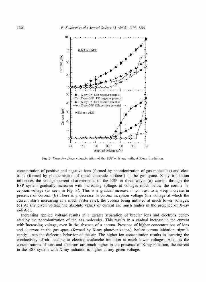
Fig. 3 shows voltage–current characteristics of the ESP with two DE (0.575 and 0:323 mm �diameters), with and without X-ray irradiation. In all the experiments with the presence of X-rayirradiation, the current is signi*cantly higher at a given voltage. For the 0:575 mm � DE, the positivecorona inception voltage is about 9:0 kV without X-ray irradiation and goes down to 8:6 kV inpresence of X-ray radiation. The negative corona inception voltage decreases from 8.9 to about7:0 kV on irradiating with X-rays. The current is higher in case of negative corona (negative potentialapplied to DE) compared to that with positive corona at any given voltage. As expected, the currentvalues are higher for the 0:323 mm � DE compared to that with 0:575 mm � DE at any given

P. Kulkarni et al. / Aerosol Science 33 (2002) 1279–1296 1285
Soft X-rayemitter
hν ν
Photoemission of electrode surface
Discharge Electrode (DE)
Collectingelectrode of ESP
e
hν ν
Unipolar ion atmospheredue to corona around DE
iffusion charging
Particle
Free ion
Bipolar ion atmospherecreated by photoionizationof gas molecules
hν ν
Photoionizationof particle
Particle
e
e
e e
Fig. 2. Aerosol photoemission and charging processes inside the ESP in the presence of X-ray radiation and corona.Particles can acquire charge either by direct photoionization or by diBusion charging. The metal electrodes surfaces canalso be ionized by the incident radiation, resulting in emission of electrons. The unipolar corona formed by the externalapplied voltage (above inception voltage) is a source of unipolar ions. Photoionization of molecules in the gas space leadsto a bipolar atmosphere containing free positive and negative ions and electrons.
voltage. The variability in corona current is relatively higher for the negative corona compared tothat with the positive corona for both discharge electrodes.
Under the regular ESP operation positive corona (discharge electrode positive) results in excesspositive ions in the gas space while that under negative corona results in excess negative ions.However when X-ray radiation is present along with the unipolar corona, there is an increased
1286 P. Kulkarni et al. / Aerosol Science 33 (2002) 1279–1296
Cur
rent
(µ A
)
0
25
50
75
100
Applied voltage (kV)7.0 7.5 8.0 8.5 9.0 9.5 10.0
Cur
rent
(µ A
)
0
10
20
30
40
50 X-ray ON, DE: negative potential X-ray OFF, DE: negative potential X-ray ON, DE: positive potentialX-ray OFF, DE: positive potential
0.575 mm φ DE
0.323 mm φ DE
Fig. 3. Current–voltage characteristics of the ESP with and without X-ray irradiation.
concentration of positive and negative ions (formed by photoionization of gas molecules) and elec-trons (formed by photoemission of metal electrode surfaces) in the gas space. X-ray irradiationinRuences the voltage–current characteristics of the ESP in three ways: (a) current through theESP system gradually increases with increasing voltage, at voltages much below the corona in-ception voltage (as seen in Fig. 3). This is a gradual increase in contrast to a steep increase inpresence of corona. (b) There is a decrease in corona inception voltage (the voltage at which thecurrent starts increasing at a much faster rate), the corona being initiated at much lower voltages.(c) At any given voltage the absolute values of current are much higher in the presence of X-rayradiation.
Increasing applied voltage results in a greater separation of bipolar ions and electrons gener-ated by the photoionization of the gas molecules. This results in a gradual increase in the currentwith increasing voltage, even in the absence of a corona. Presence of higher concentrations of ionsand electrons in the gas space (formed by X-ray photoionization), before corona initiation, signi*-cantly alters the dielectric behavior of the air. The higher ion concentration results in lowering theconductivity of air, leading to electron avalanche initiation at much lower voltages. Also, as theconcentrations of ions and electrons are much higher in the presence of X-ray radiation, the currentin the ESP system with X-ray radiation is higher at any given voltage.
P. Kulkarni et al. / Aerosol Science 33 (2002) 1279–1296 1287
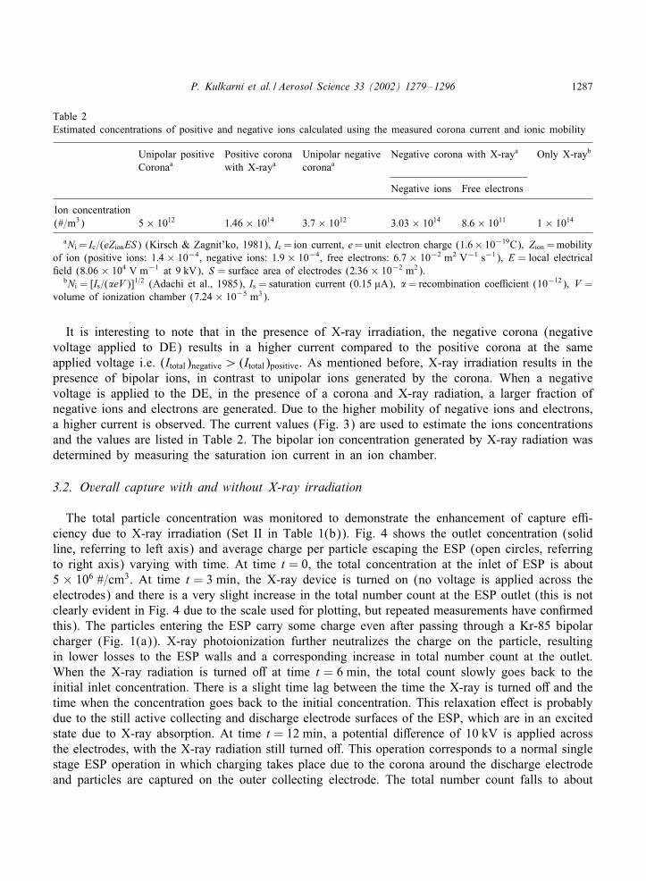
Table 2Estimated concentrations of positive and negative ions calculated using the measured corona current and ionic mobility
Unipolar positive Positive corona Unipolar negative Negative corona with X-raya Only X-rayb
Coronaa with X-raya coronaa
Negative ions Free electrons
Ion concentration(#=m3) 5 × 10¹² 1:46 × 1014 3:7 × 10¹² 3:03 × 1014 8:6 × 10¹¹ 1 × 10¹⁴
aNi = Ic=(eZionES) (Kirsch & Zagnit’ko, 1981), Ic = ion current, e=unit electron charge (1:6× 10⁻¹⁹C); Zion =mobilityof ion (positive ions: 1:4 × 10⁻⁴, negative ions: 1:9 × 10⁻⁴, free electrons: 6:7 × 10⁻² m2 V−1 s−1); E = local electrical*eld (8:06 × 104 V m−1 at 9 kV); S = surface area of electrodes (2:36 × 10−2 m2).
bNi = [Is=(�eV )]1=2 (Adachi et al., 1985), Is = saturation current (0:15 �A); �= recombination coe)cient (10−12); V =volume of ionization chamber (7:24 × 10−5 m3).
It is interesting to note that in the presence of X-ray irradiation, the negative corona (negativevoltage applied to DE) results in a higher current compared to the positive corona at the sameapplied voltage i.e. (Itotal)negative¿ (Itotal)positive. As mentioned before, X-ray irradiation results in thepresence of bipolar ions, in contrast to unipolar ions generated by the corona. When a negativevoltage is applied to the DE, in the presence of a corona and X-ray radiation, a larger fraction ofnegative ions and electrons are generated. Due to the higher mobility of negative ions and electrons,a higher current is observed. The current values (Fig. 3) are used to estimate the ions concentrationsand the values are listed in Table 2. The bipolar ion concentration generated by X-ray radiation wasdetermined by measuring the saturation ion current in an ion chamber.
3.2. Overall capture with and without X-ray irradiation
The total particle concentration was monitored to demonstrate the enhancement of capture e)-ciency due to X-ray irradiation (Set II in Table 1(b)). Fig. 4 shows the outlet concentration (solidline, referring to left axis) and average charge per particle escaping the ESP (open circles, referringto right axis) varying with time. At time t = 0, the total concentration at the inlet of ESP is about5 × 106 #=cm3. At time t = 3 min, the X-ray device is turned on (no voltage is applied across theelectrodes) and there is a very slight increase in the total number count at the ESP outlet (this is notclearly evident in Fig. 4 due to the scale used for plotting, but repeated measurements have con*rmedthis). The particles entering the ESP carry some charge even after passing through a Kr-85 bipolarcharger (Fig. 1(a)). X-ray photoionization further neutralizes the charge on the particle, resultingin lower losses to the ESP walls and a corresponding increase in total number count at the outlet.When the X-ray radiation is turned oB at time t = 6 min, the total count slowly goes back to theinitial inlet concentration. There is a slight time lag between the time the X-ray is turned oB and thetime when the concentration goes back to the initial concentration. This relaxation eBect is probablydue to the still active collecting and discharge electrode surfaces of the ESP, which are in an excitedstate due to X-ray absorption. At time t = 12 min, a potential diBerence of 10 kV is applied acrossthe electrodes, with the X-ray radiation still turned oB. This operation corresponds to a normal singlestage ESP operation in which charging takes place due to the corona around the discharge electrodeand particles are captured on the outer collecting electrode. The total number count falls to about

1288 P. Kulkarni et al. / Aerosol Science 33 (2002) 1279–1296
Time (min)0 3 6 9 12 15 18 21 24 27 30
Part
icle
num
ber
conc
entr
atio
n (#
/cm
3)
10 5
10 6
10 7 Average charge per particle (unit electron charge)
0.00
0.05
0.10
0.15
0.20
0.25
0.30
0.35
0.40
Vol
tage
,V=0
kV
X-r
ay O
FF
V=0
kV
X-r
ay O
N
V=0 kVX-ray OFF
V=1
0 kV
X-r
ay O
FF
V=10 kVX-ray ON
V=0 kVX-ray OFF
V=10 kVX-ray OFF
Fig. 4. Variation in particle number concentration (left axis) at the outlet of ESP under diBerent operating conditions forFe2O3 aerosol. Diameter of the discharge electrode was 0:575 mm and was maintained at positive potential. Also shownare curves representing average particle charge (right axis) under these conditions.
106 #=cm3. At time t = 15 min, the X-ray radiation is turned on, with the applied voltage still at10 kV. In this case, ionization is taking place in corona region around the discharge electrode aswell as due to X-ray photoionization. The total number count rapidly drops to 2× 10⁵ #=cm3. Whenthe X-ray is turned oB at time t = 18 min, the total count goes back to the initial value prior toturning on the X-ray (t = 12–15 min). When the applied voltage across the electrodes is reduced to0 kV at time t = 24 min, the total count again increases to its initial inlet count at time t = 0.
Also shown on the same graph (open circles, referring to the right y-axis) is the variation ofaverage net charge per particle. The average charge per particle is computed from the electrometercurrent (I), Row rate (Q) and the total number concentration (N ) using the relation:
qavg =INQe
; (1)
where, qavg is average charge per particle (in unit electron charge units) and e is the unit electroncharge. It should be noted that the average charge measured is that of the aerosol escaping collectionin the ESP, and hence only provides qualitative information.
The particles entering the ESP at time t = 0 carry a small average charge. When the X-ray isturned on at time t=3 min, the average charge on the particles coming out of the ESP decreases byan insigni*cant amount, indicating that some further neutralization takes place due to X-ray bipolarcharging. When a potential diBerence of 10 kV is applied across the electrodes at time t = 12 min,corresponding to a decrease in total number count, the average charge on the particle leaving theESP increases by a factor of 1.5. When the X-ray is turned on at time t = 15 min, the charge onthe particles leaving the ESP goes up by a factor of 4.3, clearly indicating the increased charginge)ciency of particles due to X-ray photoionization.
It is important to note that the net average charge per particle due to X-ray irradiation decreasesin the absence of corona (i.e. 0 kV applied voltage across electrodes) whereas it increases in the

P. Kulkarni et al. / Aerosol Science 33 (2002) 1279–1296 1289
presence of corona (10 kV). The average charge decreases by 14% for the Fe2O3 aerosol in theabsence of corona, whereas it increases by a factor of 6.5 (550%) in the presence of corona (at10 kV). This indicates that X-ray radiation in the presence of an unipolar corona is resulting in animproved charging e)ciency. As mentioned before, there are two mechanisms by which a particlecan acquire charges—(a) direct charging due to photoionization and (b) diBusion charging. Whilethe former mechanism depends on the intensity and wavelength of incident radiation, composition ofaerosols, the latter depends on the extent of ion atmosphere and the mobility of ions. By applyinga high voltage across the electrodes, as is done in the above experiment, conditions aBecting onlythe second mechanism (diBusion charging) are changed. The external applied voltage aids in thebetter separation of bipolar ions promoting the extent of unipolar ion atmosphere (owing to diBerentelectrical mobility of ions). Increasing the voltage in the presence of X-ray radiation may alsosigni*cantly change the photoelectric behavior of metal electrode surfaces, especially near the coronainception voltage or photoelectric threshold potential of the metal electrode surface. The presence ofcorona further enhances the unipolar ion atmosphere leading to a more e)cient diBusion charging.Direct charging by photoionization may also play a role in increasing the charge on the particles.But considering the fact that presence of corona is very critical for obtaining high charge per particleit is surmised that diBusion charging is a major charging mechanism in our system. Comparing sizeresolved capture e)ciencies measured at varying voltage levels with and without X-ray radiation arepresented in the next section to further support this conclusion.
3.3. Capture e:ciencies
Set III in Table 1(b) summarizes the experiments that were performed to study the enhancement ofcapture e)ciencies due to X-ray radiation under diBerent operating conditions, i.e. varying voltagelevels and the polarity of DE. Capture e)ciencies for the Fe2O3 aerosol are presented as modelresults to elucidate the salient features of particle charging by X-ray irradiation followed by thediscussion for the other particles.
Fig. 5(a) shows a typical size distribution of Fe2O3 aerosol at the inlet and outlet of ESPobtained from the SMPS. All the aerosols used in this study were polydisperse with a distributionbetween 20 and 600 nm (Table 1(a)). The *gure clearly shows the eBect of X-ray irradiation and theconcentration at the outlet of the ESP goes down by an order of magnitude. These size distributionpro*les were then used to compute the capture e)ciencies of aerosols and are presented in Fig. 5(b)and are discussed below.
Fig. 5(b) shows capture e)ciencies (�) as a function of diameter for the Fe2O3 aerosol at fourdiBerent applied voltage levels. A 0:575 mm � DE at positive potential was used in the experiment.The capture e)ciency curves without X-ray irradiation (open symbols, Fig. 5) show a decreasingcapture as the size decreases below 60 nm. The e)ciency, for example, at 10 kV applied voltage(without X-ray, denoted by open diamonds) continually decreases with decreasing diameter below∼60 nm, being about 31% at 50 nm, 23% at 35 nm and 11% at 20 nm. Similar observations weremade by Zhuang et al. (2000) and were attributed to partial charging eBects. Lower e)ciencieshave been mainly attributed to poor diBusion charging of the ultra*ne particles (Yoo et al., 1997;Zhuang et al., 2000). These particles have a low saturation charge and are also more di)cult tocharge because of the lower ion attachment coe)cients (Adachi et al., 1985).

1290 P. Kulkarni et al. / Aerosol Science 33 (2002) 1279–1296
Diameter (nm)101 102 103
dN/d
Log
d p (
#/cm
3 )
100
101
102
103
104
105
Inlet OutletWithout X-ray irradiation
OutletWith X-ray irradiation
Fe 2 O 3 aerosolsApplied voltage: 10 kVDE: 0.575mm φ and wasmaintained at positive potential
(b)(a)
Fe O AerosolsPositive corona
DE: 0.575mm
Diameter (nm)100
Cap
ture
eff
icie
ncy,
η (%
)
0
20
40
60
80
100
10kV X-ray OFF 10kV X-ray ON
9 kV X-ray ON8 kV X-ray OFF
8 kV X-ray ON5 kV X-ray OFF
5 kV X-ray ON
30 600
Fe2O3 Aerosols
DE: 0.575mm φ maintainedat positive potential
Fig. 5. (a) Typical size distribution of Fe2O3 aerosol at the inlet and outlet of ESP with and without X-ray irradiation. (b)Collection e)ciencies as a function of diameter for Fe2O3 aerosol with and without X-ray irradiation at diBerent appliedvoltage levels. Diameter of the discharge electrode was 0:575 mm and was maintained at positive potential. (E)ciencycurve at 9 kV with X-ray OFF is similar to that at 10 kV with X-ray OFF and is not shown to improve the legibility).
The e)ciencies of capture with X-ray irradiation are shown with solid symbols in Fig. 5(b). At anygiven voltage, the e)ciency of capture with X-ray radiation is much higher compared to that withoutX-ray radiation for all the particle diameters. Enhancement, i.e. increase in e)ciency due to X-rayrelative to that without X-ray irradiation, is seen to be highest for smaller particles and relativelylower for larger particles. For instance, at 10 kV, e)ciencies due to X-ray irradiation increase bya factor of 10 for 20 nm particles, by a factor of 3 for 50 nm and 3.2 for 100 nm particles. Thisclearly indicates that X-ray radiation is very eBective in charging the ultra*ne particles, especially inthe size ranges below 50 nm where charging by a unipolar corona is ine)cient. It is also importantto note that the capture e)ciencies at 10 kV with X-ray irradiation decrease with increasing diameterand roughly plateau out after 200 nm.
Table 3 shows capture e)ciencies of a 40 nm particle at varying voltage levels for the Fe2O3
aerosol using a 0:575 mm � DE. Also listed in the same table are e)ciencies of other aerosols, i.e.NaCl, SiO2; TiO2, using 0:575 and 0:323 mm � DE for comparison. The numbers in parenthesis inTable 3 indicate capture e)ciencies without X-ray irradiation. Comparing e)ciencies of 40 nm Fe2O3
particles (using a 0:575 mm � DE) at diBerent voltage levels shows that e)ciency due to X-rayirradiation increases from 51% at 5 kV, 60% at 8 kV, 62% at 9 kV to 86% at 10 kV. However,under the normal ESP operation without X-ray it only increases from 18% at 5 kV to 29% at 10 kV.It is interesting to note that at lower voltages, i.e. 5 and 8 kV when there is no corona, the e)ciencies(with X-ray) even though higher in magnitude, follow a bell-shaped trend, indicating lower charginge)ciency of smaller particles (¡ 60 nm). However at higher voltages the bell-shape disappears andthere is enhanced charging e)ciency below 60 nm. This clearly indicates the eBectiveness of theX-ray irradiation in charging the particles is enhanced by the presence of corona. This was alsonoted earlier in the Set II tests where the average charge per particle escaping the collection rapidlyincreased in the presence of X-ray radiation and corona.

P. Kulkarni et al. / Aerosol Science 33 (2002) 1279–1296 1291
Table 3Capture e)ciencies (%) for a 40 nm diameter particle with (and without) X-ray irradiation at diBerent voltage levels withpositive polarity discharge electrode (DE)
Voltage DE : 0:575 mm � DE : 0:323 mm �(kV ↓)
Fe2O3 NaCl SiO2 TiO2 Fe2O3 NaCl SiO2 TiO2
5 50.8 (17.9)a 47.7 (36.4) 48.5 (12.3) 57.6 (9.81) 55.8 (19.8) 48.2 (38.9) 35.1 (11.9) 71.2 (20.4)8 59.1 (23.4) 78.3 (42.1) 74.9 (14.8) 85.8 (13.3) 95.6 (24.8) 96.7 (45.4) 92.1 (14.1) 98.2 (23.6)9 80.4 (26.7) 93.1 (42.3) 95.3 (16.8) 97.4 (18.4) 99.1 (99.1) 99.2 (99.4) 99.3 (92.1) 99.8 (99.7)
10 86.4 (29.2) 96.4 (43.2) 99.0 (16.0) 99.5 (98.6) 99.9 (99.9) 99.7 (99.8) 99.6 (99.8) 99.9 (99.9)aValues in the parenthesis indicate capture e)ciencies without X-ray irradiation, keeping all other operating conditions
the same.
Experiments were also performed to study increase in capture e)ciencies due to X-ray irradiationwith other aerosols, viz. NaCl, SiO2 and TiO2 and the representative results are presented in Figs.6(a)–(c). The e)ciencies exhibit a similar qualitative trend—charging due to X-ray is very e)cientin the ultra*ne fraction and presence of corona rapidly increases charging e)ciency. A pronouncedeBect of applied voltage is also seen in all cases. Comparing e)ciencies for diBerent particles, it isseen that e)ciencies are normally higher for TiO2. The charging e)ciency of the particle depends onvarious factors such as their physical characteristics (inRuences the direct photoionization as well asthe diBusion charging e)ciency), chemical composition (photoionization threshold could be diBerentfor each material; aBects direct photo ionization) and the number concentration of the aerosol inthe gas space (aBects the scattering of the X-ray radiation and hence direct photoionization, as wellas photoemission from surface of the metal electrodes). More controlled experimentation addressingeach of the above factors to understand material dependent charging e)ciency is necessary to furtherestablish the reasons for observed variations.
Table 3 also lists the capture e)ciencies of all the aerosols, with and without X-ray irradiation, fora 0:323 mm � DE. Comparing capture e)ciencies for the 0:323 mm � DE to those for 0:575 mm �DE, indicate higher e)ciencies with the former. Reducing the diameter of DE decreases the coronainception voltage. Hence the extent of corona is higher at any given voltage (above inception voltage)resulting in higher capture e)ciency with smaller diameter DE. Also enhancement in the capturee)ciency is less in case of 0:323 mm � DE, compared to 0:575 mm � DE at higher applied voltagesin presence of X-ray irradiation due to higher ion concentration in the latter case.
3.4. E;ect of type of corona
Capture e)ciencies were measured by changing the polarity of the discharge electrode to studythe eBect of type of corona on the capture e)ciency in the presence of X-ray radiation. Fig. 7compares capture e)ciency curves for Fe2O3 particles with positive and negative coronas (withX-ray radiation) at three voltage levels (using a 0:575 mm diameter DE). The *gure clearly showthat capture e)ciencies with X-ray irradiation in a positive corona are higher compared to thosein a negative corona in all cases (at 8 kV there is no signi*cant diBerence, the corona is not fullydeveloped at this voltage). Table 4 compares capture e)ciencies of 40 and 100 nm particles ofall aerosols for a positive and negative corona. Collection e)ciencies of Fe2O3 under regular ESP

1292 P. Kulkarni et al. / Aerosol Science 33 (2002) 1279–1296
NaCl AerosolsPositive coronaDE: 0.575mm
Diameter (nm)100
Cap
ture
eff
icie
ncy,
η (%
)
0
20
40
60
80
100
10kV X-ray OFF10kV X-ray ON
9 kV X-ray OFF9 kV X-ray ON
8 kV X-ray OFF8 kV X-ray ON
5 kV X-ray OFF 5 kV X-ray ON
20 60050 300
NaCl AerosolsDE: 0.575mm φ φ maintainedat positive potential
Diameter (nm)100
Cap
ture
eff
icie
ncy,
η (%
)
0
20
40
60
80
100
SiO2 Aerosols
DE: 0.575mm φ φ maintainedat positive potential
30 600
TiO Aerosols
Positive coronaDE: 0.575mm
Diameter (nm)100
Cap
ture
eff
icie
ncy,
η (%
)
0
20
40
60
80
100
20 600
TiO2 Aerosols
DE: 0.575mm φ φ maintainedat positive potential
(a) (b)
(c)
Fig. 6. Collection e)ciencies with and without X-ray at diBerent voltage levels for (a) NaCl, (b) SiO2 and (c) TiO2.Diameter of the discharge electrode was 0:575 mm and was maintained at positive potential in all cases.
Table 4Comparison of capture e)ciencies with negative and positive corona at two diBerent particle diameters. Capture e)cienciescorrespond to experiments performed on Fe2O3 aerosol at 9 kV with X-ray irradiation using a 0:575 mm � wire
Dp = 40 nm Dp = 100 nm
Positive corona Negative corona Positive corona Negative corona
Fe2O3 80.4 (26.7)a 61.4 (26.4) 65.1 (24.2) 46.0 (40.6)NaCl 93.1 (42.3) 72.9 (48.4) 74.5 (29.9) 50.3 (58.8)SiO2 95.3 (16.8) 74.0 (17.7) 90.2 (15.4) 70.1 (23.1)TiO2 97.4 (18.4) 78.7 (19.4) 99.1 (18.8) 78.9 (35.5)
aValues in the parenthesis indicate capture e)ciencies without X-ray irradiation, keeping all other operating conditionsthe same.

P. Kulkarni et al. / Aerosol Science 33 (2002) 1279–1296 1293
Diameter, nm100
% C
aptu
re e
ffic
ienc
y, η
0
20
40
60
80
100
8 kV, DE: positive potential 8 kV, DE: negative potential
9 kV, DE: positive potential 9 kV, DE: negative potential
10 kV, DE: positive potential10 kV, DE: negative potential
Fe2O3 Aerosol
DE: 0.575mm φ φX-ray ON in all cases
20 600
Fig. 7. Comparison of capture e)ciencies at positive and negative corona for Fe2O3 aerosol.
operation (without X-ray irradiation) are slightly higher for negative corona compared to that atpositive corona. However, a reverse trend is observed in the presence of X-ray irradiation. Capturee)ciency (with X-ray irradiation) of a 40 nm particle with positive corona is about 80% and 61%in case of a negative corona, keeping all other conditions the same. The capture e)ciencies with apositive corona are higher by a factor of 1.3–1.5 in all cases in this study.
For a positive corona only operation (without X-ray), excess positive ions are present in the gasspace while for a negative corona (without X-ray), excess negative ions are present. Thus particlesacquire positive charge in a positive corona and negative charge in a negative corona. As negativeions have higher electrical mobility, negative corona yields higher diBusion charging e)cienciesas compared to that of a positive corona. This leads to higher capture e)ciencies in a negativecorona only (without X-ray) experiment. However, when X-ray radiation is present along with theunipolar corona discussed above, the charging process is augmented by (i) direct photoionizationof the particle (plays a minor role in this study, as collection e)ciency with X-ray irradiationonly is low) and (ii) enhanced unipolar atmosphere created by separation of bipolar ion atmosphere(generated by X-ray radiation) due to the applied electrical *eld and (iii) enhanced ion- and electronconcentration in the gas space due to photoionization of the metal electrode surfaces. Of the factorsmentioned above the extent of unipolar atmosphere enhanced by separation in the applied electrical*eld greatly depends on the polarity of electrodes and positioning of collecting electrodes withrespect to the X-ray radiation source. In case of the negative corona, due to the higher mobility,negative ions and electrons (especially near the collecting electrodes owing to particular positioningof X-ray in our ESP) are rapidly removed from the gas space leaving behind a lower concentrationof ions, resulting in a lower diBusion charging e)ciency in comparison to a positive corona + X-raycombination. The lifetime (or residence time) of ions in the positive corona + X-ray combinationis greater resulting in more eBective diBusion charging.V–I characteristics reported in Fig. 3 show that (Itotal)negative¿ (Itotal)positive. The total current is
composed of two components—one due to Row of particles to the electrodes and the other due to

1294 P. Kulkarni et al. / Aerosol Science 33 (2002) 1279–1296
Table 5Theoretically computed relative fractions of 40 nm uncharged particles under diBerent conditions
Ion concentration (#=m3) Relative fractiona
N+i N−
i
Unipolar positive corona 5 × 10¹² — 42.5Unipolar negative corona — 3:7 × 10¹² 20.0Pure bipolar 1 × 10¹⁴ 1 × 10¹⁴ 2.3Bipolar plus negative corona 1 × 10¹⁴ 1:3 × 10¹⁴ 1.8Bipolar plus positive corona 1:46 × 1014 1 × 10¹⁴ 1.0 (0.04%)
aRelative to fraction of uncharged particles remaining after 0:05 s for the bipolar + positive corona case.
Row of ions (and electrons), i.e., Itotal=Iparticle+Iion. As the capture e)ciencies are higher with positivecorona (Fig. 7) than those with negative corona, it implies that (Iparticle)positive¿ (Iparticle)negative. But asstated above, in order to have (Itotal)negative¿ (Itotal)positive it is necessary that (Iion)negative¿ (Iion)positive.This means that more number of free ions (or electrons) are reaching the electrodes when thedischarge electrode is at negative potential, instead of charging the particles. This results in a lowerdiBusion charging e)ciency and hence less capture e)ciency with negative corona as compared tothat with positive corona.
Also the photoelectric behavior of metal electrode surfaces, especially the collecting electrode,could behave diBerently under the reverse polarities. The photoemission from the metal surface ofcollecting electrode takes place in three steps (Burtscher et al., 1982)—(i) excitation of an electronby absorption of radiation (ii) the escape of photoexcited electron from the surface barrier potential(iii) escape of an electron from the coulomb and image potential. In case of positive corona theapplied electrical *eld reduces the net photo threshold potential of the collecting electrode surfaceby reducing the coulomb potential component. This results in a higher yield of electrons=ions inthe gas space and thus higher capture e)ciencies. The net photoionization yield of gas moleculescould also be signi*cantly diBerent under reverse polarity of electrodes, resulting in diBerent ionsconcentrations with positive and negative corona.
To further qualitatively investigate these trends, calculations were carried out to determine thecharging e)ciency using Fuchs formula for attachment of particles with ions, similar to that pro-posed by Adachi et al. (1985). The results are summarized in Table 5. The calculations were *rstdone for a case of unipolar ions, as would be expected in case of only a corona being present.Using the ion concentrations listed in Table 2, the negative corona is more e)cient at chargingparticles (see relative fractions in Table 4), and this is consistent with the capture e)ciency mea-surements. Irradiating with X-ray only results in bipolar ions and the calculated fraction of unchargedparticles is lower than that of the unipolar corona, and hence capture e)ciency of such particlesshould be higher. The results in Figs. 5(b) and 6 support these calculations, wherein higher ef-*ciencies were observed in case of X-ray irradiation only (in the absence of corona) relative tothat with a unipolar corona (at 9 kV). Calculations are then carried out for a case when there arebipolar ions present in addition to the unipolar ions generated by corona. For the positive coronacase, the fraction uncharged is the lowest, consistent with the highest measured e)ciencies (Fig. 7,Table 4). In case of a negative corona and X-ray irradiation, it is di)cult to get an accurate estimate

P. Kulkarni et al. / Aerosol Science 33 (2002) 1279–1296 1295
of the concentration of negative ions and electrons. The X-ray irradiated positive corona is moreeBective at removing particles compared to X-ray irradiated negative corona (Fig. 7, Table 4). Thusthe fraction of uncharged particles in the negative corona case should be greater. This is true forion concentrations less than approximately 1:3 × 10¹⁴ m−3. This value is in between the estimatedconcentrations of negative ions and electrons (based on V–I measurements) reported in Table 2.
4. Conclusions
The study revealed that combination of soft X-ray irradiation with unipolar corona generated byan applied electrical *eld results in enhanced ion concentrations and eBective charging of ultra*neparticles. Soft X-ray irradiation incident on electrode surfaces and the gas space results in loweringof inception voltage for initiation of the corona. Furthermore, presence of an electrical *eld results inion separation that increases their lifetime and hence more eBectively charges the particles. A positivecorona irradiated with the soft X-ray irradiation was most eBective at charging particles, followedby a negative corona irradiated with soft X-rays. The trends were explained qualitatively by calcu-lations done using Fuchs theory for particle–ion attachment. Several diBerent particles, Fe2O3; SiO2,NaCl, TiO2, exhibited increased charging e)ciencies in the presence of soft X-rays in a unipolarcorona. The eBectiveness of charging also depends on the chemical and physical characteristics ofthe particles, though this was not studied in detail in this work.
Acknowledgements
The work was partially supported by the Jens Endowment, Washington University in Saint Louis.Dr. Namiki acknowledges partial support by the Ministry of Education, Sports, Culture and Tech-nology, Japan Fellowship.
References
Adachi, M., Kousaka, Y., & Okuyama, K. (1985). Unipolar and bipolar diBusion charging of ultra*ne aerosol particles.Journal of Aerosol Science, 16, 109–123.
Biswas, P., & Wu, C. Y. (1998). Control of toxic metal emission from combustors using sorbents: A review. Journal ofthe Air & Waste Management Association, 48, 113–127.
Boisdron, Y., & Brock, J. R. (1970). On the stochastic nature of the acquisition of electrical charge and radioactivity byaerosol particles. The Atmospheric Environment, 4, 35–39.
Bricard, J. (1949). L’equilibre Ionique de la Basse Atmosphere. Journal of Geophysical Research, 39, 42.Burtscher, H., Scherrer, L., Siegmann, H. C., Schmidt-Ott, A., & Federer, B. (1982). Probing aerosols by photoelectric
charging. Journal of Applied Physics, 53(5), 3787–3791.EPA, (1996). National air quality and emissions trends reports, 1996, Research Triangle Park, NC 27711, US EPA.Fisher, B. O., Thomas, M. K., Hatherly, P. A., Codling, K., Stankiewicz, M., Karawajczyk, A., & Roper, M. (1999).
Soft X-ray photoionization and fragmentation of SO2 studied by threshold photoelectron–photoion–photoion coincidence(TPEPIPICO) spectroscopy. Journal of Physics B, 32(18), 4437–4446.
Fuchs, N. A. (1963). On the stationery charge distribution on aerosol particles in a bipolar ionic atmosphere. GeoCsicaPura e Applicata, 56, 185.
Hussin, A., Scheibel, H. G., Becker, K. H., & Porstendorfer, J. (1983). Bipolar diBusion charging of aerosol particles—I: Experimental results within the diameter range 4–30 nm. Journal of Aerosol Science, 14, 671–677.

1296 P. Kulkarni et al. / Aerosol Science 33 (2002) 1279–1296
Inaba, H., Tohmi, T., Yoshida, T., & Okada, T. (1994). Neutralization of static electricity by soft X-ray and vacuum UVradiation. Journal of Electrostatics, 33, 15–42.
Jung, T., Burtscher, H., & Schmidt-Ott, A. (1988). Multiple charging of ultra*ne aerosol particles by aerosol photoemission(APS). Journal of Aerosol Science, 19, 485.
Kim, Y. J., & Biswas, P. (1996). Controlled studies of submicron particle capture in electrostatic precipitators. Presentedat the 15th AAAR annual conference, Orlando, FL.
Kirsch, A. A., & Zagnit’ko, A. V. (1981). DiBusion charging of submicrometer aerosol particles by unipolar ions. Journalof Colloid and Interface Science, 80, 111.
Liu, B. Y. H., & Kapadia, A. (1978). Combined *eld and diBusion charging of aerosol particles in the continuum regime.Journal of Aerosol Science, 9, 227–242.
Liu, B. Y. H., & Pui, D. Y. H. (1977). On unipolar diBusion charging of aerosols in the continuum regime. Journal ofColloid and Interface Science, 58, 142–149.
Matthews, D. L. (1996). Possibility of short wavelength X-ray lasers and their applications. Institute of Physics.Conference Series, 151, 32–39.
Miller, C. R., Biswas, P., & Leikauf, G. D. (2001). Combustion generated nickel species aerosols: Role of chemical andphysical properties on lung injury. Aerosol Science and Technology, 35(4), 829–839.
Oberdorster, G., Ferin, J., Finkelstein, G., Wade, P., & Corson, N. (1990). Increased pulmonary toxicity of ultra*neparticles 2. Lung lavage studies. Journal of Aerosol Science, 21, 384–387.
Ondov, J., & Wexler, A. (1998). Where do particulate toxins reside? An improved paradigm for the structure and dynamicsof the urban mid-Atlantic aerosol. Environmental Science and Technology, 32, 2547–2555.
Pauthenier, M. M., & Moreau-Hanot, M. (1932). Charging of spherical particles in an ionizing *eld. J. de Physique, 7,590–613.
Watanabe, T., Tochikobu, F., Koizumi, Y., Tsuchida, T., Hautanen, J., & Kauppinen, I. E. (1995). Submicron particleagglomeration by an electrostatic agglomerator. Journal of Electrostatics, 34, 367–383.
Wiedensolher, A., & Fissan, H. J. (1991). Bipolar charge distributions of aerosol particles in high purity argon andnitrogen. Aerosol Science and Technology, 14, 358–364.
Yoo, K. H., Lee, J. S., & Oh, M. D. (1997). Charging and collection of submicron particles in two-stage parallel-plateelectrostastic precipitators. Aerosol Science and Technology, 27, 308–323.
Zhuang, Y. (2001). Experimental and theoretical studies of submicron particle formation and capture mechanism incoal combustion environments. Ph.D. thesis, AAQRL, University of Cincinnati, OH 45219, USA.
Zhuang, Y., Kim, Y. J., Lee, T. G., & Biswas, P. (2000). Experimental and theoretical studies of ultra-*ne particlebehavior in electrostatic precipitators. Journal of Electrostatics, 48, 245–260.