chemical & radiation leukemogenesis in humans & rodents & the value of rodent models for
TRANSCRIPT

EPA/600/R-97/090May 1997
Chemical and Radiation Leukemogenesis in Humans and Rodentsand the Value of Rodent Models for Assessing Risks of
Lymphohematopoietic Cancers
National Center for Environmental Assessment-Washington OfficeOffice of Research and Devleopment
U.S. Environmental Protection AgencyWashington, DC 20460

ii
DISCLAIMER
This document has been reviewed in accordance with U.S. Environmental Protection Agency policyand approved for publication. Mention of trade names or commercial products does not constituteendorsement or recommendation for use.

iii
TABLE OF CONTENTS
TABLE OF CONTENTS . . . . . . . . . . . . . . . . . . . . . . . . . . . . . . . . . . . . . . . . . . . . . . . . . . . . . . . . . . . . . . . . . iiiLIST OF TABLES . . . . . . . . . . . . . . . . . . . . . . . . . . . . . . . . . . . . . . . . . . . . . . . . . . . . . . . . . . . . . . . . . . . . . viiLIST OF FIGURES . . . . . . . . . . . . . . . . . . . . . . . . . . . . . . . . . . . . . . . . . . . . . . . . . . . . . . . . . . . . . . . . . . . . . viiiLIST OF KEY ABBREVIATIONS . . . . . . . . . . . . . . . . . . . . . . . . . . . . . . . . . . . . . . . . . . . . . . . . . . . . . . . . . ixPREFACE . . . . . . . . . . . . . . . . . . . . . . . . . . . . . . . . . . . . . . . . . . . . . . . . . . . . . . . . . . . . . . . . . . . . . . . . . . . . . xiAUTHORS, CONTRIBUTORS, AND REVIEWERS . . . . . . . . . . . . . . . . . . . . . . . . . . . . . . . . . . . . . . . . . xiiEXECUTIVE SUMMARY . . . . . . . . . . . . . . . . . . . . . . . . . . . . . . . . . . . . . . . . . . . . . . . . . . . . . . . . . . . . . . xiii
1. INTRODUCTION . . . . . . . . . . . . . . . . . . . . . . . . . . . . . . . . . . . . . . . . . . . . . . . . . . . . . . . . . . . . . . . . . . . 1
2. OVERVIEW OF HUMAN HEMATOPOIESIS AND LYMPHOPOIESIS . . . . . . . . . . . . . . . . . . . . . . . 42.1. HUMAN HEMATOPOIESIS AND LYMPHOPOIESIS . . . . . . . . . . . . . . . . . . . . . . . . . . . . . . . . 42.2. COMPARISON OF HEMATOPOIESIS AND LYMPHOPOIESIS IN RODENTS
AND HUMANS . . . . . . . . . . . . . . . . . . . . . . . . . . . . . . . . . . . . . . . . . . . . . . . . . . . . . . . . . . . . . . . . 6
3. OVERVIEW OF CHEMICALLY AND RADIATION-INDUCED HEMATOPOIETIC NEOPLASIA IN HUMANS AND RODENTS . . . . . . . . . . . . . . . . . . . . . . . . . . . . . . . . . . . . . . . . . . . . 9
4. LEUKEMIAS INDUCED BY SELECTED CLASSES OF ENVIRONMENTAL AND THERAPEUTIC AGENTS IN HUMANS . . . . . . . . . . . . . . . . . . . . . . . . . . . . . . . . . . . . . . . . . . . . . . . 164.1. GENERAL COMMENTS . . . . . . . . . . . . . . . . . . . . . . . . . . . . . . . . . . . . . . . . . . . . . . . . . . . . . . . 164.2. IONIZING RADIATION . . . . . . . . . . . . . . . . . . . . . . . . . . . . . . . . . . . . . . . . . . . . . . . . . . . . . . . . 18
4.2.1. Background . . . . . . . . . . . . . . . . . . . . . . . . . . . . . . . . . . . . . . . . . . . . . . . . . . . . . . . . . . . . . 184.2.2. Hematopoietic and Lymphoid Neoplasia Seen in Humans . . . . . . . . . . . . . . . . . . . . . . . . 19
4.2.2.1. Acute Nonlymphocytic Leukemia (ANLL) . . . . . . . . . . . . . . . . . . . . . . . . . . . . . 214.2.2.2. Chronic Myelogenous Leukemia (CML) . . . . . . . . . . . . . . . . . . . . . . . . . . . . . . 214.2.2.3. Acute Lymphocytic Leukemia (ALL) . . . . . . . . . . . . . . . . . . . . . . . . . . . . . . . . . 224.2.2.4. Adult T-Cell Leukemia (ATL) . . . . . . . . . . . . . . . . . . . . . . . . . . . . . . . . . . . . . . . 224.2.2.5. Multiple Myeloma . . . . . . . . . . . . . . . . . . . . . . . . . . . . . . . . . . . . . . . . . . . . . . . . 224.2.2.6. Non-Hodgkin's Lymphoma (NHL) . . . . . . . . . . . . . . . . . . . . . . . . . . . . . . . . . . . 23
4.2.3. Chromosomal Alterations Observed in Model Systems and Human Biomonitoring Studies . . . . . . . . . . . . . . . . . . . . . . . . . . . . . . . . . . . . . . . . . . . . . . . . . . . . 23
4.2.4. Genetic Alterations in Cancer Patients . . . . . . . . . . . . . . . . . . . . . . . . . . . . . . . . . . . . . . . . 244.2.4.1. Chromosomal Alterations . . . . . . . . . . . . . . . . . . . . . . . . . . . . . . . . . . . . . . . . . 244.2.4.2. Other Genetic Changes . . . . . . . . . . . . . . . . . . . . . . . . . . . . . . . . . . . . . . . . . . . 24

TABLE OF CONTENTS (continued)
iv
4.2.5. Hematopoietic and Lymphoid Neoplasia Seen in Rodent Models . . . . . . . . . . . . . . . . . . . 254.2.5.1. Thymic Lymphoma . . . . . . . . . . . . . . . . . . . . . . . . . . . . . . . . . . . . . . . . . . . . . . . 254.2.5.2. Myeloid Leukemia . . . . . . . . . . . . . . . . . . . . . . . . . . . . . . . . . . . . . . . . . . . . . . . . 27
4.3. ALKYLATING AGENTS . . . . . . . . . . . . . . . . . . . . . . . . . . . . . . . . . . . . . . . . . . . . . . . . . . . . . . . 294.3.1. Background . . . . . . . . . . . . . . . . . . . . . . . . . . . . . . . . . . . . . . . . . . . . . . . . . . . . . . . . . . . . . 294.3.2. Hematopoietic and Lymphoid Neoplasia Seen in Humans . . . . . . . . . . . . . . . . . . . . . . . . 29
4.3.2.1. Factors Influencing Risk of Leukemia . . . . . . . . . . . . . . . . . . . . . . . . . . . . . . . . 314.3.3. Chromosomal Alterations Observed in In Vitro and Human Biomonitoring
Studies . . . . . . . . . . . . . . . . . . . . . . . . . . . . . . . . . . . . . . . . . . . . . . . . . . . . . . . . . . . . . . . . . 324.3.4. Genetic Alterations in Cancer Patients . . . . . . . . . . . . . . . . . . . . . . . . . . . . . . . . . . . . . . . . 33
4.3.4.1. Chromosomal Alterations . . . . . . . . . . . . . . . . . . . . . . . . . . . . . . . . . . . . . . . . . 334.3.4.2. Other Genetic Changes . . . . . . . . . . . . . . . . . . . . . . . . . . . . . . . . . . . . . . . . . . . 33
4.3.5. Hematopoietic and Lymphoid Neoplasia Seen in Rodent Models . . . . . . . . . . . . . . . . . . . 344.3.5.1. Genetic Alterations in Animals . . . . . . . . . . . . . . . . . . . . . . . . . . . . . . . . . . . . . 34
4.4. EPIPODOPHYLLOTOXIN-TYPE TOPOISOMERASE INHIBITORS . . . . . . . . . . . . . . . . . . 354.4.1. Hematopoietic and Lymphoid Neoplasia Seen in Humans . . . . . . . . . . . . . . . . . . . . . . . . 354.4.2. Factors Influencing Risk of Leukemia . . . . . . . . . . . . . . . . . . . . . . . . . . . . . . . . . . . . . . . . 354.4.3. Chromosomal Alterations Observed in In Vitro and Human Biomonitoring
Studies . . . . . . . . . . . . . . . . . . . . . . . . . . . . . . . . . . . . . . . . . . . . . . . . . . . . . . . . . . . . . . . . . 374.4.4. Genetic Alterations in Cancer Patients . . . . . . . . . . . . . . . . . . . . . . . . . . . . . . . . . . . . . . . . 37
4.4.4.1. Chromosomal Alterations . . . . . . . . . . . . . . . . . . . . . . . . . . . . . . . . . . . . . . . . . 374.4.4.2. Hematopoietic and Lymphoid Neoplasia Seen in Rodent Models . . . . . . . . . . 38
4.5. DIOXOPIPERAZINE-TYPE TOPOISOMERASE INHIBITORS . . . . . . . . . . . . . . . . . . . . . . . 384.5.1 Hematopoietic and Lymphoid Neoplasia Seen in Humans . . . . . . . . . . . . . . . . . . . . . . . . 384.5.2. Chromosomal Alterations Observed in Model Systems and Human
Biomonitoring Studies . . . . . . . . . . . . . . . . . . . . . . . . . . . . . . . . . . . . . . . . . . . . . . . . . . . . 404.5.3. Genetic Alterations in Cancer Patients . . . . . . . . . . . . . . . . . . . . . . . . . . . . . . . . . . . . . . . . 40
4.5.3.1. Chromosomal Alterations . . . . . . . . . . . . . . . . . . . . . . . . . . . . . . . . . . . . . . . . . 404.5.4. Hematopoietic and Lymphoid Neoplasia Seen in Rodent Models . . . . . . . . . . . . . . . . . . 40
4.6. BENZENE . . . . . . . . . . . . . . . . . . . . . . . . . . . . . . . . . . . . . . . . . . . . . . . . . . . . . . . . . . . . . . . . . . . 414.6.1. Background . . . . . . . . . . . . . . . . . . . . . . . . . . . . . . . . . . . . . . . . . . . . . . . . . . . . . . . . . . . . . 414.6.2. Metabolic Studies Utilizing Animal and Vitro Systems . . . . . . . . . . . . . . . . . . . . . . . . . . 41
TABLE OF CONTENTS (continued)
v
4.6.3. Metabolism Studies in Humans . . . . . . . . . . . . . . . . . . . . . . . . . . . . . . . . . . . . . . . . . . . . . 434.6.4. Genotoxicity Studies Utilizing Animal or In Vitro Systems . . . . . . . . . . . . . . . . . . . . . . . 444.6.5. Genotoxicity Studies in Benzene-Exposed Workers . . . . . . . . . . . . . . . . . . . . . . . . . . . . . 464.6.6. Myelotoxic and Immunotoxic Effects in Humans . . . . . . . . . . . . . . . . . . . . . . . . . . . . . . . 474.6.7. Hematopoietic and Lymphoid Neoplasia Seen in Humans . . . . . . . . . . . . . . . . . . . . . . . . 48
4.6.7.1. Acute Nonlymphocytic Leukemia . . . . . . . . . . . . . . . . . . . . . . . . . . . . . . . . . . . . 494.6.7.2. Lymphoma . . . . . . . . . . . . . . . . . . . . . . . . . . . . . . . . . . . . . . . . . . . . . . . . . . . . . . 54
4.6.8. Genetic Alterations in Cancer Patients . . . . . . . . . . . . . . . . . . . . . . . . . . . . . . . . . . . . . . . . 544.6.9. Hematopoietic and Lymphoid Neoplasia Seen in Rodent Models . . . . . . . . . . . . . . . . . . . 55
4.7. 1,3-BUTADIENE . . . . . . . . . . . . . . . . . . . . . . . . . . . . . . . . . . . . . . . . . . . . . . . . . . . . . . . . . . . . . . 564.7.1. Background . . . . . . . . . . . . . . . . . . . . . . . . . . . . . . . . . . . . . . . . . . . . . . . . . . . . . . . . . . . . . 564.7.2. Metabolic Studies Utilizing Animal and In Vitro Systems . . . . . . . . . . . . . . . . . . . . . . . . 574.7.3. Metabolism Studies in Humans . . . . . . . . . . . . . . . . . . . . . . . . . . . . . . . . . . . . . . . . . . . . . 594.7.4. Genotoxicity Studies Utilizing Animal or In Vitro Systems . . . . . . . . . . . . . . . . . . . . . . . 594.7.5. Genotoxicity Studies in Butadiene-Exposed Workers . . . . . . . . . . . . . . . . . . . . . . . . . . . . 614.7.6. Hematopoietic and Lymphoid Neoplasia Seen in Humans . . . . . . . . . . . . . . . . . . . . . . . . 624.7.7. Genetic Alterations in Cancer Patients . . . . . . . . . . . . . . . . . . . . . . . . . . . . . . . . . . . . . . . . 634.7.8. Hematopoietic and Lymphoid Neoplasia Seen in Rodent Models . . . . . . . . . . . . . . . . . . 63
4.7.8.1. Mechanisms Involved in Butadiene-induced Thymic Lymphomas in Mice . . . . . . . . . . . . . . . . . . . . . . . . . . . . . . . . . . . . . . . . . . . . . . 64
5. MECHANISMS OF CHEMICALLY OR RADIATION-INDUCED LEUKEMOGENESIS IN HUMANS . . . . . . . . . . . . . . . . . . . . . . . . . . . . . . . . . . . . . . . . . . . . . . . . . . . . . . . . . . . . . . . . . . . . . . 665.1. KEY GENES IMPLICATED IN CHEMICALLY INDUCED LEUKEMOGENESIS . . . . . . . . 67
5.1.1. Loss of Chromosome 5 or Portions of the Long Arm (-5 and 5q-) . . . . . . . . . . . . . . . . . . 675.1.2. Loss of Chromosome 7 or Portions of the Long Arm (-7 and 7q-) . . . . . . . . . . . . . . . . . . 675.1.3. Translocation Between Chromosomes 8 and 21 and Related Alterations . . . . . . . . . . . . . 685.1.4. Alterations Involving the Long Arm of Chromosome 11 (11q23) . . . . . . . . . . . . . . . . . . 695.1.5. Translocation Between Chromosome 15 and 17 . . . . . . . . . . . . . . . . . . . . . . . . . . . . . . . . 715.1.6. Additional Alterations . . . . . . . . . . . . . . . . . . . . . . . . . . . . . . . . . . . . . . . . . . . . . . . . . . . . 71
5.2. THE TARGET CELL IN DE NOVO AND CHEMICALLY INDUCED HEMATOPOIETIC NEOPLASIA . . . . . . . . . . . . . . . . . . . . . . . . . . . . . . . . . . . . . . . . . . . . . . . . 72

TABLE OF CONTENTS (continued)
vi
6. MECHANISMS OF CHEMICAL- OR RADIATION-INDUCED LYMPHOHEMATOPOIETICNEOPLASIA IN MICE . . . . . . . . . . . . . . . . . . . . . . . . . . . . . . . . . . . . . . . . . . . . . . . . . . . . . . . . . . . . . . 766.1. THYMIC LYMPHOMA OR T-CELL LEUKEMIA . . . . . . . . . . . . . . . . . . . . . . . . . . . . . . . . . . 766.2. MYELOID LEUKEMIA . . . . . . . . . . . . . . . . . . . . . . . . . . . . . . . . . . . . . . . . . . . . . . . . . . . . . . . . 78
7. RELEVANCE OF RODENT MODELS OF INDUCED LEUKEMIA OR LYMPHOMA . . . . . . . . . 80
8. SUMMARY AND CONCLUSIONS . . . . . . . . . . . . . . . . . . . . . . . . . . . . . . . . . . . . . . . . . . . . . . . . . . . . 83
9. RECOMMENDATIONS FOR FUTURE RESEARCH . . . . . . . . . . . . . . . . . . . . . . . . . . . . . . . . . . . . . 85
10. REFERENCES . . . . . . . . . . . . . . . . . . . . . . . . . . . . . . . . . . . . . . . . . . . . . . . . . . . . . . . . . . . . . . . . . . . . . 88
vii
LIST OF TABLES
1. Classification of major lymphohematopoietic neoplastic diseases in humans . . . . . . . . . . . . . . . . . . . . . 2
2. Inspecies comparison of the composition of major nucleated cell types in bone marrow and blood . . . . . . . . . . . . . . . . . . . . . . . . . . . . . . . . . . . . . . . . . . . . . . . . . . . . . . . . . . . . . . . 8
3. Characteristics of selected known and probable human leukemia-inducing agents . . . . . . . . . . . . . . . . 10
4. Types of hematopoietic neoplasia observed in three studies of benzene-exposed workers . . . . . . . . . 50
5. Comparative aspects of human leukemias induced by the six agents . . . . . . . . . . . . . . . . . . . . . . . . . . . 52
6. Comparison of genetic alterations in human and mouse acute leukemias or lymphomas . . . . . . . . . . . . 53
viii
LIST OF FIGURES
1. Hierarchical relationships between major cell types involved in hematopoiesis. For simplicity, a number of steps in maturation pathways have been omitted . . . . . . . . . . . . . . . . . . . . . . . . . 5
2. Types of lymphohematopoietic neoplasia in Nagasaki survivors of the atomic-bomb blast. Types of cancer for which evidence is sufficient for association with radiation are labeled with an S. Data are from Matsuo et al., 1988. No information was available from this source for multiple myeloma or lymphomas . . . . . . . . . . . . . . . . . . . . . . . . . . . . . . . . . . . . . . . . . . . . . . 20
3. Types of lymphohematopoietic neoplasia observed in Hodgkin’s disease patients following chemotherapy and radiation. Alkylating agents ommonly are used for this type of chemotherapy. Types of cancer for which evidence is sufficient for association with alkylating agent–based chemotherapy are labeled with an S. Data are from Kaldor et al., 1990. No information was available from this source for myelodysplastic syndromes, multiple myeloma, or lymphomas . . . . . . . . . . . . . . . . . . . . . . . . . . . . . . . 30
4. Types of lymphohematopoietic neoplasia observed in children treated with epipodophyllotoxin-type antineoplastic drugs, etoposide, and teniposide. Types of cancer for which evidence is sufficient for association with these drugs are labeled with an S. Data are combined from Stark et al., 1994, and Sandoval et al., 1993. No information was available from this source for multiple myeloma or lymphomas . . . . . . . . . . . . . . . . . 36
5. Types of lymphohematopoietic neoplasia observed in psoriasisare patients treated with bimolane. Types of cancer for which evidence is sufficient for association with this dioxopiperazine-type drug are labeled with an S. Data are from Zhang et al., 1993. Data for multiple myeloma and lymphomas do not appear to be presented . . . . . . . . . . . . . . . . . . . . . . 39
6. Types of lymphohematopoietic neoplasia observed in benzene-exposed workers. Typesof cancer for which evidence is sufficient for association with benzene exposure are labeled with an S. Data are combined from Aksoy, 1988a, Paxton et al., 1994b, and Yin et al., 1996 . . . . . . . . . . . . . . . . . . . . . . . . . . . . . . . . . . . . . . . . . . . . . . . . . . . . . . . . . . . . . . . . . . 51

ix
LIST OF KEY ABBREVIATIONS
Acute lymphoblastic leukemia ALLAcute lymphoblastic leukemia with B-cell lineage B-ALLAcute lymphoblastic leukemia with T-cell lineage T-ALLAdult T-cell leukemia ATLAcute myeloid leukemia AMLAcute nonlymphocytic leukemia ANLLBurst-forming unit - erythroid BFU-EBurst-forming unit - megakaryocyte BFU-megButadiene monoepoxide (monoepoxybutene) BDOButadiene diepoxide (diepoxybutane) BDO2Chronic lymphocytic leukemia CLLChronic myelogenous leukemia CMLChronic myelomonocytic leukemia CMMLColony-forming units of basophils CFU-basoColony-forming units - eosinophil CFU-EosColony-forming units of granulocytes and macrophages CFU-GMColony-forming units - monocytes CFU-MColony-forming units - neutrophil CFU-GErythroleukemia M6Fluorescence in situ hybridization FISHGlycophorin A GPAGray GyInterleukin-3 IL-3Immunoglobulin IgMegakaryoblastic leukemia M7Monocytic leukemia M5Multipotent myeloid progenitor cell CFU-GEMMMyelodysplastic syndromes MDSMyeloblastic leukemia with maturation M2Myeloblastic leukemia with minimal differentiation M0Myeloblastic leukemia without maturation M1Myelomonocytic leukemia M4Natural killer cells NK cellsNon-Hodgkin's lymphoma NHLPromyelocytic leukemia M3

x
Radiation leukemia virus RadLVRefractory anemia RARefractory anemia with excess blasts RAEBRefractory anemia with ring sideroblasts RARSRefractory anemia with excess blasts in transformation RAEBSister chromatid exchange SCESievert SvStem-cell factor SCFT-cell receptor TCR

xi
PREFACE
The National Center for Environmental Assessment-Washington Office (NCEA-W) was responsiblefor having this document prepared under contract by an expert in the field of leukemogenesis.
This document consists of the following sections: (1) a brief overview of human hematopoiesis andlymphopoiesis, (2) a comparison of these processes to those seen in rodents, (3) a description of selectedcharacteristics of chemical- and radiation-induced hematopoietic neoplasia in humans and rodents, (4) asummary of the current information on the leukemias and lymphomas induced by six major classes ofleukemia-inducing agents, and (5) a description of the key genetic alterations and genes involved inchemically induced leukemias. In the final section, a number of key issues related to chemicalleukemogenesis and the use of rodent models for human risk assessment are discussed. This document isintended to serve as useful background information for risk assessors who are dealing with cancers of thehematopoietic system. The literature search for this review is current through December 1996.

xii
AUTHORS AND REVIEWERS
The EPA’s National Center for Environmental Assessment-Washington Office (NCEA-W) wasresponsible for preparing this document. The document was prepared under an IAG (DW89935173-01) withthe Oak Ridge National Research Laboratory.
AUTHOR Dr. David A. EastmondEnvironmental Toxicology Graduate programUniversity of CaliforniaRiverside, CA
PROJECT OFFICERCharalingayya Hiremath, Ph.D.Effects Identification and Characterization GroupNational Center for Environmental Assessment (NCEA-W)Office of Research and DevelopmentU.S. Environmental Protection Agency
REVIEWERSThis document has been reviewed by the following members of the National Center for
Environmental Assessment-Washington Office.Arthur Chiu, M.D., Ph.D.Charalingayya Hiremath, Ph.D.Sheila Rosenthal, Ph.D.Cheryl Scott, M.S.Dharm Singh, D.V.M., Ph.D.Bob Sonawane, Ph.D.James Walker, Ph.D.

xiii
EXECUTIVE SUMMARY
Lymphohematopoietic neoplasia are characterized by an uncontrolled proliferation or expansion ofcells originating from the bone marrow or lymphoid tissues that do not retain the capacity to differentiatenormally to form mature blood cells. These neoplasms represent clonal expansions of hematopoietic cellswithin either the myeloid or lymphoid lineage and are further identified as chronic or acute depending on therate of clonal expansion and the stage of differentiation that dominates the leukemic clone. In recent years, alarge amount of information has emerged that is providing an increased understanding of the mechanismsunderlying the development of hematopoietic neoplasia in humans and rodents. This report is intended toprovide an up-to-date overview of the lymphoid and hematopoietic diseases induced in humans and rodentsfollowing exposure to chemical and physical agents.
Following a brief introduction to hematopoiesis and lymphopoiesis in humans and rodents, selectedcharacteristics of known leukemia-inducing agents and their effects in mice and rats are compared, allowingsome generalizations to be made about leukemogenesis and the value of rodent models. Four main patternsare outlined as follows: (1) The primary type of lymphohematopoietic cancer induced by chemicals andradiation in humans is myeloid leukemia, with the exception of the immunosuppressive agents, which areassociated almost exclusively with the development of lymphomas. (2) Potent human leukemia-inducingagents induce significant myelotoxicity and structural chromosomal aberrations in exposed humans. Similareffects are seen when these agents are administered to animal models. (3) Administration of humanleukemia-inducing agents to mice results in more lymphohematopoietic tumors. However, in contrast to thehuman, these tumors are primarily lymphoid in origin. (4) The rat is considerably less responsive than themouse to the induction of lymphohematopoietic neoplasia following administration of human leukemogens. When induced, resulting neoplasms in rat also are primarily lymphoid in origin.
In the next section of the report is a more detailed examination of leukemias and related effects seenfollowing treatment by a number of classes of human leukemia-inducing agents. The objective of this sectionis to identify similarities and differences between classes of agents and to provide insights into mechanismsunderlying their hematopoietic effects. Six different classes of established leukemia-inducing agents wereselected; ionizing radiation; alkylating agents, epipodophyllotoxin-type topoisomerase inhibitors;dioxopiperazine-type topoisomerase inhibitors; benzene; and 1,3-butadiene, a probable human leukemia-inducing agent.
Although similarities are seen among various agents (or classes of agents), each of these agentsexhibits a unique profile of lymphoid or hematopoietic neoplasia. Ionizing radiation is unique among the sixagents in that it is associated with increased incidence of chronic myelogenous leukemia and acutelymphoblastic leukemia, in addition to acute nonlymphocytic leukemia. The other agents are associatedprimarily with acute nonlymphocytic leukemias, although some associations between butadiene exposure andthe development of lymphoid neoplasia in humans have been seen. Leukemias induced by radiation andalkylating agents are characterized by latency periods of about 5 years, the common appearance of a

xiv
myelodysplastic phase, and cells that frequently exhibit loss of all or part of chromosomes 7 and 5. Incontrast, leukemias induced by the topoisomerase inhibitors have latency periods of around 3 years, do notexhibit a myelodysplastic phase, and are characterized by cells that contain balanced translocations ratherthan deletions or losses. Benzene exhibits some similarities to the other classes but possesses enough uniquecharacteristics to indicate that it is induced by additional or different mechanisms. Butadiene is quitedifferent from other specific agents examined. Although detailed information on types of induced leukemiaand related genetic changes is lacking, butadiene appears to have a significant number of similarities toethylene oxide and vinyl chloride. All three are small-molecular–weight compounds that appear to actthrough the formation of DNA adducts by epoxide intermediates. Similarities between these agents in theirmetabolism, DNA adducts, mutational spectra, and the nature of the induced rodent tumors have been notedpreviously.
In the report’s latter portion, the current understanding of mechanisms underlying acutenonlymphocytic leukemia (the major leukemia type seen in chemically exposed individuals) in humans andthymic lymphoma and myeloid leukemia in mice is presented. The relevance of mouse models for humanleukemias and lymphomas is then discussed. These sections indicate that chemical- and radiation-inducedlymphomagenesis and leukemogenesis are complex processes involving multiple genes, chromosomalalterations, and, probably, altered differentiation. In addition, such other factors as metabolic capabilities,DNA repair, and genetic susceptibilities also have been shown to influence cancer incidence. Given thecomplexity and multiplicity of steps, animal models are unlikely to reproduce precisely all the critical stagesinvolved in development of chemical-induced leukemias or lymphomas in humans. Indeed, significantinterspecies differences can be seen in humans and rodents for such basic biological processes ashematopoiesis and in response to leukemia-inducing agents. In spite of these limitations, rodent models haveproved valuable for identifying metabolic pathways and immunotoxic and myelotoxic effects as well as theprocesses and genes involved in hematopoiesis and leukemogenesis. Furthermore, chronic animal bioassaysusing mouse models have been shown to be effective in identifying human leukemia-inducing agents. However, in evaluating results of animal bioassays, consideration should be given to processes ormechanisms that do not operate in humans but may be responsible for or contribute to the incidence of cancerin rodents.
In the final section, a series of recommendations is made for future research in this area.

1
1. INTRODUCTION
Lymphohematopoietic neoplasia can be defined as an uncontrolled proliferation or expansion oflymphohematopoietic cells that do not retain the capacity to differentiate normally to form mature blood cells(Sawyers et al., 1991). These neoplasms represent clonal expansions of hematopoietic cells, almost alwayswithin either the myeloid or lymphoid lineage (Nowell, 1991). The myeloid clones are designated as chronicor acute leukemias, depending on the rate of clonal expansion and the stage of differentiation that dominatesthe leukemic clone. Lymphoid neoplasms typically manifest themselves in the blood as chronic or acutelymphoblastic leukemias or remain confined to lymphoid proliferative sites such as the lymph nodes orspleen; they are designated as lymphomas (Nowell, 1991). Acute leukemias tend to have a rapid onset with apredominance of immature cells, whereas chronic leukemias have a more insidious onset that progresses overa period of months or years to a blast or acute leukemic phase. Within these general classifications,leukemias and lymphomas represent a heterogeneous group of diseases. Heterogeneity can be seen evenwithin such discrete clinical or pathological types of leukemia as acute lymphoblastic leukemia or acutemyelogenous leukemia, which include subgroups involving different molecular mechanisms and etiologies. The myelodysplastic syndromes (MDS) are a series of blood disorders characterized by maturation defectsresulting in ineffective hematopoiesis. These syndromes are commonly considered as preleukemic because avariable, but significant, proportion (8 to 60%) of various disorders progress to frank leukemia (Wright,1995). An outline of major types of human leukemias and lymphomas with their subtypes is shown in Table1. For the most common of these human cancers, tumors affecting the same cell type and exhibiting similarcharacteristics have been identified in rodents (Pattengale, 1994; Pattengale and Taylor, 1983; Perkins, 1989;Ward et al., 1990).
As seen with other cancers, leukemogenesis and lymphomagenesis are multistep processes involvinga series of genetic and possibly epigenetic alterations in the transformation of a normal cell into a malignantcell. Because of the heterogeneous nature of the lymphohematopoietic neoplastic diseases, a large number ofgenes are likely to be involved. As many as 200 separate genes may be involved in the origin of allleukemias (Greaves, 1995). Some of these genetic alterations can be detected only at the molecular level.
However, a large number of these genetic changes can be found directly through the use of cytogenetic andmolecular cytogenetic techniques. Nonrandom chromosomal alterations are detected in theneoplastic cells of a majority of patients with leukemias or lymphomas, and identification of genesinvolved in these alterations has provided

2
Table 1. Classification of major lymphohematopoietic neoplastic diseases in humans
I. Neoplasms of multipotent stem-cell originChronic myelogenous leukemia CML
II. Neoplasms possibly originating in the multipotent stem cellMyelodysplastic syndromes MDS
Refractory anemia RARefractory anemia with ring sideroblasts RARSRefractory anemia with excess blasts RAEBChronic myelomonocytic leukemia CMMLRefractory anemia with excess blasts in transformation RAEBChronic myeloproliferative disorders
III. Neoplasms possibly originating in myeloid-committed precursorsAcute nonlymphocytic leukemia or Acute myeloid leukemia ANLL/AML
Myeloblastic leukemia with minimal differentiation M0Myeloblastic leukemia without maturation M1Myeloblastic leukemia with maturation M2Promyelocytic leukemia M3Myelomonocytic leukemia M4Monocytic leukemia M5Erythroleukemia M6Megakaryoblastic leukemia M7
Malignant histiocytosisIV. Neoplasms of lymphoid-committed precursors
Immature phenotype: Acute lymphoblastic leukemia ALL, L1,L2B-cell lineage b-ALLT-cell lineage t-ALL
Intermediate or mature phenotype: Non-Hodgkin's lymphoma NHLNodal/splenic phaseLeukemic phase
B-cell lineageNon-Burkitt's
Burkitt's L3T-cell lineage
Lymphoblastic lymphomaAdult T-cell leukemia/lymphoma
Mature lymphocytic phenotypeProlymphocytic leukemiaChronic lymphocytic leukemia CLL
B-cell lineageT-cell lineage
Hairy cell leukemiaPlasmacytoid phenotype: Marrow phase predominant
Macroglobulinemia Heavy chain diseases
MyelomaV. Neoplasms of uncertain (possibly lymphoid) origin: Hodgkin's Disease
Modified from Sullivan (1993).

3
valuable insights into leukemogenesis and lymphomagenesis in humans (Rowley, 1990; Sandberg,1990).
Leukemias induced by therapeutic, occupational, or environmental exposure to chemicalsand radiation appear to have characteristics different from those typically seen in individualswithout a history of chemical or radiation exposure, suggesting variations in origin and etiology(Pedersen-Bjergaard and Rowley, 1994). In recent years, a number of animal and human studiesprovided significant new information on mechanisms underlying hematopoietic neoplasia inducedby chemical and physical agents. This information has important implications for assessing therisk of leukemias, and to a lesser degree lymphomas, resulting from chemical exposure; it alsoprovides insights into the value of rodent models for predicting chemically induced leukemias andlymphomas in humans.
The objective of this article is to provide an overview of the types of lympho-hematopoietic neoplasia induced by chemical agents and radiation in humans and rodents and tosummarize current information on chemical leukemogenesis mechanisms in these organisms. Dueto the complexity of leukemogenesis, the large number of potential agents involved, and theextensive literature on leukemogenesis, this review will focus primarily on the types andmechanisms underlying leukemias induced by six extensively studied classes of leukemia-inducingagents. These classes are ionizing radiation; alkylating agents; epipodophyllotoxin-type anddioxopiperazine-type topoisomerase inhibitors; benzene; and 1,3-butadiene, a probable humanleukemia-inducing agent.
The body of the review consists of the following sections: (1) A brief overview of humanhematopoiesis and lymphopoiesis; (2) A comparison of these processes to those seen in rodents;(3) A description of selected characteristics of chemical- and radiation-induced hematopoieticneoplasia in humans and rodents; (4) A summary of current information on the leukemias andlymphomas induced by six major classes of leukemia-inducing agents; and (5) A description of thecurrent understanding of mechanisms involved in chemically and radiation-induced leukemias inhumans and mice. In the final section, a number of key issues related to chemical leukemogenesisand the use of rodent models for risk assessment are discussed.
4
2. OVERVIEW OF HUMAN HEMATOPOIESIS AND LYMPHOPOIESIS
2.1. HUMAN HEMATOPOIESIS AND LYMPHOPOIESISWith the exception of lymphocytes, blood-cell formation in normal human adults occurs
exclusively in the bone marrow. All mature blood cells have a finite life, with the majority of cellsbeing terminally differentiated and unable to replicate (Bagby, 1994). To maintain steady-statelevels, formation of cells in the marrow must equal the rate of cellular senescence and elimination. As a result, the hematopoietic system has a tremendous proliferative capacity. Estimates of cellturnover range from 200 billion to 1 trillion cells per day in a 70-kg man (Irons, 1991; Ogawa,1993). Furthermore, the hematopoietic system must respond to a variety of environmentalstresses by increasing the blood-cell counts of specific lineages when needed (Bagby, 1994). Forexample, upon exposure to a hypoxic environment, erythrocyte production will increase without achange in neutrophil production. Similar lineage-specific responses are required followingexposure to myelotoxic agents. To maintain steady-state blood-cell levels and respond toenvironmental pressures, hematopoiesis must be a highly regulated process.
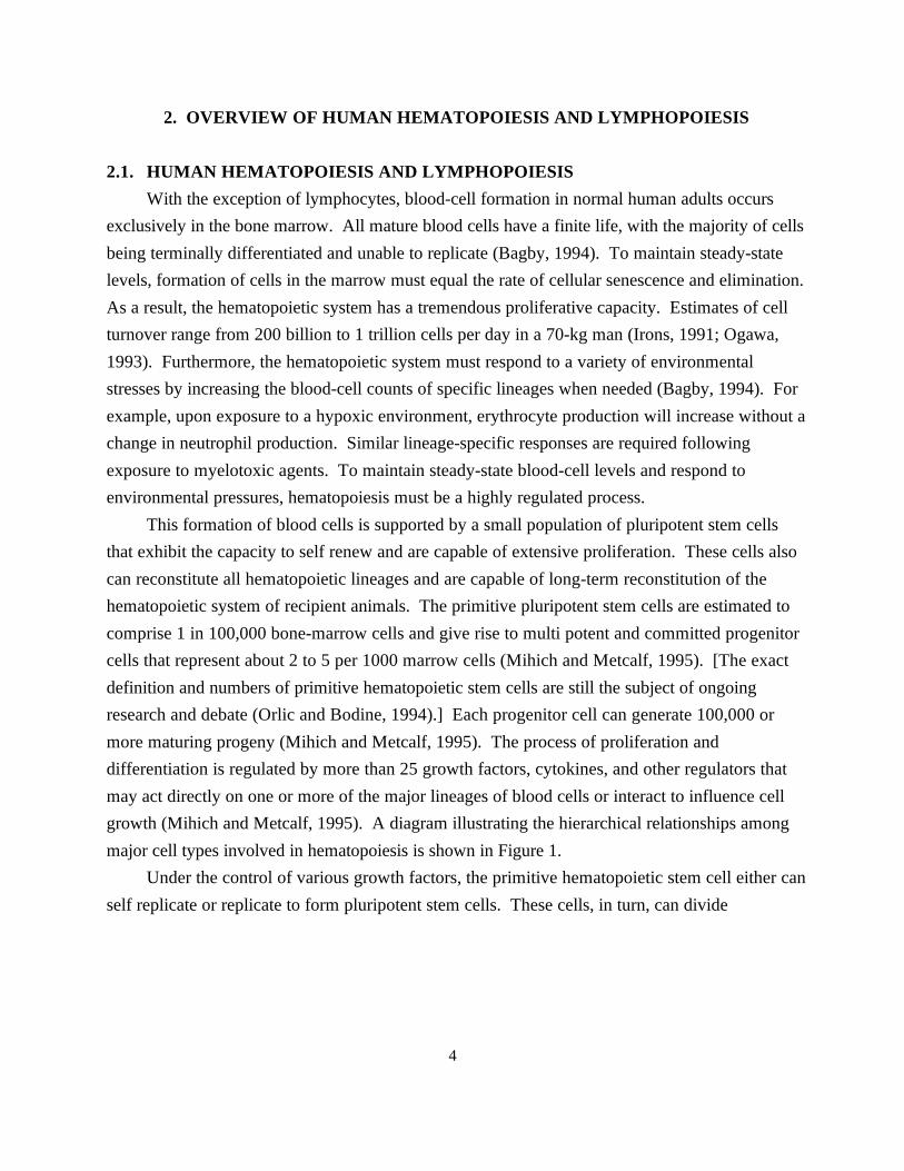
This formation of blood cells is supported by a small population of pluripotent stem cellsthat exhibit the capacity to self renew and are capable of extensive proliferation. These cells alsocan reconstitute all hematopoietic lineages and are capable of long-term reconstitution of thehematopoietic system of recipient animals. The primitive pluripotent stem cells are estimated tocomprise 1 in 100,000 bone-marrow cells and give rise to multi potent and committed progenitorcells that represent about 2 to 5 per 1000 marrow cells (Mihich and Metcalf, 1995). [The exactdefinition and numbers of primitive hematopoietic stem cells are still the subject of ongoingresearch and debate (Orlic and Bodine, 1994).] Each progenitor cell can generate 100,000 ormore maturing progeny (Mihich and Metcalf, 1995). The process of proliferation anddifferentiation is regulated by more than 25 growth factors, cytokines, and other regulators thatmay act directly on one or more of the major lineages of blood cells or interact to influence cellgrowth (Mihich and Metcalf, 1995). A diagram illustrating the hierarchical relationships amongmajor cell types involved in hematopoiesis is shown in Figure 1.
Under the control of various growth factors, the primitive hematopoietic stem cell either canself replicate or replicate to form pluripotent stem cells. These cells, in turn, can divide

5
Figure 1. Hierachical relationships betw
een major cell types involved in hem
atopoiesis. For simplicity, a num
ber of steps inm
aturation pathways have been om
itted.

6
to form multi potent progenitor cells that are committed to either the myeloid or lymphoidlineages (Bagby, 1994). The multi potent myeloid progenitor cell (CFU-GEMM) can give rise tocolony-forming cells of each myeloid lineage (erythrocyte, BFU-E; megakaryocyte, BFU-meg;monocyte/neutrophil/eosinophil, CFU-GM; basophil, CFU-baso). The CFU-GM cell can undergofurther differentiation to form colony-forming cells that are restricted to the monocyte (CFU-M),neutrophil (CFU-G), and eosinophil (CFU-Eos) lineages.
The multi potent lymphoid progenitor cell undergoes further specialization to form naturalkiller (NK) cells, T-cells, and B-cells (see Burns et al., 1996, for additional details). Shortly aftercommitment to the T-cell lineage, pre-T-cells migrate from the bone marrow to the thymus wherethey begin T-cell receptor (TCR) rearrangement. Through a process of sequential positive andnegative selection, cells that can properly recognize major histocompatibility class surface proteinsand foreign peptides survive and are released to the blood. These mature T-cells then migrate tothe lymph nodes and elsewhere through the circulatory system and body. Following commitmentto the B-cell lineage, the pro-B-cells in the bone marrow begin a process of rearranging their V(variable), D (diversity), J (joining), and C (constant) gene segments to form antigen-receptorgenes. The resulting pre-B-cells expressing: heavy chains in their cytoplasm undergo furthermaturation and eventually are characterized by the presentation of IgM and IgD surfaceimmunoglobulins. These mature B-cells then migrate from the bone marrow to the lymph nodesand other secondary lymphoid organs. Relatively little is known about development of NK cells,apart from the observation that mature NK cells are localized primarily in the spleen, blood, andperitoneal exudate.
2.2. COMPARISON OF HEMATOPOIESIS AND LYMPHOPOIESIS IN RODENTSAND HUMANS
In general, hematopoiesis and lymphopoiesis in rodents is similar to that described above forhumans. All blood cells originate from a pluripotent hematopoietic cell and become committedinto both myeloid and lymphoid lineages. These individual cells then differentiate into theirrespective T- and B-cells for lymphocytes and neutrophils, eosinophils, and monocytes formyeloid cells. Rodent models, and in particular mouse models, are thought to be highly relevantfor understanding most aspects of hematopoiesis (Bagby, 1994). However, a number ofdifferences between hematopoiesis and lymphopoiesis in humans and rodents are significant. Hematopoiesis in adult humans is restricted to the bone medullary spaces. Extramedullaryhematopoiesis involving the spleen, liver, and lymph nodes rarely occurs except under conditionsof extreme demand (Irons, 1991). In contrast, small groups of hematopoietic cells commonly are

7
found in mouse and rat spleen, and occasionally small clusters of these cells can be located in theliver (Hall, 1992; Irons, 1991). In addition, compensatory hyperplasia of various cell lineages inrodents can confound the diagnosis of lymphoma and other diseases. Furthermore, the relativenumber of lymphocytes and macrophages in rodents can vary considerably, depending on theamount of splenic hematopoiesis (Hall, 1992).
There are also significant differences in the composition of nucleated cells in the blood andbone marrow of adult rodents and humans. Illustrated in Table 2 are average values reported forhumans, rats, and mice from a variety of sources. For example, lymphocytes make up about 35%of nucleated cells in adult human blood and around 70% of nucleated cells in rodent blood. Thisfrequency also can be influenced by age and strain. For example, in rats the percentage oflymphocytes decreases from 90% at 2 months of age to about 65% at 30 months (Valli et al.,1990). In a corresponding fashion, the percentage of neutrophils increases with age from about9% at 2 months to around 30% at 30 months. A similar but less-pronounced change in thepercentage of lymphoid cells can also be seen in the bone marrow. The proportion of myeloid anderythroid cells in the marrow increases over the first year of life, whereas the proportion oflymphocytes and monocytes decreases between 2 and 12 months (Valli et al., 1990).
This high frequency of lymphocytes in the peripheral blood of rats and mice is among thehighest percentage seen in mammals. In a report by Smith (1990), in which around 100mammalian species were compared, the frequency of lymphocytes reported for the rat rankedamong the highest 5 species when compared on a per-liter basis. Among the top 5, the rat had thehighest percentage of circulating white blood cells (71%) compared with the pig (51%), the crab-eating macaque (63%), the guinea pig (65%), and the elephant (54%) (Smith, 1990). Correspondingly lower percentages of neutrophils, eosinophils, and basophils are seen in the rat ascompared to the human.
In addition to this high percentage of lymphocytes, other differences in the blood profile canbe seen. Both mice and rats have smaller erythrocytes than humans, with a survival time of 40 to68 days compared to about 120 days in humans (Hall, 1992; Irons, 1991). A large proportion ofthe neutrophils of both mice and rats exhibit a "ring" type of nucleus with a donut shape ratherthan the multilobulated type seen in humans (Andrew, 1965; Hall, 1992; Hulse, 1964). Furthermore,
unlike human neutrophils, the granules are small and difficult to stain (Bannerman, 1983) In addition,.
platelet counts in rats and mice are very high, averaging about 1 million/:l in the rat and 1.5 million/:l in themouse (Hall, 1992).

8
Table 2. Inspecies comparison of the composition of major nucleated cell types in bone marrow and blooda
Bone Marrow
Myleloid Erythroid Lymphoid
Human 56 26 13
Mouse 44 24 31
Rat 36 34 23
Blood
Neutrophilis Monocytes Eosinophils Basophils Lymphocytes
Human 54 7 3 1 36
Mouse 19 5 0.7 0 76
Rat 22 3 2 0.1 73
All values represent percentages of nucleated cells. Values are averages obtained from the following sources: Bannerman, 1983; Bergemann anda
Rastletter, 1979; Chervenick et al., 1968; Hulse, 1964; Parmley, 1988; Ringler and Dabich, 1979; Smith, 1990; Snyder et al., 1975; and Valli et al.,1990.
The different characteristics of rodent blood and bone marrow combined with differences in splenichematopoiesis indicate that in spite of their similarities, significant differences in hematopoiesis exist amongmice, rats, and humans. A recent article by Irons and associates also pointed out that species differences arelikely to exist in the organization of the hematopoietic stem-cell compartment, particularly in regard to thestage at which the primitive hematopoietic progenitor cells become restricted to a specific differentiationpathway (Irons et al., 1995). Primitive human hematopoietic progenitor cells are supported in Go by IL-3 orGM-CSF but not SCF, whereas the comparable murine primitive hematopoietic progenitor cells require IL-3or SCF for survival. These differences in cell composition, number, proliferation rates, or organization of thestem-cell compartment are likely to influence the hematotoxic and carcinogenic effects observed in humanand rodent systems following exposure to carcinogenic agents.
3. OVERVIEW OF CHEMICALLY AND RADIATION-INDUCED HEMATOPOIETIC NEOPLASIA IN HUMANS AND RODENTS
Over the past 50 years, considerable evidence has demonstrated the involvement of radiation, selectedtherapeutic drugs, and occupational chemicals in the etiology of human leukemias and lymphomas. Agentsrecognized by the International Agency for Research on Cancer (IARC) as Group 1 carcinogens, meaningthat sufficient evidence exists of their carcinogenicity in humans, are listed in Table 3. This list comprises

9
only the IARC Group 1 chemicals associated with lympho- or hematopoietic neoplasia. Also listed areionizing radiation and several topoisomerase II inhibitors not reviewed by IARC but for which there isconvincing evidence of leukemogenicity in humans. In addition, a group of chemicals is classified by IARCas probable human carcinogens (Group 2A) based on either animal studies, mechanistic information, orlimited epidemiology; these chemicals are likely to be associated with lymphohematopoietic cancers. Foreach agent, information summarizes the primary type of leukemia or lymphoma observed, whether the agentinduces significant myelotoxicity in humans, and whether increases in structural chromosomal aberrationshave been seen in the peripheral blood lymphocytes of humans exposed to each agent. In addition, theprimary types of lymphohematopoietic tumors seen in various mouse and rat cancer bioassays are listed. Formost, a summary description from a recent IARC monograph is also presented.
At present 16 agents show sufficient evidence of lymphohematopoietic neoplasia in humans, and 10are probable human leukemogens. A number of patterns emerge from the information in the table. Of theGroup 1 carcinogens, 12 of the 16 are associated primarily with acute nonlymphocytic leukemia (ANLL). The two strong immunosuppressive agents, cyclosporin and azathioprine, are associated primarily with Non-Hodgkin's lymphoma (NHL) and two agents, ethylene oxide and vinyl chloride, have been associated withboth ANLL and lymphoid malignancies. However, the leukemogenic effects of these last two appear to berelatively weak, and their classification as Group 1 carcinogens seems to rely primarily on tumors seen inother tissues (vinyl chloride) or on mechanistic data (ethylene oxide). In addition to its ability to induceANLL, radiation also has been strongly associated with the induction of chronic myelogenous leukemia andacute lymphoblastic leukemia in humans (BEIR V, 1990; UNSCEAR, 1994).

Table 3. Characteristics of selected known and probable human leukemia-inducing agents
Human Myelotoxic Aberrations Mouse Rat Sourcev t u
Agents carcinogenic to humans (IARC Group 1)
DNA-reactive
1,4-Butanediol dimethanesulphonate ANLL H SCA Leukemia/lymphoma NLHT IARC, 1987a (Busulfan, Myleran) Thymic lymphoma Conklin et al., 1965
w y
Thymic lymphoma Robin et al., 1981
l
NLHT Schmahl and Osswald, 1970l
Chlorambucil ANLL H SCA Hematopoeitic Hematopoietic + Lymphatic IARC, 1987am a
Lymphosarcoma Leukemia Kaldor et al., 1988Lymphosarcoma Lymphoma Weisburger et al., 1975
Lymphoma + granulocytic Cavaliere et al., 1990 leukemia
Hematopoietic + Lymphatic Berger et al., 1985a
n
(1-(2-)Chlorethyl)-3- ANLL H NA NLHT NLHT IARC, 1987a (4-methylcyclohexyl) nitrosurea – Schmahl and Habs, 1982 (Methyl-CCNU, Semustine)
o a,l a,l
e
Cyclophosphamide ANLL H SCA NLHT NLHT IARC, 1987aLeukemia Kaldor et al., 1988LymphosarcomaLymphocytic leukemia Petru et al., 1989
Lymphoid hematopoietic Schmahl and Habs, 1978 leukemiasVariety Schmahl and Osswald, 1970p
Lymphold + hematopoietic Schmahl and Habs, 1979 leukemiasHematopoietic + lymphoid tumors Schmahl and Habs, 1982NLHT Schmahl and Habs, 1976l
n
Melphalan ANLL H SCA Lymphosarcoma Lymphosarcoma IARC, 1987aLymphosarcoma Lymphosarcoma Kaldor et al., 1988Lymphosarcoma NLHT Weisburger et al., 1975l
Lymphosarcoma Gold et al., 1984
n
n
Treosulphan ANLL H NA NA NA IARC, 1987a
Vinyl chloride Mixed – SCA NLHT NLHT IARC, 1987ax g l f,l
– Radike et al., 1981d

Table 3. Continued
Human Myelotoxic Aberrations Mouse Rat Sourcev t u
DNA-reactive (continued)
Thio-TEPA ANLL H SCA Lymphoid Lymphoid IARC, 1990d (tris(1-aziridinyl)-phosphine) Lymphoid Lymphoid + granulocytic leukemia NCI, 1978c
NLHT Schmahl and Osswald, 1970d
Ethylene Mixed – SCA Lymphoma Lymphoid IARC, 1994ax g
Lymphoma NTP, 1987
r
Lymphoid Lynch et al., 1984r
Lymphoid Snellings et al., 1984r
Topoisomerase II inhibitors
Etoposide ANLL H Na NA NAz q i
Teniposide ANLL H NA NA NAz
Bimolane ANLL Na SCA Granulocytic leukemia NA Ye et al., 1994z h
Immunosuppressive agents
Cyclosporin NH Lymphoma – SCA Lymphoma – IARC, 1990cg k j
Azathioprine NH Lymphoma H SCA Lymphoma Lymphoma IARC, 1987as a
Thymic lymphoma Cohen et al., 1983
Other
Benzene ANLL H SCA Lymphoma NLHT IARC, 1987aLymphoma NLHT NTP, 1986bLymphoma NLHT (see text)
l
l
b,c
Ionizing radiation ANLL H SCA Thymic Lymphoma Yokoro et al., 1986CML Myeloid leukemia Storer et al., 1982ALL Myeloid leukemia NLHT Conklin et al., 1965
Thymic Lymphoma NLHT (see text)
b,c
l
Lymphoid+Myeloid Gross and Dreyfuss, 1979(see text) Schmahl and Osswald, 1970
Agents probably carcinogenic to humans (IARC Group 2A)
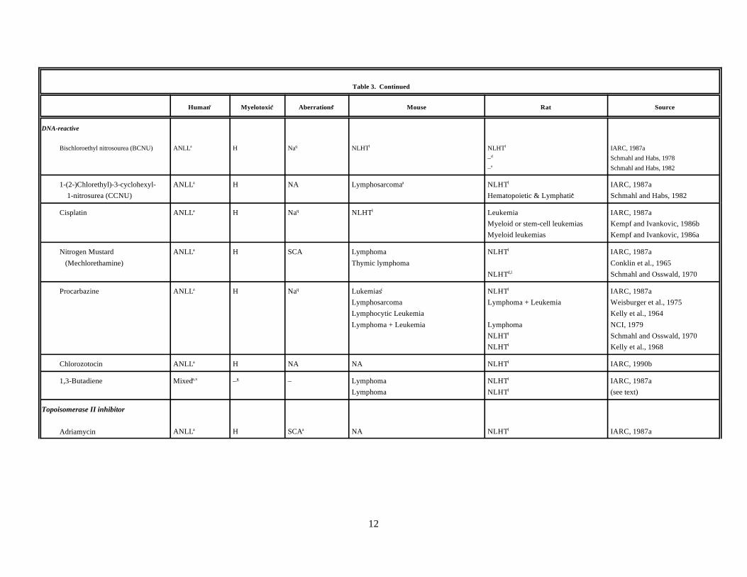
Table 3. Continued
Human Myelotoxic Aberrations Mouse Rat Sourcev t u
12
DNA-reactive
Bischloroethyl nitrosourea (BCNU) ANLL H Na NLHT NLHT IARC, 1987aa q l l
– Schmahl and Habs, 1978d
– Schmahl and Habs, 1982e
1-(2-)Chlorethyl)-3-cyclohexyl- ANLL H NA Lymphosarcoma NLHT IARC, 1987a 1-nitrosurea (CCNU) Hematopoietic & Lymphatic Schmahl and Habs, 1982
a a l
a
Cisplatin ANLL H Na NLHT Leukemia IARC, 1987aa q l
Myeloid or stem-cell leukemias Kempf and Ivankovic, 1986bMyeloid leukemias Kempf and Ivankovic, 1986a
Nitrogen Mustard ANLL H SCA Lymphoma NLHT IARC, 1987a (Mechlorethamine) Thymic lymphoma Conklin et al., 1965
a l
NLHT Schmahl and Osswald, 1970d,l
Procarbazine ANLL H Na Lukemias NLHT IARC, 1987aa q c
Lymphosarcoma Lymphoma + Leukemia Weisburger et al., 1975Lymphocytic Leukemia Kelly et al., 1964Lymphoma + Leukemia Lymphoma NCI, 1979
l
NLHT Schmahl and Osswald, 1970l
NLHT Kelly et al., 1968l
Chlorozotocin ANLL H NA NA NLHT IARC, 1990ba l
1,3-Butadiene Mixed – – Lymphoma NLHT IARC, 1987aa,x g
Lymphoma NLHT (see text)
l
l
Topoisomerase II inhibitor
Adriamycin ANLL H SCA NA NLHT IARC, 1987aa a l

Table 3. Continued
Human Myelotoxic Aberrations Mouse Rat Sourcev t u
Other
Azacytidine ANLL H Na Granulocytic + Lymphocytic NLHT IARC, 1990aa q
Granulocytic sarcoma + Lymphoma – NCI, 1978aLymphoma Cavaliere et al., 1987Lymphatic leukemia + Lymphoma Schmahl et al., 1985 + Myeloid leukemia
l
a,d
Chloramphenicol ANLL H NA Lymphoma NA IARC, 1987aa a
Limited evidence.a
Influenced by strain.b
Hemopoietic and lymphatic tumors showed a statistically nonsignificant increase.d
No data presented, increases reported at other sites.e
Reported to induce lymphomas in hamster [Registry of Toxic Effects of Chemical Substances (RTECS)].f
Myelotoxicity is either not seen or infrequently seen (Arky, 1996; ATSDR, 1992b; Handschumacher, 1990; IARC, 1994a).g
Reported to alter leukocyte count in treated dogs (RTECS, 1996).h
Minor increases of tumors at various sites have been reported (RTECS, 1996).i
Study had limited sensitivity.j
Increases observed in AKR mouse, a strain highly susceptible to lymphomas. Cyclosporin also has been shown to accelerate lymphoma development in mice treated with radiation ork
N-methyl-N-nitrosourea and accelerate the formation of lymphomas in grafted macaques, a species of monkey with an extremely low frequency of lymphoma (IARC, 1990c).No or minimal increase in lymphohematopoietic tumors reported. In many cases, tumors in other tissues were reported.l
Information from Seiber and Adamson, 1975.m
Secondary reference.n
Information from Calabresi and Chabner, 1990.o
Slight increase in lymphohematopoietic neoplasia.p
Increase seen in experimental animals (RTECS,1996).q
Mononuclear cell leukemia classified as lymphoid based on the classification scheme of Ward et al., 1990. r
From IARC, 1981.s
Myelotoxic to humans. Information from Arky, 1996, unless otherwise indicated.t
SCA - Increases in structural chromosomal aberrations observed in human lymphocytes. Information from Sorsa, et al., 1992, unless otherwise indicated.u
Primary classification based on IARC Monographs or BEIR V unless otherwise specified.v
ANLL - Acute nonlymphocytic leukemia.w
Table 3 Footnotes (continued)
Leukemias of both myeloid and lymphoid lineages frequently reported.x
Effect frequently observed in humans.y

14
Classification based on Pedersen-Bjergaard and Rowley, 1994, for etoposide and teniposide and Zhang et al., 1993, for bimolane.z
15
In regard to myelotoxicity, 12 of the 16 agents for which information was available showed significantevidence of myelotoxicity in humans who either received the drug therapeutically or were exposed to theagent occupationally. Most of these agents, such as benzene, busulfan, chlorambucil, and radiation, are wellknown for their myelotoxic effects. Ethylene oxide, vinyl chloride, and cyclosporin were not classified asmyelotoxic; although bone marrow toxicity is seen occasionally following exposure to these agents, ittypically is an infrequent event (ATSDR, 1992b; IARC, 1994a). Of the Group 2 leukemogenic agents, nineof the ten have been observed to induce significant myelotoxicity in humans. The one agent for whichmyelotoxic effects commonly have not been seen in humans is 1,3-Butadiene. Although not listed in thetable, most of these Group 1 and 2 carcinogens have been shown to exhibit myelotoxic effects in rodentmodels.
Of the Group 1 agents for which data could be found (11 total), all were reported to induce structuralchromosomal aberrations in the peripheral blood lymphocytes of exposed individuals. Reports of structuralchromosomal aberrations in humans were rarely found for the Group 2 agents. However, most Group 1 andGroup 2 carcinogens have been shown to induce chromosomal aberrations in rodent models.
Testing results for most Group 1 and 2 agents in rodent bioassays for carcinogenicity are also shown inTable 3. Based on the number of studies and reported frequencies for various lymphohematopoietic tumortypes, the predominant tumor type associated with each agent was determined. For simplicity and because of the close relationship between tumor types such as T-cell lymphomas and T-cell leukemias (Pattengale,1990), the tumors are summarized as primarily lymphoid, myeloid, or of multiple lineages. In addition, manyof these studies were conducted using a variety of strains and administration routes and were done before theimplementation of standardized testing protocols. For the 16 Group 1 agents, the primary tumor type in themouse was lymphoid for 10 of the chemicals and myeloid for 1 agent, bimolane. Either no information wasavailable or the studies were of inadequate quality for 4 agents (methyl CCNU, treosulphan, etoposide, andteniposide). Only for vinyl chloride had significant increases in lymphohematopoietic tumors not beenreported. [An increase in lymphoid tumors has been seen for vinyl chloride in hamsters (ATSDR, 1992b)]. Radiation was listed as inducing primarily lymphoid tumors because this is the most frequently observedtumor type in most strains of mouse (Storer et al., 1982; Yokoro et al., 1986). However, myeloid leukemia isinduced by ionizing radiation in some mouse strains (Riches, 1995; Yokoro et al., 1986). Of the 10 Group 2agents, 5 induced primarily lymphoid neoplasms in the mouse, 0 myeloid, and 1 mixed (azacytidine). Twoagents showed no evidence of lymphohematopoietic cancers (BCNU, and cisplatin), and inadequateinformation was available for two agents (chlorozotocin and adriamycin).
An overview of tumor types induced in rat by Group 1 carcinogens revealed that threeagents—melphalan, ethylene oxide, and azathioprine—caused primarily lymphoid neoplasms, andthree—chlorambucil, cyclophosphamide, and thio-TEPA—induced tumors with both lymphoid and myeloidlineages. No rat cancer bioassay data was available for treosulphan, etoposide, teniposide, and bimolane. Significant increases in lymphohematopoietic cancers were not generally seen for six agents: busulfan,methyl CCNU, vinyl chloride, cyclosporin, benzene, and radiation. Radiation has been reported to induce
16
lymphoid and myeloid leukemias in rats in a few reports, but most studies using this species have failed to seesignificant increases in these tumor types (Gross and Dreyfuss, 1979; Ward et al., 1990). Similar resultshave been reported for the cancer bioassays of benzene performed in the rat (Maltoni et al., 1989; NTP,1986a).
Based on the above information, a number of patterns seem apparent. (1) The primary type oflymphohematopoietic cancer induced by chemicals in humans is myeloid leukemia (ANLL), with theexception of immunosuppressive agents associated almost exclusively with lymphoma development. (2)Potent leukemia-inducing agents also induce significant myelotoxicity and structural chromosomal aberra-tions in exposed humans. (3) Administration of human leukemia-inducing agents to mice results in morelymphohematopoietic tumors. However, in contrast to human, these tumors are primarily lymphoid in origin. (4) The rat is considerably less responsive than the mouse for the induction of lymphohematopoieticneoplasia following administration of human leukemogens. When induced, resulting neoplasms in the rat areprimarily lymphoid in origin.
4. LEUKEMIAS INDUCED BY SELECTED CLASSES OF ENVIRONMENTAL AND THERAPEUTIC AGENTS IN HUMANS
4.1. GENERAL COMMENTSA number of studies have shown an association between exposure to petroleum, solvents, pesticides,
and other chemical agents and increased risks of hematopoietic neoplasia (Brandt, 1992). In most of thesestudies, the increased risks have been for ANLL, but increases in other types of leukemias have also beenreported (Brandt, 1992; Malone et al., 1989; Persson et al., 1989; Weisenburger, 1994). In some cases,increases have been attributed to benzene exposure whereas in other cases, the agent responsible for theneoplastic effects is not readily apparent (Checkoway et al., 1984; Ott et al., 1989; Persson et al., 1989). There is also evidence that such other chemicals as ethylene oxide, vinyl chloride, and 1,3-butadiene mayhave been responsible for some increases in observed lymphohematopoietic cancers (Finch and Linet, 1992;IARC, 1987a; IARC, 1992; IARC, 1994a). However, for most studies on these agents, the hematopoieticeffects have been relatively weak, and some inconsistencies have been seen (Cole et al., 1993; Shore et al.,1993). Although not a chemical, ionizing radiation has been included because it has been extensively studiedand represents an important leukemia-inducing agent. Differences in effect have been seen for different typesof ionizing radiation and for applications at different dose rates. However, for simplicity and ease ofpresentation, radiation has been treated as one agent with uniform characteristics. For a more detaileddescription of the effects of ionizing radiation, the reader is referred to the following sources: BEIR V, 1990;Hendry and Lord, 1995; UNSCEAR, 1993; and UNSCEAR, 1994. The following section will review sixagents, for five of which there is strong and consistent epidemiological evidence. The six agents are ionizingradiation; alkylating agents; epipodophyllotoxin-topoisomerase and dioxopiperazine-topoisomeraseinhibitors; benzene; and 1,3-butadiene, an agent for which human data is more limited and controversial.
17
However, before describing what is currently known about the mechanisms of genotoxicity andleukemogenesis for the individual agents, some general observations about solvent and chemically inducedlymphohematopoietic cancers will be discussed.
Over the past 20 years, cytogenetic studies of bone-marrow cells of leukemia patients have becomeimportant for diagnosing the disease, as prognostic indicators, and for mechanistic information (Pedersen-Bjergaard and Philip, 1987). Karyotype comparison between cases with de novo leukemia (ANLL) andpatients previously treated with alkylating chemotherapeutic agents revealed that the latter groups had asignificantly higher frequency of leukemic cells with abnormal karyotypes, primarily loss and deletions ofchromosomes 5 and 7 (Rowley, 1983). Based on these results, a series of studies was conducted to determineif a similar pattern could be seen in patients with a history of exposure to occupational and environmentalchemicals (Golomb et al., 1982; Mitelman et al., 1978; Mitelman et al., 1979; Mitelman et al., 1981). Thesestudies generally have shown that patients with a history of chemical exposure have significantly higherfrequencies of karyotypically aberrant leukemic cells than nonexposed patients. A variety of abnormalkaryotypes were originally reported, including -5/5q-, -7/7q-, +8, +21, t(8;21) and t(9;22) (Mitelman et al.,1978; Mitelman et al., 1981). Differences in frequency between exposed and nonexposed were substantial(83% to 24%) in initial studies. In follow-up studies, the differences, although still significant, generally havenot been as strong. This issue was addressed at the Fourth International Workshop on Chromosomes inLeukemia, and a significantly higher frequency of karyotypically abnormal leukemic cells was seen in theexposed (64%) when compared to the nonexposed (49%) (Mitelman et al., 1984). In this study, -5/5q-, -7/7q- and t(8;21) appeared to be associated with previous occupational exposure. More recent studiesgenerally have seen similar associations for chromosomes 5 and 7 (Cuneo et al., 1992; Fagioli et al., 1992;Zedginidze et al., 1990). Association of lifestyle exposures such as smoking and alcohol consumption withleukemic karyotype have been reported in several studies (Crane et al., 1989; Sandler et al., 1993). In boththese studies, cigarette smoking was associated with -7/7q-. Although less information is available for othertypes of lymphohematopoietic cancers, one study reported that higher frequencies of cytogeneticabnormalities were seen in the lymphoma cells of patients with non-Hodgkin's lymphoma who had a historyof exposure to organic solvents (Brandt et al., 1989). The exposed patients had a higher frequency oftranslocations involving the 14q32 band. Aberrations of 6q- appeared to occur more frequently in thenonexposed, but the difference was not statistically significant (p=0.08).
One recent morphologic, immunologic, and cytogenetic study of leukemia (AML) patients with historyof exposure to pesticides and organic solvents is particularly noteworthy (Cuneo et al., 1992; Fagioli et al.,1992). In this study, clonal chromosomal aberrations involving chromosomes 5 or 7 were seen morefrequently among exposed patients. Myelodysplasia involving multiple cell lineages was seen in assessablepatients with chemical exposure but was seen only in a minority of nonexposed individuals. In addition,immunological studies revealed that leukemic cells of 80% of exposed patients were positive for the CD34stem-cell marker, whereas only 22% of leukemic cells of nonexposed patients were positive for this marker. Exposed patients showed a much lower frequency of remission following conventional chemotherapy. These
18
studies suggest that leukemias caused by chemical exposure have higher frequencies of chromosomalabnormalities, affect multiple lineages, and more frequently involve the more primitive hematopoietic stemcells.
Activation of one of the ras oncogenes, primarily N-ras or K-ras, has been observed frequently andconsistently in MDS and human leukemias (Bishop, 1991; Bos, 1989; Sandberg, 1993). Although mutationsin ras were detected in only a minority of leukemias induced by alkylated agents (Pedersen-Bjergaard et al.,1988; Yunis et al., 1989), there is some evidence that leukemic cells in other chemically exposed individualsexhibit higher frequencies of ras activation than similar cells from nonexposed individuals (Taylor et al.,1992). In the case control studies by Taylor and associates (Taylor et al., 1992), patients with ras-mutation–positive AML were more likely to have worked in an occupation with chemical exposure, and therisk was higher in those who had worked for 5 or more years in an exposed profession. The ras-positivepatients also were more likely to have had dermal exposures to chemicals, to have breathed chemical vapors,and to have worked in a dusty environment.
4.2. IONIZING RADIATION4.2.1. Background
Shortly after the discovery of X rays by Roentgen in 1895, injuries resulting from overexposure toradiation became apparent. Initial reports primarily were skin reactions. Since that time, ionizing radiationhas been shown to affect a wide range of tissues and organs, with the bone marrow and lymphoid tissuesamong the most severely affected (Upton, 1993). A rapidly delivered whole-body dose of 2 to 3 Sievert (Sv.;1 Sv = 100 rem) results in extensive killing of lymphocytes and their precursors, with manifestations ofsevere lymphopenia and immunosuppression within 48 hours (Upton, 1993). Extensive killing ofhematopoietic cells also occurs, leading to aplasia within the marrow and a decrease in granulocyte andplatelets. Regeneration of the bone marrow varies markedly among individuals and is related to the dose,type of radiation, and extent of exposure. A number of studies have shown that regeneration takes placeprimarily during the first 1 to 2 years following radiotherapy, but in some cases the irradiated area may showhypoplasia for as long as 13 years after exposure (Parmentier et al., 1988).
4.2.2. Hematopoietic and Lymphoid Neoplasia Seen in HumansChronic exposure to lower levels or high exposures over a short time period has been associated with a
variety of cancers: skin carcinomas were seen in X-ray workers; leukemias, breast cancer, and thyroid cancerin radiologists; osteogenic sarcomas in radium dial painters; and lung cancer in miners (Upton, 1993; Wanget al., 1988). The strongest association between radiation and cancer has been seen for the induction ofleukemia. Numerous population studies have shown an association between ionizing radiation and varioustypes of leukemia and lymphoma (BEIR V, 1990; UNSCEAR, 1994). The actual risk of these malignanciesfollowing exposure to radiation appears to be complex and is related to the type of radiation, the dose, theproportion of the body exposed, and the extent of cell killing and DNA repair (Curtis et al., 1994). Such

19
factors as age, gender, genetic background, and physiological condition of exposed individuals also caninfluence the risk of radiation-induced cancer (BEIR V, 1990). In lymphoid or hematopoietic tissues that are distributed diffusely, a significant proportion of tissue must be irradiated to increase the incidence ofneoplasia (Storer et al., 1982). Reduced risks of leukemia also have been seen in atomic-bomb survivorswho received high doses [above 3 to 4 Gray (Gy)] of primarily gamma radiation that has been attributed toextensive killing of marrow-containing progenitor cells (BEIR V, 1990). A similar reduced risk at high doseshas been seen in recent years; changes in therapeutic strategy employing high doses within limited fieldsappear to have reduced the risk of leukemia and altered the shape of the dose-response curve in the high-doseregion (Boice et al., 1987; Curtis et al., 1994).
The most extensive information on induction of leukemias and lymphomas has come from studies ofJapanese atomic-bomb survivors who were exposed to both gamma, and to a lesser extent, neutron radiation. The latest update of this study group, which was concluded in 1987, included 93,696 survivors andencompassed 2,778,000 person-years of study (Preston et al., 1994). Unfortunately, data collection began 5years after the bombing, so little information is available about leukemias occurring during that period orabout the minimum latency period (Mole, 1990; Preston et al., 1994). In addition, dose estimates for manyindividuals remain imprecise. In spite of this, a great deal of valuable information on types of neoplasia andvariables affecting responses is available from this population. In the 1980s, most cases in the leukemiaregistry were reclassified using more modern criteria and nomenclature such as the French-American-British(FAB) classification scheme. This reclassification allowed a more accurate identification of the types oflymphoid and hematopoietic neoplasia induced by radiation. Results of previous and recent analysis indicatethat acute lymphocytic leukemia (ALL), chronic myelogenous leukemia (CML), and acute myelogenousleukemia (AML) were the major contributors to the total leukemias seen (Matsuo et al., 1988; Preston et al.,1994). No increase in risk was seen for adult T-cell leukemia (ATL), chronic lymphocytic leukemia (CLL) orHodgkin's disease (HD) (BEIR V, 1990; Matsuo et al., 1988; Preston et al., 1994; UNSCEAR, 1994). Asummary of the hematopoietic and lymphoid neoplasia seen in the Nagasaki A-bomb survivors is shown inFigure 2. The types of leukemia

20
Figure 2. Types of lymphohem
atopoietic neoplasia in Nagasaki survivors of the atom
ic-bomb blast. Types of cancer for w
hichevidence is sufficient for association w
ith radiation are labeled with an S
. Data are from
Matsuo et al., 1988. N
o information w
asavailable from
this source for multiple m
yeloma or lym
phomas.
21
and related disorders that have been consistently or occasionally associated with radiation exposure areindicated.
In bombing survivors, the incidence of total leukemia appeared to increase in a nonlinear fashion andwas influenced both by gender and age at exposure (Preston et al., 1994). Young men had the highest excessrisks during the period 5 to 10 years after exposure, but these risks decreased rapidly with time. Excess riskduring the early time period was not as high in older men but decreased more slowly. Exposed women tendedto have lower excess risks than men until about 20 years after exposure. Interestingly, the risk of leukemiaappears to decrease for women who were young at the time of exposure, whereas no decrease in risk has beenseen in women who were older then (Preston et al., 1994). Evidence also shows that the latency period can beaffected by the intensity of radiation exposure (Cadman et al., 1977). Latency periods of around 5 years wereobserved in survivors located within 1500 meters of the hypocenter, whereas latency periods of 10 years orlonger were seen for individuals located at greater distances (Cadman et al., 1977). Additional details foreach type of hematopoietic or lymphoid malignancies are described below.
4.2.2.1. Acute Nonlymphocytic Leukemia (ANLL)There is strong evidence for the induction of ANLL in individuals exposed to ionizing radiation as a
result of atomic-bombing or therapeutic uses of radiation (BEIR V, 1990;Preston et al., 1994; UNSCEAR, 1994). All FAB subtypes were represented at lower and intermediatedoses. However, at the highest doses promyelocytic leukemia (M3) and erythroleukemia (M6) were not seenamong those exposed. Acute myelogenous leukemia (M2) was the FAB subtype most strongly associatedwith radiation exposure (Matsuo et al., 1988). Recent studies conducted on atomic-bomb survivors haveindicated that risk was highest in those exposed when young, and those risks decreased with time. Considerably lower risks were seen in survivors who were over 20 years of age at the time of the bombing,but there is no evidence that those risks have decreased with time (Preston et al., 1994). Similar age-relatedfindings have been seen in other studies (UNSCEAR, 1994).
4.2.2.2. Chronic Myelogenous Leukemia (CML)Significant increases in CML were seen in survivors of both Hiroshima and Nagasaki, and the risk
appears to be linear with dose (Preston et al., 1994). Men had greater CML risks that decreased rapidly withtime. Risks for females were much lower but continued to be elevated 25 years after exposure. Similarly toANLL, CML occurrence was pronounced in the young (Ichimaru et al., 1986). In addition, the risk of CML,which was higher for residents of Hiroshima than Nagasaki, may be related to differences in the backgroundincidence of CML in the two areas (Preston et al., 1994). Increased risks of CML also have been seen in patients treated with Xrays for ankylosing spondylitis during the mid-1930s to 1950s, as well as in radiologists using X rays beforethe implementation of modern safety standards (BEIR V, 1990; Finch and Linet, 1992).

22
4.2.2.3. Acute Lymphocytic Leukemia (ALL)Significant increased risks for ALL were seen in exposed survivors. Both age of exposure and gender
(to a lesser extent) seemed to influence the excess risk that was seen, with somewhat lower risks observed inolder individuals and in women (marginal significance)(Preston et al., 1994). The increases in ALL occurredmost frequently in those who were less than 15 years old at the time of the bombing, with the incidencepeaking within about 8 years and then decreasing. In adults, the induced ALL occurred late in life at abouthalf the peak frequency of children. These periods of elevated risk correspond to periods during which ALLtypically is seen in humans (Pendergrass, 1985).
4.2.2.4. Adult T-Cell Leukemia (ATL)No significant association was seen between radiation exposure and adult T-cell leukemia (Preston et
al., 1994). This is particularly interesting in that the human T-cell leukemia virus, HTLV-1, is endemic to theNagasaki region. About 30% of leukemia cases diagnosed in Nagasaki as part of the long-term survivorstudy were diagnosed as ATL whereas about 0.5% of leukemias in Hiroshima were ATL (Preston et al.,1994). The lack of an observed association between ATL and radiation exposure suggests that radiationexposure does not interact with HTLV-1 to increase the incidence of leukemia. In addition, recent studieshave seen no evidence for an association between radiation exposure and infection with HTLV-1 (Matsuo etal., 1995).
4.2.2.5. Multiple MyelomaAt present, there appears to be limited evidence for an association between multiple myeloma and
radiation exposure. Although a considerable number of studies have reported increased frequencies ofmultiple myeloma in radiation-exposed individuals, the increases generally have been modest and frequentlyhave not achieved statistical significance (BEIR V, 1990; UNSCEAR, 1994). More recent evaluations andreanalyses of previous studies have found much less evidence for an association (Preston et al., 1994;UNSCEAR, 1994). As an example, recent analysis of atomic-bomb survivors have found no significantassociation between radiation exposure from the bombs and the risk of multiple myeloma (Preston et al.,1994). This finding is in contrast to previous reports that were based on mortality rather than incidence. Thedifferences are believed to be related to reliance on questionable diagnoses in the earlier studies as well as theinclusion of high-dose cases and cases with second primary tumors that were excluded from the more recentanalysis (Preston et al., 1994). A more detailed review of myeloma and lymphoma cases is planned to clarifythese issues (Preston et al., 1994).
4.2.2.6. Non-Hodgkin's Lymphoma (NHL)There is limited evidence that exposure to high doses of radiation increases the risk of NHL (Boice et
al., 1988; Ichimaru et al., 1986; Kobayashi et al., 1990; Neglia et al., 1991; Preston et al., 1994). Initially,some evidence was provided from early prevalence studies (Ichimaru et al., 1986). More recent incidence

23
studies also have provided some evidence for an association between male atomic-bomb survivors and thedevelopment of lymphoma (Preston et al., 1994). No association was seen for females, and little evidencewas seen for an influence of time or age at exposure (Preston et al., 1994). Elevated risks of lymphoma alsohave been seen following radiotherapy to treat cervical cancer (Boice et al., 1988) and ankylosing spondylitis(Darby et al., 1985). However, other studies have not seen any increase (UNSCEAR, 1994). Thebackground frequency of lymphoma is significantly influenced by age and gender (Preston et al., 1994). Although the specific etiology of NHL is currently unknown, it is likely to be related to disturbances of theimmune system. Increased risks of NHL have been seen in individuals with immunosuppression resultingfrom HIV infection, Hodgkin's disease, genetic syndromes, and treatment with immunosuppressive agentsassociated with organ transplantation (Tucker, 1993). Recent results have indicated that atomic-bombsurvivors exhibit an increased prevalence of Epstein-Barr virus reactivation (Akiyama et al., 1993). This isof interest, as previous studies have suggested an association between infection with Epstein-Barr virus andsubsequent development of secondary NHL following radiotherapy (Evans and Mueller, 1990; List et al.,1986). This suggests that immunosuppression and immunomodulation (Akiyama et al., 1991) induced inradiation-exposed individuals may play a role in the etiology of radiation-induced NHL. However, studies inlaboratory animals have failed to demonstrate a direct relationship between immunomodulating agents andthe development of cancer (Schmahl, 1986).
4.2.3. Chromosomal Alterations Observed in Model Systems and Human Biomonitoring StudiesRadiation is a highly potent clastogen that induces structural chromosomal aberrations animals and in
human cells both in vitro and in vivo (Bender et al., 1988; Rithidech et al., 1995; Tanaka et al., 1983; Waldand Conner, 1988). Elevated frequencies of chromosomal aberrations such as acentric fragments, micronuclei, dicentric chromosomes, inversions, and translocations have been seen immediately in lymphocytes andbone marrow of individuals exposed to high levels of radiation (Bender et al., 1988; Lucas et al., 1992;Tanaka et al., 1983). Unstable alterations such as acentric fragments, micro nuclei, and dicentric chromo-somes decrease with time, whereas the frequency of stable alterations, primarily translocations and inver-sions, remains relatively stable for years and may be detected 30 to 40 years later (Lucas et al., 1992;Straume et al., 1992). Studies of chromosomal alterations in peripheral blood lymphocytes of highly exposedatomic-bomb survivors conducted many years after bombing have shown that the breakpoints in lymphocytechromosomes of these individuals did not occur randomly throughout the genome (Kamada et al., 1988;Tanaka et al., 1983). Of particular note is that a number of these nonrandom breakpoints (e.g., 5q31, 7q32,11q23, 21q2) lie within or immediately adjacent to regions that are altered in therapy-related and de novoleukemias and are believed to indicate the location of genes involved in leukemogenesis (Pedersen-Bjergaardand Rowley, 1994). The 7q32 region also has also been associated with lymphomagenesis (Offit et al., 1991;Offit et al., 1995). Elevated frequencies of alterations affecting 5q31, 7q22, and 21q22 also have been seenin the lymphocytes of ankylosing spondylitis patients many years after radiotherapy (Buckton, 1983). Translocations involving 7q32-36 also were seen in the lymphocytes of technicians with long-term radiation

24
exposure (Kumagai et al., 1991). In addition, nonrandom breaks have been observed at 6q21 in lymphocytesof atomic-bomb survivors. This region has been implicated in a variety of lymphoid malignancies (Johanssonet al., 1993).
4.2.4. Genetic Alterations in Cancer Patients4.2.4.1. Chromosomal Alterations
Cytogenetic analysis were performed on 25 leukemic patients with a history of radiation exposuregreater than 0.01 Gy (Kamada et al., 1988). The ten patients with the highest exposure (>2 Gy) exhibitedabnormal clones, and the aberration frequency was higher than that of patients with lower exposures. Abnormalities affecting chromosomes 5 and 8 seemed to predominate. There is also a report of a radiation-exposed preleukemia case that exhibited a 7q- (Ichimaru et al., 1986). Other studies have been performed onpatients who have developed ANLL following radiotherapy. Although relatively few studies are available,karyotypeas of these patients generally exhibit the same pattern of nonrandom aberrations (-7, -5, 5q-, 7q-) asseen in patients treated with alkylating agents (Kantarjian and Keating, 1987; Le Beau et al., 1986; Philip andPedersen-Bjergaard, 1988; Sandberg, 1990). Although alterations affecting chromosomes 5 and 7 have beenreported to occur less frequently than in patients treated with chemotherapy (Kantarjian and Keating, 1987),this is difficult to evaluate because relative risks are low following radiotherapy as currently performed(Philip and Pedersen-Bjergaard, 1988). Consequently, many observed cases are likely to represent de novoleukemias. In the relatively few cases for which cytogenetic analysis are available, the 9;22 translocationgenerally seen in leukemic cells of radiation-exposed CML patients is similar to what is commonly seen in denovo CML (Ichimaru et al., 1986; Kamada et al., 1988; Philip and Pedersen-Bjergaard, 1988; Sandberg,1990). Interestingly, a 9;22 translocation also was seen in a radiation-exposed leukemia patient with AML(M2)(Kamada et al., 1988).
4.2.4.2. Other Genetic ChangesThe association between chromosomal abnormalities and leukemia development has been investigated
further. The transforming activity of DNA extracted from bone marrow of A-bomb survivors with highfrequencies of nonclonal chromosomal aberrations and nine leukemia patients previously exposed toradiation, (three with CML, five with AML, and one with ALL) was tested using an in vivo selection assay(Kamada et al., 1988). The DNA was transfected into NIH3T3 cells and subsequently injected into nudemice. Tumors were formed in all mice injected with DNA from the leukemic patients and in 3 of 4 miceinjected with DNA from heavily exposed survivors. Activated N-ras genes were detected in 7 of 9 tumorsformed from leukemic patient DNA and 2 of 3 tumors from exposed-survivor DNA. As a control 5 DNAsamples from healthy medical students were tested in the same way, and no positive results were obtained.

25
4.2.5. Hematopoietic and Lymphoid Neoplasia Seen in Rodent Models4.2.5.1. Thymic Lymphoma
Although ionizing radiation is myelotoxic in all tested mammalian models, considerable variability hasbeen observed in the incidence of tumors and types of lymphohematopoietic neoplasia observed in animalexperiments (see UNSCEAR, 1977, UNSCEAR, 1993, and references therein for reviews). Differences inresponse have been seen for different species, genders, and strains, thus raising questions about theappropriateness of any one strain for predicting response and dose-response in humans (UNSCEAR, 1993). The mouse has been used most widely to study radiation-induced leukemias and lymphomas, whereas the rathas been used more widely for studies of radiation-induced breast cancer (Yokoro et al., 1986). In general,the rat is considered to be a poor model for radiation-induced lymphohematopoietic cancers (Gross andDreyfuss, 1979; Ward et al., 1990). As a result, this article will focus primarily on the induction of leukemiasand lymphomas in mice.
As observed in humans, lymphoid and hematopoietic tissues exhibit severe damage following highdoses of whole-body irradiation to rodents (Storer et al., 1982). Decreases are seen in all cell lineages, withlymphocytes showing high sensitivity and related effects on both humoral and cellular immune responses. Cell regeneration begins rapidly and takes place primarily during the first 2 years after exposure (Parmentieret al., 1988). However, studies have shown that the bone-marrow multipotential stem-cell pool is reduced 6to 18 months after whole- or partial-body irradiation and that hematopoietic activity of irradiated areas doesnot recover completely (Parmentier et al., 1988).
The thymus is also one of the most sensitive tissues to tumor induction by radiation in many mousestrains (Yokoro et al., 1986). For example, doses of certain types of ionizing radiation as low as 0.25 Gy caninduce thymic lymphoma in sensitive mouse strains such as the RFM strain (Storer et al., 1982). TheC57Bl/6 strain is another radiation-sensitive strain that has been widely used for mechanistic studies. Following whole-body X irradiation administered in fractionated doses, a high incidence of thymic lymphoma(>90%) was observed in C57BL/Ka mice (Sen-Majumdar et al., 1994b). Although the lymphoma developsin the thymus, the initial step in leukemogenesis occurs in bone marrow (Kotler et al., 1994; UNSCEAR,1977). The relationship between thymus and bone marrow appears to be complex. As described byFurmanski and Rich (1982), irradiation of the thymus itself does not result in lymphocytic leukemias, nordoes whole-body irradiation of thymus-intact and bone marrow-shielded–mice. Implants of normal thymusesin previously irradiated, athymic mice serve as the focus for development of thymic leukemia. Preleukemicbone-marrow cells (but not thymocytes) from irradiated mice can transfer the disease to normal recipients. Based on these observations, it appears that the primary leukemogenic event occurs in the bone marrow; theprogenitor cells then migrate to the thymus, where maturation and expression of neoplasia occur.
Initially, a viral hypothesis was presented to explain the high incidence of thymic lymphoma followingfractionated whole-body irradiation. In initial studies by Kaplan and associates, a thymotropic andlymphomagenic retrovirus, the radiation leukemia virus (RadLV), was isolated from cell extracts of one of thelymphomas (Kaplan, 1967). The original isolate was weakly leukemogenic upon intrathymic injection into

26
C57Bl/Ka mice. However, following serial passage, the virus became highly leukemogenic and capable ofinducing lymphomas in adult mice (Yefenof and Kotler, 1995). Based on the work of Kaplan, the key stepsin lymphomagenesis following radiation were outlined by Janowski as described below (Janowski et al.,1990):
1. Activation of a potentially leukemogenic retroviral genome from endogenous proviruses of thehost.
2. Involution of the thymus, followed by a regeneration that leads to the reappearance of virus-susceptible cells, normally present in neonates.
3. Impairment of immune surveillance;4. Possible functional impairment of bone-marrow cells that might allow normal” reconstruction of
the thymus.
However, follow-up studies by a number of investigators failed to demonstrate retroviral expressionduring X-ray lymphomagenesis (Kotler et al., 1994). It currently appears that activation of RadLV may becritical for only a minority of lymphomas observed following X irradiation and may not be the causativeagent (Sen-Majumdar et al., 1994a). As a result, two additional theories have been postulated to explain theinduction of T-cell lymphomas by radiation (Kotler et al., 1994): (1) Haran-Ghera proposed that irradiationinduces a genetic lesion in premature bone-marrow cells that, upon migration to the thymus, undergomalignant transformation. (2) Lieberman and associates suggested that fractionated radiation activates aleukemogenic factor in the bone marrow that is transmitted to target cells in the thymus. In both these cases,the thymus provides the right microenvironment that permits the development of a mature lymphoma.
More recent studies also have indicated significant differences between murine lymphomas induced byX rays and by RadLV (Kotler et al., 1994; Sen-Majumdar et al., 1994a). Initiation and maturation of virallyinduced lymphomas appear to occur within the thymus, whereas X-ray lymphomagenesis involves a gradualefflux of progenitor cells (or the leukemogenic factor) from the bone marrow to the thymus (Kotler et al.,1994). One of these bone-marrow–derived preleukemic cells then becomes dominant and eventuallydevelops into a lymphoma. Recent studies have indicated that 1C11 CD3 4 8 cells that represent a veryhigh + - -
small subset of normal thymocytes are either targeted for neoplastic transformation after irradiation orrepresent the earliest identifiable thymocyte population after the transforming event (Sen-Majumdar et al.,1994a).
4.2.5.1.1. Genetic alterations in animals.4.2.5.1.1.1. Chromosomal alterations . Cytogenetic studies of radiation-induced murine thymic
lymphomas indicate that trisomy of chromosome 15 is a frequent nonrandom alteration (Adler et al., 1994;Janowski et al., 1990; Klein, 1981). Trisomy 15 has been reported to appear late in the development ofneoplastic cells, indicating that this alteration may contribute to tumor progression (McMorrow et al., 1988). McMorrow and associates also have reported the presence of a t(1;5) in lymphomas obtained from irradiatedmice (McMorrow et al., 1988). This alteration occurred in 43% of tumors from irradiated animals and

27
occurred at an earlier stage than trisomy15, suggesting that it may be a relatively early event in radiationlymphomagenesis.
4.2.5.1.1.2. Other genetic changes . Similar to that observed in humans, the involvement of the ras
oncogene has been seen in murine lymphomas induced by radiation in a variety of mice strains (Newcomb etal., 1988). For example, in the C57Bl/6J strain, thymic lymphomas developed in over 90% of mice exposedto gamma radiation, with a latency period of 6 months (Newcomb et al., 1988). Of 12 radiation-inducedlymphomas, 2 showed transforming activity in nude mice or in a focus-forming assay. Southern-blot analysisof these transformants demonstrated K-ras transforming sequences in 2 of 12 radiation-induced lymphomaDNAs and N-ras transforming sequences in 5 of the 12 (Newcomb et al., 1988). Interestingly, exposure ofthis same strain to neutron radiation induced a lower frequency of activated ras oncogenes (4 of 24) andinduced a spectrum of mutations different from that seen with gamma irradiation (Sloan et al., 1990). Overexpression of the N-ras proto-oncogene in mice has been associated with an increased formation ofthymic lymphomas (Mangues et al., 1994). Additional studies of irradiated C57Bl/6J mice showed that 2 of15 tumors induced by ionizing radiation exhibited mutations in the p53 tumor-suppressor gene, suggestingthat p53 mutations played only a minor role in development of radiation-induced thymic lymphomas(Brathwaite et al., 1992). However, a more recent study has provided evidence for mutant p53 involvementin a large proportion of radiation-induced lymphoid cancers (10 of 17 evaluated), particularly in those that aredisseminated into a leukemic form (Hsiao et al., 1995).
4.2.5.2. Myeloid LeukemiaIn certain strains of mice such as RFM, SLJ/J, or CBA/H, significant increases of myeloid leukemia
have been seen at doses in the range of 1 to 5 Gy (Storer et al., 1982). These leukemias generally are myeloidor myelomonocytic in origin, although induction of erythroblastic and megakaryoblastic leukemias has beenreported following irradiation (Riches, 1995). The incidence of these leukemias is influenced by geneticbackground, hormonal status, and rearing environment (UNSCEAR, 1993). In addition, induced frequenciesgenerally are higher in males than in females and tend to be relatively low (Janowski et al., 1990; UNSCEAR,1993). Peak incidences of 20 to 25% were seen at doses of about 3 Gy and declined at higher doses(UNSCEAR, 1993). At this time, little evidence indicates the involvement of murine retroviruses or otherindirect mechanisms in induction of myeloid leukemias following radiation treatment (Janowski et al., 1990).
4.2.5.2.1. Genetic Alterations in Animals.4.2.5.2.1.1. Chromosomal alterations . Alterations affecting mouse chromosome 2 have been seenconsistently in myeloid leukemias induced in a number of strains (Hayata, 1983; Hayata et al., 1983;Rithidech et al., 1995; Trakhtenbrot et al., 1988). For example, Hayata evaluated the chromosomes of 52myeloid leukemias (5 myeloblastic, 22 granulocytic, 17 myelomonocytic, and 8 monocytic) occurring in 5different strains of male and female mice, 2 of which developed spontaneously and 50 that developed

28
following radiation exposure (Hayata, 1983; Hayata et al., 1983). In 49 of these cases including the 2nonirradiated animals, a partially deleted copy of chromosome 2 was observed. The chromosomal segmentlying between the C and D regions was commonly missing from all deleted No. 2 chromosomes. In addition,an alteration affecting chromosome 6 was observed in 16 cases. More recent studies by Rithidech andcoworkers have shown that a clone of bone-marrow cells containing deletions and translocations affectingmouse chromosome 2 could be seen in mice at various times after radiation exposure (Rithidech et al., 1995). This fraction of cells with stable chromosome 2 lesions progressively increased from 8% of analyzed cells at3 months to 38% at 24 months, suggesting an expansion of cells containing the chromosome 2 abnormality. All nine mice that developed myeloid leukemia exhibited a deletion of the D and E regions of chromosome 2. These results suggest that genes within or close to mouse chromosome regions D and E play a significant rolein developing radiation-induced myeloid leukemia in the mouse. In addition to chromosome 2, loss of the Ychromosome frequently occurs as a late event in leukemogenesis in male mice (Wright, 1995).
Little is known about the genes involved in myeloid leukemogenesis in the mouse. A homeobox geneHox 4.1 has been mapped to the D region of mouse chromosome 2, and a potential role for its involvement inmyeloid transformation has been suggested (Janowski et al., 1990; Rithidech et al., 1995). In addition, Silverand colleagues have reported a deregulation of IL-1 in 3 murine AMLs involving a rearrangement of mousechromosome 2 (Janowski et al., 1990; Silver et al., 1989). No such effect was seen in the closely linked IL-1alpha gene, the beta 2 microglobulin gene, or the c-abl oncogene located on chromosome 2 (Janowski et al.,1990; Silver et al., 1988). Based on the genetic homology of D and E regions on mouse chromosome 2 withthe 2q24-32 and 11p11-13 regions of human chromosomes, the possible involvement of the WT1 tumor-suppressor gene located at 11p13 has also been proposed (Rithidech et al., 1995). In addition, somehomology between the distal portion of mouse chromosome 2 and the long arm of human chromosome 20suggests a possible relationship to the 20q13 deletion seen occasionally in de novo and therapy-relatedhuman leukemias (Pedersen-Bjergaard and Rowley, 1994; Williamson et al., 1995).
4.3. ALKYLATING AGENTS4.3.1. Background
Several years after the successful initiation of intensive chemotherapy with genotoxic agents, anincrease in treatment-related cancers began to appear (Seiber and Adamson, 1975). Initially, this increaseconsisted primarily of ANLL. However, with increased periods of follow-up, increases in other types of solidtumors have been observed (Boffetta and Kaldor, 1994; Loescher et al., 1989; Tucker et al., 1988; vanLeeuwen et al., 1994). With implementation of new treatment combinations and therapeutic strategies, anincreasing number of agents have been recognized as inducing hematopoietic and lymphopoietic neoplasia inhumans. Indeed, drugs used in cancer chemotherapy comprise most agents identified by IARC and othergroups as human leukemia-inducing agents (see Table 3). Currently therapy-related leukemias comprise 10to 20% of leukemia cases seen at major institutions (Pedersen-Bjergaard et al., 1995). Identification ofspecific agents involved in leukemogenesis and interpretation of many of the studies can be challenging due
29
to the use of multiple therapeutic agents, varying dosing regimens, supplemental use of radiotherapy, andvariable periods of patient follow-up. Based on a large number of studies, two main classes of leukemogenicchemotherapeutic agents have been identified—alkylating agents and topoisomerase inhibitors (Pedersen-Bjergaard and Philip, 1991; Pedersen-Bjergaard and Rowley, 1994). This section will focus on the types andunique characteristics of hematopoietic neoplasia induced by alkylating agents, whereas the following twosections will describe leukemias resulting from treatment with two different classes of topoisomeraseinhibitors.
4.3.2. Hematopoietic and Lymphoid Neoplasia Seen in HumansNumerous studies conducted over many years have demonstrated that patients treated with alkylating
chemotherapeutic agents are at an increased risk of MDS and ANLL (Levine and Bloomfield, 1992; Smith etal., 1994a). These agents exhibit varying degrees of myelotoxicity and immunotoxicity, which are affectedby the agent, dose, and route of administration (Gale, 1988). Increased risks of leukemia appear 1 to 2 yearsafter treatment and remain elevated for 6 to 8 years after chemotherapy ends (Pedersen-Bjergaard and Philip,1991). An example of the types of lymphohematopoietic neoplasia observed in Hodgkin's disease patientsfollowing treatment with alkylating agents is shown in Figure 3. FAB subtypes M1 and M2, acutemyeloblastic leukemia with or without maturation, have been associated most commonly with prior treatmentwith alkylating agents although all FAB subtypes of

30
Figure 3. Types of lymphohem
atopoietic neoplasia observed in Hodgkin’s disease patients follow
ing chemotherapy and
radiation. Alkylating agents com
monly are used for this type of chem
otherapy. Types of cancer for which evidence is sufficient
for association with alkylating agent–based chem
otherapy are labeled with an S
. Data are from
Kaldor et al., 1990. N
oinform
ation was available from
this source for myelodysplastic syndrom
es, multiple m
yeloma, or lym
phomas.

31
ANLL, with the exception of promyelocytic leukemia (M3), have been observed periodically (Ellis et al.,1993; Pedersen-Bjergaard and Rowley, 1994). A weak but significant increase in NHL has been reported innumerous studies of Hodgkin's disease patients following treatment with chemotherapeutic agents andradiotherapy (Boffetta and Kaldor, 1994; Boivin et al., 1995; Koletsky et al., 1986; Sont et al., 1992; vanLeeuwen et al., 1994; van Leeuwen et al., 1989). Although elevated risk may be related to geneticsusceptibility and related immunosuppression (Tucker, 1993), a significant role for chemotherapy(particularly in combination with radiotherapy) has been seen in a number of studies (Boivin et al., 1995;Koletsky et al., 1986; Sont et al., 1992; van Leeuwen et al., 1994; van Leeuwen et al., 1989). Therapy-related NHL generally has a latency period of 5 to 6 years, similar to that seen with myelogenous leukemias(Weisenburger, 1994). However, treatment-related NHL cases can be seen 15 or more years after treatment(Henry-Amar and Somers, 1990; Tucker et al., 1988; van Leeuwen et al., 1989; Weisenburger, 1994). Inaddition, a recent review of the literature has indicated that therapy-related ALL makes up 5 to 10% ofobserved cases of secondary leukemias (Hunger et al., 1992). As described by Ellis and colleagues (Ellis et al., 1993), clinically the myelogenous leukemias can bedescribed as panmyelosis with trilineage involvement. A significant proportion of patients exhibitpancytopenia, and examination of bone marrow reveals features of myelodysplastic syndrome. Transformation into an acute leukemia typically occurs within 12 months following the diagnosis ofmyelodysplastic phase. Observed leukemias generally are refractory to treatment, progress rapidly, and areoften lethal. Secondary ANLL is preceded by MDS much more commonly (30 to 80% of the time) than seenwith de novo ANLL (20%) (Levine and Bloomfield, 1992). The median latency period for this type oftherapy-related ANLL is about 4 to 5 years including the myelodysplastic phase (Levine and Bloomfield,1992; Smith et al., 1994a). However, evidence shows that the latency period can be affected by treatmentintensity, with shorter latency periods observed in higher doses and more intensive chemotherapy, whereasless intensive treatment is associated with longer latency periods (Cadman et al., 1977).
4.3.2.1. Factors Influencing Risk of LeukemiaThe risk of developing ANLL has been shown to be proportional to dose; at least for patients treated
for Hodgkin's disease, increasing age (over 40) is associated with increased risk (Levine and Bloomfield,1992; Pedersen-Bjergaard and Rowley, 1994). The risk appears to be less in children than adults, withyounger children exhibiting lower risks than older children (Levine and Bloomfield, 1992). In many cohortsof patients treated with alkylating agents, the cumulative risk of developing treatment-related leukemias hasbeen shown to increase by about 0.5 to 1% per year (over the first 8 to 10 years) (Pedersen-Bjergaard andRowley, 1994). Patients receiving alkylating agents have a much higher risk of therapy-related leukemia thande novo leukemia, with reported relative risks of 100 to 300 (Pedersen-Bjergaard and Philip, 1991; Pedersen-Bjergaard and Rowley, 1994). Recent studies have investigated the potential role of genetic polymorphismsaffecting genes involved in detoxication of alkylating agents or in repair of DNA adducts and their relation torisks of treatment-related myelodysplastic syndrome (MDS) and ANLL (Chen et al., 1996; Kyrtopoulos,
32
1995). In one recently conducted study, individuals exhibiting the glutathione-S-transferase theta 1 nullphenotype exhibited a 4.3-fold increased risk of MDS following chemotherapy (Chen et al., 1996; Chen etal., 1995). No increased risk was seen for patients with the glutathione-S-transferase M1 genotype. In otherstudies, a protective role of O -methylguanine-DNA methyltransferase, an enzyme involved in repair of6
alkylating agent–induced DNA damage, was seen in the accumulation of O -methylguanine, a premutagenic6
lesion, in patients treated with alkylating agents (Kyrtopoulos, 1995). In addition, Sagher and associatesreported that Hodgkin's disease survivors who were treated with an alkylating agent and later developedANLL tended to have lower blood lymphocyte levels of O -methylguanine-DNA methyltransferase than6
similarly treated patients who did not develop ANLL and untreated control subjects (Kyrtopoulos, 1995;Sagher et al., 1988).
4.3.3. Chromosomal Alterations Observed in In Vitro and Human Biomonitoring StudiesCytogenetic investigations of cancer patients following administration of alkylating agents have
detected significantly elevated frequencies of structural chromosomal aberrations in peripheral bloodlymphocytes at various times following chemotherapy (Lambert et al., 1984; Mamuris et al., 1989a; Reeveset al., 1985; Robison et al., 1982; Seiber and Adamson, 1975). Aberration frequency has been reported to berelated to chemotherapy intensity and duration (Lambert et al., 1984; Lawler et al., 1982). As with radiation,many of these aberrations are unstable and decrease with time (Haglund et al., 1980; Robison et al., 1982). However, elevated frequencies of chromosomal aberrations, principally translocations, can be detected inpatients’ cultured lymphocytes for years following treatment (Haglund et al., 1980; Lambert et al., 1984). Some studies have indicated that induced aberrations occurred nonrandomly, with higher frequencies inchromosomes 5, 7, 9, and 11 (Mamuris et al., 1989a). A few of these persistent breakage-prone regions(7q31, 5q23) were same or immediately adjacent to regions implicated in therapy-related leukemogenesis(Mamuris et al., 1989a). Similar findings have been reported in in vitro studies showing that alkylatingagents used clinically can be highly clastogenic, with breakage affecting regions implicated in carcinogenesis(Aurias, 1993; Brogger, 1977; Mamuris et al., 1989b; Seiber and Adamson, 1975).
Alkylating agents generally exhibit a wide range of such genotoxic effects as potent mutagenicity instandard mutation assays, sister chromatid exchanges (SCEs), and clastogenicity in cytogenetic assays(Ashby, 1992; IARC, 1987b; Sorsa et al., 1992). Alkylation of both proteins and nucleotides can occur, butbinding to DNA, particularly at such sites involved with base pairing as the O position of guanine, is6
believed to play a critical role in carcinogenesis (Dumenco et al., 1993). Recent studies have shown theimportance of DNA alkylation and repair in formation of murine thymic and splenic lymphomas (Dumenco etal., 1993). The expression of a single human O -alkylguanine-DNA alkyltransferase gene in transgenic mice6
reduced lymphoma frequency from 71% in the MNU-treated nontransgenic mice to 16% in the treatedtransgenic littermates (Dumenco et al., 1993). A relationship between human O -alkylguanine-DNA6
alkyltransferase activity and SCE formation in human lymphocytes treated in vitro was also seen forbifunctional, but not monofunctional, alkylating agents (Schwartz et al., 1989).

33
4.3.4. Genetic Alterations in Cancer Patients4.3.4.1. Chromosomal Alterations
Cytogenetic studies of therapy-related leukemias resulting from treatment with alkylating agents haveshown that a high frequency (70 to 97%) of leukemic cells exhibit clonal chromosomal aberrations (Levineand Bloomfield, 1992; Pedersen-Bjergaard and Philip, 1991). In contrast, a large percentage (~45%) of cellsfrom de novo MDS and ANLL exhibit a normal karyotype (Pedersen-Bjergaard et al., 1995; Pedersen-Bjergaard et al., 1993). Karyotypes of these therapy-related leukemias are characterized by unbalanced chromosome aberrations, most commonly loss of chromosomes 7 and 5 or loss of their long arms (5q- and7q-) (Kantarjian and Keating, 1987; Pedersen-Bjergaard et al., 1995). Deletions affecting 17p, 12p, and 20q;loss of chromosome 18; trisomy 8 (usually as a subclone); and a 1q duplication (usually associated with achromosome 7 alteration) also have been reported (Pedersen-Bjergaard et al., 1995). These alterations, withthe exception of trisomy 8, typically are seen in a much smaller proportion of de novo leukemias (Pedersen-Bjergaard and Rowley, 1994; Walker et al., 1994). Deletions breakpoints are variable but almost alwaysinclude one region believed to be critical for leukemogenesis (Pedersen-Bjergaard and Rowley, 1994). Thechromosome 5 critical region has been determined to be 5q31, whereas several regions including 7q22 and7q31-qter have been proposed for chromosome 7 (Johansson et al., 1993; Kantarjian and Keating, 1987;Kere et al., 1989; Pedersen and Ellegaard, 1994). Identification of critical genes deleted in these regions iscurrently the focus of intensive investigation. Interestingly, a large number of genes involved inhematopoiesis and lymphopoiesis have been localized within or adjacent to these critical regions (Huebner etal., 1985; Lemieux et al., 1994; Logan et al., 1989; Offit et al., 1995; Pedersen-Bjergaard et al., 1995;Pedersen-Bjergaard and Rowley, 1994; Pettenati et al., 1987; Simeone et al., 1994; Willman et al., 1993).
4.3.4.2. Other Genetic ChangesAmong the many genes involved in carcinogenesis, activation of one of the ras oncogenes, primarily N-
ras or K-ras, has been frequently and consistently observed in MDS and human leukemias (Bishop, 1991;Bos, 1989; Sandberg, 1993). Mutations in N- and K- ras genes observed in about 20 to 30% of de novoleukemias, represent some of the most frequent genetic aberrations in these tumors (Neubauer et al., 1994). However, there is much less evidence for the involvement of ras oncogenes in chemical-induced leukemias;activation of these genes does not appear to play an essential role in leukemogenesis (Levine and Bloomfield,1992; Yunis et al., 1989). In a study of patients with treatment-related ANLL, Pedersen-Bjergaard andcolleagues detected a ras mutation—a G-T transversion in codon 13 of the N-ras gene—in only 1 of the 13patients studied (Pedersen-Bjergaard et al., 1988). Consistent with this is the observation that ras mutationsare found preferentially in myelomonocytic (M4) and monocytic (M5) subtypes of leukemia rather than themyelogenous forms (M1 and M2) that are more commonly seen following chemotherapy with alkylatingagents (Bos, 1989; Pedersen-Bjergaard and Rowley, 1994).

34
4.3.5. Hematopoietic and Lymphoid Neoplasia Seen in Rodent ModelsMany alkylating agents used therapeutically have been studied in chronic animal cancer bioassays. As
summarized in Table 3, significant increases in lymphoid and hematopoietic cancers have been seen in mostbioassays. As indicated above, lymphohematopoietic effects seem to be seen more consistently in mice thanrats, and the tumors seen are more commonly lymphoid in lineage rather than myeloid.
4.3.5.1. Genetic Alterations in Animals4.3.5.1.1. Chromosomal alterations . Limited information is available about types of chromosomal
alterations seen in these leukemias or lymphomas. Following treatment with chemical agents, trisomy 15 andtrisomy 2 (as well as other alterations affecting this chromosome) have been reported in mouse lymphomasand rat leukemias, respectively (McMorrow et al., 1988; Sugiyama et al., 1978; Wiener et al., 1978). However, an absence of trisomy 15 and the occurrence of a diploid karyotype have been reported in otherstudies of murine tumors induced by chemical agents (Goodenow et al., 1986).
4.3.5.1.2. Other genetic changes . Although a number of oncogenes have been reported in spontaneous ortransgenic rat and mouse leukemias, much less information is available from studies of chemically inducedleukemias and lymphomas. The ras oncogene has been the gene most commonly associated with chemicallyinduced murine leukemias and lymphomas. Following a single injection of a potent alkylating agent, N-methylnitrosourea (NMU), to C57Bl/6J mice, thymic lymphomas with a latency period of 3 monthsdeveloped in over 90% of the mice (Newcomb et al., 1988). Eight of the ten NMU-induced lymphomasshowed transforming activity in nude mice and in a focus-forming assay. Southern-blot analysis of thesetransformants demonstrated K-ras transforming sequences in all eight NMU-induced lymphoma DNAs. Asingle-base transition from a G to A at the second position of codon 12 (exon I) was seen in five of the sixtumors that could be evaluated. Follow-up studies indicated that in about half the tumors, the mutant allelewas expressed predominantly, suggesting that the normal allele had been lost or that the mutant allele hadbeen amplified (Corominas et al., 1991). Overexpression of the N-ras protooncogene in transgenic mice hasalso been associated with an increased formation of thymic lymphoma (Mangues et al., 1994). Additionalstudies of NMU-treated C57Bl/6J mice for p53 mutations showed detectable mutations in 2 of 15 tumorsinduced by NMU (Brathwaite et al., 1992). Based on these results, the authors concluded that p53 mutationswere not likely to play a major role in NMU-induced thymic lymphomas.
4.4. EPIPODOPHYLLOTOXIN-TYPE TOPOISOMERASE INHIBITORS4.4.1. Hematopoietic and Lymphoid Neoplasia Seen in Humans
Toward the end of the 1980s, patients began to appear with therapy-related leukemia that exhibitedfeatures different from those previously seen following treatment with alkylating agents and radiation (Pui etal., 1989). These patients had been treated with etoposide or teniposide, two of the recently developedepipodophyllotoxin-type chemotherapy agents (Pedersen-Bjergaard and Philip, 1991). Since then a large

35
number of studies have been published indicating an association between these drugs and the subsequentdevelopment of leukemia (Haupt et al., 1993; Pedersen-Bjergaard et al., 1995; Smith et al., 1994a). Theseagents exhibit a moderate degree of myelosuppression and genotoxicity that is mediated by inhibition oftopoisomerase II through a process that involves stabilization of the DNA-enzyme complex (Gale, 1988;Smith et al., 1994b). Topoisomerase II is an enzyme involved in a wide variety of cellular functions,including DNA replication, recombination, and repair (Anderson and Berger, 1994). In recent years, evidencehas indicated that DNA-intercalating topoisomerase inhibitors, such as the anthracycline derivatives, alsomay induce secondary leukemias (Blatt, 1995; Quesnel et al., 1993). Leukemias developing after treatmentwith epipodophyllotoxin-type topoisomerase inhibitors appear from 10 months to 8 years following theinitiation of chemotherapy, with a median latency period of 24 to 36 months (Smith et al., 1994b). Inaddition, the leukemias were primarily of the monocytic or myelomonocytic subtype (M4 and M5) and wererarely preceded by a clinically observed myelodysplastic phase, a pattern that differs significantly fromleukemias seen following therapy with alkylating agents or radiation (Pedersen-Bjergaard and Rowley, 1994;Smith et al., 1994b). Occasionally, myelogenous (M2) and promyelocytic leukemias (M3) have been seen(Bhavnani et al., 1994; Haupt et al., 1993; Hoffmann et al., 1995; Horibe et al., 1993; Quesnel et al., 1993). Less frequently, treatment-related ALL has been reported (Felix et al., 1995a; Pui, 1992; Zhang et al., 1996). Although these lymphocytic leukemias are not common, molecular and cytogenetic evidence indicates thatthey were caused by treatment with topoiSomerase inhibitors (Felix et al., 1995a; Haupt et al., 1993;Kobayashi et al., 1995; Quesnel et al., 1993; Zhang et al., 1996). A profile of the types of hematopoieticneoplasia observed in children following treatment with epipodophyllotoxin-type topoisomerase II inhibitorsis shown in Figure 4.
4.4.2. Factors Influencing Risk of LeukemiaLeukemia risk following therapy was reported in to be very high in early studies, with cumulative
incidences approaching 19% in some treatment groups (Pui et al., 1989). Interestingly, the risk of a

36
Figure 4. Types of lymphohem
atopoietic neoplasia observed in children treated with epipodophyllotoxin-type antineoplastic
drugs, etoposide, and teniposide. Types of cancer for which evidence is sufficient for association w
ith these drugs are labeledw
ith an S. D
ata are combined from
Stark et al., 1994, and S
andoval et al., 1993. No inform
ation was available from
this sourcefor m
ultiple myelom
a or lymphom
as.

37
secondary cancer appears to be more closely related to the treatment regimen than to total dose (Pui et al.,1991). Patients receiving epipodophyllotoxins on a weekly or twice-weekly basis had much highercumulative risks (12.4%) than those receiving the drugs on a biweekly schedule (1.6%). Withimplementation of newer treatment protocols, the risk of ANLL is lower and within the range of that seenwith alkylating chemotherapeutic agents (Smith et al., 1993). There also is some evidence that the combinedtreatment with topoisomerase inhibitors and alkylating agents (or cisplatin) confers a greater risk ofsecondary leukemia than with either type of chemotherapeutic agent (Blatt, 1995; Sandoval et al., 1993;Smith et al., 1994a).
4.4.3. Chromosomal Alterations Observed in In Vitro and Human Biomonitoring StudiesIn vitro studies and short-term animal bioassays have shown that these agents are highly clastogenic,
induce aneuploidy, and are effective at inducing deletion-type mutations (Anderson and Berger, 1994; Ashbyet al., 1994; Ferguson and Baguley, 1994). Cytogenetic studies of peripheral blood lymphocytes have shownthat in vitro etoposide induces a high frequency of reciprocal translocations with elevated alterations affectingchromosomes 1, 11, and 17 (Maraschin et al., 1990; Pedersen-Bjergaard and Rowley, 1994). More recently,in vitro studies have investigated the mechanisms underlying chromosomal breakage at band 11q23 inhuman chromosomes (Felix et al., 1995b). In these studies, etoposide and teniposide were assayed in thepresence of limiting dilutions of the topoisomerase enzyme using subclones containing the normal 11q23homologue and a t(9;11) translocation breakpoint junction. The strongest cleavage sites coincided with thet(9;11) and two other translocation breakpoint sites within the normal homologue, indicating a directrelationship between DNA cleavage by topoisomerase II and the 11q23 translocation.
4.4.4. Genetic Alterations in Cancer Patients4.4.4.1. Chromosomal Alterations
Cytogenetic studies of patients with these epipodophyllotoxin-related leukemias have shown that theleukemic clone in over 50% of these cases involved a balanced translocation affecting the 11q23 region andanother chromosomal partner, usually t(6;11), t(9;11), and t(11;19) (Canaani et al., 1995; Pedersen-Bjergaard and Rowley, 1994; Smith et al., 1994b). In children who have received prior treatment withtopoisomerase inhibitors, 80 to 90% of secondary leukemias have a 11q23 alteration (Canaani et al., 1995). Identification of topoisomerase II recognition sites adjacent to translocation breakpoints of participatingchromosomes has provided evidence for direct involvement of topoisomerase enzymes in translocationformation (Domer et al., 1995; Gu et al., 1994; Negrini et al., 1993). In addition, the presence ofrecombinase recognition sites and Alu sequences in some cases suggests that aberrant recombination alsomay contribute to generation of observed translocations (Domer et al., 1995; Gu et al., 1994; Gu et al., 1992;Negrini et al., 1993; Schichman et al., 1994). Less frequently, such translocations involving the 21q22region as t(8;21) and t(3;21), as well as those affecting chromosomes 15 and 17 (t15;17), have been seen(Haupt et al., 1993; Hoffmann et al., 1995; Horibe et al., 1993; Pedersen-Bjergaard and Rowley, 1994). The

38
high frequency of translocations affecting the 11q23 region is particularly interesting in that translocationsinvolving this region are frequently seen in de novo leukemias, particularly in children with ALL (Canaani etal., 1995). Translocations affecting the 11q23 region also occur in about 5% of de novo cases of ANLL(Smith et al., 1994b). The 11q23 alterations affecting both treatment-related and de novo leukemias appearto involve common breakpoints, although as many as 27 different cytogenetic abnormalities have been seen(Canaani et al., 1995; Smith et al., 1994b). The 8;12 translocation also is seen frequently in de novoleukemias (Nucifora and Rowley, 1995; Walker et al., 1994).
4.4.4.2. Hematopoietic and Lymphoid Neoplasia Seen in Rodent ModelsFew, if any, chronic cancer bioassays have been performed to assess the carcinogenic activity of these
types of topoisomerase inhibitors in laboratory animals (Anderson and Berger, 1994). Clearly there is a needfor animal bioassays, particularly in mice, for these classes of topoisomerase II inhibitors.
4.5. DIOXOPIPERAZINE-TYPE TOPOISOMERASE INHIBITORS4.5.1 Hematopoietic and Lymphoid Neoplasia Seen in Humans
In recent years, a large number of case reports have been published indicating that a new pattern oftherapy-related leukemia occurs in patients who have been administered bis-dioxopiperazine derivatives. These agents also have been shown to be effective inhibitors of topoisomerase II (Ishida et al., 1995; Tanabeet al., 1991) but are believed to exhibit their inhibitory effects through direct effects on the topoisomeraseenzyme,rather than through formation of the cleavable complex (Tanabe et al., 1991). Razoxane (ICRF 159),bimolane, and related derivatives (ICRF 154, ICRF 187, ICRF 193, MST-16) have been shown to exhibitantipsoriatic and antineoplastic activity and currently are being used therapeutically or in clinical trials(Caffrey et al., 1985; Gilbert et al., 1986; Li et al., 1989; Xue et al., 1992c; Zhang et al., 1993). Razoxaneand related compounds, also reported to reduce cardiotoxicity in cancer patients who were administeredadriamycin, are being used increasingly in this manner (Bu'Lock et al., 1993; Jelic et al., 1995; Kolaric et al.,1995).
Bimolane has been used widely in China as an antipsoriatic agent. Over the past 10 years, more than140 cases of bimolane-induced leukemias have been reported following the therapeutic use of this drug(Zhang et al., 1993). These leukemias exhibit a latency period of 12 to 96 months after therapy initiation,with a median of about 36 months (Xue et al., 1992c; Zhang et al., 1993). However, in a few cases leukemiashave developed with latency periods of more than 108 months (9 years). A profile of the types ofhematopoietic neoplasia observed in Chinese patients taking bimolane for psoriasis is shown in Figure 5. Inthis large case series, the subtypes of leukemia reported were promyelocytic (M3 in 67

39
Figure 5. Types of lymphohem
atopoietic neoplasia observed in psoriasisare patients treated with bim
olane. Types of cancer forw
hich evidence is sufficient for association with this dioxopiperazine-type drug are labeled w
ith an S. D
ata are from Zhang et al.,
1993. Data for m
ultiple myelom
a and lymphom
as do not appear to be presented.
40
cases for 48%) followed by acute myeloblastic with maturation (M2 in 35 cases for 25%), monocytic (M5 in24 cases for 17%), myelomonocytic (M4 in 7 cases for 5%), acute myeloblastic without maturation (M1 in 4cases for 3%), acute lymphocytic (ALL in 2 cases for 1.4%), and acute erythroblastic (M6 in 1 case for0.7%). An association between bimolane treatment and the development of acute myeloblastic leukemia(M2a) also was reported based on a case control study involving more than 1200 leukemia patients fromChina (Anemia, 1992). Elevated nonsignificant risks also were seen in this study for other types of ANLL aswell as ALL using a conditional logistic model. Although the number of reported cases is much higher forbimolane, similar reports also have indicated an association between therapy with razoxane and ICRF 154and the development of ANLL (Bhavnani et al., 1994; Caffrey et al., 1985; Gilbert et al., 1986; Kingston etal., 1993; Li et al., 1989). Subtypes reported in these studies were the M3, M2, M4, and M5 subtypes similarto those reported in bimolane-treated leukemic patients.
4.5.2. Chromosomal Alterations Observed in Model Systems and Human Biomonitoring StudiesA number of Chinese studies have demonstrated that bimolane exhibits cytotoxic and genotoxic effects
in human lymphocytes treated in vitro (Xue et al., 1992a; Xue et al., 1992b; Xue et al., 1994). Followingbimolane treatment, increased frequencies of micronuclei were seen, as were cells exhibiting increases innuclear abnormalities, cell cycle delay, and abnormal numbers of nuclei indicating that this agent interfereswith cytokinesis. Consistent with these observations, increased frequencies of chromosomal aberrations andmicronuclei have been observed in lymphocytes of psoriatic patients taking bimolane (Ye et al., 1994). Inaddition to cytogenetic changes, treated patients were reported to have reduced levels of CD4 T-lymphocytesand IgM levels in their blood (Ye et al., 1994).
4.5.3. Genetic Alterations in Cancer Patients4.5.3.1. Chromosomal Alterations
Cytogenetic analyses were performed on a series of 14 consecutive patients whose leukemia wasbelieved to be caused by bimolane therapy (Xue et al., 1992c). Clonal karyotypic abnormalities were seen inall cases. Eight cases exhibited the t(15;17) characteristic of M3 leukemias and t(8;21) was seen in fourcases of M2 leukemia. A deletion of 7q was seen in one case of M4 and one case of M5 leukemia. Preleukemia was observed in only one instance. The reported presence of topoisomerase recognition sitesimmediately adjacent to t(15;17) translocation breakpoints indicates the direct involvement of topoisomeraseII inhibition in the formation of this abnormality (Dong et al., 1993).
4.5.4. Hematopoietic and Lymphoid Neoplasia Seen in Rodent ModelsTwo studies were located in which the dioxopiperazine-type of topoisomerase II inhibitors were tested
in experimental animals. A chronic cancer bioassay on ICRF-159 conducted underthe auspices of the National Cancer Institute was published in 1978 (NCI, 1978b). In this study, B6C3F1mice and Sprague-Dawley rats were administered ICRF-159 by intraperitoneal injection three times a week

41
for 52 weeks and sacrificed at 86 weeks. A significant increase in uterine adenocarcinomas was seen infemale rats. No increase in tumors was seen in the male rats. In the female mice, a significant dose-relatedincrease in lymphoid neoplasms (histiocytic lymphomas, lymphocytic lymphomas, and lymphocyticleukemias) was observed. No significant increase in tumors was seen in the male mice. In a recentlypublished study from China, a significant increase in leukemia was reported (Ye et al., 1994). Of the 40 micetreated with bimolane, 10 developed granulocytic leukemia whereas no leukemias were seen in the controlgroup of 20 mice. Seven of the ten leukemias were described as the immature granulocytic type.
4.6. BENZENE4.6.1. Background
Benzene is a widely used industrial chemical and ubiquitous environmental pollutant (IARC, 1982;IPCS, 1993) with an annual production of 11.8 billion pounds in the United States. Due to benzene’spresence in gasoline and tobacco smoke as well as other consumer products, a large proportion of thepopulation experiences low-level exposure to this agent. Chronic exposure to benzene is associated withpancytopenia, aplastic anemia, and lymphohematopoietic neoplasia in humans (ATSDR, 1992a; IARC,1982; IPCS, 1993). Chronic exposure of laboratory animals to benzene results in myelotoxicity as well asthe formation of tumors at multiple sites, including lymphoid neoplasia (Huff et al., 1989; IARC, 1982;IPCS, 1993).
4.6.2. Metabolic Studies Utilizing Animal and In Vitro SystemsAlthough benzene have a simple structure, identification of the mechanisms by which benzene exerts
its toxic and carcinogenic effects has remained elusive (see Kalf, 1987; Snyder et al., 1981; andSubrahmanyam et al., 1991 for more detailed description of the metabolism of benzene). Early animalstudies showed that benzene was unlikely to be the actual toxicant but rather required metabolism to exert itstoxic effects (Andrews et al., 1977; Gad-El Karim et al., 1986; Sammett et al., 1979; Sawahata et al., 1Õ85). The metabolism of benzene is more rapid and extensive in the mouse than in the rat, and this appears tocorrelate with the sensitivity of these two species to benzene's myelotoxic and genotoxic effects (Hendersonet al., 1992; Sabourin et al., 1988). Early animal studies by Sammett and associates showed that partialhepatectomy resulted in higher levels of benzene in the bone marrow yet protected against toxicity (Sammettet al., 1979). In addition, Irons and coinvestigators showed that very little metabolism of benzene occurred inthe bone marrow (Irons et al., 1980; Sawahata et al., 1985). This combination of metabolism in the liver andtoxicity in bone marrow suggests that a relatively stable metabolite formed in the liver is transported to bonemarrow and exerts its toxic effects.
Subsequent studies have shown that bioactivation in the liver occurs primarily via oxidation by thecytochrome P450 2E1 isozyme leading to the formation of a number of primary metabolites including phenol,hydroquinone, catechol, benzene dihydrodiol, 1,2,4-trihydroxybenzene, and trans, trans-muconic acid(Guengerich et al., 1991; Schlosser et al., 1993; Seaton et al., 1994; Snyder et al., 1981). However, the

42
administration of these metabolites to mice has failed to produce the myelotoxicity characteristic of benzene(Eastmond et al., 1987). This failure of known benzene metabolites to exhibit extensive myelotoxicity hasled scientist to investigate the role of reactive intermediates formed during ring opening such as trans,trans-muconaldehyde, a reactive dialdehyde that rearranges to form trans,trans-muconic acid (Goldstein et al.,1982b; Witz et al., 1985). The administration of high doses of trans,trans-muconaldehyde to mice resulted inmoderate bone-marrow damage. The high reactivity of trans,trans-muconaldehyde and such numerouscellular defenses within the liver as glutathione and aldehyde dehydrogenase make it unlikely that this reactivemetabolite would be unable to travel from liver interior to bone marrow in significant quantities without beingdetoxified or reacting with blood constituents (Kline et al., 1993). Recently Witz and coworkers isolated ametabolite formed from trans,trans-muconaldehyde (6-hydroxy-trans,trans-2,4-hexadienal) that is much lessreactive and forms a reversible complex with proteins, allowing it to travel to distant sites within the body(Kline et al., 1993). This metabolite also has been shown to be myelotoxic when administered to mice(Zhang et al., 1995b).
Other investigators have proposed that myelotoxic effects of benzene result from interactive effects ofvarious metabolites. Studies by Eastmond, Smith, and Irons demonstrated that coadministration of phenoland hydroquinone to mice resulted in potent myelotoxic effects (Eastmond et al., 1987). Subsequent studieshave shown that this combination, as well as other combinations of metabolites, also exhibits significantmyelotoxic and genotoxic effects (Barale et al., 1990; Chen and Eastmond, 1995a; Dimitriadis et al., 1988;Guy et al., 1990; Guy et al., 1991; Hu et al., 1990; Kolachana et al., 1993; Marrazzini et al., 1994;Subrahmanyam et al., 1990). A number of mechanisms have been suggested as being responsible for thesynergistic effects of phenol and hydroquinone. Eastmond and coinvestigators proposed that the observedsynergism might be due to a phenol-induced stimulation of the conversion of hydroquinone to 1,4-benzoquinone in a reaction mediated by myeloperoxidase, an enzyme found at high concentrations in bonemarrow (Eastmond et al., 1987; Smith et al., 1989). Toxicity was believed to be mediated through increasedformation of 1,4-benzoquinone, a reactive metabolite previously proposed to be involved in benzene's toxiceffects (Dustin, 1950; Irons, 1985; Kracke and Parker, 1934). Due to the metabolite’s highly reactive andtoxic nature, attempts to measure directly its toxic and genotoxic effects have been largely unsuccessful (El-Mofty et al., 1992; Guy et al., 1991; Irons and Sawahata, 1985; Kracke and Parker, 1934; Yager et al.,1990). Alternatively, genotoxic effects might be related to a pharmacokinetic interaction resulting in higherlevels of hydroquinone reaching the bone marrow (Barale et al., 1990; Legathe et al., 1994). Recent studiesby Chen and Eastmond on genotoxicity of the phenol-hydroquinone combination have indicated that theinteractive effects may not be due simply to higher concentrations of hydroquinone reaching the bonemarrow; rather, they may involve an inhibition of such enzymes involved in DNA replication and repair astopoisomerase enzymes, byphenol, hydroquinone, or their metabolites (Chen and Eastmond, 1995a; Chen andEastmond, 1995b).
43
4.6.3. Metabolism Studies in HumansAlthough much less is known about benzene metabolism in humans, studies have indicated that
humans and laboratory animals metabolize benzene along similar metabolic pathways (Cooper and Snyder,1988; Inoue et al., 1988a; Inoue et al., 1988b; IPCS, 1993; Sabourin et al., 1989). However, some reactionrates may differ (Henderson et al., 1989; IPCS, 1993; Sabourin et al., 1989).
A recent series of investigations conducted by Smith, Rothman, and coinvestigators on Chineseworkers highly exposed to benzene are providing valuable insights into the mechanisms underlying benzenemyelotoxicity in humans. These studies also are helping to identify groups of individuals who are atincreased risk for benzene toxicity. In one series of studies, individual susceptibility to benzenemyelotoxicity was determined by evaluating the ability of benzene-exposed workers to metabolizechlorzoxazone to 6-OH-chlorzoxazone, a reaction catalyzed primarily by the cytochrome P450 2E1 isozyme(Rothman et al., 1995a). After administration of small doses of this muscle relaxant, urine samples werecollected in 50 workers with a history of benzene poisoning and compared with 50 age- and sex-matchedcontrols. Overall, individuals with the rapid hydroxylator phenotype exhibited a 2.5-fold increased risk ofbenzene poisoning as compared to those with the slow hydroxylator phenotype. Among cases with a historyof high-level benzene exposure, the rapid metabolizer phenotype was associated with a 22.8-fold increasedrisk. In the second series of studies, Ross and colleagues utilized DNA collected from the Chinese workerstudy to investigate the potential role of genetic polymorphisms affecting NAD(P)H:quinone oxidoreductase(NQO1) in the susceptibility of workers to benzene (Rothman et al., 1996b). NQO1 (also known as DT-diaphorase), an enzyme that catalyzes a two-electron reduction of quinones to hydroquinones, has beenimplicated in several in vitro and animal experiments as protecting against quinones formed during benzenemetabolism (Ross et al., 1990; Smart and Zannoni, 1984; Zhu et al., 1995). A point mutation in the NQO1gene results in a loss of enzyme activity in homozygous individuals. Analyses performed on 38 workers and35 controls showed that individuals homozygous for this mutation had a 3.2-fold increased risk ofmyelotoxicity. These results also are consistent with previous animal and in vitro results and identify groupsof individuals with increased susceptibility to benzene's hematopoietic effects (Seaton et al., 1994; Smart andZannoni, 1984). Recent biochemical studies also have suggested a role for NQO1 in the differentialsensitivity of mice and rats to benzene’s myelotoxic effects (Zhu et al., 1995). Metabolic activation by P4502E1 and detoxication of benzene-derived quinones appear to be important steps in benzene hematotoxicity. Levels of P450 2E1 are known to vary substantially among individuals and ethnic groups and may partiallyexplain response differences seen in different studies (Stephens et al., 1994). Furthermore, P450 2E1 isinvolved in ethanol metabolism and is readily inducible, indicating that alcohol consumption may affect aperson's susceptibility to benzene poisoning (Koop et al., 1989; Stephens et al., 1994).
4.6.4. Genotoxicity Studies Utilizing Animal or In Vitro SystemsDue to its well-known leukemogenic properties, benzene has been the object of a large number of
investigations on genotoxicity (Dean, 1978; Dean, 1985; Snyder and Kalf, 1994). As with its myelotoxic
44
effects, benzene appears to require bioactivation to exert it genotoxic effects (Gad-El-Karim et al., 1984;Plappert et al., 1994; Roh et al., 1987; Tice et al., 1982). Considerable effort has focused on the ability ofbenzene and its metabolites to interact with DNA. In vitro incubations with benzene metabolites have beenused to identify (1) metabolites capable of reacting with DNA and (2) DNA adducts formed in animalexperiments (Bleasdale et al., 1993; Bodell et al., 1993; Chenna et al., 1995; Pongracz and Bodell, 1991;Pongracz et al., 1990; Reddy et al., 1989b; Reddy et al., 1990). Most adducts identified to date from in vivoexperiments have been formed from hydroquinone/1,4-benzoquinone, although adducts derived from suchother benzene metabolites as phenol, catechol, 1,2,4-benzenetriol, or trans,trans-muconaldehyde have beenseen in in vitro studies. Studies using radiolabeled benzene that employed a single or daily dosing regimenhave detected significant radiolabel binding to both liver and bone-marrow DNA (Arfellini et al., 1985; Gilland Ahmed, 1981; Lutz and Schlatter, 1977; Mazzullo et al., 1989; Snyder et al., 1978). Similar studiesusing the P postlabeling technique have had mixed results, with some investigators detecting DNA adducts32
and others failing to detect binding (Bauer et al., 1989; Reddy et al., 1989a; Reddy et al., 1989c; Reddy et al.,1990; Reddy et al., 1994). Even in studies where detectable binding to DNA has been detected, themagnitude of covalent binding observed has been low and ranks among the weakest of genotoxic agents,approaching the level of agents that are believed to act though indirect genotoxic mechanisms (Lutz, 1986). Recent studies by Bodell and associates have shown that if benzene is administered twice daily for 1 to 7days, adduct formation is substantially increased, allowing enhanced detection using the P postlabeling32
technique (Levay et al., 1996; Pathak et al., 1995). The major adduct identified in these studiescochromatographed with an adduct detected in HL-60 cells treated with hydroquinone. A second in vivoadduct corresponded to a DNA adduct formed in HL-60 cells treated with 1,2,4-benzenetriol.
In addition, Smith and colleagues have shown that following the administration of benzene to mice,increased levels of 8-hydroxy-deoxyguanosine were recovered in the bone marrow cells, demonstrating thatoxygen radicals were formed in the bone marrow following benzene administration. These results alsoindicate that reactive oxygen species could contribute to benzene’s genotoxic and mutagenic effects(Kolachana et al., 1993). These oxygen radical-derived DNA adducts also were seen followingadministration of 1,2,4-trihydroxybenzene as well as phenol and hydroquinone in combination. Time-coursestudies indicated that the adducts disappeared rapidly from bone marrow, presumably due to efficient repairof this type of adduct.
The relatively weak binding of benzene to DNA, combined with its potent clastogenic effects, have ledsome investigators to investigate the role of protein and enzymes involved in DNA replication and repair aspotential targets for benzene's reactive metabolites. In addition, most in vivo studies that have usedradiolabeled benzene to investigate binding have recovered substantially more radiolabel bound to proteinsthan to DNA (Arfellini et al., 1985; Mazzullo et al., 1989).
Other investigators have investigated the inhibitory effects of benzene and its metabolites on specificenzymes involved in DNA replication and repair as well as cell division and homeostasis. These studies haveshown that various quinone and quinone-forming metabolites of benzene are capable of inhibiting

45
topoisomerase II, DNA, and RNA polymerases and interfering with microtubule assembly (Chen andEastmond, 1995b; Epe et al., 1990; Irons, 1985; Lee et al., 1989; Post et al., 1984; Schwartz et al., 1985).
In vitro mutagenicity testing generally has indicated that benzene and its major metabolites are weaklymutagenic or nonmutagenic in most standard gene-mutation assays (IPCS, 1993; Waters et al., 1988). Interpreting these studies is complicated due to benzene’s potentially complex metabolism and the fact thatmost studies have not employed proper metabolic activation. However, in vivo mutagenicity studies have notdetected dose-related increases in mutation frequency in reporter genes in the bone marrow of treated mice(Stratagene, 1994). A significant increase (<twofold) was seen at one dose. Modest 1.5 to 2-fold increasesin mutation frequency have been measured in reporter genes from cells isolated from the spleen of transgenicmice (Mullin et al., 1995; Stratagene, 1994). In a 6-week inhalation study in which native CD-1 mice wereexposed to benzene, somewhat higher frequencies of 6-thioguanine–resistant splenocytes were observed(Ward et al., 1992). Higher frequencies seen in the latter study may reflect the increased ability of the HPRTassay to detect deletions as well as intragenic or point mutations commonly detected in the lacI transgenicsystem. Other in vitro genotoxicity studies focused on chromosome-level alterations have indicated thatbenzene and its metabolites are capable of inducing chromosome breakage and may interfere withchromosome segregation (IARC, 1982; Waters et al., 1988; Yager et al., 1990). Significant increases instructural chromosomal aberrations, micronuclei, and sister chromatid exchanges (SCEs) also have beendetected in the bone marrow and spleen of benzene-treated animals (Chen et al., 1994; IPCS, 1993; Tice etal., 1980; Tice et al., 1981). In addition, significant increases in aneuploidy have been seen in recent studiesof benzene-exposed mice and following incubation of cells with benzene metabolites (Chen and Eastmond,1995a; Chen et al., 1994; Eastmond et al., 1994; Natarajan, 1993; Zhang et al., 1994). These results suggestthat large chromosome-level alterations are likely to be major contributors to benzene’s genotoxic effects.
4.6.5. Genotoxicity Studies in Benzene-Exposed WorkersCytogenetic studies of benzene-exposed workers have shown a profile of genotoxicity similar to that
seen in laboratory animals. Increased frequencies of structural chromosomal aberrations in lymphocytes ofbenzene-exposed workers have been reported by numerous investigators (Aksoy, 1988a; Sasiadek, 1992;Sasiadek et al., 1989; Tompa et al., 1994). No obvious breakage pattern has been seen, although someinvestigators have reported that break distribution occurred in a nonrandom fashion throughout the genome(Li and Ding, 1990; Sasiadek, 1992; Sasiadek et al., 1989). Excess breaks or gaps have been reported toaffect chromosomes 1, 2, 4, 6, 7, 9, and 14 (Li and Ding, 1990; Sasiadek, 1992; Sasiadek and Jagielski,1990; Sasiadek et al., 1989). In one limited cytogenetic study of Chinese patients with chronic benzene poisoning, deletions and gaps in the long arm of chromosome 6 (as well as other aberrations) were seen inperipheral blood lymphocytes of four patients and in a bone-marrow cell of one patient (Li and Ding, 1990). This is particularly interesting in that deletions in 6q- are seen frequently in many types of lymphoidneoplasia, including NHL (Johansson et al., 1993). Elevated frequencies of aneuploidy also have beendetected in lymphocytes of exposed workers (Eastmond, 1993). These increases were due to both

46
chromosome loss and hyperdiploidy and were reported to occur at relatively high frequencies in previouslypoisoned individuals (Ding et al., 1983; Haberlandt and Mente, 1971). Studies investigating an associationbetween benzene exposure and increased SCE frequencies have yielded both positive and negative results(Karacic et al., 1995; Major et al., 1994; Popp et al., 1992; Sarto et al., 1984; Tompa et al., 1994; Yardley-Jones et al., 1988). No increase in SCE has been seen in several studies where increases in chromosomalaberrations were detected, suggesting that chromosomal aberrations are a more sensitive and consistentendpoint than SCEs for benzene's genotoxic effects (Sarto et al., 1984; Yardley-Jones et al., 1990).
Recent studies of highly exposed Chinese workers are providing a number of significant insights intothe genotoxic effects of benzene. Smith and associates have utilized fluorescence in situ hybridization (FISH)with probes specific to centromeric regions of chromosomes 7, 8, and 9 to detect aneuploidy in culturedinterphase lymphocytes obtained from 43 benzene-exposed Chinese workers and 44 age- and sex-matchedcontrols (Zhang et al., 1995a). Significant increases in both hyperdiploidy and hypodiploidy were observedusing each probe in the cells of workers with the highest benzene exposure (> 31 ppm). Using thechromosome 7 probe, chromosomal alterations also were observed in the lymphocytes of workers exposed atlower levels, suggesting a more frequent involvement of this chromosome in benzene-induced aneuploidy. This observation is noteworthy in that monosomy of chromosome 7 is one of the earliest and most commongenetic alterations observed in individuals with chemically induced leukemias (Pedersen-Bjergaard et al.,1995). The 8;21 translocation is another nonrandom alteration commonly seen in ANLL (Pedersen-Bjergaard and Rowley, 1994). As an extension of this previous study, investigators used FISH with paintingprobes to detect translocations affecting chromosomes 8 and 21 in metaphase cells (Smith et al., 1996a;Smith et al., 1996b). An average of 483 metaphases per person were scored (>77,000 total metaphases). Adose-related 15-fold increase in translocation frequency involving these two chromosomes was seen inexposed workers when compared to controls, suggesting that translocations relevant to leukemia inductioncan be detected in workers with current benzene exposure.
In addition, these investigators used the glycophorin A (GPA) mutation assay, which measures geneloss, to evaluate the nature of DNA damage produced by benzene in 24 benzene-exposed Chinese workersand 23 matched controls heterozygous for MN alleles (Rothman et al., 1995b). A significant increase in NNvariant cell frequency was found in benzene-exposed workers as compared with unexposed controlindividuals, whereas no significant difference in NØ variant frequency was seen. Increase in NN variants wasassociated significantly with lifetime cumulative occupational exposure to benzene, suggesting that NNmutations had occurred in longer-lived bone-marrow stem cells. NN variants result from loss of the GPA Mallele combined with duplication of the N allele, presumably through mechanisms of recombination ornondisjunction combined with chromosome loss, whereas NØ variants arise from gene inactivation,presumably due to point mutations and deletions. These results originating in the bone marrow of exposedhumans suggest that benzene induces chromosome-level mutations rather than producing inactivatingmutations within the GPA locus. These findings, which are consistent with previous studies on benzene’s

47
genetic toxicology and its metabolites, provide further evidence for the hypothesis that chromosome-levelgenetic alterations contribute to benzene-induced leukemia.
4.6.6. Myelotoxic and Immunotoxic Effects in HumansThe myelotoxic effects of benzene have been recognized for more than a century. Chronic exposure to
high benzene concentrations is characterized by bone-marrow toxicity affecting all lineages of blood cells(Aksoy, 1988b; Goldwater and Tewksbury, 1941; IPCS, 1993). Clinical features have been described byVigliani and Forni (1976) as follows: "Chronic benzene intoxication was described as an aplastic orhypoplastic anemia, either with the clinical feature of a chronic progressive hyporegenerative anemia, or withthe clinical pattern of a rapidly fatal hemorrhage, necrotic angina, and septicemia. Many times a progressivehypoplastic anemic was seen to precipitate abruptly with severe granulocytopenia and thrombocytopenia, andto terminate with hemorrhage, septicemia, and death." A number of studies have indicated that, althoughbenzene can affect all major blood elements, decreases in frequency of circulating white blood cells,particularly lymphocytes, is one of the more sensitive indicators of benzene toxicity (Kipen et al., 1988;Moszczynski and Lisievwicz, 1983; Moszczynski and Lisiewicz, 1983; Rothman et al., 1996a). Probably asa consequence of direct myelotoxicity, benzene also has been shown to be immunotoxic, affecting bothhumoral and cellular acquired immunity. Workers exposed to benzene have been reported to exhibit alteredlevels of immunoglobulins, autoimmunity, and increased susceptibility to allergies as well as other conditionsrelated to altered lymphocyte function (ATSDR, 1992a; IPCS, 1993; Moszczynski and Lisiewicz, 1983;Zeman et al., 1990).
4.6.7. Hematopoietic and Lymphoid Neoplasia Seen in HumansBenzene is a widely recognized human leukemogen (IARC, 1982). Initial association between benzene
exposure and subsequent leukemia development was proposed in 1928 by Delore and Borgomano, whodescribed a case of acute lymphoblastic leukemia in a worker who had been exposed to benzene for 5 years. Since that time, a large number of leukemia cases in benzene-exposed individuals have been reported (seeAksoy, 1988a; Goldstein, 1977; IARC, 1982 for reviews). These case reports have been followed by a seriesof epidemiological studies that clearly have linked benzene exposure to hematological neoplasia (Austin etal., 1988; IPCS, 1993; McMichael, 1988; OSHA, 1985). Although many types of hematological malignancyhave been reported to be associated with benzene exposure, the most consistent association has beenobserved with ANLL (Goldstein, 1977; Lamm, 1988; Richardson et al., 1992; Rinsky et al., 1987; Rinsky etal., 1981; Yin et al., 1987).
Within the past few years, updates to these earlier studies, as well as the results of new studies, havebecome available (Aksoy, 1988a; Anemia, 1992; Paxton et al., 1994a; Paxton et al., 1994b; Richardson etal., 1992; Travis et al., 1994; Vai et al., 1989). In each of these studies, significantly increased risks ofANLL were observed in benzene-exposed workers, consistent with what had been seen in previous studies. However, in the Chinese worker study, probably the most comprehensive epidemiological study on benzene

48
performed to date, a significant association was seen between benzene exposure and lymphoid and myeloidmalignancies (Dosemeci et al., 1994; Travis et al., 1994; Yin et al., 1994). In this case, elevated risks oflymphoma (RR for incidence = 3.5; CI 1.2-14.9) were seen in addition to leukemia (RR for incidence = 2.6;CI 1.3-5.7). Mortality due to lymphoid and hematopoietic cancers also was associated with increasingcumulative benzene exposure, providing additional evidence for the association between benzene exposureand these malignancies (Hayes et al., 1996). Among leukemia subtypes, only AML (RR = 2.6, CI 1.2-10.7)was elevated significantly, although nonsignificant increases also were seen for CML (RR = 2.6, CI 0.7-16.9)and ALL (RR = 2.8, CI 0.5-54.5). In addition, a significantly elevated risk in lymphoid neoplasia was seenwhen all lymphocytic and histiocytic malignancies were combined (RR = 2.7, CI 1.1-25.7). Increased risksfor MDS and aplastic anemia also were seen among exposed workers. This large cohort study, conducted byinvestigators from several Chinese agencies and the U.S. National Cancer Institute, involved more than110,000 individuals (74,828 benzene-exposed workers and 35,805 controls) and collected data over a 15-year period (1972 to 1987). Even though the reported increase in lymphoid malignancies was somewhatunexpected, associations between benzene exposure and lymphocytic leukemias and lymphomas have beenindicated from other studies (Aksoy, 1988a; Blair et al., 1993; Franco and Fonte, 1984; Vianna and Polan,1979; Young, 1989; Zorn, 1992). In addition, the general pattern of malignancies seen in the Chinese studyis fairly consistent with those observed in the case series of Turkish shoe workers reported by Aksoy (Aksoy,1988a) and the recent update of the U.S. Pliofilm cohort (Paxton et al., 1994a; Paxton et al., 1994b). Acomparison of types of lymphoid and hematopoietic neoplasia reported in these three studies is shown inTable 4, and the pooled results are shown in Figure 6. The frequency of lymphoma (and related diseases)observed in the three studies ranges from 29% in the Chinese study to 14% in the Pliofilm study. The actualincidence of lymphoid malignancies in the Pliofilm study may actually be higher, as one exposed workercommitted suicide due to "intolerable itching" resulting from lymphosarcoma (OSHA, 1985). Since suicidewas listed as the cause of death on the death certificate without mention of the lymphosarcoma, this case wasnot included among those dying from benzene-related malignancies.
Additional details about each of these two major types of malignancies associated with benzeneexposure are provided below:
4.6.7.1. Acute Nonlymphocytic LeukemiaAs indicated above, ANLL consistently has been associated with benzene exposure. In about 22% of
patients, leukemia was preceded by pancytopenia; in 12%, it was preceded by preleukemia or MDS (Aksoy,1988a; Goguel et al., 1967). Among the types of frank leukemias, the most common subtypes of ANLLassociated with benzene exposure have been reported to be acute myelogenous leukemia (M1 and M2),myelomonocytic leukemia (M4), and erythroleukemia (M6)(Aksoy, 1988a; Crane et al., 1992; Infante, 1995;Jandl, 1987). However, such other subtypes as monocytic (M5) and promyelocytic leukemia (M3) also havebeen reported (Aksoy, 1988a; Infante, 1995; Mele et al., 1995; Paxton et al., 1994b; Yin et al., 1996). Inaddition, considerable variability in subtypes has been reported in studies from different geographical

49
regions. For example, erythroleukemia was very prominent in cases reported from Italy and Turkey but hasbeen seen much less frequently in more recent studies from the United States and China. These differencesare believed to be due, at least in part, to changes in hematological classification over time. Based onpublished descriptions, Cirasino and Invernizzi have proposed that a number of these erythroleukemias wouldbe classified today as MDS (Cirasino and Invernizzi, 1995). Interestingly, the latency period (4 years)reported for cases with acute myelomonocytic leukemia is considerably shorter than that reported for acutemyeloblastic leukemia (11.5 years)(Aksoy, 1988a). The difference in latency periods between these twotypes of leukemia is similar to those seen in therapy-related leukemias induced by other agents (Pedersen-Bjergaard and Rowley, 1994). Latency periods for benzene-induced leukemias have ranged from 1 to morethan 40 years with an average of about 11 years being reported by several investigators (Infante, 1995;OSHA, 1985). In the recent update of the Pliofilm study, an average latency period of 24.9 years wasobserved for leukemias in the benzene-exposed cohort (Infante, 1995).

50
Table 4. Types of hematopoietic neoplasia observed in three studies of benzene-exposed workers
Type of Leukemia Total Overall %Case series Cohort studies
Aksoy 1988a Paxton et al. 1994b Yin et al. 1996
Preleukemia/MDS 7 (9) 7 (10) 14 8.1a
ANLL 40 (51) 9 (41) 23 (33) 72 42.1b
Undifferentiated (M0) 1
Myelogenous (M1&M2) 23 6 3
Promyelocytic (M3) 1 4
Myelomonocytic (M4) 5 1c
Monocytic (M5) – 2 1d
Erythroleukemia (M6) 10 1
ALL 5 (6) 1 (5) 5 (7) 11 6.4
CML 3 (4) 3 (14) 9 (13) 15 8.8
CLL 2 (3) 2 1.2
9 5.3e fMultiple Myeloma 4 (5) 4 (18) 1 (1)
Lymphoma 17 (22) 3 (14) 20 (29) 40 23.4e g
Nonspecified or rare leukemias 1 (1) 2 (9) 5 (7) 8 4.7
TOTALS 79 (100) 22 (100) 70 (100) 171 100
Percentages are in parentheses.a
Information was not available for all leukemias by subtype.b
May have been M2.c
May have been M4.d
Tabulated separately.e
Includes one plasma-cell sarcoma.f
Includes one case of other malignant lymphoid or histicytic neoplasia and one reticulosarcoma.g

51
Figure 6. Types of lymphohem
atopoietic neoplasia observed in benzene-exposed workers. Types of cancer for w
hich evidenceis sufficient for association w
ith benzene exposure are labeled with an S
. Data are com
bined from A
ksoy, 1988a, Paxton et al.,
1994b, and Yin et al., 1996.

52
TABLE 5. Comparative aspects of human leukemias induced by the six agents
Disease FAB MDSa b Chromosome LatencyAbnormalities (yrs)
c
8-35Ionizing radiation ANLL M1-M7 ++ -7,-5,7q-,5q- d
ALL L1-L2 8
CML t(9;22) 5
Alkylating agents ANLL M1,M2 ++ -7,-5,7q-,5q- 4-5
[NHL] 5-6e
Epipodophyllotoxin topo II nhibitors ANLL M4, M5 – 11q23 2-3t(8:21)
Dioxopiperazine topo II inhibitors ANLL M2,M3 – t(15;17) 3t(8:21)
Benzene ANLL M1,M2,M4,M6 + Present 11f
[NHL]e
1,3-Butadiene ANLL NA NA NA 30e g
ALLe
[NHL]e
French-American-British classification of leukemia subtypes.a
Significant proportion exhibit myelodysplastic phase.b
Approximate peak or mode.c
Significantly influenced by age at exposure.d
Results across studies have been inconsistent. e
Clonal chromosomal abnormalities are present. However, the karyotypes observed to date have been inconsistent. Based on clinicalf
descriptions and some case reports, involvement of chromosomes 5 and 7 is likely. In addition, there is some evidence for involvement of translocations similar to those seen with topoisomerase inhibitors. No information is available.g
NA = not available
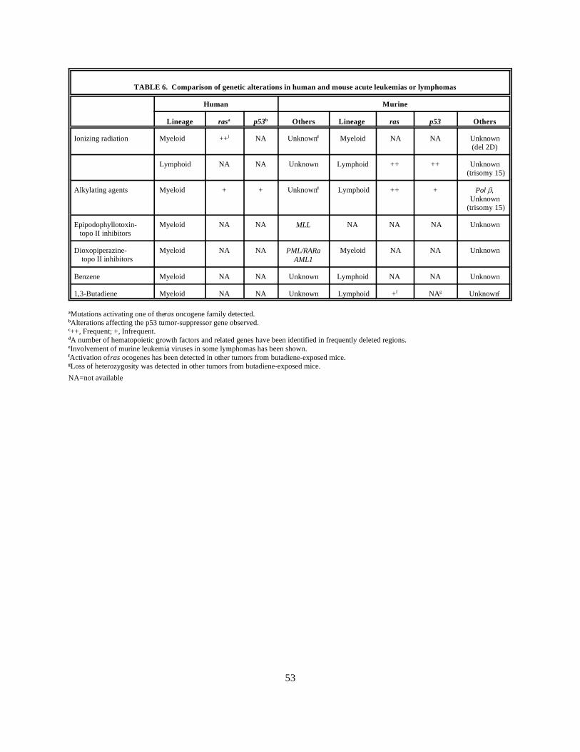
53
TABLE 6. Comparison of genetic alterations in human and mouse acute leukemias or lymphomas
Human Murine
Lineage Others Lineage Othersras p53 ras p53a b
Ionizing radiation Myeloid ++ NA Unknown Myeloid NA NA Unknownl d
(del 2D)
Lymphoid NA NA Unknown Lymphoid ++ ++ Unknown (trisomy 15)
Alkylating agents Myeloid + + Unknown Lymphoid ++ +d Pol ,Unknown
(trisomy 15)
Epipodophyllotoxin- Myeloid NA NA NA NA NA Unknown topo II inhibitors
MLL
Dioxopiperazine- Myeloid NA NA Myeloid NA NA Unknown topo II inhibitors
PML/RARaAML1
Benzene Myeloid NA NA Unknown Lymphoid NA NA Unknown
1,3-Butadiene Myeloid NA NA Unknown Lymphoid + NA Unknownf g e
Mutations activating one of the ras oncogene family detected.a
Alterations affecting the p53 tumor-suppressor gene observed. b
++, Frequent; +, Infrequent.c
A number of hematopoietic growth factors and related genes have been identified in frequently deleted regions.d
Involvement of murine leukemia viruses in some lymphomas has been shown.e
Activation of ras ocogenes has been detected in other tumors from butadiene-exposed mice.f
Loss of heterozygosity was detected in other tumors from butadiene-exposed mice.g
NA=not available

54
4.6.7.2. LymphomaIncreases in lymphoma have been observed repeatedly in studies of benzene-exposed workers.
This type of malignancy has been seen less consistently than ANLL, and there are inconsistencies in specifictypes of lymphoma in various reports. Of the 20 lymphomas observed in the Chinese worker study, 17 wereNHL (Yin et al., 1995). Relative risks for nodal and extranodal NHL were 4.0 (CI 1.1-25.7) and 1.0 (CI 0.1-22.3), respectively. No cases of Hodgkin's disease were identified. In addition, a case control studyconducted in Minnesota and Iowa provided some evidence that benzene exposure was associated withincreased risks 17 lymphomas reported by Aksoy in his case series were classified as Hodgkin's disease(Aksoy, 1988a). In addition, an early study by Vianna and Polan indicated that individuals working inoccupations with potential benzene exposure had increased risks of lymphosarcoma, reticulum-cell sarcoma,and Hodgkin's disease among older workers (Vianna and Polan, 1979).
4.6.8. Genetic Alterations in Cancer PatientsA significant number of cytogenetic studies have been performed on patients exhibiting benzene-
related leukemias and related diseases. Unfortunately, most were conducted before the development ofsensitive chromosome-banding techniques. As a result, detailed karyotypic information is available forrelatively few individuals. This makes identification of chromosomal regions and genes involved in benzene-induced leukemias very difficult. In general, karyotypes of patients with benzene-induced leukemia exhibitabnormal chromosomal number and structure (Forni, 1979; Wolman, 1977). Beyond this, however, no clearpattern emerges from published karyotypes. Chromosomal alterations similar to those seen following treat-ment with alkylating chemotherapeutic agents, such as loss of all or part of the long arms of chromosomes 5and 7, have been reported to occur in leukemia patients previously exposed to of both follicular and diffuseNHL (Blair et al., 1993). Although confidence intervals for each category encompassed 1.0, risk increasedwith increasing exposure intensity. In contrast, 10 of benzene (Pollini and Colombi, 1964; Van den Berghe etal., 1979). In addition, trisomy or tetrasomy of a C-group chromosome, occasionally identified aschromosome 8 or 9, has been associated with benzene exposure (Antonucci et al., 1989; Erdogan and Aksoy,1973; Forni and Moreo, 1967). Occasionally, these clonal alterations were observed before the developmentof frank leukemia (Erdogan and Aksoy, 1973; Forni and Moreo, 1967). Furthermore, translocations havebeen reported in cells of leukemic patients following exposure to benzene or benzene-containing solventssuch as petroleum (Li et al., 1989; Sole et al., 1990; Tasaka et al., 1992; Van den Berghe et al., 1979). Occasionally, these translocations have been identical to those observed in patients following chemotherapywith topoisomerase II inhibitors (Sole et al., 1990; Tasaka et al., 1992).
Due in part to the mixed pattern of karyotypic alterations described above, little is known aboutgenes involved in benzene leukemogenesis. However, genes involved are likely to be similar to thosecontributing to other types of therapy-related leukemias and some de novo leukemias. Occasional reportshave implicated or suggested that a gene might be involved (Snyder and Kalf, 1994; Taylor et al., 1995). Forexample, a mutation in k-ras was seen in DNA isolated from peripheral blood of a worker occupationally

55
exposed to petrochemicals; the authors postulated that this might be related to benzene exposure (Taylor etal., 1995). However, at this time there is little direct evidence for the involvement of any specific gene ortypes of genes. Different patterns of aberrations seen in benzene leukemia cases, as well as those seen in
genotoxicity studies of exposed workers, suggest that multiple types and mechanisms of genotoxicity may be
occurring. One current hypothesis is that both the alkylating-type of chromosomal alterations (aneuploidyand deletions) as well as topoisomerase-type alterations (translocations and deletions) are occurring inbenzene-exposed individuals (Frantz et al., 1996). Combination of these types of chromosomal alterationsconfers increased leukemia risks similar to those reported for cancer patients treated with both alkylatingagents and topoisomerase inhibitors (Pedersen-Bjergaard et al., 1992; Sandoval et al., 1993; Smith et al.,1994b).
4.6.9. Hematopoietic and Lymphoid Neoplasia Seen in Rodent ModelsA large number of chronic bioassays have been performed in an attempt to identify an animal
model for benzene-induced myelogenous leukemia (for a summary see IPCS, 1993). Although minorincreases in myelogenous leukemias occasionally have been seen in rodent bioassays (Cronkite et al., 1989;Goldstein et al., 1982), consistent and reproducible increases in these leukemias have not been seen in eithermice or rats (Farris et al., 1993; Maltoni et al., 1989; NTP, 1986a). In mouse bioassays, benzene has beenreported more consistently to induce increases in lymphoblastic leukemias or lymphomas (Cronkite et al.,1984; Farris et al., 1993; Maltoni et al., 1989; NTP, 1986a; Snyder et al., 1980). In rats, Maltoni andcoworkers reported that in some experiments benzene was capable of inducing "reticuloendothelial" tumorson chronic administration of benzene (Maltoni et al., 1983). These tumors were reported to be a combinationof hematosarcomas, lymphoblastic leukemias, and one myelogenous leukemia. However, no increase in thesetumor types was seen in the rat bioassay conducted by the National Toxicology Program (NTP, 1986a).
Somewhat in contrast to its leukemogenic effects, chronic benzene exposure has been shown toinduce solid tumors at a large number of sites in mice and rats, including the Zymbal gland, Harderian gland,mammary glands, oral cavity, skin, lung, preputial gland, and ovary (Haseman et al., 1984). Recent studieshave reported on the metabolic capacity and genotoxicity occurring in these target tissues and their proposedrelevance in benzene carcinogenicity. All tissues examined had significant levels of peroxidase enzymes, aclass of enzymes believed to be involved in metabolic activation of benzene in the bone marrow (Low et al.,1995). In addition, detectable sulfatase levels were observed. The authors suggested that the somewhatunique metabolic capabilities of the target tissues might play a role in the target organ specificity of benzene. Additional studies by Angelosanto and colleagues detected micronuclei in primary Zymbal gland culturesestablished from benzene-treated animals, demonstrating that genetic damage occurred in these target organsfollowing benzene exposure (Angelosanto, 1995).
Recently, benzene has been tested in two different strains of transgenic mice to determine thesensitivity of mouse models to various types of carcinogenic agents. Benzene was administered for 40 weeksto E mu-pim1 transgenic mice, which overexpress the pim-1 oncogene in lymphoid tissues and have been

56
shown to exhibit increased sensitivity to T-cell lymphoma induction by the alkylating agent, N-ethyl-N-nitrosourea (Storer et al., 1995). At the end of a 40-week treatment period, a statistically significant increasein lymphomas was not seen, suggesting the limited usefulness of this transgenic model for benzene-carcinogenicity testing. Other studies have indicated that this transgenic strain was sensitive to benzene’sgenotoxic effects as measured by micronuclei induction in bone marrow and blood erythrocytes (Armstrongand Galloway, 1993). In a similar study using a different strain of transgenic mouse, administration ofbenzene by dermal application to the TG.AC transgenic mouse strain, which carries a v-ras construct,induced a high frequency of skin papillomas in a dose-related fashion (Tennant et al., 1995). A similar effectwas not seen when phenol, one of benzene's primary metabolites, was applied to the mouse skin. Cytogeneticstudies were performed for the epidermal sarcomas or squamous cell carcinomas in six benzene-treatedTG.AC transgenic animals (French et al., 1994). Two of the six tumors were characterized by trisomy 15,and two exhibited normal karyotypes. The presence of trisomy 15, is somewhat interesting in that thisalteration frequently occurs in mouse T-cell lymphomas. No information has been published on thekaryotype of the leukemias and lymphomas induced in mice (or rats) by benzene.
4.7. 1,3-BUTADIENE4.7.1. Background
Butadiene is a widely used industrial chemical and common environmental pollutant. Annual U.S.production in 1990 was estimated to be 3.2 billion pounds, most of which was used for the manufacture ofstyrene-butadiene rubber and polybutadiene rubber (NTP, 1994). National Institute of Safety and Health(NIOSH) has estimated that 69,555 U.S. workers were potentially exposed to butadiene (NTP, 1994). Currently the permissible exposure level for butadiene is 1000 ppm, but actual occupational exposures areconsiderably lower, with measured values ranging from 0.005 to 374 ppm (Fajen et al., 1993). Of samplestaken in a recent occupation-exposure survey, 85% were below 2 ppm, and only 3.7% exceeded 10 ppm(Fajen et al., 1993). Butadiene also has been detected in cigarette smoke (approx. 0.4 mg/cigarette) and inautomobile exhaust (approx. 0.35% of total hydrocarbons in exhaust emissions). Historically, butadiene wasconsidered a safe chemical with no adverse effects seen in mice following acute exposures up to 100,000 ppm(ACGIH, 1993). The permissible exposure level was accordingly set at 1000 ppm (RTECS). However,more recent findings that butadiene is carcinogenic at multiple sites in rats and mice on chronic exposure hasraised concerns about the human health risks from occupational and environmental exposures (Huff et al.,1985; Melnick and Huff, 1992). In animal bioassays, significant differences have been seen between miceand rats in the number, types of neoplasia, and concentrations at which tumors have been seen. A significantincrease in lung cancers was seen at exposure concentrations as low as 6.25 ppm in B6C3F1 mice, whereasexposures of 1000 and 8000 ppm were necessary to induce mammary gland tumors in rats (Melnick andHuff, 1992). Particularly striking was the increase in thymic leukemias and lymphomas observed in B6C3F1mice but not rats (Melnick and Huff, 1992). This difference in species sensitivity has been used to provideinsights into critical steps involved in butadiene toxicity and carcinogenesis.
57
In addition to animal data, in recent years an increasing number of epidemiological studies haveshown an association between butadiene exposure and the incidence of lymphohematopoietic disease inhumans. These studies have raised additional concern about adverse effects of butadiene exposure tohumans. Butadiene has been the focus of a number of recent reviews (Bond et al., 1995; IARC, 1992; IARC,1993; Melnick and Kohn, 1995) to which the reader is referred for additional details. The primary focus ofthis section will be on mechanisms of toxicity and genotoxicity that are believed to contribute to formation oflymphohematopoietic neoplasms in humans and rodents.
4.7.2. Metabolic Studies Utilizing Animal and In Vitro SystemsThe metabolism of butadiene has been shown to be qualitatively similar among the various species
studied (Henderson et al., 1993). Butadiene is metabolized via cytochrome P450-dependent monooxygenasesinitially to butadiene monoepoxide (BDO; 1,2-epoxy-3-butene) and 3-butenal, which tautomerizes to producecrotonaldehyde (Elfarra et al., 1991). The P450 2E1 isozyme appears to be primarily responsible for thisreaction, although the 2A6 isozyme can also metabolize butadiene when butadiene is present at highconcentrations (Duescher and Elfarra, 1994; Seaton et al., 1995). BDO can be further oxidized to butadienediepoxide (BDO2; 1,2,3,4-diepoxybutane) in a reaction apparently catalyzed by the hepatic 2E1 isozyme inmice, rats, and humans (Seaton et al., 1995). BDO can be transformed via epoxide hydralase to formbutenediol (1,2-dihydroxy-3 butene) in what is believed to be a detoxication process (Henderson et al., 1993;IARC, 1992). This latter product can be metabolized further by P450 enzymes to form 3,4-epoxy-1,2-butanediol. Both BDO and BDO2 also can be metabolized by glutathione-S-transferases to form glutathioneconjugates. These are presumed to be detoxication products, although there have been some suggestions thatthese conjugates might be involved in bioactivation (Guengerich et al., 1995; Thier et al., 1995). BDO andBDO2 are both highly mutagenic and carcinogenic when administered to mice and rats (IARC, 1992). Basedon in vivo mutagenicity studies in mouse and human cells, the mutagenic activity of BDO2 appears to be 10to 100 times higher than the primary BDO metabolite. Other potential reactive metabolites aredihydroxyepoxybutane, crotonaldehyde, and hydroxymethylvinylketone (Waters and Nolan, 1995). However,the most current metabolic schemes seem to favor the epoxide metabolites, BDO and BDO2, as mutagenicand carcinogenic metabolites. The production of BDO2 is higher in female rats than male rats; this has beenproposed to play a role in the increased incidence of mammary tumors seen in female rats following butadieneexposure (Thornton-Manning et al., 1995b).
Although bone marrow is one of the key organs for butadiene toxicity in mice, the absence ofsignificant cytochrome P450 2E1 activity in this organ in mouse or human cells indicates that hepaticmetabolism or another type of bioactivation is likely to be involved (Genter and Recio, 1994; Maniglier-Poulet et al., 1995). B one marrow contains high levels of peroxidase enzymes, which have been shown to becapable of converting butadiene to BDO (Duescher and Elfarra, 1992; Maniglier-Poulet et al., 1995). However, BDO formation was about 20 times greater in rat and mouse liver microsomes than in mouse bone-
58
marrow cells and lysates, suggesting the importance of hepatic metabolism in the butidiene bioactivation(Maniglier-Poulet et al., 1995).
Significant metabolism differences in mice and rats are believed to be largely responsible for thetoxicity differences between the two species (Bond et al., 1995; Melnick and Kohn, 1995). A number ofstudies have demonstrated that mice are much more efficient in butadiene uptake and bioactivation and lessefficient in detoxication. Studies in liver and lung microsomes have shown that the rate of BDO formation ishighest in liver microsomes from B6C3F1 mice. High activation rates were also seen in mouse lungmicrosomes. Lower reaction rates were seen in the microsomes of Sprague-Dawley rats, humans, and rhesusmonkeys (Henderson et al., 1993). Pharmacokinetic studies have indicated that uptake and metabolism ofbutadiene was greater in the mouse than in the rat or the monkey and that the mouse metabolized much higherpercentages to the epoxide metabolites in vivo (Henderson et al., 1993). Recent studies also have shown thatBDO, and in most tissues BDO2, can be detected in the blood, bone marrow, heart, lung, fat and thymusfollowing in vivo exposure of B6C3F1 mice to butadiene (Thornton-Manning et al., 1995a). BDO2 was alsodetected in the heart, lung, fat, spleen, and thymus of Sprague-Dawley rats but at levels 40- to 160-fold lowerthan those seen in mice. In addition to more efficient bioactivation, the mouse has been shown to have lowerepoxide hydrolase activity than the rat, hamster, or monkey; as a result, more glutathione metabolites can beseen in mouse urine. The combination of more efficient activation and lower epoxide hydralase activityresults in greater depletion of nonprotein sulfhydryls in the various mouse tissues than in rat tissues, evenwhen rats are exposed to much higher concentrations (Bolt, 1993). Binding of radioactive butadieneequivalents to liver DNA and nucleoproteins could be detected in both rats and mice under these sameconditions but was about twofold higher in the mice (Bolt, 1993). Higher levels (about fivefold) of the BDOhemoglobin adduct were also seen in mice than in rats after exposure to butadiene (Albrecht et al., 1993;Osterman-Golkar et al., 1993).
4.7.3. Metabolism Studies in HumansEvidence indicates that the same metabolites detected in laboratory animals will be formed in
humans. Butadiene was reported to be metabolized to BDO by human liver and lung preparations at one-halfto one-tenth the rates seen in mouse microsomes (Bond et al., 1993). Studies with human liver microsomesindicated that the rate of hydrolysis by epoxide hydrolase was higher in human liver samples than in mouse orrat samples (Bechtold et al., 1994; Bond et al., 1993). However, lower rates of glutathione conjugation byglutathione-S-transferase were seen in human hepatic microsomes than in mouse or rat (Bechtold et al.,1994). Equal amounts of BDO were formed in human and mouse bone-marrow cells following exposure tobutadiene in a reaction that appeared to be catalyzed by myeloperoxidase (Maniglier-Poulet et al., 1995). This indicates that formation of this bioactive metabolite can occur in a target organ for butadiene toxicity. However, the amount of metabolite formed appeared to be significantly lower than seen in liver incubations.
Urinary metabolites resulting from BDO detoxication were identified in samples from rats, mice,hamsters, monkeys, and humans exposed to butadiene (Bechtold et al., 1994). The presence of only the 1,2-

59
dihydroxy-4-(N-acetylcysteinyl-S-)-butane metabolite in the urine of exposed workers suggests that humansexposed to butadiene in the 3- to 4-ppm range metabolize BDO by hydrolysis to butanediol rather thanthrough direct conjugation with glutathione. This pattern is more closely related to that seen in the monkeythan in either rats or mice (Bechtold et al., 1994). The BDO hemoglobin adduct also was detected at lowlevels in the blood of occupationally exposed workers (Osterman-Golkar et al., 1993). More recent studieshave confirmed that blood hemoglobin adduct levels were increased among worker groups with higherpotential butadiene exposure than among less-exposed workers (Osterman-Golkar et al., 1996; Sorsa et al.,1996b). However, the measured N-terminal valine hemoglobin adducts were 10 to 20 times lower than thoseextrapolated for the same exposure dose in mice (Sorsa et al., 1996b). These results indicate that humans canmetabolize butadiene to the monoepoxide. However, whether or not humans are able to form the diepoxide insignificant quantities at current occupational exposure levels remains uncertain.
4.7.4. Genotoxicity Studies Utilizing Animal or In Vitro SystemsLike benzene, butadiene requires metabolic activation to exert its genotoxic effects (Norppa and
Sorsa, 1993). However, in contrast to benzene, bioactivated butadiene binds readily to DNA and is efficientat inducing specific gene mutations. Results of most standard in vitro studies of butadiene mutagenesis andgenotoxicity have been negative, probably due to lack of metabolic activation and the difficulty in testing agas that requires bioactivation (IARC, 1992; Norppa and Sorsa, 1993). Several studies have demonstratedthe ability of butadiene to bind to DNA and induce DNA breaks in vivo (Jacobson-Kram and Rosenthal,1995; Vangala et al., 1993). Following in vivo exposure to butadiene, male B6C3F1 and NIH Swiss miceshow macrocytic-megaloblastic anemia (Irons et al., 1986a; Irons et al., 1986b). A leukopenia, relatedprimarily to a decrease in neutrophils, and an increase in bone-marrow erythrocyte micronuclei also has beenobserved. Consistent treatment-related alterations in bone-marrow cellularity were not seen. In addition tomicronuclei, butadiene has been shown consistently to induce chromosomal breakage and sister chromatidexchanges (SCEs) in mouse bone marrow and germ cells (Adler and Anderson, 1994; Adler et al., 1994;IARC, 1992; Irons et al., 1987a; Shelby, 1990; Tice et al., 1987); no increases in micronuclei or aberrationswere seen in rat (Autio et al., 1994; IARC, 1992; Norppa and Sorsa, 1993; Sorsa et al., 1996b). A weakinduction in SCEs was seen in rat lymphocyte and lung cells (Norppa and Sorsa, 1993). Apparently thesedifferences are due largely to metabolic differences in formation of bioactive metabolites, as both BDO andBDO2 exhibit significant genotoxic effects in both rats and mice (Sharief et al., 1986; Xiao and Tates, 1995). Increases in SCEs were seen following butadiene exposures for 10 days at 6.25 ppm in B6C3F1 mice (Ticeet al., 1987). Significant increases in micronuclei were also seen at this concentration but only followingexposure for a longer period of Time (13 weeks) and by scoring a large number of cells (10,000 RBC peranimal) (Jauhar et al., 1988; Shelby, 1990).
A variety of butadiene-DNA adducts have been detected following the in vitro incubation ofbutadiene metabolites with isolated nucleotides, DNA, or cell cultures (Leuratti et al., 1994; Leuratti et al.,1993; Neagu et al., 1995; Peltonen et al., 1993). Adducts to the N6 position of adenine and N7 position of
60
guanine have been detected (Leuratti et al., 1994; Neagu et al., 1995). Recent studies have detected the N6-alkyldeoxyadenosine adduct of BDO in lungs of mice and rats exposed to butadiene by inhalation (Sorsa etal., 1996b). Similar levels were seen in rat and mouse lungs, but the levels were about 10 times lower thanthose seen in rat liver. In a preliminary report, two BDO-DNA adducts were detected in the urine of abutadiene-exposed worker (Peltonen et al., 1993).
A number of studies have detected increased mutation frequencies in endogenous mouse genes orin reporter genes in transgenic mice, consistent with the ability of bioactive butadiene metabolites to reactwith DNA. Studies by two groups have demonstrated that three- to fivefold increased frequencies of 6-thioguanine-resistant cells indicative of mutation at the HPRT locus can be detected in the splenic T-lymphocytes of B6C3F1 mice following multiple exposures to butadiene at 625-ppm concentrations andhigher (Cochrane and Skopek, 1993; Tates et al., 1994). Somewhat higher frequencies also were seen in thisstudy following the intraperitoneal administration of BDO and BDO2 to mice (Cochrane and Skopek, 1993). Follow-up studies by Cochrane and Skopek have shown that, for each of these compounds, about half themutations induced within HPRT in vivo were frameshift mutations (Cochrane and Skopek, 1994b). Transition and transversion mutations also were seen, with a tendency toward substitutions at AT base pairs. The authors commented that the spectrum of mutations induced by butadiene and metabolites was verysimilar to that seen with ethylene oxide, suggesting that these agents might be working through similarmutagenic mechanisms.
Similar results have been seen with butadiene in studies of transgenic B6C3F1 mice carrying arecoverable lacI bacterial gene. Following a 2-week exposure to butadiene at concentrations similar to thoseseen in the mouse cancer bioassay, the lacI mutant frequency in the bone marrow was increased in aconcentration-related manner 2- to 3.5-fold over that seen in the controls (Sisk et al., 1994). DNA sequenceanalysis suggested that the exposed mice had higher frequencies of point mutations at AT sites (Recio andMeyer, 1995).
The three epoxide metabolites of butadiene, BDO, BDO2, and 3,4-epoxy-1,2-butanediol weretested for mutagenicity at HPRT and tk loci in the TK6 human lymphoblastoid cells (Cochrane and Skopek,1993; Cochrane and Skopek, 1994a). All three epoxides were mutagenic, but the mutagenicity of BDO2 wasmuch greater than that of BDO, which in turn was greater than 3,4-epoxy-1,2-butanediol. Each compoundwas also capable of inducing slow growth tk-/ mutants, indicating the induction of large-scale mutations bythese compounds (Cochrane and Skopek, 1993; Cochrane and Skopek, 1994a). An increase in deletion-typemutants was seen with BDO2 but not BDO, suggesting that the deletions might be related to BDO2's abilityto form DNA-DNA and DNA-protein crosslinks (Cochrane and Skopek, 1994a).
Both BDO and BDO2 have been shown to induce SCEs in in vitro incubations with humanlymphocytes (IARC, 1992). With exposure to BDO2, blood donors can be classified into BDO2-sensitive orBDO2-resistant (Norppa et al., 1995; Wiencke and Kelsey, 1993; Wiencke et al., 1995). Recent studies haveshown that variability in the cytotoxicity and SCE response of different individuals to BDO2 correlates withan individual's genotype for glutathione-S-transferase theta (GSST1) (Norppa et al., 1995; Wiencke et al.,

61
1995). Individuals expressing an active GSTT1 enzyme were all BDO2-resistant, whereas GSST1 null(homozygous recessive) individuals were BDO2 sensitive. In similar studies, lymphocytes from blood donorswith a homozygous recessive genotype for glutathione-S-transferase M1 exhibited higher frequencies ofSCEs upon exposure to BDO (Uuskula et al., 1995). These results indicate that polymorphisms in enzymesinvolved in butadiene detoxication are likely to influence an individual's in vivo sensitivity to genotoxiceffects of this compound.
4.7.5. Genotoxicity Studies in Butadiene-Exposed WorkersA recent assessment of biomonitoring studies for butadiene concluded that they provide no
convincing evidence for formation of SCA, micronuclei, or SCEs in lymphocytes of occupationally exposedindividuals (Sorsa et al., 1994; Waters and Nolan, 1995). In Sorsa and colleagues’ initial studies ofbutadiene workers in two production facilities, significant increases in aberrations, micronuclei, and SCEswere not seen in exposed workers (Sorsa et al., 1994). However, a more recent study by these sameinvestigators found significantly higher frequencies of chromosomal aberrations in a group of butadiene-exposed workers who lacked the GSST1 gene, as compared to butadiene workers with the gene. Thisindicates that a person's genotype may affect his or her susceptibility to this agent’s genotoxic effects (Sorsaet al., 1996a). A slightly elevated but not statistically significant increase in chromosomal aberrations wasalso seen in another group of butadiene workers (Au et al., 1995). In this study by Au and associates,significantly higher frequencies of aberrations were seen in lymphocytes of butadiene-exposed workersfollowing lymphocyte exposure to a challenge dose of ionizing radiation. This suggests that the exposedworkers had an altered DNA-repair capacity (Au et al., 1995). Recently, Kelsey and associates investigated agroup of workers involved in butadiene monomer production and attempted to correlate butadiene exposurewith baseline frequencies of SCE and BDO2-induced SCE (Kelsey et al., 1995). No association was seenbetween butadiene exposure and the baseline of induced SCE frequencies. The lack of correlation persistedwhen workers’ GSST1 genotypes were included in the analysis. However, for each of these studies,butadiene-exposure levels for most workers were relatively low (~1 ppm or less).
Ward and colleagues have performed a series of studies of workers at a butadiene monomerproduction plant to determine whether current exposure was associated with increased variant frequency atHPRT (Legator et al., 1993). In the initial study of eight "high" exposed workers, five "low" exposedworkers, and six nonexposed controls, a significant increase in HPRT variant frequency was seen in the high-exposure group (Legator et al., 1993). Exposures in the high-exposed group averaged about 1 ppm. Inaddition, a highly significant correlation (r= 0.85) was seen between HPRT variant frequency and the urinaryconcentration of 1,2-dihydroxy-4-(N-acetlycysteinyl-S)butane, a urinary metabolite of butadiene. In thisstudy, only one worker was of the GSTT1-null phenotype. Interestingly, this worker had the highest HPRTvariant frequency (Kelsey et al., 1995). A second study has recently been reported in abstract form in whichthe HPRT variant frequency of workers at high (0.30 ppm), intermediate (0.21 ppm) and low (0.12 ppm)

62
exposures was determined (Ward et al., 1995b). The authors reported that a significant increase in variantfrequency was seen in the high-exposure group.
4.7.6. Hematopoietic and Lymphoid Neoplasia Seen in HumansSince the initial report of increased lymphomas in butadiene-exposed mice, the carcinogenic effects
of butadiene have been the subject of numerous epidemiological studies and considerable controversy. Results of these studies, as well as their consistencies and inconsistencies, have been the focus of a number ofrecent reviews and commentaries (Bond et al., 1995; Cole et al., 1993; IARC, 1992; IARC, 1993; Landrigan,1993; Melnick and Kohn, 1995). In general, exposure to butadiene has exhibited some association withincreased frequencies of lymphosarcomas, reticulosarcomas, and leukemias. Recently, three studiesexamined an association between butadiene exposure and lymphohematological malignancies. In a mortalitystudy of 614 workers conducted by Cowles and associates, no deaths due to lymphohematopoietic cancerwere observed (Cowles et al., 1994). Industrial hygiene data collected between 1979 and 1992 indicated thatmost exposures were below 1 ppm. In addition, no differences in hematological results were seen betweenbutadiene-exposed workers and other employees at the manufacturing complex. In contrast, a study ofbutadiene production workers conducted by Ward and collaborators detected a significant increase in thestandardized mortality ratio for lymphosarcomas and reticulosarcomas (Ward et al., 1995a). Three of thefour deaths from lymphosarcoma and reticulosarcoma occurred in workers who had been employed for morethan 2 years and showed a latency period of more than 30 years. A slightly elevated increase (SMR = 1.23)was also seen for workers developing leukemia (including aleukemia) in this study. There was no monitoringdata at these facilities.
In the largest butadiene study to date, Delzell and coworkers conducted a retrospective study on15,649 men employed at 8 styrene-butadiene rubber manufacturing facilities to determine whether exposureto butadiene was associated with increases of lymphohematopoietic cancers (Delzell et al., 1995). The groupencompassed a total of 386,172 person-years of follow-up and averaged 25 years of follow-up per person. Inall, 11 excess deaths were from leukemia, based on 48 observed and 37 expected (SMR=131, CI 97-174). More deaths from hematological disorders were seen in individuals who had worked more than 10 years andwhere more than 30 years had elapsed since hire. Among the 45 hourly workers who died with leukemia, 14(31%) were lymphocytic in origin, 17 (38%) were myeloid, and 14 (31%) were unspecified. The SMR forthese workers by leukemia subtype were 210 (CI 115-352) for lymphocytic, 140 (CI 78-231) for myeloid,and 196 (101-342) for unspecified. Those who died of leukemia had a median period of 28 years from timeof hire to death. Mortality was elevated among subjects employed in the polymerization, labor, andlaboratories process groups, areas that had the potential for relatively high butadiene monomer exposure. Ananalysis showed that those dying from leukemia had higher exposures than for all workers. The associationwith butadiene exposure was stronger for ANLL and unspecified leukemia than for CML or CLL, but therelative risk estimates were imprecise. In summary and as indicated by the authors, the strength and internalconsistency of the leukemia findings indicate that employment in the styrene-butadiene industry produced an
63
excess of this disease. However, the authors emphasize that the risk should be kept in perspective; an excessof 11 deaths was seen in a group that experienced 4000 deaths (with 4500 expected), indicating that at theseexposure concentrations butadiene is a relatively weak human leukemogen.
4.7.7. Genetic Alterations in Cancer PatientsNo information is available about genetic changes in leukemias or lymphomas associated with
butadiene exposure.
4.7.8. Hematopoietic and Lymphoid Neoplasia Seen in Rodent ModelsDuring the course of a 104-week chronic inhalation bioassay of butadiene conducted by the
National Toxicology Program in B6C3F1 mice, the study was terminated at 60 to 61 weeks due to reducedsurvival resulting from fatal tumors (Huff et al., 1985). Significant increases were seen in tumors affectingthe heart, lung, forestomach, mammary gland, ovary, and liver. Thymic lymphomas in both males andfemales comprised the most prominent type of cancer increased by butadiene, with frequencies of 46% and58% seen at the 625- and 1250-ppm exposure concentrations in male mice. Elevated but lower (20%)frequencies were seen in exposed female mice. These lymphomas were seen as early as week 20, and most ofthem possessed surface markers indicative of early T-lymphocytes (Miller and Boorman, 1990). Subsequently, experiments were conducted in Sprague-Dawley rats (Owen and Glaister, 1990). Relativelyweak but significant carcinogenic effects were observed, with increases in tumors seen in a variety of organsincluding the pancreas, uterus, Zymbal gland, mammary gland, and testes. Most tumors were increased onlyat the high 8000-ppm exposure concentration. These results indicated that butadiene was carcinogenic inboth mice and rats and that there was a clear difference in response between these two rodent species,particularly in the formation of lymphomas.
Additional studies were performed by the National Toxicology Program (NTP) to provide a bettercharacterization of concentration- and time-dependent responses for butadiene-induced neoplasms (NTP,1993). B6C3F1 mice were exposed to butadiene at 6.25, 20, 62.5, 200, and 625 ppm for 2 years and forshorter durations at 200, 312, and 625 ppm (male mice). The follow-up studies confirmed butadienecarcinogenicity to B6C3F1 mice, with increased tumor incidences seen for hematopoietic system, heart, lung,forestomach, liver, Harderian gland, preputial gland, ovary, brain, and kidney. An increase in lung tumorswas seen at exposure concentrations as low as 6.25 ppm. Significant dose-related increases in both malignantlymphoma and histiocytic sarcoma were seen, with significant increases being detected at exposureconcentrations as low as 20 ppm (Melnick et al., 1993; NTP, 1993). Significant increases in frequencies oflymphomas and other tumors were seen in the discontinued- exposure groups. These studies indicated that atsimilar total exposures, lymphoma insidence was greater in mice exposed to a higher concentration ofbutadiene for a shorter time than in those exposed to a lower concentration for a longer time (34% incidenceat 625 ppm for 13 weeks vs 2% at 200 ppm for 40 weeks; 60% incidence at 625 ppm for 26 weeks vs. 3 % at312 ppm for 52 weeks). However, a single 2-hour exposure to high concentrations of butadiene (1000, 5000,

64
and 10,000 ppm) was not sufficient to induce significant neoplasm increases in B6C3F1 mice (Bucher et al.,1993).
4.7.8.1. Mechanisms Involved in Butadiene-induced Thymic Lymphomas in Mice4.7.8.1.1. Involvement of murine leukemia viruses . Due to the high incidence of thymic lymphomas in
these butadiene-exposed mice, an incidence much higher than that seen with other chemicals tested by NTP(Sanders et al., 1995), a series of investigations has been undertaken to understand the mechanismsunderlying butadiene-induced lymphomagenesis. As described above, considerable differences in metabolismand pharmacokinetics can be seen in mice and rats following exposure to butadiene. One possibleexplanation for the high incidence of lymphomas observed in butadiene-exposed mice is Related to thepossible activation of a murine retrovirus. Murine retroviruses have been proposed to play an important rolein induction of thymic lymphomas by a number of agents, including radiation and urethane. Thymiclymphomas induced by butadiene exhibited increased expression of the murine leukemia virus env antigen(Irons et al., 1987b). Chronic exposure to butadiene increased markedly the amount of ecotropic retrovirusrecoverable from the bone marrow, thymus, and spleen of B6C3F1 mice (Irons et al., 1987b). Expression ofother endogenous retroviruses (such as the xenotropic type) was not elevated. Furthermore, no viruses of anykind were found in similarly treated NIH Swiss mice. This strain only rarely expresses endogenousretroviruses and does not possess intact endogenous ecotropic retrovirus sequences.
To explore further the role of this ecotropic retrovirus in the formation of thymic lymphomasinduced by butadiene, B6C3F1 mice and NIH Swiss mice were exposed to 1250-ppm butadiene for up to 1year (Irons et al., 1989). At the end of the study, the B6C3F1 mice exhibited a fourfold incidence of thymiclymphoma (57%) over that seen in NIH Swiss mice (14%). Tumors detected in the NIH Swiss mouse weremorphologically similar to those seen in the B6C3F1 mouse and expressed T-cell but not murine leukemiaviral env surface antigens (Irons, 1990). As mentioned by Sanders and coauthors, these results were taken aspresumptive evidence for involvement of an endogeneous ecotropic retrovirus in the etiology of thelymphomas, although no attempt was made to analyze the tumors for mutations or viral insertions (Sanders etal., 1995). Additional studies by Irons and associates have proposed that BDO exerts its toxic effects on aspecific primitive hematopoietic stem cell, resulting in a pattern of hematopoietic pathology similar to thatassociated with mice bearing mutations at the W and Sl loci (Colagiovanni et al., 1993; Irons et al., 1993). These results indicate that butadiene, most likely through one or more metabolites, is involved directly inlymphomagenesis and that the expression of an endogenous ecotropic virus significantly increases theincidence of tumors induced by this agent.
4.7.8.1.2. Involvement of other cancer-related genes . Additional studies on selected solid tumors inducedby butadiene has provided evidence for the involvement of several cancer-associated genes in thecarcinogenesis induced by this agent. In a study of the potential involvement of tumor-suppressor genes inbutadiene-induced lung and mammary tumors, losses of heterozygosity were observed at several loci on
65
mouse chromosome 11 surrounding the p53 tumor-suppressor gene (Wiseman et al., 1994). These losseswere seen in 12 of 17 mammary tumors and 2 of 8 lung tumors. Most losses appeared to result fromnondisjunction, but at least two were believed to arise from recombination or deletion. Loss of heterozygositywas also observed at the Rb-1 tumor-suppressor gene in 7 of 17 mammary tumors and 1 lung tumor. Someevidence also was seen for the loss of an additional putative tumor-suppressor gene on mouse chromosome 4. In two additional studies, activated K- and H-ras genes were observed in a portion of lung (6 of 9) and liver(4 of 12) tumors induced by butadiene (Goodrow et al., 1990). Of the 29 Harderian gland tumors examinedfrom butadiene-treated mice, 16 exhibited an H-ras mutation at codon 61 (Goodrow et al., 1994). Inaddition, 11 butadiene-induced lymphomas were examined for transforming genes using the nude mousetumorigenicity assay (Goodrow et al., 1990). Activated K-ras genes were detected in 2 of the 11 tumorsassayed. Sequence analysis of the activated K-ras genes for various tumors showed that most of theseresulted from G-to-C transversions at codon 13. This type of mutation was not seen in tumors isolated fromnonexposed animals, providing additional evidence that the tumors were induced by butadiene (Goodrow etal., 1990). Results of these analyses are consistent with in vitro and in vivo mutagenicity studies and indicatethat mutagenic activation of oncogenes and inactivation of tumor-suppressor genes is likely to contribute totumors induced by butadiene.
5. MECHANISMS OF CHEMICALLY OR RADIATION-INDUCED LEUKEMOGENESIS IN HUMANS
Increasing molecular and cytogenetic evidence indicates that human cancers are a consequence ofan accumulation of genetic and possibly epigenetic changes that are critical for the transformation of a normalcell into a malignant cell (Fearon and Vogelstein, 1990; Sandberg, 1993; Solomon et al., 1991). Leukemogenesis and, in particular, chemical- and radiation-induced leukemogenesis seems to involvemultiple genes and chromosomal alterations. In recent years, a number of models have been described in anattempt to integrate various genetic alterations seen in leukemias and to indicate temporal relationshipsbetween specific steps in leukemogenesis (Luna-Fineman et al., 1995; Pedersen-Bjergaard et al., 1995). Inthe model proposed by Pedersen-Bjergaard and colleagues, treatment-related MDS and ANLL develop bydifferent pathways involving varying numbers of genetic alterations, some of which are primary and somesecondary (Pedersen-Bjergaard et al., 1995). In general, genes affected by translocations in leukemic cellsappear to be transcription factors and may therefore represent master genes that control the expression ofother genes involved in cell proliferation, differentiation, and transformation (Pedersen-Bjergaard et al.,1995). The loss of entire chromosomes or chromosomal regions, as in chromosomes 5 and 7, is believed toplay a role in the inactivation of tumor-suppressor genes that influence such important cellular functions ascell cycling, DNA repair, and apoptosis. However, the loss of a single gene copy may be sufficient to cause asignificant dysregulation of key steps in hematopoiesis and lymphopoiesis. This condition, known as

66
haploinsufficiency, where two copies of a gene are needed to maintain a normal phenotype, has been shown toplay a role in a variety of genetic disorders (Fisher and Scambler, 1994).
In addition to genetic alterations, it is important to understand the nature of the target cell in whichinitial alterations occur. The first section will overview what is known about major chromosomal alterationsand associated genes involved in secondary ANLL in humans. The second will describe the nature of thetarget cell in various leukemia types.
5.1. KEY GENES IMPLICATED IN CHEMICALLY INDUCED LEUKEMOGENESIS5.1.1. Loss of Chromosome 5 or Portions of the Long Arm (-5 and 5q-)
Loss of chromosome 5 or a deletion in the long arm (5q-) has been shown to accompany MDS andANLL in patients treated with alkylating agents and radiation. Evidence is that these alterations areassociated with leukemias induced by solvents, pesticides, and other chemical agents (Cuneo et al., 1992;Fagioli et al., 1992; Mitelman et al., 1981; Zedginidze et al., 1990). Loss of a portion of the long arm occurssomewhat more frequently than loss of the entire chromosome and appears as a middle-to-late step intreatment-related MDS and ANLL (Pedersen-Bjergaard et al., 1995). Deletions of 5q- can encompass asignificant portion of the long arm (Rowley and Le Beau, 1989). However, the region believed to be critical for leukemogenesis has been narrowed to 5q31 (Le Beau et al., 1993). Although the distal region of 5qcontains a large number of genes involved in hematopoiesis and lymphopoiesis, such as interleukin 3,interleukin 4, interleukin 5, interleukin 9, granulocyte/macrophage colony- stimulating factor, the CD14myeloid antigen, and a T-cell specific transcription factor, most of these do not appear to be located withinthe critical region (Le Beau et al., 1993). Only the early growth response 1 gene (EGR1), a DNA-bindingzinc finger protein that functions as a transcriptional regulator, was deleted in all patients in this particularstudy. Another putative tumor-suppressor gene, interferon regulatory factor-1 (IRF-1), which was localizedto 5q31.1, was suggested to be the critically deleted gene in ANLL and MDS (Willman et al., 1993). However, more recent refinements have suggested that both EGR1 and IRF-1 lie outside the critical region(Boultwood et al., 1993; Fairman et al., 1995; Kroef et al., 1994). At present identity of the putative tumor-suppressor gene in this region remains unknown but is believed to lie within a small region at band 5q31.1(Fairman et al., 1995).
Nonrandom breakage affecting the 5q31 region has been seen in otherwise normal lymphocytesfrom patients with NHL and therapy-related ANLL (Johansson and Mertens, 1988; Mamuris et al., 1990) aswell as in individuals with a genetic predisposition for leukemia (Brogger, 1977). In addition, breakageaffecting this region has been seen following in vitro exposure of lymphocytes or fibroblasts in normalindividuals and cancer patients to variOus chemical agents and radiation (Aurias, 1993; Brogger, 1977;Mertens and Johansson, 1989; Tedeschi et al., 1992; Wu et al., 1995; Yunis et al., 1987). This suggests thatthe breakage may occur early in the carcinogenic process. Moreover, it suggests that this region is unusuallyprone to breakage and that cells may be able to tolerate breakage in this region and remain viable.
67
5.1.2. Loss of Chromosome 7 or Portions of the Long Arm (-7 and 7q-)Monosomy of chromosome 7 and 7q- are the most common alterations seen in leukemias following
treatment with alkylating-type chemotherapy and radiotherapy (Pedersen-Bjergaard and Rowley, 1994). There is also some evidence for the involvement of these alterations in solvent-induced leukemias, includingthose induced by benzene, and such other agents as pesticides and tobacco (Crane et al., 1989; Cuneo et al.,1992; Fagioli et al., 1992; Mitelman et al., 1981; Sandler et al., 1993; Zedginidze et al., 1990). Loss of theentire chromosome or part of the long arm occurs frequently as an early step in treatment-related MDS(Pedersen-Bjergaard et al., 1995). Among deletions critical regions appear to be within the 7q22-qter area,which is involved in both adult and child leukemias (Luna-Fineman et al., 1995). Within this largechromosomal segment, the critical involvement of 7q22 has been implicated in some studies, whereas othershave implicated the 7q32 region (Johnson et al., 1996; Velloso et al., 1996). Three different categories ofalterations affecting this chromosome also have been seen: deletions of 7q that have either retained or lost the7q-specific telomeric sequences, unbalanced translocations in which the telomeric sequences have been lost,and complex rearrangements involving 7q (Tosi et al., 1995).
In addition to myeloid leukemias, alterations affecting 7q are seen infrequently in lymphoidneoplasms (Leukemia-Lymphoma, 1987; Offit et al., 1995). In spite of considerable interest and research,critical genes affecting leukemogenesis located in this region that have been are not known (Pedersen-Bjergaard et al., 1995). A number of genes demonstrated to play a role in hematopoiesis or proposed to playa role in carcinogenesis have been localized to the critical region. For example, a series of homeobox geneshas been mapped to the deleted region (Lemieux et al., 1994; Logan et al., 1989; Simeone et al., 1994). Inaddition, several potential tumor-suppressor genes have been identified or postulated to lie within thischromosomal region (Kumar et al., 1995; Oakahashi et al., 1995; Pedersen and Ellegaard, 1994).
Involvement of other genes that cooperate with monosomy 7 and deletion 7q- has been suggestedby several investigators. The presence of ras mutations in the bone marrow of some patients with monosomy7 was suggested by Luna-Fineman and coauthors to indicate that aberrant Ras-mediated signaling andalterations of chromosome 7 may cooperate in leukemogenesis, possibly by deregulating differentbiochemical pathways involved in myeloid growth and differentiation (Luna-Fineman et al., 1995). Anassociation between ras activation and monosomy 7 was also reported in patients with de novo MDS(Stephenson et al., 1995). Furthermore, activation of EVI1, a developmental gene that is not normallyexpressed in hematopoietic cells and is involved in the 3;21 translocation, has been reported in the leukemiccells of patients whose karyotypes exhibit monosomy 7 or 7q- (Nucifora and Rowley, 1995).
5.1.3. Translocation Between Chromosomes 8 and 21 and Related AlterationsTranslocations affecting the long arms of chromosomes 8 and 21 commonly are seen in de novo
leukemias, and recent evidence indicates that leukemias with this translocation also are associated withtreatment with topoisomerase inhibitors (Nucifora and Rowley, 1995; Pedersen-Bjergaard and Rowley,1994). In addition, some evidence points to involvement of this translocation in leukemias associated with

68
exposure to solvents, including benzene and other agents (Crane et al., 1989; Fourth International Workshopon Chromosomes in Leukemia, 1984; Smith et al., 1996b). This translocation is seen in about 40% ofcytOgenetically abnormal cases of acute myelogenous leukemia (M2) and is almost exclusively seen with thisleukemia subtype (Nucifora and Rowley, 1995). Based on three-way translocations and other evidence, thecritical alteration appears to be a fusion gene formed by the juxtaposition of AML1, located at 21q22, andETO (CDR or MTG8) at 8q22 (Nucifora and Rowley, 1995). The AML1 gene also has been shown to befused with the EVI1 gene in the t(3;21), an uncommon aberration seen in topoisomerase-related leukemias, aswell as some other rare translocations seen in MDS and AML (Nucifora and Rowley, 1995). AML1 also hasbeen shown to be involved in the t(12;21), an aberration frequently associated with childhood B-lineageacute lymphoblastic leukemia (Nucifora and Rowley, 1995; Romana et al., 1995).
AML1 shows a 67% homology with runt, a segmentation gene in Drosophila, with close homologyin domains believed to be involved in DNA binding and protein-protein interaction (Miyoshi et al., 1995;Nucifora and Rowley, 1995). Two other human genes exhibiting homology to runt and AML1 (named AML2and AML3) have been identified. These genes are located at 1p36 and 6p21 (Levanon et al., 1994). Thethree AML genes correspond to the murine Cbf (or PEBP2) family of genes, which have been shown to beheterodimeric transcription factors that interact with the core enhancer region of the polyoma DNA tumorvirus and are involved in viral transcription and replication (Nucifora and Rowley, 1995). These genes arebelieved to regulate growth and differentiation of hematopoietic cells through transcriptional regulation ofgenes involved in hematopoiesis (Miyoshi et al., 1995; Nucifora and Rowley, 1995; Satake et al., 1995;Takahashi et al., 1995). Following the fusion of AML1 to either the ETO or EVI1 gene, the dimerizedproteins are postulated to lose the ability to properly activate the promoters that they control andconsequently inhibit differentiation of translocation-containing myeloid cells (Nucifora and Rowley, 1995). The major role of fusion proteins would be to act as negative dominant competitors of normal AML1(Nucifora and Rowley, 1995). Other models have been proposed in which fusion genes exert effects otherthan binding to AML1, allowing them to act as direct transforming genes (Kurokawa et al., 1995; Nuciforaand Rowley, 1995; Tanaka et al., 1995).
5.1.4. Alterations Involving the Long Arm of Chromosome 11 (11q23)Translocations involving 11q23 are a common characteristic of leukemias associated with
epipodophyllotoxin-based chemotherapy. These transformations alter a gene known as MLL (also known asALL-1, HRX or Htrx). This gene has been cloned and bears a strong similarity to the Drosophila trithoraxgene, which has the characteristics of a homeobox gene (Ross et al., 1994; Yu et al., 1995). Homeoboxgenes act as master switches that regulate different developmental pathways (Kehrl, 1994). These genes playan important role in regulating pattern formation in embryogenesis and hematopoiesis (Kehrl, 1994; Ross etal., 1994; Yu et al., 1995). Breakpoints involved in the translocation in the leukemic cells are clustered in a8.3-kb region, and studies of these cells have shown that this gene or chromosomal region is involved in atleast 29 different translocations, insertions, and inversions (Rowley, 1993). Translocations result in fusion

69
genes, all of which have the same region of MLL at their 5' ends. However, deletions and trisomy 11affecting this region also have been described (Canaani et al., 1995). The genes located at the most commontranslocation partners have been cloned, contain nuclear targeting regions, and at least some probably aretranscription factors (Canaani et al., 1995).
As discussed by Canaani and associates (Canaani et al., 1995), the large number of partner genesinvolved with the 11q23 region indicates that alteration of MLL gene function is critical. The observationthat, in the six translocation products studied thus far, proteins encode a chimeric protein and not a truncatedprotein indicates that partner polypeptides have a valuable function. However, the observation of leukemiasin which the MLL protein has undergone partial duplication with no evidence of partner peptide involvementsuggests that the partner is not critical and that MLL rearrangements result simply in MLL proteininactivation. MLL homology to the Drosophila trithorax gene makes it very likely that MLL controls geneexpression in hematopoiesis (Canaani et al., 1995).
As indicated in the section on epipodophyllotoxin-type topoisomerase inhibitors, most therapy-related leukemias exhibiting 11q23 alterations are diagnosed as myelomonocytic or monocytic leukemias,although other types of ANLL or ALL are seen occasionally. In addition to involvement in therapy-relatedleukemias, rearrangements of this chromosomal region and the MLL gene have been seen in a largeproportion (up to 70%) of de novo infant ALL cases as well as lymphomas (primarily NHL), CML at blastcrisis, and MDS (Leukemia-Lymphoma, 1987; Rowley, 1993). Nonrandom breakage affecting the 11q23region also has been observed in lymphocytes isolated from patients with therapy-related ANLL (Mamuris etal., 1990).
The murine homologue of MLL recently has been cloned and sequenced (Ma et al., 1993). Itexhibits greater than 93% homology to the human MLL gene in four domains and 66 to 69% homology inthree shorter regions (Canaani et al., 1995). The murine gene is located in the proximal region of mousechromosome 9, a region not reported to be commonly associated with murine leukemias or lymphomas (Ma etal., 1993). Recently, the mouse homologue (Mll or All-1) was targeted by homologous recombination inembryonic stem cells to allow gene’s function in pattern development to be assessed in the resulting mutantmice (Yu et al., 1995). Complete deficiency of the Mll gene was lethal, with homozygous mice dying duringembryogenesis. Mll heterozygous mice exhibited retarded growth, axial and sternal malformations, and somehematopoietic abnormalities including anemia, decreased platelet counts, and reduced numbers of B-cells. These results demonstrate that MLL positively regulates homeobox gene expression, displayshaploinsufficiency, and influences normal hematopoiesis.
5.1.5. Translocation Between Chromosome 15 and 17A translocation between chromosome 15 and 17 [t(15;17)(q22;q12-21)] is present in nearly 100%
of cases with acute promyelocytic leukemia (M3) and is considered diagnostic for this leukemia subtype(Borrow et al., 1990; Nichols and Nimer, 1992). In addition to de novo leukemias, leukemias exhibiting thistranslocation have been shown to be induced by topoisomerase inhibitors, particularly such dioxopiperazine

70
types as bimolane and razoxane (Hoffmann et al., 1995; Zhang et al., 1993). Acute promyelocytic leukemiasare rare and have not been commonly associated with occupational or environmental exposures. However,two recent studies have implicated benzene exposure as a risk factor for this type of leukemia. Acutepromyelocytic leukemia (M3) was the most common subtype (of the limited number that could be classified)seen in recent Chinese studies on benzene, suggesting a possible involvement of this translocation type inbenzene leukemogenesis (Yin et al., 1996). In addition, a significant association between employment as ashoemaker and acute promyelocytic leukemia was reported recently in an Italian case-control study (Mele etal., 1995). Exposure to high levels of benzene occurred commonly among Italian shoemakers. In thistranslocation, the retinoic acid receptor alpha (RARa) gene located on chromosome 17 is fused with the PML(for promyelocytic leukemia) gene on chromosome 15, forming a chimeric PML-RARA gene. Followingtreatment with retinoic acid, APL cells undergo terminal differentiation in vitro, and the use of retinoic acid invivo has resulted in a 85 to 90% remission rate among APL patients (Chen and Chen, 1992). RARa genebreakpoints are localized consistently within intron 2, whereas those of the PML gene are clustered primarilyin introns 3 and 6 (Yoshida et al., 1995). The critical role of the RARa gene has been demonstrated in a smallgroup of patients with APL by its rearrangement and fusion to a zinc finger gene on chromosome 11q(Hogge, 1994; Love and Gudas, 1994). The PML/RARa fusion protein is believed to lead to a block inpromyelocytes maturation and differentiation, but the mechanism by which this occurs is not known (Loveand Gudas, 1994). Recent evidence indicates that normal RARa protein is involved in regulating the Hoxb1gene located within the b chromosomal cluster of homeobox genes in the mouse (Boylan et al., 1995; Loveand Gudas, 1994). One model proposes that the PML/RARa fusion protein exerts a dominant negative effectby diverting the retinoid X receptor and other nuclear antigens into aberrant structures that are tightly boundto chromatin (Weis et al., 1994). This aberrant nuclear arrangement is believed to result in altered myeloiddifferentiation (Dyck et al., 1994). Treatment with retinoic acid causes a relocalization of these proteins torestore normal subnuclear structure (Dyck et al., 1994; Weis et al., 1994).
5.1.6. Additional AlterationsA variety of such chromosomal alterations as trisomy 8, trisomy 21, inv (16), del (17p), del (12p),
monosomy 18, del (20q) and dup (1q) have been reported to occur in chemically induced leukemias(Pedersen-Bjergaard et al., 1995). For some of these, genes likely to be involved in preleukemic celltransformation, such as the p53 gene at 17p13 or the MCL1 gene at 1q21 have been identified; for others,such as trisomy 8, little is known about the genes involved (Craig et al., 1994; Pedersen-Bjergaard et al.,1995). In addition, activation of N-ras has been reported to occur in patients in radiation-induced leukemias. (The study included patients with AML, CML, and ALL). Involvement of the t(9;22) forming the abl-bcrfusion protein has been implicated in radiation-induced CML. For additional information about these genesand p53, the reader is recommended to the following sources: Bishop, 1991; Bos, 1989; Hesketh, 1994.

71
5.2. THE TARGET CELL IN DE NOVO AND CHEMICALLY INDUCED HEMATOPOIETIC NEOPLASIAGenerally, diagnosis of acute myelogenous leukemia is based on the presence of excess blast cells
in the blood or bone marrow (Mehrotra et al., 1995). Histochemical, immunological, and cytogenetic studiesare often used to provide confirmatory evidence for the diagnosis. In spite of the observation that AML is adisease of blast cells, it remains controversial whether genetic changes underlying clonal expansion occur in aprimitive pluripotent hematopoietic stem cell or whether transformation occurs later in progenitor cellscommitted to a specific lineage (or, occasionally, two lineages in the case of biphenotypic leukemias)(Mehrotra et al., 1995). Through the use of cytogenetic analyses, molecular probes, and studies on patientsheterozygous for the X-chromosome–linked glucose-6-phosphate dehydrogenase gene, a number ofinvestigators have shown that myeloid leukemias and other chronic myeloproliferative disorders are clonaldiseases (Fialkow, 1990). These studies also have indicated that chronic myelogenous leukemia,polycythemia vera, and certain other myeloproliferative disorders originate in stem cells capable ofdeveloping into myeloid, erythroid, and lymphoid lineages (Fialkow, 1990). This is perhaps most evidentwith CML in which, following the blast crisis, many leukemic cells show lymphoid characteristics and surfaceantigens and are positive for lymphocyte-specific gene expression such as expression of terminaldeoxynucleotidyltransferase (Erslev and Gabuzda, 1985).
With respect to acute myeloid leukemias, Fialkow and colleagues have also provided evidence thattwo different types of leukemia exist, one in which the transforming event occurs in the primitivepluripotential compartment and another in which the transforming event occurs in a more mature progenitorcompartment (Fialkow, 1990; Fialkow et al., 1987). In general, aberrant cells involved in AML have beenidentified as occurring in progenitor cells restricted to myeloid and erythroid lineages, with occasionalinvolvement of the megakaryocyte lineage (see Mehrotra et al., 1995 for discussion). One study indicatedthat in one-third of leukemias, the leukemic clone expressed the surface markers of the multipotent myeloidprogenitor cell (CFU-GEMM) and that two-thirds exhibited the markers of a more restricted lineage (CFU-GM) (Sullivan, 1993). Occasionally, alterations have been seen in lymphoid cells, indicating that thealteration occurred at the more primitive pluripotent stem-cell stage. However, others have argued that theprimitive multipotential stem cells are more commonly involved in AML (Greaves, 1993).
Recently, a number of studies have provided additional evidence for involvement of earlyhematopoietic stem cells in malignant transformation. Studies by Pallavicini and colleagues using FISH andimmunofluorescent sorting of hematopoietic subpopulations have demonstrated that high frequencies ofaberrant (aneuploid) cells were present in the primitive stem-cell compartment (CD34 ) of patients with+
AML, even in cases in which the leukemic cells did not express the CD34 antigen (CD34 ) (Mehrotra et al.,-
1995). Aberrations were also seen in lymphoid and erythroid cells in cases where the predominant leukemiccells lacked lymphoid or erythroid differentiation markers. Based on an analysis of aberrant-cell frequencyand compartment size, the authors proposed that these cytogenetically abnormal primitive stem cellsrepresent preleukemic cells requiring the expression of aberrant genes or additional transforming events (i.e.,
72
clonal expansion, decreased apoptosis, etc.) for the leukemic phenotype to be expressed. Similar results wereseen by Haase and coinvestigators indicated that malignant transformation could occur at the level of theearly hematopoietic stem cell [CD34+/CD38-] (Haase et al., 1995). Other studies have indicated that, insome samples obtained from MDS or AML patients, monosomy 7 and trisomy 8 seen in leukemic cells maybe present in single or multiple lineages, including the lymphoid lineage (Knuutila et al., 1994; van Lom etal., 1995). Interestingly, in studies by Knuutila and associates, leukemias in which the clonal karyotypicabnormality was a translocation or other structural chromosome abnormality seemed to be more restricted intheir lineage.
Most of the above studies were performed on patients with de novo leukemias. Much less isknown about the origin of alterations and transforming events in leukemic cells resulting from exposure tochemicals and radiation. The induction of CML, ALL, and ANLL by ionizing radiation clearly indicates thatthe pluripotent stem cell is the target in radiation-induced leukemias (Preston et al., 1994). For alkylatingagents, ANLL is the predominant type of lymphohematopoietic neoplasm induced, indicating that the criticaltransforming event most likely occurs at the level of the committed myeloid progenitor cell. Most treatment-related leukemias show multiple lineage involvement, consistent with an origin at the multipotent myeloidprogenitor cell (CFU-GEMM) (Keinanen et al., 1988). The critical event possibly occurs in the moreprimitive pluripotential stem cell but, as part of the leukemic process, the cells become lineage-restricted anddifferentiate within the myeloid lineage (Irons and Stillman, 1993).
In addition to ANLL, ALL is infrequently but consistently observed following treatment withalkylating agents (Hunger et al., 1992). This indicates either that the pluripotent stem cell is affected or thatthe multipotent lymphoid progenitor cells also are targeted by these agents. In a similar fashion, a largemajority of leukemias induced by topoisomerase inhibitors are myeloid in nature, suggesting that criticalalterations are occurring at the myeloid stem-cell level. However, secondary ALL also has been seen in asmall proportion of patients treated with topoisomerase II inhibitors (Felix et al., 1995a; Hawkins et al.,1992; Hunger et al., 1993; Narayanan et al., 1994; Zhang et al., 1993). In several cases of ALL,translocations or deletions affecting 11q23 and confirmed rearrangements within the MLL gene haveprovided additional evidence that these cases were related to treatment with topoisomerase II inhibitors (Felixet al., 1995a; Hunger et al., 1993; Zhang et al., 1996). In one case of a pro-B-ALL, the leukemic cell wasalso CD34 positive (Zhang et al., 1996). Collectively, these data indicate that, at least part of the time, thetransforming event occurred at the level of the early progenitor cell or pluripotent stem cell.
Other evidence lends some support for involvement of the pluripotent stem cell in chemicalleukemogenesis. The relatively long latency periods of 5 to 6 years for alkylating agents and over 10 yearsfor benzene provide some evidence for involvement of an early stem cell. Observations of long-termhematopoiesis derived from retrovirally marked transplanted cells in mouse models have indicated that asuccession of clones occurs during hematopoiesis; new clones emerging about every 4 to 6 months areresponsible for virtually 100% of steady-state hematopoiesis (Bagby, 1994). However, other mouseexperiments have indicated that an individual stem cell can reconstitute hematopoiesis in a lethally irradiated

73
mouse and give rise to a significant fraction of blood cells for periods up to 15 months—a periodrepresenting over one-half the animal’s life (Keller and Snodgrass, 1990; Lemischka, 1992; Ogata et al.,1995). Similar results recently have been reported in hematopoiesis experiments in larger animals (safaricats), indicating that hematopoiesis involving one or a few cells is not restricted to the mouse model(Abkowitz et al., 1995). These studies indicate that one (or a few) stem cells have a vast self-renewalcapacity and can contribute to hematopoiesis for long periods of time. Note that these experiments wereconducted in animals in which the normal bone marrow had been destroyed by lethal irradiation. Therelevance of these observations to hematopoiesis in normal animals or humans, or under conditions whereless severe toxicity occurs, remains to be determined. However, recent studies of an atomic-bomb survivormay provide some insight into the involvement of individual stem cells in humans following radiationexposure. In this report by Kusunoki and associates, a clonal translocation was detected in the T- and B-lymphocytes as well as the myeloid cells of one atomic bomb survivor (Kusunoki et al., 1995). Results fromthis individual imply that a single altered stem cell from an adult can generate long-lived myeloid andlymphoid progeny that can amount to several percent of the total population of circulating lymphocytes andhematopoietic precursors.
Involvement of an early myeloid progenitor cell or pluripotent stem cell in chemically inducedleukemias is supported by studies of Cuneo and associates. These studies found that 20 of 25 AML patients(80%) with reported exposure to pesticides and solvents were positive for the CD34 stem-cell marker,whereas only 22% of the nonexposed patients showed positive staining for this marker (Cuneo et al., 1992). More frequent involvement of the CD34 marker in leukemic clones of exposed patients, when compared tononexposed individuals, indicates that chemical exposure was associated with a leukemic transforming eventwithin a more primitive cell. Additionally, stem-cell involvement has been associated with bone-marrowtoxicity induced by alkylating agents (Gale, 1988). With agents such as busulfan, mitomycin C, or thenitrosoureas, a considerable length of time between dosing and the onset of toxicity suggests that early hematopoietic stem cells are being targeted (Gale, 1988). Moreover, the observation that aplastic anemiafollowing exposure to alkylating agents and some times to benzene frequently precedes ANLL developmentprovides evidence for involvement of the committed myeloid stem cell (CFU-GEMM) as all four myeloidlineages, (granulocytes, erythrocytes, monocytes, and megakaryocytes) are affected by aplastic anemia (Jandl,1987).
Involvement of bone-marrow–derived stem or progenitor cells also has been implicated inleukemogenesis and lymphomagenesis in mouse models. Due to the myeloid lineage involvement, criticaltransforming event in radiation-induced myeloid leukemogenesis almost certainly involve bone-marrowmyeloid progenitor or pluripotent stem cells. However, the stage at which critical events occur has not beenestablished. Studies of radiation-induced myeloid leukemias have indicated that they were monoclonal inorigin (Wright, 1995). In some mice, the leukemias appeared to originate in a cell that had been committed toa specific myeloid lineage whereas in other mice, the target cell appeared to be capable of differentiating intoboth myelomonocytic and erythroid cells. In radiation and probably in chemically induced lymphomagenesis,

74
recent evidence indicates that the initial effect occurs in the bone marrow (Kotler et al., 1994). Followingirradiation, the preleukemic thymocyte precursors then migrate from bone marrow to the thymus (detectable 6weeks after irradiation), remain relatively constant in number during most of the premalignant latency period,and emerge as a monoclonal lymphoma (Kotler et al., 1994). In both chemical- and radiation-inducedmodels, depletion of bone-marrow cells capable of populating the thymus appears to be an obligatory step inlymphomagenesis (Irons et al., 1993). Following irradiation and exposure to agents such as butadiene, thecapacity of the bone marrow to repopulate the thymus is dramatically impaired (Irons et al., 1993). Althoughthis scenario appears to be relatively simple, cellular events leading to transformation T-cell in the thymusfollowing radiation or chemical treatment appear to be complex, interactions among the thymic cells, thymicmicroenvironment, and bone marrow (Sen-Majumdar et al., 1994a).
The role of stem cells in murine leukemogenesis also is suggested by involvement of these cells intoxic effects induced by leukemogenic agents (Botnick et al., 1979; Cronkite et al., 1982). For example, anumber of studies have demonstrated adverse effects of benzene exposure on committed and noncommittedstem cells of mice (Cronkite et al., 1982; Gill et al., 1980). In some cases, defects in the proliferativepotential of stem cells appeared to persist throughout the life of the animal (Botnick et al., 1979).
Evidence for NHL induction following exposure to radiation, alkylating agents, and benzene alsosuggests that lymphoid cells can be targeted by chemical agents and radiation (Henry-Amar and Somers,1990; Koletsky et al., 1986; van Leeuwen et al., 1994; van Leeuwen et al., 1989; Yin et al., 1996). Thesetrends, while still somewhat tentative, are important in that NHL generally is thought to originate withinmature lymphoid cells (Greaves, 1993). NHL, which is seen as a secondary cancer in patients with Hodgkin'sdisease, appears to be related to a complex interaction between chemotherapy with alkylating agents andextended field radiation (Sont et al., 1992; van Leeuwen et al., 1989). More recent studies have providedadditional evidence for the contribution of chemotherapy in the etiology of NHL in patients treated forHodgkin's disease (Boivin et al., 1995; van Leeuwen et al., 1994). In these studies, significantly elevatedrisks were seen in patients treated only with chemotherapy (van Leeuwen et al., 1994), and significantcorrelations were seen between specific chemotherapeutic agents and NHL risk (Boivin et al., 1995). Increased risks were seen for previous treatment with mechlorethamine and a combined group of "other raredrugs" (Boivin et al., 1995). However, NHL is consistently observed in immunosuppressed individuals, andimmunosuppression may be involved in the induction of chemical- or radiation-induced NHL becauseradiation, alkylating agents, and benzene all have immunosuppressive effects (Tucker, 1993; Weisenburger,1994).

75
6. MECHANISMS OF CHEMICAL- OR RADIATION-INDUCED LYMPHOHEMATOPOIETIC NEOPLASIA IN MICE
Two major types of lymphohematopoietic neoplasia, thymic lymphomas and myeloid leukemias,are induced in mice by radiation and chemical agents. As in human cancers, lymphomagenesis andleukemogenesis in the mouse appear to involve multiple pathways involving genetic and, possibly, epigeneticevents. In contrast with secondary leukemias in humans, much less is known about the genes involved,particularly in murine myeloid leukemias. This is particularly true for topoisomerase inhibitors, where verylittle is known about types of induced neoplasia and the genes involved. An overview of mechanismsinvolved in thymic lymphomas and myeloid leukemias in the mouse following exposure to radiation andchemical agent is presented below.
6.1. THYMIC LYMPHOMA OR T-CELL LEUKEMIAThymic lymphoma (or T-cell leukemia) is generally the most common type of hematopoietic
neoplasm to develop in the mouse, either spontaneously or following exposure to ionizing radiation, chemicalagents, or viruses (Furmanski and Rich, 1982). Some strains of mice, such as the AKR strain, exhibit a veryhigh spontaneous incidence (90% by 12 months) of thymic lymphoma, whereas this neoplasm occursinfrequently or rarely in other strains (Krueger, 1990). The biology and pathogenesis of thymic lymphoma,including the role of murine leukemia viruses in its induction and progression, has been the object ofextensive investigation. Although murine leukemia viruses undoubtedly are capable of inducing lymphomasin sensitive mice, their role in lymphomas observed following treatment with radiation or chemical agents isless certain. Current evidence indicates that these leukemia viruses may influence the final incidence ofleukemia but do not appear to play a significant role in their initiation (Irons et al., 1993). Fundamentaldifferences have been seen between lymphomas induced by radiation and most chemical agents and thoseinduced by viruses (Kotler et al., 1994; Richie et al., 1985; Sen-Majumdar et al., 1994a). As a result, thisdiscussion will focus on known mechanisms of lymphomas induced by radiation or chemicals and not onvirally induced lymphomas. For additional information on the general biology of rodent lymphomas or theaction of murine leukemia viruses, recommended sources are Krueger, 1990; Pattengale, 1990; Pattengale,1994; and Pattengale and Taylor, 1983 for rodent lymphomas; and Kaplan, 1967; and Yefenof and Kotler,1995 for murine leukemia viruses.
Development of thymic lymphoma induced by radiation or chemicals is currently believed to benearly identical in most mouse models with a requirement for a preleukemia event in bone marrow,accompanied by macrocytic anemia, thymic hypoplasia, and subsequent development of lymphoma in thethymus (Irons et al., 1995).
A number of different models describing early and late stages of murine lymphomagenesis havebeen proposed. Models by Irons and Hsiao have described epigenetic or phenotypic changes, whereas thoseof Newcomb and Hsiao (to some extent) have focused on genetic changes. Based on their recent studies,

76
Irons and coworkers have suggested that the critical initial event or preleukemic lesion is suppression of asubpopulation of primitive hematopoietic progenitor cells that is common to the radiation, chemical, genetic,and spontaneous models of mouse leukemogenesis (Colagiovanni et al., 1993; Irons et al., 1995). Hsiao andcoworkers have argued that early nondisseminated leukemic cells differ from normal thymic pre-T cells intheir ability to differentiate in a normal fashion (Hsiao et al., 1995). They have proposed that inhibition ofdifferentiation coupled with unscheduled self renewal is the mechanism underlying the induction of murinethymic lymphomas and leukemias (Hsiao et al., 1995). Their recent results have indicated that loss of wild-type p53 plays a significant role in the conversion of a thymus-confined lymphoma into a widely dispersedT-cell leukemia. In their study, 10 of 17 murine thymic leukemias induced by radiation possessed mutationsin the p53 gene. These ten were widely disseminated throughout the body, whereas the seven with wild-typep53 were confined to the thymus (Hsiao et al., 1995).
Newcomb and associates have studied genetic events occurring in C57Bl/6J mice followingadministration of N-methylnitrosourea (Newcomb et al., 1995). In these studies, lymphomas have beenclassified as stage I, II, or III representing lymphomas at early, middle, and late stages of development. Usingthis approach, the investigators found that mutations the ras, primarily Ki-ras, oncogene appear as an earlyevent and eventually can be detected in 90% of the lymphomas. Trisomy of chromosome 15 was an earlyinvolved alteration that was seen in 90% of stage III lymphomas. In contrast, p53 mutations occurred lessfrequently and appeared to be late events in lymphomagenesis. However, the observation that somelymphomas lacked mutations in ras and manifested trisomy 15 showed that additional oncogenes couldsubstitute for mutations in Ki-ras. These results indicate that chemically induced murine lymphoma canresult from multiple genetic alterations affecting multiple pathways.
6.2. MYELOID LEUKEMIAMyelogenous leukemias are uncommon in mice but can be induced in some strains by viruses,
radiation, and some chemicals (Storer et al., 1982). For a general description of murine myelogenousleukemias, recommended sources are Perkins, 1989; and Seki and Inoue, 1990. The spontaneous incidence ofmyeloid leukemias ranges from 0 in most strains of mice to 2 to 6% in the RFM strain (Storer et al., 1982;Wright, 1995). Following exposure to 3 Gy of X irradiation, the incidence of myeloid leukemia in the RFMstrain was increased from 2 to 6% to 30 to 40% (Wright, 1995). In the CBA/H, SJL/j, or C3H strains,around 20 to 25% of the mice exposed to whole-body radiation eventually develop myeloid leukemia. Athigher doses, the incidence of leukemia decreases and returns to near-control levels (Riches, 1995). Theseleukemias have a latency of 6 to 30 months, with a peak incidence at 18 months (Wright, 1995). Using FABcriteria, these myeloid leukemias have been classified (Wright, 1995): M2 and M4 were the most commonsubtypes; M5 were less common; and a few examples of M6, M0, and M1 were seen. No examples of M3or M7 leukemias were observed.
Studies of radiation-induced myeloid leukemias have indicated that they were monoclonal in origin(Wright, 1995). In some mice, leukemia appeared to originate in a single cell that already had been

77
committed to specific lineage, whereas in other animals the target cell appeared to be capable ofdifferentiating into both myelomonocytic and erythroid cells. These results indicate that there may bedifferent target cells in murine myelogenous leukemia, in a fashion similar to that seen in human myeloidleukemias (Fialkow, 1990; Fialkow et al., 1987).
Irons and associates have conducted a series of mechanistic studies on the origin of myelogenousleukemias induced by chemicals in mice (Irons et al., 1995; Irons and Stillman, 1993). Their research hasfocused on alterations in cell proliferation and differentiation that may contribute to early events inleukemogenesis. This is based on the premise that in rapidly dividing tissues such as bone marrow,proliferation and differentiation must be tightly regulated by various growth factors and cytokines. Agentsaltering the rates of proliferation or differentiation can change the size of the bone-marrow target cellpopulation and restrict its lineage pathway, thereby increasing their likelihood of subsequent leukemiadevelopment. In these studies, agents known or likely to be associated with leukemia in humans were all ableto act synergistically with recombinant GM-CSF in vitro to alter the recruitment of a resting population ofhematopoietic progenitor cells that are normally unresponsive to this cytokine (Irons and Stillman, 1993). This resulted in a twofold increase in the size of the proliferating cell compartment generally regarded to be atrisk of transformation in leukemogenesis. These effects were seen in in vitro experiments with busulfan,melphalan, lomustine, doxorubicin and etoposide as well as the 4-hydroxycyclophosphamide andhydroquinone (metabolites of cyclophosphamide and benzene respectively) that are believed to be involved inleukemogenesis (Irons and Stillman, 1993; Irons et al., 1992). Interestingly, the coadministration of thebenzene metabolite phenol with hydroquinone did affect the magnitude of the hydroquinone-inducedenhancement of CFU-GM but rather decreased the optimal concentration of hydroquinone from 10 M to 10-6 -
M (Irons et al., 1992). In contrast to results seen with leukemogenic chemicals or their metabolites, an10
enhancement of CFU-GM was not seen with hydroxyurea, vincristine, and—colchicine, myelotoxic agentsnot associated with leukemogenesis. As a continuation of these studies, the authors have demonstrated thatthis phenomenon can be observed in bone-marrow cells isolated from animals administeredcyclophosphamide and busulfan in vivo (Irons and Stillman, 1993). In addition, alterations in the number ofgranulocytic precursors and bone-marrow cell kinetics have been seen in vivo following benzene exposure(Dempster and Snyder, 1991; Irons et al., 1979; Niculescu and Kalf, 1995). These intriguing results indicatethat a common property among leukemogenic agents, regardless of their mechanisms of cytotoxicity orgenotoxicity, is their ability to alter the dynamics of resting hematopoietic progenitor cells in response torecombinant GM-CSF. Genetic studies of mouse leukemogenesis have indicated that deletion of a portion of mousechromosome 2 is a frequent event occurring in murine myelogenous leukemias. Interstitial deletionsreportedly affect the C2-E5/F1 regions, whereas terminal deletion break points are generally clustered in theC2 and C3/D regions (Wright, 1995). Cells containing these aberrations, which can be detected shortly aftera leukemogenic dose of radiation, increase in proportion with time. Chromosome 2 aberrations were seen in

78
bone-marrow cultures established 4 months after irradiation in 80 to100% of treated animals (Wright, 1995). In addition, loss of the Y chromosome frequently occurs as a late event in leukemogenesis (Wright, 1995).
As indicated in the radiation section, little is known about genes involved in myeloidleukemogenesis in the mouse. Due to its location on mouse chromosome 2, the homeobox gene Hox 4.1 hasbeen suggested to have a potential role in myeloid transformation (Janowski et al., 1990; Rithidech et al.,1995). Silver and colleagues have reported that a deregulation of IL-1 was seen in three murine AMLsinvolving a rearrangement of mouse chromosome 2 (Janowski et al., 1990; Silver et al., 1989). A similareffect was not seen for the closely linked IL-1 alpha gene, the beta 2 microglobulin gene, or the c-abloncogene located on chromosome 2 (Janowski et al., 1990; Silver et al., 1988). Based on the genetichomology between the D and E regions of mouse chromosome 2 and the 2q24-32 and 11p11-13 regions ofhuman chromosomes, possible involvement of the WT1 tumor-suppressor gene located at 11p13 also hasbeen proposed (Rithidech et al., 1995). In addition, homology between the distal portion of mousechromosome 2 and the long arm of human chromosome 20 has been seen, suggesting a possible relationship;deletion of 20q13 is seen occasionally in de novo and therapy-related human leukemias (Pedersen-Bjergaardand Rowley, 1994; Williamson et al., 1995).

79
7. RELEVANCE OF RODENT MODELS OF INDUCED LEUKEMIA OR LYMPHOMA
As indicated above, rodent models have proven to be valuable models for understandinghematopoiesis and xenobiotic metabolism, for identifying myelotoxic and genotoxic effects of carcinogenicagents, and for identifying critical genes involved in leukemogenesis (Kociba and Kociba, 1990). For themost common human lymphohematopoietic cancers, tumors affecting the same cell type and exhibitingsimilar characteristics have been identified in rodents (Pattengale, 1994; Pattengale and Taylor, 1983;Perkins, 1989; Ward et al., 1990). Pattengale and associates have argued that close parallels can be seenbetween certain human B- and T-cell lymphomas and lymphoid neoplasms seen in murine systems and thatthe majority of murine lymphomas and related leukemias have close clinicopathologic similaritiescorresponding to analogous human NHL and related leukemias (Pattengale, 1994; Pattengale and Taylor,1983). Parallels also can be seen between human myeloid leukemias and those seen in mice (Perkins, 1989). Statements can be made that the majority of rat leukemias and lymphomas have similar counterparts inhumans (Ward et al., 1990).
In spite of these similarities, a few notable concerns have been raised about rodent models. One isthe dissimilarity in the B-T ratio of lymphocytes and in the lymphoma-leukemia incidence in mice comparedto humans. Humans have been reported to have more B-cell neoplasms than T-cell lymphomas, whereasmice have more T-cell lymphomas than B. However, as Pattengale has argued, the differences are not great ifone compares the B-T ratio in older mice with that of the adult human population (Pattengale, 1994). Additionally, some strains such as the AKR mouse or Fischer 344 rat have very high spontaneous incidencesof lymphomas or leukemias, which raises questions about the relevance of these models for estimating humanrisk (Furmanski and Rich, 1982; Ward et al., 1990). Certainly as recommended by IARC, caution should beused in interpreting of results for species that exhibit high spontaneous frequeNcies for a particular tumor(IARC, 1980). High spontaneous incidences of lymphohematopoietic neoplasia are not unique to selectedstrains of rodents; high incidences of leukemia can be seen in such genetically susceptible humans as thosewith Fanconi's anemia or Down's syndrome (Butturini et al., 1994; Zipursky et al., 1992).
One notable concern related to chemically induced leukemias involves the difference in lineageobserved in induced lymphohematopoietic neoplasia in rodents as compared to humans. As shown in Table3, for most agents associated with lymphohematopoietic cancers in humans including radiation, benzene,alkylating agents, and the topoisomerase inhibitors, ANLL is the predominant type of cancer seen. Incontrast, lymphoid neoplasms, usually thymic lymphomas or T-cell leukemias, generally are seen for thesesame agents in mice and to a lesser extent in rats. This indicates that rodent models are less-than-perfectmodels for leukemias induced in humans by chemicals or radiation. The reason for this lineage difference isunknown but may be related to fundamental differences in hematopoiesis, DNA repair, or possibly apoptosisbetween the two species (Dumenco et al., 1993; Erexson et al., 1991; Kligerman et al., 1992). As presentedin Table 2, lymphocytes comprise a much larger proportion of nucleated cells in mouse blood and bone
80
marrow than is seen in comparable human blood and bone-marrow samples. This suggests either that thesecells have divided more than similar human cells or they do not undergo apoptosis as readily. Since bothactive cell division and inefficient ability to undergo apoptosis often confer increased susceptibility to geneticlesions and carcinogenesis, the increased proportion of lymphoid cells in rodent blood and bone marrow maybe relevant to the frequent involvement of lymphoid neoplasms seen in rodents. In addition, significantdifferences in the extent of DNA damage and subsequent repair have been seen in studies of rodent andhuman cells (Erexson et al., 1991).
However, in spite of its limitations, the mouse model has been highly effective at detecting theknown human leukemia-inducing agents. For example, of the 12 agents for which data was available inTable 3, 11 induced lymphomas and leukemias in mice; vinyl chloride was the exception, and it was clearlycarcinogenic in other tissues. These studies indicate that the mouse model is useful in identifying humanleukemogens and can be an important contributor to the hazard-identification step of the risk-assessmentparadigm.
A related concern about the rodent model is that magnitude of response seen in animal bioassays issometimes much higher than that seen in humans. For most human carcinogens tested, a fairly goodquantitative correlation is seen between estimates of lymphohematopoietic cancers in animal models andthose in humans (Allen et al., 1988; Dedrick and Morrison, 1992; Kaldor et al., 1988; Perera et al., 1989). For example, Dedrick and Morrison saw a good correlation between carcinogenic potency for leukemias inhumans induced by melphalan, chlorambucil, and cyclophosphamide as compared to lymphosarcomas in miceand rats when potency was based on total lifetime exposure to the bioactive compounds (Dedrick andMorrison, 1992). However, for some agents such as butadiene, the response seen in animals appears to bemuch greater than that seen in studies of occupationally exposed human populations (Bond et al., 1995). Several factors, including metabolism, pharmacokinetics, and genetic susceptibility, appear to contribute tothe difference in response seen in mice, rats, and humans. However, there is evidence that the involvement ofa murine leukemia virus in butadiene-induced lymphomas in the B6C3F1 mouse is largely responsible forhigh incidences of lymphoma observed (Irons et al., 1989). Murine leukemia viruses also have beenimplicated in T-cell lymphomas induced by radiation and chemicals in mice (Janowski et al., 1990; Kaplan,1967). As explained by Irons and associates, little or no evidence suggests that murine leukemia is virusesplay a role in the initiation of radiation and chemical-induced leukemia, but evidence substantial that theyhave a major influence on the final incidence of lymphoma and leukemia in this mouse model (Irons et al.,1993). Butadiene exposure to NIH Swiss mice, a strain which does not possess intact endogenous ecotropicviruses, results in T-cell lymphomas, albeit at a lower incidence than seen in the B6C3F1 experiments (Ironset al., 1989). These results indicate that, even in cases where murine leukemia viruses are involved intumorigenesis, mouse experiments can be used to identify carcinogenic agents. However, incidence data insuch experiments are unlikely to allow accurate dose-response estimates to be made. In these cases, it wouldseem wiser to model tumor incidences based on the incidences seen in bioassays where murine leukemiaviruses were not involved. Alternatively and preferably, mechanism-based risk assessment should
81
incorporate information on metabolism and pharmacokinetics as well as such other biologically relevantendpoints as altered differentiation, genetic alterations, and DNA adducts should be performed.
The above discussion highlights the need for mechanistic information providing an accurateunderstanding of the relevance of chemically induced tumors in rodents to cancers seen in humans,particularly in relation to an assessment of dose response. As described above, rapid progress is being madein identifying genes and chromosomal regions involved in chemically induced leukemias in humans. Inaddition, some basic research has been conducted showing parallels for genes and chromosomal regionsinvolved in leukemogenesis and lymphomagenesis in rodents and humans (Pattengale, 1994; Spira, 1983). However, for the most part these parallels are seen for de novo leukemias and not chemically inducedleukemias. With the exception of the ras oncogene, very little is known about genes involved in chemicallyinduced leukemias and lymphomas in rodents. Trisomy 15 has been seen frequently in chemically orradiation-induced lymphomas, and at least three oncogenes, myc, sis and ghr, are located on this mousechromosome (French et al., 1994). The role, if any, that these play in chemically induced lymphomagenesis isunclear. A series of studies has been conducted in an attempt to identify the critical region and genesinvolved in lymphomagenesis (Spira, 1983; Uno et al., 1989; Wirschubsky et al., 1986). However, criticalgenes located on the long arm of this chromosome remain to be identified.
Homeobox genes have been shown to play an important role in hematopoiesis and in the formationof lymphohematopoietic cancers in humans and mice (Kehrl, 1994; Yu et al., 1995). Recent confirmationthat the MLL gene located at 11q23 on human chromosomes is a homeobox gene as well as the appearance ofhematopoietic abnormalities in mice heterozygous for the murine homologue of this gene clearly indicate arole for homeobox genes in hematopoiesis and in leukemias induced by the epipodophyllotoxin-type oftopoisomerase II inhibitors in humans (Pedersen-Bjergaard and Rowley, 1994; Yu et al., 1995). In addition,recent evidence that RARa protein is involved in regulation of a homeobox gene points to an involvement ofhomeobox genes in human leukemias induced by dioxopiperazine-type topoisomerase inhibitors that exhibitthe t(15;17) (Love and Gudas, 1994). However, there is no information is available on the role that thesespecific genes play in chemically induced murine leukemias or lymphomas.
The homeobox gene Hox 4.1 has been mapped to the D region of mouse chromosome 2, which isconsistently deleted in radiation-induced myelogenous leukemias in the mouse (Rithidech et al., 1995). Inaddition, a cluster of homeobox genes has been mapped to the long arm of chromosome 7, which is deletedin ANLL in humans following treatment with alkylating agents (Lemieux et al., 1994; Logan et al., 1989;Simeone et al., 1994). Although this is interesting observation, a direct relationship has not beendemonstrated between specific human and mouse homeobox genes in these cases. The human homologue forHox4.1 lies on human chromosome 2, whereas murine homologues for human homeobox genes onchromosome 7 have been mapped to mouse chromosome 6 (Kehrl, 1994). Structural and numericalaberrations affecting mouse chromosome 6 were the second most common type of alteration seen in at leastone study of mouse myeloid leukemias (Hayata, 1983). Based on these observations, homeobox genesappear likely to play an important role in human and murine leukemias. This will continue to be a promising

82
area for research on mechanisms of leukemogenesis and may provide a bridge between the human experienceand the mouse model.
As indicated above, a fairly good correlation in carcinogenic potency also was seen in comparisonsof various carcinogenic agents in rodents and humans, with the exception that carcinogenicity was seen foractinomycin D in the rat but not in humans (Kaldor et al., 1988). Reports of compounds yielding positiveresults in animal bioassays for which there is no evidence of carcinogenicity in humans are problematic anddifficult to address, usually due to limitations inherent in human epidemiological studies and the limitedamount of mechanistic information available for most agents. In some cases, these results may be truly falsepositives (due to type-1 error) related to unique species-specific metabolic pathways or mechanisms ofcarcinogenesis. For example, Irons and associates suggested by that, in some cases, thymic lymphomasobserved in mice may not be relevant for estimating human risk due to species-specific mechanisms oftoxicity (Irons et al., 1995). In addition to these mechanistic differences, discrepancies in outcome may berelated to differences in exposure patterns, exposure concentrations, or sample sizes. Given this situation, therecommendation presented in the preamble to the IARC monographs would appear to be relevant (IARC,1994b). It states, "All known human carcinogens that have been studied adequately in experimental animalshave produced positive results in one or more animal species. . . . Although this association cannot establishthat all agents and mixtures that cause cancer in experimental animals also cause cancer in humans,nevertheless, in the absence of adequate data on humans, it is biologically plausible and prudent to regardagents and mixtures for which there is sufficient evidence of carcinogenicity in experimental animals as ifthey presented a carcinogenic risk to humans. The possibility that a given agent may cause cancer through aspecies-specific mechanism which does not operate in humans should also be taken into consideration."
8. SUMMARY AND CONCLUSIONS
Chemical- and radiation-induced lymphomagenesis and leukemogenesis are complex processesinvolving multiple genes, chromosomal alterations, and, probably altered differentiation. In addition, suchother factors as metabolic capabilities, DNA repair, and genetic susceptibilities can influence cancerincidence. Given the complexity and multiplicity of steps, animal models are unlikely to reproduce preciselyall the critical stages involved in development of chemical-induced leukemias or lymphomas in humans. However, in spite of these limitations, rodent models have proved valuable for identifying and understandingalmost all stages in leukemogenesis. Mouse models have been important for identifying metabolic pathways,immunotoxic and myelotoxic effects, and the processes and genes involved in hematopoiesis andleukemogenesis. In addition, chronic animal bioassays using mouse models appear to be effective inidentifying human leukemia–inducing agents.
By evaluating the characteristics of known leukemia-inducing agents, a number of generalizationsappear to be warranted. (1) The primary type of lymphohematopoietic cancer induced by chemicals and

83
radiation in humans is myeloid leukemia (ANLL), with the exception of immunosuppressive agentsassociated almost exclusively with lymphoma development. (2) Potent human leukemia–inducing agentsinduce significant myelotoxicity and structural chromosomal aberrations in exposed humans. Similar effectsare seen when these agents are administered to animal models. (3) Administration of humanleukemia–inducing agents to mice results in increases in lymphohematopoietic tumors. However, in contrastto the human, these tumors are primarily lymphoid in origin. (4) The rat is considerably less responsive thanthe mouse for induction of lymphohematopoietic neoplasia following administration of human leukemogens. However, the resulting neoplasms in the rat also are primarily lymphoid in origin.
A more detailed examination of the six different types of leukemia-inducing agents revealedadditional insights. For ease of comparison, many key characteristics of each class of agent is presented inTables 5 and 6. Although there were similarities among a number of agents, classes of agents appeared toexert their carcinogenic effects through different basic mechanisms. For example, radiation and alkylatingagents exhibited similarities in types of nonrandom chromosomal aberrations observed in leukemic clonesand in the frequent appearance of MDS prior to ANLL. However, they differed in the frequency involvementof ras oncogene and in the more common occurrence of ALL and CML induced by radiation. Theepipodophyllotoxin- and dioxopiperazine-type topoisomerase inhibitors exhibited some similarities to eachother—the frequent involvement of translocations, short latency periods, and absence of MDS precedingANLL. However, they differed from each other by the types of translocations induced and subtypes of ANLLdeveloping in patients. Leukemias induced by topoisomerase inhibitors were considerably different fromthose induced by either radiation or alkylating agents. Benzene exhibits some similarities to the radiation andalkylating agents (occurrence of MDS, longer latency period, etc.) but enough differences exist, at leastcompared to the alkylating agents (absence of DNA adducts, lack of specific gene mutations), to indicate thatit acts through an additional or different mechanism.
Butadiene is quite different from the other specific agents examined. Although detailedinformation is lacking on types of induced leukemia and related genetic changes, butadiene appears to have asignificant number of similarities to ethylene oxide and vinyl chloride (see Table 3). This is not particularlysurprising because all three are small-molecular-weight compounds that appear to act through the formationof DNA adducts by epoxide intermediates. The reaction of these agents to form N-alkyl DNA adducts maybe related to their relatively weak leukemogenicity; efficient repair of these types of adducts has beenproposed (Vogel and Natarajan, 1995). Similarities among these agents in their metabolism, DNA adducts,mutational spectra, and induced rodent tumors has been mentioned by a number of investigators (Cochraneand Skopek, 1994b; Miller and Boorman, 1990; Vogel and Natarajan, 1995).
Using available information on toxicity, genotoxicity, and types of leukemia, scientist may be ableto identify classes of leukemia-inducing agents and derive risk estimates for each class and agents within eachclass. However, due to significant differences among classes of leukemia-inducing agents, that more than onemodel probably will be needed as biologically based models are incorporated into future risk-assessmentapproaches. Current rapid progress in the field of cancer research undoubtedly will increase our
84
understanding of leukemogenesis in humans and rodents and allow more accurate use of rodent models in therisk-assessment process.
9. RECOMMENDATIONS FOR FUTURE RESEARCH
In reviewing information on the six different classes of leukemia-inducing agents, a large number ofareas for research are apparent. A number of areas that I consider to be of high priority are listed below.
1. In recent years, great progress has been made in identifying a number of genes involved in humanleukemogenesis, particularly those involved in leukemias induced by the topoisomerase inhibitors. However, almost nothing is known about the genotoxic effects of these agents in humans or aboutthe carcinogenic effects of the topoisomerase inhibitors in rodents. Cancer bioassays in themouse should be performed for etoposide, teniposide, bimolane, and related compounds that havebeen widely used clinically or currently are being used. Cytogenetic studies and molecular geneticstudies should be performed at various times during the bioassay to identify chromosomal regionsand genes involved in murine leukemogenesis and the stages at which they are involved. Thesestudies should focus on the involvement of the homeobox or homeobox-regulating genes thathave been implicated in human leukemias induced by these agents. Similar cytogenetic andmolecular genetic studies should be performed at various time intervals in patients using thesedrugs therapeutically.
2. Two recently published prospective studies have shown a significant association betweenfrequency of chromosomal alterations in the peripheral blood lymphocytes and an increased riskof cancer (Bonassi et al., 1995; Hagmar et al., 1994). Indications are that myelotoxicity, geneticsusceptibility, and metabolic capabilities, as well as age and gender, can play critical roles incarcinogenesis. One important area for future research would be to conduct prospective studiesfor groups exposed to human leukemogens at high, medium, and low concentrations to determinethe predictive value of chromosomal aberrations in general; specific aberrations affectingchromosomes 5, 7, 11q23, etc.; activation of ras; immunotoxicity, or myelotoxicity forsubsequent development of leukemia. DNA and urine samples also should be obtained fromthese individuals to allow genetic and metabolic polymorphisms to be correlated with toxicity,genotoxicity, and the eventual onset of leukemia.
3. Most genes identified as involved in leukemogenesis or lymphomagenesis have been identified bycytogenetic studies of nonrandom chromosomal alterations in the cells of leukemia patients. Although information of this type has been obtained for leukemias induced by radiation,

85
alkylating agents, and topoisomerase inhibitors, very little cytogenetic information is availablefrom leukemias or lymphomas induced by benzene and butadiene. Obtaining this information willbe critical for identifying the mechanisms underlying lymphohematopoietic cancers induced bythese agents. In addition, permanent cell lines and DNA samples of both normal cells andleukemic cells should be obtained from these patients to allow additional analyses to beperformed. As a practical comment, for highly exposed individuals this will almost certainlyinvolve obtaining samples and conducting studies in less-developed countries where exposure tobenzene and butadiene is still high. In addition, an international registry for benzene- andbutadiene-induced leukemias may need to be established, similar to that of uncommon leukemiassuch as those associated with Fanconi's anemia (Auerbach and Allen, 1991; Butturini et al.,1994).
4. Metabolic activation plays a critical role in toxic and genotoxic effects of benzene and butadieneand probably in the induction of lymphohematopoietic cancers by these agents. Detailedcomparative studies on metabolic capabilities of humans, mice, and rats should be performed, andfactors influencing metabolism should be identified. A particular emphasis should be placed onmetabolic activation by the cytochrome P450 2E1 isozyme and other genes (NQO1, GSST1, etc.)likely to influence the effects of these agents.
5. Cancer bioassays employing mouse strains that exhibit myelogenous leukemia following radiationexposure (CBA, RFM, etc.) should be performed for the alkylating agents and butadiene to see ifmyelogenous leukemia can be induced by these agents in these strains. This approach has beenattempted for benzene using the CBA strain without clear results (Cronkite, 1987; Farris et al.,1993).
6. Fundamental information on normal hematopoiesis, and particularly hematopoiesis followingexposure to leukemogenic agents, will need to be obtained for both humans and rodents. Somequestions that should be addressed include: Can the initial transforming steps occur in restingcells or do they require cell replication? What is the contribution on average and in extreme casesof an individual stem cell to the total formation of blood cells? For how long does an individualcell participate in active hematopoiesis? Following the recruitment of quiescent pluripotent stemcells into an active proliferating state, can these dividing stem cells return to a quiescent state and,if so, in what proportion? Which genes and processes are involved in apoptosis of myeloid cells,and how is this process altered by exposure to leukemic agents? Why and in what specific waysdoes hematopoiesis in humans and rodent differ? Answers to these questions undoubtedly willcontribute to our understanding of the processes contributing to development oflymphohematopoietic neoplasia and allow more accurate estimates of human risks to be madefrom rodent models.
86
10. REFERENCES
Abkowitz, J. L., M. T. Persik, G. H. Shelton, R. L. Ott, J. V. Kiklevich, S. N. Catlin, and P. Guttorp, 1995,Behavior of hematopoietic stem cells in a large animal: Proc Natl Acad Sci U S A, v. 92, p. 2031-5.
ACGIH, 1993, 1993-1994, Threshold limit values for chemical substances and physical agents andbiological exposure indices, Cincinnati, OH, American Conference of Governmental IndustrialHygienists.
Adler, I. D., and D. Anderson, 1994, Dominant lethal effects after inhalation exposure to 1,3-butadiene:Mutat Res, v. 309, p. 295-7.
Adler, I. D., J. Cao, J. G. Filser, P. Gassner, W. Kessler, U. Kliesch, A. Neuhauser-Klaus, and M. Nusse,1994, Mutagenicity of 1,3-butadiene inhalation in somatic and germinal cells of mice: Mutat Res, v.309, p. 307-14.
Akiyama, M., Y. Kusunoki, and S. Kyoizumi, 1991, Overview of immunological studies on A-bomb survivors:J Radiat Res (Tokyo), v. 32 Suppl, p. 301-9.
Akiyama, M., Y. Kusunoki, S. Kyoizumi, K. Ozaki, S. Mizuno, and J. B. Cologne, 1993, Study of the titers ofanti-Epstein-Barr virus antibodies in the sera of atomic bomb survivors: Radiat Res, v. 133,p. 297-302.
Aksoy, M., 1988a, Benzene carcinogenicity, in M. Aksoy, ed., Benzene Carcinogenicity, Boca Raton, FL,CRC Press, p. 113-144.
Aksoy, M., 1988b, Benzene hematoxicity, in M. Aksoy, ed., Benzene Carcinogenicity, Boca Raton, FL, CRCPress, p. 113-144.
Albrecht, O. E., J. G. Filser, and H. G. Neumann, 1993, Biological monitoring of 1,3-butadiene: speciesdifferences in haemoglobin binding in rat and mouse: IARC Sci Publ, p. 135-42.
Allen, B. C., K. S. Crump, and A. M. Shipp, 1988, Correlation between carcinogenic potency of chemicals inanimals and humans: Risk Analysis, v. 8, p. 531-544.
Anderson, R. D., and N. A. Berger, 1994, International Commission for Protection Against EnvironmentalMutagens and Carcinogens. Mutagenicity and carcinogenicity of topoisomerase-interactive agents:Mutat Res, v. 309, p. 109-42.
Andrew, W., 1965, Comparative hematology: New York, Grune & Stratton, p. 188.Andrews, L. S., E. W. Lee, C. M. Witmer, J. J. Kocsis, and R. Snyder, 1977, Effects of toluene on the
metabolism, disposition and hemopoietic toxicity of [3H]-benzene: Biochem Pharmacol, v. 26,p. 293-300.
Angelosanto, F. A., 1995, Tissues other than bone marrow that can be used for cytogenetic analyses:Environ Mol Mutagen, v. 25, p. 338-43.
Antonucci, R., R. Walker, and J. Herion, 1989, Myelofibrosis and aplastic anemia: first report of the twodisorders occurring sequentially in the same person, American Journal of Medicine, v. 86, p. 352-5.
Arfellini, G., S. Grilli, A. Colacci, M. Mazzullo, and G. Prodi, 1985, In vivo and in vitro binding of benzene tonucleic acids and proteins of various rat and mouse organs: Cancer Letters, v. 28, p. 159-168.
Arky, R., 1996, Physicians' desk reference for nonprescription drugs: Montvale, N.J, Medical EconomicsCo., 888 p.
Armstrong, M. J., and S. M. Galloway, 1993, Micronuclei induced in peripheral blood of E mu-PIM-1transgenic mice by chronic oral treatment with 2-acetylaminofluorene or benzene but not withdiethyl-nitrosamine or 1,2-dichloroethane: Mutat Res, v. 302, p. 61-70.

87
Ashby, J., 1992, Use of short-term tests in determining the genotoxicity or nongenotoxicity of chemicals:IARC Sci Publ, p. 135-64.
Ashby, J., H. Tinwell, P. Glover, P. Poorman-Allen, R. Krehl, R. D. Callander, and D. Clive, 1994, Potentclastogenicity of the human carcinogen etoposide to the mouse bone marrow and mouse lymphomaL5178Y cells: comparison to Salmonella responses: Environ Mol Mutagen, v. 24, p. 51-60.
ATSDR, 1992a, Toxicological Profile for Benzene (Update), Atlanta, Georgia, Agency for Toxic Substancesand Disease Registry.
ATSDR, 1992b, Toxicological Profile for Vinyl Chloride (Update), Atlanta, Georgia, Agency for ToxicSubstances and Disease Registry.
Au, W. W., W. E. Bechtold, E. B. Whorton, Jr., and M. S. Legator, 1995, Chromosome aberrations andresponse to gamma-ray challenge in lymphocytes of workers exposed to 1,3-butadiene: Mutat Res,v. 334, p. 125-30.
Auerbach, A. D., and R. G. Allen, 1991, Leukemia and preleukemia in Fanconi anemia patients, A review ofthe literature and report of the International Fanconi Anemia Registry: Cancer Genet Cytogenet, v.51, p. 1-12.
Aurias, A., 1993, Acquired chromosomal aberrations in normal individuals, in I. R. Kirsch, ed., The causesand consequences of chromosomal aberrations, Boca Raton, CRC Press, p. 125-139.
Austin, H., E. Delzell, and P. Cole, 1988, Benzene and leukemia, a review of the literature and a riskassessment: Amer J Epidemiol, v. 127, p. 419-439.
Autio, K., L. Renzi, J. Catalan, O. E. Albrecht, and M. Sorsa, 1994, Induction of micronuclei in peripheral bloodand bone marrow erythrocytes of rats and mice exposed to 1,3-butadiene by inhalation: Mutat Res, v.309, p. 315-20.
Bagby, G. C. J., 1994, Hematopoiesis, in G. Stamatoyannopoulos, A. W. Nienhuis, P. W. Majerus, and H.Varmus, eds., The Molecular Basis of Blood Diseases, Philadelphia, W.B. Saunders Company,p. 71-103.
Bannerman, R. M., 1983, Hematology, in H. L. Foster, J. D. Small, and J. G. Fox, eds., The Mouse inBiomedical Research, New York, Academic Press, p. 294-312.
Barale, R., A. Marrazzini, C. Betti, V. Vangelisti, N. Loprieno, and I. Barrai, 1990, Genotoxicity of two metabolitesof benzene: phenol and hydroquinone show strong synergistic effects in vivo: Mutat Res, v. 244, p. 15-20.
Bauer, H., E. A. Dimitriadis, and R. Snyder, 1989, An in vivo study of benzene metabolite DNA adduct formationin liver of male New Zealand rabbits: Arch Toxicol, v. 63, p. 209-213.
Bechtold, W. E., M. R. Strunk, I. Y. Chang, J. B. Ward, Jr., and R. F. Henderson, 1994, Species differences inurinary butadiene metabolites: comparisons of metabolite ratios between mice, rats, and humans: ToxicolAppl Pharmacol, v. 127, p. 44-9.
Begemann, H., and J. Rastetter, 1979, Atlas of Clinical Hematology: Berlin, Springer-Verlag.BEIR V (Biological Effects of Ionizing Radiations V), 1990, Health effects of exposure to low levels of ionizing
radiation: Washington, DC, National Academy Press, 421 p.Bender, M. A., A. A. Awa, A. L. Brooks, H. J. Evans, P. G. Groer, L. G. Littlefield, C. Pereira, R. J. Preston, and
B. W. Wachholz, 1988, Current status of cytogenetic procedures to detect and quantify previousexposures to radiation: Mutat Res, v. 196, p. 103-59.
Berger, M. R., M. Habs, and D. Schmahl, 1985, Comparative carcinogenic activity of prednimustine,chlorambucil, prednisolone and chlorambucil plus prednisolone in Sprague-Dawley rats: ArchGeschwulstforsch, v. 55, p. 429-42.

88
Bhavnani, M., S. A. Azzawi, J. A. Yin, and G. S. Lucas, 1994, Therapy-related acute promyelocytic leukaemia:Br J Haematol, v. 86, p. 231-2.
Bishop, J. M., 1991, Molecular themes in oncogenesis: Cell, v. 64, p. 235-48.Blair, A., A. Linos, P. A. Stewart, L. F. Burmeister, R. Gibson, G. Everett, L. Schuman, and K. P. Cantor, 1993,
Evaluation of risks for non-Hodgkin's lymphoma by occupation and industry exposures from acase-control study: American Journal of Industrial Medicine, v. 23, p. 301-12.
Blatt, J., 1995, Second malignancies associated with doxorubicin (editorial comment): Pediatr Hematol Oncol,v. 12, p. 111-3.
Bleasdale, C., B. T. Golding, G. Kennedy, J. O. MacGregor, and W. P. Watson, 1993, Reactions ofmuconaldehyde isomers with nucleophiles including tri-O-acetylguanosine: formation of 1,2-disubstitutedpyrroles from reactions of the (Z,Z)-isomer with primary amines: Chem Res Toxicol, v. 6, p. 407-12.
Bodell, W. J., G. Levay, and K. Pongracz, 1993, Investigation of benzene-DNA adducts and their detection inhuman bone marrow: Environ Health Perspect, v. 99, p. 241-244.
Boffetta, P., and J. M. Kaldor, 1994, Secondary malignancies following cancer chemotherapy: Acta Oncol, v.33, p. 591-8.
Boice, J., Jr., M. Blettner, R. A. Kleinerman, M. Stovall, W. C. Moloney, G. Engholm, D. F. Austin, A. Bosch, D.L. Cookfair, E. T. Krementz, H. Latourette, L. Peters, M. Schultz, et al., 1987, Radiation dose andleukemia risk in patients treated for cancer of the cervix: J Natl Cancer Inst, v. 79, p. 1295-311.
Boice, J., Jr., G. Engholm, R. A. Kleinerman, M. Blettner, M. Stovall, H. Lisco, W. C. Moloney, D. F. Austin, A.Bosch, D. L. Cookfair, et al., 1988, Radiation dose and second cancer risk in patients treated for cancerof the cervix: Radiat Res, v. 116, p. 3-55.
Boivin, J. F., G. B. Hutchison, A. G. Zauber, L. Bernstein, F. G. Davis, R. P. Michel, B. Zanke, C. T. Tan, L. M.Fuller, P. Mauch, et al., 1995, Incidence of second cancers in patients treated for Hodgkin's disease (seecomments): J Natl Cancer Inst, v. 87, p. 732-41.
Bolt, H. M., 1993, Interspecies differences in metabolism and kinetics of 1,3-butadiene, isobutene and styrene:IARC Sci Publ, p. 37-44.
Bonassi, S., A. Abbondandolo, L. Camurri, L. Dal Pra, M. De Ferrari, F. Degrassi, A. Forni, L. Lamberti, C.Lando, P. Padovani, I. Sbrana, D. Vecchio, and R. Puntoni, 1995, Are chromosome aberrations incirculating lymphocytes predictive of future cancer onset in humans?: Cancer Genet Cytogenet, v. 79,p. 133-135.
Bond, J. A., G. A. Csanady, T. Leavens, and M. A. Medinsky, 1993, Research strategy for assessing targettissue dosimetry of 1,3-butadiene in laboratory animals and humans: IARC Sci Publ, p. 45-55.
Bond, J. A., L. Recio, and D. Andjelkovich, 1995, Epidemiological and mechanistic data suggest that1,3-butadiene will not be carcinogenic to humans at exposures likely to be encountered in theenvironment or workplace: Carcinogenesis, v. 16, p. 165-71.
Borrow, J., A. D. Goddard, D. Sheer, and E. Solomon, 1990, Molecular analysis of acute promyelocyticleukemia breakpoint cluster region on chromosome 17: Science, v. 249, p. 1577-80.
Bos, J. L., 1989, ras oncogenes in human cancer: a review [published erratum appears in Cancer Res 1990Feb 15;50(4):1352]: Cancer Res, v. 49, p. 4682-9.
Botnick, L. E., E. C. Hannon, and S. Hellman, 1979, Nature of the hemopoietic stem cell compartment and itsproliferative potential: Blood Cells, v. 5, p. 195-210.
Boultwood, J., C. Fidler, S. Lewis, A. MacCarthy, H. Sheridan, S. Kelly, D. Oscier, V. J. Buckle, and J. S.Wainscoat, 1993, Allelic loss of IRF1 in myelodysplasia and acute myeloid leukemia: retention of IRF1 onthe 5q- chromosome in some patients with the 5q- syndrome: Blood, v. 82, p. 2611-6.

89
Boylan, J. F., T. Lufkin, C. C. Achkar, R. Taneja, P. Chambon, and L. J. Gudas, 1995, Targeted disruption ofretinoic acid receptor alpha (RAR alpha) and RAR gamma results in receptor-specific alterations inretinoic acid-mediated differentiation and retinoic acid metabolism: Mol Cell Biol, v. 15, p. 843-51.
Brandt, L., 1992, Exposure to organic solvents and risk of haematological malignancies: Leukemia Res, v. 16,p. 67-70.
Brandt, L., U. Kristoffersson, H. Olsson, and F. Mitelman, 1989, Relation between occupational exposure toorganic solvents and chromosome aberrations in non-Hodgkin's lymphoma: Eur J Haematol, v. 42,p. 298-302.
Brathwaite, O., W. Bayona, and E. W. Newcomb, 1992, p53 mutations in C57BL/6J murine thymic lymphomasinduced by gamma-irradiation and N-methylnitrosourea: Cancer Res, v. 52, p. 3791-5.
Brogger, A., 1977, Non-random localization of chromosome damage in human cells and targets for clastogenicaction: Chromosomes Today, v. 6, p. 297-306.
Bu'Lock, F. A., H. M. Gabriel, A. Oakhill, M. G. Mott, and R. P. Martin, 1993, Cardioprotection by ICRF187against high dose anthracycline toxicity in children with malignant disease: Br Heart J, v. 70, p. 185-8.
Bucher, J. R., R. L. Melnick, and P. K. Hildebrandt, 1993, Lack of carcinogenicity in mice exposed once to highconcentrations of 1,3-butadiene (letter): J Natl Cancer Inst, v. 85, p. 1866-7.
Buckton, K. E., 1983, Chromosome aberrations in patients treated with x-irradiation for ankylosing spondylitis, inT. Ishihara, and M. S. Sasaki, eds., Radiation-induced chromosome damage in man, p. 491-511.
Burns, L. A., B. J. Meade, and A. E. Munson, 1996, Toxic responses of the immune system, in C. D. Klaassen,M. O. Amdur, and J. Doull, eds., Casarett and Doull's Toxicology The Basic Science of Poisons, NewYork, McGraw-Hill, p. 355-402.
Butturini, A., R. P. Gale, P. C. Verlander, B. Adler-Brecher, A. P. Gillio, and A. D. Auerbach, 1994, Hematologicabnormalities in Fanconi anemia: an International Fanconi Anemia Registry study: Blood, v. 84,p. 1650-5.
Cadman, E. C., R. L. Capizzi, and J. R. Bertino, 1977, Acute nonlymphocytic leukemia: a delayed complicationof Hodgkin's disease therapy: analysis of 109 cases: Cancer, v. 40, p. 1280-96.
Caffrey, E. A., M. G. Daker, and J. J. Horton, 1985, Acute myeloid leukaemia after treatment with razoxane: BrJ Dermatol, v. 113, p. 131-4.
Calabresi, P., and B. A. Chabner, 1990, Antineoplastic agents, in A. G. Gilman, T. Rall, A. S. Nies, andP. Taylor, eds., Goodman and Gilman's The Pharmacological Basis of Therapy, New York, PergamonPress, p. 1209-1263.
Canaani, E., P. C. Nowell, and C. M. Croce, 1995, Molecular genetics of 11q23 chromosome translocations:Adv Cancer Res, v. 66, p. 213-34.
Cavaliere, A., A. Bufalari, and R. Vitali, 1987, 5-Azacytidine carcinogenesis in BALB/c mice: Cancer Lett, v. 37,p. 51-8.
Cavaliere, A., N. Pietropaoli, P. F. Alberti, and R. Vitali, 1990, Chlorambucil carcinogenesis in BALB/c mice:Cancer Lett, v. 55, p. 115-20.
CESGLAA., 1992, Risk factors analysis of leukemia and aplastic anemia in China. Chinese Epidemiologic StudyGroup of Leukemia and Aplastic Anemia: Chung Kuo I Hsueh Ko Hsueh Yuan Hsueh Pao, v. 14,p. 185-9.
Checkoway, H., T. Wilcosky, P. Wolf, and H. Tyroler, 1984, An evaluation of the associations of leukemia andrubber industry solvent exposures: Am J Ind Med, v. 5, p. 239-249.
90
Chen, H., and D. A. Eastmond, 1995a, Synergistic increase in chromosomal breakage within the euchromatininduced by an interaction of the benzene metabolites phenol and hydroquinone in mice: Carcinogenesis,v. 16, p. 1963-9.
Chen, H., D. S. Rupa, R. Tomar, and D. A. Eastmond, 1994, Chromosomal loss and breakage in mouse bonemarrow and spleen cells exposed to benzene in vivo: Cancer Res, v. 54, p. 3533-9.
Chen, H., D. Sandler, J. A. Taylor, D. L. Shore, E. Liu, C. D. Bloomfield, and D. A. Bell, 1996, Increased risk formyelodysplastic syndromes in individuals with glutathione transferase theta 1 (GSTT1) gene defect:Lancet, v. 347, p. 295-297.
Chen, H., D. Sandler, J. A. Taylor, M. Watson, D. L. Shore, E. Liu, CALB, and D. A. Bell, 1995, Carcinogenmetabolism polymorphism and myelodysplastic syndrome: Proceedings American Association CancerResearch, v. 36, p. 133.
Chen, H. W., and D. A. Eastmond, 1995b, Topoisomerase Inhibition By Phenolic Metabolites - a PotentialMechanism For Benzenes Clastogenic Effects: Carcinogenesis, v. 16, p. 2301-2307.
Chen, Z., and S. J. Chen, 1992, RARA and PML genes in acute promyelocytic leukemia: Leuk Lymphoma, v. 8,p. 253-60.
Chenna, A., B. Hang, B. Rydberg, E. Kim, K. Pongracz, W. J. Bodell, and B. Singer, 1995, The benzenemetabolite p-benzoquinone forms adducts with DNA bases that are excised by a repair activity fromhuman cells tht differs from an ethenoadenine glcosylase: Proc. Natl. Acad. Sci USA, v. 92,p. 5890-5894.
Chervenick, P. A., D. R. Boggs, J. C. Marsh, G. E. Cartwright, and M. M. Wintrobe, 1968, Quantitative studies ofblood and bone marrow neutrophils in normal mice: American Journal of Physiology, v. 215, p. 353-360.
Cirasino, L., and R. Invernizzi, 1995, Hematological changes in occupational exposure to benzene: Advances inOccupational Medicine & Rehabilitation, v. 1, p. 121-133.
Cochrane, J. E., and T. R. Skopek, 1993, Mutagenicity of 1,3-butadiene and its epoxide metabolites in humanTK6 cells and in splenic T cells isolated from exposed B6C3F1 mice: IARC Sci Publ, p. 195-204.
Cochrane, J. E., and T. R. Skopek, 1994a, Mutagenicity of butadiene and its epoxide metabolites: I. Mutagenicpotential of 1,2-epoxybutene, 1,2,3,4-diepoxybutane and 3,4-epoxy-1,2-butanediol in cultured humanlymphoblasts: Carcinogenesis, v. 15, p. 713-7.
Cochrane, J. E., and T. R. Skopek, 1994b, Mutagenicity of butadiene and its epoxide metabolites: II. Mutationalspectra of butadiene, 1,2-epoxybutene and diepoxybutane at the hprt locus in splenic T cells fromexposed B6C3F1 mice: Carcinogenesis, v. 15, p. 719-23.
Cohen, S. M., E. Erturk, J. L. Skibba, and G. T. Bryan, 1983, Azathioprine induction of lymphomas andsquamous cell carcinomas in rats: Cancer Res, v. 43, p. 2768-72.
Colagiovanni, D. B., W. S. Stillman, and R. D. Irons, 1993, Chemical suppression of a subpopulation of primitivehematopoietic progenitor cells: 1,3-butadiene produces a hematopoietic defect similar to steel or whitespotted mutations in mice [published erratum appears in Proc Natl Acad Sci U S A 1993Jul;90(13):6377]: Proc Natl Acad Sci U S A, v. 90, p. 2803-6.
Cole, P., E. Delzell, and J. Acquavella, 1993, Exposure to butadiene and lymphatic and hematopoietic cancer:Epidemiology, v. 4, p. 96-103.
Conklin, J. W., A. C. Upton, and K. W. Christenberry, 1965, Further Observations on Late Somatic Effects ofRadiomimetic Chemicals and X-Rays in Mice: Cancer Research, v. 25, p. 20-28.
Cooper, K. R., and R. Snyder, 1988, Benzene Metabolism (Toxicokinetics and the Molecular Aspects ofBenzene Toxicity), in M. Aksoy, ed., Benzene Carcinogenicity, Boca Raton, Florida, CRC Press, Inc.,p. 33-58.

91
Corominas, M., M. Perucho, E. W. Newcomb, and A. Pellicer, 1991, Differential expression of the normal andmutated K-ras alleles in chemically induced thymic lymphomas: Cancer Res, v. 51, p. 5129-33.
Cowles, S. R., S. P. Tsai, P. J. Snyder, and C. E. Ross, 1994, Mortality, morbidity, and haematological resultsfrom a cohort of long-term workers involved in 1,3-butadiene monomer production: Occup Environ Med,v. 51, p. 323-9.
Craig, R. W., E. W. Jabs, P. Zhou, K. M. Kozopas, A. L. Hawkins, J. M. Rochelle, M. F. Seldin, and C. A. Griffin,1994, Human and mouse chromosomal mapping of the myeloid cell leukemia-1 gene: MCL1 maps tohuman chromosome 1q21, a region that is frequently altered in preneoplastic and neoplastic disease:Genomics, v. 23, p. 457-63.
Crane, M.M., J.E. Godwin, J.E. Annegers, and M.J. Keating, 1992, Is histological subtype a marker forenvironmental exposures in acute myelogenous leukemia?: Cancer Epidemiology, Biomarkers andPrevention, v. 1, p. 183-188.
Crane, M. M., M. J. Keating, J. M. Trujillo, D. R. Labarthe, and R. F. Frankowski, 1989, Environmentalexposures in cytogenetically defined subsets of acute nonlymphocytic leukemia [published erratumappears in JAMA 1990 Feb 2;263(5):662]: JAMA, v. 262, p. 634-9.
Cronkite, E., R. Drew, T. Inoue, Y. Hirabayashi, and J. Bullis, 1989, Hematotoxicity and Carcinogenicity ofInhaled Benzene: Environ Health Perspect, v. 82, p. 97-108.
Cronkite, E. P., 1987, Chemical leukemogenesis: Benzene as a model: Semin Hematol, v. 24, p. 2-11.Cronkite, E. P., J. E. Bullis, T. Inoue, and R. T. Drew, 1984, Benzene inhalation produces leukemia in mice:
Toxicol Appl Pharmacol, v. 75, p. 358-371.Cronkite, E. P., T. Inoue, A. L. Carsten, M. E. Miller, J. E. Bullis, and R. T. Drew, 1982, Effects of benzene
inhalation on murine pluripotent stem cells: J Toxicol Envir Health, v. 9, p. 411-421.Cuneo, A., F. Fagioli, I. Pazzi, A. Tallarico, R. Previati, N. Piva, M. G. Carli, M. Balboni, and G. Castoldi, 1992,
Morphologic, immunologic and cytogenetic studies in acute myeloid leukemia following occupationalexposure to pesticides and organic solvents: Leuk Res, v. 16, p. 789-96.
Curtis, R. E., J. Boice, Jr., M. Stovall, L. Bernstein, E. Holowaty, S. Karjalainen, F. Langmark, P. C. Nasca, A. G.Schwartz, M. J. Schymura, et al., 1994, Relationship of leukemia risk to radiation dose following cancer ofthe uterine corpus: J Natl Cancer Inst, v. 86, p. 1315-24.
Darby, S. C., E. Nakashima, and H. Kato, 1985, A parallel analysis of cancer mortality among atomic bombsurvivors and patients with ankylosing spondylitis given X-ray therapy: J Natl Cancer Inst, v. 75, p. 1-21.
Dean, B. J., 1978, Genetic toxicology of benzene, toluene, xylenes and phenols: Mutat Res, v. 47, p. 75-97.Dean, B. J., 1985, Recent findings on the genetic toxicology of benzene, toluene, xylenes and phenols: Mutat
Res, v. 154, p. 153-181.Dedrick, R. L., and P. F. Morrison, 1992, Carcinogenic potency of alkylating agents in rodents and humans:
Cancer Res, v. 52, p. 2464-7.Delzell, E., N. Sathiakumar, M. Macaluso, M. Hovinga, R. Larson, F. Barbone, C. Beall, P. Cole, J. Julian, M.
Math, and D. C. F. Muir, 1995, A follow-up study of synthetic rubber workers, International Institute ofSynthetic Rubber Producers.
Dempster, A. M., and C. A. Snyder, 1991, Kinetics of granulocytic and erythroid progenitor cells are affecteddifferently by short-term, low-level benzene exposure: Arch Toxicol, v. 65, p. 556-561.
Dimitriadis, E., R. L. Guy, P. Hu, K. R. Cooper, and R. Snyder, 1988, Effects of benzene metabolites incombination on FE uptake into erythrocytes in mice: Toxicologist, v. 8, p. 72.
Ding, X., Y. Li, Y. Ding, and H.Yang, 1983, Chromosome changes in patients with chronic benzene poisoning:Chinese Medical J, v. 96, p. 681-685.
92
Domer, P., M. Atlas, F. Erfurth, F. Behm, and D. Head, 1995, Use of long accurate-PCR for the detection of thet(9;11) and the cloning of translocation breakpoints in secondary leukemia: Blood, v. 86 Suppl 1, p. 756a.
Dong, S., J. Geng, J. Tong, T. Wu, J. Cai, G. Sun, S. Chen, Z. Wang, C. Larsen, R. Berger, S. Chen, and Z.Chen, 1993, Breakpoint clusters of the PML gene in acute promyelocytic leukemia: primary structure ofthe reciprocal products of the PML-RARA gene in a patient with t(15;17): Genes, Chromosomes &Cancer, v. 6, p. 133-139.
Dosemeci, M., G.-L. Li, R. B. Hayes, S.-N. Yin, M. Linet, W.-H. Chow, Y.-Z. Wang, Z.-L. Jiang, T.-R. Dai, W.-U.Zhang, X.-F. Lin, J.-F. Meng, J.-S. Zho, S. Wacholder, R. Kneller, and W. J. Blot, 1994, Cohort studyamong workers exposed to benzene in China: II. Exposure assessment: Am J Ind Med, v. 26, p. 401-11.
Duescher, R. J., and A. A. Elfarra, 1992, 1,3-Butadiene oxidation by human myeloperoxidase. Role of chlorideion in catalysis of divergent pathways: J Biol Chem, v. 267, p. 19859-65.
Duescher, R. J., and A. A. Elfarra, 1994, Human liver microsomes are efficient catalysts of 1,3-butadieneoxidation: evidence for major roles by cytochromes P450 2A6 and 2E1: Arch Biochem Biophys, v. 311,p. 342-9.
Dumenco, L. L., E. Allay, K. Norton, and S. L. Gerson, 1993, The prevention of thymic lymphomas in transgenicmice by human O6-alkylguanine-DNA alkyltransferase: Science, v. 259, p. 219-22.
Dustin, P. J., 1950, The action of mitotic poisons on normal and pathological blood-cell formation: Le Sang,v. 21 No. 4, p.297-330.
Dyck, J. A., G. G. Maul, W. H. Miller, Jr., J. D. Chen, A. Kakizuka, and R. M. Evans, 1994, A novelmacromolecular structure is a target of the promyelocyte-retinoic acid receptor oncoprotein: Cell, v. 76,p. 333-43.
Eastmond, D. A., 1993, Induction of micronuclei and aneuploidy by the quinone-forming agents benzene ando-phenylphenol: Toxicol Lett, v. 67, p. 105-18.
Eastmond, D. A., D. S. Rupa, and L. S. Hasegawa, 1994, Detection of hyperdiploidy and chromosomebreakage in interphase human lymphocytes following exposure to the benzene metabolite hydroquinoneusing multicolor fluorescence in situ hybridization with DNA probes: Mutation Research, v. 322, p. 9-20.
Eastmond, D. A., M. T. Smith, and R. D. Irons, 1987, An interaction of benzene metabolites reproduces themyelotoxicity observed with benzene exposure: Toxicol Appl Pharmacol, v. 91, p. 85-95.
El-Mofty, M. M., V. V. Khudoley, S. A. Sakr, and N. G. Fathala, 1992, Flour infested with tribolium casteneumbiscuits made of this flour, and 1,4-benzoquinone induce neoplastic lesions in swiss albino mice: Nutritionand Cancer, v. 17, p. 97-104.
Elfarra, A. A., R. J. Duescher, and C. M. Pasch, 1991, Mechanisms of 1,3-butadiene oxidations to butadienemonoxide and crotonaldehyde by mouse liver microsomes and chloroperoxidase: Arch Biochem Biophys,v. 286, p. 244-51.
Ellis, M., M. Ravid, and M. Lishner, 1993, A comparative analysis of alkylating agent andepipodophyllotoxin-related leukemias: Leuk Lymphoma, v. 11, p. 9-13.
Epe, B., U. Harttig, H. Stopper, and M. Metzler, 1990, Covalent binding of reactive estrogen metabolites tomicrotubular protein as a possible mechanism of aneuploidy induction and neoplastic cell transformation:Environ. Health Perspec., v. 88, p. 123-127.
Erdogan, G., and M. Aksoy, 1973, Cytogenetic studies in thirteen patients with pancytopenia and leukaemiaassociated with long-term exposure to benzene: New Istanbul Contrib Clin Sci, v. 10, p. 230-247.
Erexson, G. L., A. D. Kligerman, M. F. Bryant, M. R. Sontag, and E. C. Halperin, 1991, Induction of micronucleiby X-radiation in human, mouse and rat peripheral blood lymphocytes: Mutat Res, v. 253, p. 193-8.
Erslev, A. J., and T. G. Gabuzda, 1985, Pathophysiology of Blood: Philadelphia, W.B. Saunders Co., 254 p.
93
Evans, A. S., and N. E. Mueller, 1990, Viruses and cancer. Causal associations: Ann Epidemiol, v. 1, p. 71-92.Fagioli, F., A. Cuneo, N. Piva, M. G. Carli, R. Previati, M. Balboni, P. Tomasi, D. Cariani, G. Scapoli, and G.
Castoldi, 1992, Distinct cytogenetic and clinicopathologic features in acute myeloid leukemia afteroccupational exposure to pesticides and organic solvents: Cancer, v. 70, p. 77-85.
Fairman, J., I. Chumakov, A. C. Chinault, P. C. Nowell, and L. Nagarajan, 1995, Physical mapping of theminimal region of loss in 5q- chromosome: Proc Natl Acad Sci U S A, v. 92, p. 7406-10.
Fajen, J. M., R. A. Lunsford, and D. R. Roberts, 1993, Industrial exposure to 1,3-butadiene in monomer,polymer and end-user industries: IARC Sci Publ, p. 3-13.
Farris, G. M., J. I. Everitt, R. D. Irons, and J. A. Popp, 1993, Carcinogenicity of inhaled benzene in CBA mice:Fundam Appl Toxicol, v. 20, p. 503-507.
Fearon, E. R., and B. Vogelstein, 1990, A genetic model for colorectal tumorigenesis: Cell, v. 61, p. 759-767.Felix, C. A., M. R. Hosler, N. J. Winick, M. Masterson, A. E. Wilson, and B. J. Lange, 1995a, ALL-1 gene
rearrangements in DNA topoisomerase II inhibitor-related leukemia in children: Blood, v. 85, p. 3250-6.Felix, C. A., B. J. Lange, M. R. Hosler, J. Fertala, and M. A. Bjornsti, 1995b, Chromosome band 11q23
translocation breakpoints are DNA topoisomerase II cleavage sites: Cancer Res, v. 55, p. 4287-92.Ferguson, L. R., and B. C. Baguley, 1994, Topoisomerase II enzymes and mutagenicity: Environmental and
Molecular Mutagenesis, v. 24, p. 245-261.Fialkow, P. J., 1990, Stem cell origin of human myeloid blood cell neoplasms: Verh Dtsch Ges Pathol, v. 74,
p. 43-7.Fialkow, P. J., J. W. Singer, W. H. Raskind, J. W. Adamson, R. J. Jacobson, I. D. Bernstein, L. W. Dow, V.
Najfeld, and R. Veith, 1987, Clonal development, stem-cell differentiation, and clinical remissions in acutenonlymphocytic leukemia: N Engl J Med, v. 317, p. 468-73.
Finch, S. C., and M. S. Linet, 1992, Chronic leukaemias: Bailliere's Clinical Haematology, v. 5, Bailliere Tindall.Fisher, E., and P. Scambler, 1994, Human haploinsufficiency--one for sorrow, two for joy: Nat Genet, v. 7,
p. 5-7.F I W CLL, 1987, Correlation of chromosome abnormalities with histologic and immunologic characteristics in
non-Hodgkin's lymphoma and adult T cell leukemia-lymphoma. Fifth International Workshop onChromosomes in Leukemia-Lymphoma: Blood, v. 70, p. 1554-64.
Forni, A., 1979, Chromosome changes and benzene exposure. A review: Rev Environ Health, v. 3, p. 5-17.Forni, A., and L. Moreo, 1967, Cytogenetic studies in a case of benzene leukaemia: Europ J Cancer, v. 3,
p. 251-255.Fourth International Workshop on Chromosomes in Leukemia, 1984, Correlation of karyotype and occupational
exposure to potential mutagenic/carcinogenic agents in acute nonlymphocytic leukemia: Cancer GenetCytogenet, v. 11, p. 326-331.
Franco, G., and R. Fonte, 1984, Hodgkin's disease and occupation: Giornale Italiano di Medicina del Lavoro, v.6, p. 177-83.
Frantz, C., H. Chen, and D. Eastmond, 1996, Inhibition of human topoisomerase II in vitro by bioactive benzenemetabolites: Environ Health Perspect, p. in press.
French, J. E., B. L. Libbus, L. Hansen, J. Spalding, R. R. Tice, J. Mahler, and R. W. Tennant, 1994, Cytogeneticanalysis of malignant skin tumors induced in chemically treated TG-AC transgenic mice: Mol Carcinogen,v. 11, p. 215-226.
Furmanski, P., and M. A. Rich, 1982, Neoplasms of the Hematopoietic System, in H. L. Foster, J. D. Small, andJ. G. Fox, eds., The Mouse in Biomedical Research, New York, Academic Press, p. 351-371.

94
Gad-El Karim, M. M., V. M. Sagadopa Ramanujam, and M. S. Legator, 1986, Correlation between the inductionof micronuclei in bone marrow by benzene exposure and the excretion of metabolites in urine of CD-1mice: Toxicol Appl Pharmacol, v. 85, p. 464-477.
Gad-El-Karim, M. M., B. L. Harper, and M. S. Legator, 1984, Modifications in the myeloclastogenic effect ofbenzene in mice with toluene, phenobarbital, 3-methylcholanthrene, Aroclor 1254 and SKF-525A: MutatRes, v. 135, p. 225-243.
Gale, R. P., 1988, Myelosuppressive Effects of Antineoplastic Chemotherapy, in N. G. Testa, and R. P. Gale,eds., Hematopoiesis : long-term effects of chemotherapy and radiation: Hematology (New York, N.Y.); v.8, New York, M. Dekker, p. 63-73.
Genter, M. B., and L. Recio, 1994, Absence of detectable P450 2E1 in bone marrow of B6C3F1 mice:relevance to butadiene-induced bone marrow toxicity: Fundam Appl Toxicol, v. 22, p. 469-73.
Gilbert, J. M., K. Hellmann, M. Evans, P. G. Cassell, R. H. Taylor, B. Stoodley, H. Ellis, and C. Wastell, 1986,Randomized trial of oral adjuvant razoxane (ICRF 159) in resectable colorectal cancer: five-yearfollow-up: Br J Surg, v. 73, p. 446-50.
Gill, D. P., and A. E. Ahmed, 1981, Covalent binding of [14C]benzene to cellular organelles and bone marrownucleic acids: Biochem Pharmacol, v. 30, p. 1127-1131.
Gill, D. P., V. K. Jenkins, R. R. Kempen, and S. Ellis, 1980, The importance of pluripotential stem cells inbenzene toxicity: Toxicology, v. 16, p. 163-171.
Goguel, A., A. Cavineaux, and J. Bernard, 1967, Les leucemies benzeniques: Bull INSERM, v. 22, p. 421-440.Gold, L. S., C. B. Sawyer, R. Magaw, G. M. Backman, M. de Veciana, R. Levinson, N. K. Hooper, W. R.
Havender, L. Bernstein, R. Peto, et al., 1984, A carcinogenic potency database of the standardizedresults of animal bioassays: Environ Health Perspect, v. 58, p. 9-319.
Goldstein, B. D., 1977, Hematotoxicity in Humans: J Tox Env Health Suppl 2, p. 69-105.Goldstein, B. D., C. A. Snyder, S. Laskin, I. Bromberg, R. E. Albert, and N. Nelson, 1982, Myelogenous
leukemia in rodents inhaling benzene: Toxicol Lett, v. 13, p. 169-173.Goldwater, L. J., and M. P. Tewksbury, 1941, Recovery following exposure to benzene (benzol): J Ind Hyg
Toxicol, v. 23, p. 217-231.Golomb, H. M., G. Alimena, J. D. Rowley, J. W. Vardiman, J. R. Testa, and C. Sovik, 1982, Correlation of
occupation and karyotype in adults with acute nonlymphocytic leukemia: Blood, v. 60, p. 404-11.Goodenow, M., K. Kessler, L. Leinwand, and F. Lilly, 1986, Absence of trisomy 15 in chemically induced murine
T-cell lymphomas: Cancer Genet Cytogenet, v. 19, p. 205-11.Goodrow, T., S. Reynolds, R. Maronpot, and M. Anderson, 1990, Activation of K-ras by codon 13 mutations in
C57BL/6 X C3H F1 mouse tumors induced by exposure to 1,3-butadiene: Cancer Res, v. 50, p. 4818-23.Goodrow, T. L., W. W. Nichols, R. D. Storer, M. W. Anderson, and R. R. Maronpot, 1994, Activation of H-ras is
prevalent in 1,3-butadiene-induced and spontaneously occurring murine Harderian gland tumors:Carcinogenesis, v. 15, p. 2665-7.
Greaves, M. F., 1993, Stem cell origins of leukaemia and curability: Br J Cancer, v. 67, p. 413-23.Greaves, M. F., 1995, Molecular genetics of leukaemia, in D. S. Latchman, ed., from Genetics to Gene
Therapy, Oxford, Bios, p. 71-81.Gross, L., and Y. Dreyfuss, 1979, Spontaneous tumors in Sprague-Dawley and Long-Evans rats and in their F1
hybrids: carcinogenic effect of total-body x-irradiation: Proc Natl Acad Sci U S A, v. 76, p. 5910-3.Gu, Y., H. Alder, T. Nakamura, S. A. Schichman, R. Prasad, O. Canaani, H. Saito, C. M. Croce, and E.
Canaani, 1994, Sequence analysis of the breakpoint cluster region in the ALL-1 gene involved in acuteleukemia: Cancer Res, v. 54, p. 2326-30.

95
Gu, Y., G. Cimino, H. Alder, T. Nakamura, R. Prasad, O. Canaani, D. T. Moir, C. Jones, P. C. Nowell, C. M.Croce, and E. Canaani, 1992, The (4;11)(q21;q23) chromosome translocations in acute leukemiasinvolve the VDJ recombinase: Proc Natl Acad Sci USA, v. 89, p. 10464-10468.
Guengerich, F. P., D. Kim, and M. Iwasaki, 1991, Role of human cytochrome P-450 IIE1 in the oxidation ofmany low molecular weight cancer suspects: Chem Res Toxicol, v. 4, p. 168-179.
Guengerich, F. P., R. Thier, M. Persmark, J. B. Taylor, S. E. Pemble, and B. Ketterer, 1995, Conjugation ofcarcinogens by theta class glutathione s-transferases: mechanisms and relevance to variations in humanrisk: Pharmacogenetics, v. 5 Spec No, p. S103-7.
Guy, R., E. Dimitriadis, P. Hu, K. Cooper, and R. Snyder, 1990, Interactive inhibition of erythroid 59Fe utilizationby benzene metabolites in female mice: Chem. -Biol. Interact., v. 74, p. 55 - 62.
Guy, R. L., H. Peidi, G. Witz, B. D. Goldstein, and R. Snyder, 1991, Depression of Iron uptake into erythrocytesin mice by treatment with the combined benzene metabolites p-benzoquinone, muconaldehyde andhydroquinone: J Appl Toxicol, v. 11, p. 443-446.
Haase, D., M. Feuring-Buske, S. Konemann, C. Fonatsch, C. Troff, W. Verbeek, A. Pekrun, W. Hiddemann,and B. Wormann, 1995, Evidence for malignant transformation in acute myeloid leukemia at the level ofearly hematopoietic stem cells by cytogenetic analysis of CD34+ subpopulations: Blood, v. 86,p. 2906-12.
Haberlandt, V. W., and B. Mente, 1971, Abberrationen der Chromosomenzahl und -struktur beibenzolexponierten Industriearbeitern: Zbl Arbeitsmed, v. 11, p. 338-341.
Haglund, U., S. Hayder, and L. Zech, 1980, Sister chromatid exchanges and chromosome aberrations inchildren after treatment for malignant lymphoma: Cancer Res, v. 40, p. 4786-90.
Hagmar, L., A. Brogger, I. L. Hansteen, S. Heim, B. Hogstedt, L. Knudsen, B. Lambert, K. Linnainmaa, F.Mitelman, I. Nordenson, et al., 1994, Cancer risk in humans predicted by increased levels ofchromosomal aberrations in lymphocytes: Nordic study group on the health risk of chromosome damage:Cancer Res, v. 54, p. 2919-22.
Hall, R. L., 1992, Clinical Pathology of Laboratory Animals, in S. C. Gad, and C. P. Chengelis, eds., AnimalModels in Toxicology, New York, Marcel Dekker, Inc., p. 765-812.
Handschumacher, R. E., 1990, Drugs used for immunosuppression, in A. G. Gilman, T. Rall, A. S. Nies, andP. Taylor, eds., Goodman and Gilman's The Pharmacological Basis of Therapy, New York, PergamonPress, p. 1264-1276.
Haseman, J. K., D. D. Crawford, J. E. Huff, G. A. Boorman, and E. E. McConnell, 1984, Results from 86two-year carcinogenicity studies conducted by the National Toxicology Program: J Toxicol Environ Health,v. 14, p. 621-639.
Haupt, R., A. Comelli, C. Rosanda, M. Sessarego, and B. De Bernardi, 1993, Acute myeloid leukemia aftersingle-agent treatment with etoposide for Langerhans' cell histiocytosis of bone: American Journal ofPediatric Hematology/Oncology, v. 15, p. 255-7.
Hawkins, M. M., L. M. Wilson, M. A. Stovall, H. B. Marsden, M. H. Potok, J. E. Kingston, and J. M. Chessells,1992, Epipodophyllotoxins, alkylating agents, and radiation and risk of secondary leukaemia afterchildhood cancer: Brit. Med. J. v. 304, p. 951-8.
Hayata, I., 1983, Partial deletion of chromosome 2 in radiation-induced myeloid leukemia in mice, in T. Ishihara,and M. S. Sasaki, eds., Radiation-Induced Chromosome Damage in Man, New York, Alan R. Liss,p. 277-297.
Hayata, I., M. Seki, K. Yoshida, K. Hirashima, T. Sado, J. Yamagiwa, and T. Ishihara, 1983, Chromosomalaberrations observed in 52 mouse myeloid leukemias: Cancer Res, v. 43, p. 367-73.
96
Hayes, R. B., S.-N. Yin, M. Dosemeci, G.-L. Li, S. Wacholder, W.-H. Chow, N. Rothman, Y.-Z. Wang, T.-R. Dai,X. J. Chao, Z. L. Jiang, P.-Z. Ye, X.-C. Zhang, Q. R. Kou, W. Y. Zhang, J.-F. Meng, J. S. Zho, X. F. Lin, C.Y. Ding, C. Y. Li, Z.-N. Zhang, D.-G. Li, L. B. Travis, W. J. Blot, and M. S. Linet, 1996, Mortality amongbenzene-exposed workers in China: Environ Health Perspect, v. in press.
Henderson, R. F., W. E. Bechtold, P. J. Sabourin, K. R. Maples, and A. R. Dahl, 1993, Species differences inthe metabolism of 1,3-butadiene in vivo: IARC Sci Publ, p. 57-64.
Henderson, R. F., P. J. Sabourin, W. E. Bechtold, W. C. Griffith, M. A. Medinsky, L. S. Birnbaum, and G. W.Lucier, 1989, The effect of dose, dose rate, route of administration, and species on tissue and bloodlevels of benzene metabolites: Environ Health Perspect, v. 82, p. 9-17.
Henderson, R. F., P. J. Sabourin, M. A. Medinsky, L. S. Birnbaum, and G. L. Lucier, 1992, Benzene dosimetry inexperimental animals: relevance for risk assessment, in R. e. a. D'Amato, ed., Relevance of AnimalStudies to the Evaluation of Human Cancer Risk, New York, New York, Wiley-Liss, Inc., p. 93-105.
Hendry, J. H., and B. I. Lord, 1995, Radiation toxicology: bone marrow and leukaemia: London, Taylor &Francis, 360 p.
Henry-Amar, M., and R. Somers, 1990, Survival outcome after Hodgkin's disease: a report from theinternational data base on Hodgkin's disease: Semin Oncol, v. 17, p. 758-68.
Hesketh, R., 1994, The Oncogene Handbook: London, Academic Press, 620 p.Hoffmann, L., P. Moller, J. Pedersen-Bjergaard, A. Waage, M. Pedersen, and F. R. Hirsch, 1995,
Therapy-related acute promyelocytic leukemia with t(15;17)(q22;q12) following chemotherapy with drugstargeting DNA-topoisomerase II: Blood, v. 86 Suppl 1, p. 763a.
Hogge, D. E., 1994, Cytogenetics and oncogenes in leukemia: Curr Opin Oncol, v. 6, p. 3-13.Horibe, K., T. Matsushita, S. Numata, Y. Miyajima, I. Katayama, T. Kitabayashi, M. Yanai, N. Sekiguchi, and S.
Egi, 1993, Acute promyelocytic leukemia with t(15;17) abnormality after chemotherapy containingetoposide for Langerhans cell histiocytosis (see comments): Cancer, v. 72, p. 3723-6.
Hsiao, M., C. Y. Wu, J. Low, P. Pattengale, J. Yeargin, and M. Haas, 1995, Dissemination and tissueinvasiveness in murine acute leukemia associated with acquisition of p53 mutation and loss of wild-typep53: Mol Carcinog, v. 13, p. 112-21.
Hu, P., R. Guy, G. Witz, B. Goldstein, and R. Snyder, 1990, Suppression of Erythropoiesis in Mice by CombinedTreatment with the Benzene Metabolites p-Benzoquinone (BQ), Muconaldehyde (MUC) andHydroquinone (HQ): Toxicologist, v. 10, p. 58.
Huebner, K., M. Isobe, C. Croce, D. Golde, S. Kaufman, and J. Gasson, 1985, The human gene encodingGM-CSF is at 5q21-q32, the chromosome region deleted in the 5q- anomaly: Science, v. 230,p. 1282-1285.
Huff, J. E., J. K. Haseman, D. M. DeMarini, S. Eustis, R. R. Maronpot, A. C. Peters, R. L. Persing, C. E. Chrisp,and A. C. Jacobs, 1989, Multiple-site carcinogenicity of benzene in Fischer 344 rats and B6C3F1 mice:Environ. Health Perspect., v. 82, p. 125-163.
Huff, J. E., R. L. Melnick, H. A. Solleveld, J. K. Haseman, and M. Powers, 1985, Multiple organ carcinogenicityof 1,3-butadiene in B6C3F1 mice after 60 weeks of inhalation exposure: Science, v. 227, p. 548-549.
Hulse, E. V., 1964, Quantitative Cell Counts of the Bone Marrow and Blood and their Secular Variations in theNormal Adult Rat: Acta Haemat, v. 31, p. 50-63.
Hunger, S. P., J. Sklar, and M. P. Link, 1992, Acute lymphoblastic leukemia occurring as a second malignantneoplasm in childhood: report of three cases and review of the literature: J Clin Oncol, v. 10, p. 156-63.

97
Hunger, S. P., D. C. Tkachuk, M. D. Amylon, M. P. Link, A. J. Carroll, J. L. Welborn, C. L. Willman, and M. L.Cleary, 1993, HRX involvement in de novo and secondary leukemias with diverse chromosome 11q23abnormalities (see comments): Blood, v. 81, p. 3197-203.
IARC, 1980, Long-term and short-term screening assays for carcinogens: a critical appraisal, Lyons, France,International Agency for Reseach on Cancer.
IARC, 1981, Azathioprine, IARC monographs on the the evaluation of carcinogenic risks to man, Lyon, France,International Agency for Research on Cancer, v. 26, p. 47-78.
IARC, 1982, Benzene: IARC Monographs on the Evaluation of Carcinogenic Risks to Man, International Agencyfor Reseach on Cancer, vol. 29, p. 93-148.
IARC, 1987a, Evaluation of the carcinogenic risk of chemicals to humans. Overall evaluations ofcarcinogenicity: An updating of IARC monographs v. 1-42., Supplement 7, Lyons, France, InternationalAgency for Reseach on Cancer.
IARC, 1987b, Genetic and related effects: an updating of selected IARC monographs from volumes 1 to 42:IARC monographs on the evaluation of carcinogenic risks to humans: Suppl 6., International Agency forReseach on Cancer.
IARC, 1990a, Azacitidine, IARC monographs on the evaluation of carcinogenic risks to humans:Pharmaceutical Drugs, Lyons, France, International Agency for Research on Cancer, v. 50, p. 47-63.
IARC, 1990b, Chlorozotocin, IARC monographs on the evaluation of carcinogenic risks to humans:Pharmaceutical Drugs, Lyons, France, International Agency for Research on Cancer, v. 50, p. 65-75.
IARC, 1990c, Ciclosporin, IARC monographs on the evaluation of carcinogenic risks to humans:Pharmaceutical Drugs, Lyons, France, International Agency for Research on Cancer, v. 20, p. 77-114.
IARC, 1990d, Thiotepa, IARC monographs on the evaluation of carcinogenic risks to humans: PharmaceuticalDrugs, Lyons, France, International Agency for Research on Cancer, v. 20, p. 123-141.
IARC, 1992, 1,3-Butadiene, IARC Monographs, Lyon, International Agency for Research on Cancer, v. 54p. 237-285.
IARC, 1993, Butadiene and styrene: assessment of health hazards, v. 127: Lyon, France, International Agencyfor Research on Cancer IARC Scientific Publications, 412 p.
IARC, 1994a, Ethylene oxide, IARC Monographs, Lyon, France, International Agency for Research on Cancer,v. 60, p. 73-159.
IARC, 1994b, Preamble, IARC Monographs, Lyon, International Agency for Research on Cancer, v. 60, p. 13-33.
Ichimaru, M., T. Ohkita, and T. Ishimaru, 1986, Leukemia, multiple myeloma, and malignant lymphoma, in A.Kagan, and I. Shigematsu, eds., Cancer in atomic bomb survivors, p. 113-127.
Infante, P., 1995, Benzene and leukemia: cell types, latency and amount of exposure associated with leukemia:Advances in Occupational Medicine & Rehabilitation, v. 1, p. 107-120.
Inoue, I., K. Seiji, T. Watanabe, M. Kasahara, H. Nakatsuka, S. Yin, G. Li, S. Cia, C. Jin, and M. Ikeda, 1988a,Mutual metabolic suppression between benzene and toluene in man: Arch Occup Environ Health, v. 60,p. 15-20.
Inoue, O., K. Seiji, M. Kasahara, H. Nakatsuka, T. Watanabe, and S. G. Yin, 1988b, Determination of catecholand quinol in the urine of workers exposed to benzene: Br J Ind Med, v. 45, p. 487-492.
IPCS, 1993, Benzene: Environmental health criteria 150: Geneva, World Health Organization, InternationalProgramme on Chemical Safety.
Irons, R., M. Oshimura, and J. Barrett, 1987a, Chromosome aberrations in mouse bone marrow cells followingin vivo exposure to 1,3-butadiene: Carcinogenesis, v. 8, p. 1711-1714.

98
Irons, R. D., 1985, Quinones as toxic metabolites of benzene: J Toxicol Environ Health, v. 16, p. 673-678.Irons, R. D., 1990, Studies on the mechanism of 1,3-butadiene-induced leukemogenesis: the potential role of
endogenous murine leukemia virus: Environ Health Perspect, v. 86, p. 49-55.Irons, R. D., 1991, Blood and Bone Marrow, in W. M. Haschek, and C. G. Rousseaux, eds., Handbook of
Toxicologic Pathology, San Diego, Academic Press, Inc., p. 389-419.Irons, R. D., H. P. Cathro, W. S. Stillman, W. H. Steinhagen, and R. S. Shah, 1989, Susceptibility to
1,3-butadiene-induced leukemogenesis correlates with endogenous ecotropic retroviral background inthe mouse: Toxicol Appl Pharmacol, v. 101, p. 170-6.
Irons, R. D., D. B. Colagiovanni, and W. S. Stillman, 1993, Toxicity of 1,3-butadiene to bone marrow mimicshaematopoietic defects observed in mice bearing white spotted or steel mutations: IARC Sci Publ,p. 205-15.
Irons, R. D., J. G. Dent, T. S. Baker, and D. E. Rickert, 1980, Benzene is metabolized and covalently bound inbone marrow in situ: Chem Biol Interact, v. 30, p. 241-245.
Irons, R. D., H. D. A. Heck, B. J. Moore, and K. A. Muirhead, 1979, Effects of short-term benzene administrationon bone marrow cell cycle kinetics in the rat: Toxicol Appl Pharmacol, v. 51, p. 399-409.
Irons, R. D., A. T. Le, D. B. Som, and W. S. Stillman, 1995, 2'3'-Dideoxycytidine-induced thymic lymphomacorrelates with species-specific suppression of a subpopulation of primitive hematopoietic progenitor cellsin mouse but not rat or human bone marrow: J Clin Invest, v. 95, p. 2777-82.
Irons, R. D., and T. Sawahata, 1985, Phenols, catechols, and quinones, in M. W. Anders, ed., Bioactivation ofForeign Compounds, New York, Academic Press, p. 259-281.
Irons, R. D., C. N. Smith, W. S. Stillman, R. S. Shah, W. H. Steinhagen, and L. J. Leiderman, 1986a,Macrocytic-megaloblastic anemia in male B6C3F1 mice following chronic exposure to 1,3-butadiene:Toxicol Appl Pharmacol, v. 83, p. 95-100.
Irons, R. D., C. N. Smith, W. S. Stillman, R. S. Shah, W. H. Steinhagen, and L. J. Leiderman, 1986b,Macrocytic-megaloblastic anemia in male NIH Swiss mice following repeated exposure to 1,3-butadiene:Toxicol Appl Pharmacol, v. 85, p. 450-5.
Irons, R. D., and W. S. Stillman, 1993, Cell proliferation and differentiation in chemical leukemogenesis: StemCells (Dayt), v. 11, p. 235-42.
Irons, R. D., W. S. Stillman, and M. W. Cloyd, 1987b, Selective activation of endogenous ecotropic retrovirus inhematopoietic tissues of B6C3F1 mice during the preleukemic phase of 1,3-butadiene exposure:Virology, v. 161, p. 457-62.
Irons, R. D., W. S. Stillman, D. B. Colagiovanni, and V. A. Henry, 1992, Synergistic action of the benzenemetabolite hydroquinone on myelopoietic stimulating activity of granulocyte/macrophagecolony-stimulating factor in vitro: Proc Natl Acad Sci U S A, v. 89, p. 3691-5.
Ishida, R., M. Hamatake, R. A. Wasserman, J. L. Nitiss, J. C. Wang, and T. Andoh, 1995, DNA topoisomerase IIis the molecular target of bisdioxopiperazine derivatives ICRF-159 and ICRF-193 in Saccharomycescerevisiae: Cancer Res, v. 55, p. 2299-303.
Jacobson-Kram, D., and S. L. Rosenthal, 1995, Molecular and genetic toxicology of 1,3-butadiene: Mutat Res,v. 339, p. 121-30.
Jandl, J. H., 1987, Blood: Textbook of Hematology: Boston/Toronto, Little, Brown and Company, 117-120 p.Janowski, M., R. Cox, and P. G. Strauss, 1990, The molecular biology of radiation-induced carcinogenesis:
thymic lymphoma, myeloid leukaemia and osteosarcoma: Int J Radiat Biol, v. 57, p. 677-91.

99
Jauhar, P. P., P. R. Henika, J. T. MacGregor, C. M. Wehr, M. D. Shelby, S. A. Murphy, and B. H. Margolin,1988, 1,3-Butadiene: induction of micronucleated erythrocytes in the peripheral blood of B6C3F1 miceexposed by inhalation for 13 weeks: Mutat Res, v. 209, p. 171-6.
Jelic, S., S. Radulovic, Z. Neskovic-Konstantinovic, M. Kreacic, Z. Ristovic, S. Bosnjak, N. Milanovic, and L.Vuletic, 1995, Cardioprotection with ICRF-1 87 (Cardioxane) in patients with advanced breast cancerhaving cardiac risk factors for doxorubicin cardiotoxicity, treated with the FDC regimen: Support CareCancer, v. 3, p. 176-82.
Johansson, B., and F. Mertens, 1988, The breakpoints of spontaneous chromosome aberrations inlymphocytes from patients with non-hodgkin's lymphoma are nonrandomly distributed: Hereditas, v. 108,p. 243-249.
Johansson, B., F. Mertens, and F. Mitelman, 1993, Cytogenetic deletion maps of hematologic neoplasms:circumstantial evidence for tumor suppressor loci: Genes Chromosom Cancer, v. 8, p. 205-18.
Johnson, E. J., S. W. Scherer, L. Osborne, L. C. Tsui, D. Oscier, S. Mould, and F. E. Cotter, 1996, Moleculardefinition of a narrow interval at 7q22.1 associated with myelodysplasia: Blood, v. 87, p. 3579-86.
Kaldor, J. M., N. E. Day, and K. Hemminki, 1988, Quantifying the carcinogenicity of antineoplastic drugs: Eur JCancer Clin Oncol, v. 24, p. 703-11.
Kaldor, J. M., N. E. Day, F. Pettersson, E. A. Clarke, D. Pedersen, W. Mehnert, J. Bell, H. Host, P. Prior, S.Karjalainen, F. Neal, M. Koch, P. Band, W. Choi, V. P. Kirn, A. Arslan, B. Zaren, A. R. Belch, H. Storm, B.Kittelnammn, P. Fraser, and M. Stovall, 1990, Leukemia following Hodgkin's disease: N Eng J Med, v.322, p. 7.
Kalf, G. F., 1987, Recent advances in the metabolism and toxicity of benzene: CRC Crit Rev Toxicol, v. 18,p. 141-159.
Kamada, N., K. Tanaka, and A. Kasegawa, 1988, Chromosome Aberrations and transforming genes inleukemic and non-leukemic patients with a history of atomic bomb exposure, in R. W. Miller, S.Watanabe, J. F. J. Fraumeni, T. Sugimura, S. Takayama, and H. Sugano, eds., Unusual occurrences asclues to cancer etiology : proceedings of the 18th International Symposium of the Princess TakamatsuCancer Research Fund, Tokyo, 1987, p. 125-134.
Kantarjian, H. M., and M. J. Keating, 1987, Therapy-related leukemia and myelodysplastic syndrome: Cancer,v. 40, p. 435-43.
Kaplan, H., 1967, On the natural history of the murine leukemias: Presidential address: Cancer Res, v. 27,p. 1325-1340.
Karacic, V., L. Skender, B. Bosner-Cucancic, and A. Bogadi-Sare, 1995, Possible genotoxicity in low levelbenzene exposure: Am J Ind Med, v. 27, p. 379-88.
Kehrl, J. H., 1994, Homeobox genes in hematopoiesis: Crit Rev Oncol Hematol, v. 16, p. 145-56.Keinanen, M., J. D. Griffin, C. D. Bloomfield, J. Machnicki, and A. de la Chapelle, 1988, Clonal chromosomal
abnormalities showing multiple-cell-lineage involvement in acute myeloid leukemia: N Engl J Med, v. 318,p. 1153-8.
Keller, G., and R. Snodgrass, 1990, Life span of multipotential hematopoietic stem cells in vivo: J Exp Med, v.171, p. 1407-18.
Kelly, G. K., R. W. O'Gara, K. Gadekar, S. T. Yancey, and V. T. Oliverio, 1964, Carcinogenic Activity of a NewAntitumor Agent, N-Isopropyl-a-(2-Methyl-Hydrazino)-r-Toluamide, Hydrochloride (NSC-77213): CancerChemotherapy Reports, v. 39, p. 77-80.
Kelly, M. G., R. W. O'Gara, S. T. Yancey, and C. Botkin, 1968, Induction of tumors in rats with procarbazinehydrochloride: J Natl Cancer Inst, v. 40, p. 1027-51.

100
Kelsey, K. T., J. K. Wiencke, J. Ward, W. Bechtold, and J. Fajen, 1995, Sister-chromatid exchanges,glutathione S-transferase theta deletion and cytogenetic sensitivity to diepoxybutane in lymphocytes frombutadiene monomer production workers: Mutat Res, v. 335, p. 267-73.
Kempf, S. R., and S. Ivankovic, 1986a, Carcinogenic effect of cisplatin (cis-diammine-dichloroplatinum (II),CDDP) in BD IX rats: J Cancer Res Clin Oncol, v. 111, p. 133-6.
Kempf, S. R., and S. Ivankovic, 1986b, Chemotherapy-induced malignancies in rats after treatment withcisplatin as single agent and in combination: preliminary results: Oncology, v. 43, p. 187-91.
Kere, J., T. Ruutu, K. A. Davies, I. B. Roninson, P. C. Watkins, R. Winqvist, and A. de la Chapelle, 1989,Chromosome 7 long arm deletion in myeloid disorders: a narrow breakpoint region in 7q22 defined bymolecular mapping: Blood, v. 73, p. 230-4.
Kingston, R. D., J. W. Fielding, and M. K. Palmer, 1993, An evaluation of the effectiveness and safety ofrazoxane when used as an adjunct to surgery in colo-rectal cancer. Report of a controlled randomisedstudy of 603 patients: Int J Colorectal Dis, v. 8, p. 106-10.
Kipen, H. M., R. P. Cody, K. S. Crump, B. C. Allen, and B. D. Goldstein, 1988, Hematologic effects of benzene:a thirty-five year longitudinal study of rubber workers: Toxicol Ind Health, v. 4, p. 411-430.
Klein, G., 1981, The role of gene dosage and genetic transpositions in carcinogenesis: Nature, v. 294,p. 313-316.
Kligerman, A. D., M. F. Bryant, C. L. Doerr, E. C. Halperin, P. Kwanyuen, M. R. Sontag, and G. L. Erexson,1992, Interspecies cytogenetic comparisons: studies with X-radiation and bleomycin sulfate: Environ MolMutagen, v. 19, p. 235-43.
Kline, S. A., Q. Xiang, B. D. Goldstein, and G. Witz, 1993, Reaction of (E,E)-muconaldehyde and its aldehydicmetabolites, (E,E)-6-oxohexadienoic acid and (E,E)-6-hydroxyhexa-2,4-dienal, with glutathione: ChemRes Toxicol, v. 6, p. 578-583.
Knuutila, S., L. Teerenhovi, M. L. Larramendy, E. Elonen, K. O. Franssila, S. J. Nylund, T. Timonen, K.Heinonen, E. Mahlamaki, R. Winqvist, et al., 1994, Cell lineage involvement of recurrent chromosomalabnormalities in hematologic neoplasms: Genes Chromosom Cancer, v. 10, p. 95-102.
Kobayashi, Y., H. Arimoto, and S. Watanabe, 1990, Second malignant lymphomas and leukemias in theNational Cancer Center from 1962 to 1987: Jpn J Cancer Res, v. 81, p. 570-7.
Kobayashi, Y., Y. Hayashi, K. Ozawa, and S. Asano, 1995, HRX gene rearrangement in secondary acutelymphoblastic leukemia: Leuk Lymphoma, v. 17, p. 391-9.
Kociba, R. J., and G. J. Kociba, 1990, Assessment of Toxicologic Effects upon Bone Marrow and RelatedTissues, in T. C. Jones, J. M. Ward, U. Mohr, and R. D. Hunt, eds., Hemopoietic System, Berlin,Springer-Verlag, p. 79-87.
Kolachana, P., V. V. Subrahmanyam, K. B. Meyer, L. Zhang, and M. T. Smith, 1993, Benzene and its phenolicmetabolites produce oxidative DNA damage in HL60 cells in vitro and in the bone marrow in vivo: CancerRes, v. 53, p. 1023-1026.
Kolaric, K., V. Bradamante, J. Cervek, A. Cieslinska, E. Cisarz-Filipcak, L. E. Denisov, D. Donat, K. Drosik, M.Gershanovic, P. Hudziec, et al., 1995, A phase II trial of cardioprotection with Cardioxane (ICRF-187) inpatients with advanced breast cancer receiving 5-fluorouracil, doxorubicin and cyclophosphamide:Oncology, v. 52, p. 251-5.
Koletsky, A. J., J. R. Bertino, L. R. Farber, L. R. Prosnitz, D. S. Kapp, D. Fischer, and C. S. Portlock, 1986,Second neoplasms in patients with Hodgkin's disease following combined modality therapy--the Yaleexperience: J Clin Oncol, v. 4, p. 311-7.

101
Koop, D. R., C. L. Laethem, and G. C. Schnier, 1989, Identification of ethanol-inducible P450 isozyme3a(P450IIE1) as a benzene and phenol hydroxylase: Toxicol Appl Pharmocol, v. 98, p. 278-288.
Kotler, M., R. Ruchlemer, O. Avni, and E. Yefenof, 1994, Radiation leukemogenesis: quantitative relationshipbetween pre-leukemic cells in the thymus and lymphoma induction: Int J Cancer, v. 56, p. 761-5.
Kracke, R. R., and F. P. Parker, 1934, The etiology of granulopenia (agranulocytosis) with particular referenceto drugs containing the benzene ring: Am J Clin Pathol, v. 4, p. 451-469.
Kroef, M. J., M. W. Bolk, R. Willemze, and J. E. Landegent, 1994, Absence of loss of heterozygosity of the IRF1gene in some patients with a 5q31 deletion (letter): Blood, v. 83, p. 2382.
Krueger, G. R. F., 1990, Lymphoblastic Lymphomas, Mouse, in T. C. Jones, J. M. Ward, U. Mohr, and R. D.Hunt, eds., Hemopoietic System, Berlin, Springer-Verlag, p. 264-275.
Kumagai, E., R. Tanaka, M. Onomichi, and S. Sawada, 1991, Effects of long-term low doseradiation--relationship between chromosomal translocation and the cellular oncogenes: Rinsho Byori, v.39, p. 639-44.
Kumar, S., D. L. White, S. Takai, S. Turczynowicz, C. A. Juttner, and T. P. Hughes, 1995, Apoptosis regulatorygene NEDD2 maps to human chromosome segment 7q34-35, a region frequently affected inhaematological neoplasms: Hum Genet, v. 95, p. 641-4.
Kurokawa, M., S. Ogawa, T. Tanaka, K. Mitani, Y. Yazaki, O. N. Witte, and H. Hirai, 1995, The AML1/Evi-1fusion protein in the t(3;21) translocation exhibits transforming activity on Rat1 fibroblasts withdependence on the Evi-1 sequence: Oncogene, v. 11, p. 833-40.
Kusunoki, Y., Y. Kodama, Y. Hirai, S. Kyoizumi, N. Nakamura, and M. Akiyama, 1995, Cytogenetic andimmunologic identification of clonal expansion of stem cells into T and B lymphocytes in oneAtomic-bomb survivor: Blood, v. 86, p. 2106-12.
Kyrtopoulos, S. A., 1995, Variability in DNA repair and individual susceptibility to genotoxins: Clin Chem, v. 41,p. 1848-53.
Lambert, B., K. Holmberg, and N. Einhorn, 1984, Persistence of chromosome rearrangements in peripherallymphocytes from patients treated with melphalan for ovarian carcinoma: Hum Genet, v. 67, p. 94-8.
Lamm, S. H., 1988, Consistencies and inconsistencies underlying the quantitative assessment of leukemia riskfrom benzene exposure: Environ Health Perspect, v. 82, p. 289-287.
Landrigan, P. J., 1993, Critical assessment of epidemiological studies on the carcinogenicity of 1,3-butadieneand styrene: IARC Sci Publ, p. 375-88.
Lawler, S. D., B. M. Summersgill, and T. J. McElwain, 1982, Cytogenetic studies in patients previously treatedfor Hodgkin's disease: Cancer Genet Cytogenet, v. 5, p. 25-35.
Le Beau, M. M., K. S. Albain, R. A. Larson, J. W. Vardiman, E. M. Davis, R. R. Blough, H. M. Golomb, and J. D.Rowley, 1986, Clinical and cytogenetic correlations in 63 patients with therapy-related myelodysplasticsyndromes and acute nonlymphocytic leukemia: further evidence for characteristic abnormalities ofchromosomes no. 5 and 7: J Clin Oncol, v. 4, p. 325-45.
Le Beau, M. M., R. Espinosa III, W. L. Neuman, W. Stock, D. Roulston, R. A. Larson, M. Keinanen, and W. C.A.,1993, Cytogenetic and molecular delineation of the smallest commonly deleted region of chromosome 5in malignant myeloid diseases: Proc Natl Acad Sci, v. 90, p. 5484-88.
Lee, E. W., J. T. Johnson, and C. D. Garner, 1989, Inhibitory effect of benzene metabolites on nuclear DNAsynthesis in bone marrow cells: J Toxicol Environ Health, v. 26, p. 277-291.
Legathe, A., B.-A. Hoener, and T. N. Tozer, 1994, Pharmacokinetic interaction between benzene metabolites,phenol and hydroquinone, in B6C3F1 mice: Toxicol Appl Pharmacol, v. 124, p. 131-138.
102
Legator, M. S., W. W. Au, M. Ammenheuser, and J. B. Ward, Jr., 1993, Elevated somatic cell mutantfrequencies and altered DNA repair responses in nonsmoking workers exposed to 1,3-butadiene: IARCSci Publ, p. 253-63.
Lemieux, N., X. X. Zhang, D. Dufort, and A. Nepveu, 1994, Assignment of the human homologue of theDrosophila Cut homeobox gene (CUTL1) to band 7q22 by fluorescence in situ hybridization: Genomics,v. 24, p. 191-3.
Lemischka, I. R., 1992, What we have learned from retroviral marking of hematopoietic stem cells: Curr TopMicrobiol Immunol, v. 177, p. 59-71.
Leuratti, C., N. J. Jones, E. Marafante, R. Kostiainen, K. Peltonen, and R. Waters, 1994, DNA damage inducedby the environmental carcinogen butadiene: identification of a diepoxybutane-adenine adduct and itsdetection by 32P-postlabelling: Carcinogenesis, v. 15, p. 1903-10.
Leuratti, C., E. Marafante, N. J. Jones, and R. Waters, 1993, Detection by HPLC-32P-postlabelling of DNAadducts formed after exposure to epoxybutene and diepoxybutane: IARC Sci Publ, p. 277-82.
Levanon, D., V. Negreanu, Y. Bernstein, I. Bar-Am, L. Avivi, and Y. Groner, 1994, AML1, AML2, and AML3, thehuman members of the runt domain gene-family: cDNA structure, expression, and chromosomallocalization: Genomics, v. 23, p. 425-32.
Levay, G., D. N. Pathak, and W. J. Bodell, 1996, Detection of DNA adducts in the white blood cells of B6C3F1mice treated with benzene: Carcinogenesis, v. 17, p. 151-3.
Levine, E. G., and C. D. Bloomfield, 1992, Leukemias and myelodysplastic syndromes secondary to drug,radiation, and environmental exposure: Seminars in Oncology, v. 19, p. 47-84.
Li, Y., and Y. Ding, 1990, (Chromosome changes by G-banding in patients with chronic benzene poisoning):Chinese J Indust Med, v. 3, p. 29-31.
Li, Y.-S., Y.-L. Zhao, Q.-P. Jiang, and C.-L. Yang, 1989, Specific chromosome changes and non-occupationalexposure to potentially carcinogenic agents in acute leukemia in China: Leuk Res, v. 13, p. 367-376.
List, A. F., J. P. Greer, J. B. Cousar, R. S. Stein, J. M. Flexner, F. Sinangil, J. Davis, D. J. Volsky, and D. T.Purtilo, 1986, Non-Hodgkin's lymphoma after treatment of Hodgkin's disease: association withEpstein-Barr virus: Ann Intern Med, v. 105, p. 668-73.
Loescher, L. J., D. Welch-McCaffrey, S. A. Leigh, B. Hoffman, and F. Meyskens, Jr., 1989, Surviving adultcancers. Part 1: Physiologic effects [published erratum appears in Ann Intern Med 1990 Apr15;112(8):634]: Ann Intern Med, v. 111, p. 411-32.
Logan, C., H. F. Willard, J. M. Rommens, and A. L. Joyner, 1989, Chromosomal localization of the humanhomeo box-containing genes, EN1 and EN2: Genomics, v. 4, p. 206-9.
Love, J. M., and L. J. Gudas, 1994, Vitamin A, differentiation and cancer: Curr Opin Cell Biol, v. 6, p. 825-31.Low, L. K., C. E. Lambert, J. R. Meeks, P. A. Naro, and C. R. Mackerer, 1995, Tissue-specific metabolism of
benzene in zymbal gland and other solid tumor target tissues in rats: J Am Coll Toxicol, v. 14, p. 40-60.Lucas, J. N., A. Awa, T. Straume, M. Poggensee, Y. Kodama, M. Nakano, K. Ohtaki, H. Weier, D. Pinkel, J.
Gray, and G. Littlefield, 1992, Rapid translocation frequency analysis in humans decades after exposureto ionizing radiation: Int J Radiat Biol, v. 62, p. 53-63.
Luna-Fineman, S., K. M. Shannon, and B. J. Lange, 1995, Childhood monosomy 7: epidemiology, biology, andmechanistic implications: Blood, v. 85, p. 1985-99.
Lutz, W. K., 1986, Quantitative evaluation of DNA binding data for risk estimation and for classification of directand indirect carcinogens: Journal of Cancer Research and Clinical Oncology, v. 112, p. 85-91.
Lutz, W. K., and C. Schlatter, 1977, Mechanism of the carcinogenic action of benzene: irreversible binding to ratliver DNA: Chem-Biol Interactions, v. 18, p. 241-245.

103
Lynch, D. W., T. R. Lewis, W. J. Moorman, J. R. Burg, D. H. Groth, A. Khan, L. J. Ackerman, and B. Y. Cockrell,1984, Carcinogenic and toxicologic effects of inhaled ethylene oxide and propylene oxide in F344 rats:Toxicol Appl Pharmacol, v. 76, p. 69-84.
Ma, Q., H. Alder, K. K. Nelson, D. Chatterjee, Y. Gu, T. Nakamura, E. Canaani, C. M. Croce, L. D. Siracusa,and A. M. Buchberg, 1993, Analysis of the murine All-1 gene reveals conserved domains with humanALL-1 and identifies a motif shared with DNA methyltransferases: Proc Natl Acad Sci U S A, v. 90,p. 6350-4.
Major, J., M. Jakab, G. Kiss, and A. Tompa, 1994, Chromosome aberration, sister-chromatid exchange,proliferative rate index, and serum thiocyanate concentration in smokers exposed to low-dose benzene:Environ Mol Mutagen, v. 23, p. 137-142.
Malone, K. E., T. D. Koepsell, J. R. Daling, N. S. Weiss, P. D. Morris, J. W. Taylor, G. M. Swanson, and J. L.Lyon, 1989, Chronic lymphocytic leukemia in relation to chemical exposures: Am J Epidemiol, v. 130,p. 1152-1158.
Maltoni, C., A. Ciliberti, G. Cotti, B. Conti, and F. Belpoggi, 1989, Benzene, an experimental multipotentialcarcinogen: results of the long-term bioassays performed at the Bologna Institute of Oncology: EnvironHealth Perspect, v. 82, p. 109-124.
Maltoni, C., B. Conti, and G. Cotti, 1983, Benzene: A multipotential carcinogen. Results of long-term bioassaysperformed at the Bologna Institute of Oncology: Am J Ind Med, v. 4, p. 589-630.
Mamuris, Z., J. Dumont, B. Dutrillaux, and A. Aurias, 1990, Specific chromosomal mutagenesis observed instimulated lymphocytes from patients with S-ANLL: Int J Cancer, v. 46, p. 563-568.
Mamuris, Z., M. Gerbault-Seureau, M. Prieur, P. Pouillart, B. Dutrillaux, and A. Aurias, 1989a, Chromosomalaberrations in lymphocytes of patients treated with melphalan: Int. J. Cancer, v. 43, p. 80-86.
Mamuris, Z., M. Prieur, B. Dutrillaux, and A. Aurias, 1989b, The chemotherapeutic drug melphalan inducesbreakage of chromosomes regions rearranged in secondary leukemia: Cancer Genet Cytogenet, v. 37,p. 65-77.
Mangues, R., J. M. Kahn, I. Seidman, and A. Pellicer, 1994, An overexpressed N-ras proto-oncogenecooperates with N-methylnitrosourea in mouse mammary carcinogenesis: Cancer Res, v. 54,p. 6395-401.
Maniglier-Poulet, C., X. Cheng, J. A. Ruth, and D. Ross, 1995, Metabolism of 1,3-butadiene to butadienemonoxide in mouse and human bone marrow cells: Chem Biol Interact, v. 97, p. 119-29.
Maraschin, J., B. Dutrillaux, and A. Aurias, 1990, Chromosome aberrations induced by etoposide (VP-16) arenot random: Int J Cancer, v. 46, p. 808-812.
Marrazzini, A., L. Chelotti, I. Barrai, N. Loprieno, and R. Barale, 1994, In vivo genotoxic interactions among threephenolic benzene metabolites: Mutat Res, v. 341, p. 29-46.
Matsuo, T., E. Nakashima, R. L. Carter, K. Neriishi, K. Mabuchi, M. Akiyama, K. Shimaoka, K. Kinoshita, M.Tomonaga, and M. Ichimaru, 1995, Anti-Human T-lymphotropic virus type-I antibodies in atomic-bombsurvivors: J Radiat Res (Tokyo), v. 36, p. 8-16.
Matsuo, T., M. Tomonaga, J. M. Bennett, K. Kuriyama, F. Imanaka, A. Kuramoto, N. Kamada, M. Ichimaru, S.C. Finch, A. V. Pisciotta, et al., 1988, Reclassification of leukemia among A-bomb survivors in Nagasakiusing French-American-British (FAB) classification for acute leukemia: Jpn J Clin Oncol, v. 18, p. 91-6.
Mazzullo, M., S. Bartoli, B. Bonora, A. Colacci, S. Grilli, G. Lattanzi, A. Niero, M. P. Turina, and S. Parodi, 1989,Benzene adducts with rat nucleic acids and proteins: Dose-response relationship after treatment in vivo:Environ Health Perspect, v. 82, p. 259-266.

104
McMichael, A. J., 1988, Carcinogenicity of benzene, toluene and xylene: Epidemiological and experimentalevidence: IARC Sci Publ, v. 85, p. 3-18.
McMorrow, L. E., E. W. Newcomb, and A. Pellicer, 1988, Identification of a specific marker chromosome earlyin tumor development in gamma-irradiated C57BL/6J mice: Leukemia, v. 2, p. 115-9.
Mehrotra, B., T. I. George, K. Kavanau, H. Avet-Loiseau, D. n. Moore, C. L. Willman, M. L. Slovak, S. Atwater,D. R. Head, and M. G. Pallavicini, 1995, Cytogenetically aberrant cells in the stem cell compartment(CD34+lin-) in acute myeloid leukemia: Blood, v. 86, p. 1139-47.
Mele, A., M. A. Stazi, A. Pulsoni, G. Visani, B. Monarca, G. Castelli, L. Rocchi, G. Avvisati, and F. Mandelli,1995, Epidemiology of acute promyelocytic leukemia: Haematologica, v. 80, p. 405-8.
Melnick, R. L., and J. Huff, 1992, 1,3-butadiene: toxicity and carcinogenicity in laboratory animals and inhumans, in N. I. o. E. H. Sciences, ed., Reviews of Environmental Contamination and Toxicology, NewYork, Springer-Verlag, p. 111-144.
Melnick, R. L., and M. C. Kohn, 1995, Mechanistic data indicate that 1,3-butadiene is a human carcinogen:Carcinogenesis, v. 16, p. 157-63.
Melnick, R. L., C. C. Shackelford, and J. Huff, 1993, Carcinogenicity of 1,3-butadiene: Environ Health Perspect,v. 100, p. 227-36.
Mertens, F., and B. Johansson, 1989, Frequency and distribution of n-methyl-n'-nitro-n-nitrosoguanidine_(MNNG)-induced structural chromosome aberrations in fibroblasts from sarcoma and non-hodgkin'slymphoma patients: Mutat Res, v. 210, p. 63-70.
Mihich, E., and D. Metcalf, 1995, Sixth Annual Pezcoller Symposium: normal and malignanthematopoiesis--new advances: Cancer Res, v. 55, p. 3462-6.
Miller, R. A., and G. A. Boorman, 1990, Morphology of neoplastic lesions induced by 1,3-butadiene in B6C3F1mice: Environ Health Perspect, v. 86, p. 37-48.
Mitelman, F., L. Brandt, and P. G. Nilsson, 1978, Relation among occupational exposure to potentialmutagenic/carcinogenic agents, clinical findings, and bone marrow chromosomes in acutenonlymphocytic leukemia: Blood, v. 52, p. 1229-1237.
Mitelman, F., P. Nilsson, L. Brandt, G. Alimena, A. Montuoro, and B. Dallapiccola, 1979, Chromosomes,leukaemia, and occupational exposure to leukaemogenic agents: Lancet, p. 1195-1196.
Mitelman, F., P. G. Nilsson, and L. Brandt, 1984, Fourth International Workshop on Chromosomes in Leukemia. Correlation of karyotype and occupational exposure to potential mutagenic/ carcinogenic agents in acutenonlymphocytic leukemia: Cancer Genetics and Cytogenetics, v. 11, p. 326-331.
Mitelman, F., P. G. Nilsson, L. Brandt, G. Alimena, R. Gastaldi, and B. Dallapiccola, 1981, Chromosomepattern, occupation, and clinical features in patients with acute nonlymphocytic leukemia: Cancer GenetCytogenet, v. 4, p. 197-214.
Miyoshi, H., M. Ohira, K. Shimizu, K. Mitani, H. Hirai, T. Imai, K. Yokoyama, E. Soeda, and M. Ohki, 1995,Alternative splicing and genomic structure of the AML1 gene involved in acute myeloid leukemia: NucleicAcids Res, v. 23, p. 2762-9.
Mole, R. H., 1990, Ionizing radiations and human leukemia, in W. Dameshek, F. W. Gunz, E. S. Henderson,and T. A. Lister, eds., William Dameshek and Frederick Gunz's leukemia, p. 253-269.
Moszczynski, P., and J. Lisievwicz, 1983, Occupational exposure to benzene, toluene and xylene and the Tlymphocyte functions: J Clin Hematol Oncology, v. 13, p. 37-41.
Moszczynski, P., and J. Lisiewicz, 1983, Effect of environmental contamination of the work place with benzene,toluene and xylene on human lymphocyte-associated immunity: Medicina del Lavoro, v. 74, p. 492-498.

105
Mullin, A. H., R. Rando, F. Esmundo, and D. A. Mullin, 1995, Inhalation of benzene leads to an increase in themutant frequencies of a lacI transgene in lung and spleen tissues of mice: Mutat Res, v. 327, p. 121-9.
Narayanan, M. N., G. R. Morgenstern, J. C. Chang, C. J. Harrison, M. Ranson, and J. H. Scarffe, 1994, Acutelymphoblastic leukaemia following Hodgkin's disease is associated with a good prognosis: Br J Haematol,v. 86, p. 867-9.
Natarajan, A. T., 1993, An overview of the results of testing of known or suspected aneugens using mammaliancells in vitro: Mutat Res, v. 287, p. 113-118.
NCI, 1978a, Bioassay of 5-azacytidine for possible carcinogenicity, National Cancer Institute, p.86. NCI, 1978b, Bioassay of ICRF-159 for possible carcinogenicity (CAS No. 21416-87-5), National Cancer Institute
NCI.NCI, 1978c, Bioassay of thio-TEPA for possible carcinogenicity, National Cancer Institute, p. 168.NCI, 1979, Bioassay of procarbazine for possible carcinogenicity, National Cancer Institute, p.124. Neagu, I., P. Koivisto, C. Neagu, R. Kostiainen, K. Stenby, and K. Peltonen, 1995, Butadiene monoxide and
deoxyguanosine alkylation products at the N7-position: Carcinogenesis, v. 16, p. 1809-13.Neglia, J. P., A. T. Meadows, L. L. Robison, T. H. Kim, W. A. Newton, F. B. Ruymann, H. N. Sather, and G. D.
Hammond, 1991, Second neoplasms after acute lymphoblastic leukemia in childhood: N Engl J Med, v.325, p. 1330-6.
Negrini, M., C. A. Felix, C. Martin, B. J. Lange, T. Nakamura, E. Canaani, and C. M. Croce, 1993, Potentialtopoisomerase II DNA-binding sites at the breakpoints of a t(9;11) chromosome translocation in acutemyeloid leukemia: Cancer Res, v. 53, p. 4489-92.
Neubauer, A., R. K. Dodge, S. L. George, F. R. Davey, R. T. Silver, C. A. Schiffer, R. J. Mayer, E. D. Ball, D.Wurster-Hill, C. D. Bloomfield, et al., 1994, Prognostic importance of mutations in the rasproto-oncogenes in de novo acute myeloid leukemia: Blood, v. 83, p. 1603-11.
Newcomb, E. W., W. Bayona, and S. Pisharody, 1995, N-methylnitrosourea-induced Ki-ras codon 12 mutations:early events in mouse thymic lymphomas: Mol Carcinog, v. 13, p. 89-95.
Newcomb, E. W., J. J. Steinberg, and A. Pellicer, 1988, ras oncogenes and phenotypic staging inN-methylnitrosourea- and gamma-irradiation-induced thymic lymphomas in C57BL/6J mice: Cancer Res,v. 48, p. 5514-21.
Nichols, J., and S. D. Nimer, 1992, Transcription factors, translocations, and leukemia: Blood, v. 80,p. 2953-2963.
Niculescu, R., and G. F. Kalf, 1995, A morphological analysis of the short-term effects of benzene on thedevelopment of the hematological cells in the bone marrow of mice and the effects of interleukin-1 alphaon the process: Arch Toxicol, v. 69, p. 141-8.
Norppa, H., A. Hirvonen, H. Jarventaus, M. Uuskula, G. Tasa, A. Ojajarvi, and M. Sorsa, 1995, Role of GSTT1and GSTM1 genotypes in determining individual sensitivity to sister chromatid exchange induction bydiepoxybutane in cultured human lymphocytes: Carcinogenesis, v. 16, p. 1261-4.
Norppa, H., and M. Sorsa, 1993, Genetic toxicity of 1,3-butadiene and styrene: IARC Sci Publ, p. 185-93.Nowell, P. C., 1991, Origins of human leukemia: an overview., in J. Brugge, T. Curran, E. Harlow, and F.
McCormick, eds., Origins of human cancer. A comprehensive review., Cold Spring Harbor LaboratoryPress.
NTP, 1986a, NTP technical report on the toxicology and carcinogenesis studies of benzene (CAS no. 71-43-2)in F344/N rats and B6C3F1 mice (gavage studies), National Toxicology Program, p. 277.
NTP, 1986b, Toxicology and Carcinogenesis Studies of Benzene (CAS No. 71-43-2) in F344/N Rats andB6C3F1 Mice (Gavage Studies), Research Triangle Park, North Carolina, National Toxicology Program.

106
NTP, 1987, Toxicology and carcinogenesis studies of ethylene oxide (CAS no. 75-21-8) in B6C3F1 mice(inhalation studies), National Toxicology Program.
NTP, 1993, NTP technical report on the toxicology and carcinogenesis studies of 1,3-butadiene : (CAS no.106-99-0) in B6C3F1 mice (inhalation studies), National Toxicology Program, p. 391.
NTP, N. T. P., 1994, Seventh Annual Report on Carcinogens: Research Traingle Park, U.S. Dept. of Health andHuman Services, National Toxicology Program, 473 p.
Nucifora, G., and J. D. Rowley, 1995, AML1 and the 8;21 and 3;21 Translocations in Acute and Chronic MyeloidLeukemia: Blood, v. 86, p. 1-14.
Oakahashi, S., A. L. Shan, S. R. Ritland, K. A. Delacey, D. G. Bostwick, M. M. Lieber, S. N. Thibodeau, and R.B. Jenkins, 1995, Frequent loss of heterozygosity at 7q31.1 in primary prostate cancer is associated withtumor aggressiveness and progression: Cancer Res, v. 55, p. 4114-9.
Offit, K., S. C. Jhanwar, M. Ladanyi, D. A. Filippa, and R. S. Chaganti, 1991, Cytogenetic analysis of 434consecutively ascertained specimens of non-Hodgkin's lymphoma: correlations between recurrentaberrations, histology, and exposure to cytotoxic treatment: Genes Chromosom Cancer, v. 3, p. 189-201.
Offit, K., D. C. Louie, N. Z. Parsa, A. Noy, and R. S. Chaganti, 1995, Del (7)(q32) is associated with a subset ofsmall lymphocytic lymphoma with plasmacytoid features: Blood, v. 86, p. 2365-70.
Ogata, H., W. G. Bradley, M. Inaba, N. Ogata, S. Ikehara, and R. A. Good, 1995, Long-term repopulation ofhematolymphoid cells with only a few hemopoietic stem cells in mice: Proc Natl Acad Sci U S A, v. 92,p. 5945-9.
Ogawa, M., 1993, Differentiation and proliferation of hematopoietic stem cells: Blood, v. 81, p. 2844-53.Orlic, D., and D. M. Bodine, 1994, What defines a pluripotent hematopoietic stem cell (PHSC): will the real
PHSC please stand up! (editorial): Blood, v. 84, p. 3991-4.OSHA, 1985, Occupational exposure to benzene; proposed rule and notice of hearing: Federal Register, v. 50,
p. 50512-50586.Osterman-Golkar, S., K. Peltonen, T. Anttinen-Klemetti, H. H. Landin, V. Zorcec, and M. Sorsa, 1996,
Haemoglobin adducts as biomarkers of occupational exposure to 1,3-butadiene: Mutagenesis, v. 11,p. 145-149.
Osterman-Golkar, S. M., J. A. Bond, J. B. Ward, Jr., and M. S. Legator, 1993, Use of haemoglobin adducts forbiomonitoring exposure to 1,3-butadiene: IARC Sci Publ, p. 127-34.
Ott, M., M. Teta, and H. Greenberg, 1989, Lymphatic and hematopoietic tissue cancer in a chemicalmanufacturing environment: Am J Ind Med, v. 16, p. 631-643.
Owen, P. E., and J. R. Glaister, 1990, Inhalation toxicity and carcinogenicity of 1,3-butadiene in Sprague-Dawleyrats: Environ Health Perspect, v. 86, p. 19-25.
Parmentier, N. M., N. Morardet, and M. Tubiana, 1988, Long-Term Bone Marrow Damage After Treatment forLymphomas, in N. G. Testa, and R. P. Gale, eds., Hematopoiesis : long-term effects of chemotherapyand radiation: Hematology (New York, N.Y.) ; v. 8, New York, M. Dekker, p. 301-317.
Parmley, R. T., 1988, Mammals, in A. F. Rowley, and N. A. Ratcliffe, eds., Vertebrate blood cells, Cambridge,Cambridge University Press, p. 337-424.
Pathak, D. N., G. Levay, and W. J. Bodell, 1995, DNA adduct formation in the bone marrow of B6C3F1 micetreated with benzene: Carcinogenesis, v. 16, p. 1803-8.
Pattengale, P. K., 1990, Classification of Mouse Lymphoid Cell Neoplasms, in T. C. Jones, J. M. Ward, U. Mohr,and R. D. Hunt, eds., Hemopoietic System, Berlin, Springer-Verlag, p. 137-143.
Pattengale, P. K., 1994, Tumours of the lymphohaematopoietic system: IARC Sci Publ, p. 651-70.
107
Pattengale, P. K., and C. R. Taylor, 1983, Experimental models of lymphoproliferative disease. The mouse as amodel for human non-Hodgkin's lymphomas and related leukemias: Am J Pathol, v. 113, p. 237-65.
Paxton, M. B., V. M. Chinchilli, S. M. Brett, and J. V. Rodricks, 1994a, Leukemia risk associated with benzeneexposure in the pliofilm cohort.. II. Risk estimates: Risk Analysis, v. 14, p. 155-161.
Paxton, M. B., V. M. Chinchilli, S. M. Brett, and J. V. Rodricks, 1994b, Leukemia risk associated with benzeneexposure in the pliofilm cohort: I. Mortality update and exposure distribution: Risk Analysis, v. 14,p. 147-154.
Pedersen, B., and J. Ellegaard, 1994, A factor encoded by 7q31 suppresses expansion of the 7q- clone anddelays cytogenetic progression: Cancer Genet Cytogenet, v. 78, p. 181-8.
Pedersen-Bjergaard, J., M. Pedersen, D. Roulston, and P. Philip, 1995, Different Genetic Pathways InLeukemogenesis For Patients Presenting With Therapy-Related Myelodysplasia and Therapy-RelatedAcute Myeloid Leukemia: Blood, v. 86, p. 3542-3552.
Pedersen-Bjergaard, J., J. Janssen, J. Lyons, P. Philip, and C. Bartram, 1988, Point mutation of the ras protooncogenes and chromosome aberrations in acute nonlymphocytic leukemia and preleukemiarelated to therapy with alkylating agents: Cancer Res, v. 48, p. 1812-1817.
Pedersen-Bjergaard, J., and P. Philip, 1987, Cytogenetic characteristics of therapy-related acutenonlymphocytic leukaemia, preleukaemia, and acute myeloproliferative syndrome: correlation withclinical data for 61 consucutive cases: Brit J Haemat, v. 66, p. 199-207.
Pedersen-Bjergaard, J., and P. Philip, 1991, Two different classes of therapy-related and denovo acute myeloidleukemia?: Cancer Genet Cytogenet, v. 55, p. 119-124.
Pedersen-Bjergaard, J., and J. D. Rowley, 1994, The balanced and the unbalanced chromosome aberrationsof acute myeloid leukemia may develop in different ways and may contribute differently to malignanttransformation: Blood, v. 83, p. 2780-2786.
Pedersen-Bjergaard, J., T. C. Sigsgaard, D. Nielsen, S. B. Gjedde, P. Philip, M. Hansen, S. O. Larsen, M. Rorth,H. Mouridsen, and P. Dombernowsky, 1992, Acute monocytic or myelomonocytic leukemia with balancedchromosome translocations to band 11q23 after therapy with 4-epi-doxorubicin and cisplatin orcyclophosphamide for breast cancer: J Clin Oncol, v. 10, p. 1444-51.
Pedersen-Bjergaard, J., P. Phillip, S.O. Larsen, M. Anderson, G. Daugaard, J. Ersbill, S.W. Hansen, K. Hou-Jensen, D. Nielson, T.C. Sigsgaard, L. Specht, and K. Osterlind, 1993, Therapy-relatedmyelodysplasia and acute myeloid leukemia, Cytogenetic characteristics of 115 consecutive cases andrisk in seven cohorts of patients treated intensively for malignant diseases in the Copenhagen series:Leukemia, v. 7, p. 1975-1986.
Peltonen, K., P. Koivisto, I. Neagu, R. Kostiainen, I. Kilpelainen, and M. Sorsa, 1993, Estimating internal dose of1,3-butadiene: preliminary data on use of modified purine bases as markers of exposure: IARC Sci Publ,p. 119-26.
Pendergrass, T. W., 1985, Epidemiology of acute lymphoblastic leukemia: Semin Oncol, v. 12, p. 80-91.Perera, F. P., P. Boffetta, and I. C. T. Nisbet, 1989, What are the major carcinogens in the etiology of human
cancer, in V. T. DeVita, S. Hellman, and S. A. Rosenberg, eds., Important Advances in Oncology,Philadelphia, J.B. Lippincott Company, p. 249-265.
Perkins, A. S., 1989, The pathology of murine myelogenous leukemias: Curr Top Microbiol Immunol, v. 149,p. 3-21.

108
Persson, B., A. M. Dahlander, M. Fredriksson, H. N. Brage, C. G. Ohlson, and O. Axelson, 1989, Malignantlymphomas and occupational exposures: Br J Ind Med, Petersen-Bjergaard, J.P. Phillip, v. 46,p. 516-20.
Petru, E., M. R. Berger, and D. Schmahl, 1989, Long-term carcinogenicity of cyclophosphamide in two mousestrains with different spontaneous leukemia incidence: Cancer Lett, v. 44, p. 221-6.
Pettenati, M., M. LeBeau, R. Lemons, E. Shima, E. Kawasaki, R. Larson, C. Sherr, M. Diaz, and J. Rowley,1987, Assignment of CSF-1 to 5q33.1: Evidence for clustering of genes regulating hematopoiesis and fortheir involvement in the deletion of the long arm of chromosome 5 in myeloid disorders: Proc. Natl. Acad.Sci. USA, v. 84, p. 2970-2974.
Philip, P., and J. Pedersen-Bjergaard, 1988, Cytogenetic, clinical, and cytologic characteristics ofradiotherapy-related leukemias: Cancer Genet Cytogenet, v. 31, p. 227-36.
Plappert, U., E. Barthel, and H. J. Seidel, 1994, Reduction of benzene toxicity by toluene: Environ Mol Mutagen,v. 24, p. 283-292.
Pollini, G., and R. Colombi, 1964, Il Danno Cromosomico Midollare Nell'anemia Aplastica Benzolica: Med Lav,v. 55, p. 241 - 255.
Pongracz, K., and W. J. Bodell, 1991, Detection of 3'-hydroxy-1, n6-benzetheno-2'-deoxyadenosine3'-phosphate by 32p postlabeling of DNA reacted with p-benzoquinone: Chem. Res. Toxicol., v. 4,p. 199-202.
Pongracz, K., S. Kaur, A. Burlingame, and W. Bodell, 1990, Detection of(3'-hydroxy)-3,N4-Benzetheno-2'-deoxycytidine-3'-phosphate by 32P-Postlabeling of DNA Reacted withp-Benzoquinone: Carcinogenesis, v. 11, p. 1469 - 1472.
Popp, W., C. Vahrenholz, S. Yaman, C. Muller, G. Muller, W. Schmieding, K. Norpoth, and R. Fahnert, 1992,Investigations of the frequency of DNA strand breakage and cross-linking and of sister chromatidexchange frequency in the lymphocytes of female workers exposed to benzene and toluene:Carcinogenesis, v. 13, p. 57-61.
Post, G. B., R. Snyder, and G. F. Kalf, 1984, Inhibition of mRNA synthesis in rabbit bone marrow nuclei in vitroby quinone metabolites of benzene: Chem Biol Interact, v. 50, p. 203-211.
Preston, D. L., S. Kusumi, M. Tomonaga, S. Izumi, E. Ron, A. Kuramoto, N. Kamada, H. Dohy, T. Matsuo, T.Matsui, et al., 1994, Cancer incidence in atomic bomb survivors. Part III. Leukemia, lymphoma andmultiple myeloma, 1950-1987 [published erratum appears in Radiat Res 1994 Jul;139(1):129]: RadiatRes, v. 137, p. S68-97.
Pui, C.-H., 1992, Acute leukemias with the t(4;11)(q21;q23): Leukemia and Lymphoma, v. 7, p. 173-179.Pui, C.-H., R. C. Ribeiro, M. L. Hancock, G. K. Rivera, W. E. Evans, S. C. Raimondi, D. R. Head, F. G. Behm, M.
H. Mahmoud, J. T. Sandlund, and W. M. Crist, 1991, Acute myeloid leukemia in children treated withepipodophyllotoxins for acute lymphoblastic leukemia: New Eng J Med, v. 325, p. 1682-1687.
Pui, C. H., F. G. Behm, S. C. Raimondi, R. K. Dodge, S. L. George, G. K. Rivera, J. Mirro, Jr., D. K. Kalwinsky,G. V. Dahl, and S. B. Murphy, 1989, Secondary acute myeloid leukemia in children treated for acutelymphoid leukemia (see comments): N Engl J Med, v. 321, p. 136-42.
Quesnel, B., H. Kantarjian, J. P. Bjergaard, P. Brault, E. Estey, J. L. Lai, H. Tilly, A. M. Stoppa, E. Archimbaud,J. L. Harousseau, et al., 1993, Therapy-related acute myeloid leukemia with t(8;21), inv(16), and t(8;16):a report on 25 cases and review of the literature: J Clin Oncol, v. 11, p. 2370-9.
Radike, M. J., K. L. Stemmer, and E. Bingham, 1981, Effect of ethanol on vinyl chloride carcinogenesis: EnvironHealth Perspect, v. 41, p. 59-62.

109
Recio, L., and K. G. Meyer, 1995, Increased frequency of mutations at A:T base pairs in the bone marrow ofB6C3F1 lacI transgenic mice exposed to 1,3-butadiene: Environ Mol Mutagen, v. 26, p. 1-8.
Reddy, M. V., G. R. Blackburn, S. E. Irwin, W. T. Bleicher, C. A. Schreiner, and C. R. Mackerer, 1989a, A ratZymbal gland tissue culture system that mimics in vivo activation of carcinogens to adduct-formingspecies, as detected by the 32P-assay: Proc Am Assoc Cancer Res, v. 30, p. 126.
Reddy, M. V., G. R. Blackburn, S. E. Irwin, C. Kommineni, C. R. Mackerer, and M. A. Mehlman, 1989b, Amethod for in vitro culture of rat Zymbal gland: use in mechanistic studies of benzene carcinogenesis incombination with 32P-postlabeling: Environ Health Perspect, v. 82, p. 239-47.
Reddy, M. V., G. R. Blackburn, C. A. Schreiner, M. A. Mehlman, and C. R. Mackerer, 1989c, 32P Analysis ofDNA adducts in tissues of benzene-treated rats: Environ. Health Perspect., v. 82, p. 253-257.
Reddy, M. V., W. T. Bleicher, G. R. Blackburn, and C. R. Mackerer, 1990, DNA adduction by phenol,hydroquinone, or benzoquinone in vitro but not in vivo: nuclease P1-enhanced 32P-postlabeling ofadducts as labeled nucleoside bisphosphates, dinucleotides and nucleoside monophosphates:Carcinogenesis, v. 11, p. 1349-1357.
Reddy, M. V., S. C. Schultz, G. R. Blackburn, and C. R. Mackerer, 1994, Lack of DNA adduct formation in micetreated with benzene: Mutat Res, v. 325, p. 1994.
Reeves, B., G. Casey, H. Harris, and W. Dinning, 1985, Long-term cytogenetic follow-up study of patients withuveitis treated with chlorambucil: Carcinogenesis, v. 6, p. 1615-1619.
Richardson, S., R. Zittoun, S. Bastuji-Garin, V. Lasserre, C. Guihenneuc, M. Cadiou, F. Viguie, and I.Laffont-Faust, 1992, Occupational risk factors for acute leukaemia: a case-control study: InternationalJournal of Epidemiology, v. 21, p. 1063-73.
Riches, A. C., 1995, Experimental radiation leukaemogenesis, in J. H. Hendry, and B. I. Lord, eds., Radiationtoxicology: bone marrow and leukemia, London, Taylor & Francis, p. 311-334.
Richie, E. R., R. S. Nairn, and F. F. Becker, 1985, Proviral structure and differentiation antigen phenotype ofspontaneous and chemically induced AKR lymphomas: Cancer Res, v. 45, p. 2802-6.
Ringler, D. H., and L. Dabich, 1979, Hematology and clinical biochemistry, in H. J. Baker, J. R. Lindsey, and S.H. Weisbroth, eds., The Laboratory Rat, New York, Academic Press, p. 108-121.
Rinsky, R. A., A. B. Smith, R. F. Hornung, T.G. Filloon, R. J. Young, A. H. Okun, and P. J. Landrigan, 1987,Benzene and leukemia. An epidemiologic risk assessment: N Engl J Med, v. 316, p. 1044-1050.
Rinsky, R. A., R. J. Young, and A. B. Smith, 1981, Leukemia in benzene workers: Amer J Indust Med, v. 2,p. 217-245.
Rithidech, K., V. P. Bond, E. P. Cronkite, M. H. Thompson, and J. E. Bullis, 1995, Hypermutability of mousechromosome 2 during the development of x-ray-induced murine myeloid leukemia: Proc Natl Acad Sci US A, v. 92, p. 1152-6.
Robin, E., M. Berman, N. Bhoopalam, H. Cohen, and W. Fried, 1981, Induction of lymphomas in mice bybusulfan and chloramphenicol: Cancer Res, v. 41, p. 3478-82.
Robison, L. L., D. C. Arthur, D. W. Ball, T. J. Danzl, and M. E. Nesbit, 1982, Cytogenetic studies of long-termsurvivors of childhood acute lymphoblastic leukemia: Cancer Res, v. 42, p. 4289-92.
Roh, J., Y. H. Moon, and K. Y. Kim, 1987, The cytogenetic effects of benzene and toluene on bone marrowcells in rats: Yonsei Med J, v. 28, p. 297-309.
Romana, S. P., H. Poirel, M. Leconiat, M. A. Flexor, M. Mauchauffe, P. Jonveaux, E. A. Macintyre, R. Berger,and O. A. Bernard, 1995, High frequency of t(12;21) in childhood B-lineage acute lymphoblasticleukemia: Blood, v. 86, p. 4263-9.
110
Ross, D., D. Siegel, N. Gibson, D. Pacheco, D. Thomas, M. Reasor, and D. Wierda, 1990, Activation andDeactivation of Quinones Catalyzed by DT-Diaphorase. Evidence for Bioreductive Activation ofDiaziquone (AZQ) in Human Tumor Cells and Detoxification of Benzene Metabolites in Bone MarrowStroma: Free Radic Res Commun, v. 8, p. 373 - 381.
Ross, J. A., J. D. Potter, and L. L. Robison, 1994, Infant leukemia, topoisomerase II inhibitors, and the MLLgene: J Natl Cancer Inst, v. 86, p. 1678-1680.
Rothman, N., S. Capleman, R. B. Hayes, B. Hoener, S.-N. Yin, M. Dosemeci, K. B. Meyer, G.-L. Li, L. P. Zhang,and M. T. Smith, 1995a, High CYP2E1 activity is a risk factor for benzene poisoning: Proceedings of theAmerican Association for Cancer Research, v. 36, p. 282.
Rothman, N., R. Haas, R. B. Hayes, G. L. Li, J. Wiemels, S. Campleman, P. J. Quintana, L. J. Xi, M. Dosemeci,N. Titenko-Holland, et al., 1995b, Benzene induces gene-duplicating but not gene-inactivating mutationsat the glycophorin A locus in exposed humans: Proc Natl Acad Sci U S A, v. 92, p. 4069-73.
Rothman, N., G.-L. Li, M. Dosemeci, W. Bechtold, G. Marti, Y.-Z. Wang, M. Linet, L.-Q. Xi, W. Lu, M. T. Smith,N. Titenko-Holland, L. Zhang, W. Blot, S.-N. Yin, and R. B. Hayes, 1996a, Hematotoxicity among workersheavily exposed to benzene: Am J Ind Med, v. 236-246.
Rothman, N., R. D. Traver, M. T. Smith, R. B. Hayes, G.-L. Li, S. Campleman, M. Dosemeci, L. Zhang, M. Linet,S. Wacholder, S.-N. Yin, and D. Ross, 1996b, Lack of NAD(P)H:quinone oxidoreductase activity (NQO1)is associated with increased risk of benzene hematotoxicity: Proceedings of Amer. Assoc. Canc. Res.(AACR), v. 37, p. 258.
Rowley, J., and M. Le Beau, 1989, Cytogenetic and molecular analysis of therapy-related leukemia: Ann N YAcad Sci, v. 567, p. 130-140.
Rowley, J. D., 1983, Chromosome changes in leukemic cells as indicators of mutagenic exposure, in J. D.Rowley, and J. E. Uttmann, eds., Chromosomes and Cancer, New York, Academic Press, Inc.,p. 139-159.
Rowley, J. D., 1990, Molecular cytogenetics: Rosetta Stone for understanding cancer-twenty-ninth G.H.A.Clowers Memorial Award Lecture: Cancer Res, v. 50, p. 3816-3825.
Rowley, J. D., 1993, Rearrangements involving chromosome band 11Q23 in acute leukaemia: Seminars inCancer Biology, v. 4, p. 377-385.
RTECS, Registry of Toxic Effects of Chemical Substances, 1996, National Institute for Occupational Safety andHealth U.S. Dept. of Health and Human Services, Public Health Service, Centers for Disease Control andPrevention, Atlanta, GA.
Sabourin, P. J., W. E. Bechtold, L. S. Birnbaum, G. Lucier, and R. F. Henderson, 1988, Differences in themetabolism and disposition of inhaled [3H]benzene by F344/N rats and B6C3F1 mice: Toxicol ApplPharmacol, v. 94, p. 128-140.
Sabourin, P. J., M. A. Medinsky, W. E. Bechtold, and R. F. Henderson, 1989, Disposition of benzene in animalsand man, in M. A. Mehlman, ed., Advances in Modern Environmental Toxicology; Vol. 16. Benzene:Occupational and Environmental Hazards - Scientific Update, Princeton, New Jersey, Princeton ScientificPublishing Co., Inc., p. 153-176.
Sagher, D., T. Karrison, J. L. Schwartz, R. Larson, P. Meier, and B. Strauss, 1988, Low O6-alkylguanine DNAalkyltransferase activity in the peripheral blood lymphocytes of patients with therapy-related acutenonlymphocytic leukemia: Cancer Res, v. 48, p. 3084-9.
Sammett, D., E. W. Lee, J. J. Kocsis, and R. Snyder, 1979, Partial hepatectomy reduced both the metabolismand toxicity of benzene: J Toxicol Environ Health, v. 5, p. 785-792.
111
Sandberg, A. A., 1993, Chromosome changes in leukemia and cancer and their molecular limning, in I. R.Kirsch, ed., The Causes and Consequences of Chromosomal Aberrations, Bethesda, Maryland, CRCPress, Inc., p. 141-163.
Sandberg, A. M., 1990, The Chromosomes in Human Cancer and Leukemia: New York, Elsevier, 1315 p.Sanders, V. M., M. R. Elwell, J. E. Heath, B. J. Collins, J. K. Dunnick, G. N. Rao, D. Prejean, C. Lindamood, and
R. D. Irwin, 1995, Induction Of Thymic Lymphoma In Mice Administered the Dideoxynucleoside Ddc:Fundamental and Applied Toxicology, v. 27, p. 263-269.
Sandler, D. P., D. L. Shore, J. R. Anderson, F. R. Davey, D. Arthur, R. J. Mayer, R. T. Silver, R. B. Weiss, J. O.Moore, C. A. Schiffer, et al., 1993, Cigarette smoking and risk of acute leukemia: associations withmorphology and cytogenetic abnormalities in bone marrow: J Natl Cancer Inst, v. 85, p. 1994-2003.
Sandoval, C., C. H. Pui, L. C. Bowman, D. Heaton, C. A. Hurwitz, S. C. Raimondi, F. G. Behm, and D. R. Head,1993, Secondary acute myeloid leukemia in children previously treated with alkylating agents,intercalating topoisomerase II inhibitors, and irradiation: J Clin Oncol, v. 11, p. 1039-45.
Sarto, F., I. Cominato, A. M. Pinton, P. G. Brovedani, E. Merler, M. Peruzzi, V. Bianchli, and A. G. Levis, 1984, Acytogenetic study on workers exposed to low concentrations of benzene: Carcinogenesis, v. 5,p. 827-832.
Sasiadek, M., 1992, Nonrandom distribution of breakpoints in the karyotypes of workers occupationally exposedto benzene: Environ Health Perspect, v. 97, p. 255-257.
Sasiadek, M., and J. Jagielski, 1990, Genotoxic effects observed in workers occupationally exposed to organicsolvents: Polish Journal of Occupational Medicine, v. 3, p. 103-108.
Sasiadek, M., J. Jagielski, and R. Smolik, 1989, Localization of breakpoints in the karotype of workersprofessionally exposed to benzene: Mutat Res, v. 224, p. 235-240.
Satake, M., S. Nomura, Y. Yamaguchi-Iwai, Y. Takahama, Y. Hashimoto, M. Niki, Y. Kitamura, and Y. Ito, 1995,Expression of the Runt domain-encoding PEBP2 alpha genes in T cells during thymic development: MolCell Biol, v. 15, p. 1662-70.
Sawahata, T., D. E. Rickert, and W. F. Greenlee, 1985, Metabolism of benzene and its metabolites in bonemarrow, in R. D. Irons, ed., Toxicology of the Blood and Bone Marrow, New York, Raven Press,p. 141-148.
Sawyers, C. L., C. T. Denny, and W. N. Witte, 1991, Leukemia and the disruption of normal hematopoiesis:Cell, v. 64, p. 337-350.
Schichman, S. A., M. A. Caligiuri, M. P. Strout, S. L. Carter, Y. Gu, E. Canaani, C. D. Bloomfield, and C. M.Croce, 1994, ALL-1 tandem duplication in acute myeloid leukemia with a normal karyotype involveshomologous recombination between Alu elements: Cancer Res, v. 54, p. 4277-80.
Schlosser, P. M., J. A. Bond, and M. A. Medinsky, 1993, Benzene and phenol metabolism by mouse and ratliver microsomes: Carcinogenesis, v. 14, p. 2477-86.
Schmahl, D., 1986, Carcinogenicity of anticancer drugs and especially alkylating agents: Iarc Sci Publ, v. 78,p. 29-35.
Schmahl, D., and M. Habs, 1976, Life-span investigations for carcinogenicity of some immune-stimulating,immunodepressive and neurotropic substances in Sprague-Dawley-rats: Z Krebsforsch Klin OnkolCancer Res Clin Oncol, v. 86, p. 77-84.
Schmahl, D., and M. Habs, 1978, Experimental carcinogenesis of antitumour drugs: Cancer Treat Rev, v. 5,p. 175-84.
Schmahl, D., and M. Habs, 1979, Carcinogenic action of low-dose cyclophosphamide given orally toSprague-Dawley rats in a lifetime experiment: Int J Cancer, v. 23, p. 706-12.

112
Schmahl, D., and M. Habs, 1982, Cancer after the use of alkylating and non-alkylating cytotoxic agents inanimals: Cancer Surveys, v. 1, p. 585-597.
Schmahl, D., and H. Osswald, 1970, (Experimental studies on the carcinogenic effects of anticancerchemotherapeutics and immunosuppressive agents): Arzneimittelforschung, v. 20, p. 1461-7.
Schmahl, W., E. Geber, and W. Lehmacher, 1985, Diaplacental carcinogenic effects of 5-azacytidine inNMRI-mice: Cancer Lett, v. 27, p. 81-90.
Schwartz, C. S., R. Snyder, and G. F. Kalf, 1985, The inhibition of mitochondrial DNA replication in vitro by themetabolites of benzene, hydroquinone and p-benzoquinone: Chem Biol Interact, v. 53, p. 327-350.
Schwartz, J. L., T. Turkula, D. Sagher, and B. Strauss, 1989, The relationship between O6-alkylguaninealkyltransferase activity and sensitivity to alkylation-induced sister chromatid exchanges in humanlymphoblastoid cell lines: Carcinogenesis, v. 10, p. 681-5.
Seaton, M. J., M. H. Follansbee, and J. A. Bond, 1995, Oxidation of 1,2-epoxy-3-butene to1,2:3,4-diepoxybutane by cDNA-expressed human cytochromes P450 2E1 and 3A4 and human, mouseand rat liver microsomes: Carcinogenesis, v. 16, p. 2287-93.
Seaton, M. J., P. M. Schlosser, J. A. Bond, and M. A. Medinsky, 1994, Benzene metabolism by human livermicrosomes in relation to cytochrome P450 2E1 activity: Carcinogenesis, v. 15, p. 1799-1806.
Seiber, S. M., and R. H. Adamson, 1975, Toxicity of antineoplastic agents in man: chromosomal aberrations,antifertility effects, congenital malformations, and carcinogenic potential: Advances in Cancer Research,v. 22, p. 57-155.
Seki, M., and T. Inoue, 1990, Granulocytic Leukemia, Mouse, in T. C. Jones, J. M. Ward, U. Mohr, and R. D.Hunt, eds., Hemopoietic System, Berlin, Springer-Verlag, p. 46-50.
Sen-Majumdar, A., C. Guidos, T. Kina, M. Lieberman, and I. L. Weissman, 1994a, Characterization ofpreneoplastic thymocytes and of their neoplastic progression in irradiated C57BL/Ka mice: J Immunol, v.153, p. 1581-92.
Sen-Majumdar, A., I. L. Weissman, G. Hansteen, J. Marian, E. K. Waller, and M. Lieberman, 1994b, Radiationleukemia virus-induced thymic lymphomas express a restricted repertoire of T-cell receptor V beta geneproducts: J Virol, v. 68, p. 1165-72.
Sharief, Y., A. M. Brown, L. C. Backer, J. A. Campbell, B. Westbrook-Collins, A. G. Stead, and J. W. Allen,1986, Sister chromatid exchange and chromosome aberration analyses in mice after in vivo exposure toacrylonitrile, styrene, or butadiene monoxide: Environ Mutagen, v. 8, p. 439-48.
Shelby, M. D., 1990, Results of NTP-sponsored mouse cytogenetic studies on 1,3-butadiene, isoprene, andchloroprene: Environ Health Perspect, v. 86, p. 71-3.
Shore, R. E., M. J. Gardner, and B. Pannett, 1993, Ethylene oxide: an assessment of the epidemiologicalevidence on carcinogenicity: Br J Ind Med, v. 50, p. 971-97.
Silver, A., J. Boultwood, G. Breckon, W. Masson, J. Adam, A. R. Shaw, and R. Cox, 1989, Interleukin-1 betagene deregulation associated with chromosomal rearrangement: a candidate initiating event for murineradiation-myeloid leukemogenesis: Mol Carcinog, v. 2, p. 226-32.
Silver, A. R., W. K. Masson, G. Breckon, and R. Cox, 1988, Preliminary molecular studies on two chromosome2 encoded genes, c-abl and beta 2M, in radiation-induced murine myeloid leukaemias: Int J Radiat BiolRelat Stud Phys Chem Med, v. 53, p. 57-63.
Simeone, A., D. Acampora, M. Pannese, M. D'Esposito, A. Stornaiuolo, M. Gulisano, A. Mallamaci, K. Kastury,T. Druck, K. Huebner, et al., 1994, Cloning and characterization of two members of the vertebrate Dlxgene family: Proc Natl Acad Sci U S A, v. 91, p. 2250-4.
113
Sisk, S. C., L. J. Pluta, J. A. Bond, and L. Recio, 1994, Molecular analysis of lacI mutants from bone marrow ofB6C3F1 transgenic mice following inhalation exposure to 1,3-butadiene: Carcinogenesis, v. 15, p. 471-7.
Sloan, S. R., E. W. Newcomb, and A. Pellicer, 1990, Neutron radiation can activate K-ras via a point mutation incodon 146 and induces a different spectrum of ras mutations than does gamma radiation: Mol Cell Biol,v. 10, p. 405-8.
Smart, R., and V. Zannoni, 1984, DT-diaphorase and peroxidase influence the covalent binding of themetabolites of phenol, the major metabolite of benzene: Mol Pharmacol, v. 26, p. 105-111.
Smith, J. E., 1990, Comparative Hematology, in W. J. Williams, E. Beutler, A. J. Erslev, and M. A. Lichtman,eds., Hematology, New York, McGraw-Hill, Health Professions Division, p. 118-125.
Smith, M. A., L. Rubinstein, L. Cazenave, R. S. Ungerleider, H. M. Maurer, R. Heyn, F. M. Khan, and E. Gehan,1993, Report of the Cancer Therapy Evaluation Program monitoring plan for secondary acute myeloidleukemia following treatment with epipodophyllotoxins: Journal of the National Cancer Institute, v. 85,p. 554-8.
Smith, M. A., L. Rubinstein, and R. S. Ungerleider, 1994a, Therapy-related acute myeloid leukemia followingtreatment with epipodophyllotoxins: Estimating the risks: Medical and Pediatric Oncology, v. 23, p. 86-98.
Smith, M. A., L. Rubinstein, and R. S. Ungerleider, 1994b, Therapy-related acute myeloid leukemia followingtreatment with epipodophyllotoxins: estimating the risks: Med Pediatr Oncol, v. 23, p. 86-98.
Smith, M. T., N. Rothman, L. Zhang, Y. Wang, R. B. Hayes, and S.-N. Yin, 1996a, Molecular cytogenetics ofhumans exposed to benzene: Fund. Appl. Toxicology, v. 30, p. 178.
Smith, M. T., J. W. Yager, K. M. Steinmetz, and D. A. Eastmond, 1989, Peroxidase-dependent metabolism ofbenzene's phenolic metabolites and its potential role in benzene toxicity and carcinogenicity: EnvironHealth Perspect, v. 82, p. 23-29.
Smith, M. T., L. Zhang, L. Wang, G.-L. Li, R. B. Hayes, P. Kolachana, M. Dosemeci, N. Titenko-Holland, L.-Q.Xi, S.-N. Yin, and N. Rothman, 1996b, Translocations between chromosomes 8 and 21 in workersexposed to benzene: Proceedings of AACR, v. In Press.
Snellings, W. M., C. S. Weil, and R. R. Maronpot, 1984, A two-year inhalation study of the carcinogenic potentialof ethylene oxide in Fischer 344 rats: Toxicol Appl Pharmacol, v. 75, p. 105-17.
Snyder, C. A., B. D. Goldstein, A. R. Sellakumar, I. Bromberg, S. Laskin, and R. E. Albert, 1980, The inhalationtoxicology of benzene: Incidence of hematopoietic neoplasms and hematotoxicity in AKR/J andC57BL/6J mice: Toxicol Appl Pharmacol, v. 54, p. 323-331.
Snyder, R., and G. F. Kalf, 1994, A perspective on benzene leukemogenesis: Crit Rev Toxicol, v. 24,p. 177-209.
Snyder, R., E. W. Lee, and J. J. Kocsis, 1978, Binding of labeled benzene metabolites to mouse liver and bonemarrow: Res Commun Chem Pathol Pharmacol, v. 20, p. 191.
Snyder, R., S. L. Longacre, Witmer, C.M., and J. J. Kocsis, 1981, Biochemical toxicology of benzene: RevBiochem Tox, v. 3, p. 123-154.
Snyder, W. S., M. J. Cook, E. S. Nasset, L. R. Karhausen, G. P. Howells, and I. H. Tipton, 1975, Report of theTask Group on Reference Man: International Commission on Radiological Protection No. 23: Oxford,Pergammon Press.
Sole, F., M. Caballin, M. Coll, S. Woessner, and J. Egozcue, 1990, Acute lymphoblastic leukemia with t(4;11) ina patient previously exposed to a carcinogen: Cancer Genet Cytogenet, v. 49, p. 133-136.
Solomon, E., J. Borrow, and A. D. Goddard, 1991, Chromosome aberrations and cancer: Science, v. 254,p. 1153-1160.
114
Sont, J. K., W. A. van Stiphout, E. M. Noordijk, J. Molenaar, J. H. Zwetsloot-Schonk, R. Willemze, and J.P. Vandenbroucke, 1992, Increased risk of second cancers in managing Hodgkins disease: the 20-yearLeiden experience: Ann Hematol, v. 65, p. 213-8.
Sorsa, M., K. Autio, N. A. Demopoulos, H. Jarventaus, P. Rossner, R. J. Sram, G. Stephanou, and D.Vlachodimitropoulos, 1994, Human cytogenetic biomonitoring of occupational exposure to 1,3-butadiene:Mutat Res, v. 309, p. 321-6.
Sorsa, M., S. Osterman-Golkar, K. Peltonen, S. T. Saarikoski, and R. Sram, 1996a, Assessment of exposure tobutadiene in the process industry: Toxicology, v. in press.
Sorsa, M., K. Peltonen, D. Anderson, N. A. Demopoulos, H.-G. Neumann, and S. Osterman-Golkar, 1996b,Assessment of environmental and occupational exposures to butadiene as a model for risk estimation ofpetrochemical emissions: Mutagenesis, v. 11, p. in press.
Sorsa, M., J. Wilbourn, and H. Vainio, 1992, Human cytogenetic damage as a predictor of cancer risk: IARC SciPubl, p. 543-54.
Spira, J., 1983, Similar translocation pattern in B cell derived tumors of human, mouse, and rat origin, in J. D.Rowley, and J. R. Ultmann, eds., Chromosomes and Cancer, N.Y., Academic Press Inc, p. 85-97.
Stark, B., M. Jeison, M. Shohat, Y. Goshen, R. Vogel, I. J. Cohen, I. Yaniv, C. Kaplinsky, and R. Zaizov, 1994,Involvement of 11p15 and 3q21q26 in therapy-related myeloid leukemia (t-ML) in children. Case reportsand review of the literature: Cancer Genet Cytogenet, v. 75, p. 11-22.
Stephens, E. A., J. A. Taylor, N. Kaplan, C. H. Yang, L. L. Hsieh, G. W. Lucier, and D. A. Bell, 1994, Ethnicvariation in the CYP2E1 gene: polymorphism analysis of 695 African-Americans, European-Americansand Taiwanese: Pharmacogenetics, v. 4, p. 185-92.
Stephenson, J., H. Lizhen, and G. J. Mufti, 1995, Possible co-existence of RAS activation and monosomy 7 inthe leukaemic transformation of myelodysplastic syndromes: Leuk Res, v. 19, p. 741-8.
Storer, J. B., R. J. M. Fry, and R. L. Ullrich, 1982, Somatic Effects of Radiation, in H. L. Foster, J. D. Small, andJ. G. Fox, eds., The Mouse in Biomedical Research, New York, Academic Press, p. 138-144.
Storer, R. D., M. E. Cartwright, W. O. Cook, K. A. Soper, and W. W. Nichols, 1995, Short-term carcinogenesisbioassay of genotoxic procarcinogens in PIM transgenic mice: Carcinogenesis, v. 16, p. 285-93.
Stratagene, 1994, The Big Blue Transgenic® Rodent Mutagenesis Assay Systems, Standardization & ValidationData Summary, p. 13-16.
Straume, T., J. N. Lucas, J. D. Tucker, W. L. Bigbee, and R. G. Langlois, 1992, Biodosimetry for a radiationworker using multiple assays: Health Phys, v. 62, p. 122-130.
Subrahmanyam, V., P. Doane-Setzer, K. Steinmetz, D. Ross, and M. Smith, 1990, Phenol-Induced Stimulationof Hydroquinone Bioactivation in Mouse Bone Marrow In Vivo: Possible Implications in BenzeneMyelotoxicity: Toxicology, v. 62, p. 107-116.
Subrahmanyam, V. V., D. Ross, D. A. Eastmond, and M. T. Smith, 1991, Potential role of free radicals inbenzene-induced myelotoxicity and leukemia: Free Radic Biol Med, v. 11, p. 495-515.
Sugiyama, T., H. Uenaka, N. Ueda, S. Fukuhara, and S. Maeda, 1978, Reproducible chromosome of polycyclichydrocarbon-induced rat leukemia: incidence and chromosome banding pattern: J Natl Cancer Inst, v.60, p. 153-160.
Sullivan, A. K., 1993, Classification, pathogenesis, et iology of neoplastic diseases of the hematopoietic system,in G. R. Lee, T. C. Bithell, J. Foerster, J. W. Athens, and J. N. Lukens, eds., Wintrobe's ClinicalHematology, Philadelphia, Lea & Febiger, p. 1725-1791.

115
Takahashi, A., M. Satake, Y. Yamaguchi-Iwai, S. C. Bae, J. Lu, M. Maruyama, Y. W. Zhang, H. Oka, N. Arai, K.Arai, et al., 1995, Positive and negative regulation of granulocyte-macrophage colony-stimulating factorpromoter activity by AML1-related transcription factor, PEBP2: Blood, v. 86, p. 607-16.
Tanabe, K., Y. Ikegami, R. Ishida, and T. Andoh, 1991, Inhibition of topoisomerase II by antitumor agentsbis(2,6-dioxopiperazine) derivatives: Cancer Res, v. 51, p. 4903-4908.
Tanaka, K., N. Kamada, T. Ohkita, and A. Kuramoto, 1983, Nonrandom distribution of chromosome breaks inlymphocytes of atomic bomb survivors: J Radiat Res (Tokyo), v. 24, p. 291-304.
Tanaka, T., K. Mitani, M. Kurokawa, S. Ogawa, K. Tanaka, J. Nishida, Y. Yazaki, Y. Shibata, and H. Hirai, 1995,Dual functions of the AML1/Evi-1 chimeric protein in the mechanism of leukemogenesis in t(3;21)leukemias: Mol Cell Biol, v. 15, p. 2383-92.
Tasaka, T., M. Nagai, K. Sasaki, M. Murata, T. Taoka, K. Ikeda, T. Tanaka, T. Abe, J. Takahara, and S. Irino,1992, Translocation (3;21)(q26.2;q22.1) found in a patient with myelodysplastic syndrome and long-termexposure to organic solvents: British J Haematology, v. 82, p. 473-475.
Tates, A. D., F. J. van Dam, F. A. de Zwart, C. M. van Teylingen, and A. T. Natarajan, 1994, Development of acloning assay with high cloning efficiency to detect induction of 6-thioguanine-resistant lymphocytes inspleen of adult mice following in vivo inhalation exposure to 1,3-butadiene: Mutat Res, v. 309, p. 299-306.
Taylor, C., D. C. Hughes, E. Zappone, M. Cazzola, G. Carter, A. Jacobs, and R. A. Pauda, 1995, A screen forras mutations in individuals at risk of secondary leukemia due to occupational exposure topetrochemicals: Leukemia Research, v. 19, p. 299-301.
Taylor, J. A., D. P. Sandler, C. D. Bloomfield, D. L. Shore, E. D. Ball, A. Neubauer, O. R. McIntyre, and E. Liu,1992, ras oncogene activation and occupational exposures in acute myeloid leukemia: Journal of theNational Cancer Institute, v. 84, p. 1626-32.
Tedeschi, B., P. Vernole, M. L. Sanna, and B. Nicoletti, 1992, Population cytogenetics of aphidicolin-inducedfragile sites: Hum Genet, v. 89, p. 543-7.
Tennant, R. W., J. E. French, and J. W. Spalding, 1995, Identifying Chemical Carcinogens and AssessingPotential Risk in Short-term Bioassays Using Transgenic Mouse Models: Environmental HealthPerspectives, v. 103, p. 942-950.
Thier, R., M. Muller, J. B. Taylor, S. E. Pemble, B. Ketterer, and F. P. Guengerich, 1995, Enhancement ofbacterial mutagenicity of bifunctional alkylating agents by expression of mammalian glutathioneS-transferase: Chem Res Toxicol, v. 8, p. 465-72.
Thornton-Manning, J. R., A. R. Dahl, W. E. Bechtold, W. C. Griffith, Jr., and R. F. Henderson, 1995a,Disposition of butadiene monoepoxide and butadiene diepoxide in various tissues of rats and micefollowing a low-level inhalation exposure to 1,3-butadiene: Carcinogenesis, v. 16, p. 1723-31.
Thornton-Manning, J. R., A. R. Dahl, W. E. Bechtold, W. C. Griffith, Jr., L. Pei, and R. F. Henderson, 1995b,Gender differences in the metabolism of 1,3-butadiene in Sprague-Dawley rats following a low levelinhalation exposure: Carcinogenesis, v. 16, p. 2875-8.
Tice, R. R., R. Boucher, C. A. Luke, and M. D. Shelby, 1987, Comparative cytogenetic analysis of bone marrowdamage induced in male B6C3F1 mice by multiple exposures to gaseous 1,3-butadiene: EnvironMutagen, v. 9, p. 235-50.
Tice, R. R., D. L. Costa, and R. T. Drew, 1980, Cytogenetic effects of inhaled benzene in murine bone marrow:Induction of sister chromatid exchanges, chromosomal aberrations, and cellular proliferation inhibition inDBA/2 mice: Proc Natl Acad Sci, v. 77, p. 2148-2152.

116
Tice, R. R., T. F. Vogt, and D. L. Costa, 1981, Effect of sex, strain, age and route of exposure onbenzene-induced sister chromatic exchanges (SCE) in murine bone marrow: Environ Mutagen, v. 3, p. 338.
Tice, R. R., T. F. Vogt, and D. L. Costa, 1982, Cytogenetic effects of inhaled benzene in murine bone marrow,in R. R. Tice, Vogt, T.F., and Schaich, N.M., ed., Genotoxic Effects of Airbourne Agents, Plenum Press,p. 257-275.
Tompa, A., M. J. Majorand, and M.G. Jakab, 1994, Monitoring of benzene-exposed workers for genotoxiceffects of benzene: improved-working-condition-related decrease in the frequencies of chromosomalaberrations in peripheral blood lymphocytes: Mutation Research, v. 304, p. 159-65.
Tosi, S., J. Harbott, O. A. Haas, A. Douglas, D. Hughes, F. Ross, A. Biondi, S. W. Scherer, and L. Kearney,1995, Molecular cytogenetic analysis of 7q deletions in haematological malignancies: Blood, v. 86,p. 744a.
Trakhtenbrot, L., R. Krauthgamer, P. Resnitzky, and N. Haran-Ghera, 1988, Deletion of chromosome 2 is anearly event in the development of radiation-induced myeloid leukemia in SJL/J mice: Leukemia, v. 2,p. 545-50.
Travis, L. B., C.-Y. Li, Z.-N. Zhang, D.-G. Li, S.-N. Yin, W.-H. Chow, G.-L. Li, M. Dosemeci, W. Blot, J. Fraumeni,J.F., R. B. Hayes, and M. S. Linet, 1994, Hematopoietic malignancies and related disorders amongbenzene-exposed workers in China: Leuk Lymph, v. 14, p. 91-102.
Tucker, M. A., 1993, Solid second cancers following Hodgkin's disease: Hematol Oncol Clin North Am, v. 7,p. 389-400.
Tucker, M. A., C. N. Coleman, R. S. Cox, A. Varghese, and S. A. Rosenberg, 1988, Risk of second cancersafter treatment for Hodgkin's disease: N Engl J Med, v. 318, p. 76-81.
Uno, M., Z. Wirschubsky, F. Wiener, and G. Klein, 1989, Relationship between tumorigenicity and the dosage oflymphoma- vs. normal-parent-derived chromosome 15 in somatic cell hybrids between lymphoma cellswith rearranged pvt-1 gene and normal cells: Int J Cancer, v. 44, p. 353-9.
UNSCEAR, 1977, Sources and effects of ionizing radiation : United Nations Committee on the Effects of AtomicRadiation 1977 report to the General Assembly, with annexes, 725 p.p.
UNSCEAR, 1993, Sources and effects of ionizing radiation : United Nations Committee on the Effects of AtomicRadiation : UNSCEAR 1993 report to the General Assembly, with scientific annexes, 922 p.p.
UNSCEAR, 1994, Sources and effects of ionizing radiation : United Nations Scientific Committee on the Effectsof Atomic Radiation : UNSCEAR 1994 report to the General Assembly, with scientific annexes, 272 p.
Upton, A. C., 1993, Ionizing radiation, in J. E. Craighead, ed., Environmental and occupational disease, St.Louis, Mosby, p. 205-214.
Uuskula, M., H. Jarventaus, A. Hirvonen, M. Sorsa, and H. Norppa, 1995, Influence of GSTM1 genotype onsister chromatid exchange induction by styrene-7,8-oxide and 1,2-epoxy-3-butene in cultured humanlymphocytes: Carcinogenesis, v. 16, p. 947-50.
Vai, T., L. Radice, G. Catenacci, G. P. Biscaldi, S. Guercilena, A. C. Pesatori, and P. A. Bertazzi, 1989, (Afollow-up study of 304 cases of suspected pathology caused by benzene seen in 1950-71): Medicina delLavoro, v. 80, p. 397-404.
Valli, V. E., D. C. Villeneuve, B. Reed, N. Barsoum, and G. Smith, 1990, Evaluation of blood and bone marrow,rat, in T. C. Jones, J. M. Ward, U. Mohr, and R. D. Hunt, eds., Hemopoietic System, Berlin,Springer-Verlag, p. 9-26.
Van den Berghe, H., A. Louwagie, A. Broeckaert-Van Orshoven, G. David, and R. Verwilghen, 1979,Chromosome analysis in two unusual malignant blood disorders presumably induced by benzene: Blood,v. 53, p. 558-566

117
van Leeuwen, F. E., W. J. Klokman, A. Hagenbeek, R. Noyon, A. W. van den Belt-Dusebout, E. H. vanKerkhoff, P. van Heerde, and R. Somers, 1994, Second cancer risk following Hodgkin's disease: a20-year follow-up study: J Clin Oncol, v. 12, p. 312-25.
van Leeuwen, F. E., R. Somers, B. G. Taal, P. van Heerde, B. Coster, T. Dozeman, S. J. Huisman, and A. A.Hart, 1989, Increased risk of lung cancer, non-Hodgkin's lymphoma, and leukemia following Hodgkin'sdisease: J Clin Oncol, v. 7, p. 1046-58.
van Lom, K., A. Hagemeijer, E. Smit, K. Hahlen, K. Groeneveld, and B. Lowenberg, 1995, Cytogenetic clonalityanalysis in myelodysplastic syndrome: monosomy 7 can be demonstrated in the myeloid and in thelymphoid lineage: Leukemia, v. 9, p. 1818-21.
Vangala, R. R., R. J. Laib, and H. M. Bolt, 1993, Evaluation of DNA damage by alkaline elution technique afterinhalation exposure of rats and mice to 1,3-butadiene: Arch Toxicol, v. 67, p. 34-8.
Velloso, E. R., L. Michaux, A. Ferrant, J. M. Hernandez, P. Meeus, J. Dierlamm, A. Criel, A. Louwagie, G.Verhoef, M. Boogaerts, J. L. Michaux, A. Bosly, C. Mecucci, and H. Van den Berghe, 1996, Deletions ofthe long arm of chromosome 7 in myeloid disorders: loss of band 7q32 implies worst prognosis: Br JHaematol, v. 92, p. 574-81.
Vianna, N. J., and A. Polan, 1979, Lymphomas and occuational benzene exposure: Lancet, v. June 30,p. 1394-1395.
Vigliani, E. C., and A. Forni, 1976, Benzene and leukemia: Environ Res, v. 11, p. 122-127.Vogel, E. W., and A. T. Natarajan, 1995, DNA damage and repair in somatic and germ cells in vivo: Mutat Res,
v. 330, p. 183-208.Wald, N., and M. K. Conner, 1988, Induced Chromosome Damage After Irradiation and Cytotoxic Drugs, in N.
G. Testa, and R. P. Gale, eds., Hematopoiesis : long-term effects of chemotherapy and radiation:Hematology (New York, N.Y.) ; v. 8, New York, M. Dekker, p. 159-202.
Walker, H., F. J. Smith, and D. R. Betts, 1994, Cytogenetics in acute myleoid leukaemia: Blood Reviews, v. 8,p. 30-36.
Wang, J. X., J. D. Boice, Jr., B. X. Li, J. Y. Zhang, and J. F. Fraumeni, Jr., 1988, Cancer among medicaldiagnostic x-ray workers in China: J Natl Cancer Inst, v. 80, p. 344-50.
Ward, E. M., J. M. Fajen, A. M. Ruder, R. A. Rinsky, W. E. Halperin, and C. A. Fessler-Flesch, 1995a, Mortalitystudy of workers in 1,3-butadiene production units identified from a chemical workers cohort: EnvironHealth Perspect, v. 103, p. 598-603.
Ward, J. B., M. M. Ammenheuser, V. M. S. Ramanujam, D. L. Morris, E. B. Whorton, and M. S. Legator, 1992,The mutagenic effects of low level sub-acute inhalation exposure to benzene in CD-1 mice: Mutat Res, v.268, p. 49-57.
Ward, J. B. J., M. M. Ammenheuser, W. E. Bechtold, E. B. Whorton, and M. S. Legator, 1995b, Biologicalmonitoring for mutation at the hprt locus in lymphocytes: Environ Molecul Mutagen, v. 25 Suppl 25, p. 56.
Ward, J. M., S. Rehm, and C. W. Reynolds, 1990, Pathology of tumours in laboratory animals. Tumours of therat. Tumours of the haematopoietic system: IARC Sci Publ, p. 625-57.
Waters, M. D., and C. Nolan, 1995, EC/US workshop report: assessment of genetic risks associated withexposure to ethylene oxide, acrylamide, 1,3-butadiene and cyclophosphamide (editorial): Mutat Res, v.330, p. 1-11.
Waters, M. D., H. F. Stack, A. L. Brady, P. H. M. Lohman, L. Haroun, and H. Vainio, 1988, Use of computerizeddata listings and activity profiles of genetic and related effects in the review of 195 compounds: MutatRes, v. 205, p. 295-312.

118
Weis, K., S. Rambaud, C. Lavau, J. Jansen, T. Carvalho, M. Carmo-Fonseca, A. Lamond, and A. Dejean,1994, Retinoic acid regulates aberrant nuclear localization of PML-RAR alpha in acute promyelocyticleukemia cells: Cell, v. 76, p. 345-56.
Weisburger, J. H., D. P. Griswold, J. D. Prejean, A. E. Casey, H. B. Wood, and E. K. Weisburger, 1975, Thecarcinogenic properties of some of the principal drugs used in clinical cancer chemotherapy: RecentResults Cancer Res, p. 1-17.
Weisenburger, D. D., 1994, Epidemiology of non-hodgkin's lymphoma: recent findings regarding an emergingepidemic: Ann Oncol, v. 5 (Suppl 1), p. S19-S24.
Wiencke, J. K., and K. T. Kelsey, 1993, Susceptibility to induction of chromosomal damage by metabolites of1,3-butadiene and its relationship to 'spontaneous' sister chromatid exchange frequencies in humanlymphocytes: IARC Sci Publ, p. 265-73.
Wiencke, J. K., S. Pemble, B. Ketterer, and K. T. Kelsey, 1995, Gene deletion of glutathione S-transferasetheta: correlation with induced genetic damage and potential role in endogenous mutagenesis: CancerEpidemiol Biomarkers Prev, v. 4, p. 253-9.
Wiener, F., J. Spira, S. Ohno, N. Haran-Ghera, and G. Klein, 1978, Chromosome changes (Trisomy 15) inmurine T-cell leukemia induced by 7,12-dimethylbenz(a)anthracene (DMBA): Int. J. Cancer, v. 22,p. 447-453.
Williamson, C. M., E. R. Dutton, C. M. Abbott, C. V. Beechey, S. T. Ball, and J. Peters, 1995, Thirteen genes(Cebpb, E2f1, Tcf4, Cyp24, Pck1, Acra4, Edn3, Kcnb1, Mc3r, Ntsr, Cd40, Plcg1 and Rcad) that probablylie in the distal imprinting region of mouse chromosome 2 are not monoallelically expressed: Genet Res,v. 65, p. 83-93.
Willman, C. L., C. E. Sever, M. G. Pallavicini, H. Harada, N. Tanaka, M. L. Slovak, H. Yamamoto, K. Harada, T.C. Meeker, A. F. List, and T. Taniguchi, 1993, Deletion of IRF-1, mapping to chromosome 5q31.1, inhuman leukemia and preleukemic myelodysplasia: Science, v. 259, p. 968-970.
Wirschubsky, Z., P. Tsichlis, G. Klein, and J. Sumegi, 1986, Rearrangement of c-myc, pim-1 and Mlvi-1 andtrisomy of chromosome 15 in MCF- and Moloney-MuLV-induced murine T-cell leukemias: Int J Cancer, v.38, p. 739-45.
Wiseman, R. W., C. Cochran, W. Dietrich, E. S. Lander, and P. Soderkvist, 1994, Allelotyping ofbutadiene-induced lung and mammary adenocarcinomas of B6C3F1 mice: frequent losses ofheterozygosity in regions homologous to human tumor-suppressor genes: Proc Natl Acad Sci USA, v. 91,p. 3759-63.
Witz, G., G.S. Rao, and B.D. Goldstein, 1985, Short-term toxicity of trans,trans-muconaldehyde: Toxicol ApplPharmacol, v. 80, 511-516.
Wolman, S. R., 1977, Cytologic and cytogenetic effects of benzene: J Toxicol Environ Health, v. suppl. 2,p. 63-68.
Wright, E. C., 1995, The pathogenesis of leukaemia, in J. H. Hendry, and B. I. Lord, eds., Radiation toxicology:bone marrow and leukemia, London, Taylor & Francis, p. 245-274.
Wu, X., T. C. Hsu, J. F. Annegers, C. I. Amos, J. J. Fueger, and M. R. Spitz, 1995, A case-control study ofnonrandom distribtion of bleomycin-induced chromatid breaks in lymphocytes of lung cancer cases:Cancer Res, v. 55, p. 557-561.
Xiao, Y., and A. D. Tates, 1995, Clastogenic effects of 1,3-butadiene and its metabolites 1,2-epoxybutene and1,2,3,4-diepoxybutane in splenocytes and germ cells of rats and mice in vivo: Environ Mol Mutagen, v.26, p. 97-108.
119
Xue, K., G. Ma, J. Wu, and Z. Shan, 1992a, Preliminary studies of the relationship between micronucleusformation and cell cycle. IV. Micronucleus formation induced by chemical mutagens at G0, G1, S and G2phases of human lymphocytes: I Chuan Hsueh Pao, v. 19, p. 17-21.
Xue, K. X., G. J. Ma, S. Wang, and Y. P. Wang, 1992b, Nuclear anomaly test in human lymphocytes in vitro:Chung Kuo Yao Li Hsueh Pao, v. 13, p. 464-7.
Xue, K. X., G. J. Ma, and J. Z. Wu, 1994, Effects of bimolane on cell cycle cytokinesis in human lymphocytes invitro: Chung Kuo Yao Li Hsueh Pao, v. 15, p. 87-9.
Xue, Y., D. Lu, Y. Guo, and B. Lin, 1992c, Specific chromosomal translocations and therapy-related leukemiainduced by bimolane therapy for psoriasis: Leuk Res, v. 16, p. 1113-23.
Yager, J. W., D. A. Eastmond, M. L. Robertson, W. M. Paradisin, and M. T. Smith, 1990, Characterization ofmicronuclei induced in human lymphocytes by benzene metabolites: Cancer Res, v. 50, p. 393-399.
Yardley-Jones, A., D. Anderson, P. C. Jenkinson, D. P. Lovell, S. D. Blowers, and M. J. Davies, 1988, Genotoxiceffects in peripheral blood and urine of workers exposed to low level benzene: Br J Ind Med, v. 45,p. 694-700.
Yardley-Jones, A., D. Anderson, D. Lovell, and P. Jenkinson, 1990, Analysis of Chromosomal Aberrations inWorkers Exposed to Low Level Benzene: Br J Ind Med, v. 47, p. 48 - 51.
Ye, H., Z. H. Tu, and X. F. Li, 1994, Leukemia associated with bimkolane: Chung Hua Nei Ko Tsa Chih, v. 33,p. 669-71.
Yefenof, E., and M. Kotler, 1995, Radiation leukemia virus-induced leukemogenesis: a paradigm ofpreleukemia and its control by preventive therapy: Adv Cancer Res, v. 66, p. 293-312.
Yin, S.-N., R. B. Hayes, M. S. Linet, G.-L. Li, M. Dosemeci, L. Travis, C.-Y. Li, X.-C. Zhang, D.-C. Li, W.-H.Chow, S. Wacholder, Y.-Z. Wang, Z.-L. Jiang, T.-R. Dai, W.-U. Zhang, X.-J. Chao, P.-Z. Ye, Q.-R. Kou,X.-C. Zhang, X.-F. Lin, J.-F. Meng, C.-Y. Ding, J.-S. Zho, and W. J. Blot, 1996, Cohort study amongworkers exposed to benzene in China: Overall results: Am J Ind Med, v. 28, p. 227-235.
Yin, S.-N., M. S. Linet, R. B. Hayes, G.-L. Li, M. Dosemeci, Y.-Z. Wang, W.-H. Chow, Z.-L. Jiang, S. Wacholder,W.-U. Zhang, T.-R. Dai, X.-J. Chao, X.-C. Zhang, P.-Z. Ye, Q.-R. Kou, J.-F. Meng, J.-S. Zho, X.-F. Lin,C.-Y. Ding, R. Kneller, and W. J. Blot, 1994, Cohort study among workers exposed to benzene in China: I.General Methods and resources: Am J Ind Med, v. 26, p. 383-400.
Yin, S. N., G. L. Li, F. D. Tain, Z. I. Fu, C. Jin, Y. J. Chen, S. J. Luo, P. Z. Ye, J. Z. Zhang, and G. C. Wang,1987, Leukaemia in benzene workers: A retrospective cohort study: Br J Ind Med, v. 44, p. 124-128.
Yokoro, K., T. Seyama, and K. Yanagihara, 1986, Experimental radiation carcinogenesis in rodents, in A.Kagan, and I. Shigematsu, eds., Cancer in atomic bomb survivors, p. 89-112.
Yoshida, H., T. Naoe, H. Fukutani, H. Kiyoi, K. Kubo, and R. Ohno, 1995, Analysis of the joining sequences ofthe t(15;17) translocation in human acute promyelocytic leukemia: sequence non-specific recombinationbetween the PML and RARA genes within identical short stretches: Genes Chromosom Cancer, v. 12,p. 37-44.
Young, N., 1989, Benzene and lymphoma: Am J Ind Med, v. 15, p. 495-498.Yu, B. D., J. L. Hess, S. E. Horning, G. A. Brown, and S. J. Korsmeyer, 1995, Altered Hox expression and
segmental identity in Mll-mutant mice: Nature, v. 378, p. 505-8.Yunis, J. J., A. J. Boot, M. G. Mayer, and J. L. Bos, 1989, Mechanisms of ras mutation in myelodysplastic
syndrome: Oncogene, v. 4, p. 609-14.Yunis, J. J., A. L. Soreng, and A. E. Bowe, 1987, Fragile sites are targets of diverse mutagens and carcinogens:
Oncogene, v. 1, p. 59-69.

120
Zedginidze, A. G., L. V. Chakvetadze, R. G. Abdushelishvili, T. Chinchaladze, M. D. Dzhanashiia, and L. M.Glonti, 1990, Characteristics of chromosomal and mitotic disorders in patients with acutenon-lymphoblastic leukemia exposed to chemical factors: Gematol Transfuziol, v. 35, p. 25-7.
Zeman, K., H. Tchorzewski, Z. Baj, Z. Nowak, E. Majewska, L. Pokoca, E. Kocur, and J. Kantorski, 1990, Theeffects of occupational exposure to hydrocarbons on some immune parameters of workers of the phenoldivision of a petrochemical plant: Pol J Occup Med, v. 3, p. 399-407.
Zhang, L., N. Rothman, Y. Wang, R. B. Hayes, S.-N. Yin, G.-L. Li, and M. T. Smith, 1995a, Aneuploidy ofchromosomes 7,8 and 9 detected by fluorescence in situ hybridization in workers exposed to benzene:Proceedings of the American Association for Cancer Research, v. 36, p. 112.
Zhang, L., P. Venkatesh, M. L. Creek, and M. T. Smith, 1994, Detection of 1,2,4-benzenetriol inducedaneuploidy and microtubule disruption by fluorescence in situ hybridization and immunocytochemistry:Mutat Res, v. 320, p. 315-27.
Zhang, M. H., X. Y. Wang, and L. S. Gao, 1993, 140 cases of acute leukemia caused by bimolane: Chung HuaNei Ko Tsa Chih, v. 32, p. 668-72.
Zhang, Y., M. Poetsch, K. Weber-Matthiesen, K. Rohde, M. Winkemann, T. Haferlach, W. Gassmann, W. D.Ludwig, W. Grote, H. Loffler, and B. Schlegelberger, 1996, Secondary acute leukaemias with 11q23rearrangement: clinical, cytogenetic, FISH and FICTION studies: Br J Haematol, v. 92, p. 673-80.
Zhang, Z., F. Schafer, H. Schoenfeld, K. Cooper, R. Snyder, B. D. Goldstein, and G. Witz, 1995b, Thehematotoxic effects of 6-hydroxy-trans,trans-2,4-hexadienal, a reactive metabolite of trans,trans-muconaldehyde, in CD-1 mice: Toxicology and Applied Pharmacology, v. 132, p. 213-219.
Zhu, H., Y. Li, and M. A. Trush, 1995, Differences in xenobiotic detoxifying activities between bone marrowstromal cells from mice and rats: implications for benzene-induced hematotoxicity: J Toxicol EnvironHealth, v. 46, p. 183-201.
Zipursky, A., A. Poon, and J. Doyle, 1992, Leukemia in Down syndrome: a review: Pediatr Hematol Oncol, v. 9,p. 139-49.
Zorn, H., 1992, Etiology of malignant lymphomas: Deutsche Medizinische Wochenschrift, v. 117, p. 683.