chemical and mechanical nerve root insults induce...
TRANSCRIPT

B R A I N R E S E A R C H 1 1 8 1 ( 2 0 0 7 ) 3 0 – 4 3
ava i l ab l e a t www.sc i enced i rec t . com
www.e l sev i e r. com/ loca te /b ra in res
Research Report
Chemical and mechanical nerve root insults induce differentialbehavioral sensitivity and glial activation that areenhanced in combination
Sarah M. Rothmana, Beth A. Winkelsteina,b,⁎aDepartment of Bioengineering, University of Pennsylvania, Philadelphia, PA 19104, USAbDepartment of Neurosurgery, University of Pennsylvania, Philadelphia, PA 19104, USA
A R T I C L E I N F O
⁎ Corresponding author.Department of Bioeng19104 6321, USA. Fax: +1 215 573 2071.
E-mail address: [email protected]: http://spinepain.seas.upenn.edu/ (B.A
0006-8993/$ – see front matter © 2007 Elsevidoi:10.1016/j.brainres.2007.08.064
A B S T R A C T
Article history:Accepted 28 August 2007Available online 6 September 2007
Both chemical irritation and mechanical compression affect radicular pain from discherniation. However, relative effects of these insults on pain symptoms are unclear. Thisstudy investigated chemical and mechanical contributions for painful cervical nerve rootinjury. Accordingly, the C7 nerve root separately underwent chromic gut exposure, 10gfcompression, or their combination. Mechanical allodynia was assessed, and glial reactivityin the C7 spinal cord tissue was assayed at days 1 and 7 by immunohistochemistry usingGFAP and OX-42 as markers of astrocytes and microglia, respectively. Both chromic gutirritation and 10gf compression produced ipsilateral increases in allodynia over sham(p<0.048); combining the two insults significantly (p<0.027) increased ipsilateral allodyniacompared to either insult alone. Behavioral hypersensitivity was also produced in thecontralateral forepaw for all injuries, but only the combined insult was significantlyincreased over sham (p<0.031). Astrocytic activation was significantly increased overnormal (p<0.001) in the ipsilateral dorsal horn at 1 day after either compression or thecombined injury. By day 7, GFAP-reactivity was further increased for the combined injurycompared to day 1 (p<0.001). In contrast, spinal OX-42 staining was generally variable, withonly mild activation at day 1. By day 7 after the combined injury, there were significant(p<0.003) bilateral increases in OX-42 staining over normal. Spinal astrocytic and microglialreactivity follow different patterns after chemical root irritation, compression, and acombined insult. The combination of transient compression and chemical irritationproduces sustained bilateral hypersensitivity, sustained ipsilateral spinal astrocyticactivation and late onset bilateral spinal microglial activation.
© 2007 Elsevier B.V. All rights reserved.
Keywords:RadiculopathyDisc herniationPainGliaNerve rootSpinal cord
1. Introduction
Nearly one-third of Americans suffer from acute, chronic, orchronic intermittent pain; chronic pain affects as many as 1/2
ineering, University of Pe
(B.A. Winkelstein).. Winkelstein).
er B.V. All rights reserved
of the general population (Hasselstrom et al., 2002; Elliott et al.,1999). In particular, disc herniation with nerve root compres-sion is the most common cause of radicular pain (Frymoyer,1988; Hart et al., 1995; Ohnmeiss et al., 1997; Atlas et al., 2005).
nnsylvania, 240 Skirkanich Hall, 210 S. 33rd Street, Philadelphia, PA
.

31B R A I N R E S E A R C H 1 1 8 1 ( 2 0 0 7 ) 3 0 – 4 3
Low back pain and sciatica often result from disc herniation(Boos et al., 1995); cervical disc herniation, while not ascommon as lumbar disc herniation, is nonetheless a leadingcause of cervical radiculopathy (Ellenberg et al., 1994). Bothchemical and mechanical factors contribute to radicular painfrom disc herniation; the inflammatory material of the disccomes in contactwith thenerve root, causing impingement andcompression (Wall and Melzak, 1994; Rydevik et al., 1996;Olmarker andMyers, 1998). Disc tissue, particularly the nucleuspulposus, initiates an inflammatory response andproducesandreleases pro-inflammatory cytokines (Weiler et al., 2004; Saal,1995; Goupille et al., 1998; Murata et al., 2004; Mulleman et al.,2005; Yoshida et al., 2005). For these reasons, the commonapproach to clinical treatment for painful disc herniation issurgery to remove the inflammatory material and unload theroot (Loupasis et al., 1999; DeLeo and Winkelstein, 2002).
Despite the commonnotion that removing a putative insultcan alleviate pain, there remains uncertainty about whethermechanical or chemical factors contributemore to pain from adisc herniation. In fact, surgery to relieve pressure to nerveroots does not always provide pain relief (Loupasis et al., 1999;Mulleman et al., 2005). These surgical studies imply that eitherchronic radicular pain is mediated by a chemical componentthat is not detectable through conventional imaging, or that atransient compressive insult may induce persistent pain.Chemical mediators, transient compression, or their combi-nationmay have amechanistic role in cervical nerve root pain.
Many pain models have been developed to mimic clinicallumbar disc herniation (McCarron et al., 1987; Olmarker andMyers, 1998; Hou et al., 2003; Murata et al., 2004, 2005). Eithernucleuspulposus is expelledbya small injectionof air, or excisedand applied directly to the nerve root (Olmarker et al., 1993, 1995,1997, 2002; Otani et al., 1997; Olmarker and Myers, 1998; Yabukiet al., 1998; Skouen et al., 1999; Kawakami et al., 2003; Cornefjordet al., 2004; Kallakuri et al., 2005; Onda et al., 2005). Thesemodelsinduce behavioral hypersensitivity, as measured by allodynia,(pain provoked by a normally non-painful stimulus) andhyperalgesia (an increased response to a painful stimulus).While autologous disc material may be a clinically relevantmodel of the local injury scenario, such approaches presentlogistical challenges and do not enable separation of the specificand relative effects of either the mechanical and/or chemicalcontributions of the insult. As such, lumbar radiculopathymodels have also separated these injury components toinvestigate their effectsonbehavioralhypersensitivity (Olmarkeret al., 1993; Maves et al., 1993; Kawakami et al., 1994a,b; Kajanderet al., 1996; Colburn et al., 1999; Hashizume et al., 2000; Hou et al.,2003; Murata et al., 2004; Winkelstein and DeLeo, 2004; Hubbardand Winkelstein, 2005). Though radicular pain models imple-ment ligation with chromic gut to simultaneously applycompression and chemical irritation in low back pain (Maveset al., 1993; Kawakami et al., 1994a,b; Kajander et al., 1996; Xuet al., 1996; Colburn et al., 1999; Hashizume et al., 2000;Winkelstein and DeLeo, 2004; Robinson and Meert, 2005),mechanistic information for cervical disc herniation is limited.
Spinal glia contribute to the onset and development ofneuropathic and radiculopathic pain (Colburn et al., 1999;Hashizumeet al., 2000;Winkelstein andDeLeo, 2002;Newman,2003; Watkins and Maier, 2003; McMahon et al., 2005; DeLeoet al., 2006; Narita et al., 2006). Activation of spinal microglia,
the resident macrophages of the central nervous system,occurs before astrocytic activation, and includes their trans-formation to a phagocytic state, increases in cell surfacemarkers, and release of inflammatory mediators (Watkinsand Maier, 2003; McMahon et al., 2005; DeLeo et al., 2006).Ligation with chromic gut suture elicits greater spinal micro-glial activation than silk suture, despite similar astrocyticactivation for the two suture types (Hashizume et al., 2000). Itremains to be seen whether spinal glial activation is producedfollowing cervical nerve root chemical irritation alone, and ifso, whether it is modulated with the addition of transientcompression.
While many studies have utilized chromic gut suture tomimic disc herniation, no report to date has separated mecha-nically and chemically induced behavioral sensitivity; nor havethese effects been studied for a transient compression. Thisstudy examined the hypothesis that exposure of the cervicalnerve root to either a chemical stimulus (chromic gut suturematerial) or transientmechanical compressionof thenerve rootproduces behavioral hypersensitivity and spinal glial activationthat are increased when the two insults are combined. Thishypothesiswas tested by applying either transient compressionor chromic gut directly to the cervical nerve root, or both insultssimultaneously. Rats underwent separate procedures: cervicalnerve root exposure to chromic gut suture material (chromicexposure), 10gf transient compression (10gf compression), chromicgut exposure with supplemental compression (chromic+10gfcompression), or root exposure (sham). Mechanical allodynia wasmeasured in bilateral forepaws and cervical spinal cord tissuewas harvested on days 1 and 7 for immunohistochemicalanalysis to detect astrocytic and microglial reactivity in thebilateral dorsal horns at C7. Both qualitative and quantitativemethodswereused toassess glial activation inorder to comparethe findings to the literature (Colburn et al., 1999; Hubbard andWinkelstein, 2005) and to provide objective quantification ofactivation. The goal of this study was to identify the relativeroles of chemical irritation and transient mechanics in mediat-ing behavioral sensitivity from cervical nerve root injury and toprovide insight into relationships between spinal glial activa-tion and behavioral outcomes.
2. Results
2.1. Behavioral hypersensitivity
Ipsilateral mechanical allodynia was present following all typesof insults: chromic exposure, 10gf compression and chromic+10gfcompression. Sham procedures did not produce increases inallodynia over baseline in either forepaw (Figs. 1 and 2). Expo-sure of the nerve root to chromic gut material produced animmediate, albeit slight, increase inmechanical allodynia in theipsilateral forepaw (Fig. 1). This increase was significant oversham (p<0.041) onday 1 for testingwith the 2 g and 4 g filamentsand on day 3 for testing with all filaments (Fig. 1). Yet, theincrease in allodynia following chromic exposure was onlytransient, and by day 5 allodynia was not significantly differentthan sham (Fig. 1). Transient compression produced increases inmechanical allodynia in the ipsilateral forepaw that weresustained (Fig. 1). This increase was significant over sham at

Fig. 1 – Average mechanical allodynia in the ipsilateralforepaw for sham (○), chromic exposure ( ), 10gf compression (□)and chromic+10gf compression (•). Allodynia is measured bythe number of paw withdrawals for stimulation with eachvon Frey filament: (A) 1.4 g, (B) 2 g, and (C) 4 g. Chromicexposure produced a slight increase in mechanical allodyniathat was significant over sham (p<0.041) on day 1 for testingwith the 2 g and 4 g filaments and on day 3 for testingwith allfilaments. By day 5 chromic exposure was not significantlydifferent than sham. Following 10gf compression, ipsilateralallodynia was sustained, displaying significant elevationover sham on day 7 (p<0.047). Chromic+10gf compressionproduced significant increases in ipsilateral allodynia overchromic exposure (p<0.01), 10gf compression (p<0.033) andsham (p<0.043). Asterisks indicate significant differencebetween chromic+10gf compression and sham (*), chromicexposure (**) and 10gf compression (***). Pound sign (#) indicatessignificant difference between 10gf compression and sham,and delta (δ) indicates significant difference between chromicexposure and sham.
Fig. 2 – Average mechanical allodynia in the contralateralforepaw for sham (○), chromic exposure ( ), 10gf compression (□)and chromic+10gf compression (•) for (A) 1.4 g, (B) 2 g, and(C) 4 g von Frey filaments. Chromic exposure and 10gfcompression produced slight increases in allodynia oversham. However, chromic+10gf compression producedincreased contralateral allodynia compared to sham, chromicexposure and 10gf compression, for testing with the (A) 1.4 g,(B) 2 g, and (C) 4 g filaments. Asterisks indicate significantdifferences between chromic+10gf compression and sham (*),chromic exposure (**) and 10gf compression (***). Pound sign (#)indicates significant difference between 10gf compressionand sham.
32 B R A I N R E S E A R C H 1 1 8 1 ( 2 0 0 7 ) 3 0 – 4 3
days 3 and 5 for testing with the 1.4 g and 4 g filaments and onday 7 for testing with all filaments (p<0.047) (Fig. 1). Nosignificant differences in mechanical allodynia were foundbetween chromic exposure and 10gf compression. However, thecombinationof compressionand chromic gut irritation together
produced a robust increase in allodynia in the ipsilateralforepaw at all time points (Fig. 1). Allodynia for chromic+10gfcompression was significantly greater (p<0.01) than that forchromic exposure at all time points with all filaments (Fig. 1).Allodynia for chromic+10gf compression was also significantlygreater than that for 10gf compression at days 1 and 3 for allfilaments (p<0.007), and at day 5 for testingwith the 4g filament(p<0.027) and day 7 for testing with the 2 g filament (p<0.033).Allodynia produced by the combination of insults was alsosignificantly greater than sham at all timepoints for testingwiththe2 gand4 g filaments (p<0.043), andat days 1 and3 for testingwith the 1.4 g filament (p<0.001) (Fig. 1).

33B R A I N R E S E A R C H 1 1 8 1 ( 2 0 0 7 ) 3 0 – 4 3
Mechanical allodynia was produced in the contralateralforepaw of rats following chemical insult with the chromic gutor transient compression, despite being remote to the injurysite. Contralateral mechanical allodynia was not as profound
Fig. 3 – Representative ipsilateral (B, D, F, H, J, L) and contralateraafter chromic exposure (B, C, H, I), 10gf compression (D, E, J, K) or chday 7 (H–M) following injury. A matching normal naive sample (Apanel A is 100 μm and applies to all panels.
as that produced on the side ipsilateral to the nerve root injuryand was not sustained (Figs. 1 and 2). Nonetheless, contralat-eral allodynia was slightly elevated over baseline and sham forboth the chromic exposure and 10gf compression groups, and
l (C, E, G, I, K, M) C7 spinal cord sections stained against GFAPromic+10gf compression (F, G, L, M) on day 1 (B–G) or) was assigned baseline levels of staining. Scale bar shown in
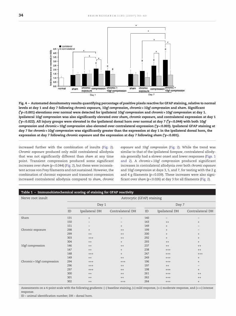
Fig. 4 – Automated densitometry results quantifying percentage of positive pixels reactive for GFAP staining, relative to normallevels at day 1 and day 7 following chromic exposure, 10gf compression, chromic+10gf compression and sham. Significant(#p<0.001) elevations over normal were detected for ipsilateral 10gf compression and chromic+10gf compression at day 1.Ipsilateral 10gf compression was also significantly elevated over sham, chromic exposure, and contralateral expression at day 1(*p<0.022). All injury groups were elevated in the ipsilateral dorsal horn over normal at day 7 (#p<0.044) with both 10gfcompression and chromic+10gf compression also elevated over contralateral expression (*p<0.003). Ipsilateral GFAP staining atday 7 for chromic+10gf compression was significantly greater than the expression at day 1 in the ipsilateral dorsal horn, theexpression at day 7 following chromic exposure and the expression at day 7 following sham (*p<0.001).
34 B R A I N R E S E A R C H 1 1 8 1 ( 2 0 0 7 ) 3 0 – 4 3
increased further with the combination of insults (Fig. 2).Chromic exposure produced only mild contralateral allodyniathat was not significantly different than sham at any timepoint. Transient compression produced some significantincreases over sham (p<0.044) (Fig. 2), but these were inconsis-tent across von Frey filaments andnot sustained.However, thecombination of chromic exposure and transient compressionincreased contralateral allodynia compared to sham, chromic
Table 1 – Immunohistochemical scoring of staining for GFAP r
Nerve root insult
Day 1
ID Ipsilateral DH Contra
Sham 131 +150 −151 −
Chromic exposure 298 +299 ++303 +++304 ++
10gf compression 146 ++147 ++148 +++149 ++
Chromic+10gf compression 294 +++296 +++297 +++300 ++301 ++302 ++
Assessments on a 4-point scale with the following gradients: (−) baselineresponse.ID – animal identification number; DH – dorsal horn.
exposure and 10gf compression (Fig. 2). While the trend wassimilar to that of the ipsilateral forepaw, contralateral allody-nia generally had a slower onset and lower responses (Figs. 1and 2). A chromic+10gf compression produced significantincreases in contralateral allodynia over both chromic exposureand 10gf compression at days 3, 5, and 7, for testing with the 2 gand 4 g filaments (p<0.039). These increases were also signi-ficant over sham (p<0.026) at day 3 for all filaments (Fig. 2).
eactivity
Astrocytic (GFAP) staining
Day 7
lateral DH ID Ipsilateral DH Contralateral DH
− 140 − −− 143 ++ ++ 149 + −++ 199 + −++ 200 + +++ 292 + ++ 293 ++ +++ 237 ++ +++ 238 +++ +++ 247 +++ +++++ 249 +++ ++++ 196 +++ +++ 197 ++ −++ 198 +++ +++ 261 +++ ++++ 262 +++ +++++ 264 +++ +
staining, (+) mild response, (++) moderate response, and (+++) intense

35B R A I N R E S E A R C H 1 1 8 1 ( 2 0 0 7 ) 3 0 – 4 3
2.2. Spinal glia
Separate groups of rats were used for analysis of glial activationat days 1 and 7 following surgery. Tissue damage during harvest
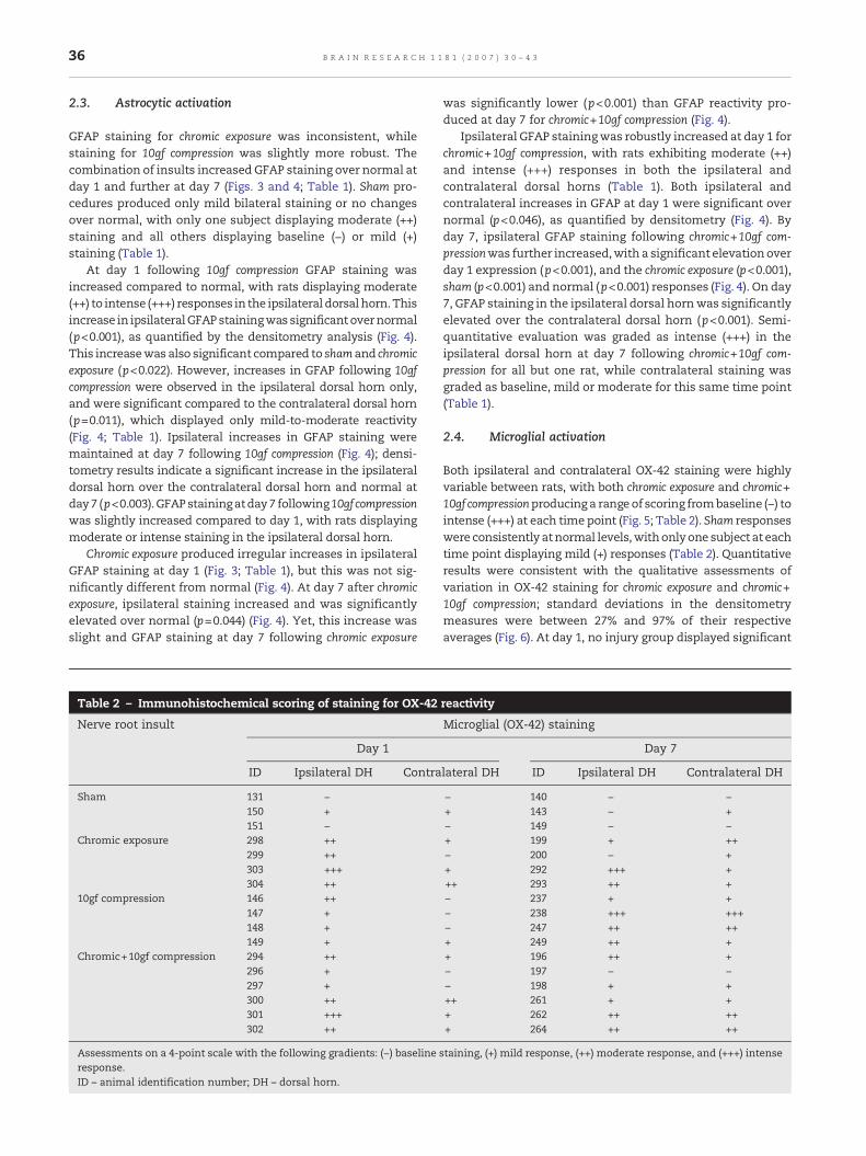
Fig. 5 – Representative ipsilateral (B, D, F, H, J, L) and contralateraafter chromic exposure (B, C, H, I), 10gf compression (D, E, J, K) or chday 7 (H–M) following injury. A matching normal naive sample (Apanel A is 100 μm and applies to all panels.
procedures precluded the use of spinal cord tissue from somerats in the behavioral study. Glial reactivity data at day 7 reflectonly a subset of rats used for behavioral assays in each of thechromic exposure (n=4) and chromic+10gf compression (n=6) groups.
l (C, E, G, I, K, M) C7 spinal cord sections stained against OX-42romic+10gf compression (F, G, L, M) on day 1 (B–G) or) was assigned baseline levels of staining. Scale bar shown in
36 B R A I N R E S E A R C H 1 1 8 1 ( 2 0 0 7 ) 3 0 – 4 3
2.3. Astrocytic activation
GFAP staining for chromic exposure was inconsistent, whilestaining for 10gf compression was slightly more robust. Thecombination of insults increased GFAP staining over normal atday 1 and further at day 7 (Figs. 3 and 4; Table 1). Sham pro-cedures produced only mild bilateral staining or no changesover normal, with only one subject displaying moderate (++)staining and all others displaying baseline (−) or mild (+)staining (Table 1).
At day 1 following 10gf compression GFAP staining wasincreased compared to normal, with rats displaying moderate(++) to intense (+++) responses in the ipsilateral dorsal horn.Thisincrease in ipsilateralGFAP stainingwas significant overnormal(p<0.001), as quantified by the densitometry analysis (Fig. 4).This increasewas also significant compared to sham and chromicexposure (p<0.022). However, increases in GFAP following 10gfcompression were observed in the ipsilateral dorsal horn only,and were significant compared to the contralateral dorsal horn(p=0.011), which displayed only mild-to-moderate reactivity(Fig. 4; Table 1). Ipsilateral increases in GFAP staining weremaintained at day 7 following 10gf compression (Fig. 4); densi-tometry results indicate a significant increase in the ipsilateraldorsal horn over the contralateral dorsal horn and normal atday7 (p<0.003).GFAPstainingatday7 following10gf compressionwas slightly increased compared to day 1, with rats displayingmoderate or intense staining in the ipsilateral dorsal horn.
Chromic exposure produced irregular increases in ipsilateralGFAP staining at day 1 (Fig. 3; Table 1), but this was not sig-nificantly different from normal (Fig. 4). At day 7 after chromicexposure, ipsilateral staining increased and was significantlyelevated over normal (p=0.044) (Fig. 4). Yet, this increase wasslight and GFAP staining at day 7 following chromic exposure
Table 2 – Immunohistochemical scoring of staining for OX-42
Nerve root insult
Day 1
ID Ipsilateral DH Contra
Sham 131 −150 +151 −
Chromic exposure 298 ++299 ++303 +++304 ++
10gf compression 146 ++147 +148 +149 +
Chromic+10gf compression 294 ++296 +297 +300 ++301 +++302 ++
Assessments on a 4-point scale with the following gradients: (−) baselineresponse.ID – animal identification number; DH – dorsal horn.
was significantly lower (p<0.001) than GFAP reactivity pro-duced at day 7 for chromic+10gf compression (Fig. 4).
Ipsilateral GFAP stainingwas robustly increased at day 1 forchromic+10gf compression, with rats exhibiting moderate (++)and intense (+++) responses in both the ipsilateral andcontralateral dorsal horns (Table 1). Both ipsilateral andcontralateral increases in GFAP at day 1 were significant overnormal (p<0.046), as quantified by densitometry (Fig. 4). Byday 7, ipsilateral GFAP staining following chromic+10gf com-pressionwas further increased,with a significant elevationoverday 1 expression (p<0.001), and the chromic exposure (p<0.001),sham (p<0.001) and normal (p<0.001) responses (Fig. 4). On day7, GFAP staining in the ipsilateral dorsal hornwas significantlyelevated over the contralateral dorsal horn (p<0.001). Semi-quantitative evaluation was graded as intense (+++) in theipsilateral dorsal horn at day 7 following chromic+10gf com-pression for all but one rat, while contralateral staining wasgraded as baseline, mild or moderate for this same time point(Table 1).
2.4. Microglial activation
Both ipsilateral and contralateral OX-42 staining were highlyvariable between rats, with both chromic exposure and chromic+10gf compressionproducing a range of scoring frombaseline (−) tointense (+++) at each time point (Fig. 5; Table 2). Sham responseswere consistently atnormal levels,withonlyone subject at eachtime point displaying mild (+) responses (Table 2). Quantitativeresults were consistent with the qualitative assessments ofvariation in OX-42 staining for chromic exposure and chromic+10gf compression; standard deviations in the densitometrymeasures were between 27% and 97% of their respectiveaverages (Fig. 6). At day 1, no injury group displayed significant
reactivity
Microglial (OX-42) staining
Day 7
lateral DH ID Ipsilateral DH Contralateral DH
− 140 − −+ 143 − +− 149 − −+ 199 + ++− 200 − ++ 292 +++ +++ 293 ++ +− 237 + +− 238 +++ +++− 247 ++ +++ 249 ++ ++ 196 ++ +− 197 − −− 198 + +++ 261 + ++ 262 ++ +++ 264 ++ ++
staining, (+) mild response, (++) moderate response, and (+++) intense

Fig. 6 – Automated densitometry results quantifying relative OX-42 staining at day 1 and day 7 following chromic exposure, 10gfcompression, chromic+10gf compression and sham. At day 1, reactivity in the bilateral dorsal horns for all injuries was notsignificantly different from sham or normal. By day 7, ipsilateral OX-42 staining following 10gf compression or chromic+10gfcompressionwas significantly increased compared to day 1 (*p<0.001). These day 7 increases were also significant compared tonormal (#p<0.001), sham (*p<0.001), and day 7 chromic exposure (*p<0.004). Contralateral increases were also significantcompared to normal following chromic+10gf compression (#p=0.003) and day 1 chromic+10gf compression (*p=0.008).
37B R A I N R E S E A R C H 1 1 8 1 ( 2 0 0 7 ) 3 0 – 4 3
increases in ipsilateral OX-42 staining in the dorsal horn oversham or normal (Fig. 6).
By day 7, ipsilateral staining for chromic exposure remainedgenerally unchanged compared to reactivity at day 1 (Fig. 6;Table 2). In contrast, at day 7 after 10gf compression, ipsilateralOX-42 reactivity was significantly increased compared to day 1(p<0.001). This ipsilateral increase between days 1 and 7 wasalso observed following chromic+10gf compression (p<0.001).Increases in ipsilateral OX-42 on day 7 following 10gf com-pression or chromic+10gf compression were significant overchromic exposure ( p<0.004), sham (p<0.001) and normal(p<0.001) (Figs. 5 and 6). In addition, at this later time point,there was no significant difference in OX-42 responses detect-ed between ipsilateral and contralateral dorsal horns for anyinjury type (Fig. 6). Further, at day 7 the chromic+10gf com-pression contralateral OX-42 responses were significantlyelevated over normal (p=0.003) and the contralateral spinalresponses for that same injury at day 1 (p=0.008) (Figs. 5 and 6).
3. Discussion
Many studies have demonstrated a dependence of radicularpain on mechanical and chemical inputs (Kawakami et al.,1994a,b; Kajander et al., 1996; Olmarker et al., 1997; Hashizumeet al., 2000; Olmarker et al., 2002; Hou et al., 2003; Winkelsteinand DeLeo, 2004; Cornefjord et al., 2004; Hubbard andWinkelstein, 2005). This study is the first to our knowledgeto examine such factors of chemical irritation and nerve rootcompression for transient loading to the cervical nerve roots.In particular, these results demonstrate a role for transientcompression in mediating bilateral mechanical allodyniawhen combined with chemical factors. While allodynia wasproduced for chromic exposure, and also for 10gf compression,behavioral hypersensitivity was significantly increased andsustained when these injuries were combined (chromic+10gfcompression) (Figs. 1 and 2). Coupled with mechanical allody-
nia, the combination of chemical irritation and transientcompression of the nerve root also produced significantincreases in both astrocytic and microglial reactivity in thedorsal horn of the spinal cord at day 7 (Figs. 4 and 6).
Although the combination of chromic gut exposure andtransient compression induced robust increases inmechanicalallodynia, chromic exposure alone also produced significantlyelevated ipsilateral mechanical allodynia over sham at earlytime points. Allodynia following chromic exposure returned tosham levels by day 5 (Fig. 1), despite the fact that the chromicgut pieces remained in contact with the nerve root for theduration of the study. These results are in agreement withprevious studies demonstrating slight and non-sustainedincreases in behavioral hypersensitivity following chromicexposure or a loose ligation of chromic suture around thelumbar nerve root (Lee et al., 2003; Winkelstein and DeLeo,2004). Maves et al. (1993) demonstrated a ‘dose response’ forthermal sensitivity from sciatic nerve ligations using chromicgut suture; it is, therefore, likely that the amount or diameter ofsuture used in our study may not have been sufficient toinduce a sustained behavioral effect. However, in studies usinga chromic gut ligation it is difficult to separate the respectiveroles of the inflammatory andmechanical effects. The chromicgut suture can resorb and ligation with this material mayinduce further tightening as a consequence of tissue swelling,with both effects changing the local mechanics over time.Some studies of low back pain have utilized what thoseauthors defined as a ‘loose’ ligation of the nerve root to placechromic suture in contactwith thenerve rootwithout applyingadditional pressure and a ‘tight’ ligation to apply mechanicalcompression in addition to the chemical irritation of thechromic suture (Kawakami et al., 1994a; Hashizumeet al., 2000;Rutkowski et al., 2002). While described as ‘loose’ and ‘tight’respectively, the mechanical conditions of the ligation groupswerenot explicitly definedby thoseauthors.Yet, it is likely thatthe ‘loose’ chromic ligation did impose mechanical loadingsince gross pathology demonstrated narrowing in the nerve

38 B R A I N R E S E A R C H 1 1 8 1 ( 2 0 0 7 ) 3 0 – 4 3
root following ‘loose’ ligations (Kawakami et al., 1994b;Hashizume et al., 2000). In our study, it is possible that thechromic gut pieces may have also imposed physical irritationto the nerve root, but this is very unlikely. The chromic piecescontacted the nerve root when gently placed along the root.They are very small and have little mass to provide any directcompression. In addition, visual inspection of the root at thetime of tissue harvest showed no evidence of its narrowing ordeformation in samples from the chromic exposure group.
The increased behavioral hypersensitivity for the combi-nation of insults compared to a single insult (Figs. 1 and 2) isconsistent with other reports of increases in mechanical andthermal hyperalgesia when these insults are combinedcompared to the application of disc material without com-pression of the root (Olmarker et al., 1997; Hou et al., 2003).Results presented here demonstrate that the combination ofmechanical and chemical factors induces allodynia that isrobustly increased compared to either chemical ormechanicalinjury alone (Figs. 1 and 2). Findings also suggest that amechanical insultmay be required for behavioral symptoms tobe chronic, as chemical irritation alone does not produce sus-tained allodynia while the transient compression produceallodynia that is significant at day 7. Our data lend additionalsupport to thehypothesis that the load threshold for producingpersistent hypersensitivity in nerve root compression islowered when chemical factors are present at injury (Winkel-stein and DeLeo, 2004).
While bilateral allodynia has been previously reported for aunilateral insult (Tabo et al., 1999; Rutkowski et al., 2002; Araujoet al., 2003; Hou et al., 2003; Hubbard and Winkelstein, 2005),such mirror-image behavioral hypersensitivity was only signif-icant for the combined mechanical and chemical insult (Fig. 2).This production of contralateral allodynia for a combined injurycould imply that functional changes in the spinal cord arespecifically induced in the interneurons that project to the non-operated contralateral side only for specific injury paradigmsand not for the chemical or mechanical injuries used in thisstudy. Contralateral sensitivity has been previously reported forthe combination of these insults, but not for exposure of thenerve root to chromic material or intervertebral disc samples(Maves et al., 1993; Rutkowski et al., 2002; Hou et al., 2003).Although contralateral allodynia has been induced by mechan-ical insults in theabsence of any chemical loading, those studiesimplementedeither ligationofmultiplenerve rootsor thespinalnerve, imposing a more severe sustainedmechanical compres-sion and one which may induce either a more robust, orcontinuous, barrage to the nociceptive cascades (Tabo et al.,1999; Araujo et al., 2003). While we did not specifically probeelectrophysiologic properties of spinal interneurons, neuronalsensitization in the spinal cord can be initiated by increasedneurotransmitter release, inflammatory cytokines, or directlyby glial cells (Schiefer et al., 1999; Tabo et al., 1999; Cuellar et al.,2004; Inoue, 2005; Tsuda et al., 2005). Ipsilateral astrocyticactivation was present at day 7 following compression and wasfurther increased for the combined injury (Figs. 3 and 4; Table 1).Microglial activation at day 7 was also increased followingcompression, but was only on the side of injury; in contrast, forthe addition of chromic suture,microglial reactivity at day 7wasobserved bilaterally in the spinal cord (Figs. 5 and 6; Table 2).These increases in spinal glial activation following the com-
bined injuryatday 7 compared toeithermechanical or chemicalinjuries alone imply that spinal inflammation is greatlyincreased when these factors are combined. Robust bilateralspinal inflammation, including increases incell surfacemarkersand inflammatory cytokines, and behavioral hypersensitivityhave been reported for chromic ligation of lumbar nerve rootsfurther supporting a role for inflammation in mediating spinalglial responses in painful nerve root injury (Rutkowski et al.,2002). Biochemical and functional immune changes in thespinal cord correlatewithmechanical parameters for nerve rootloading (i.e. applied loading rate, magnitude and duration); forexample, imposed nerve root strain is significantly correlatedwith spinal mRNA levels for a host of inflammatory cytokines,implying sensitive biochemical responses to mechanical nerveroot injury (Winkelstein et al., 2001a,b). Further, there is a directrelationship between root compression and neuronal function(Olmarker et al., 1989, 1990; Pedowitz et al., 1992;Mao et al., 1998;Yamaguchi et al., 1999). These results provide a host ofcandidate spinal responses that may be induced by injuries tothe nerve root and are likely sensitive to the specific profile ofthemechanical injury.
In the current study, the time course of activation ofastrocytes and microglia was differentially modulated for thethree types of injuries. At day 1, spinal astrocytic activationwas evident in the ipsilateral horn for 10gf compression andchromic+10gf compression, with an increase in activation onthat side at day 7 following the combined injury (Fig. 4).However, microglial activation was not significantly elevateduntil day 7, and it was bilateral (Fig. 6). This is consistent withresults from studies using silk ligations in which microglialactivation was bilateral, whereas astrocytic activation wasonly apparent on the ipsilateral side of the spinal cord(Winkelstein andDeLeo, 2002). Bilateral increases inmicroglia,but not astrocytes, may be produced because microglia aremore sensitive to ionic changes caused by neuronal sensiti-zation which may occur on the contralateral side. Althoughlittle is known about what triggers glial activation, it has beenshown that both microglia and astrocytes can be activated bydepolarization of neighboring neurons, consistent with atetrapartite model of the synapse (Kreutzberg, 1996; DeLeoet al., 2006). Yet, microglia may respond to these neuronalchanges before astrocytes, and may be more sensitive tochanges in neurotransmitters (Caggiano and Kraig, 1996). It islikely that in our combined injury microglia in the contralat-eral dorsal horn respond to small perturbations in extracellu-lar ion homeostasis and this microglial activation then leadsto further neuronal sensitization. Astrocytic activationmay bepresent in the contralateral dorsal horn at a time point notprobed in this study, as microglia produce and releasemolecules that can cause astrocytic activation (Kreutzberg,1996; Newman, 2003; DeLeo et al., 2006). Studies of kinaseactivation and transcriptional changes demonstrate thatspinal microglial activation can precede astrocytic activation(Tanga et al., 2004; Zhuang et al., 2005). While this study didprobe the time course of glial activation, additional studies areneeded of both acute and intermediate time points not studiedhere.
Differential spinal glial activation may also result fromthese cells having different thresholds for activation. Spinalastrocytic activation after ligation using silk is only slightly

39B R A I N R E S E A R C H 1 1 8 1 ( 2 0 0 7 ) 3 0 – 4 3
lower than that for a ligation with chromic gut (Hashizume etal., 2000; Winkelstein and DeLeo, 2002). This suggests that theinitiation of spinal astrocytic reactivity may require a me-chanical insult, and that it is only enhanced when thismechanical injury is accompanied by a chemical insult. Incontrast, in those models, spinal microglial activation wasgreater for ligation with chromic suture relative to silk,suggesting that spinal microglia are responsive to chemicalirritation. Microglial reactivity in our study increased bilater-ally between days 1 and 7 for chromic+10gf compression, andunilaterally following compression alone (Fig. 6; Table 2),suggesting that microglia respond sensitively to the type andintensity of injury. Results presented here demonstrate onlymoderate microglial activation following chromic exposure(Fig. 6; Table 2) implying that it is the combination of insults,rather than simply a chemical insult alone, that causes robustmicroglial activation.
Our results, in the context of other studies, imply thatmicroglia are more sensitive to chemical inputs than astro-cytes are. This is in agreement with studies demonstratingthat microglia respond tominor changes in their environment(Caggiano and Kraig, 1996; Kreutzberg, 1996). The apparentdisparity in responses between microglia and astrocytes fordifferent types of stimuli may be due to their varying roles inthe central nervous system. Activated microglia releasecytokines (TNFα, IL-1β, IL-6), which can then activate astro-cytes, and also induces changes in neuronal excitability, aswell as transcriptional changes in neurons that alter theirsensitivity to stimuli (McMahon et al., 2005). The findingspresented here are contradictory to this, with astrocyticreactivity significantly elevated over normal at day 1 followingchromic+10gf compression and microglia not exhibiting signifi-cant elevations until later at day 7 (Figs. 4 and 6). However, it ispossible that the peak of microglial activation precedes thepeak of astrocytic activation and occurs at an intermediatetime point not probed in this study. In fact, following a painfullumbar nerve ligation, spinal extracellular signal-regulatedprotein kinases are activated in microglia with maximumactivation on day 2, followed by a peak of pERK activation inastrocytes at day 21 (Zhuang et al., 2005). The bilateralmicroglial activation produced at day 7 for chromic+10gfcompression indicates prolonged microglial reactivity whenchemical irritation is combined with transient compression inour model (Fig. 4). This could also suggest sustained cytokinerelease in a combined mechanical and chemical injury.
While our results demonstrate that the combination ofputative insults causes robust and widespread behavioral andspinal glial responses (Figs. 1–6), additional studies directed atspecific mechanical and chemical dose-effects are needed tofully define the relationship between injury parameters andpain. Although the present study used a repeatable, reliablechemical irritation as a discrete and uniform amount ofchromic gut suture, the precise chemicals delivered andtheir effect on behavioral sensitivity were not specificallyidentified here. Quantitative studies specifically probing therole of spinal glia and their biochemical contributions, such ascytokine production, for different degrees of painful mechan-ical and/or chemical injury are necessary at additional timepoints. While the time points in this study were chosen as theonset and maintenance of behavioral hypersensitivity in this
cervical radiculopathymodel (Hubbard andWinkelstein, 2005;Rothman et al., 2005), it may be necessary to examinemicroglial activation at other time points. Further, it isrecognized that qualitative microglial activation data showeda wide range of variability (Table 2) and associated densitom-etry results showed correspondingly highly standard devia-tions (Fig. 6); larger group sizes may also be necessary. Whileexposure of the nerve root to chromic gut suture does notdirectly mimic the clinical condition of nerve root irritation bythe disc material, the chromic salts and pyrogallol that areused to produce chromic suture are known to be inflammatoryto nervous tissue (Maves et al., 1993). Chromic suture,therefore, provides a basis for modeling the chemical compo-nent of painful disc herniation. In fact, chromic gut suture hasbeen used extensively inmodels of low back pain tomimic theinflammatory effects of disc material (Kawakami et al., 1994a,b; Maves et al., 1993; Hashizume et al., 2000; Rutkowski et al.,2002; Lee et al., 2003; Winkelstein and DeLeo, 2004). Thesemodels also demonstrate a robust increase in behavioralhypersensitivity for chromic ligations (mechanical and chem-ical injury) compared to a mechanical ligation injury alone(Kim and Chung, 1992; Kawakami et al., 1994b; Hashizumeet al., 2000), similar to the behavioral effects achieved byapplying nucleus pulposus directly to the compressed nerveroot (McCarron et al., 1987; Hou et al., 2003; Kawakami et al.,2003; Murata et al., 2004; Kallakuri et al., 2005). Further,behavioral outcomes from both chromic suture ligation anddisc herniation models are similar to clinical symptoms fromdisc herniation, which show long-term sensitivity in theextremities (Ellenberg et al., 1994; Boos et al., 1995).
These results demonstrate that either chemical irritationor mechanical compression to the cervical nerve root caninduce mechanical allodynia and spinal glial activation andthat those responses are increased by the combination ofthese insults. Behavioral results from our study support thatpain can be produced in the absence of a compression (Figs. 1and 2). However, while chemical irritation alone may beresponsible for some pain cases, a compressive event, even atransient one, can dramatically enhance behavioral sensitivityand such sensitivity may persist. Microglial activation washighly variable both between injury types and for individualrats in each injury group (Table 2); this may explain, in part,the individual variation in pain symptoms observed forhumans. Studies are needed to specifically define the relation-ships between mechanical and chemical parameters andbehavioral, cellular, and biochemical responses in quantita-tive approaches.
4. Experimental procedure
Experiments were performed using male Holtzman rats(Harlan Sprague-Dawley; Indianapolis, ID), weighing 250–350 g at the start of the study. Rats were housed under USDA-and AAALAC-compliant conditions with a 12-h:12-h light/darkcycle and free access to food and water. All experimentalprocedures were approved by the University of PennsylvaniaInstitutional Animal Care and Use Committee and carried outaccording to the guidelines of the Committee for Research andEthical Issues of the IASP (Zimmermann, 1983).

40 B R A I N R E S E A R C H 1 1 8 1 ( 2 0 0 7 ) 3 0 – 4 3
4.1. Surgical procedures
All procedures were performed under inhalation anesthesia(4% halothane for induction, 2% for maintenance). Each ratreceived either nerve root exposure to chromic gut suturematerial (chromic exposure) (n=10), transient nerve root com-pression (10gf compression) (n=8), chromic gut exposure withsupplemental cervical nerve root compression (chromic+10gfcompression) (n=13), or sham exposure (sham) (n=6). For thisstudy, previously described procedures of cervical dorsal rootcompression were used and modified to incorporate inflam-matory components at the time of insult (Hubbard andWinkelstein, 2005; Rothman et al., 2005). Rats were placed ina prone position, and an incision was made in the skin fromthe base of the skull to the second thoracic vertebra. Muscleand soft tissuewere cleared exposing the C6 and C7 laminae. AC6/C7 hemilaminectomy and partial facetectomy were per-formed on the right side to expose the spinal cord and right C7dorsal nerve root. Procedures for the chromic exposure involvedplacing four pieces of 3-0 chromic gut suture (2 mm in length)(Surgical Specialties; Reading, PA) on the right C7 dorsal nerveroot proximal to the dorsal root ganglia (DRG). Procedures for10gf compression match those previously published (Hubbardand Winkelstein, 2005; Rothman et al., 2005). Briefly, a 10gfmicrovascular clip (World Precision Instruments, Inc) wasapplied to the right C7 dorsal nerve root proximal to the dorsalroot ganglion. Compression was imposed for 15 min, beforeclip removal. Procedures for chromic+10gf compression involvedthose described for chromic exposure in addition to thosedescribed for 10gf compression. Compression was applied tothe right C7 dorsal nerve root proximal to the DRG, removedafter 15 min, and then four chromic gut suture pieces wereplaced on the dorsal root as described above. Procedures forsham involved nerve root exposure only, as previouslypublished (Rothman et al., 2005). Following surgery, allwounds were closed using 3-0 polyester suture and surgicalstaples. Rats were recovered in room air and monitoredcontinuously.
4.2. Behavioral assessment
Following surgery, a subset of rats (n=6 chromic exposure, n=410gf compression, n=7 chromic+10gf compression, n=3 sham) wasevaluated for behavioral hypersensitivity after injury. Mechan-ical allodynia was measured in the ipsilateral and contralateralforepaws on each of postoperative days 1, 3, 5, and 7. Methodsfor quantifying forepaw allodynia have been previously validat-ed for cervical nerve root injury (Hubbard andWinkelstein, 2005;Rothman et al., 2005). Prior to surgery, rats were acclimated tothe tester and environment, and baseline measurements wererecorded for 2 days. For each session, after 20 min of accli-mation, rats were stimulated on the plantar surface of theipsilateral and contralateral forepaws, separately, using threevon Frey filaments (1.4, 2, 4 g) (StoeltingCo.;WoodDale, IL). Eachtesting sessionconsistedof three roundsof 10stimulationseachto each forepaw, separated by 10 min. For each session with agiven filament, the total number of withdrawals was countedfor each paw and averages for each injury group were deter-mined. A single tester performed all behavioral testing blindedto surgical procedures.
4.3. Immunohistochemistry procedures
In order to assess the temporal pattern of glial activation in thedorsal horn, C7 spinal cord was assayed for astrocytic andmicroglial activation using antibodies to glial fibrillary acidicprotein (GFAP) and CR3/CD11b (OX-42), respectively. Cervicalspinal cord tissue was harvested on each of days 1 and 7following injury. Spinal cord tissue evaluated at day 1 wastaken from a separate set of rats than that used for allodyniaassessments (n=4 chromic exposure, n=4 10gf compression, n=6chromic+10gf compression, n=3 sham). For tissue harvest, ratswere deeply anesthetized followed by transcardiac perfusionwith 200 ml of phosphate-buffered saline (PBS) and 300 ml of4% paraformaldehyde in PBS (pH 7.4). Following perfusion, thecervical spinal cord was exposed by laminectomy, the C7segment of the cervical cord was harvested, and tissue waspost-fixed in the 4% paraformaldehyde solution for 20 min(Hubbard and Winkelstein, 2005). Tissue was transferred to30% sucrose/PBS and stored for 3 days at 4 °C. Samples werefreeze-mounted with OCT medium (Triangle BiomedicalSciences; Durham, NC) for axial cryosectioning. Matchedspinal cord tissue from a naïve (un-operated) rat was alsoprocessed for normalization and comparison.
Four serial C7 spinal cord sections (20 μm) from each ratwere prepared for free-floating immunohistochemical stain-ing. A polyclonal antibody to GFAP (Dako; Carpinteria, CA) wasused as a marker of activated astrocytes. Slices were blockedwith normal goat serum (Vector Labs; Burlingame, CA) for20 min followed by incubation in a primary antibody directedagainst GFAP (1:20,000) overnight. Sections were then treatedwith a biotinylated goat anti-rabbit IgG, quenched in 0.3%peroxide solution, and developed using 3,3-diaminobenzidine(Vector Labs; Burlingame, CA). For a second set of matchedaxial sections, amonoclonal antibody toCR3/CD11b (OX-42; BDPharmingen; San Diego, CA) was used as a marker of activatedmicroglia. Spinal cord slices were blocked with normal goatserum (Vector Labs; Burlingame, CA) for 20 min and incubatedovernight in an antibody directed against OX-42 (1:500). AnAlexa546-conjugated goat anti-mouse secondary antibody(1:500) was used for immunofluorescent detection (Invitrogen;Carlsbad, CA). Previous studies were performed to determineoptimal antibody dilutions. A negative controlwith no primaryantibody staining was always included for verification ofspecificity of immunohistochemical techniques.
Two techniques were used to analyze glial activation.Semiquantitative scoring was used to grade the glial activationin each of the ipsilateral and contralateral dorsal horns to enablecomparison to normal sections and results in the literature. Inaddition, automated densitometry was also used to quantifystaining to enable quantitative and statistical comparisons. Foreach glialmarker, two representative cord sections fromeach ratwere photographed at 100×magnification using a digital cameraand stereomicroscope system and Axiovision software (Zeiss;Thornwood, NY). For both analysis methods, the ipsilateral andcontralateral dorsal horns relative to the injury site wereassessed. For the semi-quantitative grading, two observers,blinded to the injury group graded the degree of activation.Separate scores were given to ipsilateral and contralateral sides.Gradingwasperformedusing anestablished 4-point scale, basedoncell numbers, compactness, and intensity of staining (Colburn

41B R A I N R E S E A R C H 1 1 8 1 ( 2 0 0 7 ) 3 0 – 4 3
et al., 1999; Winkelstein and DeLeo, 2002; Hubbard and Winkel-stein, 2005). Assessments were made using the following levelsof gradation: baseline staining (−), mild response (+), moderateresponse (++), and intense response (+++) (Colburn et al., 1999).Results from the first observer were verified by the secondobserver, and tabulated. For automated densitometry analysis,the same sections were used for semi-quantitative scoring.Images were cropped (800×200 pixels) to include the superficiallamina I–III only; fluorescent images were also inverted. Imageswere analyzed for percentage of pixels above a defined threshold(Abbadie et al., 1996). Thresholdpixel intensitywas chosenbasedon staining in normal tissue. This approach gives a quantitativemeasure of the degree and amount of staining above what ispresent in normal naïve tissue. Values were normalized,averaged and compared.
4.4. Statistical analysis
Significantdifferences inallodyniabetweengroupsweredetectedby a one-way ANOVA at each time point (SYSTAT; Richmond,CA). For quantitative densitometry, a one-way ANOVA wasperformed to compare differences over time (day 1, day 7), bet-ween injury groups (chromic exposure, 10gf compression, chromic+10gf compression, sham), and between sides of the spinal cord(ipsilateral, contralateral). Significance was defined at p<0.05.
Acknowledgments
The authors thank Dr. Steven B. Nicoll for use of hismicroscope and imaging system and Raymond D. Hubbardfor assessment of glial activation.
This work was funded by grant support from NIH (NIAMS)AR047564-02 and the Catharine D. Sharpe Foundation.
R E F E R E N C E S
Abbadie, C., Brown, J.L., Mantyh, P.W., Basbaum, A.I., 1996. Spinalcord substance P receptor immunoreactivity increases in bothinflammatory and nerve injury models of persistent pain.Neuroscience 70, 201–209.
Araujo, M.C., Sinnott, C.J., Strichartz, G.R., 2003. Multiple phases ofrelief from experimental mechanical allodynia by systemiclidocaine: responses to early and late infusions. Pain 103,21–29.
Atlas, S.J., Keller, R.B., Wu, Y.A., Deyo, R.A., Singer, D.E., 2005.Long-term outcomes of surgical and nonsurgical managementof sciatica secondary to a lumbar disc herniation: 10 yearresults from the Maine lumbar spine study. Spine 30, 927–935.
Boos, N., Rieder, R., Schade, V., Spratt, K.F., Semmer, N., Aebi, M.,1995. 1995 Volvo award in clinical sciences: the diagnosticaccuracy ofmagnetic resonance imaging, work perception, andpsychosocial factors in identifying symptomatic discherniations. Spine 20, 2613–2625.
Caggiano, A.O., Kraig, R.P., 1996. Eicosanoids and nitric oxideinfluence induction of reactive gliosis from spreadingdepression in microglia but not astrocytes.J. Comp. Neurol. 369, 93–108.
Colburn, R.W., Rickman, A.J., DeLeo, J.A., 1999. The effect of siteand type of nerve injury on spinal glial activation andneuropathic pain behavior. Exp. Neurol. 157, 289–304.
Cornefjord, M., Nyberg, F., Rosengren, L., Brisby, H., 2004.Cerebrospinal fluid biomarkers in experimental spinal nerveroot injury. Spine 29, 1862–1868.
Cuellar, J.M., Montesano, P.X., Carstens, E., 2004. Role of TNF-alphain sensitization of nociceptive dorsal horn neurons induced byapplication of nucleus pulposus to L5 dorsal root ganglion inrats. Pain 110, 578–587.
DeLeo, J.A., Winkelstein, B.A., 2002. Physiology of chronic spinalpain syndromes: from animal models to biomechanics.Spine 27, 2526–2537.
DeLeo, J.A., Tawfik, V.L., LaCroix-Fralish, M.L., 2006. Thetetrapartite synapse: path to CNS sensitization and chronicpain. Pain 122, 17–21.
Ellenberg, M.R., Honet, J.C., Treanor, W.J., 1994. Cervicalradiculopathy. Arch. Phys. Med. Rehabil. 75, 342–352.
Elliott, A., Smith, B., Penny, K., Smith, W., Chambers, W., 1999. Theepidemiology of chronic pain in the community. Lancet 354,1248–1252.
Frymoyer, J.W., 1988. Back pain and sciatica. N. Engl. J. Med. 318,291–300.
Goupille, P., Jayson, M., Valat, J.P., Freemont, A.J., 1998. The role ofinflammation in disk herniation-associated radiculopathy.Semin. Arthritis Rheum. 28, 60–71.
Hart, L.G., Deyo, R.A., Cherkin, D.C., 1995. Physician office visits forlow back pain. Frequency, clinical evaluation, and treatmentpatterns from a U.S. national survey. Spine 20, 11–19.
Hashizume, H., DeLeo, J.A., Colburn, R.W., Weinstein, J.N., 2000.Spinal glial activation and cytokine expression after lumbarroot injury in the rat. Spine 25, 1206–1217.
Hasselstrom, J., Liu-Palmgren, J., Rasjo-Wraak, G., 2002. Prevalenceof pain in general practice. Eur. J. Pain 6, 375–385.
Hou, S.X., Tang, J.G., Chen, H.S., Chen, J., 2003. Chronicinflammation and compression of the dorsal root contribute tosciatica induced by the intervertebral disc herniation in rats.Pain 105, 255–264.
Hubbard, R.D., Winkelstein, B.A., 2005. Transient cervical nerveroot compression in the rat induces bilateral forepaw allodyniaand spinal glial activation: mechanical factors in painful neckinjuries. Spine 30, 1924–1932.
Inoue, K., 2005. The function of microglia through purinergicreceptors: neuropathic pain and cytokine release.Pharmacol. Ther. 109, 210–226.
Kajander, K.C., Pollock, C.H., Berg, H., 1996. Evaluation of hindpawposition in rats during chronic constriction injury (CCI)produced with different suture materials. Somatosens.Mot. Res. 13, 95–101.
Kallakuri, S., Takebayashi, T., Ozaktay, A.C., Chen, C., Yang, S.,Wooley, P.H., Cavanaugh, J.M., 2005. The effects of epiduralapplication of allografted nucleus pulposus in rats on cytokineexpression, limb withdrawal and nerve root discharge.Eur. Spine J. 14, 956–964.
Kawakami, M., Weinstein, J.N., Spratt, K.F., Chatani, K., Traub, R.,Meller, S.T., Gebhart, G.F., 1994a. Experimental lumbarradiculopathy: immunohistochemical and quantitativedemonstrations of pain induced by lumbar root irritation of therat. Spine 19, 1780–1794.
Kawakami, M.,Weinstein, J.N., Chatani, K., Spratt, K.F., Meller, S.T.,Gebhart, G.F., 1994b. Experimental lumbar radiculopathy:behavioral and histologic changes in a model of radicular painafter spinal nerve root irritation with chromic gut ligatures inthe rat. Spine 19, 1795–1802.
Kawakami, M., Hashizume, H., Nishi, H., Matsumoto, T., Tamaki,T., Kuribayashi, K., 2003. Comparison of neuropathic paininduced by the application of normal and mechanicallycompressed nucleus pulposus to lumbar nerve roots in the rat.J. Orthop. Res. 21, 535–539.
Kim, S.H., Chung, J.M., 1992. An experimental model for peripheralneuropathy produced by segmental spinal nerve ligation in therat. Pain 50, 355–363.
42 B R A I N R E S E A R C H 1 1 8 1 ( 2 0 0 7 ) 3 0 – 4 3
Kreutzberg, G., 1996. Microglia: a sensor for pathological events inthe CNS. Trends Neurosci. 19, 312–318.
Lee, D.H., Iyengar, S., Lodge, D., 2003. The role of uninjured nerve inspinal nerve ligated rats points to an improved animal modelof neuropathic pain. Eur. J. Pain 7, 473–479.
Loupasis, G.A., Stamos, K., Katonis, P.G., Sapakis, G., Korres, D.S.,Hartofilakidis, G., 1999. Seven- to 20-year outcome of lumbardisectomy. Spine 24, 2313–2317.
Mao, G.P., Konno, S., Arai, I., Olmarker, K., Kikuchi, S., 1998.Chronic double-level cauda equina compression: anexperimental study on the dog cauda equina with analyses ofnerve conduction velocity. Spine 23, 1641–1644.
Maves, T.J., Pechman, P.S., Gebhart, G.F., Meller, S.T., 1993. Possiblechemical contribution from chromic gut sutures producesdisorders of pain sensation like those seen in man. Pain 54,57–69.
McCarron, R.F.,Wimpee, M.W., Hudkins, P.G., Laros, G.S., 1987. Theinflammatory effect of nucleus pulposus. A possible element inthe pathogenesis of low-back pain. Spine 12, 760–764.
McMahon, S.B., Cafferty, W.B.J., Marchand, F., 2005. Immune andglial cell factors as pain mediators and modulators. Exp.Neurol. 192, 444–462.
Mulleman, D., Mammou, S., Griffoul, I., Watier, H., Goupille, P.,2005. Pathophysiology of disk-related sciatica: I. Evidencesupporting a chemical component. Jt. Bone Spine 73, 151–158.
Murata, Y., Onda, A., Rydevik, B., Takahashi, K., Olmarker, K., 2004.Distribution and appearance of tumor necrosis factor-α in thedorsal root ganglion exposed to experimental disc herniationin rats. Spine 29, 2235–2241.
Murata, Y., Olmarker, K., Takahashi, I., Takahashi, K., Rydevik, B.,2005. Effects of selective tumor necrosis factor-alpha inhibitionto pain-behavioral changes caused by nucleuspulposus-induced damage to the spinal nerve in rats.Neurosci. Lett. 382, 148–152.
Narita, M., Yoshida, T., Nakajima, M., Narita, M., Miyatake, M.,Takagi, T., Yajima, Y., Suzuki, T., 2006. Direct evidence forspinal cord microglia in the development of a neuropathicpain-like state in mice. J. Neurochem. 97l, 1337–1348.
Newman, E.A., 2003. New roles for astrocytes: regulation ofsynaptic transmission. Trends Neurosci. 26, 536–542.
Ohnmeiss, D.D., Vanharanta, H., Ekholm, J., 1997. Degree of discdisruption and lower extremity pain. Spine 22, 1600–1605.
Olmarker, K., Myers, R.R., 1998. Pathogenesis of sciatic pain: role ofherniated nucleus pulposus and deformation of spinal nerveroot and dorsal root ganglion. Pain 78, 99–105.
Olmarker, K., Rydevik, B., Holm, S., 1989. Edema formation inspinal nerve roots induced by experimental, gradedcompression. An experimental study on the pig cauda equinawith special reference to differences in effects between rapidand slow onset of compression. Spine 14, 569–573.
Olmarker, K., Holm, S., Rydevik, B., 1990. Importance ofcompression onset rate for the degree of impairment ofimpulse propagation in experimental compression injury ofthe procine cauda equina. Spine 15, 416–419.
Olmarker, K., Rydevik, B., Nordborg, C., 1993. Autologous nucleuspulposus induces neurophysiologic and histologic changes inporcine cauda equina nerve roots. Spine 18, 1425–1432.
Olmarker, K., Blomquist, M., Stromberg, J., Nannmark, U.,Thomsen, P., Rydevik, B., 1995. Inflammatogenic properties ofnucleus pulposus. Spine 20, 665–669.
Olmarker, K., Brisby, H., Yabuki, S., Nordborg, C., Rydevik, B., 1997.The effects of normal, frozen, and hyaluronidase-digestednucleus pulposus on nerve root structure and function.Spine 22, 471–475.
Olmarker, K., Storkson, R., Berge, O.G., 2002. Pathogenesis of sciaticpain. A study of spontaneous behavior in rats exposed toexperimental disc herniation. Spine 27, 1312–1317.
Onda, A., Murata, Y., Rydevik, B., Larsson, K., Kikuchi, S.,Olmarker, K., 2005. Nerve growth factor content in dorsal root
ganglion as related to changes in pain behavior in a rat modelof experimental lumbar disc herniation. Spine 30, 188–193.
Otani, K., Itaru, A., Mao, G.P., Konno, S., Olmarker, K., Kikuchi, S.,1997. Experimental disc herniation: evaluation of the naturalcourse. Spine 22, 2894–2899.
Pedowitz, R.A., Garfin, S.R., Massie, J.B., Hargens, A.R.,Swenson, M.R., Myers, R.R., Rydevik, B.L., 1992. Effects ofmagnitude and duration of compression on spinal nerve rootconduction. Spine 17, 194–199.
Robinson, I., Meert, T.F., 2005. Stability of neuropathic painsymptoms in partial sciatic nerve ligation in rats is affected bysuture material. Neurosci. Lett. 373, 125–129.
Rothman, S.M., Kreider, R.A., Winkelstein, B.A., 2005. Spinalneuropeptide responses in persistent and transient painfollowing cervical nerve root injury. Spine 30, 2491–2496.
Rutkowski, M.D., Winkelstein, B.A., Hickey, W.F., Pahl, J.L.,DeLeo, J.A., 2002. Lumbar nerve root injury induces centralnervous system neuroimmune activation andneuroinflammation in the rat: relationship to painfulradiculopathy. Spine 27, 1604–1613.
Rydevik, B., Hasue, M., Wehling, P., 1996. Etiology of sciatic painand mechanisms of nerve root compression. In: Wiesel, S.W.,Weinsten, J.N., Herkowitz, H., Dvorak, J., Bell, G. (Eds.), TheLumbar Spine. WB Saunders Co., Philadelphia, pp. 123–141.
Saal, J.S., 1995. The role of inflammation in lumbar pain. Spine 20,1821–1827.
Schiefer, J., Kampe, K., Dodt, H.U., Zieglgansberger,W., Kreutzberg,G.W., 1999. Microglial motility in the rat facial nucleusfollowing peripheral axotomy. J. Neurocytol. 28, 439–453.
Skouen, J.S., Brisby, H., Otani, K., Olmarker, K., Rosengren, L.,Rydevik, B., 1999. Protein markers in cerebrospinal fluid inexperimental nerve root injury. Spine 24, 2195–2200.
Tabo, E., Jinks, S.L., Eisele Jr., J.H., Carstens, E., 1999. Behavioralmanifestations of neuropathic pain and mechanical allodynia,and changes in spinal dorsal horn neurons, following L4–L6dorsal root constriction in rats. Pain 80, 503–520.
Tanga, F.Y., Raghavendra, V., DeLeo, J.A., 2004. Quantitativereal-time RT–PCR assessment of spinal microglial andastrocytic activation markers in a rat model of neuropathicpain. Neurochem. Int. 45, 397–407.
Tsuda, M., Inoue, K., Salter, M.W., 2005. Neuropathic pain andspinal microglia: a big problem from molecules in ‘small’ glia.Trends Neurosci. 28, 101–107.
Wall, P., Melzak, R., 1994. Textbook of Pain, 3rd edition. ChurchillLivingstone, London.
Watkins, L.R., Maier, S.F., 2003. Glia: a novel drug discovery targetfor clinical pain. Nat. Rev. Drug Discov. 2, 973–985.
Weiler, C., Nerlich, A.G., Bachmeier, B.E., Boos, N., 2004. Expressionand distribution of tumor necrosis factor alpha in humanlumbar intervertebral discs: a study in surgical specimen andautopsy controls. Spine 30, 44–54.
Winkelstein, B.A., DeLeo, J.A., 2002. Nerve root injury severitydifferentially modulates spinal glial activation in a rat lumbarradiculopathy model: considerations for persistent pain. BrainRes. 956, 294–301.
Winkelstein, B.A., DeLeo, J.A., 2004. Mechanical thresholds forinitiation and persistence of pain following nerve root injury:mechanical and chemical contributions at injury. J. Biomech.Eng. 126, 258–263.
Winkelstein, B.A., Rutkowski, M.D., Sweitzer, S.M., Pahl, J.L., DeLeo,J.A., 2001a. Nerve injury proximal or distal to the DRG inducessimilar spinal glial activation and selective cytokine expressionbut differential behavioral responses to pharmacologictreatment. J. Comp. Neurol. 439, 127–139.
Winkelstein, B.A., Rutkowski, M.D., Weinstein, J.N., DeLeo, J.A.,2001b. Quantification of neural tissue injury in a ratradiculopathy model: comparison of local deformation,behavioral outcomes, and spinal cytokine mRNA for twosurgeons. J. Neurosci. Methods 11, 49–57.

43B R A I N R E S E A R C H 1 1 8 1 ( 2 0 0 7 ) 3 0 – 4 3
Xu, J., Pollack, C.H., Kajander, K.C., 1996. Chromic gut suturereduces calcitonin-gene-related-peptide and substance Plevels in the spinal cord following chronic constriction injury inthe rat. Pain 64, 503–509.
Yabuki, S., Kawaguchi, Y., Nordborg, C., Kikuchi, S., Rydevik, B.,Olmarker, K., 1998. Effects of lidocaine on nucleuspulposus-induced nerve root injury: a neurophysiologic andhistologic study of the pig cauda equina. Spine 23, 2383–2389.
Yamaguchi, K., Murakami, M., Takahashi, K., Moriya, H., Tatsuoka,H., Chiba, T., 1999. Behavioral and morphologic studies of thechronically compressed cauda equina. Spine 24, 845–851.
Yoshida, M., Nakamura, T., Sei, A., Kikuchi, T., Takagi, K.,Matsukawa, K., 2005. Intervertebral disc cells produce tumornecrosis factor α, interleukin-1β, and monocytechemoattractant protein-1 immediately after herniation: anexperimental study using a new hernia model. Spine 30, 55–61.
Zhuang, Z.Y., Gerner, P., Woolf, C.J., Ji, R.R., 2005. ERK issequentially activated in neurons, microglia, and astrocytes byspinal nerve ligation and contributes to mechanical allodyniain this neuropathic pain model. Pain 114, 149–159.
Zimmermann, M., 1983. Ethical guidelines for investigations ofexperimental pain in conscious animals. Pain 16, 109–110.