chemical physics letters - vilniaus...
TRANSCRIPT

Chemical Physics Letters 653 (2016) 67–72
Contents lists available at ScienceDirect
Chemical Physics Letters
journal homepage: www.elsevier .com/ locate /cplet t
Research paper
A femtosecond stimulated Raman spectroscopic study on the oxazinering opening dynamics of structurally-modified indolobenzoxazines
http://dx.doi.org/10.1016/j.cplett.2016.04.0300009-2614/� 2016 Elsevier B.V. All rights reserved.
⇑ Corresponding author.E-mail address: [email protected] (K. Redeckas).
1 For interpretation of color in figures, the reader is referred to the web vthis article.
Kipras Redeckas a,⇑, Stepas Toliautas b, Rasa Steponavici�ute c, Algirdas Šackus c, Juozas Sulskus b,Mikas Vengris a
aDepartment of Quantum Electronics, Faculty of Physics, Vilnius University, Sauletekio av. 10, LT-10223 Vilnius, LithuaniabDepartment of Theoretical Physics, Faculty of Physics, Vilnius University, Sauletekio av. 9, LT-10222 Vilnius, LithuaniacDepartment of Organic Chemistry, Kaunas University of Technology, K. Baršausko St. 59, LT-50270 Kaunas, Lithuania
a r t i c l e i n f o
Article history:Received 23 February 2016Revised 25 March 2016In final form 9 April 2016Available online 20 April 2016
a b s t r a c t
Steady-state and time-resolved femtosecond stimulated Raman scattering spectroscopic methods wereapplied to elucidate the photodynamics and the oxazine ring opening contingency in phenyl-substituted indolobenzoxazine systems. Using wavelength- and pulse duration-tunable multi-pulse tech-niques, we have measured the (static) stimulated Raman spectra of the chemically ring-openedindolobenzoxazines, and the (dynamic) femto-to-nanosecond time- and wavenumber-resolved spectraof their photo-generated species. The two experimental realizations show a notable vibronic disparity,thereby indicating the structural difference between the chemically bond-cleaved and the UV excitationproduced species.
� 2016 Elsevier B.V. All rights reserved.
1. Introduction
Indolobenzoxazines (IBs) are a relatively new class of organiccompounds, consisting of structurally fused indoline and benzox-azine moieties (Fig. 1(a)). IBs are normally assumed to be posi-tively photochromic and their photochromism is purportedlyassociated with UV excitation-induced CAO bond breakage andthe sub-nanosecond formation of 3H-indolium and 4-nitrophenolate chromophores, the latter of which exhibits absorp-tion in the blue part of the visible spectrum [1–4] (even thoughmolecular substitutions to the basic chemical structure can heavilyalter the transient absorption spectra [4–6]). While numerous sub-microsecond [1–5,7] and sub-nanosecond [6,8,9] time-resolvedstudies have been carried out on IB compounds, the mechanismof their photodynamics remains unclear. Spectral discrepanciesbetween the chemically ring-opened and the optical excitation-induced forms [1,5], prominent influence of the molecular substi-tutions [4–6], and the dependence of ground-state recovery timeson molecular oxygen level [10,11] have recently led to suggestionsthat an intersystem crossing process effectively competes with theoxazine ring opening. A certain level of ambiguity stems from thefact that all time-resolved studies on IBs performed until nowwerebased on electronic absorption spectra [1–13] that inherently
provide very little explicit information on the structural changesof the molecule, thus suggesting that structure-sensitive spectro-temporal methods are necessary to provide additional insight onthe intricate IB photodynamics. Moreover, the current knowledgeof the vibrational behavior of the IB species is rather scarce. In thisletter we aim to address these issues by presenting an experimen-tal study, elucidating the vibrational statics and dynamics of IBs.We employ femtosecond stimulated Raman scattering (FSRS) spec-troscopic techniques [14–17] to analyze the ground- and thephotoexcited-state vibrational development of several selectedIBs. These experiments shed additional insight to the IB photo-physics and, to our knowledge, this is the first vibrational studyon compounds of the IB family.
2. Materials and methods
For the study, we have selected several previously investigated[5,6,8,10] IB compounds with phenylic substituents in the para-position, relative to the nitrogen of the indole ring: IB1 (4-methoxyphenyl substitution, orange structure in Fig. 1(a)) andIB2 (3-chlorphenyl1 substitution, blue structure in Fig. 1(a)). Thesecompounds were chosen mainly for their relatively high quantumyield [5], compared to numerous other IBs [1–3,7,13], and excellent
ersion of

Fig. 1. (a) Steady-state (solid lines, opaque plot area) and excited-state [5,6] (dashed lines, transparent plot area) absorption spectra of the phenyl-substitutedindolobenzoxazine compounds IB1 (orange1) and IB2 (blue) dissolved in acetonitrile (the dotted line/semi-transparent red plot represents the NUV/VIS part of thechemically-opened form steady-state absorption spectrum [5]). The molecular structures of the compounds are presented adjacent to the proper spectra; arrows on thebottom abscissa indicate spectral positions of the actinic and the Raman pump pulses. (b) Steady state spontaneous Raman scattering spectra of compounds IB1, IB2, and theunsubstituted indolobenzoxazine IB0 [1,2,9]. The spectra are offset vertically and normalized with respect to the ca. 1330 cm�1 ANO2 band for better viewing. Numbersindicate the positions of several significant spectral peaks (Table 1).
68 K. Redeckas et al. / Chemical Physics Letters 653 (2016) 67–72
photodynamical stability [5]. The unsubstituted (‘‘base”) indoloben-zoxazine compound IB0 [1–3,9] was, for comparative purposes,studied only via steady-state (i.e., SRS) techniques, since its lowerquantum yield [1–3] and fatigue resistance [5] proved to be inade-quate for time-resolved FSRS measurements (which necessitatedhigher excitation intensities to produce a feasible amount of theexcited state population). The ground state absorption of thephenyl-substituted IBs peaks at ca. 290–300 nm, while the transientabsorption of their photo-induced forms is distinctive for its double-band structure, peaking in the UV/VIS (415 nm for IB1 and 375 nmfor IB2) and the VIS/NIR (750 nm for IB1 and 600 nm for IB2) bound-aries. These spectra are in striking contrast to the ones of thechemically-induced ring-opened forms (produced via addition oftetrabutylammonium hydroxide (TBAH) to the sample solutions[1–3,5,10]) that exhibit only a single spectral maximum in the VIS/NIR, peaking for both compounds at ca. 430 nm [5,6,10] (red spec-trum in Fig. 1(a)).
FSRS experiments were performed using a home-built spectro-scopic setup introduced in [18]. In short, FSRS is a multi-pulsetechnique in which a narrowband (picosecond) pulse, resonantto an electronic transition of the sample, induces resonant Ramanscattering, while a time-coincident broadband (femtosecond)pulse triggers stimulated Raman emission, thereby giving rise toStokes (and anti-Stokes) features on top of the probe field[15,16,19]. If an auxiliary, temporally-variable femtosecond pulseis used to excite the sample (and the Raman pump is set to cor-respond to an excited state resonance), FSRS can be utilized tomeasure the time-dependent vibrational dynamics of the evolvingsystem with both excellent temporal and spectral resolution[15–17,19]. Two different types of SRS measurements wereperformed on the IB compounds: (a) steady state SRS (withoutthe actinic pulse) on the chemically ring-opened IB forms (i.e.,investigation of the characteristic Raman frequencies of the
blue-absorbing ring-opened molecule); and (b) time-resolvedFSRS on the photoexcited IB species (i.e., investigation of thetime-dependent vibrational changes, occurring after the UV pho-ton absorption). Crystalline IB samples were dissolved in acetoni-trile (Lichrosolv Gradient Grade) and diluted to an appropriateconcentration for the either of the experiments. In the time-resolved FSRS experiments both of the IB samples, concentratedto 1 OD at 325 nm in a 1 mm optical pathway, were excited withkAP = 325 nm, EAP = 1 lJ, sAP = 70 fs actinic pulses, while the Ramanpumps were set to either kRP = 795 nm, ERP = 5 lJ, sRP = 3.5 ps forIB1, or kRP = 610 nm, ERP = 4 lJ, sRP = 4 ps for IB2. In the steady-state SRS experiments the concentrations were slightly increasedin order to produce a sufficient amount of ring-opened species viaintroduction of a small amount (ca. 10 lL) of highly-concentratedTBAH (1.0 M in methanol, Alfa Aeser) to the IB solutions. Samplesof 1 OD at 450 nm were used in the steady-state SRS measure-ments. The ground-to-excited state SRS resonance was achievedwith kRP = 460 nm, ERP = 2 lJ, sRP = 2 ps spectrally-narrowedpulses.
A computational study was conjointly performed to character-ize the fundamental vibrational modes of the investigated IBs.Molecular structures of the IB compounds in ground electronicstate were optimized using density-functional theory [20] withB3LYP functional [21] and cc-pVTZ basis set [22]. Vibrational fre-quency analysis including anharmonic corrections [23] and calcu-lation of Raman scattering activities were carried out for theoptimized structures. Calculations were performed using Gaus-sian09 package [24], using computational resources at the HighPerformance Computing Center ‘‘HPC Sauletekis” (Vilnius Univer-sity, Faculty of Physics). Obtained scattering activities were subse-quently converted to simulated Raman intensities by calculatingscattering cross-section [25] (k0 and T values were taken fromcorresponding experiments).

Table 1Calculated properties of steady-state Raman scattering of IB compounds. The relative intensities can be assessed from Fig. 1(b). A full list of characteristic vibrational frequenciesis presented in SM.
No. Intensity IB0 IB1 IB2 Description
1. Very strong 1334 1331 1332 NO2 deformation2. Very strong – 1604 1588, 1609 Biphenyl ring stretching along axis3. Strong 1585, 1615 1582 1583 Phenyl ring and NO2 stretching4. Strong 1315–1360 1315–1360 1315–1360 CH2 out-of-plane deformations5. Strong – 1282, 1306 1282 Biphenyl ring CH rocking/twisting6. Strong 1233 1237 1237 Various CH bends
The values in italics denote peaks of a given character that are less pronounced in the corresponding compounds.
Fig. 2. Baseline- and solvent line-corrected ground state SRS spectra of thechemically ring-opened forms (via addition of TBAH to the sample solutions) ofIB1, IB2 and the unsubstituted indolobenzoxazine compound IB0. The resultinganionic nitrophenolate-like forms exhibit strong absorption at ca. 430 nm (red1
spectrum in Fig. 1(a)) that allows us to exploit the 460 nm resonant enhancementconditions (RP0 in Fig. 1a). The SRS spectra are normalized and offset vertically forbetter viewing. Red spectrum depicts the properly scaled (according to the2250 cm�1 C„N line) spectrum of acetonitrile.
K. Redeckas et al. / Chemical Physics Letters 653 (2016) 67–72 69
3. Results and discussion
The ground state spontaneous Raman scattering spectra2 of thephenyl-substituted IBs (Fig. 1(b)) feature a multitude of vibrationalbands, most prominent of which surface at ca. 1330 cm�1 and1600 cm�1. The intense vibrations at 1330 cm�1 are observed inall compounds of the IB variety and can be ascribed to thesymmetricANO2 stretching in the nitrophenole moiety [26,27]. Whilethe 1600 cm�1 region also envelops the relatively weaker antisym-metric ANO2 vibrations [26] (as exhibited by the unsubstituted IB0),the substantially intensified vibrations of IB1 and IB2 that arise inthe particular spectral vicinity are of a different origin. The intense�1600 cm�1 vibrations in the substituted IBs stem from the phenylicextensions of the molecule and can be ascribed to the biphenyl-likesymmetric inter-aromatic ring stretching in the phenyl-indole moiety[28–31]. The phenylic substituents also give rise to reasonably intensearomatic ring rocking/twisting vibrations appearing in the closeproximity of the ANO2 peak at ca. 1280–1300 cm�1. Ascription ofthe key vibrational modes is outlined in Table 1, whereas a full listcan be found in supplementary material (SM).
In Fig. 2 we can see that in spite of the alterations to the molec-ular backbone (and the eventual differences emanating in the elec-tronic excited state evolution [5,6,8,9]), the chemically-inducedopen-ring conformations of all the investigated compounds exhibitvirtually identical SRS spectra. This equivalence allows us toassume that the SRS features in Fig. 2 belong mainly to the 4-nitrophenolate moiety, considering that the blue-absorbing ionicchromophore is predominantly resonant with the 460 nm Ramanpump (for explicitness, properly scaled SRS lines of the solventare presented in Fig. 2; the indole segment of the cleaved moleculeis an unlikely candidate to be stimulated by the utilized Ramanpump, as it absorbs principally in the UV [9,32,33]). The majorityof the higher frequency (>1000 cm�1) vibrational bands, thataccompany the chemical ring opening, generally correlate withthe ones observed upon the deprotonation of 4-nitrophenole[34,35]. While there are some discrepancies between the absolutepositions and the relative amplitudes, in view of the main 4-nitrophenolate spectral peaks [34,35], the general outline of theRaman spectrum is, by and large, similar. Most notably, formationof 4-nitrophenolate prompts a decline of the intense �1330 cm�1
ANO2 vibrations that redshift [34] and, likely, change their vibra-tional character [35] following the reaction. Moreover, the SRS sig-nals at ca. 1400–1600 cm�1 are common to all of the IBcompounds, including the unsubstituted IB0. This suggests thatin the ring-opened forms they stem from the 4-nitrophenolate-like CAO�, CAC and CAH [34] vibrations (since the intensephenyl-indole vibrations, that occupy the same spectral region in
2 Tunability of narrowband Raman pump in our FSRS setup is limited to �400 nmon the short-wave side, which disallows resonant enhancement of the UV-absorbingIB solutions (Fig. 1(a)). Spontaneous Raman scattering spectra of crystalline IBsamples were measured as an alternative. Refer to the supplementary material for theexperimental details on the spontaneous Raman scattering measurements.
Fig. 1(b), cannot surface in the SRS spectrum of the unsubstitutedIB0). The chemically-opened forms are also characteristic for theirtwo intense Raman bands, emerging at 790 and 930 cm�1. Interest-ingly, none of the particular bands can be associated exclusivelywith formation of the 4-nitrophenolate moiety. While 4-nitrophenolate does in fact exhibit a singular intense lower-frequency Raman peak [34,35] (i.e., joint ANO2 and CAC vibrationsat 858 cm�1 [34]) the SRS peaks in Fig. 2 are interspersed from the‘‘expected” spectral location by ±70 cm�1 (which is somewhat toolarge to be interpreted as Fermi resonance splitting of the band).While it is unclear whether these two bands appear due to eitherthe probable upshift of the said mode, accompanied by an intensi-fication of the ring-breathing in the indole fragment at ca.760 cm�1 [36], or the possible intra-chromophore vibrations,developing intrinsically from the CAO bond dissociation, it isnonetheless evident that the emergence of these intensive Ramanmodes is one of the key ‘‘identifiers” of the ring-opening of an IBmolecule.
In contrast to themultitude of well-defined spectral peaks of thechemically-induced open-ring forms, the time- and wavenumber-resolved FSRS datamaps in Fig. 3 indicate the presence of a singledominant vibrational band that prevails through both the earlyand the late stages of the IB photoevolution. It should be noted that
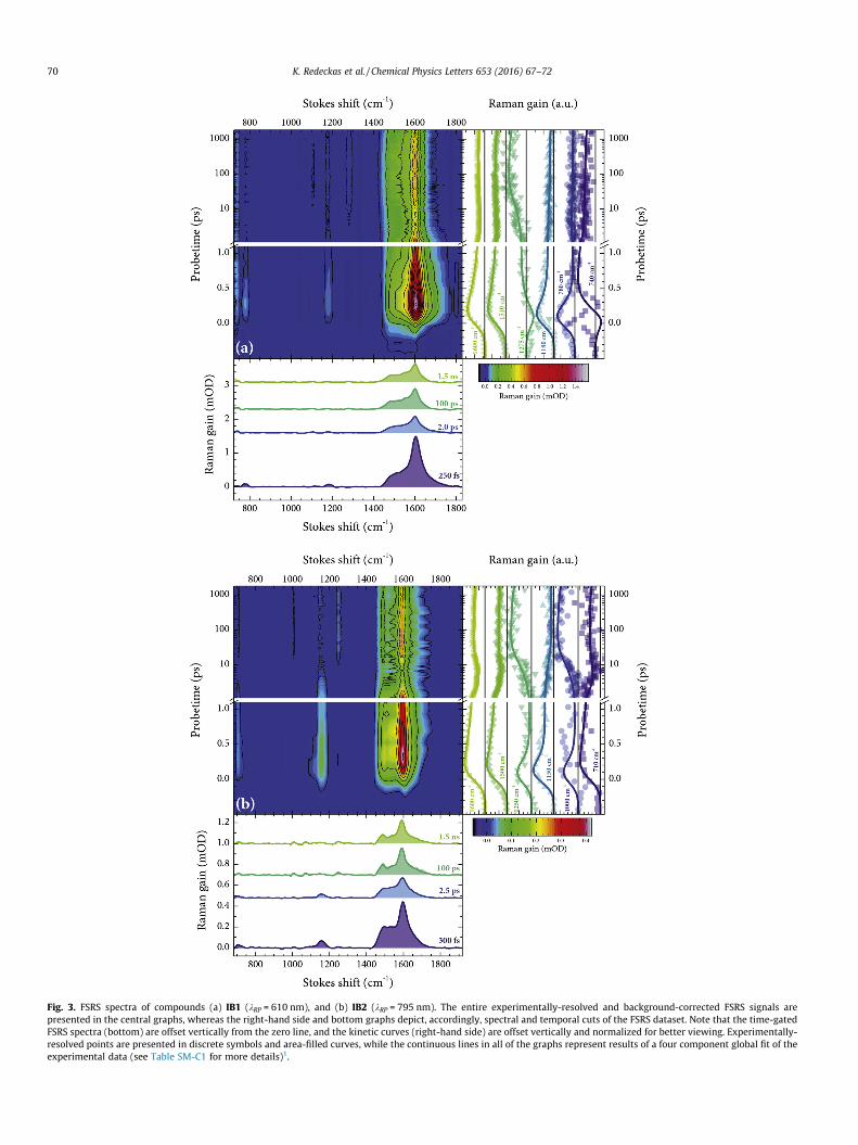
Fig. 3. FSRS spectra of compounds (a) IB1 (kRP = 610 nm), and (b) IB2 (kRP = 795 nm). The entire experimentally-resolved and background-corrected FSRS signals arepresented in the central graphs, whereas the right-hand side and bottom graphs depict, accordingly, spectral and temporal cuts of the FSRS dataset. Note that the time-gatedFSRS spectra (bottom) are offset vertically from the zero line, and the kinetic curves (right-hand side) are offset vertically and normalized for better viewing. Experimentally-resolved points are presented in discrete symbols and area-filled curves, while the continuous lines in all of the graphs represent results of a four component global fit of theexperimental data (see Table SM-C1 for more details)1.
70 K. Redeckas et al. / Chemical Physics Letters 653 (2016) 67–72

K. Redeckas et al. / Chemical Physics Letters 653 (2016) 67–72 71
due to the relatively low Raman yield of the IB samples, as well asthe significant deviation of both 610 and 795 nm Raman pumpsfrom the ground state resonances (>15,000 cm�1), bleaching contri-butions do not surface in either of the time-gated FSRS spectra. Thisobservation, along with the fact that the pre-actinic pump interac-tion FSRS signals are zero, allows us to designate all the Raman gainsignals in Fig. 3 entirely to the excited and/or photoproduct states[6,8] of the investigated IBs. The distinguishing broad Raman peakemerges at 1606 cm�1 for IB1 and at 1595 cm�1 for IB2, and isadjoined by a cluster of near-lying lower-frequency vibrationalbands at ca. 1400–1500 cm�1. At least two clear-cut Ramanmaxima on the red wing of the main peak—at 1490 and1540 cm�1—can be discerned for IB2, whereas the correspondingmaxima of IB1 are slightly more dispersed spectrally, indicating apotential contribution of hot luminescence [14,37], instigated bythe repopulation of higher excited states of the compound [8]. Fem-tosecond time-resolved studies have previously shown that IB-typecompounds exhibit a sub-picosecond decay and consequent sub-nanosecond growth of the transient absorption signal [6,9]. Asobserved in Fig. 3, the temporal behavior of the IB1 and IB2 FSRSbands in the spectral vicinity of �1600 cm�1 (and, to an extent, atthe lower-frequency shoulder at �1500 cm�1) clearly mimics thefamiliar rise-fall–rise-fall dynamics of the transient electronicabsorption signals [6,8,9] and is well described by the same kineticmodel (Table SM-C1). Moreover, spectral evolution during the sub-50-picosecond period distinctly shows the peak-shift to lower fre-quencies (by �7 cm�1) and band-narrowing phases. These FSRSdynamics, similarly to those observed in other molecules (e.g., invarious carotenoids [38–40]), can be attributed to the vibrationalrelaxation, which, in our case, accompanies the formation of thephotoproduct (also suggested in Ref. [6]). The ratios between theinitial (�300 fs) and the final (�100 ps) spectral amplitudes at1600 cm�1 slightly differ from the ones of the ‘‘pure” electronicsignals [6,8] (also a discrepancy between the intermediate statelifetimes (see Table SM-C1) and the ones from [6] can be acknowl-edged), which can be explained by the fact that the Raman pumpscan likewise irreversibly relocate the excited state/photoproductpopulation, thereby, partially altering the ‘‘standard” photoevolu-tion [8] (also observed in other multi-pulse experiments [41,42]).Some of the more notable vibrational modes, accompanying theexcited-state-to-photoproduct transition, are observed in the spec-tral region of ca. 750–1200 cm�1. The vibrational frequencies at ca.780 and 1180 cm�1 (IB1) and ca. 1000 and 1250 cm�1 (IB2) can beassociated with the initial Frank–Condon and the singlet excitedstates, as suggested in [6], seeming as they rapidly decay withinthe first picosecond of the photoevolution. Likewise, the mostprominent vibrational modes of the final forms can be identifiedat ca. 1275 cm�1 (IB1), and 1000 and 1250 cm�1 (IB2), acknowledg-ing their growth in amplitude (and the escalation above their initialamplitudes) for the first 100 ps, which coincides with the formationof the final evolutionary forms [6]. On the whole, the majority ofthese Raman signals, as indicated by bottom graphs of Fig. 3, arealmost an order of magnitude less intense than the main1600 cm�1 maxima. It should also be mentioned that no clear-cutspectro-temporal activity is observed in the spectral vicinity ofthe main ANO2 band at �1330 cm�1. The post-excitation behavior(i.e., bleach recovery and peak-shift) of the nitro functionalgroup was shown to accompany the ring-opening dynamics of therelated photochromic nitro-spiropyrans [43,44], and, bearing inmind the steady state vibrational changes of 4-nitrophenol[34,35], a similar effect can also be expected upon the ring-opening of IBs. Nonetheless, the data in this particular spectralregion is less ‘‘reliable”, as it lies in the close proximity of thecomparatively strong 1375 cm�1 CAH deformation frequency ofacetonitrile and is more susceptible to the solvent line subtractionartifacts [17,19].
Comparing all the presented data, one can notice an inherentdissimilarity between the chemically and optically generated IBforms. While the contrast between the steady-state and transientelectronic absorption of the chemically- and photo-generated spe-cies has been addressed previously (Refs. [5,6,10] and Fig. 1(a)), theSRS data in Fig. 3 indicate that the particular molecular forms alsobear very little vibrational semblance. This implies that althoughthe general vibronic features of the chemically-induced formscan be ascribed to 4-nitrophenolate [34,35], the same assumptioncannot be explicitly made about the forms produced via a UV pho-ton absorption. Essentially, none of the characteristic low-to-mid-frequency (<1400 cm�1) components correlate between the SRS/FSRS spectra of the two (expectedly equivalent) molecular forms.Principally, all time-resolved FSRS spectra in Fig. 3 peak in the closeproximity of the characteristic higher-frequency ground state max-ima (Fig. 1(b)). Moreover, this tendency is exhibited not only bycompounds IB1 and IB2, but also by many other para-substitutedIBs, including the ones investigated in Ref. [5] (see Fig. SM-B1).Acknowledging the key contribution of phenyl-indole inter-ringvibrations to the ground state Raman spectra (Fig. 1(b)), it is safeto assume that the signals at ca. 1600 cm�1, emerging in thepost-excitation FSRS dynamics, also originate from vibrations ofthe same molecular moiety. Since these biphenyl-like vibrationsare not bleached after the optical excitation (as the FSRS signal inFig. 3 is always positive), the temporal development of the1600 cm�1 region can be interpreted as an intensification of molec-ular vibrations (that stems from the possible electronic excitationre-localization) within the phenyl-indolic moiety. The signals thatcould be potentially linked to oxazine ring opening (i.e., the sub-100-picosecond growth at �1250 cm�1 in Fig. 3) are much weakerthan the ones assumedly associated with the phenyl-bearing frag-ments. These observations agree well with the earlier predictionsthat the UV excitation does not, in fact, cause a CAO bond breakageor (spectrally-resolvable) 4-nitrophenolate formation in photoex-cited IB systems (especially in an acetonitrile environment [10]).It has been suggested that intersystem crossing competes with ringopening in both the substituted and unsubstituted IBs [10]. Whilethe presented FSRS data supports the notion that ring-opening isnot a likely outcome from UV excitation, the overwhelmingbiphenyl-like nature of the ground- and excited-state FSRS dynam-ics of the phenyl-substituted IBs allows us to propose yet anotherpossible photoevolutionary route. The transient electronic absorp-tion spectra in Fig. 1(a) bear a striking semblance to the radicalforms of biphenyl and its various derivatives [29–31,45,46](obtained both via optical excitation [29,30] and pulse radiolysis[31,45] experiments). Most specifically, both electronic species dis-play a presence of two characteristic spectral bands – a sharp NUV/VIS peak and a broad NIR maximum. Moreover, the transientRaman spectra [30,47,48] of the said radicals also share a great dealof likeness to the ones depicted in Fig. 3 (i.e., an increased Ramanactivity at m > 1500 cm�1). Acknowledging the earlier observations,concerning the dependence of the sub-nanosecond dynamics onthe UV excitation wavelength [6] and the susceptibility of thesub-microsecond relaxation rate to molecular oxygen [10,11], itis possible to predict that the photoexcited phenyl-substituted IBsystems develop a biphenyl radical-like (ionic) state (or, possibly,a charge-separated form, in which the electron traverses fromone of the chromophoric segment to the other) that predominatesthroughout the entire photoevolution. The radical formation, mostlikely, develops not from the excited singlet state, familiar to mostIBs [6,8]. The unsubstituted IB0 has also been found to decay fasterin the presence of oxygen [10], which suggests that the photo-pathway of the substituted systems is, presumably, singlet? tri-plet? radical. The proposed radical nature, all things considered,would elucidate why the basic photophysical properties of thephenyl-substituted IBs—the noticeably higher yield [5,6], the exci-

72 K. Redeckas et al. / Chemical Physics Letters 653 (2016) 67–72
tation saturation dynamics [5], the atypical multi-peak transientabsorption spectra [5,6], and the strong contrast to 4-nitrophenolate [34,35]—are drastically different from numerousclose counterparts of the IB family [1–7,9–13].
4. Conclusions
We have performed an experimental FSRS study on thechemically-induced and the optically-generated molecular formsof phenyl-substituted IBs. The vibrational (as well as the electronic[5,6,8,10]) spectra of the ‘‘factual” ring-opened molecules show aninherent similarity to 4-nitrophenolate [1–3,34,35], whereas theUV excitation generated species exhibit a singular dominant vibra-tional peak at ca. 1600 cm�1. The latter results suggest anincreased vibrational activity within the phenyl-indolic (and notthe nitrophenolic) moiety, thus indicating that the substituentsheavily alter the vibronic photodynamics of IB systems. The FSRSdata support the earlier assumptions [10,11] that the ring-opening competes with an auxiliary electronic process. Moreover,the peculiar FSRS activity within the indolic fragment of themolecule allows us to estimate that the UV-generated excited statespecies might actually be not of a triplet (as suggested in [10,11]),but of a ionic radical-like [30,46,47] character.
Acknowledgements
The authors would like to thank prof. Valdas Šablinskas fromDepartment of General Physics and Spectroscopy (Vilnius Univer-sity) and prof. Gediminas Niaura from Department of OrganicChemistry (Center for Physical Sciences and Technology) for thesteady state spontaneous Raman scattering measurements.
Appendix A. Supplementary material
Supplementary data associated with this article can be found, inthe online version, at http://dx.doi.org/10.1016/j.cplett.2016.04.030.
References
[1] M. Tomasulo, S. Sortino, F.M. Raymo, J. Photochem. Photobiol. A 200 (2008) 44.[2] M. Tomasulo, S. Sortino, F.M. Raymo, J. Org. Chem. 73 (2008) 118.[3] M. Tomasulo, S. Sortino, A.J.P. White, F.M. Raymo, J. Org. Chem. 70 (2005) 8180.[4] E. Deniz, M. Tomasulo, S. Sortino, F.M. Raymo, J. Phys. Chem. C 113 (2009)
8491.[5] V. Voiciuk, K. Redeckas, V. Martynaitis, R. Steponavici�ute, A. Šackus, M. Vengris,
J. Photochem. Photobiol. A 278 (2014) 60.[6] K. Redeckas, V. Voiciuk, R. Steponavici�ute, V. Martynaitis, A. Šackus, M. Vengris,
J. Photochem. Photobiol. A 285 (2014) 7.[7] M. Tomasulo, E. Deniz, T. Benelli, S. Sortino, F.M. Raymo, Adv. Funct. Mater. 19
(2009) 3956.[8] K. Redeckas, V. Voiciuk, R. Steponavici�ute, V. Martynaitis, A. Šackus, M. Vengris,
J. Phys. Chem. A 118 (2014) 5642.[9] M. Barkauskas, V. Martynaitis, A. Šackus, R. Rotomskis, V. Sirutkaitis, M.
Vengris, Lith. J. Phys. 48 (2008) 231.
[10] V. Voiciuk, K. Redeckas, V. Martynaitis, R. Steponaviciute, A. Sackus, M.Vengris, PCCP 17 (2015) 17828.
[11] F.M. Raymo, J. Phys. Chem. A 116 (2012) 11888.[12] M. Tomasulo, S. Sortino, F.M. Raymo, Adv. Mater. 20 (2008) 832.[13] M. Tomasulo, S. Sortino, A.J.P. White, F.M. Raymo, J. Org. Chem. 71 (2006) 744.[14] S.-Y. Lee, D. Zhang, D.W. McCamant, P. Kukura, R.A. Mathies, J. Chem. Phys. 121
(2004) 3632.[15] P. Kukura, D.W. McCamant, R.A. Mathies, Annu. Rev. Phys. Chem. 58 (2007)
461.[16] D.W. McCamant, P. Kukura, R.A. Mathies, Appl. Spectrosc. 57 (2003) 1317.[17] D.W. McCamant, P. Kukura, S. Yoon, R.A. Mathies, Rev. Sci. Instrum. 75 (2004)
4971.[18] K. Redeckas, V. Voiciuk, M. Vengris, Photosynth. Res. 128 (2016) 169.[19] D.W. McCamant, P. Kukura, R.A. Mathies, J. Phys. Chem. A 107 (2003) 8208.[20] R.G. Parr, Density-Functional Theory of Atoms and Molecules, Oxford
University Press, USA, 1989.[21] P.J. Stephens, F.J. Devlin, C.F. Chabalowski, M.J. Frisch, J. Phys. Chem. 98 (1994)
11623.[22] R.A. Kendall, T.H. Dunning, R.J. Harrison, J. Chem. Phys. 96 (1992) 6796.[23] V. Barone, J. Chem. Phys. 122 (2005) 014108.[24] M.J. Frisch, G.W. Trucks, H.B. Schlegel, G.E. Scuseria, M.A. Robb, J.R. Cheeseman,
G. Scalmani, V. Barone, B. Mennucci, G.A. Petersson, H. Nakatsuji, M. Caricato,X. Li, H.P. Hratchian, A.F. Izmaylov, J. Bloino, G. Zheng, J.L. Sonnenberg, M.Hada, M. Ehara, K. Toyota, R. Fukuda, J. Hasegawa, M. Ishida, T. Nakajima, Y.Honda, O. Kitao, H. Nakai, T. Vreven, J.A. Montgomery Jr., J.E. Peralta, F. Ogliaro,M.J. Bearpark, J. Heyd, E.N. Brothers, K.N. Kudin, V.N. Staroverov, R. Kobayashi,J. Normand, K. Raghavachari, A.P. Rendell, J.C. Burant, S.S. Iyengar, J. Tomasi, M.Cossi, N. Rega, N.J. Millam, M. Klene, J.E. Knox, J.B. Cross, V. Bakken, C. Adamo, J.Jaramillo, R. Gomperts, R.E. Stratmann, O. Yazyev, A.J. Austin, R. Cammi, C.Pomelli, J.W. Ochterski, R.L. Martin, K. Morokuma, V.G. Zakrzewski, G.A. Voth,P. Salvador, J.J. Dannenberg, S. Dapprich, A.D. Daniels, Ö. Farkas, J.B. Foresman,J.V. Ortiz, J. Cioslowski, D.J. Fox, Gaussian 09, Revision D.01. Gaussian Inc,Wallingford, CT, USA, 2013.
[25] D. Michalska, R. Wysokinski, Chem. Phys. Lett. 403 (2005) 211.[26] D. Lin-Vien, N.B. Colthup, W.G. Fateley, J.G. Grasselli, The Handbook of Infrared
and Raman Characteristic Frequencies of Organic Molecules, Academic Press,San Diego, USA, 1991.
[27] A. Kovács, V. Izvekov, G. Keresztury, G. Pongor, Chem. Phys. 238 (1998) 231.[28] P. Sett, S. Chattopadhyay, P.K. Mallick, Chem. Phys. Lett. 331 (2000) 215.[29] C. Kato, H.-O. Hamaguchi, M. Tasumi, Chem. Phys. Lett. 120 (1985) 183.[30] G. Buntinx, O. Poizat, J. Chem. Phys. 91 (1989) 2153.[31] J. Choi, D.W. Cho, S. Tojo, M. Fujitsuka, T. Majima, J. Phys. Chem. A 119 (2015)
851.[32] M.W. Allen, J.R. Unruh, B.D. Slaughter, S.J. Pyszczynski, T.R. Hellwig, T.J.
Kamerzell, C.K. Johnson, J. Phys. Chem. A 107 (2003) 5660.[33] L. Serrano-Andrés, B.O. Roos, JACS 118 (1996) 185.[34] R.A. Ando, A.C. Borin, P.S. Santos, J. Phys. Chem. A 111 (2007) 7194.[35] M. Muniz-Miranda, Appl. Catal. B 146 (2014) 147.[36] S.D. Dieng, J.P.M. Schelvis, J. Phys. Chem. A 114 (2010) 10897.[37] B. Zhao, K. Niu, X. Li, S.-Y. Lee, Sci. China Chem. 54 (2011) 1989.[38] S. Shim, R.A. Mathies, J. Phys. Chem. B 112 (2008) 4826.[39] T.M. Kardas, B. Ratajska-Gadomska, A. Lapini, E. Ragnoni, R. Righini, M. Di
Donato, P. Foggi, W. Gadomski, J. Chem. Phys. 140 (2014) 204312.[40] M. Kloz, R.v. Grondelle, J.T.M. Kennis, PCCP 13 (2011) 18123.[41] A. Weigel, A. Dobryakov, B. Klaumünzer, M. Sajadi, P. Saalfrank, N.P. Ernsting, J.
Phys. Chem. B 115 (2011) 3656.[42] S. Ruetzel, M. Kullmann, J. Buback, P. Nuernberger, T. Brixner, Phys. Rev. Lett.
110 (2013) 148305.[43] J. Buback, M. Kullmann, F. Langhojer, P. Nuernberger, R. Schmidt, F. Würthner,
T. Brixner, JACS 132 (2010) 16510.[44] A.-K. Holm, M. Rini, E.T.J. Nibbering, H. Fidder, Chem. Phys. Lett. 376 (2003)
214.[45] K. Sehested, E.J. Hart, J. Phys. Chem. 79 (1975) 1639.[46] C. Takahashi, S. Maeda, Chem. Phys. Lett. 24 (1974) 584.[47] T. Nakabayashi, S. Kamo, H. Sakuragi, N. Nishi, J. Phys. Chem. A 105 (2001)
8605.[48] Y. Sasaki, H.-O. Hamaguchi, Spectrochim. Acta Mol. Biomol. Spectrosc. 50
(1994) 1475.