chemopreventiveandantioxidantactivityby …josorge.com/publications/citations/cca01/030.pdf · leaf...
TRANSCRIPT
RESEARCH ARTICLE
Chemopreventive and antioxidant activity by Smilax zeylanicaleaf extract against N-nitrosodiethylamine inducedhepatocarcinogenesis in wistar albino rats
Venugopalan Rajesh & Perumal Perumal
Received: 8 April 2013 /Accepted: 28 May 2013# Institute of Korean Medicine, Kyung Hee University 2013
Abstract The chemopreventive potential of methanol extractof Smilax zeylanica leaves against N-nitrosodiethylamine(NDEA) induced hepatocarcinogenesis was investigated inwistar albino male rats. The animals were divided into sixgroups. Group I served as normal control group, group II andgroup III served as extract control received Smilax zeylanicaleaf extract 200 mg/kg and 400 mg/kg respectively once dailyorally.A single intraperitoneal injection ofNDEA (200mg/kg)to group IV, group V and group VI animals followed by asingle dose of carbon tetrachloride (CCl4, 2 ml/kg) 2 weekslater induced hepatocarcinogenesis in rats at the end of exper-imental period (16 weeks). Starting 1 week prior to NDEAadministration, group Vand group VI animals received meth-anol extract of Smilax zeylanica 200 mg/kg and 400 mg/kgrespectively once daily orally for 16 weeks. At the end of theexperimental period, the body weight, morphology of liver,relative liver weight, the levels of liver injury and liver cancermarkers, the levels of lipid peroxidation and the activities ofenzymatic and non-enzymatic antioxidants in liver tissue wereassessed. Group IV NDEA induced hepatocarcinoma animalsshowed bodyweight loss with a significant increase in relativeliver weight. The levels of liver injury and liver cancermarkers such as alanine transaminase (AST), aspartate trans-aminase (AST), alkaline phosphatase (ALP), gamma-glutamyl transferase (GGT), α-feto protein (AFT) andcarcinoembryonic antigen (CEA) were substantially increasedin group IV untreated animals. Moreover, with reference tolipid peroxidation and antioxidant system, group IV animalsshowed elevated levels of lipid peroxidation with a subse-quent decrease in activities of antioxidant enzymes viz.,
superoxide dismutase (SOD), catalase (CAT), glutathione per-oxidase (GSH-Px), glutathione reductase (GR), glutathione-S-transferase (GST), glutathione (GSH), vitamin- C andvitamin-E in liver tissue. In contrast, the NDEA inducedanimals in groupVand group VI treated with Smilax zeylanicaleaf extract showed a significant improvement in body weightgain at the end of experimental period (16 weeks). A signif-icant decrease in relative liver weight with reduced levels ofliver injury and liver cancer markers in serum were observedin treatment groups. Moreover, treatment with Smilaxzeylanica leaf extract decreased the extent of lipid peroxida-tion with concomitant increase in activities of enzymatic andnon-enzymatic antioxidants when compared with NDEA in-duced untreated group IV animals. The histological observa-tions of liver tissue too correlated the biochemical observa-tions. Furthermore, the extract did not produce any deleteriouseffects in extract alone treated groups. These findings suggestthat, the potential chemoprevention of methanol extract ofSmilax zeylanica leaves might be due to stabilization andincrease in all the components of antioxidant system attributedto antioxidant and free radical scavenging activity.
Keywords Smilax zeylanica leaves .N-nitrosodiethylamine .
Hepatocarcinogenesis . Tumour markers . Lipidperoxidation . Antioxidant activity
Introduction
Liver cancer, especially hepatocellular carcinoma (HCC), isthe fifth most common cancer and the third foremost cause ofcancer associated death globally (Befeler and Di Bisceglie2002; Bishayee and Dhir 2009). Hepatocellular carcinomacan be secondary to hepatitis B or C, cirrhosis due to alcoholconsumption, liver disease due to aflatoxin toxicity, hormon-al imbalance and certain metabolic diseases (Thorgeirssonand Grisham 2002). Hepatocarcinogenesis involves initial
V. Rajesh (*) : P. PerumalDepartment of Pharmacology, JKK Nattraja College of Pharmacy,Natarajapuram, Komarapalayam,638183, Namakkal District, Tamil Nadu, Indiae-mail: [email protected]
P. Perumale-mail: [email protected]
Orient Pharm Exp MedDOI 10.1007/s13596-013-0125-3
genotoxic insult (initiation), clonal expansion from prema-lignant to malignant lesions (promotion) and finally tumorprogression by further clonal expansion (Thorgeirsson andGrisham 2002; Halsted et al. 1996).
N-Nitrosodiethylamine (NDEA) a potent hepatocarcinogen,is known to cause perturbations in the nuclear enzymes in-volved in DNA repair/replication (Bhosale et al. 2002) and isnormally used as a carcinogen to induce liver cancer in animalmodels. Investigations have provided evidence that N-nitrosamines cause wide range of tumours in all animal speciesand these compounds are considered to be effective healthhazards to man (Loeppky 1994). The main cause for concernis that diethylnitrosamine is found in a wide variety of foodslike cheese, soybean, smoked, salted and dried fish, cured meatand alcoholic beverages (Liao et al. 2001). Metabolism ofcertain therapeutic drugs is also reported to producediethylnitrosamine (Akintonwa 1985). It is also found in to-bacco smoke at a concentration ranging from 1 to 28-ng/cigarette and in baby bottle nipples at a level of 10 ppb(IARC 1972). It has been reported that, onmetabolic activation,it produces the pro-mutagenic products, O6-ethyl deoxy gua-nosine and O4 and O6- ethyl deoxy thymidine in liver whichare responsible for its carcinogenic effects (Verna et al.1996). It is also reported that the generation of reactiveoxygen species (ROS) by NDEA causes carcinogeniceffects. Oxidative stress caused by ROS has been re-ported in membrane lipid peroxidation, DNA damageand mutagenesis associated with various stages of tumorformation process (Parola and Robino 2001). Human liverappears to metabolize nitrosamines in a manner similar to thatof rodent liver and also exhibits considerable similarities withregard to morphology, genomic alterations and gene expres-sion, despite their different disease etiologies (Feo et al. 2000).Hence the model of NDEA-induced HCC is considered as oneof the most accepted and widely used experimental models tostudy hepatocarcinogenesis (Hai et al. 2001).
Chemoprevention offers a novel approach to control theincidence of liver cancer (Wattenberg and Estensen 1996). Alarge number of chemotherapeutic agents have been identi-fied in epidemiological and experimental studies, preclinicaland clinical observations (Takuji et al. 1993; Jagadeeswaranet al. 2000). However, the toxic effects produced by some ofthe agents have limited their extensive use. There is, there-fore, a need to identify synthetic or natural compound thathave significant chemopreventive potential to block initia-tion or arrest the progression in premalignant cells withoutundesirable side effects.
In recent years, there has been considerable interest innatural products with antioxidant property to protect cellularcomponents from oxidative damage and prevent diseases.Over 50 % of anticancer drugs approved by United StatesFood and Drug Administration since 1960 were originatedfrom terrestrial plants (Kim and Park 2002; Mann 2002).
Several constituents present naturally in medicinal plants havebeen shown to modify critical reactions that cause inhibitionof chemically induced hepatocarcinogenesis (Soni et al. 1997;Bhattracharya and Chatterjee 1998) and this worldwide effortcontinues to discover new anticancer drugs from plants.Among the herbal resources is Smilax zeylanica Linn. belongsto family Smilacaceae. The genus Smilax Linn. consist ofabout 350 species in the world (Shao et al. 2007), out of which24 species are found in India (Saldhana and Wicolson 1976;Ramaswamy et al. 2001; Santapau and Henry 1976). In southIndia, the genus is represented by 4 species viz., Smilaxzeylanica Linn., Smilax aspera Linn., Smilax perfoliataRoxb. and Smilax wightii A. DC (Gamble 2004). Many ofthem have been long used as medicinal herbs, especially astraditional Chinese medicines in China (Abdala et al. 2008).The extracts of Smilax china L. tubers are known to showantitumor and antioxidant activities on mice and rats (Wanget al. 1996). Smilax zeylanica Linn. is distributed in tropicaland subtropical hills of Asia between an altitude of 500–1,800 m. Smilax zeylanica leaves were extensively used intraditional system of medicine against veneral diseases(Oommachan and Masih 1991), skin disorders, sores, swell-ings, abscess (Ambasta 2006; Nadkarni 1976) and also ap-plied for rheumatism and pain in lower extremities (Kirtikarand Basu 1991). Based on the widespread use of Smilaxspecies in traditional medicine and the traditional importanceof Smilax zeylanica Linn it was decided to select the plant forinvestigation. As previous studies mainly focused on the plantrhizomes, and there was little information about the Smilaxzeylanica leaves. In the present study, an effort was made toascertain the possible role of Smilax zeylanica Linn. leaves inchemically induced hepatocarcinogenesis.
Materials and methods
Chemicals
Chemicals used in this study including NDEA, carbon tetra-chloride (CCl4), Oxidized and reduced glutathione (GSSG andGSH), hydrogen peroxide (H2O2), dithionitrobenzene (DTNB),Sodium dodecyl sulphate (SDS), Thiobarbituric acid (TBA), 1-chloro-2, 4-dinitrobenzene (CDNB), dinitrophenyl hydrazine(DNPH), nitro blue tetrazolium (NBT), reduced nicotinamideadenine dinucleotide (NADPH), ethylenediamine tetra aceticacid (EDTA), phenyl methyl sulfonyl fluoride (PMSF) wereprocured from sigma chemicals, St. Louis, USA. Enzymeimmunoassay kit for estimation of tumor markers α- fetoprotein (AFP) and carcinoembryonic antigen (CEA) were pur-chased from UBI MAGIWELL (USA) and all other chemicalsincluding petroleum ether (60 – 80 °C), methanol (HPLC) werepurchased from SD Fine Chemicals Ltd., Mumbai, India withhighest purity grade.
V. Rajesh, P. Perumal
Plant material
The fresh leaves of Smilax zeylanica Linn were collected inthe month of September from yercaud hills. Yercaud is a hillstation in Salem District, in Tamil Nadu, India. It located inthe Shevaroys range of hills in the Eastern Ghats. It issituated at an altitude of 1,515 m (4,970 ft) above sea level(Geoposition: Latitude- 11.781997 (11° 46′ 55.19″ N),Longitude- 78.209782 (78° 12′ 35.22″ E). The plant materialwas taxonomically indentified, confirmed and authenticatedby Botanical Survery of India, Coimbatore, Tamilnadu(BSI/SRC/5/23/2011-12/Tech-1256) and the voucher speci-men was retained in our laboratory for further reference. Thecollected leaves were shade dried and the dried materialswere crushed to coarse powder with mechanical grinder. Thepowder was stored in an airtight container for extraction.
Extraction
The dried coarse powder was defatted with petroleum ether(60 – 80 °C) in a soxhlet extractor in order to remove fattysubstances. The defatted marc was dried and it was subjectedto extraction with methanol using Soxhlet apparatus for 72 h.After completion of extraction, methanol was removed bydistillation. The residue obtained was air dried. The driedmethanol extract was stored in air tight glass container forfurther investigation. The percentage yield obtained was22.03 % w/w.
Phytochemical screening
The extract obtained was subjected to preliminary phyto-chemical screening (Khandelwal and Kokate 1995).
Acute oral toxicity study of Smilax zeylanica leaf extract
Healthy, young adult albino wistar rats (150–200 g) of fe-male sex were used for the study. The animals were obtainedfrom Agricultural University, Mannuthy, Thrissur, kerala(328/99/CPCSEA) and were housed in polypropylene cages.The animals were maintained under standard laboratory con-ditions (25° ± 2 °C; 12 h light and dark cycle). The animalswere fed with standard diet and water ad libitum. Ethicalclearance (for handling of animals and the procedures used instudy) was obtained from the Institutional Animal EthicalCommittee (887/ac/05/CPCSEA) before performing thestudy on animals.
Experimental design
Acute oral toxicity study of Smilax zeylanica leaf extract wascarried out as per Organization for Economic Cooperationand Development (OECD) guideline 425 (Up and Down
procedure). Limit test was performed to determine the safetyof extract. Five female wistar rats were used for the study.Animals were fasted prior to the dosing (food but not waterwithheld for 3–4 h). Extract 2,000 mg/kg b.wt. suspendedin 0.5 % w/v carboxy methyl cellulose was administeredorally to one animal and observed for mortality. If thefirst animal survives, four additional animals were se-quentially dosed orally so that a total of five animalsare tested. Animals were observed individually at leastonce during the first 30 min after dosing, periodicallyduring the first 24 h (with special attention given duringthe first 4 h), and daily thereafter for a total of 14 days.After the experimental period, the animals were weighedand humanely killed and their vital organs includingheart, lungs, liver, kidneys, spleen, adrenals, sex organsand brain were grossly examined (OECD 2000).
Experimental design and induction of hepatocellularcarcinoma (HCC)
The experimental protocol was approved by InstitutionalAnimal Ethical Committee (887/ac/05/CPCSEA). Healthymale albino wistar rats (6–8 weeks) weighing 130–150 g wereused for the study. Randomization in selection of animals forgrouping was carried out to avoid statistical difference in thebody weight of animals. The experimental animals were di-vided into six groups, each group comprising of six animals.The experimental animals were divided into six groups of sixanimals each according to the following experimental regi-men. Group 1 animals served as normal control rats. Group 2animals received Smilax zeylanica leaf extract 200 mg/kg/day(p.o.) in 0.5 % carboxy methyl cellulose (CMC) for 16 weeks.Group 3 animals received Smilax zeylanica leaf extract400 mg/kg/day (p.o.) in 0.5 % CMC for 16 weeks. Group 4animals received vehicle 0.5 % CMC 2 ml/kg/day (p.o.)1 week prior to NDEA 200 mg/kg single i.p. and 2 weekslater received a single dose of CCl4 (2ml/kg) by gavage as 1:1dilution in corn oil. The oral administration of vehicle wascontinued after NDEA injection throughout the experimentalperiod of 16 weeks. Group 5 animals were given Smilaxzeylanica leaf extract 200 mg/kg/day (p.o.) in 0.5 % CMCstarted 1 week prior to the injection of NDEA and 2 weekslater received a single dose of CCl4 2 ml/kg by gavage as 1:1dilution in corn oil. The oral administration of extractwas continued for 16 weeks after NDEA injection.Animals in group 6 were given Smilax zeylanica leafextract 400 mg/kg/day (p.o.) in 0.5 % CMC started1 week prior to the injection of NDEA and 2 weekslater received a single dose of CCl4 2 ml/kg b.wt. bygavage as 1:1 dilution in corn oil. The oral administra-tion of extract was continued for 16 weeks after NDEAinjection. Body weight was recorded at the end of every weekfor 16 weeks.
Chemopreventive and antioxidant activity
Sample collection
At the end of the experimental period (16 weeks), bloodsamples were collected from retro-orbital plexus under an-aesthesia in EDTA tubes and centrifuged at 2,200 g for15 min at 4 °C. Plasma samples were stored at−20 °C forbiochemical analysis.
Biochemical analysis
Serum transaminases (AST and ALT) were determined by themethod of Reitman and Frankel (1957). The activity of serumalkaline phosphatase (ALP) was estimated by the method ofKind and King (1954). Serum bilirubin (SB) and total protein(TB) were estimated by the methods of Malloy and Evelyn(1937) andWooten (1964) respectively. Gamma glutamyl trans-ferase (GGT) activity was determined according to the methodof Szas (1976). Quantitative estimation of tumor markers α-fetoprotein (AFP) and carcinoembryonic antigen (CEA) were basedon solid phase enzyme linked immunosorbent assay (ELISA)using the UBI MAGIWELL (USA) enzyme immunoassay kit(Sell and Beckar 1978; Macnab et al. 1978).
After blood collection animals were sacrificed by cervicaldecapitation and the liver was excised, washed in ice coldsaline, blotted to dryness and examined for any deductablechanges. A portion of liver tissue was then homogenized forbiochemical assays.
Preparation of tissue homogenate
The tissues were weighed and 10 % tissue homogenate wasprepared with 0.025 M Tris–HCl buffer, pH 7.5. After cen-trifugation at 10,000×g for 10 min, the clear supernatant wasused to measure thiobarbituric acid reactive substances(TBARS). For the determinations of vitamin E level, thetissues were weighed and lipids were extracted from tissuesby the method of Folch et al. (1957) using chloroform–methanol mixture (CHCl3:CH3OH) (2:1; v/v). The lipidfraction was used for the estimation of vitamin E. For theestimation of non-enzymatic and enzymatic antioxidants,tissues were minced and homogenized (10 % w/v) in 0.1 Mphosphate buffer (pH 7.0) and centrifuged for 10 min and theresulting supernatant was used for enzyme assays.
Estimation of lipid peroxidation (LPO)
The levels of thiobarbituric acid reactive substances(TBARS) in the liver was measured by the method ofOhkawa et al. (1979) as a marker for lipid peroxidation. Analiquot of liver homogenates was mixed with 0.2 mL of8.1 % Sodium dodecyl sulphate (SDS), 1.5 mL of 20 %acetic acid and 1.5 mL of 0.8 % Thiobarbituric acid (TBA),then the volume was adjusted to 4.0 mL with distilled water.
After boiled at 95o for 60 min, the reaction solution wasextracted with 1.0 mL of distilled water and 5.0 mL of n-butanol and pyridine (15:1 v/v). The absorbance at 532 nmof the organic layer was determined after centrifugation.
Estimation of superoxide dismutase (SOD) activity
0.5 ml of tissue homogenate was diluted with 1 ml of water.In this mixture, 2.5 ml of ethanol and 1.5 ml of chloroform(all reagents chilled) were added and shaken for 1 min at 4 °Cthen centrifuged. The enzyme activity in the supernatant wasdetermined. The assay mixture contained 1.2 ml of sodiumpyrophosphate buffer (0.025 M, pH 8.3), 0.1 ml of 186 μMphenyl methane sulphate (PMS), 0.3 ml of 30 μM nitro bluetetrazolium (NBT), 0.2 ml of 780 μM nicotinamide adeninedinucleotide reduced (NADH), appropriately diluted en-zyme preparation and water in a total volume of 3 ml.Reaction was started by the addition of nicotinamide adeninedinucleotide reduced (NADH). After incubation at 30 °C for90 s the reaction was stopped by the addition of 1 ml glacialacetic acid. The reaction mixture was stirred vigorously andshaken with 4 ml of n-butanol. The intensity of the chromogenin the butanol layer was measured at 560 nm against butanolblank. A system devoid of enzyme served as control. One unitof the enzyme activity is defined as the enzyme reaction, whichgave 50 % inhibition of nitro blue tetrazolium (NBT) reductionin 1 min under the assay conditions (Karkar et al. 1984)
Estimation of catalase activity
The reaction mixture (1.5 ml) contained 1.0 ml of 0.01 Mphosphate buffer (pH 7.0), 0.1 ml of tissue homogenate and0.4 ml of 2 M H2O2. The reaction was stopped by theaddition of 2.0 ml of dichromate-acetic acid reagent (5 %potassium dichromate and glacial acetic acid were mixed in1:3 ratio). Then the absorbance was read at 620 nm; CATactivity was expressed as μM of H2O2 consumed/min/mgprotein (Sinha 1972).
Glutathione peroxidase (GSH-Px)
Glutathione peroxidase activity was measured by the methodof Rotruck et al. (1973). To 0.2 ml of buffer, 0.2 ml ofethylenediaminetetraacetic acid (EDTA), 0.1 ml of sodiumazide and 0.5 ml of tissue homogenate were added. To thatmixture, 0.2 ml of glutathione solution and 0.1 ml of hydro-gen peroxide were added. The contents were mixed well andincubated at 37 °C for 10 min along with the control tubescontaining all the reagents but no enzyme. After 10 min, thereaction was arrested by the addition of 0.4 ml of 10 % TCA.0.2 mL of tissue homogenate was added to the control tubes.The tubes were centrifuged and supernatant was assayed forglutathione content by adding Ellman’s reagent.
V. Rajesh, P. Perumal

Glutathione-S-transferase (GST)
Glutathione-S-transferase activity was measured by themethod of Habig et al. (1974). The reaction mixture contain-ing 1 ml of buffer, 0.1 ml of 1-chloro-2,4-dinitrobenzene(CDNB), 0.1 ml of homogenate and 1.7 ml of distilled waterwas incubated at 37 °C for 5 min. The reaction was thenstarted by the addition of 1 ml of glutathione. The increase inabsorbance was followed for 3 min at 340 nm. The reactionmixture without the enzyme was used as blank.
Glutathione reductase (GR) activity
Glutathione reductase that utilizes Nicotinamide adeninedinucleotide phosphate (NADPH) to convert metabolizedglutathione (GSSG) to the reduced form was assayed bythe method of Horn and Burns (1978).
Reduced glutathione (GSH)
Reduced glutathione was estimated by the method of Ellman(1959). 0.5 ml of tissue homogenate was precipitated with2 ml of 5 % TCA. After centrifugation, 1 ml of supernatantwas taken and added 0.5 mL of Ellman’s reagent (19.8 mg of5,5′ dithio (bis) nitrobenzoic acid in 100 ml of 1 % sodiumcitrate) and 3ml of phosphate buffer. Standards were treated ina similar way and the colour developed was read at 412 nm.
Estimation of ascorbic acid (vitamin C)
0.5 ml of tissue homogenate was mixed thoroughly with1.5 ml of 6 % TCA and centrifuged for 10 min at 3,500 g.After centrifugation, 0.5 ml of the supernatant was mixed with0.5 ml of Dinitrophenylhydrazine (DNPH) reagent andallowed to stand at room temperature for an additional 3 hthen added 2.5 ml of 85 % sulphuric acid and allowed to standfor 30 min. Then the absorbance was read at 530 nm. A set ofstandards containing 10–50 μg of ascorbic acid were takenand processed similarly along with a blank. Ascorbic acidvalues were expressed as μg/mg tissue (Omaye et al. 1979).
Estimation of vitamin E
0.1 ml of lipid extract, 1.5 ml of ethanol and 2 ml ofpetroleum ether were added, mixed and centrifuged for3,000 rpm for 10 min. The supernatant was evaporated todryness at 80 °C then 0.2 ml of 2, 2-1-dipyridyl solution and0.2 ml of ferric chloride solution was added and mixed well.This was kept in dark for 5 min and added 2 ml of butanol.Then the absorbance was read at 520 nm. Standards of α-tocopherol in the range of 10–100 μg were taken and treatedsimilarly along with blank containing only the reagent. Thevalues were expressed as μg/mgtissue (Barker et al. 1951).
Histopathological examination
Representative sections of right, left and caudate lobes ofliver were fixed in 10 % formalin and embedded in moltenparaffin wax and were ultra sectioned (5–6 μm thickness),stained with hematoxylin and eosin and were examinedunder light microscope for histopathological changes.
Statistical analysis
Results were expressed as mean±standard error of mean(SEM). The results were analysed for statistical significanceby one way ANOVA followed by dunnett’s test (GraphpadSoftware Inc,La Jolla, CA. Trial version). The criterion forstatistical significance was set at P<0.05.
Results
Percentage yield of extract
The percentage yield of extract obtained from extraction ofSmilax zeylanica leaves using methanol as solvent was foundto be 22.03 % w/w.
The phytochemical examination of methanol extract ofSmilax zeylanica leaves revealed the presence of carbohy-drates, alkaloids, phenolics, tannins, flavonoids, glycosidesand alkaloids.
Acute oral toxicity study
Acute oral toxicity study was carried out as per OECDguideline 425. From the limit test results it was observedthat, the Smilax zeylanica leaf extract is safe upto a dose levelof 2,000 mg/kg. There was no mortality and the experimentalanimals did not show any toxic effect throughout the obser-vation period of 14 days.
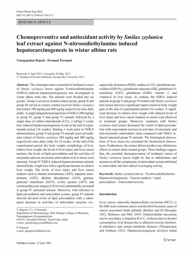
Body weight
Average body weights of different animal groups at variousintervals are shown in Fig. 1. There was no significant differ-ence in final body weight between the control group (230.8±2.0 g) and extract alone treated groups (Extract 200 mg/kg−237.5±2.81 g, Extract 400 mg/kg− 231.7±2.1 g). The finalbody weight of NDEA induced untreated animals (142.5±4.95 g) was significantly less (p<0.001) compared to controlanimals (230.8±2.0 g). The group V and group VI animalswith NDEA induced and treated with extract 200 mg/kg and400 mg/kg showed a significant increase (p<0.001) in finalbody weight (Extract 200 mg/kg− 194.2±9.61 g and Extract400 mg/kg− 210±1.82 g) compared to untreated animals. Itshows the Smilax zeylanica leaf extract prevented the body
Chemopreventive and antioxidant activity

weight loss in NDEA induced animals and maintained thegrowth rate near normal.
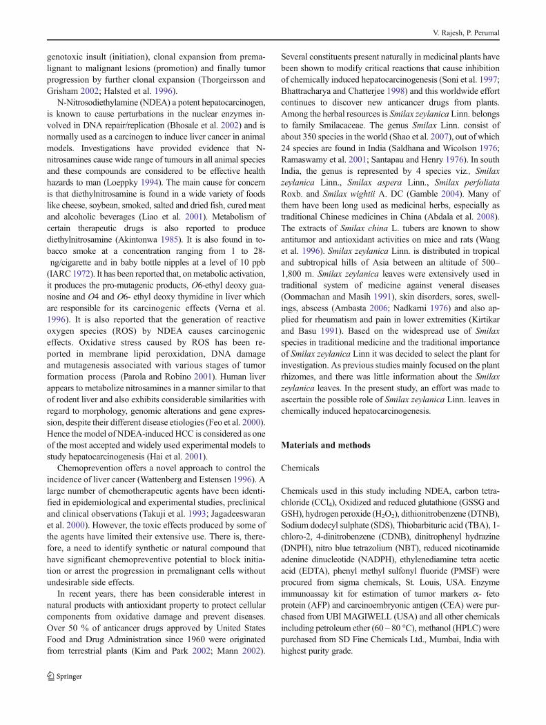
Gross morphology
As depicted in Fig. 2, the appearance of liver in controlgroup rats were normal and there were no macroscopi-cally detectable changes in liver (Fig. 2a). No obviouschanges in liver morphology were observed in extractalone treated group which is indicative of nontoxicnature of Smilax zeylanica leaf extract (Fig. 2b and c).The rats in NDEA group revealed enlarged liver andmultiple lesions (whitish) with almost the entire surfaceof liver is occupied with abnormal growth in posteriorsurface (Fig 2d Posterior surface of liver). In group ofNDEA induced animals treated with Smilax zeylanicaleaf extract 200 mg/kg, compared to NDEA controlthere was a marked reduction in damage caused by NDEAon gross examination of anterior and posterior surface. Therewere no detectable lesions or nodules (Fig 2e). The liver ofrats treated with Smilax zeylanica leaf extract 400 mg/kgappeared similar to normal rat liver. Both anterior and poste-rior surface of liver appeared smooth and there were nodetectable lesions or nodules (Fig 1f).
Relative liver weight
The relative liver weight of control and experimental group ofanimals are shown in Table 1 and the results were expressed asg liver/100 g body wt. There was no statistical difference in
relative liver weight between control (2.15±0.09) and extractalone treated groups (Extract 200 mg/kg− 2.02±0.02, Extract400 mg/kg− 2.03±0.02). The relative liver weight of group IVNDEA induced untreated animals (3.61±0.25) were signifi-cantly increased (p<0.001) compared to Group I control ani-mals. A significant decrease (p<0.001) in relative liver weightwas noted in NDEA induced group V and group VI animalstreated with extract 200 mg/kg (2.60±0.23) and 400 mg/kg(2.45±0.10).
Effect of Smilax zeylanica leaf extract on serum markerenzymes and hepatocarcinogenesis marker
The effect of Smilax zeylanica leaf extract on the activities ofmarker enzymes and serum protein levels in control andexperimental groups are shown in Table 2. A significantincrease (P<0.001) in AST (77.33±3.40), ALT (63.50±2.06), ALP (107.5±4.78) and GGT (116.7±8.02) IU/L wereobserved in NDEA induced untreated animals compared tocontrol animals with AST (36.97±1.97), ALT (33.33±1.33),ALP (46.83±1.47) and GGT (42.00±4.83) IU/L. The ani-mals treated with Smilax zeylanica leaf extract 200 mg/kgand 400 mg/kg showed a significant decrease in AST, ALT,ALP and GGT levels from (38.17±4.28)–(42.00±1.71) (p<0.001), (43.67±3.63)–(37.67±0.91) (p<0.001), (83.3±1.66)–(58±0.89) (p<0.001) and (58.00±8.18)–(42.67±2.49) (p<0.001) respectively compared to NDEA untreatedanimals . The total protein levels were decreased (P<0.001)in NDEA untreated animals (6.28±0.16) compared to con-trol animals (8.43±0.51). However, upon treatment with
0
20
40
60
80
100
120
140
160
180
200
220
240
260
1 2 3 4 5 6 7 8 9 10 11 12 13 14 15 16 17 18
Bo
dy
wei
gh
t (g
)
weeks
Normal E xtract 200 mg/kg Extract 400 mg/kg
NDEA Control NDEA + Extract 200 mg/kg NDEA + Extract 400 mg/kg
Fig. 1 Effect of Smilax zeylanica leaf extract on bodyweight gain duringN-nitrosodiethylamine (NDEA) induced hepatocarcinogenesis in rats. Averagebody weight of different animal groups at various intervals. Results are givenas mean±S.E.M. One -way ANOVA followed by Dunnett’s test was used tocompare experimental groups. There was no significant difference in finalbody weight between the control (normal) group and extract alone treatedgroups (Extract 200 mg/kg and Extract 400 mg/kg) (nsa). NDEA induced
untreated animals (NDEA control) showed a significant decrease in bodyweight (***p<0.001b) compared to control animals. The group Vand groupVI animals with NDEA induced and treated with extract 200 mg/kg(NDEA+Extract 200 mg/kg) and 400 mg/kg (NDEA+Extract 400 mg/kg)showed a significant increase (***p<0.001b) in final body weight comparedto untreated animals (NDEA control)
V. Rajesh, P. Perumal

Table 1 Relative liver weight of various groups of rats at the end of the study (16 weeks)
Groups Relative liver weight (g liver/100 g body wt)
Group I (Control) (0.5 % CMC) 2 ml/kg p.o. 2.15±0.09
Group II (Extract 200 mg/kg p.o.) 2.02±0.02ns,a
Group III (Extract 400 mg/kg p.o.) 2.03±0.02ns,a
Group IV (NDEA alone) 3.61±0.25***,a
Group V (NDEA+Extract 200 mg/kg p.o.) 2.60±0.23***,b
Group VI (NDEA+Extract 400 mg/kg p.o.) 2.45±0.10***,b
All values are expressed as mean±S.E.M, n=6 in each groupa Values are significantly different from control group; ns-non significant; *P<0.05; **P<0.01; ***P<0.001b Values are significantly different from NDEA- induced group; ns-non significant; *P<0.05; **P<0.01; ***P<0.001 (one-way ANOVA followedby Dunnett’s test)
Fig. 2 Gross morphology of liver. a Liver of control animal showingnormal morphology. b and c Liver of animal dosed with extract alone200 mg/kg and 400 mg/kg respectively showing normal appearancewhich is indicates nontoxic nature of extract. d Liver of group IVNDEA induced untreated animals (NDEA control) showing multiplelesions (whitish) with almost the entire posterior surface of liver is
occupied with abnormal growth. e Liver of NDEA induced group Vanimals treated with Smilax zeylanica leaf extract 200 mg/kg showingmarked reduction in damage caused by NDEA. f Liver of rats treatedwith Smilax zeylanica leaf extract 400 mg/kg showing normal morphol-ogy similar to control animals. No detectable lesions or nodules werenoted in anterior and posterior surface of treatment groups
Chemopreventive and antioxidant activity

Smilax zeylanica leaf extract 200 mg/kg and 400 mg/kg theprotein levels were significantly increased from (7.20±0.31)–(8.76±0.56 mg/dl) (p<0.01–p<0.001).
The effect of Smilax zeylanica leaf extract on AFP andCEA levels in serum of control and experimental groups areshown in Table 3. A significant increase in AFP (23.67±2.05) (p<0.001) and CEA (16.32±1.51) (p<0.05) were ob-served in NDEA induced untreated animals compared tocontrol animals with AFP and CEA levels 12.55±1.24 and11.55±1.23 ng/ml respectively. In group of animals treatedwith Smilax zeylanica leaf extract 200 mg/kg and 400 mg/kg,the AFP levels in serum were significantly reduced from16.43±0.85 – 13.87±0.30 (p<0.01–p<0.001) compared toNDEA untreated animals and there was no significant dif-ference in CEA levels (14.75±0.21) in serum of animalstreated with extract 200 mg/kg compared to NDEA untreatedanimals. However, CEA levels were significantly decreased(11.60±0.40) (p<0.01) in animals treated with Smilax
zeylanica leaf extract 400 mg/kg. Non-significant alterationin serum marker enzymes and hepatocarcinogenesis markerin extract alone treated group II and group III animals whencompared with control group I animals indicates the non-toxic nature of Smilax zeylanica leaf extract.
Effect of Smilax zeylanica leaf extract on LPO and levelsof antioxidant enzymes in liver
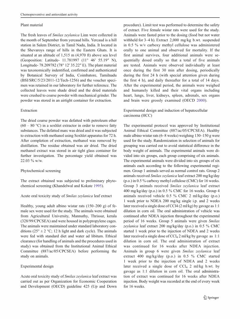
The LPO levels in liver homogenate of control and experi-mental animals are illustrated in Fig. 3a significant increase(p<0.001) in the production of MDA (1.90±0.03) was ob-served in the group IV NDEA intoxicated untreated animalscompared to control animals (1.09±0.08). Administration ofSmilax zeylanica leaf extract showed a significant reductionin LPO as evidenced by a significant fall in MDA levels to1.42±0.18 (p<0.01) and 1.10±0.05 (p<0.001) at doses 200-mg/kg and 400 mg/kg respectively.
Table 2 Effect of Smilax zeylanica leaf extract on biochemical parameters in control and experimental groups of rats
Groups AST (IU/L) ALT (IU/L) ALP (IU/L) TB (mg/dl) TP (mg/dl) GGT (IU/L)
Group I (Control) (0.5 %CMC)2 ml/kg p.o.
36.97±1.97 33.33±1.33 46.83±1.47 0.41±0.04 8.43±0.51 42.00±4.83
Group II (Extract 200 mg/kgp.o.)
46.17±1.42ns,a 35.00±1.41ns,a 53.50±1.92ns,a 0.33±0.04ns,a 8.23±0.21ns,a 37.83±2.41ns,a
Group III (Extract 400 mg/kgp.o.)
46.67±1.90ns,a 39.17±0.74ns,a 57.17±0.90ns,a 0.33±0.03ns,a 8.41±0.15ns,a 32.17±1.32ns,a
Group IV (NDEA alone) 77.33±3.40***,a 63.50±2.06 ***,a 107.5±4.78 ***,a 0.41±0.03ns,a 6.28±0.16***,a 116.7±8.02***,a
Group V (NDEA+Extract200 mg/kg p.o.)
38.17±4.28***,b 43.67±3.63***,b 83.3±1.66**,b 0.40±0.03ns,b 7.75±0.16*,b 58.00±8.18***,b
Group VI (NDEA+Extract400 mg/kg p.o.)
42.00±1.71***,b 37.67±0.91***,b 58±0.89***,b 0.36±0.02ns,b 8.76±0.56***,b 42.67±2.49***,b
All values are expressed as mean±S.E.M, n=6 in each group. One -way ANOVA followed by Dunnett’s test was used to compare experimentalgroupsa Values are significantly different from control group; ns-non significant; *P<0.05; **P<0.01; ***P<0.001b Values are significantly different from NDEA- induced group; ns-non significant; *P<0.05; **P<0.01; ***P<0.001
Table 3 Effect of Smilax zeylanica leaf extract on α-feto protein levels (AFP) and carcino embryonic antigen (CEA) levels in the serum of controland experimental groups of rats
Groups α-feto protein (AFP) ng/ml Carcino embryonic antigen (CEA) ng/ml
Group I (Control) (0.5%CMC) 2 ml/kg p.o. 12.55±1.24 11.55±1.23
Group II (Extract 200 mg/kg p.o.) 11.45±0.38ns,a 10.02±0.31ns,a
Group III (Extract 400 mg/kg p.o.) 11.82±0.32ns,a 9.90±0.15ns,a
Group IV (NDEA alone) 23.67±2.05***,a 16.32±1.51*,a
Group V (NDEA+Extract 200 mg/kg p.o.) 16.43±0.85**,b 14.75±0.21ns,b
Group VI (NDEA+Extract 400 mg/kg p.o.) 13.87±0.30***,b 11.60±0.40**,b
All values are expressed as mean±S.E.M, n=6 in each groupa Values are significantly different from control group; ns-non significant; *P<0.05; **P<0.01; ***P<0.001b Values are significantly different from NDEA- induced group; ns-non significant; *P<0.05; **P<0.01; ***P<0.001 (one-way ANOVA followedby Dunnett’s test)
V. Rajesh, P. Perumal

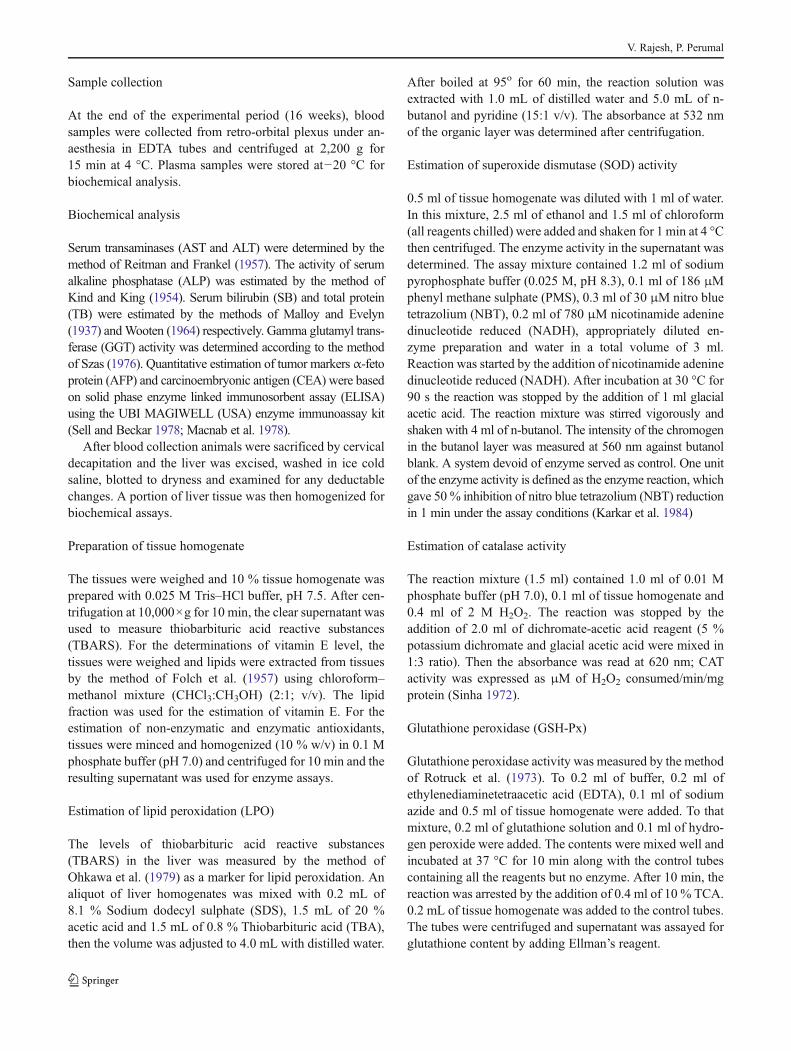
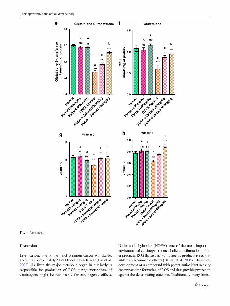
Figure 4a to h portrays the activities and the levels ofantioxidants in liver of control and experimental animals andthe results were expressed as U/mg of protein. A significantdecrease (p<0.001) in activities of SOD (0.93±0.02), cata-lase (0.92±0.03), GSH-Px (0.98±0.13) GR (0.68±0.04),GST (0.68±0.04) were noted in group IV NDEA intoxicateduntreated animals compared to control animals with SOD (1.85±0.07), CAT (1.75±0.06), GSH-Px (2.40±0.24), GR (1.08±0.10), GST (1.50±0.03). The levels of non-enzymatic antiox-idants GSH (0.59±0.09), vitamin-C (8.58±0.41) and vitamin-E (0.63±0.01) were also significantly decreased (p<0.001) inliver of group IV untreated animals compared to control ani-mals with GSH (1.08±0.07), vitamin-C (10.77±0.47) andvitamin-E (0.78±0.01). However, administration of Smilaxzeylanica leaf extract at 200mg/kg and 400mg/kg significantlyincreased the levels of SOD, CAT, GSH-Px, GR and GST to
near normal levels. The SOD levels in liver of animals treatedwith extract 200 mg/kg and 400 mg/kg were significantlyincreased (p<0.01 to p<0.001) – 1.25±0.21 and 1.18±0.07respectively compared to NDEA untreated animals. Extract200 mg/kg and 400 mg/kg significantly increased the CAT,GSH-Px, GR and GST levels from (1.41±0.08) – (1.44±0.03)(p<0.001), (1.53±0.04) – (1.73±0.08) (p<0.05 – p<0.01),(1.06±0.10) – (1.13±0.08) (p<0.01 – p<0.001) and (0.91±0.04) – (1.28±0.04) U/mg protein (p<0.01 – p<0.001), respec-tively compared to NDEA induced untreated animals.Similarly, administration of extract 200 mg/kg and 400 mg/kgsignificantly increased the non-enzymatic antioxidants, GSH,vitamin C and vitamin E levels from (0.86±0.04) – (0.95±0.02) (<0.05 – p<0.001), (10.45±0.39) – (10.63±0.37) (p<0.01) and (0.75±0.02) – (0.90±0.02) (p<0.01 – p<0.001),respectively compared to NDEA induced untreated animals.
The extract did not produce any deleterious effect on theantioxidant defense system in normal animals which isevidenced from the non-significant alteration of the enzymaticand non-enzymatic antioxidants along with the maintained rateof lipid peroxidation in group II and group III animals whencompared with the normal control group I animals. The resultsindicate that, the level of lipid peroxidation which increased inliver of carcinogen administered animals was lowered in extracttreated animals and in contrast the antioxidant status which wasfound to be decreased in carcinogen administered animals wasimproved to near normal upon Smilax zeylanica leaf extractadministration. This indicates that Smilax zeylanica leaf extractcontributes to exert antioxidant defense mechanism.
Histopathology
Histopathological examination of liver sections from controlgroup animals revealed normal architecture (Fig. 5a). Theliver sections of group II and group III animals given extractalone for 16 weeks showed normal architecture. The portaltracts were normal. The hepatic parenchyma showed normalhepatocytes. The central vein and sinusoids appeared normal.There is no evidence of inflammation or malignancy or atyp-ical cells depicting the non-toxic nature of Smilax zeylanicaleaf extract (Fig. 5b and c). Section of liver from group IVNDEA induced untreated animals revealed loss of architecturewith proliferation of tumour cells resembling bile ductularcells with suggestive adenocarcinoma (cholangio adenocarci-noma) (Fig. 5d). In contrast, NDEA induced animals treatedwith Smilax zeylanica leaf extract 200 mg/kg showed normallobular architecture with mild inflammation in portal tracts(Fig. 5e), whereas the liver sections of NDEA induced animalstreated with Smilax zeylanica leaf extract 400 mg/kg showednormal hepatic architecture (Fig. 5f). The portal tracts werenormal and the hepatic parenchyma showed normal hepato-cytes with no evidence of neoplastically transformed cells(40×, HE).
Normal
Extra
ct 20
0mg/kg
Extra
ct 40
0mg/kg
NDEA Contro
l
NDEA + E
xtra
ct 20
0mg/kg
NDEA + E
xtra
ct 40
0mg/kg
0.0
0.5
1.0
1.5
2.0
2.5
***a
b
nsa
ns
a
**
***b
Lipid peroxidation
Lip
id p
ero
xid
atio
n
MD
A n
mo
le/h
r/g
m o
f ti
ssu
e
Fig. 3 Effect of Smilax zeylanica leaf extract on lipid peroxidation inliver homogenate of experimental animals during NDEA induced livercarcinogenesis. Results are expressed as mean±S.E.M. a values aresignificantly different from control group; ns-non significant; *P<0.05; **P<0.01; ***P<0.001. bvalues are significantly different fromNDEA induced untreated group; ns-non significant; *P<0.05;**P<0.01;***P<0.001. (ANOVA, followed by Dunnett’s test). Significantincrease (***P<0.001) in lipid peroxidation is noted in NDEA induceduntreated animals (NDEA control) compared to normal animals. NDEAinduced animals treated with extract (NDEA+Extract 200 mg/kg andNDEA+Extract 400 mg/kg) showed a significant decrease in lipidperoxidation levels (200 mg/kg- **P<0.01, 400 mg/kg- (***P<0.001) compared to NDEA control animals
Chemopreventive and antioxidant activity

Normal
Extra
ct 20
0mg/kg
Extra
ct 40
0mg/kg
NDEA Contro
l
NDEA + E
xtra
ct 20
0mg/kg
NDEA + E
xtra
ct 40
0mg/kg
0.0
0.5
1.0
1.5
2.0
2.5
a
b
Superoxide dismutase
a
**
***
b**
ns ns
b
a
SO
DU
/mg
of p
rote
in
Normal
Extra
ct 20
0mg/kg
Extra
ct 40
0mg/kg
NDEA Contro
l
NDEA + E
xtra
ct 20
0mg/kg
NDEA + E
xtra
ct 40
0mg/kg
0.0
0.5
1.0
1.5
2.0
***
a
b
Catalase
aa
*** ***
b
ns ns
b
Cat
alas
en
mo
le o
f H
2O2
cosu
med
/min
/mg
of p
rote
in
Normal
Extra
ct 20
0mg/kg
Extra
ct 40
0mg/kg
NDEA Contro
l
NDEA + E
xtra
ct 20
0mg/kg
NDEA + E
xtra
ct 40
0mg/kg
0
1
2
3
***
a
b
nsa
nsa
b
*
**
Glutathione peroxidasec
GS
H-P
x
Mm
oles
/min
/mg
of p
rote
in
Normal
Extra
ct 20
0mg/kg
Extra
ct 40
0mg/kg
DENA Contro
l
DENA + E
xtra
ct 20
0mg/kg
DENA + E
xtra
ct 40
0mg/kg
0.0
0.5
1.0
1.5
nsa
b
Glutathione reductase
ns
a
b
***
a
** ***
d
Glu
tath
ion
e re
du
ctas
em
ole
of N
AD
PH
oxi
dize
d/m
in m
g pr
otei
nµ
Fig. 4 Effect of Smilax zeylanica leaf extract on the status of enzymatic andnon-enzymatic antioxidants in liver homogenate of control and experimentalgroup of animals. Results are expressed as mean±S.E.M. a values aresignificantly different from control group; ns-non significant; *P<0.05;**P<0.01; ***P<0.001. b values are significantly different from NDEAinduced untreated group; ns-non significant; *P<0.05;**P<0.01;***P<0.001. (ANOVA, followed byDunnett’s test). Significant decrease in activitesof SOD (***P<0.001), catalase (***P<0.001), GSH-Px (***P<0.001), GR(***P<0.001), GST (***P<0.001), GSH (***P<0.001), vitamin C (***P<0.01) and vitamin E (***P<0.001) were noted in NDEA induced untreated
animals (NDEA control) compared to normal animals. NDEA inducedanimals treatedwith extract (NDEA+Extract 200mg/kg andNDEA+Extract400 mg/kg) showed a significant increase in activities of SOD (200 mg/kg-**P<0.01, 400 mg/kg P**<0.01), catalase (200 mg/kg- ***P<0.001,400 mg/kg- ***P<0.001), GSH-Px (200 mg/kg- *P<0.05, 400 mg/kg-**P<0.01), GR (200 mg/kg- **P<0.01, 400 mg/kg ***P<0.001), GST(200 mg/kg- **P<0.01, 400 mg/kg ***P<0.001), GSH (200 mg/kg- *P<0.05, 400 mg/kg- ***P<0.001), vitamin C (200 mg/kg- **P<0.01, 400 mg/kg- **P<0.01) and vitamin E (200 mg/kg- **P<0.01, 400 mg/kg- ***P<0.001) compared to NDEA control animals
V. Rajesh, P. Perumal
Discussion
Liver cancer, one of the most common cancer worldwide,accounts approximately 549,000 deaths each year (Liu et al.2006). As liver, the major metabolic organ in our body isresponsible for production of ROS during metabolism ofcarcinogens might be responsible for carcinogenic effects.
N-nitrosodiethylamine (NDEA), one of the most importantenvironmental carcinogen on metabolic transformation in liv-er produces ROS that act as promutagenic products is respon-sible for carcinogenic effects (Bansal et al. 2005). Therefore,development of a compound with potent antioxidant activitycan prevent the formation of ROS and thus provide protectionagainst the deteriorating outcome. Traditionally many herbal
Normal
Extra
ct 20
0mg/kg
Extra
ct 40
0mg/kg
NDEA Contro
l
NDEA + E
xtra
ct 20
0mg/kg
NDEA + E
xtra
ct 40
0mg/kg
0.0
0.5
1.0
1.5
2.0
***a
b
Glutathione-S-transferase
ns
a
ns
a
***
b
**
e
Glu
tath
ion
e-S
-tra
nsf
eras
eu
nit
s/m
in/m
g o
f p
rote
in
Normal
Extra
ct 20
0mg/kg
Extra
ct 40
0mg/kg
DENA Contro
l
DENA + E
xtra
ct 20
0mg/kg
DENA + E
xtra
ct 40
0mg/kg
0.0
0.5
1.0
1.5
nsa
b
Glutathione
nsa
***a
*
b
***
f
GS
Hn
mo
le/m
g o
f p
rote
in
Normal
Extract 2
00mg/kg
Extract 4
00mg/kg
NDEA Contro
l
NDEA + Extra
ct 200mg/kg
NDEA + Extra
ct 400mg/kg
0
5
10
15
ns
a
Vitamin C
ns
a
b
b b
**
** **
g
Vit
amin
C
Norm
al
Extra
ct 2
00m
g/kg
Extra
ct 4
00m
g/kg
NDEA Con
trol
NDEA + E
xtra
ct 2
00m
g/kg
NDEA +
Ext
ract
400
mg/
kg0.0
0.2
0.4
0.6
0.8
1.0
Vitamin E
ns ns
a a
**
b
a
b
***
***
h
Vit
amin
E
Fig. 4 (continued)
Chemopreventive and antioxidant activity

medicines have revealed cancer chemopreventive potentialthrough antioxidant activity. The present study was aimed toevaluate the chemopreventive and antioxidant activity ofmethanol extract of Smilax zeylanica leaves against N-nitrosodiethylamine induced hepatocarcinogenesis. Doselevels were selected based on the acute oral toxicity studyand our previous invivo studies, where the extract showedprotection against streptozotozin (STZ) induced oxidativestress (Rajesh et al. 2010) and substantial evidence fromstudies on invitro and invivo antioxidant activities where themethanol extract of Smilax zeylanica leaves at the dose levels200 mg/kg and 400 mg/kg body weight exhibited free radicalscavenging effect against the different free radicals tested(DPPH, hydrogen peroxide, ABTS, nitric oxide and superox-ide free radicals) and significant increase in the levels of
glutathione, tissue proteins and enzymes viz. SOD, catalaseand peroxidase in CCl4 induced hepatotoxicity model inWistar albino rats (Anita et al. 2010).
Decrease in body weight observed during the experimen-tal period in NDEA induced untreated animals could belargely due to deterioration of liver function and nutritionaldeprivation which might be due to reduced food intake. Inaddition, assessment of relative liver weight was used aspotential tool to diagnose change in liver size. NDEA in-duced proliferation of cells in the liver was evident fromincrease in NDEA induced untreated animals. Treatmentwith Smilax zeylanica leaf extract prevented body weightloss and reduced the relative liver weight compared tountreated animals, which signify the amelioration capacityof extract upon carcinogen exposure. Significant decrease in
Fig. 5 Histopathological image of liver tissues. a (40 ×) H and Estained section of liver tissue from group I normal animals showingnormal architecture. b and c (40 ×) H and E stained section of liver fromgroup II and group III animals given extract alone for 16 weeks showingnormal hepatocytes in parenchyma. The portal traids appeared normalwith no evidence of inflammation or malignancy or atypical cellsdepicting the non-toxic nature of Smilax zeylanica leaf extract. d (40×) H and E stained section of liver from group IV NDEA induceduntreated animals showing loss of architecture with proliferation of
tumour cells resembling bile ductular cells with suggestive adenocarci-noma. e (circle, 40 ×) H and E stained section of liver from NDEAinduced group V animals treated with Smilax zeylanica leaf extract200 mg/kg showing normal lobular architecture with mild inflammationin portal tracts. f (arrows, 40 ×) H and E stained section of liver fromNDEA induced group VI animals treated with Smilax zeylanica leafextract 400 mg/kg showing normal hepatocytes and portal triads withno evidence of neoplastically transformed cells
V. Rajesh, P. Perumal

lesions in liver of treatment groups were supported by histo-logical assessment of liver. The pathological changes in livercaused by NDEAwere assessed by determining the levels ofvarious biochemical hepatic markers (Kovalsky et al. 1996).Marked elevation in AST and ALT in NDEA induceduntreated animals indicated the hepatocellular damage.Because AST and ALT are cytoplasmic in location, they arereleased into circulation after cellular damage and rupture ofplasma membrane (Wroblewski 1959; Sallie et al. 1991).Hence AST and ALT are considered as sensitive markersemployed in diagnosis of hepatic damage. Treatment withSmilax zeylanica leaf extract significantly reduced the levelsof AST and ALT in NDEA induced animals. This indicatesthat Smilax zeylanica leaf extract tends to prevent liverdamage by maintaining integrity of plasma membrane, there-by suppressing the leakage of enzymes through membrane.
The elevation in ALP in NDEA induced untreated ani-mals might be due to proliferation of bile ductular cells andresulting blockade of bile ducts. Significant reduction inALP levels in NDEA induced extract treated animals indi-cates the protective effect of Smilax zeylanica leaf extractagainst NDEA induced hepatic injury. The reduction inserum protein levels in NDEA induced untreated animalsmight be due to liver damage because hepatotoxicity impairsthe synthetic function of liver (David 1999). Treatment withSmilax zeylanica leaf extract ameliorated the imbalance.
GGT, an enzyme embedded in plasma membrane of he-patocytes, mainly in canalicular domain. Any damage inplasma membrane causes liberation of GGT into serum(Sivaramakrishnan et al. 2008). Hence, GGT is consideredas a best indicator of liver damage (Bulle et al. 1990).Elevated levels of GGT observed in NDEA induceduntreated animals might be due to liver injury. Treatmentwith Smilax zeylanica leaf extract significantly lowered theGGT level which indicates that Smilax zeylanica leaf extracttends to prevent liver damage by maintaining integrity ofplasma membrane.
A tumour associated foetal protein, AFP has long beenemployed as a serum tumour marker to monitor diseaseprogression (Abelev 1971). AFP is a 72 KDa α1 globulinsynthesized during embryonic life by foetal yolk sac, liverand intestinal tract with uncertain biological function. SinceAFP has high specificity for hepatocarcinoma (Liu et al.2006), its serum concentration can be used in diagnosis ofhepatocarcinoma and for the diagnosis of tumour response totherapy.
CEA is a glycoprotein found in different cells, but typicallyassociated with certain tumours. Increase in CEA levels ismost frequent in cancer of colon and rectum. The conditionswhich can elevate CEA are smoking, pancreatitis, inflamma-tory bowel disease, pancreatitis and cirrhosis of liver. CEAlevels increase, with an increase in tumour size (Jahan et al.2011). In the present study, significant increase in AFP and
CEA levels observed in NDEA induced untreated animalsmight have caused due to mutagenesis. The decrease in levelsof AFP and CEA following treatment with Smilax zeylanicaleaf might be due to response to therapy.
Increased ROS generation and decreased antioxidant de-fense in liver tissue has been reported in development ofhepatocarcinogenesis (Ramakrishnan et al. 2006; Kweonet al. 2003). ROS, through interaction with nucleic acids,proteins, lipids results in chromosomal instability, mutations,loss of organelle function play an important role in develop-ment of cancer (Waris and Ahsan 2006). Lipid peroxidationis one of the major mechanisms of cell injury caused by freeradicals. Enormous amount of free radicals generated bycarcinogens reacts with lipids causing lipid peroxidation(Sikkim andMulee 2000). The products of lipid peroxidationinclude malondialdehyde that interacts with various mole-cules leading to oxidative stress and has been reported to beinvolved in the formation of tumours (Ramakrishnan et al.2007). In the present study, significant increase in the levelsof lipid peroxidation observed in untreated group IV cancerbearing animals may be due to the excessive production offree radicals and due to inhibition of antioxidant enzymes.Significantly reduced levels of lipid peroxidation were seenin Smilax zeylanica leaf extract treated group Vand group VINDEA induced animals. This clearly shows that Smilaxzeylanica effectively controlled the rate of lipid peroxidation,which suggests the beneficial effect of the extract againstNDEA initiated free radical formation. The presence offlavonoids may contribute to this effect, because they areproved to be a potential inhibitor of lipid peroxidation(Siegers and Younes 1981).
The enhanced formation of lipid peroxides is furtherevidenced by decrease in activities of antioxidant enzymesin liver of NDEA induced untreated animals as comparedwith normal control animals.
SOD is said to act as the first line of defense againstsuperoxide radical generated as a by-product of oxidative phos-phorylation. SOD mediated dismutation of superoxide radical(O2
-) generates hydrogen peroxide (H2O2). Accumulation ofexcess of H2O2 causes toxic effects on cellular system. In thisregard GSH-Px and catalase converts H2O2 into water (LiShijun et al. 2000). GSH-Px catalyses the reduction of H2O2
at the expense of reduced GSH, thereby protecting cells againstoxidative damage. CAT detoxifies H2O2 into H2O and O2
(Murray et al. 2003). Thus SOD, CAT and GSH-Px act mutu-ally and constitute the enzymatic defense mechanism againstROS (Bhattacharjee and Sil 2006). In the present study de-crease in the activities of SOD, CAT and GSH-Px in untreatedcancer bearing animals could be attributed to excessive utiliza-tion of enzymes in detoxification of peroxides and hydroper-oxides generated during liver carcinogenesis. Restoration in thelevels of lipid peroxidation upon treatment with Smilaxzeylanica leaf extract might have resulted in the recoupment
Chemopreventive and antioxidant activity

in the activities of the above antioxidant enzymes to normalcy.SOD, catalase and GSH-Px require several secondary enzymeslike GR and cofactors like GSH, NADPH, to function at highefficacy. GR catalyzes the NADPH dependent reduction ofglutathione disulphide to glutathione thus maintaining glutathi-one levels (Katiyar et al. 1993). GST catalyzed GSH conjuga-tion is an important mechanism for the detoxification process.In the present study the activities of GR and GST were signif-icantly reduced in untreated NDEA induced untreated animals.Upon treatment with Smilax zeylanica leaf extract, the GR andGST levels were significantly increased. Increase in GR canprotect the liver by maintaining the basal level of GSH, whichis important for many other GSH dependent detoxificationreactions.
GSH, vitamin C and vitamin E are well known non-enzymatic antioxidant defense system of cells, act synergis-tically to scavenge free radicals in biological system. GSH isfound to be present in high concentration in cells, protectscells against free radical attack (Farombi et al. 2000). GSHacts directly as free radical scavenger by donating ahydrogen atom and thereby neutralizing hydroxyl radical.It reduces peroxides and maintains protein thiols in thereduced state (Nwanjo and Oze 2007). GSH-PX usesGSH as a substrate to catalyze the reduction of hydro-peroxide and H2O2 (Bebe and Panemangalore 2003).Reduced glutathione (GSH) in tissues maintains main-tains the cellular levels of vitamin C and Vitamin E inactive form. Vitamin C and vitamin E act synergisticallyin scavenging wide variety of ROS. Vitamin C protectsthe cell membrane from oxidative damage induced byaqueous radicals (Allen 1991). It removes free radicalsfrom cytosol and plays a vital role in protecting lipopro-tein molecules from oxidative damage by regeneratingthe reduced form of vitamin E (Das 1994). Vitamin E isa well recognized, important free radical scavenger in thecell membrane limits LPO by terminating chain reactioninitiated in the membrane lipids (Wiseman 1996). GSHacts synergistically with vitamin E against oxidativestress (Chaudiere 1994). Vitamin C also scavenges and de-toxifies free radicals in combination with Vitamin E and GSH(George 2003). The decreased level of these non-enzymaticantioxidants observed in NDEA induced untreated animalsmight be due to excessive utilization of these antioxidants forquenching enormous free radicals produced. Treatment withSmilax zeylanica leaf extract effectively restored the depletedlevels of non-enzymatic antioxidants GSH, vitamin C andvitamin E caused by NDEA. Increase in GSH levels in turnalso contributes to the recycling of other antioxidants such asvitamin C and vitamin E.
The results indicate that Smilax zeylanica leaf extract in-hibits the level of lipid peroxidation and significantly in-creases the enzymatic and non-enzymatic antioxidant de-fense mechanisms in NDEA induced liver carcinogenesis.
The histological observations clearly indicate the pres-ence of neoplastic changes in NDEA induced untreatedanimals. In animals treated with Smilax zeylanica leaf extractthe liver architecture was preserved which clearly supportsthe biochemical data. These suggest the Smilax zeylanicaleaves might have promising protective role against livercarcinogenesis.
Conclusion
Data from this study suggest that administration ofSmilax zeylanica leaf extract possesses significant pro-tection against NDEA induced liver carcinogenesis by amechanism related, at least in part, to the ability ofSmilax zeylanica leaves to decrease oxidative stress bystabilizing and increasing all the components of antioxi-dant defense system which were disturbed system duringNDEA induced oxidative stress in rats. The combinedactivity of the phytomolecules in the extract might beresponsible for oxidative defense and chemoprevention.
Acknowledgments The authors are thankful to the managementof JKK Nattraja College of Pharmacy, Komarapalayam, TamilNadu, INDIA for providing necessary facilities to carry out theresearch work.
References
Abdala S, Martin-Herrera D, Benjumea D, Perez-Paz P (2008) Diureticactivity of Smilax canariensis, an endemic canary Islend species. JEthnopharmacol 119:12–16
Abelev GI (1971) Alpha-feto protein in association with malignanttumours. Adv Cancer Res 14:295–357
Akintonwa DA (1985) The derivation of nitrosamines from some thera-peutic amines in the human environment. Ecotoxicol Environ Saf9:64–70
Allen RG (1991) Oxygen-reactive species and antioxidant responsesduring development: the metabolic paradox of cellular differenti-ation. Proc Soc Exp Biol Med 196:117–129
Ambasta SP (2006) The useful plants of India. NISCAIR, New Delhi, p578
Anita A, Purnima A, Madhavan V, Raju A (2010) In- vitro and in-vivoantioxidant activity studies on the leaves of Smilax zeylanica L.(Smilacaceae). J Pharm Res 3(10):2427–2430
Bansal AK, Trivedi R, Soni GL, Bhatnagar D (2005) Protective role ofvitamin E pre-treatment on N-nitrosodiethylamine induced oxida-tive stress in rat liver. Chem Biol Interact 156:101–111
Barker H, Frank O, Angelis B, Feingold S (1951) Plasma tocopherol inman at various times after ingesting free or acetylated tocopherol.Nutr Rep Int 21:531–536
Bebe FN, Panemangalore M (2003) Exposure to low doses of endosul-fan and chlorpyrifos modifies endogenous antioxidants in tissuesof rats. J Environ Sci Health B38:349–363
Befeler AS, Di Bisceglie AM (2002) Hepatocellular carcinoma: diag-nosis and treatment. Gasteroenterology 122(6):1609–1619
V. Rajesh, P. Perumal
Bhattacharjee R, Sil PC (2006) The protein fraction of Phyllanthusniruri plays a protective role against acetaminophen induced he-patic disorder via its antioxidant properties. Phytother 20:595–601
Bhattracharya S, Chatterjee M (1998) Protective role of Trianthemaportulacastrum against diethylnitrosamine- induced experimentalhepatocarcinogenesis. Cancer Lett 129:7–13
Bhosale P, Motiwale L, Ignle AD, Gadre RV, Rao KVK (2002)Protective effect of Rhodotorula glutinis NCIM3353 on the devel-opment of hepatic preneoplastic lesions. Curr Sci 83(3):303–308
Bishayee A, Dhir N (2009) Resveratrol-mediated chemoprevention ofdiethylnitrosamine-initiated hepatocarcinogenesis: inhibition ofcell proliferation and induction of apoptosis. Chem Biol Interact179(2–3):131–144
Bulle F, Mavier P, Zafrani ES, Preaux AM, Lescs MC, Siegrist S,Dhumeaux D, Guellaen G (1990) Mechanism of gamma-glutamyltranspeptidase release in serum during intra hepatic and extra hepaticcholestasis in the rat: a histochemical, biochemical and molecularapproach. Hepatology 11:545–550
Chaudiere J (1994) Some chemical and biochemical constrains of oxidativestress in living cells. In: Rice-Evans CA, BurdonRH (eds) Free radicaldamage and its control. Elsevier Science, Amsterdam, pp 25–26
Das S (1994) Vitamin E in the genesis and prevention of cancer. Areview. Acta Oncol 33:615–619
David EJ (1999) Special considerations in interpreting liver functiontests. Am Family Phy 59:8
Ellman GL (1959) Tissue sulfhydryl groups. Arch Biochem Biophys82:70–77
Farombi EO, Olowg BI, Emerole GO (2000) Effect of three structurallyrelated antimalarial drugs on liver microsomal components andlipid peroxidation in rats. Comp Biochem Physiol 126:217–224
Feo F, Pascale RM, Simile MM, De Miglio MR, Muroni MR, Calvisi D(2000) Genetic alterations in liver carcinogensis; implications fornew preventive and therapeutic strategies. Crit Rev Oncog 11:19–62
Folch J, Lees M, Solane SGH (1957) A simple method for isolation andpurification of total lipids from animal tissues. J Bio Chem26:497–509
Gamble JS (2004) Flora of the Presidency of Madras. Dehradun: BishenSingh, Mahendra Pal Singh eds, pp. 1518
George J (2003) Ascorbic acid concentrations in diethylnitrosamineinduced hepatic fibrosis in rats. Clin Chem Acta 335:39–47
Habig WH, Pabst MJ, Jakoby WB (1974) Glutathione S transferases.The first enzymatic step in mercapturic acid formation. J BiolChem 249:7130–7139
Hai W, Kim C, Song S, Kang C (2001) Study on mechanism of multistephepatotumorigenesis in rat: development of hepatotumorigenesis. JVet Sci 2:53–58
Halsted CH, Villanueva J, Chandler CJ, Stabler SP, Allen RH,Muskhelishvili L, James SJ, Poirier L (1996) Ethanol feeding ofmicropigs alters methionine Metabolism and increases hepatocel-lular apoptosis and proliferation. Hepatology 23(3):497–505
Horn HD, Burns FH (1978) Assay of glutathione reductase activity. In:Bergmeyer HV (ed) Methods of enzymatic analysis. Academic,New York, pp 142–146
IARC (1972) Monograph on the evaluation of carcinogenic risk ofchemicals to man, vol 1. International Agency for Research onCancer, Lyon, pp 107–124
Jagadeeswaran R, Thirunavukkarasu C, Nalini R, Gunasekaran S,Sakthisekaran D (2000) In vitro studies on the selective cytotoxiceffect of crocetin and quercetin. Fitotherapia 71(4):395–399
Jahan MS, Vani G, Shyamaladevi CS (2011) Anti-carcinogenic effect ofSolanum trilobatum in diethylnitrosamine induced and Phenobarbitalpromoted hepatocarcinogenesis in rats. Asian J Biochem 6(1):74–81
Karkar P, Das B, Viswanath PN (1984) Modified spectrophotometerassay of SOD. Ind J Biochem Biophys 95:51–58
Katiyar SK, Agarwal R, Mukhtar H (1993) Protective effects of greentea polyphenols administration by oral intubation against chemical
carcinogen induced forestomach and pulmonary neoplasia in A/Jmice. Cancer Lett 73:167–172
Khandelwal KR, Kokate CK (1995) Pratical pharmacognosy, 4th edn.Vallabh Prakashan, New Delhi, p 110
Kim J, Park EJ (2002) Cytotoxic anticancer candidates from naturalresources. Curr Med Chem Anti-cancer Agents 2:485–537
Kind PRN, King EJJ (1954) Estimation of plasma phosphatase by determi-nation of hydrolysed phenol with antipyrine. J Clin Pathol 7:322–330
Kirtikar KR, Basu BD (1991) Indian Medicinal Plants. Dehra Dun:Bishen Singh, Mahendra Pal Singh, eds, pp. 2496
Kovalsky I, Schaff Lapis K, Jeney A (1996) Marker enzymes of ratchemical hepatocarcinogenesis in human liver tumours. PatholOncol Res 2:54–55
Kweon S, Park KA, Choi H (2003) Chemopreventive effect of garlicpowder diet in diethylnitrosamine-induced rat hepatocarcinogenesis.Life Sci 73:2515–2526
Liao DJ, Blanck A, Eneroth P, Gustafsson JA, Hällström IP (2001)Diethylnitrosamine causes pituitary damage, disturbs hormonelevels and reduces sexual dimorphism of certain liver functionsin the rat. Environ Health Perspect 109:943–947
Liu JG, Zhao HJ, Liu YJ, Wang XL (2006) Effect of selenium-enrichedmalt on hepatocarcinogenesis, paraneoplastic syndrome and the hor-mones regulating blood glucose in rats treated by diethylitrosamine.Life Sci 78(20):2315–2321
Loeppky RN (1994) Nitrosamine and nitroso compound chemistry andbiochemistry. In: ACS Symposium Series 553, AmericanChemical Society, Washington, DC, pp. 1–12
Macnab GM, Urbanowicz JM, Kew MC (1978) Carcinoembryonicantigen in hepatocellular cancer. Br J Cancer 38:51–54
Malloy HT, Evelyn KA (1937) The determination of bilirubin with thephotometric colorimeter. J Biol Chem 119:481–490
Mann J (2002) Natural products in cancer chemotherapy: past, presentand future. Nat Rev Cancer 2:143–148
Murray RK, Granner DK, Mayes PA (2003) Rodwell VW. Harper’sIllustrated Biochemistry, 26th ed, The McGraw-Hill Companies Inc.
Nadkarni KM (1976) Indian Materica Medica. Bombay PopularPrakashan, Bombay, p 1145
Nwanjo HU, Oze GO (2007) Oxidative imbalance and non-enzymicantioxidant status in pulmonary tuberculosis infected. Pak J Nutr6:590–592
OECD (2000)Guidance document on acute oral toxicity 425. Environmentalhealth and safety monograph series on testing assessment. NO. 24
Ohkawa H, Ohishi N, Yagi K (1979) Assay for lipid peroxides in animaltissues by thiobarbituric acid reaction. Anal Biochem 95:351–358
Omaye ST, Turnbull JD, Sauberlich HE (1979) Selected methods forthe determination of ascorbic acid in animal cells, tissues andfluids. Methods Enzymol 62:1–11
Oommachan, Masih SK (1991) Ethnobotanical and conservationalaspects of medicinal plants of Madhya Pradesh. Indian J PureAppl Biol 6(1):39–44
Parola M, Robino G (2001) Oxidative stress-related molecules and liverfibrosis. J Hepatol 35:297–306
Rajesh V, Perumal P, Sundarrajan T (2010) Antidiabetic activity ofmethanol extract of Smilax zeylanica Linn in streptozotocin in-duced diabetic rats. Internet J Pharmacol 6(1)
Ramakrishnan G, Ragahavendran HR, Vinodkumar R, Devaki T (2006)Suppression of N-nitrosodiethylamine induced hepatocarcinogenesisby silymarin in rats. Chem Biol Interact 161:104–114
Ramakrishnan G, Augustine TA, Jagan S, Vinodhkumar R, Devaki T(2007) Effect of silymarin on N-nitrosodiethylamine inducedhepatocarcinogenesis in rats. Exp Oncol 29(1):39–44
Ramaswamy SN, Radhakrishna Rao M, Govindappa (2001) Flora ofShimoga district Karnataka. Prasaranga, Mysore, p 619
Reitman S, Frankel SA (1957) Coloirmetric method for determinationof serum glutamic oxaloacetic and glutamic pyruvic transami-nases. Am J Clin Pathol 28:56–63
Chemopreventive and antioxidant activity

Rotruck JT, Pope AL, Ganther HE, Swanson AB, Hafeman DG,Hoekstra WG (1973) Selenium: biochemical role as a componentof glutathione peroxidase. Science 179:588–590
Saldhana CJ, Wicolson DH (1976) Flora of Hassan district ofKarnataka. Amerind Pub Co Pvt Ltd, New Delhi, p 804
Sallie R, Tredger JM, Willam R (1991) Drug and the liver. BiopharmDrug Dispos 12:251–259
Santapau H, Henry AN (1976) A dictionary of the flowering plants inIndia (reprint). CSIR, New Delhi, p 58
Sell S, Beckar FF (1978) Alpha-feto protein. Natl Cancer Inst 60:19–26Shao B, Guo HZ, Cui YJ, Ye M, Han J, Guo DA (2007) Steroidal
saponins from Smilax china and their anti-inflammatory activities.Phytochemistry 68:623–630
Shijun L, Tao Y, Yang J-Q, Terry DO, Larry WO (2000) The role ofcellular glutathione peroxidase redox regulation in the suppressionof tumour cell growth by manganese superoxide dismutase. CancerRes 60:3927–3939
Siegers CP and Younes M (1981). Effect of bioflavonoids on lipidperoxidation induced by glutathione depletion. Pro IntBioflavonoid Symposium, Munich, FRG, pp. 409
Sikkim H, Mulee B (2000) Lipid peroxidation and antioxidant enzymesin the blood of rats treated with benzo[a]pyrene. Chem Biol Interact127:139–150
Sinha AK (1972) Colorimetric assay of catalase. Anal Biochem47:389–394
Sivaramakrishnan V, Shilpa PNM, Kumar VRP, Devaraj SN (2008)Attenuation of N-nitrosodiethylamine induced hepatocellular carcino-genesis by a novel flavonol - Morin. Chem Biol Interact 171:79–88
Soni KB, Lahiri M, Chakradeo P, Bhide SV, Kuttan R (1997) Protectiveeffect of food additives on aflatoxin-induced mutagenicity andhepatocarcinogenicity. Cancer Lett 115:129–133
Szas G (1976) Reaction rate method for gamma glutamyl transferaseactivity in serum. Clin Chem 22:2031–2055
Takuji T, Toshihiro K, Toshihiko K, Aijin W, Masumi S, Kiyohisa O,Hideki M (1993) Inhibition of 4-nitroquinoline-1-oxide- inducedrat tongue carcinogenesis by the naturally occurring plant pheno-lics caffeic, ellagic, chlorogenic and ferulic acids. Carcinogenesis14(7):1321–1325
Thorgeirsson SS, Grisham JW (2002) Molecular pathogenesis of hu-man hepatocellular Carcinoma. Nat Genet 31(4):339–346
Verna L, Whysner J, Williams GM (1996) N-nitrodiethylamine mech-anistic data and risk assessment: bioactivation, DNA-adductformation, mutagenicity, and tumor intiation. Pharmacol Ther71:57–81
Wang XJ, Feng P, Wen ZY (1996) Study on invitro and invivo antican-cer action of compound Smilax china L. Chin J Pathophysiol12:614–1614
Waris G, Ahsan H (2006) Reactive oxygen species: role in the devel-opment of cancer and various chronic conditions. J Carcinogen5:14
Wattenberg LW, Estensen RD (1996) Chemopreventive effects ofmyo-inositol and dexamethasone on benzo[a]pyrene and 4-(methylnitrosoamino)-1-(3-pyridyl)-1-butanone-induced pulmo-nary carcinogenesis in female A/J mice. Cancer Res 56:5132–5135
Wiseman H (1996) Dietary influences on membrane function: impor-tance in protection against oxidative damage and disease. J NutrBiochem 7:2–15
Wooten IDP (1964) Micro-analysis in medical biochemistry, 4th edn. Jand A Churchill Ltd, London, pp 138–140
Wroblewski F (1959) The clinical significance of transaminase activi-ties of serum. Am J Med 27:911–923
V. Rajesh, P. Perumal