chest radiography diagnostic value and interpretation › radiology-handout ›...
TRANSCRIPT
Chest Radiography
Diagnostic value and interpretation
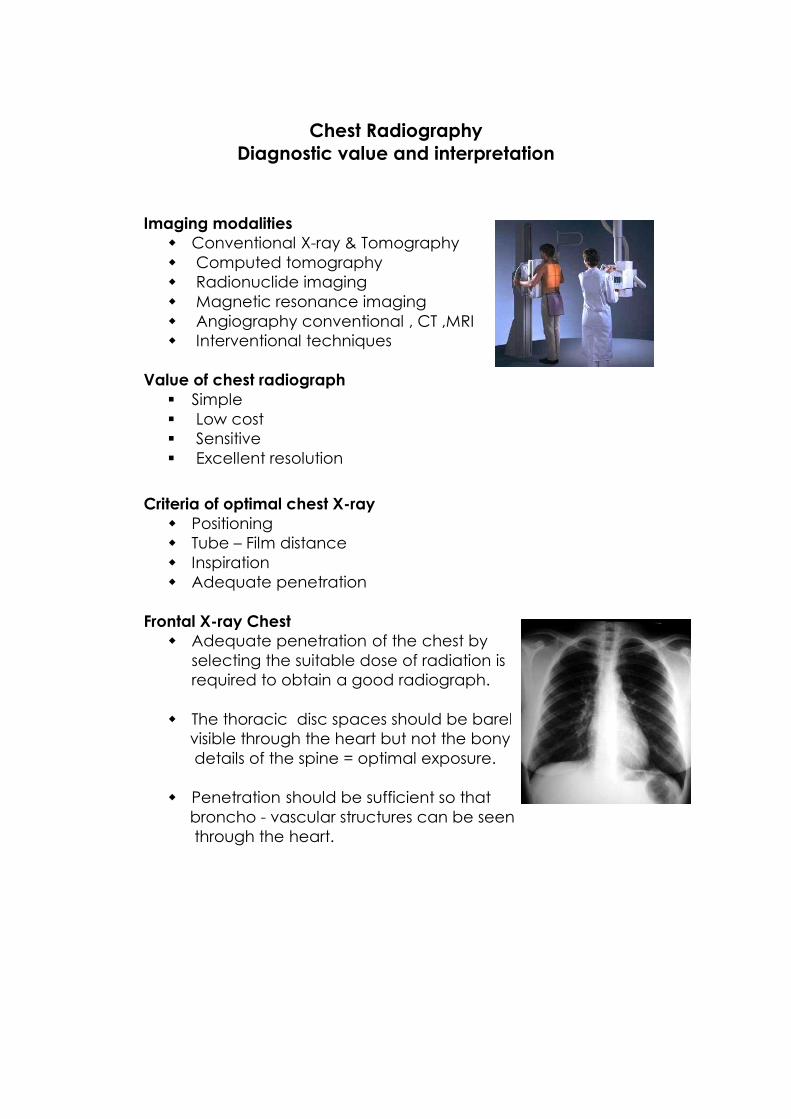
Imaging modalities
Conventional X-ray & Tomography
Computed tomography
Radionuclide imaging
Magnetic resonance imaging
Angiography conventional , CT ,MRI
Interventional techniques
Value of chest radiograph
Simple
Low cost
Sensitive
Excellent resolution
Criteria of optimal chest X-ray
Positioning
Tube – Film distance
Inspiration
Adequate penetration
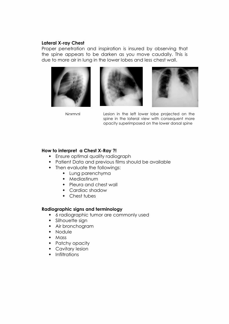
Frontal X-ray Chest
Adequate penetration of the chest by
selecting the suitable dose of radiation is
required to obtain a good radiograph.
The thoracic disc spaces should be barely
visible through the heart but not the bony
details of the spine = optimal exposure.
Penetration should be sufficient so that
broncho - vascular structures can be seen
through the heart.
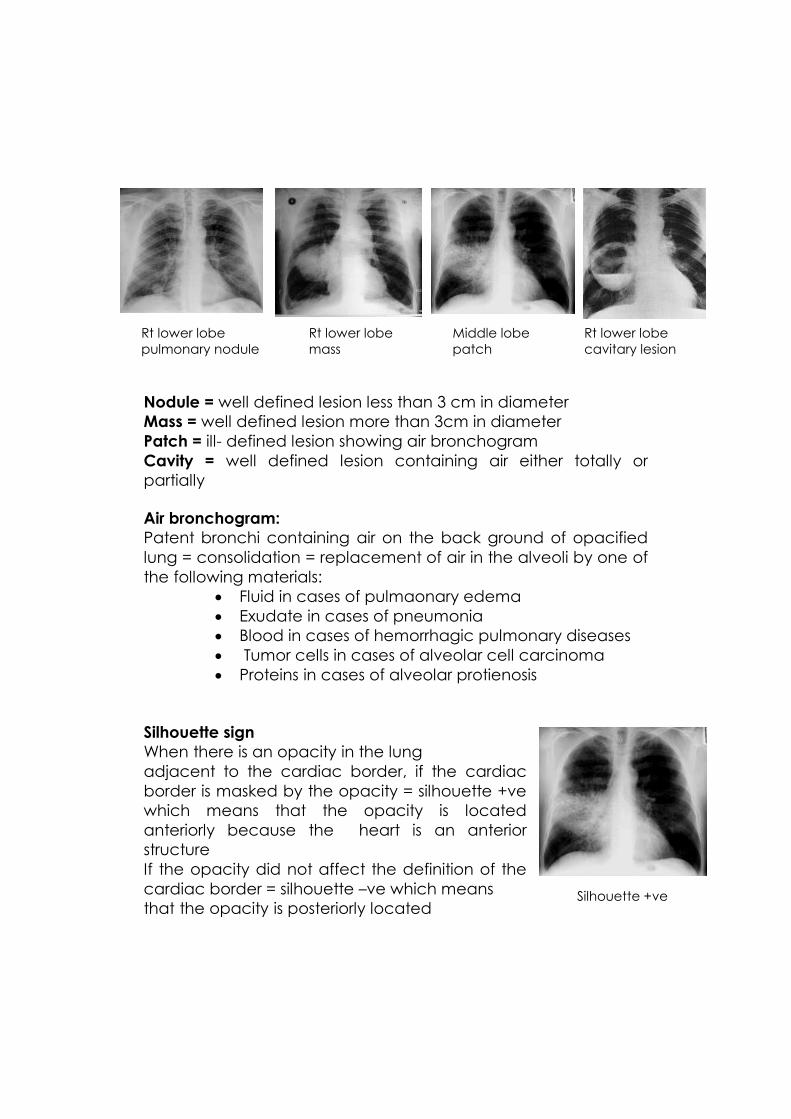
Lateral X-ray Chest
Proper penetration and inspiration is insured by observing that
the spine appears to be darken as you move caudally. This is
due to more air in lung in the lower lobes and less chest wall.
How to interpret a Chest X-Ray ?!
Ensure optimal quality radiograph
Patient Data and previous films should be available
Then evaluate the followings:
Lung parenchyma
Mediastinum
Pleura and chest wall
Cardiac shadow
Chest tubes
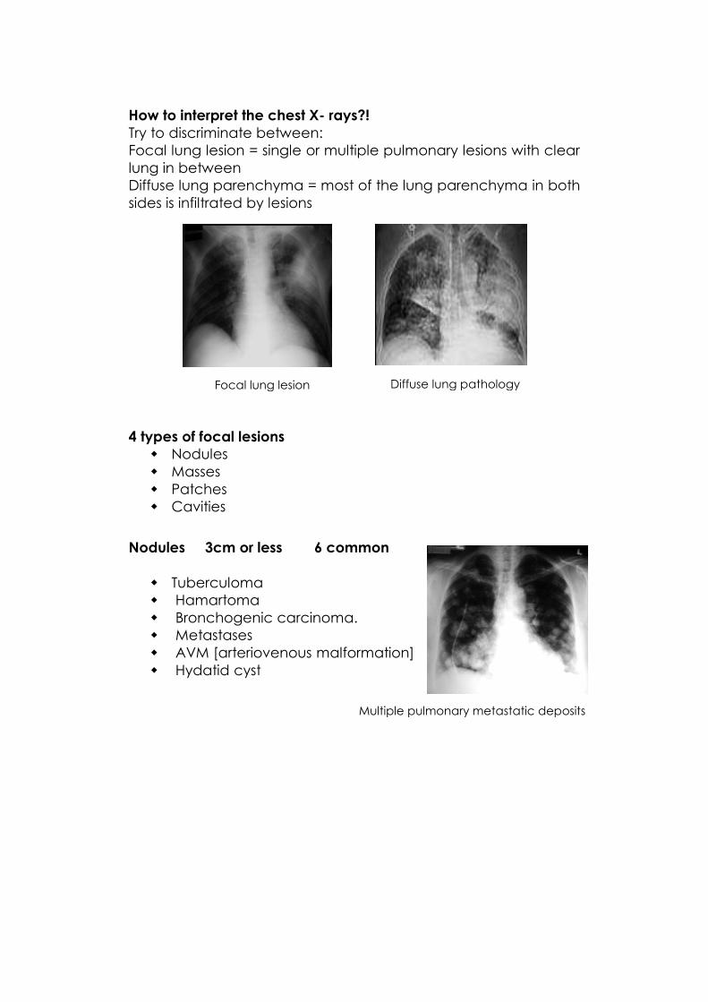
Radiographic signs and terminology
6 radiographic tumor are commonly used
Silhouette sign
Air bronchogram
Nodule
Mass
Patchy opacity
Cavitary lesion
Infiltrations
Normal Lesion in the left lower lobe projected on the
spine in the lateral view with consequent more
opacity superimposed on the lower dorsal spine
Nodule = well defined lesion less than 3 cm in diameter
Mass = well defined lesion more than 3cm in diameter
Patch = ill- defined lesion showing air bronchogram
Cavity = well defined lesion containing air either totally or
partially
Air bronchogram:
Patent bronchi containing air on the back ground of opacified
lung = consolidation = replacement of air in the alveoli by one of
the following materials:
Fluid in cases of pulmaonary edema
Exudate in cases of pneumonia
Blood in cases of hemorrhagic pulmonary diseases
Tumor cells in cases of alveolar cell carcinoma
Proteins in cases of alveolar protienosis
Silhouette sign
When there is an opacity in the lung
adjacent to the cardiac border, if the cardiac
border is masked by the opacity = silhouette +ve
which means that the opacity is located
anteriorly because the heart is an anterior
structure
If the opacity did not affect the definition of the
cardiac border = silhouette –ve which means
that the opacity is posteriorly located
Silhouette +ve
Rt lower lobe
mass
Rt lower lobe
pulmonary nodule
Rt lower lobe
cavitary lesion
Middle lobe
patch
How to interpret the chest X- rays?!
Try to discriminate between:
Focal lung lesion = single or multiple pulmonary lesions with clear
lung in between
Diffuse lung parenchyma = most of the lung parenchyma in both
sides is infiltrated by lesions
4 types of focal lesions
Nodules
Masses
Patches
Cavities
Nodules 3cm or less 6 common
Tuberculoma
Hamartoma
Bronchogenic carcinoma.
Metastases
AVM [arteriovenous malformation]
Hydatid cyst
Focal lung lesion Diffuse lung pathology
Multiple pulmonary metastatic deposits
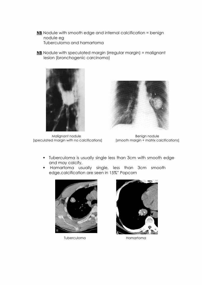
NB Nodule with smooth edge and internal calcification = benign
nodule eg
Tuberculoma and hamartoma
NB Nodule with speculated margin (irregular margin) = malignant
lesion (bronchogenic carcinoma)
Tuberculoma is usually single less than 3cm with smooth edge
and may calcify,
Hamartoma usually single, less than 3cm smooth
edge,calcification are seen in 15%” Popcorn
Malignant nodule
[speculated margin with no calcifications]
Benign nodule
[smooth margin + matrix calcifications]
Tuberculoma Hamartoma
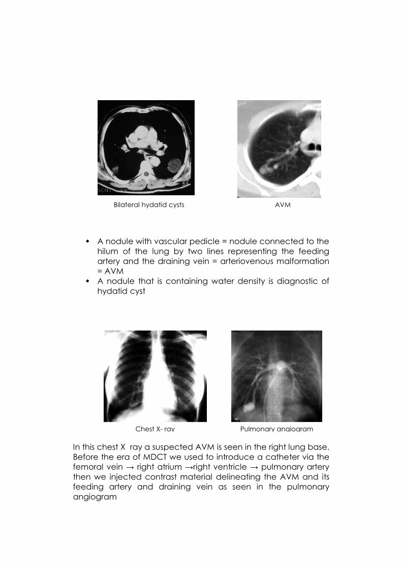
A nodule with vascular pedicle = nodule connected to the
hilum of the lung by two lines representing the feeding
artery and the draining vein = arteriovenous malformation
= AVM
A nodule that is containing water density is diagnostic of
hydatid cyst
In this chest X ray a suspected AVM is seen in the right lung base.
Before the era of MDCT we used to introduce a catheter via the
femoral vein → right atrium →right ventricle → pulmonary artery
then we injected contrast material delineating the AVM and its
feeding artery and draining vein as seen in the pulmonary
angiogram
Bilateral hydatid cysts AVM
Chest X- ray Pulmonary angiogram
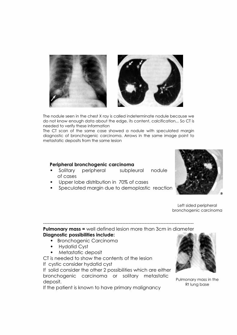
The nodule seen in the chest X ray is called indeterminate nodule because we
do not know enough data about the edge, its content, calcification,.. So CT is
needed to verify these information
The CT scan of the same case showed a nodule with speculated margin
diagnostic of bronchogenic carcinoma. Arrows in the same image point to
metastatic deposits from the same lesion
Peripheral bronchogenic carcinoma
Solitary peripheral subpleural nodule in 52%
of cases
Upper lobe distribution in 70% of cases
Speculated margin due to demoplastic reaction
-----------------------------------------------------------------------------------------------
Pulmonary mass = well defined lesion more than 3cm in diameter Diagnostic possibilities include:
Bronchogenic Carcinoma
Hydatid Cyst
Metastatic deposit
CT is needed to show the contents of the lesion
If cystic consider hydatid cyst
If solid consider the other 2 possibilities which are either
bronchogenic carcinoma or solitary metastatic
deposit.
If the patient is known to have primary malignancy
Left sided peripheral
bronchogenic carcinoma
Pulmonary mass in the
Rt lung base
(Breast cancer, RCC,..) then consider the possibility of a deposit.
If not, then a solid mass in the lung of an adult should be
considered as bronchogenic carcinoma until proved otherwise
[whatever the appearance of the lesion smooth, lobulated,
speculated and even if it contains calcium]
NB In cases of multiple pulmonary masses or nodules consider
hydatid cysts and metastatic deposits
If the lesions are cystic in CT scan, then consider hydatid
cysts. If solid, the diagnosis is metastatic deposits even in
absence of a known primary malignancy
-----------------------------------------------------------------------------------------------
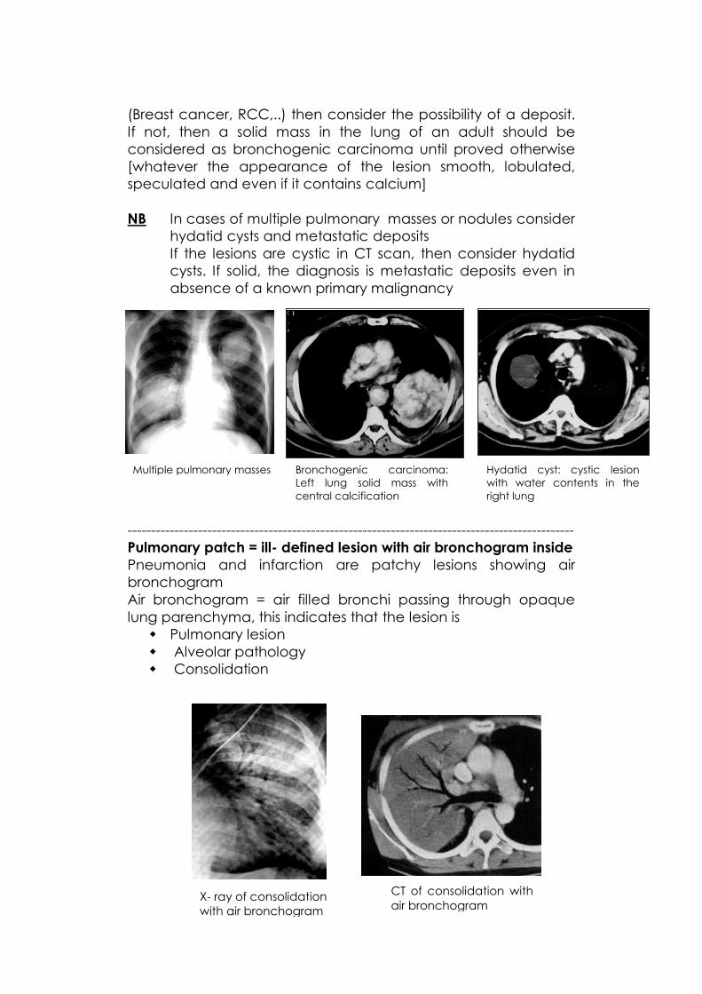
Pulmonary patch = ill- defined lesion with air bronchogram inside
Pneumonia and infarction are patchy lesions showing air
bronchogram
Air bronchogram = air filled bronchi passing through opaque
lung parenchyma, this indicates that the lesion is
Pulmonary lesion
Alveolar pathology
Consolidation
Multiple pulmonary masses
X- ray of consolidation
with air bronchogram
CT of consolidation with
air bronchogram
Bronchogenic carcinoma:
Left lung solid mass with
central calcification
Hydatid cyst: cystic lesion
with water contents in the
right lung
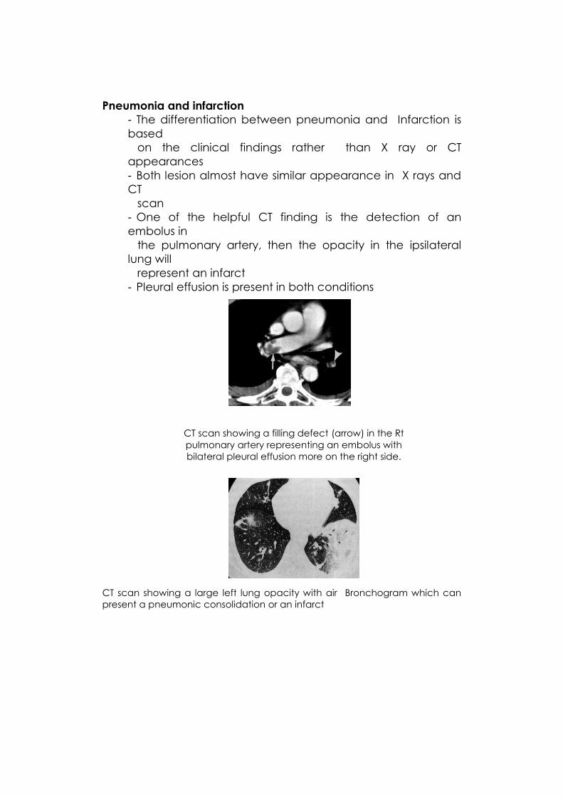
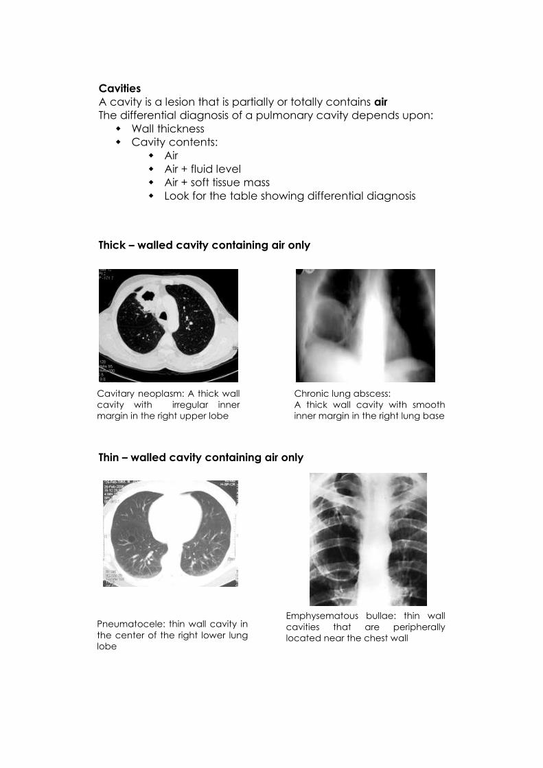
Pneumonia and infarction
- The differentiation between pneumonia and Infarction is
based
on the clinical findings rather than X ray or CT
appearances
- Both lesion almost have similar appearance in X rays and
CT
scan
- One of the helpful CT finding is the detection of an
embolus in
the pulmonary artery, then the opacity in the ipsilateral
lung will
represent an infarct
- Pleural effusion is present in both conditions
CT scan showing a large left lung opacity with air Bronchogram which can
present a pneumonic consolidation or an infarct
CT scan showing a filling defect (arrow) in the Rt
pulmonary artery representing an embolus with
bilateral pleural effusion more on the right side.
Cavities
A cavity is a lesion that is partially or totally contains air
The differential diagnosis of a pulmonary cavity depends upon:
Wall thickness
Cavity contents:
Air
Air + fluid level
Air + soft tissue mass
Look for the table showing differential diagnosis
Thick – walled cavity containing air only
Thin – walled cavity containing air only
Cavitary neoplasm: A thick wall
cavity with irregular inner
margin in the right upper lobe
Chronic lung abscess:
A thick wall cavity with smooth
inner margin in the right lung base
Pneumatocele: thin wall cavity in
the center of the right lower lung
lobe
Emphysematous bullae: thin wall
cavities that are peripherally
located near the chest wall
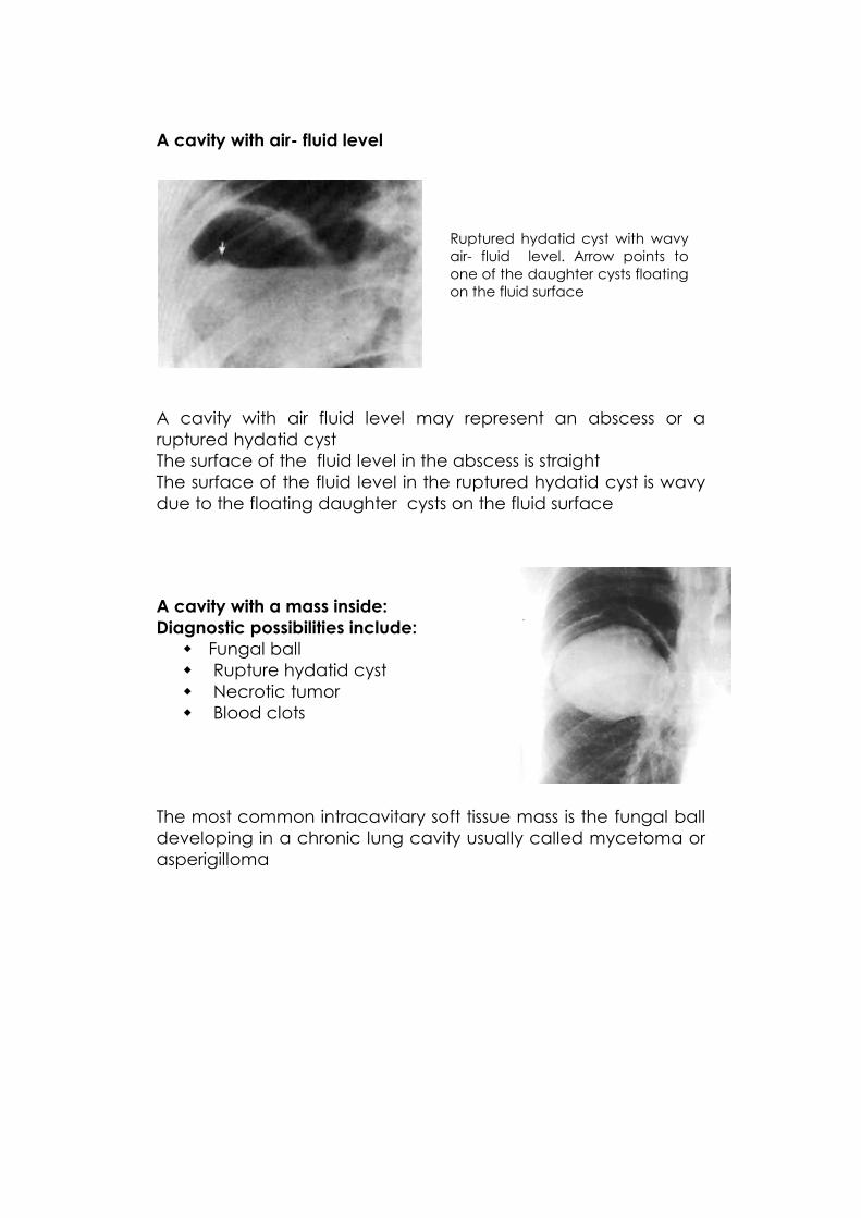
A cavity with air- fluid level
A cavity with air fluid level may represent an abscess or a
ruptured hydatid cyst
The surface of the fluid level in the abscess is straight
The surface of the fluid level in the ruptured hydatid cyst is wavy
due to the floating daughter cysts on the fluid surface
A cavity with a mass inside:
Diagnostic possibilities include:
Fungal ball
Rupture hydatid cyst
Necrotic tumor
Blood clots
The most common intracavitary soft tissue mass is the fungal ball
developing in a chronic lung cavity usually called mycetoma or
asperigilloma
Ruptured hydatid cyst with wavy
air- fluid level. Arrow points to
one of the daughter cysts floating
on the fluid surface

Cavitary
lesions
Contents
Air fluid level Air only
Surface of the fluid
level
Wall thickness
Straight Abscess
Wavy Ruptured hydatid
cyst
Thick
Thin
Site
Central in the lung
Pneumatocele
Peripheral near the chest wall Emphysematous
bulla
Inner margin of the cavity
Smooth Irregular
Chronic lung abscess
Breakdown in a bronchogenic
carcinoma