chf work up, a case presentation
TRANSCRIPT

CHF Work Up, A Case Presentation Rebecca Cogswell, MD Medical Director of Mechanical Circulatory Support University of Minnesota

Talk Overview: HFrEF
• Epidemiology • Pathophysiology • Hemodynamic concepts • Causes • Case • Summary

Prevalence 5.7 million (2009 to 2012) to 6.5 million (2011 to 2014)
Heidenreich et al, 2013

Stages of CHF

Pathophysiology – Heart Failure with Reduced Ejection Fraction
MAP- CVP = CO x SVR
Neurohormonal Activation * Renin-angiotensin-aldosterone * norepinephrine * endothilin * vasopressin - Stimulate hypertrophy, fibrosis,
• - Vasoconstriction, Na+, fluid retention
* LV overfilling - eccentric remodeling Wall stress ~ P*r/2h Oxygen demand
•

Courtesy of Barry Borlaug

CAUSES - HEART FAILURE REDUCED EJECTION FRACTION
– Pattern recognition - ECHO and ECG (LV non-compaction, concentric LVH, wall motion abnormalities in coronary distribution, LVEDD- time course)
– PET/MRI/genetic testing - enhanced classification of cardiomyopathies
– Accurate identification of the cause - implications on treatment, prognosis, family screening, and development of future therapies

NEW PATIENT - Viral symptoms - Medical history: CAD risk factors, thyroid history, radiation/chemotherapy,
arrhythmias, HTN, autoimmune disease, pregnancy - Family history - Drug history - Region of origin - HIV risk factors - Clues for sarcoid (mediastinal LAN), amyloidosis (carpel tunnel),hemochromatosis
(arthritis, bronze skin, LFTs)

Causes of HFrEF (systolic heart failure) CAD # 1 (industrialized countries)
• Class IA recommendation for definitive CAD evaluation Idiopathic dilated (when all else excluded)
Familial (20-30 % of all idiopathic dilated CM)
• LV non-compaction, ARVC, hypertrophic CM, muscular dystrophy • Transthyretin mutations(V122I in African Americans), hereditary hemochromatosis

Toxin-induced cardiomyopathy Alcohol Cocaine, methamphetamine Chemotherapeutic agents (most common anthracycline) Plaquenil
Radiation – restrictive (valves, coronary arteries, pericardium)
Cytoplasmic vacuoles – absence of inflammation

Infectious/inflammatory Coxsackie B, influenza, adenovirus, HIV Lyme Chagas- Trypanosoma cruzi
Endomyocardial fibrosis (Loeffler's myocarditis) Autoimmune
Lupus, scleroderma, rheumatoid arthritis Giant cell myocarditis
Endocrine Hyperthyroidism Acromegaly Pheochromocytoma Thiamine deficiency (wet beriberi, high output)

Stress induced • Acute systolic dysfunction, appears similar to large anterior wall MI • Catecholamine surge
Electrical causes: tachycardia, LBBB, RV pacing, high PVC burden Peri-partum
• Last month of pregnancy to first 5 months post pregnancy • ½ normalize within 6 months
Iron overload (primary or secondary) ferritin, iron/TIBC >50 %
Iron: Prussian blue staining

Infiltrative cardiomyopathies – Cardiac amyloid (AL, TTR (wild type and mutant)) –EMBx high sensitivity – Cardiac sarcoidosis – EMBx - low sensitivity
BEFORE STEROIDS AFTER STERIODS

Case: 38 M CC: SOB x 3 days, cough
+ orthopnea, +PND, + LE edema. One episode of syncope 2 years prior
No chest pain, no palpitations + nausea
PMHx: none
Social: No ETOH, no illicit drugs, no smoking. Married, full time chef
FHx: brother died in a car accident age 28, grandmother CHF in 50s

Case Vitals: Afebrile HR 110, SBP 110/90, RR 18, 93 % RA
No overt respiratory distress
JVP angle of the jaw, diffuse PMI, + summation gallop, trace +LE edema
Lungs clear
CXR: pulmonary edema
ECG: sinus tachycardia, LBBB (QRS 160 ms)
LABS: NT-pro-BNP: 10,000, Cr 1.4, Na 130, bicarb 18, LFTS normal, troponin 0.015
ECHO: LVEF 15 %, LVEDD 7.8 cm, mild mitral regurgitation

Question What are the most appropriate first steps in stabilizing this patient? A) Oral beta blocker, ace-inhibitor, IV diuretics
B) Oral beta blocker, ARNI, spironolactone, IV diuretics
C) Afterload reduction and IV diuretics
D) Sildenafil, IV diuretics, beta blocker
E) ICD, beta blocker, diuretics, ace-inhibitor

ANSWER C: Afterload reduction, IV diuretics

The ABCL GRID PCWP (LV filling pressure) < 14 > 14
>2.2 Cardiac index < 2.2
Stevenson, 1999 European Journal of Heart Failure

Back to our patient…
• HR 110, SBP 110/90 • Neck veins to jaw • Low bicarb: LACTIC ACIDOSIS • Clear lungs?
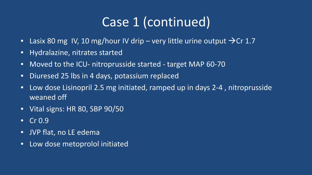
Case 1 (continued) • Lasix 80 mg IV, 10 mg/hour IV drip – very little urine output Cr 1.7 • Hydralazine, nitrates started • Moved to the ICU- nitroprusside started - target MAP 60-70 • Diuresed 25 lbs in 4 days, potassium replaced • Low dose Lisinopril 2.5 mg initiated, ramped up in days 2-4 , nitroprusside
weaned off • Vital signs: HR 80, SBP 90/50 • Cr 0.9 • JVP flat, no LE edema • Low dose metoprolol initiated

Case
Work up: Coronary angiogram: negative TSH normal, HIV negative, utox negative Cardiac MRI: negative for infiltrative processes or myocarditis Genetic testing planned as an outpatient Discharged with a life vest, followed up in CHF clinic in 3 days

Case 1 • 2-3 months- medications escalated to target doses • Cardiac rehab completed, back to work month 2 • NHYA class II symptoms, spironolactone added • Month 4 – repeat echo: LVEDD 6.0 cm, LVEF 25 %

• Always push therapy toward target doses (“make a move”)
– Lisinopril 40 mg daily – Entresto (200 mg BID (97/103mg tabs)) – Metoprolol XL 200 mg daily, carvedilol 25 mg BID – Spironolactone 25 mg daily
• Start low and go slow
• Do not up-titrate beta blocker when patient is decompensated/volume overloaded • If there is volume, diurese (even if there is renal dysfunction)
• If they still have class III (or more) symptoms REFER…. This is not normal
HFrEF Management – Clinic

Case (continued)
• Genetic testing: desmosome mutation - associated with DCM • Bi-V ICD placed • Lisinopril Entresto (PARAMOUNT TRIAL) • 2 other family members identified with low EF

Case 3 years post diagnosis Nt-bnp 300, normal end organ function LVEF 30 % No further hospitalizations Full, active life

• Neurohormonal activation adverse LV remodeling • Afterload reduction improves cardiac performance • Knowing the causes of HFrEF dictates work up and treatment, pattern recognition • Further classification important for treatment, prognosis, family screening, and
development of future therapies • Referral to cardiology/heart failure – ensure optimized
SUMMARY