chiu hs, llobet-navas d, yang x, chung wj, ambesi
TRANSCRIPT

This work is licensed under a Creative Commons Attribution-NonCommercial 4.0 International License
Newcastle University ePrints - eprint.ncl.ac.uk
Chiu HS, Llobet-Navas D, Yang X, Chung WJ, Ambesi-Impiombato A, Iyer A,
Kim HR, Seviour EG, Luo Z, Sehgal V, Moss T, Lu Y, Ram P,
Silva J, Mills GB, Califano A, Sumazin P.
Cupid: simultaneous reconstruction of microRNA-target and ceRNA networks.
Genome Research 2015, 25, 257-267.
Copyright:
© 2015 Chiu et al.; Published by Cold Spring Harbor Laboratory Press
This article is distributed exclusively by Cold Spring Harbor Laboratory Press for the first six months after the full-issue publication date (see http://genome.cshlp.org/site/misc/terms.xhtml). After six months, it is available under a Creative Commons License (Attribution-NonCommercial 4.0 International), as described at http://creativecommons.org/licenses/by-nc/4.0/.
DOI link to article:
http://dx.doi.org/10.1101/gr.178194.114
Date deposited:
02/09/2016

Method
Cupid: simultaneous reconstruction of microRNA-target and ceRNA networksHua-Sheng Chiu,1,2,3,4,12 David Llobet-Navas,5,12 Xuerui Yang,6 Wei-Jen Chung,1,2,3
Alberto Ambesi-Impiombato,7,8 Archana Iyer,1 Hyunjae Ryan Kim,9 Elena G. Seviour,10
Zijun Luo,10 Vasudha Sehgal,10 Tyler Moss,10 Yiling Lu,10 Prahlad Ram,10 Jos�e Silva,5
Gordon B. Mills,10 Andrea Califano,1,2,3,7,8,11 and Pavel Sumazin4
1Department of Systems Biology, 2Center for Computational Biology and Bioinformatics, 3Department of Biomedical Informatics,
Columbia University, New York, New York 10032, USA; 4Texas Children’s Cancer Center, Baylor College of Medicine, Houston, Texas
77030, USA; 5Department of Pathology, Icahn School of Medicine at Mount Sinai, New York, New York 10029, USA; 6MOE Key
Laboratory of Bioinformatics, Tsinghua-Peking Center for Life Sciences, School of Life Sciences, Tsinghua University, Beijing 100084,
China; 7Institute for Cancer Genetics, 8Herbert Irving Comprehensive Cancer Center, Columbia University, New York, New York 10032,
USA; 9Laboratory of RNAMolecular Biology, Rockefeller University, New York, New York 10065, USA; 10Department of Systems Biology,
The University of Texas MD Anderson Cancer Center, Houston, Texas 77030, USA; 11Department of Biochemistry and Molecular
Biophysics, Columbia University, New York, New York 10032, USA
We introduce a method for simultaneous prediction of microRNA–target interactions and their mediated competitiveendogenous RNA (ceRNA) interactions. Using high-throughput validation assays in breast cancer cell lines, we show thatour integrative approach significantly improves on microRNA–target prediction accuracy as assessed by both mRNA andprotein level measurements. Our biochemical assays support nearly 500 microRNA–target interactions with evidence forregulation in breast cancer tumors. Moreover, these assays constitute the most extensive validation platform for com-putationally inferred networks of microRNA–target interactions in breast cancer tumors, providing a useful benchmark toascertain future improvements.
[Supplemental material is available for this article.]
MicroRNAs (miRNAs) regulate RNA stability and mRNA translation
(Filipowicz et al. 2008) and their dysregulation has been implicated
in a wide range of human diseases including cancer (Garzon et al.
2009). Consequently, establishing accurate and comprehensive
repertoires of miRNA–target interactions is a necessary step toward
elucidating their mechanistic role in pathophysiology. Dissecting
miRNA regulation, however, has proven challenging because can-
didate miRNA binding sites are ubiquitous and their regulatory ef-
fects are context specific (Liu et al. 2005; Lu et al. 2005; Mukherji
et al. 2011). As a result, and despite their relatively low accuracy,
computational prediction methods that incorporate context-spe-
cific data are preferred for screening for miRNA–target interactions
in tumor contexts (Carroll et al. 2013; Erhard et al. 2014).
To address these challenges, we introduce Cupid, an in-
tegrative framework for the context-specific inference of miRNA
targets. Cupid integrates sequence-based evidence and functional
clues derived from RNA and miRNA expression analysis, predicting
candidate miRNA binding sites and associated target genes using
ensemble machine learning classifiers that are trained on validated
interactions. Candidate interactions emerging from this step are
then refined based on independent, context-specific clues, in-
cluding their predicted ability to mediate competitive endogenous
RNA (ceRNA) interactions, wheremRNA compete for sharedmiRNA
regulators (Fig. 1A; Tay et al. 2014). Thus, Cupid simultaneously
infers both interaction types (ceRNA and miRNA–target in-
teractions). In addition, we considered evidence for combinatorial
regulation by multiple miRNA species (Fig. 1B; Boissonneault et al.
2009; Xu et al. 2011) and for indirect miRNA regulation through
effector proteins (Fig. 1C). Taken individually, these clues are pre-
dictive of bona fidemiRNA–target interactions and can significantly
improve the tradeoff between precision and recall.
We show that Cupid predictions outperform other leading al-
gorithms, based on multiple experimental assays, including PAR-
CLIP data, miRNA perturbation followed by mRNA and protein
expression profiles, and 39 luciferase activity assays. Critically, while
Cupidpredicts fewer interactions thanothermethods (Fig. 1D–F), its
predictions aremuchmore likely to be consistentwith experimental
evidence. This is critical since high false-positive prediction rates are
a key limitation of current miRNA–target prediction methods.
Results
Algorithm outline and miRNA–target prediction in breastcancer tumors
Cupid is implemented in three sequential steps (Fig. 1A). First, Cupid
reevaluates candidate miRNA binding sites in 39 UTRs, as inferred by
TargetScan (Lewis et al. 2005), miRanda (John et al. 2004), and PITA
� 2015 Chiu et al. This article is distributed exclusively by Cold Spring HarborLaboratory Press for the first six months after the full-issue publication date (seehttp://genome.cshlp.org/site/misc/terms.xhtml). After six months, it is avail-able under a Creative Commons License (Attribution-NonCommercial 4.0 In-ternational), as described at http://creativecommons.org/licenses/by-nc/4.0/.
12These authors contributed equally to this work.Correspondingauthors: [email protected], [email protected] published online before print. Article, supplemental material, and publi-cation date are at http://www.genome.org/cgi/doi/10.1101/gr.178194.114.
25:257–267 Published by Cold Spring Harbor Laboratory Press; ISSN 1088-9051/15; www.genome.org Genome Research 257www.genome.org
Cold Spring Harbor Laboratory Press on September 2, 2016 - Published by genome.cshlp.orgDownloaded from

(Kertesz et al. 2007). This is accomplished by integrating their algo-
rithm-specific scores, their location in the 39 UTR, and their cross-
species conservation. Then, miRNA–target interactions are predicted
by further integrating information about selected sites, their multi-
plicity, and the statistical dependency between the expression profiles
of miRNA and putative targets.
Likelihoods for each predictive feature are computed based
on a positive gold standard set of 588 experimentally confirmed
Figure 1. Methodology. (A) Cupid first reevaluates sites predicted by TargetScan, miRanda, and PITA, selecting and rescoring each candidate site (StepI). Sites are used to select and score miRNA-target interactions (Step II), which are then examined for evidence for mediating ceRNA interactions (Step III).In addition, to support interaction prediction we considered (B) evidence for combinatorial regulation between miRNAs and (C ) evidence for indirectregulation by miRNAs through effectors. (D) The majority of site predictions by TargetScan, miRanda, and PITA are exclusive to a single algorithm; forexample, <10% of sites predicted by miRanda are also predicted by another method. (E) Cupid predicted 529K miRNA-target interactions in Step II,excluding 60% of Step I candidate interactions. As a result, it makes considerably fewer predictions than TargetScan, miRanda, and PITA. (F) Cupid Step IIIpredictions include less than a quarter of Step I candidate interactions.
258 Genome Researchwww.genome.org
Chiu et al.
Cold Spring Harbor Laboratory Press on September 2, 2016 - Published by genome.cshlp.orgDownloaded from

miRNA–target interactions, representing 1481 binding sites in
TarBase (Papadopoulos et al. 2009), TRANSFAC (Matys et al. 2006),
and miRecords (Xiao et al. 2009). They are then integrated using
a support vector machine (SVM) algorithm (Chang and Lin 2011).
Finally, Cupid assesses whether inferred targets compete for their
predicted miRNA regulators. In the following sections, we discuss
results from Cupid-inferred miRNA targets based on gene expres-
sion profile data of TCGA breast cancer samples (The Cancer
Genome Atlas Network 2012).
Step I (miRNA binding-site analysis)
miRNA binding sites in 39 UTRs were predicted and scored in-
dependently by TargetScan, miRanda, and PITA. Taken together,
these algorithms predicted a total of 37M candidate binding sites
(Fig. 1D). Each site was associated with the following features: (1)
TargetScan, miRanda, and PITA confidence scores, when available;
(2) phastCons (Siepel et al. 2005) species-conservation scores; and
(3) relative distance from the 39 and 59 ends of the target 39 UTR.
These features were used to train a support vector machine (SVM)
classifier (Chang and Lin 2011) using 10-fold cross-validation and
bootstrap aggregating (bagging), to identify consensus candidate
binding sites. In total, out of 37M candidate sites from sequence
analysis, Cupid selected ;1.6M as likely miRNA binding sites (Fig.
1D); predictions and 39 UTR sequences are provided in Supple-
mental Table S1; see Supplemental Methods for details.
Step II (miRNA–target interaction refinement)
Candidate binding sites were then used to assess the probability of
an interaction between a miRNA and a target 39 UTR. Predicted in-
teractions were associated with the following features (see Supple-
mental Methods): (1) their scores from Step I; (2) the total number
of miRNA binding sites in a target 39 UTR (i.e., multiplicity); (3)
binding-site density; and (4) the inverse correlation between the
expression of the target 39 UTR gene and that of the miRNA, as
measured by signed normalized mutual information (NMI), which
was estimated using adaptive partitioning (Darbellay and Vajda
1999) and Spearman correlation. These individual features were
used to train an SVM classifier, leading to selection of 529K candi-
date miRNA-target interactions from 1.6M original binding sites in
Step I (Fig. 1E; see Supplemental Methods for details).
Step III ( functional evidence analysis)
Finally, candidate interactions from Step II were tested for sequence-
and expression-based evidence for 39 UTR competition for shared
miRNA regulators by identifying miRNAs that are also predicted to
‘‘mediate’’ ceRNA interactions. Thus, ceRNA and miRNA–target in-
teractions are simultaneously predicted based on interaction can-
didates from Step II. We note that when evaluated individually,
other lines of functional evidence (Fig. 1B,C) significantly improved
prediction accuracy, but their contribution was relatively small; see
Supplemental Methods for description and analyses. In total, com-
bined analysis of TCGA breast cancer gene and miRNA expression
profiles supported299KmiRNA–target interactions fromCupid Step
II (Fig. 1F).
Quality of binding-site selection
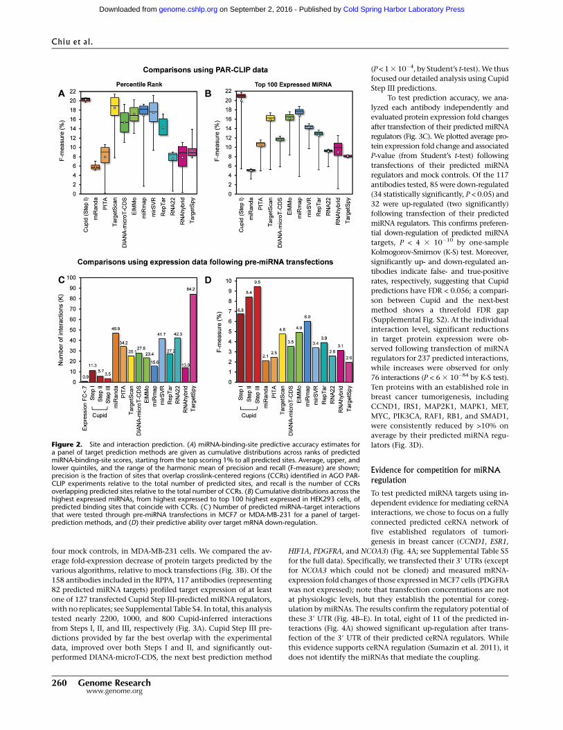
We first compared Cupid Step I performance to that of several
published algorithms, including TargetScan (Lewis et al. 2005),
miRanda (John et al. 2004), PITA (Kertesz et al. 2007), DIANA-
microT-CDS (Reczko et al. 2012), ElMMo (Gaidatzis et al. 2007),
miRmap (Vejnar and Zdobnov 2012), mirSVR (Betel et al. 2010),
RepTar (Elefant et al. 2011), rna22 (Miranda et al. 2006), RNAhybrid
(Rehmsmeier et al. 2004), and TargetSpy (Sturm et al. 2010) by
analyzing the overlap of their predictions with experimentally
assessed AGO crosslink-centered regions (CCR) in HEK293 cells
(Hafner et al. 2010). We tested both the accuracy of binding-site
scores (Fig. 2A) and the effects of miRNA expression (Fig. 2B) on
AGO localization; note that weakly expressedmiRNAs are less likely
to be associated with CCRs, and consistently, the 100 most highly
expressed miRNAs account for almost all CCRs.
Specifically, we computed cumulative F-measure distribution
statistics based on the overlap of experimentally assessed CCRs
with the k% highest confidence targets, as predicted by each al-
gorithm for the most expressed miRNAs in HEK293. The F-metric
is defined as the harmonic mean of precision and recall, i.e.,
Fk =2ðPk 3RkÞ=ðPk +RkÞ, where Pk and Rk are the method’s pre-
cision and recall for the k%most significant predicted binding sites
(Fig. 2A) or for the kmost expressed miRNAs (Fig. 2B). That is, Pk is
the frequency with which predicted binding sites overlap CCRs,
and Rk is the frequency with which CCRs overlap predicted bind-
ing sites.
Results suggest that Cupid consistently finds a good tradeoff
between precision and recall for any value of k (Fig. 2A). Similarly,
once a sufficiently large number of highly expressed miRNAs are
included in the test, Cupid outperforms other site-prediction
methods (Fig. 2B). Cupid’s predictive ability peaked at Fk =21:6%,
for the k=60 most expressed miRNAs, followed by miRmap and
ElMMo with Fk =18:6% and Fk =17:3%, for k=57 and k=50, re-
spectively. Overall, our results suggest that Cupid-predicted bind-
ing sites are in better agreement with AGO binding.
Quality of interaction prediction in breast cancer cell lines
To assess the algorithm’s ability to predict functionalmiRNA targets,
we used data from three breast cancer-specific studies that provide
gene-expressionprofiles (in duplicates) following transfectionof pre-
mir-18a, pre-mir-193b, pre-mir-206, pre-mir-302c (Leivonen et al.
2009), pre-mir-101-1 (Frankel et al. 2011), and scrambled controls in
MCF7 and pre-mir-145, and control in MDA-MB-231 (Gotte et al.
2010). In total, across the targets predicted for the six miRNAs, we
identified 869 down-regulated genes (>30% down-regulation),
supporting their role as targets of transfected miRNAs (Fig. 2C;
Supplemental Table S3; Guo et al. 2010).
Statistics were obtained separately following each of Cupid’s
steps. Specifically, we identified 11.3K (Step I), 5.7K (Step II), and 3.5K
(Step III) candidate targets of transfected miRNAs, respectively. We
calculated the F-measure for each algorithm under the assumption
that false-positive predictions would not be down-regulated follow-
ingmiRNA transfection and that false-negative predictions would be
down-regulated but not predicted. Our results suggest that Cupid’s
interaction prediction (Step II) and functional-interaction prediction
(Step III) significantly improve performance, compared to using only
binding-site predictions (Step I) (P < 0.01, FET). Critically, Cupid was
significantly more accurate (P < 0.05) than the next best algorithm
(miRmap) (Fig. 2D). Thus, Cupid inferred fewer candidate miRNA
targets, but with significantly higher F-statistics, suggesting a sub-
stantial increase in precision.
Protein expression benchmarks
We used RPPA data to measure expression fold reduction of 120
proteins following transfections of 159 miRNA mimics, including
Cupid: miRNA-target and ceRNA networks
Genome Research 259www.genome.org
Cold Spring Harbor Laboratory Press on September 2, 2016 - Published by genome.cshlp.orgDownloaded from
four mock controls, in MDA-MB-231 cells. We compared the av-
erage fold-expression decrease of protein targets predicted by the
various algorithms, relative to mock transfections (Fig. 3B). Of the
158 antibodies included in the RPPA, 117 antibodies (representing
82 predicted miRNA targets) profiled target expression of at least
one of 127 transfected Cupid Step III-predicted miRNA regulators,
with no replicates; see Supplemental Table S4. In total, this analysis
tested nearly 2200, 1000, and 800 Cupid-inferred interactions
from Steps I, II, and III, respectively (Fig. 3A). Cupid Step III pre-
dictions provided by far the best overlap with the experimental
data, improved over both Steps I and II, and significantly out-
performed DIANA-microT-CDS, the next best prediction method
(P < 13 10�4, by Student’s t-test).We thus
focused our detailed analysis using Cupid
Step III predictions.
To test prediction accuracy, we ana-
lyzed each antibody independently and
evaluated protein expression fold changes
after transfection of their predicted miRNA
regulators (Fig. 3C). We plotted average pro-
tein expression fold change and associated
P-value (from Student’s t-test) following
transfections of their predicted miRNA
regulators and mock controls. Of the 117
antibodies tested, 85 were down-regulated
(34 statistically significantly, P < 0.05) and
32 were up-regulated (two significantly)
following transfection of their predicted
miRNA regulators. This confirms preferen-
tial down-regulation of predicted miRNA
targets, P < 4 3 10�10 by one-sample
Kolmogorov-Smirnov (K-S) test. Moreover,
significantly up- and down-regulated an-
tibodies indicate false- and true-positive
rates, respectively, suggesting that Cupid
predictions have FDR < 0.056; a compari-
son between Cupid and the next-best
method shows a threefold FDR gap
(Supplemental Fig. S2). At the individual
interaction level, significant reductions
in target protein expression were ob-
served following transfection of miRNA
regulators for 237 predicted interactions,
while increases were observed for only
76 interactions (P < 6 3 10�84 by K-S test).
Ten proteins with an established role in
breast cancer tumorigenesis, including
CCND1, IRS1, MAP2K1, MAPK1, MET,
MYC, PIK3CA, RAF1, RB1, and SMAD1,
were consistently reduced by >10% on
average by their predicted miRNA regu-
lators (Fig. 3D).
Evidence for competition for miRNAregulation
To test predicted miRNA targets using in-
dependent evidence for mediating ceRNA
interactions, we chose to focus on a fully
connected predicted ceRNA network of
five established regulators of tumori-
genesis in breast cancer (CCND1, ESR1,
HIF1A, PDGFRA, and NCOA3) (Fig. 4A; see Supplemental Table S5
for the full data). Specifically, we transfected their 39 UTRs (except
for NCOA3 which could not be cloned) and measured mRNA-
expression fold changes of those expressed inMCF7 cells (PDGFRA
was not expressed); note that transfection concentrations are not
at physiologic levels, but they establish the potential for coreg-
ulation bymiRNAs. The results confirm the regulatory potential of
these 39 UTR (Fig. 4B–E). In total, eight of 11 of the predicted in-
teractions (Fig. 4A) showed significant up-regulation after trans-
fection of the 39 UTR of their predicted ceRNA regulators. While
this evidence supports ceRNA regulation (Sumazin et al. 2011), it
does not identify the miRNAs that mediate the coupling.
Figure 2. Site and interaction prediction. (A) miRNA-binding-site predictive accuracy estimates fora panel of target prediction methods are given as cumulative distributions across ranks of predictedmiRNA-binding-site scores, starting from the top scoring 1% to all predicted sites. Average, upper, andlower quintiles, and the range of the harmonic mean of precision and recall (F-measure) are shown;precision is the fraction of sites that overlap crosslink-centered regions (CCRs) identified in AGO PAR-CLIP experiments relative to the total number of predicted sites, and recall is the number of CCRsoverlapping predicted sites relative to the total number of CCRs. (B) Cumulative distributions across thehighest expressed miRNAs, from highest expressed to top 100 highest expressed in HEK293 cells, ofpredicted binding sites that coincide with CCRs. (C ) Number of predicted miRNA–target interactionsthat were tested through pre-miRNA transfections in MCF7 or MDA-MB-231 for a panel of target-prediction methods, and (D) their predictive ability over target mRNA down-regulation.
Chiu et al.
260 Genome Researchwww.genome.org
Cold Spring Harbor Laboratory Press on September 2, 2016 - Published by genome.cshlp.orgDownloaded from

We predicted that these genes compete for several miRNAs,
including seven miRNAs predicted to target at least three of the
four genes. We used 39 UTR luciferase reporter assays and miRNA
mimic transfections to test whether these miRNAs regulate their
predicted 39 UTR targets. In total, we predicted 30 miRNA-39 UTR
interactions for 10 miRNAs that were predicted to mediate ceRNA
interactions in the four-gene subnetwork (Fig. 5). Of particular
biological interest, ESR1, HIF1A, and PDGFRA were predicted to
compete for miR-17-5p, miR-106b-5p, miR-130a/b-3p, and miR-
301a-3p. Our assays tested 44 interactions, including interactions
with miR-557 as negative controls. Of the 30 predicted in-
teractions, only regulation of the HIF1A 39 UTR by miR-93-5p was
not supported by the experimental data, suggesting high precision
for Cupid’s predictions. The remaining assays tested 14 miRNA-39
UTR pairs, including four miR-557 targets that were not predicted
to interact, and the results suggest that eight of the 14 have regu-
latory potential.
ComparingCupid performance to random selection (same size)
from TargetScan, PITA, and miRanda interaction predictions, Cu-
pid had 80% accuracy and an F-measure of 0.87; random-selection
had accuracy of 22% and F-measure 0.15.
In total, our assays suggest that Cupid
predictions have high precision and
good, but lower, recall; precision was
above 95%, while recall was above 75%.
Of the 30 predictions, 10 were previously
known (Dweep et al. 2011; Hsu et al.
2011), see Supplemental Table S6, but
even after excluding these, Cupid calls
were predictive of assay results at P < 0.01
by FET, when comparing true positives
and true negatives to false positives and
false negatives. Interestingly, one pre-
viously reported interaction, CCND1 reg-
ulation bymiR-34b, was neither predicted
nor supported by our luciferase assays.
Other evidence for functionalregulation by miRNAs
We tested candidate miRNA–target in-
teractions for evidence for combinato-
rial regulation by miRNA species, and
evidence for indirect regulation through
effectors. Evidence for combinatorial reg-
ulation is complementary to evidence
derived from expression correlation be-
tweenmiRNAs and their targets. Similarly,
evidence for indirect regulation by a
miRNA examines the correlation between
the expression of this miRNA and a set
of predicted indirect targets; these were
not used to predict direct miRNA–target
interactions and are considered com-
plementary evidence. In total, these
lines of evidence produced fewer predic-
tions and had weaker predictive ability
when compared with evidence for ceRNA
regulation. Consequently, we chose to de-
scribe them independently. When com-
bined, these lines of evidence support
40,000 predicted interactions that were
not included in Step III (an additional 13%); see Supplemental
Methods.
ESR1 protein expression is correlated with miRNA regulatorexpression
We chose to focus on ESR1 for detailed validation. The analysis of
ESR1 protein expression in TCGA breast cancer tumors, profiled by
RPPA using the antibody ER.alpha.R.V_GBL.9014870, suggests that
ESR1protein expression is strongly correlatedwith the expression of
predicted miRNA regulators (Fig. 6A). Biochemical validation of se-
lect ESR1 miRNA regulators showed significantly reduced ESR1 39
UTR luciferase activity following transfection of predicted miRNA
regulators (Fig. 6B).
Results for 13 selected candidate ESR1 regulators, and miR-
557, which was chosen as negative control, are given in Figure 6A
and show significant ESR1 protein expression fold change. Eight
miRNAs were selected because their effect on ESR1 39 UTR lucif-
erase activity assays was tested in Figure 5. The other five regu-
lators where chosen at random from Supplemental Figure S4, and
Figure 3. High-throughput perturbation tests using protein-expression profiling. (A) Number ofpredictedmiRNA–target interactions that were tested through miRNAmimic transfection followed byprotein-expression profiling. (B) Average reduction in protein level following transfection of predictedmiRNA regulators for a panel of target prediction methods. (C ) P-values and average protein-ex-pression fold changes after transfection of Cupid-predicted miRNA regulators. In total, consideringexpression estimates made with 117 antibodies, 34 reported significant down-regulation P < 0.05, inred), 51 reported down-regulation (orange), 30 reported up-regulation (blue), and two reportedsignificant up-regulation (P < 0.05, in green); a comprehensive significance of P < 4 3 10�10. (D)Estimated average reduction in protein expression levels for known breast cancer regulators from C.Data are represented as mean 6 SEM.
Cupid: miRNA-target and ceRNA networks
Genome Research 261www.genome.org
Cold Spring Harbor Laboratory Press on September 2, 2016 - Published by genome.cshlp.orgDownloaded from

include previously validated regulators miR-22-5p, miR-221-3p,
and miR-222-3p, as well as previously undescribed ESR1-regula-
tors 381 and 148a-3p; 130b-5p was predicted to regulate ESR1
targets by Cupid Step II but had no evidence for mediating ESR1
ceRNA interactions. Results from biochemical testing of the pre-
dicted interactions, including results from assays described in
Figure 5, are given in Figure 6B for ease of presentation. All mimic
transfections of predicted ESR1 regulators significantly reduced
ESR1 39 UTR luciferase activity. As negative controls, we selected
miR-557 and miR-130b-5p, which were not predicted to target
ESR1; miR-130b-5p and ESR1 expression were significantly cor-
related (P < 1 3 10�8) but their interaction was not predicted by
Cupid Step III.
In total, 74miRNAswere predicted to target ESR1 byCupid Step
III. Given that ESR1mRNA expression is highly subtype specific—its
expression is high in luminal A and luminal B breast cancer tumors
and very low in basal-like tumors—we set out to test whether
miRNAs that target ESR1 may have inferred subtype-specific ac-
tivity. Our selection criteria for identifying miRNAs with subtype-
specific activity included two conditions: (1) miRNA expression
must be significantly high or low in one tumor type relative to
others (P < 13 10�3), and (2) its targets must be enriched for genes
with low or high expression in that tumor type (P < 1 3 10�4), re-
spectively, according to running sum statistics; see Supplemental
Methods. We also identified ceRNA with subtype activity, and
comprehensive lists for both are given in Supplemental Table S11.
In total, we identified 20 miRNAs that are predicted to target
ESR1 and have high or low activity in luminal tumors (Fig. 6C);
expression profiles of 12 of these miRNAs were significantly
anticorrelated with ESR1 protein expression in basal-like and
HER2-enriched tumors, but no miRNAs were significantly corre-
lated with ESR1 protein expression in luminal tumors (Fig. 6D).
This analysis suggests that ESR1 is regulated by a miRNA program
that is specific to basal-like and HER2-enriched tumors and is
absent in luminal tumors.
To test the predictive benefit of our compiled evidence for
functional regulation, we compared ESR1 protein expression in
352 samples with low (bottom 10%) and high (top 10%) expres-
sion of each of the 50 candidate miRNA regulators that were pre-
dicted in Cupid Step II and had evidence for indirect regulation
through effectors; note thatmiRNA expression and RPPA datawere
both available for only 352 of the 728 TCGAbreast cancer samples.
After removing outliers using the IQR rule for 44 miRNAs within
two interquartile ranges from the mean, ESR1 protein expression
was 2.8-fold higher in samples with low targeting miRNA expres-
sion, on average (Supplemental Fig. S4); only nine of thesemiRNAs
were not predicted by Cupid Step II. This finding is in agreement
with genome-wide statistical data that suggests that evidence for
functional regulation is significantly predictive of miRNA–target
interactions (Supplemental Fig. S4A,B).
DiscussionIdentifying and understanding pathological implications due to
miRNA dysregulation requires accurate maps of functional miRNA
targets in specific disease contexts. We describe systems-biology-
based methods that leverage previously validated interactions to-
gether with RNA and protein-expression profiles from patient
samples to predictmiRNA-39UTR target interactionswith evidence
for regulation in these samples. As a proof of principle, we pre-
dicted interactions in breast cancer using profiles from TCGA
breast cancer tumors together with perturbation data in breast
cancer cell lines. A variety of computational and biochemical
techniques demonstrated improved fidelity of resulting pre-
dictions, including evidence for hundreds of candidate miRNA–
target interactions in breast cancer cell lines. Our luciferase re-
porter assays supported >90% of interactions predicted by Cupid,
and we described evidence for functional regulation bymiRNAs in
breast cancer, including expression-based evidence for ceRNA
mediation by nearly 300K miRNA–target interactions. Other lines
of evidence that may be useful for building predictive functions in
the future failed to significantly improve predictive accuracy.
Figure 4. Competition for miRNA regulation. Cupid relies on evidencefor competition for miRNA regulation, simultaneously identifying ceRNAand miRNA–target interactions. (A) A ceRNA network of oncogenes im-plicated in breast cancer regulation. Transfections of the 39 UTRs of (B)CCND1, (C ) ESR1, (D)HIF1A, and (E) PDGFRA inMCF7 up-regulatedmRNAexpression within the network, as measured by qPCR. Data are repre-sented as mean 6 SEM; (*) P < 0.05, (**) P < 0.01, (***) P < 0.001.
Chiu et al.
262 Genome Researchwww.genome.org
Cold Spring Harbor Laboratory Press on September 2, 2016 - Published by genome.cshlp.orgDownloaded from

Focusing on predicted regulators of ESR1, we showed that
RPPA data in breast cancer tumors could be used as an effective
filter for identifying functional miRNA regulators. To further test
the effects of miRNA regulation on a select set of proteins, we
profiled protein expression after miRNA perturbation, producing
a data set that could be used to compare prediction performance,
and identifying breast cancer genes that are particularly amend-
able to miRNA regulation. In total, we identified nearly 500
miRNA–mRNA interactions with evidence for regulation in breast
cancer that were supported by miRNA perturbation assays in
breast cancer cell lines.
We observed subtype-specific activity (Supplemental Table
S11) and pathway enrichment (Supplemental Table S12) for both
miRNA and ceRNA regulators. Our analysis suggests that ESR1 is
targeted by miRNA programs that are specific to HER2-enriched
and basal-like tumors. We inferred that
the PI3K and the p53 signaling pathway
are enriched for miR-15, miR-16, and
miR-424 targets; the MAPK signaling
pathway is enriched for miR-106, miR-
20a-5p, and let-7 family targets; the TGF
beta signaling pathway is enriched for
miR-27b, miR-17-5p, and let-7 targets;
and that members of the miR-30 family
target the HER2/EGFR pathway. Inter-
estingly, members of the miR-30 family
were also identified to have the lowest
activity in HER2-enriched breast cancer
tumors, suggesting a possible tumor-sup-
pressor role for miR-30 in breast cancer
tumors; miR-30 has been recently de-
scribed as a tumor suppressor in prostate
cancer (Kao et al. 2014).We also note that
26 genes were predicted to be regulated
by at least 25% of miRNAs tested, and the
majority of these 26 are known regulatory
factors, including transcription and RNA
processing factors.
It is important to note that while
our results were based on analyses of
reference 39 UTR targeting, recent work
(Sandberg et al. 2008; Brummer and
Hausser 2014) suggests that both alter-
native polyadenylation and miRNA tar-
geting outside of 39 UTRs may alter
miRNA regulation to pathophysiolog-
ical effects. Incorporating these lines
of evidence may help further our un-
derstanding of miRNA regulation, and
we believe that these challenges can
be addressed using current technology.
Namely, the effects of alternative poly-
adenylation can be accounted for through
custom analyses of short-RNA libraries,
followed by mixture model resolution.
Similarly, methods for identifying miRNA
binding sites outside of 39 UTRs can be
used to supplement predictions presented
here. In addition, the combination of ac-
curate technology for predicting miRNA
targets and paired mRNA and protein-
expression profiles promises to enable
inquiry into miRNA mechanisms of action. For example, statisti-
cal evidence suggests that expression profiles of ESR1-regulating
miRNAs are predictive of variability in the coupling between ESR1
mRNA and protein-expression profiles. Moreover, a study focusing
on the ability of individual miRNA to predict coupling of target
mRNA and protein-expression profiles may help identify miRNAs
that primarily regulate translation and those that regulate mRNA
destabilization (Brummer and Hausser 2014).
In conclusion, the increasing body of molecular profiles in
primary disease tissues and in perturbation of disease models pres-
ents an opportunity for systems-biology-based approaches to im-
prove the accuracy of regulatory-interaction prediction methods.
Our analyses suggest that evidence for regulation by miRNAs in
given contexts can significantly improve context-specific miRNA–
target predictions, and consequently build better context-specific
Figure 5. Regulatory potential of miRNA mediators. Multiple ceRNA interactions in a network in-cluding CCND1, ESR1, HIF1A, and PDGFRA (Fig. 4) were predicted to be mediated by common miRNAs.We tested the regulatory potential of 10 of these miRNAs biochemically, with miR-557 selected asa negative control. (A) Predicted miRNA–target interactions and a summary of biochemical validation,depicting true-positive, true-negative, false-positive, and false-negative predictions; down-regulation of39 UTR luciferase activity in response to miRNA-mimic transfection at P < 0.05 was taken as evidence forregulation. Luciferase activity after miRNA mimic transfection relative to transfection of scrambledcontrol is shown for (B) CCND1, (C ) ESR1, (D) HIF1A, and (E) PDGFRA 39 UTRs. Punctuated mimics, forexample, miR-17-5p for CCND1, ESR1, HIF1A, but not PDGFRA, correspond to previously validated in-teractions. Data are represented as mean 6 SEM; (*) P < 0.05, (**) P < 0.01, (***) P < 0.001.
Cupid: miRNA-target and ceRNA networks
Genome Research 263www.genome.org
Cold Spring Harbor Laboratory Press on September 2, 2016 - Published by genome.cshlp.orgDownloaded from

Figure 6. ESR1 expression is anticorrelated with miRNA-regulator expression. (A) ESR1 protein expression in breast cancer tumors is anticorrelated withexpression profiles of previously validated (Known)andpredicted (Pred)miRNA regulators. ESR1 relative expression in the top andbottom10%of tumors rankedbased on the intensity of the expression of eachmiRNA; each rowwas ranked independently. miRNAs predicted to mediate ESR1 ceRNA regulation are marked(Step III); also marked is ESR1 protein expression fold change in tumor samples with low versus high expression for eachmiRNA. Negative controls includemiR-557 and miR-130b-5p; miR-130b-5p expression was anticorrelated with ESR1 expression. (B) 39 UTR luciferase activity fold changes after miRNA mimic trans-fections; some data replicated from Figure 5. Punctuatedmimics correspond to previously validated interactions. (C ) miRNAswith low or high activity in luminalbreast cancer tumors are enriched for predicted ESR1 regulators (P < 1.33 10�8), and (D) their expression profiles are anticorrelatedwith ESR1 protein expressionin basal-like and HER2-enriched tumors, but not in luminal tumors. Data are represented as mean 6 SEM; (*) P < 0.05, (**) P < 0.01, (***) P < 0.001.
264 Genome Researchwww.genome.org
Cold Spring Harbor Laboratory Press on September 2, 2016 - Published by genome.cshlp.orgDownloaded from

cellular wiring diagrams. Improving these has implications for efforts
to identify and interpret pathologically relevant genomic variants,
a key technical challenge in personalized genomics.
MethodsCupid multistep prediction and other functional evidencefor miRNA regulationCupid miRNA–target and ceRNA interaction predictions proceedin three sequential steps, as outlined in the Results. A detaileddescription, including Cupid site prediction (Step I), Cupid in-teraction prediction (Step II), the predictive features and machinelearning processes used in these steps, andmethods for identifyingcandidate miRNA–target interactions with evidence for mediatingceRNA interactions, are presented in the Supplemental Methods.In addition, we used evidence for combinatorial interactionsbetween miRNAs and for indirect miRNA regulation through ef-fectors to support Cupid Step II miRNA–target interaction pre-dictions; methods and results are described in the SupplementalMethods.
Genes-expression profiling following precursor transfection
Gene expression was profiled using Illumina Human-6 Expres-sion BeadChips following transfection of pre-mir-18a, pre-mir-193b, pre-mir-206, pre-mir-302c (Leivonen et al. 2009)(GSE14847), with Affymetrix GeneChip Human Genome U133Plus 2.0 Array following pre-mir-101-1 and scrambled controls inMCF7 (Frankel et al. 2011) (GSE31397), and pre-miR-145 andcontrol in MDA-MB-231 (Gotte et al. 2010) (GSE19737).
Testing sites and interactions
We used the F-measure to test the ability of binding-site predictionmethods to identify in 39UTRswith evidence formiRNA binding inHEK293 cells (Hafner et al. 2010), and when testing interactionsusing gene-expression data after transfection of precursors of pre-dicted miRNA regulators. Detailed methodology is presented in theSupplemental Methods.
39 UTR cloning, in vitro MIMIC transfection conditions, andluciferase assays
Tomeasure the targeting activity ofmicroRNAMIMICs, the 39UTRsof specific target genes were cloned downstream from the lu-ciferase reporter in the pMIR-REPORT vector (Life Technologies#AM5795M) by PCR from human genomic DNA using restrictionenzymes. 293T cells were plated at 70% confluence in 96-wellplates. Twenty-four hours later, cells were transfected with 50 ngof pMIR-REPORT constructs containing the luc-39-UTR sequences,50 ng of a Renilla normalization control, and 100 nM of each in-dividual synthetic mirVana miRNAMIMICs (Ambion #4464066) at100 nM final concentration using the TransIT-LT1 (Mirus Bio#2300A) and TransIT-TKO (Mirus Bio #2150) transfection reagentsfollowing the manufacturer’s instructions. After 24 h, relative lu-ciferase units (RLU) were measured using the Dual-Glo LuciferaseAssay System (Promega #E2949). Primer sequences are given in theSupplemental Methods.
Forward transfection of 39 UTRs
Forward transfection of the plasmid was performed with the Lipo-fectamine (Invitrogen) transfection reagent, following the manu-facturer’s protocol. Lipofectamine 2000 was used for A549, HepG2/C3A,HT-29, SK-MEL-28, and SK-OV-3 cells. LipofectamineLTXPLUS
was used for MCF7, PC-3, and U2-OS cells. In general, cells attachedto the culturing surfacewerewashedwithphosphate-buffered saline,and the medium was replaced with 100 mL of Opti-MEM with 2%fetal bovine serum. A total of 100 ng per well in a 96-well plate ofthe plasmid was then mixed with a 0.3 mL/well of Lipofectamine inOpti-MEM, and 20-min later the mixture was added to the wells.After 6 h of transfection, the cells were then cultured in regularmedium for 24 h and subsequently harvested.
Real-time quantitative RT-PCR analysis
Total RNAwas extracted fromcellswith theRNeasymini kit (Qiagen)and depleted of contaminatingDNAwithRNase-freeDNase (Qiagen).Equal amounts of total RNA (1 mg) were reverse-transcribed using theqScript cDNA Synthesis kit (Quanta Biosciences). The first-strandcDNA was used as a template. Real-time PCR was carried out usingSYBR green fluorescence. Two microliters of RT were used in a 25-mLreaction. Each sample was assayed in three independent RT reactionsand triplicate reactionswereperformedandnormalized to theGAPDHexpression levels. Negative controls included the absence of enzymein the RT reaction and the absence of template during PCR. Relativequantification of gene expression was performed with the compara-tive CT method. Primers used for quantitative RT-PCR analyses weresynthesized by Sigma-Aldrich.
High-throughput quantitative RT-PCR analysis
The first-strand cDNA, synthesized using the qScript cDNA Syn-thesis kit, was first amplified for specific target amplification (STA).Briefly, a 12-cycle preamplification reaction was performed foreach sample in 5 mL by pooling all primer pairs (final concentra-tion, 50 nM), 1.25 mL cDNA, and 2.5 mL 23 PreAmp Master Mix(Applied Biosystems) following the manufacturer’s protocol. Un-incorporated primers were then cleaned up using Exonuclease I(New England Biolabs). Briefly, 2 mL of diluted Exo I at 4 units/mLwas added to each 5-mL STA reaction, and then incubated for 30min at 37°C and 15 min at 80°C. Samples were then diluted fivetimes with low TE buffer. High-throughput qPCRs were performedon the Biomark HD (Fluidigm) in a microfluidic multiplex 48.48dynamic array chip according to the Fluidigm Advanced De-velopment Protocol with EvaGreen. For each individual assay, 5 mLAssay Mix containing 9 mM forward primer, 9 mM reverse primer,and 13 Assay Loading Reagent was loaded into the Assay Inlets onthe chip. For each sample, 5 mL Sample Mix containing 2.25 mLdiluted sample in 13 DNA binding Dye Sample Loading Reagentand 13 SsoFast EvaGreen Supermix (Bio-Rad) was loaded into thesample inlets. The Biomark’s default Fast thermo cycling programwith a melting step was used for the real time PCR reactions andfluorescence detection.
miRNA library screen by RPPA
The miRNA library was designed and synthesized by Dharmacon.MDA-MB-231 cells were seeded (3750 cells/well) and transfectedwith 50 nM miRNA mimics. After 48 h, cells were lysed and RPPAanalysis was carried out as previously described (Zhang et al. 2009;Hennessy et al. 2010; Lu et al. 2011); mimics tested and antibodiesused are given in Supplemental Table S4. Cells were washed withPBS, then lysed in 1% Triton X-100, 50 mM HEPES (pH 7.4), 150mM NaCl, 1.5 mM MgCl2, 1 mM EGTA, 100 mM NaF, 10 mM Napyrophosphate, 1 mM Na3VO4, 10% glycerol, containing freshlyadded protease and phosphatase inhibitors. Cellular proteins weredenatured by 1% SDS (with b-mercaptoethanol) and diluted in sixtwofold serial dilutions indilution buffer (lysis buffer containing 1%SDS). Serial diluted lysates were arrayed on nitrocellulose-coated
Cupid: miRNA-target and ceRNA networks
Genome Research 265www.genome.org
Cold Spring Harbor Laboratory Press on September 2, 2016 - Published by genome.cshlp.orgDownloaded from

FAST slides (Whatman Inc.). Each slide was probed with a validatedprimary antibody plus a biotinconjugated secondary antibody.
The signal was amplified using a DakoCytomation-catalyzedsystem (Dako) and visualized by DAB colorimetric reaction. Theslides were scanned, analyzed, and quantified using a customized-software Microvigene (VigeneTech Inc.) to generate spot intensity.Each dilution curve was fitted with the logistic model (‘‘SupercurveFitting’’ developed by the Department of Bioinfomatics and Com-putational Biology at the MD Anderson Cancer Center, ‘‘http://bioinformatics.mdanderson.org/OOMPA’’). The program fits a sin-gle curve using all the samples (in the dilution series) on a slide withthe signal intensity as the response variable and the dilution steps asthe independent variable. Protein expression for eachwell and eachantibody was normalized as SEM relative to mock transfections,with P-values for individual miRNA mimics calculated using a sin-gle-sample Student’s t-test against mock transfections. Mimics forRPPA experiments (159 miRNA mimics in total) were chosen froma preliminary test of 879mimics, identifying transfections that leadto highest total fold change across profiling antibodies.
Statistical analysis
All experiments were performed at least in triplicate and repre-sentative results are shown. All data are shown as the mean 6 SE.Student’s t-tests were used to evaluate statistical significances be-tween different treatment groups.
Data accessThe RPPA data from this study, including 163 RPPA experimentsfollowing miRNA transfections, where each experiment has read-outs from 158 antibodies as given in Supplemental Table S4, areavailable from The Cancer Proteome Atlas (TCPA; http://app1.bioinformatics.mdanderson.org/tcpa/_design/basic/index.html) un-der accession number TCPA00000001. Cupid source code is availablefor download fromSourceForgeprojectCupidTool athttp://cupidtool.sourceforge.net/.
AcknowledgmentsWe acknowledge the generous funding provided by the NIH underthe following grant awards: (1) Roadmap grant for a Center for theMultiscale Analysis of GeneticNetworks (MAGNet) (U54CA121852);(2) Genetic Network Inference with Combinational Phenotypes(R01CA109755); (3) In Silico Research Centers of Excellence NCI-caBIG29XS192 and 12ST1103; and (4) LINCS grants 1U01HL111566-01 and 5U01CA164184-02. RPPA was supported by an NCI CCSGgrant (P30 CA016672). G.B.M. and P.R. were supported by U54CA112970. The results published here are in part based upon datagenerated by TheCancerGenomeAtlas pilot project established bythe NCI and NHGRI as of January 2011. Information about TCGAand the investigators and institutions who constitute the TCGAresearch network can be found at http://cancergenome.nih.gov/.The dbGaP accession number for the data analyzed in this work isphs000178.v4.p4, dated January 24, 2011. P.R., G.B.M., P.S., andA.C. conceived and supervised the project and participated in itscomputational and experimental design. H.S.C. and P.S. designedand implemented the computational methods; H.S.C., P.R.,A.A.I., H.R.K., and P.S. analyzed data; P.S. and P.R. designed the ex-perimental assays. D.L.N., X.Y., and A.I. performed the experiments;H.S.C., P.S., and A.C. wrote the paper.
References
Betel D, Koppal A, Agius P, Sander C, Leslie C. 2010. Comprehensivemodeling of microRNA targets predicts functional non-conserved andnon-canonical sites. Genome Biol 11: R90.
Boissonneault V, Plante I, Rivest S, Provost P. 2009. MicroRNA-298 andmicroRNA-328 regulate expression of mouse beta-amyloid precursorprotein-converting enzyme 1. J Biol Chem 284: 1971–1981.
Brummer A, Hausser J. 2014.MicroRNA binding sites in the coding region ofmRNAs: extending the repertoire of post-transcriptional generegulation. Bioessays 36: 617–626.
The Cancer Genome Atlas Network. 2012. Comprehensive molecularportraits of human breast tumours. Nature 490: 61–70.
Carroll AP, Tooney PA, Cairns MJ. 2013. Context-specific microRNAfunction in developmental complexity. J Mol Cell Biol 5: 73–84.
Chang CC, Lin CJ. 2011. LIBSVM: a library for support vector machines.ACM Trans Intell Syst Technol 2: 27:1–27:27.
Darbellay G, Vajda I. 1999. Estimation of the information by an adaptivepartitioning of the observation space. IEEE Trans Inf Theory 45: 1315–1321.
Dweep H, Sticht C, Pandey P, Gretz N. 2011. miRWalk–database: predictionof possible miRNA binding sites by ‘‘walking’’ the genes of threegenomes. J Biomed Inform 44: 839–847.
Elefant N, Altuvia Y, Margalit H. 2011. A wide repertoire of miRNA bindingsites: prediction and functional implications. Bioinformatics 27: 3093–3101.
Erhard F, Haas J, Lieber D, Malterer G, Jaskiewicz L, Zavolan M, Dolken L,Zimmer R. 2014. Widespread context-dependency of microRNA-mediated regulation. Genome Res 24: 906–919.
Filipowicz W, Bhattacharyya SN, Sonenberg N. 2008. Mechanisms of post-transcriptional regulation by microRNAs: are the answers in sight? NatRev Genet 9: 102–114.
Frankel LB, Wen J, Lees M, Hoyer-Hansen M, Farkas T, Krogh A, Jaattela M,Lund AH. 2011. microRNA-101 is a potent inhibitor of autophagy.EMBO J 30: 4628–4641.
Gaidatzis D, van Nimwegen E, Hausser J, Zavolan M. 2007. Inference ofmiRNA targets using evolutionary conservation and pathway analysis.BMC Bioinformatics 8: 69.
Garzon R, Calin GA, Croce CM. 2009. MicroRNAs in cancer. Annu Rev Med60: 167–179.
Gotte M, Mohr C, Koo CY, Stock C, Vaske AK, Viola M, Ibrahim SA,Peddibhotla S, Teng YH, Low JY, et al. 2010. miR-145-dependenttargeting of junctional adhesion molecule A and modulation of fascinexpression are associated with reduced breast cancer cell motility andinvasiveness. Oncogene 29: 6569–6580.
Guo H, Ingolia NT, Weissman JS, Bartel DP. 2010. Mammalian microRNAspredominantly act to decrease targetmRNA levels.Nature 466: 835–840.
Hafner M, Landthaler M, Burger L, Khorshid M, Hausser J, Berninger P,Rothballer A, Ascano M Jr, Jungkamp AC, Munschauer M, et al. 2010.Transcriptome-wide identification of RNA-binding protein andmicroRNA target sites by PAR-CLIP. Cell 141: 129–141.
Hennessy BT, Lu Y, Gonzalez-Angulo AM, Carey MS, Myhre S, Ju Z, DaviesMA, Liu W, Coombes K, Meric-Bernstam F, et al. 2010. A technicalassessment of the utility of reverse phase protein arrays for the study ofthe functional proteome in non-microdissected human breast cancers.Clin Proteomics 6: 129–151.
Hsu SD, Lin FM,WuWY, Liang C, HuangWC, ChanWL, TsaiWT, Chen GZ,Lee CJ, Chiu CM, et al. 2011. miRTarBase: a database curatesexperimentally validated microRNA-target interactions. Nucleic AcidsRes 39: D163–D169.
John B, Enright AJ, Aravin A, Tuschl T, Sander C, Marks DS. 2004. HumanmicroRNA targets. PLoS Biol 2: e363.
Kao CJ, Martiniez A, Shi XB, Yang J, Evans CP, Dobi A, deVere White RW,Kung HJ. 2014. miR-30 as a tumor suppressor connects EGF/Src signal toERG and EMT. Oncogene 33: 2495–2503.
Kertesz M, Iovino N, Unnerstall U, Gaul U, Segal E. 2007. The role of siteaccessibility in microRNA target recognition. Nat Genet 39: 1278–1284.
Leivonen SK, Makela R, Ostling P, Kohonen P, Haapa-Paananen S, Kleivi K,Enerly E, Aakula A, Hellstrom K, Sahlberg N, et al. 2009. Protein lysatemicroarray analysis to identify microRNAs regulating estrogen receptorsignaling in breast cancer cell lines. Oncogene 28: 3926–3936.
Lewis BP, Burge CB, Bartel DP. 2005. Conserved seed pairing, often flankedby adenosines, indicates that thousands of human genes are microRNAtargets. Cell 120: 15–20.
Liu J, Valencia-Sanchez MA, Hannon GJ, Parker R. 2005. MicroRNA-dependent localization of targeted mRNAs to mammalian P-bodies. NatCell Biol 7: 719–723.
Lu J, Getz G,Miska EA, Alvarez-Saavedra E, Lamb J, PeckD, Sweet-Cordero A,Ebert BL, Mak RH, Ferrando AA, et al. 2005. MicroRNA expressionprofiles classify human cancers. Nature 435: 834–838.
Lu Y, Muller M, Smith D, Dutta B, Komurov K, Iadevaia S, Ruths D, Tseng JT,Yu S, Yu Q, et al. 2011. Kinome siRNA-phosphoproteomic screenidentifies networks regulating AKT signaling. Oncogene 30: 4567–4577.
Matys V, Kel-Margoulis OV, Fricke E, Liebich I, Land S, Barre-Dirrie A, ReuterI, Chekmenev D, Krull M, Hornischer K, et al. 2006. TRANSFAC and its
Chiu et al.
266 Genome Researchwww.genome.org
Cold Spring Harbor Laboratory Press on September 2, 2016 - Published by genome.cshlp.orgDownloaded from

module TRANSCompel: transcriptional gene regulation in eukaryotes.Nucleic Acids Res 34: D108–D110.
Miranda KC, Huynh T, Tay Y, Ang YS, Tam WL, Thomson AM, Lim B,Rigoutsos I. 2006. A pattern-based method for the identification ofMicroRNA binding sites and their corresponding heteroduplexes. Cell126: 1203–1217.
Mukherji S, Ebert MS, Zheng GX, Tsang JS, Sharp PA, van Oudenaarden A.2011.MicroRNAs can generate thresholds in target gene expression.NatGenet 43: 854–859.
Papadopoulos GL, Reczko M, Simossis VA, Sethupathy P, Hatzigeorgiou AG.2009. The database of experimentally supported targets: a functionalupdate of TarBase. Nucleic Acids Res 37: D155–D158.
Reczko M, Maragkakis M, Alexiou P, Grosse I, Hatzigeorgiou AG. 2012.Functional microRNA targets in protein coding sequences.Bioinformatics 28: 771–776.
Rehmsmeier M, Steffen P, Hochsmann M, Giegerich R. 2004. Fast andeffective prediction of microRNA/target duplexes. RNA 10: 1507–1517.
Sandberg R, Neilson JR, Sarma A, Sharp PA, Burge CB. 2008. Proliferatingcells express mRNAs with shortened 39 untranslated regions and fewermicroRNA target sites. Science 320: 1643–1647.
Siepel A, Bejerano G, Pedersen JS, Hinrichs AS, Hou M, Rosenbloom K,Clawson H, Spieth J, Hillier LW, Richards S, et al. 2005. Evolutionarilyconserved elements in vertebrate, insect, worm, and yeast genomes.Genome Res 15: 1034–1050.
Sturm M, Hackenberg M, Langenberger D, Frishman D. 2010. TargetSpy:a supervised machine learning approach for microRNA targetprediction. BMC Bioinformatics 11: 292.
Sumazin P, YangX,ChiuHS, ChungWJ, Iyer A, Llobet-Navas D, RajbhandariP, Bansal M, Guarnieri P, Silva J, et al. 2011. An extensive microRNA-mediated network of RNA-RNA interactions regulates establishedoncogenic pathways in glioblastoma. Cell 147: 370–381.
Tay Y, Rinn J, Pandolfi PP. 2014. The multilayered complexity of ceRNAcrosstalk and competition. Nature 505: 344–352.
Vejnar CE, Zdobnov EM. 2012. MiRmap: comprehensive prediction ofmicroRNA target repression strength. Nucleic Acids Res 40: 11673–11683.
Xiao F, Zuo Z, Cai G, Kang S, Gao X, Li T. 2009. miRecords: an integratedresource for microRNA-target interactions. Nucleic Acids Res 37: D105–D110.
Xu J, Li CX, Li YS, Lv JY, Ma Y, Shao TT, Xu LD, Wang YY, Du L, Zhang YP,et al. 2011. MiRNA-miRNA synergistic network: construction via co-regulating functional modules and disease miRNA topological features.Nucleic Acids Res 39: 825–836.
Zhang L, Wei Q, Mao L, Liu W, Mills GB, Coombes K. 2009. Serial dilutioncurve: a new method for analysis of reverse phase protein array data.Bioinformatics 25: 650–654.
Received May 9, 2014; accepted in revised form November 4, 2014.
Cupid: miRNA-target and ceRNA networks
Genome Research 267www.genome.org
Cold Spring Harbor Laboratory Press on September 2, 2016 - Published by genome.cshlp.orgDownloaded from

10.1101/gr.178194.114Access the most recent version at doi:2015 25: 257-267 originally published online November 5, 2014Genome Res.
Hua-Sheng Chiu, David Llobet-Navas, Xuerui Yang, et al. ceRNA networksCupid: simultaneous reconstruction of microRNA-target and
Material
Supplemental
http://genome.cshlp.org/content/suppl/2014/11/13/gr.178194.114.DC1.html
References
http://genome.cshlp.org/content/25/2/257.full.html#ref-list-1
This article cites 42 articles, 16 of which can be accessed free at:
License
Commons Creative
.http://creativecommons.org/licenses/by-nc/4.0/described at
a Creative Commons License (Attribution-NonCommercial 4.0 International), as ). After six months, it is available underhttp://genome.cshlp.org/site/misc/terms.xhtml
first six months after the full-issue publication date (see This article is distributed exclusively by Cold Spring Harbor Laboratory Press for the
ServiceEmail Alerting
click here.top right corner of the article or
Receive free email alerts when new articles cite this article - sign up in the box at the
http://genome.cshlp.org/subscriptionsgo to: Genome Research To subscribe to
© 2015 Chiu et al.; Published by Cold Spring Harbor Laboratory Press
Cold Spring Harbor Laboratory Press on September 2, 2016 - Published by genome.cshlp.orgDownloaded from