chlamydiosis in pen-raised bobwhite quail (colinus virginianus
TRANSCRIPT

Chlamydiosis in Pen-Raised Bobwhite Quail (Colinus virginianus) and Chukar Partridge(Alectoris chukar) with High MortalityAuthor(s): Douglas H. Erbeck and Stacey A. NunnSource: Avian Diseases, Vol. 43, No. 4 (Oct. - Dec., 1999), pp. 798-803Published by: American Association of Avian Pathologists, Inc.Stable URL: http://www.jstor.org/stable/1592752 .Accessed: 03/03/2011 11:19
Your use of the JSTOR archive indicates your acceptance of JSTOR's Terms and Conditions of Use, available at .http://www.jstor.org/page/info/about/policies/terms.jsp. JSTOR's Terms and Conditions of Use provides, in part, that unlessyou have obtained prior permission, you may not download an entire issue of a journal or multiple copies of articles, and youmay use content in the JSTOR archive only for your personal, non-commercial use.
Please contact the publisher regarding any further use of this work. Publisher contact information may be obtained at .http://www.jstor.org/action/showPublisher?publisherCode=aaap. .
Each copy of any part of a JSTOR transmission must contain the same copyright notice that appears on the screen or printedpage of such transmission.
JSTOR is a not-for-profit service that helps scholars, researchers, and students discover, use, and build upon a wide range ofcontent in a trusted digital archive. We use information technology and tools to increase productivity and facilitate new formsof scholarship. For more information about JSTOR, please contact [email protected].
American Association of Avian Pathologists, Inc. is collaborating with JSTOR to digitize, preserve and extendaccess to Avian Diseases.
http://www.jstor.org
AVIAN DISEASES 43:798-803, 1999
Case Report-
Chlamydiosis in Pen-raised Bobwhite Quail (Colinus virginianus) and Chukar Partridge (Alectoris chukar)
with High Mortality
Douglas H. Erbeck and Stacey A. Nunn
Murray State University Breathitt Veterinary Center, 715 North Drive, P.O. Box 2000, Hopkinsville, KY 42241-2000
Received 23 February 1999
SUMMARY. In a flock of 12,000 bobwhite quail (Colinus virginianus) and 7200 chukar partridge (Alectoris chukar), the owner had 100% morbidity and 40%-50% mortality in birds between the ages of 2 and 4 wk. Affected birds were stunted and anorexic and had yellow/green diarrhea. Two- and 4-wk-old birds submitted for necropsy all had slight nasal discharge. Histopathologic examination revealed mild (bobwhite) to severe (chukar) rhinitis. Immunohistochemistry was positive for Chlamydia psittaci in all birds. Chlamydia psittaci organisms were demonstrated histopathologically in hematoxylin and eosin and Gimenez- stained slides. Management sanitation and treatment with chlortetracycline stopped further excessive losses. The owners were also infected. Treatment by their local physician with tetracycline alleviated symptoms.
RESUMEN. Reporte de Caso--Clamidiosis con alta mortalidad en codornices (Colinus virginianus) y en perdices indias (Alectoris chukar) criadas en corrales de piso. En una parvada de 12000 codornices blancas (Colinus virginianus) y 7200 perdices indias (Alectoris chukar), se observ6 una morbilidad del 100% y una mortalidad del 40%-50% en aves entre dos y cuatro semanas de edad. Las aves afectadas mostraban enanismo, inapetencia y tenian diarrea de color amarillo verdosa. Las aves de dos a cuatro semanas enviadas para necropsia presentaban ligera descarga nasal. El examen histopatol6gico revel6 una rinitis suave en las codornices y severa en las perdices indias. El examen inmunohistoquimico fue positivo para Chlamydia psittaci en todas las aves. La Chlamydia psittaci fue observada histopatol6gi- camente por medio de la tinci6n de Gimenez y de la hematoxilina eosina. Medidas de manejo sanitario y el tratamiento con clortetraciclina evitaron mayores perdidas. Los propietarios tambien se infectaron y el tratamiento con tetraciclina alivi6 los sintomas.
Key words: Chlamydia psittaci, chlamydiosis in bobwhite quail, zoonotic chlamydiosis
Chlamydia psittaci is a bacteria that causes disease in birds and humans (1). The organism was originally isolated from psittacine birds and the disease was called psittacosis or parrot fever (8). Later, C. psittaci was shown to infect tur-
keys and other fowl, and the disease was called ornithosis (5,6). The organism isolated from the different species of birds is currently con- sidered the same, and the preferred term for the disease in both birds and humans is chlamydio- sis (9).
The disease is more severe in young birds (10). Clinical signs can include lethargy, nasal
and eye discharges, depression, anorexia, respi- ratory distress, and yellow diarrhea (7,10). Psit- tacines, gulls, egrets, and turkeys generally har- bor the more virulent strains (10). Older birds are often nonsymptomatic carriers and shedders (1). Avian strains of C. psittaci can also infect humans (1). Pathogenic strains of C. psittaci causing clinical signs and mortality have not previously been reported in quail (Schwartz, pers. comm.).
This case report documents chlamydiosis in a flock of bobwhite quail (Colinus virginianus) and chukar partridge (Alectoris chukar) experi-
798

Chlamydiosis in bobwhite and chukar 799
encing severe disease with mortality. The own- ers and their adult children who worked with the birds were subsequently diagnosed with Chlamydia psittaci infection.
CASE REPORT
History. Four 2-wk-old and four 4-wk-old bobwhite quail (virginianus) and six 4-wk-old chukar partridge were submitted to Murray State University Breathitt Veterinary Center in June 1998. The birds were from a game bird farm housing 12,000 bobwhite quail and 7200 chukar partridge that suddenly experienced 100% morbidity and 40%-50% mortality in birds between the ages of 2 and 4 wk. The farm incubated and hatched both species of birds. The quail were hatched from eggs purchased from an egg supplier certified clean for Salmo- nella and Mycoplasma. The chukar partridge were hatched from eggs of 300 breeder birds maintained in separate facilities on the farm. Every 2 wk, 1500 quail eggs and 900 partridge eggs were set and incubated. Eggs were incu- bated together, and the species were separated for hatching.
Both partridge and quail chicks were brood- ed and started together in a 32-ft by 75-ft house with six partitions. Chicks were brooded at one end of the house where the fresh air inlets were located. At 2-wk intervals, chicks were moved to the next partition farther from the fresh air supply. At 6 wk, both species were moved to large ground flight pens. Chicks were fed crushed quail starter pellets, 28% protein, that contained virginiamycin (Pfizer, Lee's Sum- mit, MO), 20 g/ton.
The first two hatches showed no clinical signs of infection. During the next two hatches, birds displayed no signs nor symptoms of dis- ease until about 2 wk of age. Anorexia, yellow/ green diarrhea, respiratory discharge, and stunt- ing would occur between 2 and 6 wk. Virtually 100% of the birds in the brood/start house old- er than 2 wk of age displayed clinical signs. Mortality was between 40% and 50%. Birds showing diarrhea were more severely affected. Four or 5 days after transfer to flight pens, clin- ical signs and mortality abated. There were no sequella; those birds that survived would con- tinue to normal adulthood.
Zoonosis. The owners, husband and wife, and one adult son who regularly worked with
the birds reported having stuffy head, cough, sneezing, mild sore throat, tightness of the chest, and headaches of several weeks' duration. A second adult son who worked sporadically with the birds had similar symptoms; however, his 7-yr-old daughter, who often interacted with the birds, and his pregnant wife, who had infrequent contact with the birds, did not dis- play any symptoms. An adult daughter who never went near the birds, but recalled previous contact with a normal-appearing parrot, had the most severe respiratory signs. When the owners learned that the birds had chlamydiosis, they contacted their local physician and re- quested that the family be tested for the disease. Serologic results for chlamydia antibodies, im- munoglobulin G (positive, ?1.10) (LabCorp, Louisville, KY), indicated the husband (1.92), his wife (1.33), both adult sons (2.06, 2.13), and adult daughter (3.00) tested positive. Titers from the pregnant daughter-in-law and her 7- yr-old daughter were negative (<0.91).
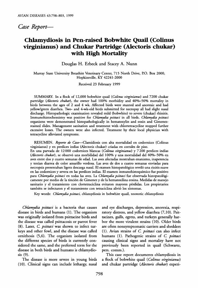
Necropsy. Antemortem findings in the 2- wk-old quail included stunting with ruffled feathers, serous to serosanguineous nasal dis- charge, and conjunctivitis with swollen eyelids (Fig. 1). The 4-wk-old quail had ruffled feath- ers with spotty feather loss over the dorsal head and body. One chick had slight serosanguine- ous, crusty discharge at the nostrils. One chu- kar was stunted and two had nasal discharge, one of which also had swollen eyelids.
Postmortem findings in the 2-wk old quail included red, hyperemic conjunctival mucous membranes; congested, hyperemic sinuses; and white urate and fibrinous streaked abdominal air sacs. The 4-wk-old quail had congested and hyperemic nasal turbinates, clear serous to cloudy fibrinous abdominal air sacs, and small streaking hemorrhages in the femoral muscles. Only two of the 6-wk-old chukar displayed vis- ible lesions. Congested and hyperemic conjunc- tival membranes and nasal turbinates were ob- served. Moderate numbers of Eimeria sp. oo- cysts were present in pooled fecal samples from each group of birds. Cryptosporidial organisms were not observed.
Toxicology. Analysis of the crushed starter feed and pellets fed to the older birds indicated that mycotoxins were within safe limits. By thin layer chromatography, aflatoxin B 1, ochratoxin A, zearalenone, T-2, and Diacetoxyscirpenol
800 D. H. Erbeck and S. A. Nunn
Fig. 1. Two-week-old bobwhite quail with crusty serosanguineous nasal discharge and swollen eyelids.
were less than 10 ppb and vomitoxin was less than 1 ppb.
Microbiology. A genus-specific nested
polymerase chain reaction procedure yielded negative results for the presence of Mycoplasma genus in lung, trachea, and choanal swabs.
Lung mycotic cultures were negative for Asper- gillus sp. Pseudomonas aeruginosa was isolated from lung and nasal swabs of each group and from liver from the chukar group. Other path- ogenic bacteria were not isolated from nasal swabs, trachea, lung, or air sacs.
Transmission electron microscopy of nasal si- nus, trachea, and lung was negative for virus
particles. Testing methanol-fixed lung impres- sion smears for the presence of Chlamydia was carried out by the fluorescent antibody tech-
nique. Fluorescein isothiocyanate-conjugated monoclonal antibody (Meridian Diagnostics, Inc., Cincinnati, OH) specific for all known
serotypes of C. psittaci, as well as Chlamydia trachomatis, was used in the procedure. Results were weakly positive and reported as equivocal.
Histopathology. Selected tissues, fixed in 10% neutral buffered formalin overnight and
processed by conventional methods, were em- bedded in paraffin, sectioned at 4 jxm, mount-
ed on precleaned glass slides, and stained with hematoxylin and eosin.
All three groups of birds had histopathologic lesions of the nasal turbinates. One of the 2- wk-old bobwhite quail had slightly hyperplastic turbinate epithelium. In the epithelial tissue were multifocal, mild to heavy accumulations of mononuclear leukocytes. Occasional hetero-
phils were within these areas. In the 4-wk-old bobwhite, in two of four nasal turbinates, small accumulations of mononuclear leukocytes ap- peared in the submucosal lamina. In the 6-wk- old chukar partridge, the turbinate tissues were edematous. The epithelial surfaces and sub- mucosal tissues were infiltrated with a fibrinous exudate containing mixed inflammatory cells. Small multifocal areas of necrosis with mixed
inflammatory cell exudate were present in both
epithelial and submucosal areas.
Lung tissues were unremarkable in the 2-wk- old quail, whereas one of four lung sections in the 4-wk-old quail had occasional areas of mononuclear leukocyte infiltration into the
capillary air spaces. The lungs of the chukar
partridge were unremarkable. Air sac sections from the chukar displayed greatly hyperplastic tissue infiltrated with a fibrinofibrous material.

Chlamydiosis in bobwhite and chukar 801
Fig. 2. Lung, capillary air space macrophage with multiple Chlamydiae psittaci elementary bodies. x400.
After immunohistochemistry studies were positive for C psittaci, retrospective histopath- ologic examination revealed C. psittaci reticulate bodies and more numerous elementary bodies (Fig. 2) in a few lung macrophages in at least one bird in each age group and in both species.
The Gimenez stain identified characteristic elementary bodies in lung tissues. Chlamydia psittaci organisms stained bright red and back- ground material stained light green.
In intestinal sections of duodenum, mild to moderate infiltration of enterocytes with coc- cidia organisms was present in each group of birds.
Sections of liver, trachea, kidney, heart and skeletal muscle were unremarkable.
Immunohistochemistry. Lung tissues were stained with a polyclonal antibody specific for C. psittaci (Chemicon, Temecula, CA). Tis- sue sections were treated with hydrogen per- oxide and digested with a proteolytic enzyme. After sections were thoroughly rinsed in water and Triton X Tris-buffered saline, the slides were transferred to an automated capillary gap system (Ventana Medical Systems, Inc., Tucson, AZ). Sections were incubated with a serum-free
protein-blocking agent (DAKO Corporation, Carpentenia, CA) and incubated with the pri- mary antibody. Bound antibodies were detected
by the avidin-biotin peroxidase method (Vector Laboratories, Burlingame, CA). Substrate was 3-amino-9-ethyl-carbazole chromagen (Bioge- nex Laboratories, San Ramon, CA). Sections were counterstained lightly with hematoxylin (Ventana Medical Systems, Inc.). Lung macro-
phages from each group of birds were positive for C. psittaci.
Treatment and results. When losses first occurred, birds were treated for 2 wk with ty- losin (Boehringer, St. Joseph, MO), 20 g/ton of feed. Mortality remained at about 50%. Af- ter laboratory diagnosis, the birds were treated with chlortetracycline (Durvet, Inc., Blue Springs, MO), 300 g/ton of feed, for chlamy- diosis and monensin (Bovatec, Hoffman-La- Roche, Inc., Nutley, NJ), 80 g/ton of feed, for coccidiosis. Thorough cleansing and disinfec- tion of the brooder/grow-out partitions and wa- ter/feed lines with 30% sodium hypochlorite (Vertex Chemical Corp., Dupo, IL) were con- ducted. Losses from later hatches dropped to less than 10%.
The humans were treated with tetracycline hydrochloride (Mylan Pharmaceuticals, Inc., Morgantown, WV), 250 mg twice daily for 15 days. The husband and the adult children had dramatic reduction of symptoms within 5 days. The wife reported only mild reduction of re- spiratory problems. Two weeks after comple- tion of treatment, the husband reported return of respiratory symptoms. Tetracycline treatment was resumed and symptoms cleared within 3 days. The wife's respiratory symptoms remained at a reduced level.
DISCUSSION
At initial submission, the clinical history of these birds included watery eyes, conjunctivitis, nasal discharge, and sinusitis, along with his- topathologic observations of moderate to severe rhinitis with mild lung lesions that were highly suggestive of Mycoplasma infection (1,4). How- ever, negative polymerase chain reaction DNA probe tests for Mycoplasma genus ruled out their possible role in these infections. Thereaf- ter, immunohistochemistry revealed positive re- actions for C psittaci in the lungs, and special stains for Chlamydia were ordered. When his- topathologic examination demonstrated Chla- mydia organisms, the owners were informed of the diagnosis. Treatment of the birds was im-

802 D. H. Erbeck and S. A. Nunn
mediately initiated. Recovery of the original tis- sue revealed it to be unsuitable for isolation at- tempts.
Once treatment was started, subsequent at-
tempts to isolate and demonstrate Chlamydia organisms were negative, even in birds continu-
ing to display typical signs. Owner discussion with personnel from the
quail breeder flocks revealed no unusual disease problems that would indicate chlamydiosis. Subsequent testing of hens from the chukar breeders failed to reveal evidence of C psittaci. Although the eggs could have harbored C. psit- taci organisms from external fecal contamina- tion, we believe this was unlikely until after the first group of chicks was severely affected.
Although chlamydiosis can be a serious dis- ease in chukar partridge (12), it has not been
reported as causing excessive mortality in bob- white quail (Schwartz, pers. comm.). The close
proximity of each species during the early grow- ing period suggests that both quail and par- tridge were infected with a pathogenic strain of
C. psittaci that contributed to excess mortality. The clinical signs of chlamydiosis vary great-
ly in severity and depend on the age of the bird and strain of the organism (1). Many avian spe- cies harbor Chlamydia organisms without show-
ing clinical signs (1). These birds act as carriers and can spread the disease. During periods of stress, carrier birds can break out with chlamy- diosis (11,13). Chlamydiosis is usually systemic; thus, infected birds may be further stressed by Chlamydia infection, and these stressed birds would be susceptible to secondary bacterial in- fections, including infection with Pseudomonas
organisms. Coinfection of C. psittaci and P ae-
ruginosa likely contributed to the high mortality of the quail and partridge chicks. Coccidia in- fection could have contributed to chick stress. The mild to moderate histopathologic lesions
suggested that this variable may also have been a coinfection factor.
In chickens and turkeys, Pseudomonas pro- duces respiratory infections (2). Because P ae-
ruginosa is an opportunist in weakened tissues (3), its isolation from the lungs of the birds was not surprising. Pseudomonas aeruginosa was later isolated from water lines in the starter facility. Vigorous cleaning and disinfection of water lines and cleaning and sanitation of the brooder and starter building were undertaken and prob- ably accounted for less secondary bacterial in-
fection. Chlamydia are very susceptible to chemicals that affect their lipid content or the
integrity of their cell walls (1). Even when
Chlamydia are in tissue debris, their infectivity is destroyed within minutes by all common dis- infectants except cresol and lime (1). Multipli- cation of all strains of Chlamydia is strongly inhibited by appropriate concentrations of tet-
racyclines (1). Treatment of Chlamydia with
chlortetracycline together with management sanitation to inhibit Pseudomonas likely contrib- uted to success in reducing mortality. Coccidi- osis treatment with monensin reduced stress.
The origin of the C. psittaci infection was never established. On the affected farm, a hu- man-to-bird zoonotic infection was suspected. The adult daughter, who worked in an estab- lishment where a client regularly brought a pet parrot, was the first to display upper respiratory symptoms. Others in the family then contract- ed "flulike" symptoms. The epornitic in the birds was believed to occur after the humans became ill. Unfortunately, circumstances did not allow testing of the parrot. Parrot-to-hu- man-to-chukar transmission is speculative but remains the suspected primary route of trans- mission.
Reservoirs of Chlamydia have been reported to include pigeons, blackbirds, grackles, house
sparrows, and killdeer (1). All of these avian
species could be found in the vicinity of the affected farm. Possibly a wild avian species transmitted the organism to chukar and bob- white on the farm. If the C psittaci infection came from birds, the chukar partridge were
likely infected first (13). Vertical transmission has been suggested to occur from infected eggs (5,6,14). Two of six chukar submitted for ex- amination displayed mild nasal and conjuncti- val signs. Commingling of the partridge and
quail likely resulted in the infection of the bob- white chicks.
This case report was interesting in that it was the first reported incident of C. psittaci epor- nitic with increased mortality in bobwhite
quail. The report also was a reminder of the zoonotic potential of chlamydiosis in game birds and their handlers.
REFERENCES
1. Andersen, A. A., J. E. Grimes, and P. B. Wyrick. Chlamydiosis (psittacosis, ornithosis). In:

Chlamydiosis in bobwhite and chukar 803
Diseases of poultry, 10th ed. B. W. Calnek, ed. Iowa State University Press, Ames, IA. pp. 333-349. 1997.
2. Barnes, H. J. Other bacterial diseases. In: Dis- eases of poultry, 10th ed. B. W. Calnek, ed. Iowa State University Press, Ames, IA. pp. 289-294. 1997.
3. Carter, G. R., M. M. Chengappa, and A. W. Roberts. Essentials of veterinary microbiology, 5th ed. Williams and Wilkins, Baltimore, MD. 1995.
4. Ley, D. H., and H. W. Yoder. Mycoplasma gallisepticum infection. In: Diseases of poultry, 10th ed. B. W. Calnek, ed. Iowa State University Press, Ames, IA. pp. 194-207. 1997.
5. Meyer, K. E Phagocytosis and immunity in psittacosis. Schweiz. Med. Wochenschr. 71:436-438. 1941.
6. Meyer, K. E, and B. Eddie. Ecology of avian psittacosis, particularly in parakeets. In: Progress in psittacosis research and control, R. Beaudette, ed. Rutgers University Press, New Brunswick, NY. pp. 52-56. 1958.
7. Mohan, R. Epidemiologic and laboratory ob- servations of Chlamydia psittaci infection in pet birds. J. Am. Vet. Med. Assoc. 184:1372-1374. 1984.
8. Morange, A. De la. Psittacoseou infection spe- ciale determinee par des parruches. M.Sc. Thesis. Ac- ademia de Paris, Paris, France. 1895.
9. Page, L. A., and J. E. Grimes. Avian chlamy- diosis (ornithosis). In: Diseases of poultry, 8th ed. M. S. Hofstad, H. J. Barnes, B. W. Calnek, W. M. Reid, and H. W. Yoder, Jr., eds. Iowa State University Press, Ames, IA. pp. 283-308. 1984.
10. Schwartz, L. D. Chlamydiosis. In: Grower's reference on gamebird health. Avicon, Inc., Okimos, MI. pp. 50-51. 1995.
11. Shewen, P. E. Chlamydial infection in ani- mals: a review. Can. Vet. J. 21:2-11. 1980.
12. Uemori, T., K. Asada, I. Kato, and R. Hara- sawa. Sensitive detection of Mycoplasma in cell cul- tures by using two step polymerase chain reaction system. Appl. Microbiol. 15:181-186. 1992.
13. Woodard, A. E. Raising chukar partridges. Cooperative Extension, University of California, Da- vis, CA. Leaflet 21321. 1982.
14. Wyrick, P. B., S. Richmond, and B. Chir. Bi- ology of Chlamydiae. Reports from the Symposium on Avian Chlamydiosis. J. Am. Vet. Med. Assoc. 195: 1507-1512. 1989.