christina liscynesky, md assistant professor of internal ... · study team. bulletin of the ......
TRANSCRIPT

EBOLA
Christina Liscynesky, MDAssistant Professor of Internal Medicine
Division of Infectious DiseasesAssociate Medical Director of Epidemiology

2
Sudan - June 27, 1976
Nazra Township Cotton factory storekeeper became ill 48 cases/ 27
deaths Contact was
admitted to local hospital in Maridi
Unrelated cases continued in cotton factory workers July-Oct.
World Health Organization. Ebola haemorrhagic fever in Sudan, 1976. Report of a WHO/International Study Team. Bulletin of the World Health Organization. 1978;56(2):247-270.
Content Provider: CDC/ Dr. Lyle Conrad; 1976
3
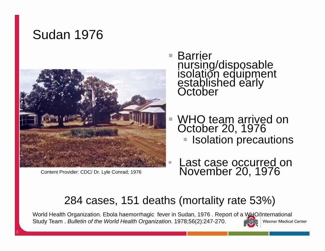
Sudan 1976
284 cases, 151 deaths (mortality rate 53%)
Barrier nursing/disposable isolation equipment established early October
WHO team arrived on October 20, 1976 Isolation precautions
• Last case occurred on November 20, 1976
World Health Organization. Ebola haemorrhagic fever in Sudan, 1976 . Report of a WHO/International Study Team . Bulletin of the World Health Organization. 1978;56(2):247-270.
Content Provider: CDC/ Dr. Lyle Conrad; 1976
4
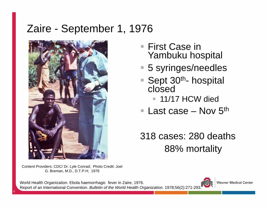
Zaire - September 1, 1976 First Case in
Yambuku hospital 5 syringes/needles Sept 30th- hospital
closed 11/17 HCW died
Last case – Nov 5th
318 cases: 280 deaths88% mortality
Content Providers: CDC/ Dr. Lyle Conrad; Photo Credit: Joel G. Breman, M.D., D.T.P.H; 1976
World Health Organization. Ebola haemorrhagic fever in Zaire, 1976. Report of an International Convention. Bulletin of the World Health Organization. 1978;56(2):271-293.

Ebolavirus
Sudan ebolavirus (SUDV) Zaïre ebolavirus (EBOV) Taï Forest (Ivory Coast) ebolavirus (TAFV) Bundibugyo ebolavirus (BDBV)
Reston ebolavirus (RESTV)
Mandell, Douglas, and Bennett’s Principals and Practice of Infectious Diseases, Seventh Edition. Gerald L. Mandell, John E. Bennett, and Raphael Dolin. 164, 2259-2263

Guinea- December 2013 Guéckédou, Guinea
2yr old Child Fever, black stool, vomiting Onset Dec. 2, 2013; died Dec. 6, 2013
• Transmitted from HCW (patient #14) to neighboring towns HCW died Feb. 10, 2014
• March 10, 2014 – Guinea Ministry of Health Notified
• March 14, 2014- Outbreak team in place
111 suspect cases: 79 deaths (71% mortality)
Baize S. et al. Emergence of Zaire Ebola Virus Disease in Guinea –Preliminary Report. N Engl J Med. 2014 Apr 16.
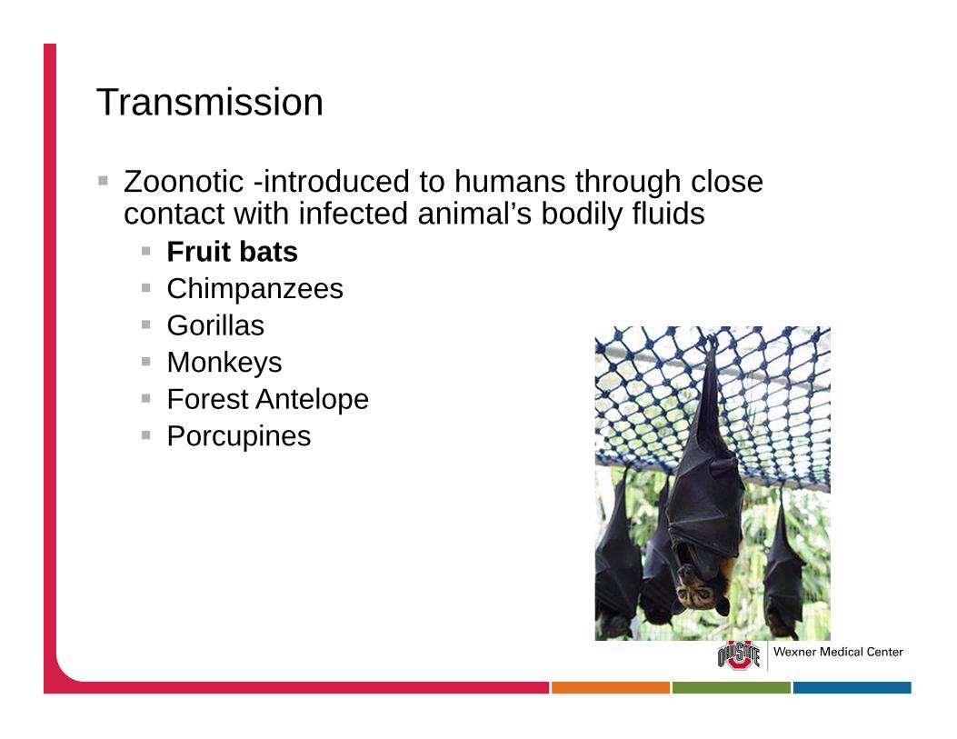
Transmission
Zoonotic -introduced to humans through close contact with infected animal’s bodily fluids Fruit bats Chimpanzees Gorillas Monkeys Forest Antelope Porcupines
Human to Human Transmission Direct contact with infected bodily secretions
Indirect contact with contaminated environments
In lab study: Ebola can remain active for up to 6 days Environmental cxs: 2/33 samples positive for Ebola
Blood stained physical glove Bloody IV insertion site
Direct contact with infected corpses
Men who survive can transmit virus via semen for up to 7 weeks
http://www.who.int/mediacentre/factsheets/fs103/en/Sagripanti JL, Rom AM, Holland LE. Persistence in darkness of virulent alphaviruses, Ebola virus, and Lassa virus deposited on solid surfaces. Arch Virol 2010; 155:2035-2039Bausch DG et al. Assessment of the Risk of Ebola Virus Transmission from Bodily Fluids and Fomites The J of Infect Dis 2007; 196:S142–7

High Risk Exposures
Percutaneous needle stick or mucous membrane exposure to body fluids
Direct care or exposure to body fluids without appropriate personal protective equipment (PPE)
Participation in funeral rites

Low Risk Exposures
Household member or other casual contact with an EVD patient
Providing patient care or casual contact without high-risk exposure with EVD patients

Ineffective Transmission
Previous epidemics have calculated that 1 primary human case of Ebola generates only 1 to 3 secondary cases on average.
1 case of Measles in West Africa generates14-17 cases.
Chowell G, Hengartner NW, Castillo-Chavez C,Fenimore PW, Hyman JM. The basic reproductive number of Ebola and the effects of public health measures: the cases of Congo and Uganda. J Theor Biol 2004;229:119-126Legrand J, Grais RF, Boelle PY, Valleron AJ,Flahault A. Understanding the dynamics of Ebola epidemics. Epidemiol Infect 2007;135:610-621

Clinical Manifestations
Incubation period of 8-10 days (range 2-21) Abrupt onset of fever, with HA and myalgia Nausea, vomiting, abdominal pain, and diarrhea Maculopapular rash by day 5-7 Chest pain, shortness of breath Hemorrhage Confusion, seizures
Kortepeter MG, Bausch DG, Bray M. Basic clinical and laboratory features of filoviralhemorrhagic fever. J Infect Dis. 2011 Nov;204 Suppl 3:S810–6

13
Differential Diagnosis
Malaria Typhoid Fever Dengue Lassa Fever Shigellosis Meningococcal
septicemia Plague Relapsing fever
Marburg Virus Yellow fever Viral hepatitis Anthrax Chikungunya fever Leptospirosis Typhus

Pathogenesis
Monocytes, macrophages, and dendritic cells are infected early Virus suppresses type 1 interferon responses and
induces cytokine and chemokine release Virus replicates, released and migrates to local lymph
nodes, travels through the lymphatic system to blood Virus disseminated throughout the body
Feldmann H, Geisbert TW. Ebola haemorrhagic fever. Lancet. 2011 Mar 5;377(9768):849-62.

Pathogenesis (cont)
Lymphocytes undergo apoptosis, which undermines adaptive immunity Hepatocellular necrosis DIC Adrenal necrosis Hypotension
Impaired Steroid Synthesis
Extensive tissue necrosis and shock
Diagnostic labs tests
Ebola virus is detectable in blood only after onset of symptoms
Detectable by real-time RT-PCR between 3 to 10 days post-onset of symptoms

Lab Abnormalities
Leukopenia Thrombocytopenia – 50 to 100K range Transaminitis: AST>ALT Proteinuria may be present PT and PTT prolonged Fibrin elevated
Fatal Illness
LFTS and D-dimer higher in fatal illness. Calcium <6mg/dL associated with death Median survival of 9 days Most patients die during the second week Alive on day 14 portends >75% survival Fatally infected patients do not develop an AB
response
Kortepeter MG, Bausch DG, Bray M. Basic clinical and laboratory features of filoviralhemorrhagic fever. J Infect Dis. 2011 Nov;204 Suppl 3:S810–6
Treatment
Supportive care Antibiotics for secondary infections
September 4-5, WHO scheduled conference on potential Ebola therapies and vaccines in Geneva

Experimental Therapeutics
ZMappTM - composed of three monoclonal antibodies directed against the Ebola Zaire virus strain. TkM-Ebola - small interfering RNAs targeting EV RNA
polymerase L AVI-7537 - which targets EV protein VP24 through an
RNA interference technology BCX-4430 - an adenosine analogue that is active
against EV in rodents chloroquine and imatinib, have shown activity against
EV in vitro and, in some cases, in rodent models.
Jesse L. Goodman, M.D., M.P.H. Studying “Secret Serums” — Toward Safe, Effective Ebola Treatments

Dr. Margaret Isaacson as she was tending to the needs of an Ebola patient in a Yambuku, Zaire hospital theatre block that was used as a temporary ICU for Ebola patients during the country’s 1976 outbreak.
Content Providers: CDC/ Dr. Lyle Conrad; Photo Credit: Joel G. Breman, M.D., D.T.P.H; 1976
Additional References
CDC Ebola Hemorrhagic Fever site: www.cdc.gov/ebola
WHO:http://www.who.int/csr/disease/ebola/en/
http://www.nejm.org/page/ebola-outbreak