chromosome analysis
TRANSCRIPT
CHROMOSOME ANALYSIS
NARENDRA YADAV
Msc II
PAPER II (MEDICAL MICROBIOLOGY)
WHAT ARE CHROMOSOMES??
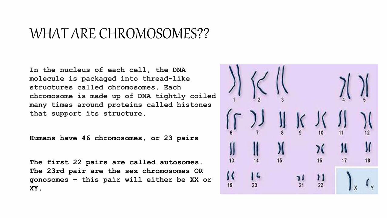
In the nucleus of each cell, the DNA
molecule is packaged into thread-like
structures called chromosomes. Each
chromosome is made up of DNA tightly coiled
many times around proteins called histones
that support its structure.
Humans have 46 chromosomes, or 23 pairs
The first 22 pairs are called autosomes.
The 23rd pair are the sex chromosomes OR
gonosomes – this pair will either be XX or
XY.
INTRODUCTION
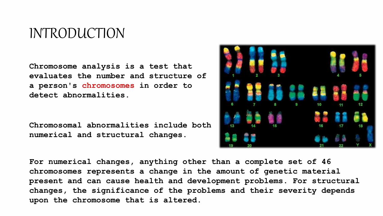
Chromosome analysis is a test that
evaluates the number and structure of
a person's chromosomes in order to
detect abnormalities.
Chromosomal abnormalities include both
numerical and structural changes.
For numerical changes, anything other than a complete set of 46
chromosomes represents a change in the amount of genetic material
present and can cause health and development problems. For structural
changes, the significance of the problems and their severity depends
upon the chromosome that is altered.
TECHNIQUES FOR
CHROMOSOME ANALYSIS
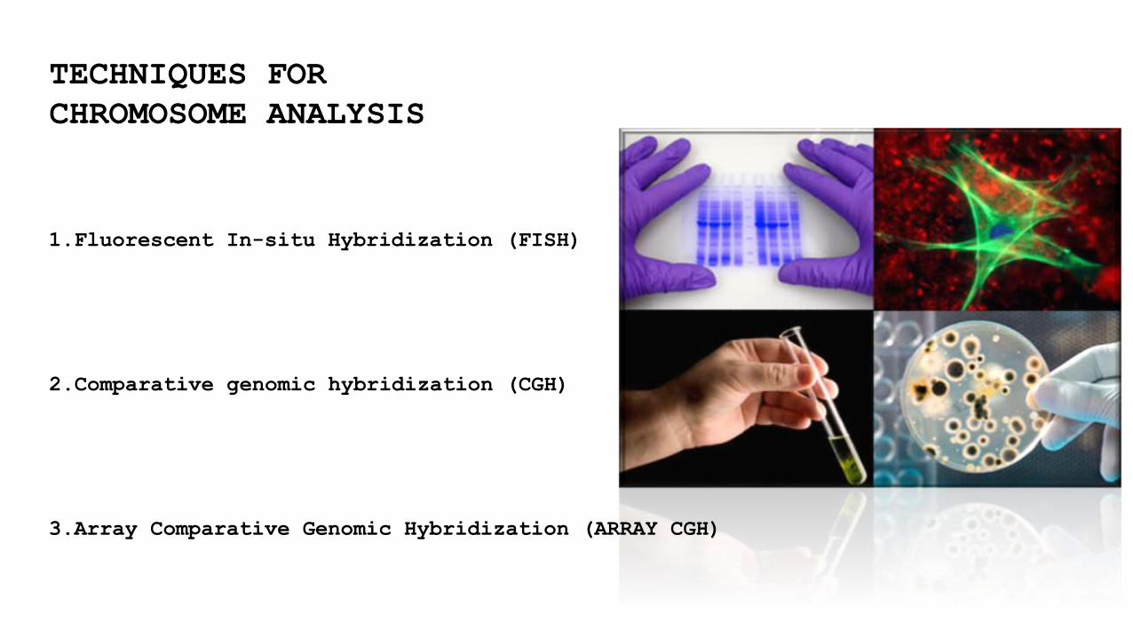
1.Fluorescent In-situ Hybridization (FISH)
2.Comparative genomic hybridization (CGH)
3.Array Comparative Genomic Hybridization (ARRAY CGH)
CHROMOSOME ANALYSIS
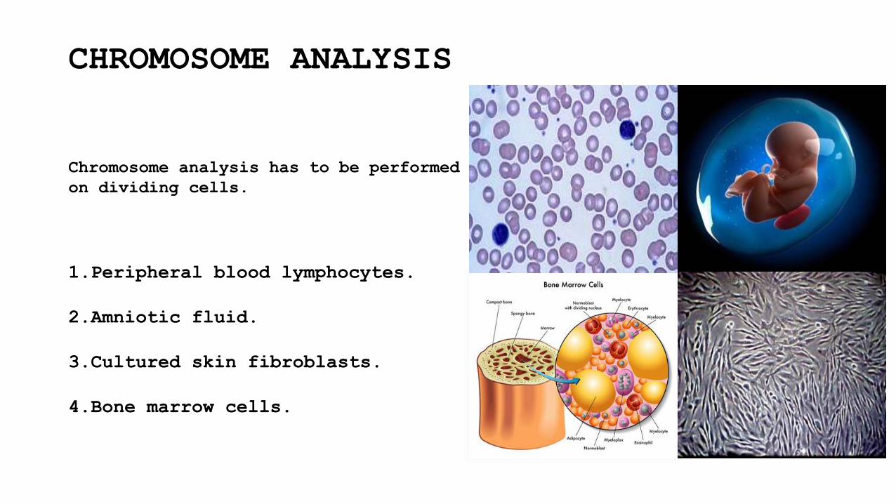
Chromosome analysis has to be performed
on dividing cells.
1.Peripheral blood lymphocytes.
2.Amniotic fluid.
3.Cultured skin fibroblasts.
4.Bone marrow cells.
FISH(Fluorescent in situ hybridization)
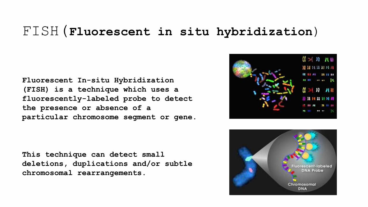
Fluorescent In-situ Hybridization
(FISH) is a technique which uses a
fluorescently-labeled probe to detect
the presence or absence of a
particular chromosome segment or gene.
This technique can detect small
deletions, duplications and/or subtle
chromosomal rearrangements.
PROCEDURE
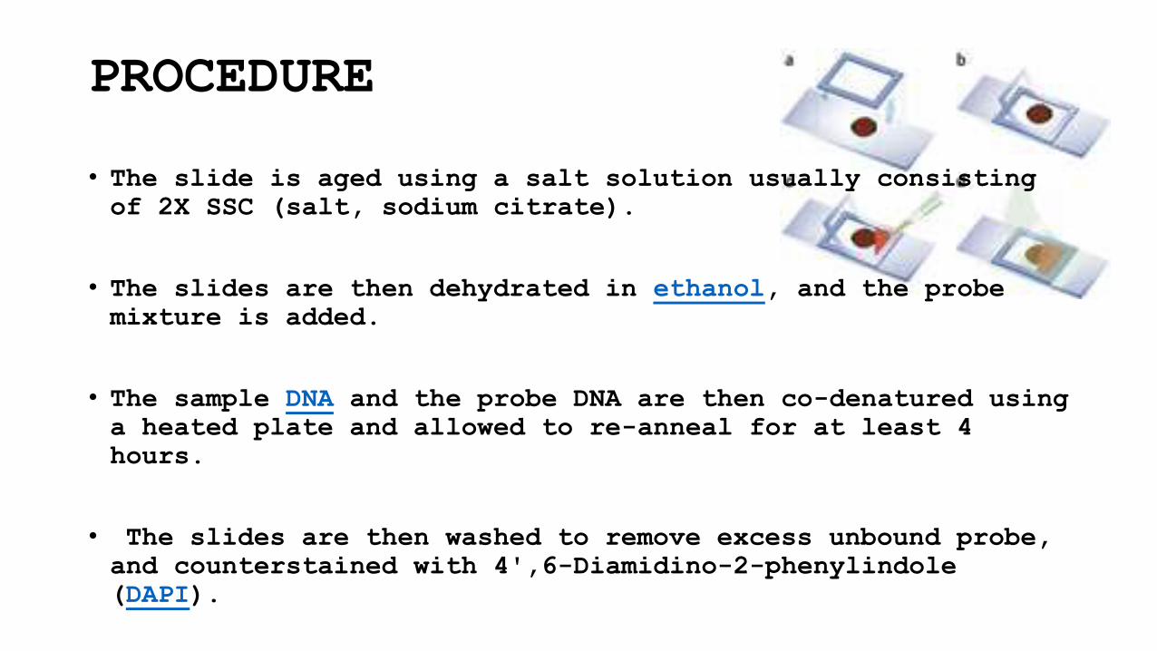
• The slide is aged using a salt solution usually consisting of 2X SSC (salt, sodium citrate).
• The slides are then dehydrated in ethanol, and the probe
mixture is added.
• The sample DNA and the probe DNA are then co-denatured using
a heated plate and allowed to re-anneal for at least 4
hours.
• The slides are then washed to remove excess unbound probe,
and counterstained with 4',6-Diamidino-2-phenylindole
(DAPI).
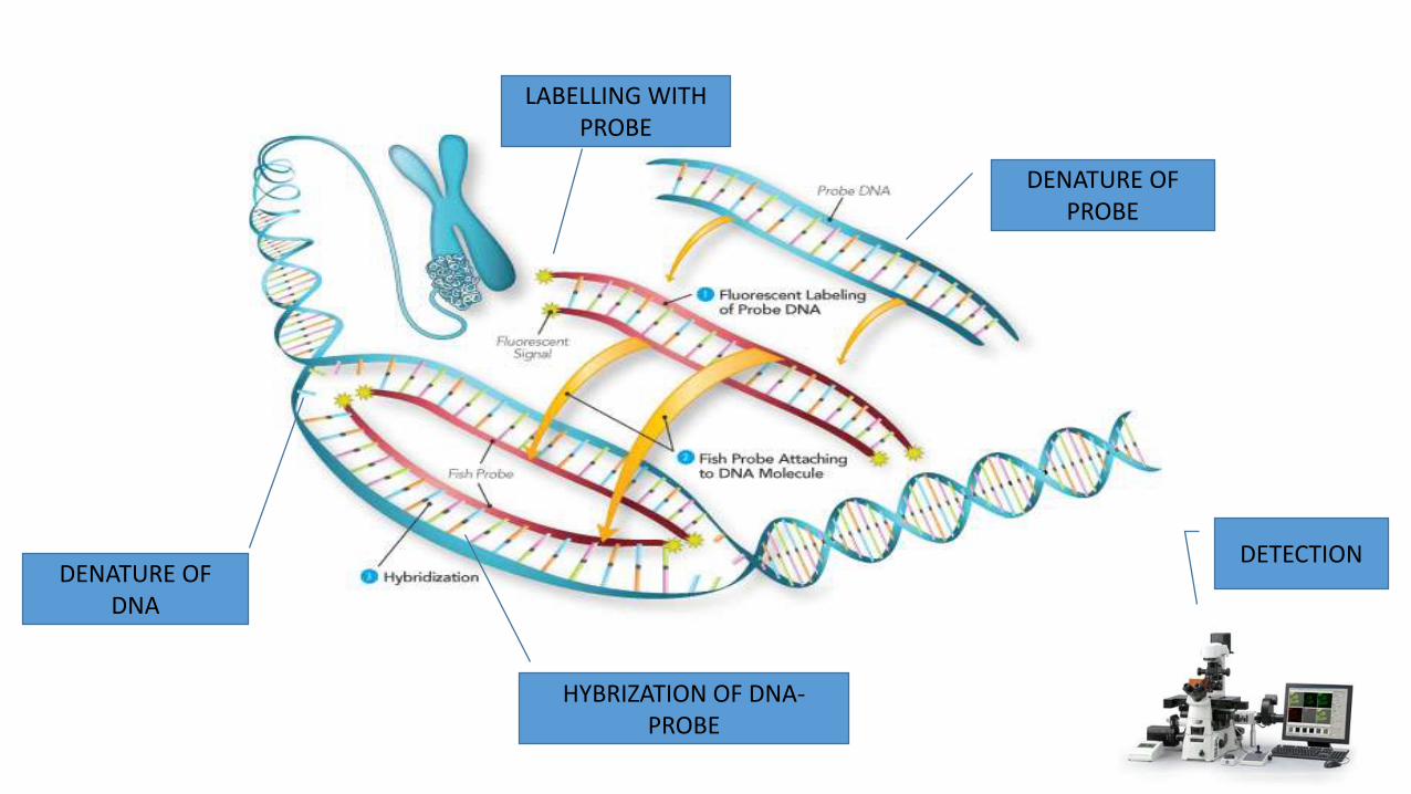
DENATURE OF DNA
DENATURE OF PROBE
LABELLING WITH PROBE
HYBRIZATION OF DNA-PROBE
DETECTION

ANALYSIS
Analysis of FISH specimens is done
by fluorescence microscopy by a clinical
laboratory specialist in cytogenetics.
For congenital problems usually
20 metaphase cells are scored.
Examples of diseases that are diagnosed using FISH include Prader-Willi
syndrome, Angelman syndrome, 22q13 deletion syndrome, chronicmyelogenous
leukemia, acute lymphoblastic leukemia, Cri-du-chat, Velocardiofacial
syndrome and Down syndrome.
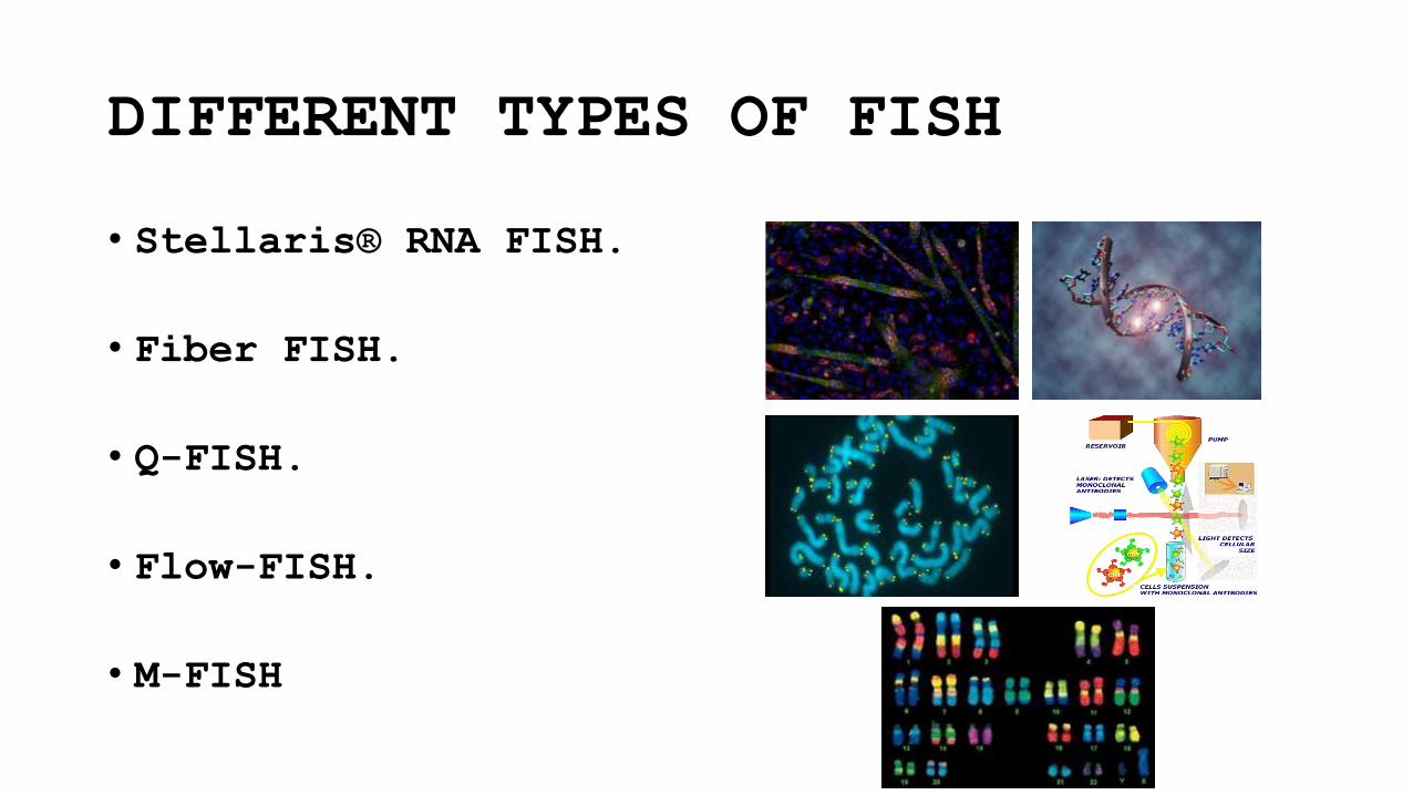
DIFFERENT TYPES OF FISH
• Stellaris® RNA FISH.
• Fiber FISH.
• Q-FISH.
• Flow-FISH.
• M-FISH
Comparative genomic
hybridization (CGH)
Comparative genomic hybridization is a molecular cytogenetic method
for analysing copy number variations (CNVs) relative
to ploidy level in the DNA of a test sample compared to a reference
sample.
The aim of this technique is to quickly and efficiently compare two
genomic DNA samples arising from two sources, which are most often
closely related, because it is suspected that they contain
differences in terms of either gains or losses of either
whole chromosomes or subchromosomal regions (a portion of a whole
chromosome).

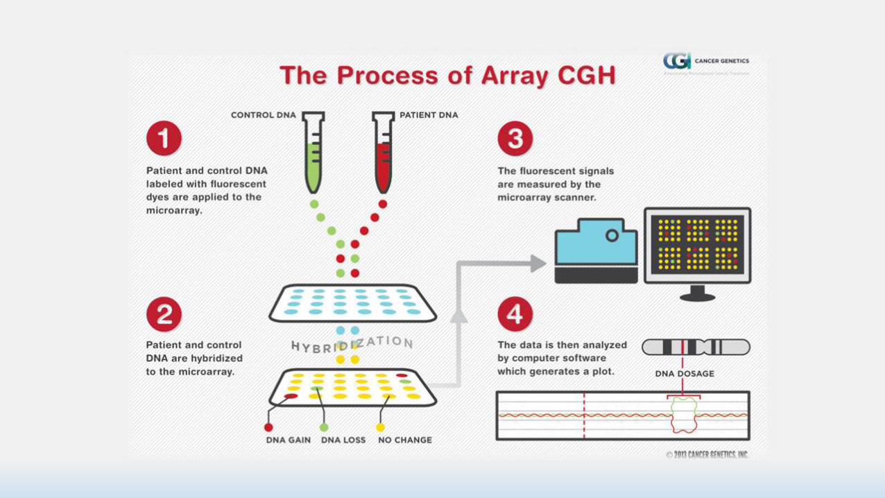
PROCEDURE
This Technique involves the isolation of DNA from the two sources to be
compared.
Most commonly a test and reference source.
Independent labelling of each DNA sample with a
different fluorophores (fluorescent molecules) of different colours
(usually red and green).
Denaturation of the DNA so that it is single stranded, and
the hybridization of the two resultant samples in a 1:1 ratio to a
normal metaphase spread of chromosomes, to which the labelled DNA samples
will bind at their locus of origin.
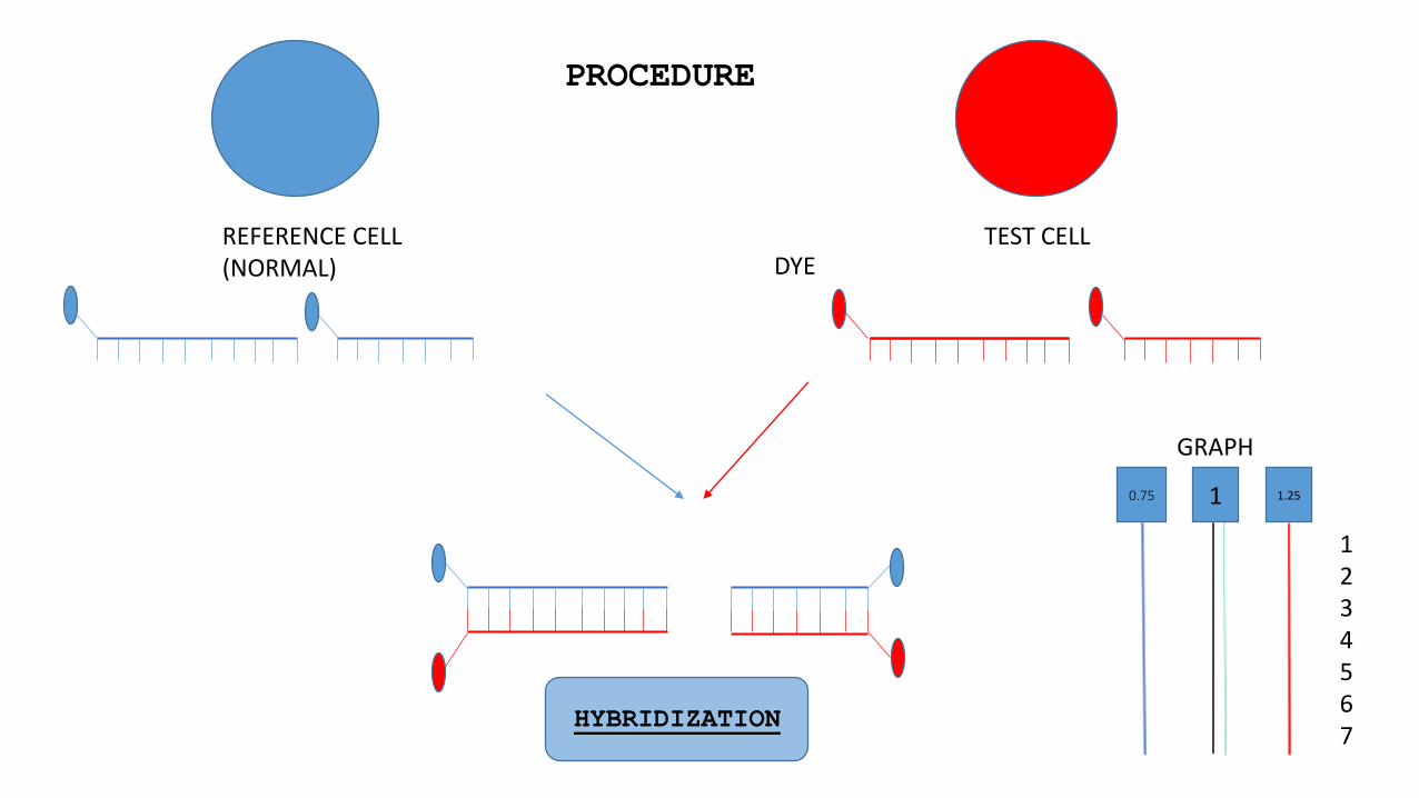
REFERENCE CELL (NORMAL)
TEST CELL
1234567
DYE
HYBRIDIZATION
PROCEDURE
GRAPH
0.75 1 1.25
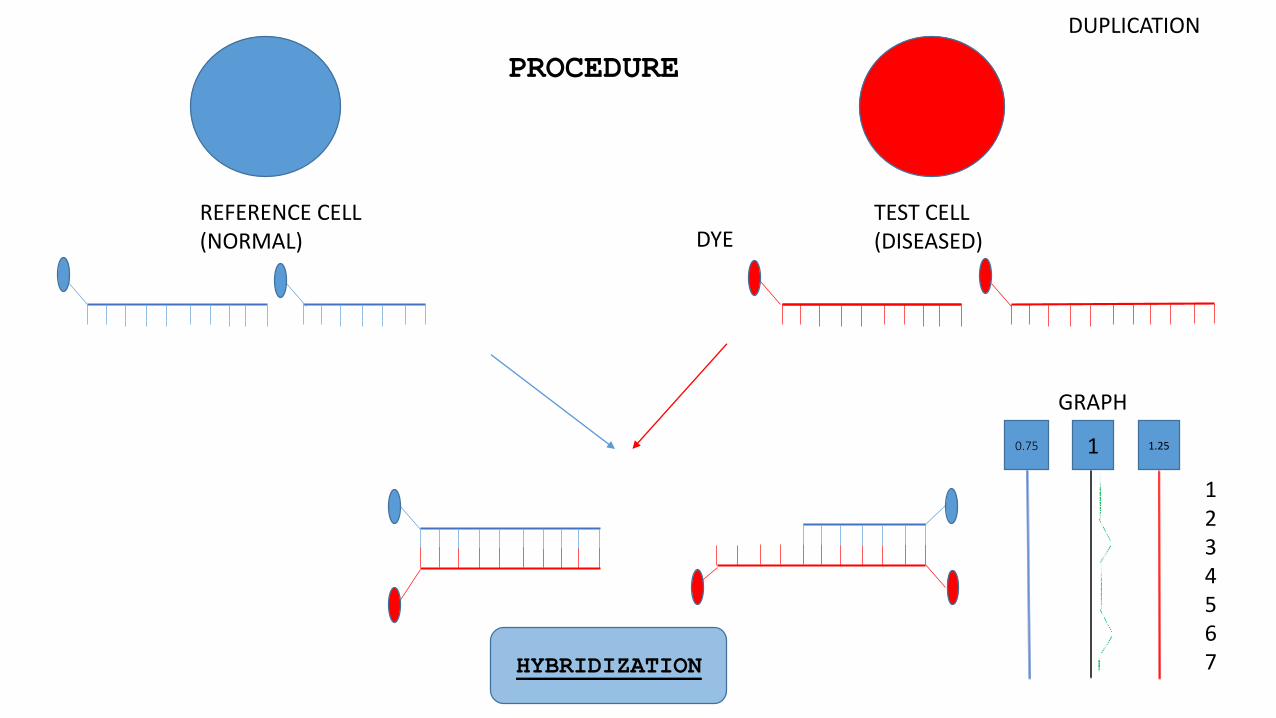
REFERENCE CELL (NORMAL)
TEST CELL (DISEASED)
1234567
DYE
HYBRIDIZATION
PROCEDURE
GRAPH
DUPLICATION
0.75 1 1.25
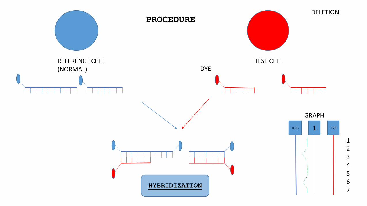
REFERENCE CELL (NORMAL)
TEST CELL
1234567
DYE
HYBRIDIZATION
PROCEDURE
GRAPH
DELETION
0.75 1 1.25

ADVANTAGE & LIMITATION
CGH is only able to detect
unbalanced chromosomal abnormalities.
Conventional CGH has been used mainly for
the identification of chromosomal regions
that are recurrently lost or gained in
tumors.
A main disadvantage of conventional CGH is
its inability to detect structural
chromosomal aberrations without copy number
changes.
for eg. balanced chromosomal
abnormalities such as reciprocal
translocations, or inversions do not
affect copy number.
Array Comparative Genomic
Hybridization (ARRAY CGH)
• Array comparative genomic hybridization (also microarray-based comparative genomic hybridization, matrix CGH, array CGH, aCGH) is a molecular cytogenetic technique for the detection of chromosomal copy number changes on a genome wide and high-resolution scale.
• With the introduction of array CGH, the main limitation of conventional CGH, a low resolution, is overcome.
• Using this method, copy number changes at a level of 5–10 kilobases of DNA sequences can be detected.

ADVANTAGE & LIMITATION
• Aberrations smaller than 5–10 Mb cannot be detected using conventional CGH. For the detection of such
abnormalities, a high-resolution technique is required.
Array CGH overcomes many of these limitations.
• The main disadvantage of array CGH is its inability to
detect aberrations that do not result in copy number
changes.

APPLICATION
1.Conventional CGH
A. CGH in cancer research
CGH data from several studies of the same tumor type show
consistent patterns of non-random genetic aberrations.
For example, 13q gain 9q loss in bladder cancer, 14q loss in renal
cancer.
B.Chromosomal Aberrations.
• Cri du Chat (CdC) is a syndrome caused by a partial
deletion of the short arm of chromosome 5.
• Several studies have shown that conventional CGH is
suitable to detect the deletion, as well as more
complex chromosomal alterations.
2.Array Comparative Genomic Hybridization (ARRAY
CGH)
A.Submicroscopic aberrations
• Prader–Willi syndrome (PWS) is a paternal structural abnormality
involving 15q11-13, while a maternal aberration in the same region
causes Angelman syndrome (AS).
• In both syndromes, the majority of cases (75%) are the result of
a 3–5 Mb deletion of the PWS/AS critical region. These small
aberrations cannot be detected using cytogenetics or conventional
CGH, but can be readily detected using array CGH.
• Array CGH applications are mainly directed at detecting genomic abnormalities in cancer.
• However, array CGH is also suitable for the analysis
of DNA copy number aberrations that cause human genetic
disorders.

• https://en.wikipedia.org/wiki/Comparative_genomic_hybridization#Array_Comparative_Genomic_Hybr
idization
• http://www.utmb.edu/pedi_ed/CORE/MedicalGenetics/page_09.htm
• https://en.wikipedia.org/wiki/Fluorescence_in_situ_hybridization#Preparation_and_hybridization
_process_.E2.80.93_DNA
REFRENCE
