chromosome localization of the gene for human terminal
TRANSCRIPT

Proc. Natl. Acad. Sci. USAVol. 82, pp. 5836-5840, September 1985Genetics
Chromosome localization of the gene for human terminaldeoxynucleotidyltransferase to region 10q23-q25
(terminal tranferase/B and T cels/leukemias and lymphomas/chromosome rearrangement/genetics of neoplasia)
MASAHARU ISOBE*, KAY HUEBNER*, JAN ERIKSON*, RONALD C. PETERSONt, F. J. BOLLUMt,Lucy M. S. CHANGt, AND CARLO M. CROCE**The Wistar Institute, 36th at Spruce Street, Philadelphia, PA 19104; and tDepartment of Biochemistry, Uniformed Services University of the HealthSciences, Bethesda, MD 20814
Communicated by Peter C. Nowell, May 3, 1985
ABSTRACT Complementary DNA clones representing the3' half, the 5' half, and the entire coding region of the humanterminal deoxynucleotidyltransferase gene (TdT; DNAnucleotidylexotransferase, nucleosidetriphosphate:DNAdeoxynucleotidylexotransferase, EC 2.7.7.31) were used toscreen a panel of mouse x human somatic cell hybrid DNAs todetermine the chromosomal location of the human TdT gene.The results of the Southern transfer analysis of hybrid DNAsindicate that the gene for TdT is located on human chromosome10. The in situ hybridization technique was then used to furtherlocalize the gene for TdT to region q23-q25 of human chro-mosome 10.
Human terminal deoxynucleotidyltransferase (TdT; DNAnucleotidylexotransferase, nucleoside triphosphate:DNAdeoxynucleotidylexotransferase, EC 2.7.7.31) is a 58-kDaprotein (1) present in a major cell population of the thymuscortex and in a minor population of bone marrow lympho-cytes (2). The normal biologic function of TdT is not fullyunderstood and the reason for its presence in pre-T and pre-Bcells remains an object of speculation. Recently, Desiderio etal. (3) have proposed that TdT might be responsible forinserting nucleotides (N regions) at the recombinationaljunctions ofimmunoglobulin heavy-chain genes during B-cellmaturation. The insertion ofN regions may also occur duringrecombinatorial joining of the T-cell-receptor genes (4). It ispossible that the insertion ofN regions reflects the increasedlevels of TdT in pre-T and pre-B cells. Approximately100,000 TdT molecules per cell have been detected in corticalthymocytes and primitive bone marrow lymphocytes (1).Interestingly, we have detected N regions at the junctionbetween chromosomes 11 and 14 in chronic lymphocyticleukemia of the B-cell type with the t(11;14) chromosometranslocation (5). If the addition ofN regions is the result ofhigh TdT activity, this finding may suggest that the t(11;14)translocation may have occurred at the pre-B-cell stage ofdifferentiation. TdT is also elevated in acute lymphoblasticleukemia and in cases of chronic granulocytic leukemia inblast crisis (6). Since the TdT gene is expressed in pre-B andpre-T cells and in certain human leukemias, this gene mighthave a role in oncogene activation in hematopoietic malig-nancies. In this study, human TdT cDNA clones isolatedfrom a cDNA expression library derived from a pre-B-cellhuman leukemic cell line (7) have been used to determine thechromosomal location of the TdT gene in order to establishwhether this gene is in a chromosome region subject torearrangements in malignant T or B cells.
MATERIALS AND METHODSTdTcDNA Clones. During the course ofthese studies, three
different cDNA clones were used: pT17, a pBR322 plasmidcontaining a 928-base-pair (bp) EcoRI (7) insert that repre-sents the 3' portion (3' half) of full-length TdT cDNA; pT106,a 967-bp EcoRI insert in pUC8 that contains the 5' half of thecDNA; and pT223, 1800 bp inserted in pUC19, which starts3 bases from the start of the coding region and extends to the3' end of the cDNA. See Fig. 1 for a sketch of the relationshipamong these cDNA clones. The 3' clone, pT17, was used forin situ hybridization. For analysis of segregation of TdT inmouse-human somatic cell hybrids, purified cDNA insertsfrom the 3' or 5' clones were usually used; total plasmidDNAfrom all three clones-3', 5', and total coding region(pT223)-were also used in Southern blot analysis, withresults identical to those obtained by using the'purified insertof the respective plasmid.
Southern Blot Analysis. DNAs from human cells, mousecells, and mouse-human hybrids were extracted by cell lysis,proteinase K digestion, phenol extraction, and ethanol pre-cipitation. Cellular DNA samples were digested with theappropriate restriction enzyme, sized in 0.8-1.0% agarosegels, and transferred to nitrocellulose as described by South-ern (8). Hybridization was carried out in 50% formamide/4xNaCi/Cit (lx NaCI/Cit is 0.15 M NaCl/0.015 M sodiumcitrate, pH 7.0)/0.2 mg of sonicated salmon sperm DNA perml/1x Denhardt's solution (0.02% bovine serumalbumin/0.02% Ficoll/0.02% polyvinylpyrrolidone) at 420Cfor 15 hr or at 370C for 24 hr. The probes were labeled by nicktranslation. After hybridization, the filters were washed andexposed to Kodak XAR-5 film with intensifying screens.
Cell Lines. Isolation, propagation, and characterization ofparental cells and the somatic cell hybrids used in this studyhave been described (9-13).
All hybrids were studied for the expression of enzymemarkers assigned to each of the human chromosomes (9).Some hybrid clones were karyotyped by trypsin/Giemsaand/or G-11 banding methods as described (9). In addition,the presence of specific human chromosomes in many of themouse-human hybrids have been confirmed by DNA hybrid-ization by using probes for genes assigned to specific humanchromosomes (9-13).In Situ Hybridization. Metaphase spreads were prepared
with normal human male phytohemagglutinin-stimulated (for72 hr) in vitro lymphocyte cultures. Total plasmid (pTl7)DNA containing the human TdT 928-bp EcoRI fragment wasnick-translated with [3H]dCTP (62 Ci/mmol; 1 Ci = 37 GBq;New England Nuclear), [3H]dGTP (39.9 Ci/mmol),[3H]dTTP (100.1 Ci/mmol), and [3H]dATP (51.9 Ci/mmol).
Abbreviations: TdT, terminal deoxynucleotidyltransferase; kbp,kilobase pair(s).
5836
The publication costs of this article were defrayed in part by page chargepayment. This article must therefore be hereby marked "advertisement"in accordance with 18 U.S.C. §1734 solely to indicate this fact.

Proc. NatL Acad. Sci. USA 82 (1985) 5837
If iE S A Be
pT223F
00 400
S E P
IpT106pT7 1 11
pT17F
800 1200
A: Apa IB: Bam Hi
Bg: BgI I
E: Eco RIP: Pvu 11S: Sat I
FIG. 1. Restriction maps of the TdT 5', 3', and total coding region cDNA clones.
The techniques used for in situ hybridization were essentiallyas described by Harper and Saunders (14). Chromosomepreparations were treated with pancreatic RNase A (Sigma)and then denatured in 70% formamide in 2x NaCl/Cit (pH7.0) at 70'C for 2 min. The chromosome preparations werethen hybridized with 3H-labeled pT17DNA (specific activity,3 x 107 cpm/ftg) at a concentration of 140-280 ng/ml in 50%formamide/2x NaClI/Cit/10%6 dextran sulfate (Pharmacia),pH 7.0, for 20 hr at 370C. A 300-fold excess of sonicatedsalmon sperm DNA was included as carrier. Slides werethoroughly rinsed in 50% formamide/2x NaCl/Cit at 390C,exposed to Kodak NTB2 nuclear track emulsion for 16 daysat 40C, and developed with Kodak Dektol at 15'C. Thechromosomes were then G-banded essentially as describedby Cannizzaro and Emanuel (15) with a mixture of 6 parts ofborate buffer (50 mM Na2SO4/2.5 mM Na2B407, pH 9.2) to1 part of Wright's/Giemsa stain solution (2.4 g of Wright'sstain per liter/1.4 g of Giemsa stain per liter in methanol).
RESULTS
Chromosomal Location of the TdT Gene. DNA from a panelof mouse-human hybrid cells was analyzed for the presenceofthe human TdTgene by cleavage with restriction enzymes,followed by fractionation of DNA in agarose, transfer tonitrocellulose, and hybridization of nitrocellulose filters witheither the 3', 5', or total TdT cDNA clones. In initialexperiments, mouse and human DNA and mouse-humanhybrid DNAs were cleaved with restriction enzyme HindIIIand the nitrocellulose-bound DNA was hybridized to eitherthe 3' (pT17) or 5' (pT106) TdT cDNA clones. An example ofhybridization of HindIII-cleaved mouse, human, and hybridDNAs to the 5' (pT106) TdT clone is shown in Fig. 2. InHindIII-cleaved mouse genomic DNA, the 5' TdT clonedetects bands of 19.3 and 5.5 kbp (lane 2), although the mouseTdTgene is not reproducibly detected after HindIII cleavageand, as will be seen in later figures, is usually not seen at allafter Pst I cleavage. The explanation for lack of detection ofthe mouse TdT is probably that small differences in strin-gency of hybridization and/or washing of filters can deter-mine whether the mouse TdT gene is seen.
In HindIII-digested human DNA, the 5' TdT probe rou-tinely detects bands of 11.8 and 4.3 kbp (probably a doublet)and, occasionally, a 1.6-kbp band (Fig. 2, lane 1). In screen-ing the mouse-human hybrid panel with the 5' probe, it wasfound that the 11.8- and 4.3-kbp band segregated indepen-dently from a 4.1-kbp (lower band of 4.3 doublet) and the1.6-kbp band (see lanes 3 and 4 versus lane 5). Since the 4.3-and 4.1-kbp HindIII fragments were not well separated in the
human genome, Pst I-digested hybrid DNAs were used toanalyze in more detail the independently segregated genes.Pst I-cleaved human and hybrid DNAs were blotted andhybridized to the 3' probe, the 5' probe, and the full-lengthprobe (pT223).
After hybridization to the 3' probe, three Pst I fragments(18.0, 8.5, and 4.9 kbp; data not shown) were detected inhuman DNA and in all hybrids showing positive hybridiza-tion; that is, with the 3' half of the TdT coding region, allfragments segregated together and were thus on only one
1 3 4 5 6
kbp
-_ w 19.3
0I p 4S
11.8
- 5.5
4.34.1
- 1.6
FIG. 2. Hybridization of the TdT 5' cDNA clone to HindIll-cleaved parental and hybrid DNAs. Ten micrograms of cellular DNAfrom human (lane 1); mouse (lane 2); hybrid clone retaining humanchromosomes 1, 3, 5, 6, 7, 8, 9, 10, 13, 14, 18, 20, 22, and X (lane 3);hybrid clone retaining human chromosomes 3, 5, 6, 9, 10, 13, 14, 18,20, 22, and X (lane 4); hybrid clone retaining human chromosomes2, 5, 12, and 19 (lane 5); and hybrid clone retaining human chromo-somes 1, 3, 5, 14, 18, 22, and X (lane 6), was digested with an excessof restriction enzyme HindIII, sized on an agarose gel, transferred tonitrocellulose, and hybridized to 32P-labeled TdT 5' cDNA. Molec-ular sizes (in kbp) of human and mouse fragments hybridizing to thisprobe are shown on the right of the figure. The 11.8- and 4.3-kbphuman fragments seen in lanes 1, 3, and 4 segregate with chromo-some 10, and the 4.1- and 1.6-kbp fragments segregate independentlyof the other two bands as can be seen in lane 5.
p E
1600I b.
2000
II
Genetics: Isobe et aL
i
Proc. NatL Acad. ScL USA 82 (1985)
chromosome, chromosome 10. Pst I-cleaved human DNAdisplayed three different fragments that hybridized to the 5'TdT probe (>23, 10.0, and 7.5 kbp; Fig. 3, lane 2). The 10.0-and 7.5-kbp bands segregated together (as did the humanHindIII 11.8- and 4.3-kbp bands; lanes 4-9) and were alwaysfound to correlate with the presence of human chromosome10 in the hybrids.
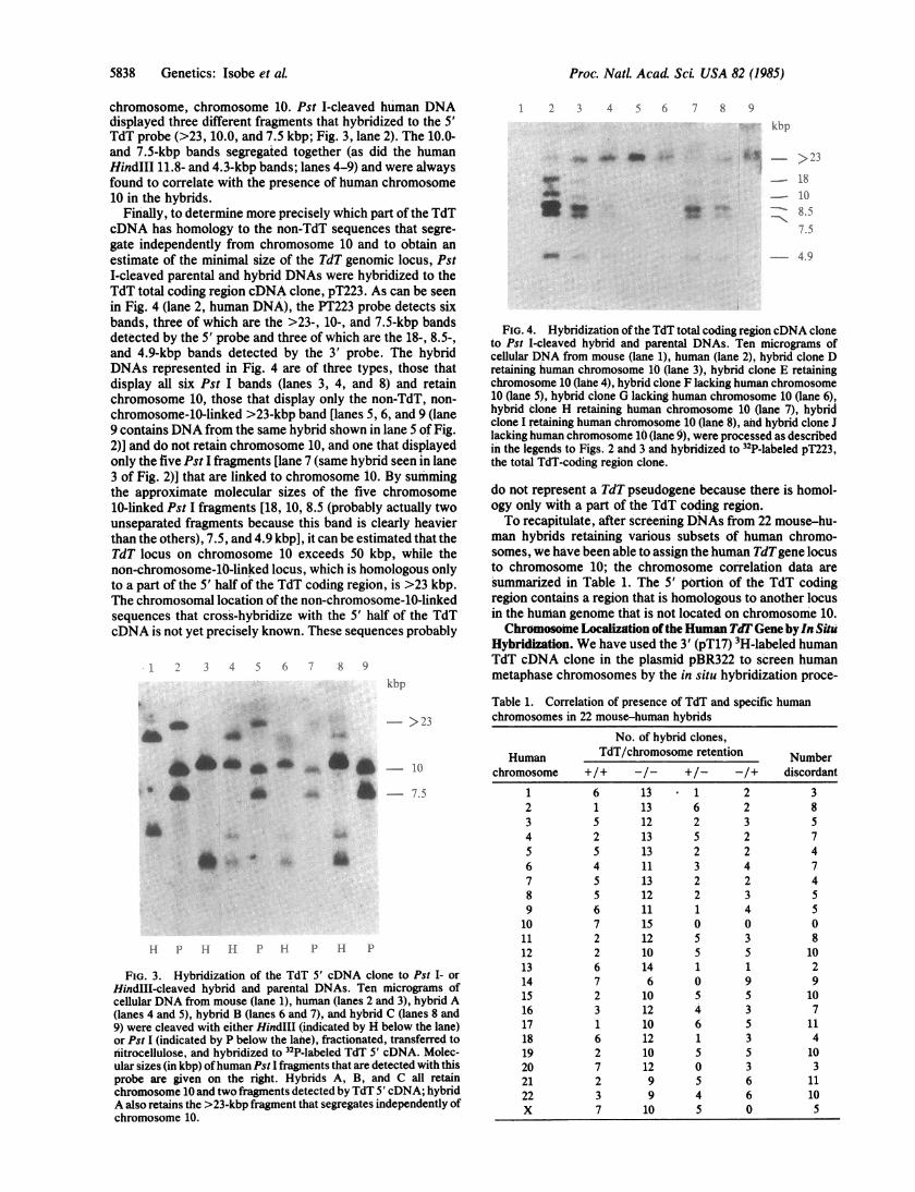
Finally, to determine more precisely which part of the TdTcDNA has homology to the non-TdT sequences that segre-gate independently from chromosome 10 and to obtain anestimate of the minimal size of the TdT genomic locus, PstI-cleaved parental and hybrid DNAs were hybridized to theTdT total coding region cDNA clone, pT223. As can be seenin Fig. 4 (lane 2, human DNA), the PT223 probe detects sixbands, three of which are the >23-, 10-, and 7.5-kbp bandsdetected by the 5' probe and three of which are the 18-, 8.5-,and 4.9-kbp bands detected by the 3' probe. The hybridDNAs represented in Fig. 4 are of three types, those thatdisplay all six Pst I bands (lanes 3, 4, and 8) and retainchromosome 10, those that display only the non-TdT, non-
chromosome-10-linked >23-kbp band [lanes 5, 6, and 9 (lane9 contains DNA from the same hybrid shown in lane 5 of Fig.2)] and do not retain chromosome 10, and one that displayedonly the five Pst I fragments [lane 7 (same hybrid seen in lane3 of Fig. 2)] that are linked to chromosome 10. By summingthe approximate molecular sizes of the five chromosome10-linked Pst I fragments [18, 10, 8.5 (probably actually twounseparated fragments because this band is clearly heavierthan the others), 7.5, and 4.9 kbp], it can be estimated that theTdT locus on chromosome 10 exceeds 50 kbp, while thenon-chromosome-10-linked locus, which is homologous onlyto a part of the 5' half of the TdT coding region, is >23 kbp.The chromosomal location ofthe non-chromosome-10-linkedsequences that cross-hybridize with the 5' half of the TdTcDNA is not yet precisely known. These sequences probably
2 3 4 5 6 7 8 9kbp
a-
:. i- ^_
>23
10
7.5
HI P1 id H P H P H P
FIG. 3. Hybridization of the TdT 5' cDNA clone to Pst I- or
HindIll-cleaved hybrid and parental DNAs. Ten micrograms ofcellular DNA from mouse (lane 1), human (lanes 2 and 3), hybrid A(lanes 4 and 5), hybrid B (lanes 6 and 7), and hybrid C (lanes 8 and9) were cleaved with either HindIll (indicated by H below the lane)or Pst I (indicated by P below the lane), fractionated, transferred tonitrocellulose, and hybridized to 32P-labeled TdT 5' cDNA. Molec-ular sizes (in kbp) ofhuman Pst I fragments that are detected with thisprobe are given on the right. Hybrids A, B, and C all retainchromosome 10 and two fragments detected by TdT 5' cDNA; hybridA also retains the >23-kbp fragment that segregates independently ofchromosome 10.
1 2 3 4 5 6 7 8 9
kbp
>23
18
1()
8.5
7.5
A. W
of- 4.9
FIG. 4. Hybridization of the TdT total coding region cDNA cloneto Pst I-cleaved hybrid and parental DNAs. Ten micrograms ofcellular DNA from mouse (lane 1), human (lane 2), hybrid clone Dretaining human chromosome 10 (lane 3), hybrid clone E retainingchromosome 10 (lahe 4), hybrid clone F lacking human chromosome10 (lane 5), hybrid clone G lacking human chromosome 10 (lane 6),hybrid clone H retaining human chromosome 10 (lane 7), hybridclone I retaining human chromosome 10 (lane 8), and hybrid clone Jlacking human chromosome 10 (lane 9), were processed as describedin the legends to Figs. 2 and 3 and hybridized to 32P-labeled pT223,the total TdT-coding region clone.
do not represent a TdT pseudogene because there is homol-ogy only with a part of the TdT coding region.To recapitulate, after screening DNAs from 22 mouse-hu-
man hybrids retaining various subsets of human chromo-somes, we have been able to assign the human TdTgene locusto chromosome 10; the chromosome correlation data aresummarized in Table 1. The 5' portion of the TdT codingregion contains a region that is homologous to another locusin the human genome that is not located on chromosome 10.Chromosome Localization ofthe Human TdO Gene by In Situ
Hybridization. We have used the 3' (pT17) 3H-labeled humanTdT cDNA clone in the plasmid pBR322 to screen humanmetaphase chromosomes by the in situ hybridization proce-
Table 1. Correlation of presence of TdT and specific humanchromosomes in 22 mouse-human hybrids
No. of hybrid clones,
Human TdT/chromosome retention Numberchromosome +/+ -/- +/- -/+ discordant
1 6 13 1 2 32 1 13 6 2 83 5 12 2 3 54 2 13 5 2 75 5 13 2 2 46 4 11 3 4 77 5 13 2 2 48 5 12 2 3 59 6 11 1 4 510 7 15 0 0 011 2 12 5 3 812 2 10 5 5 1013 6 14 1 1 214 7 6 0 9 915 2 10 5 5 1016 3 12 4 3 717 1 10 6 5 1118 6 12 1 3 419 2 10 5 5 1020 7 12 0 3 321 2 9 5 6 1122 3 9 4 6 10X 7 10 5 0 5
5838 Genetics: Isobe et aL

Proc. NatL Acad Sci. USA 82 (1985) 5839
A
S
-p
CI
200-B
s I.105.
.o . .a . I m so El
IPI lp q Ip q Tp q Ip q Ip q Ip qE6 7 8 9 10 I1 12
3IP 14 IP 511 I 17 I 81 I~0~zI II I. 3 14 15 I16 17 I18 I19 .20 21-22 X Y
2015105
CHROMOSOMES
dure (14, 15). After autoradiography, the metaphase chro-mosomes were scored for grain localization. An example isshown in Fig. 5A. The distribution of labeled sites in 100
FIG. 5. Localization of the TdT gene in the humangenome by in situ hybridization analysis. (A) Photographof a G-banded lymphocyte metaphase spread hybridizedwith the human TdT probe pT17. Arrows indicate grainsfound in the distal portion of chromosome lOq. (B) Dia-gram showing the grain distribution in 100 metaphasechromosomes. Abscissa represents the chromosomes intheir relative size proportion; ordinate shows the numberof silver grains. The distribution of grains on 100 spreadswas scored; 22% of all grains were located on the long armof chromosome 10, with most grains at 1Oq24.
metaphase chromosomes is shown in Fig. SB, where theordinate represents the number of labeled sites and theabscissa represents the chromosome regions that were ex-
z
Co
2
4%
0
U.0
w
z
3025
2015105
I
Genetics: Isobe et aL
AD
4:, 'r
% i
%ft,R*& Va.400
4t NW0
0 .::.
ab.No
I A 014"
14

Proc. NatL Acad ScL USA 82 (1985)
amined. As shown in Fig. SB, the distal portion of the longarm of chromosome 10 was significantly labeled. Of the 100cells examined, 33 (33%) exhibited labeling of one or bothchromosomes 10 (Fig. 5B). About 22% of all grains werelocated on the long arm ofchromosome 10 (Fig. SB). Ofthese,=91% of the grains on the long arm of chromosome 10 (lOq)were on region 10q23--q25, with most grains at 10q24. Thelong arm ofchromosome 10 represents =3.22% ofthe haploidgenome. Thus, our observation that >20% of the hybridiza-tion with the pT17 probe is localized on the distal half of thisregion of chromosome 10 is highly significant (P << 0.01).Thus, cytological hybridization localizes the TdT gene toregion q23-+q25 of human chromosome 10.
DISCUSSIONThe chromosome localization of the immunoglobulin genes(16-18) and of cellular oncogenes (9, 19) has been instrumen-tal to our understanding of the molecular basis of Burkittlymphoma and Burkitt-type leukemia (20, 21). Since re-arrangements involving the chromosomal region carrying theheavy-chain locus have also been observed in B-cell neo-plasms of adults, it has also been possible to isolate putativehuman oncogenes involved in these malignancies by takingadvantage of their close proximity to the heavy-chain locusdue to specific chromosome translocations (21, 22). Similar-ly, our finding that the locus of the a chain of the T-cellreceptor is located in a region of chromosome 14 (13) that isfrequently involved in inversion and translocations in humanT-cell neoplasms (23) suggests that this locus may have animportant role in T-cell neoplasia. Since the TdT gene isextremely active in pre-B and pre-T cells, it might have a rolein oncogene activation in human hematopoietic malignanciesexpressing increased levels of terminal transferase. There-fore, it was important to establish the chromosome locationof the TdT gene in order to determine whether this chromo-some region is involved in rearrangements in human hema-topoietic malignancies.The results of these studies indicate that the TdT gene is
located on region q23-*q25 of human chromosome 10. Thisfinding might be of interest because recent reports describerearrangements of this human chromosome in human T-cellneoplasms (23, 24). Analysis of these malignancies for rear-rangements within or in proximity to the TdT locus shouldprovide an answer to the question of whether the TdT locusplays a role in human leukemogenesis.
We thank Wendy Scattergood for skillful technical assistance andKathy Reinersmann for preparation of the manuscript; we are alsograteful to Josephine Romano and Dr. Pasquale Tripputi for somehybrid DNA samples. This work was supported by National Insti-
tutes of Health Grants CA16685, CA21124, CA36521, CA23262(F.J.B.), and CA31393 (L.M.S.C.).
1. Bollum, F. J. (1979) Blood 54, 1203-1215.2. Janossy, G., Bollum, F. J., Bradstock, K. F., McMichael, A.,
Rapson, N. & Greaves, M. F. (1979) J. Immunol. 123,1525-1529.
3. Desiderio, S. V., Yan Copoulos, G. D., Paskind, M., Thomas,E., Boss, M. A., Landau, N., Alt, F. A. & Baltimore, D.(1984) Nature (London) 311, 752-755.
4. Hedrick, S. M., Nielson, E. A., Kavalier, J., Cohen, D. I. &Davis, M. M. (1984) Nature (London) 308, 153-158.
5. Tsujimoto, Y., Jaffe, E., Cossman, J. & Croce, C. M. (1985)Nature (London) 315, 340-343.
6. Sarin, P. S. & Gallo, R. C. (1974) J. Biol. Chem. 249,8051-8053.
7. Peterson, R. C., Cheung, L. C., Mattaliano, R. J., Chang,L. M. S. & Bollum, F. J. (1984) Proc. Natl. Acad. Sci. USA81, 4363-4367.
8. Southern, E. M. (1975) J. Mol. Biol. 98, 503-517.9. Dalla-Favera, R., Bregni, M., Erikson, J., Patterson, D.,
Gallo, R. C. & Croce, C. M. (1982) Proc. Natl. Acad. Sci.USA 79, 7824-7827.
10. Cooper, C. S., Park, M., Blair, D. G., Tainsky, M. A.,Huebner, K., Croce, C. M. & Vande Woude, G. F. (1984)Nature (London) 311, 29-33.
11. Green, L., Van Antwerpen, R., Stein, J., Stein, G., Tripputi,P., Emanuel, B., Selden, J. & Croce, C. M. (1984) Science226, 838-840.
12. Huebner, K., Palumbo, A. P., Isobe, M., Kozak, C. A.,Monaco, S., Rovera, G., Croce, C. M. & Curtis, P. J. (1985)Proc. Natl. Acad. Sci. USA 82, 3790-3793.
13. Croce, C. M., Isobe, M., Palumbo, A. P., Puck, J., Erikson,J., Davis, M. & Rovera, G. (1985) Science 227, 1044-1047.
14. Harper, M. E. & Saunders, G. F. (1981) Chromosoma 83,431-439.
15. Cannizzaro, L. A. & Emanuel, B. (1984) Cytogenet. CellGenet. 38, 308-309.
16. Croce, C. M., Shander, M., Martinis, J., Cicurel, L.,D'Ancona, G. G. & Koprowski, H. (1979) Proc. Natl. Acad.Sci. USA 76, 3416-3419.
17. Erikson, J., Martinis, J. & Croce, C. M. (1981) Nature (Lon-don) 294, 173-175.
18. McBride, 0. W., Hieter, P. A., Hollis, G. F., Swan, D., Otey,M. C. & Leder, P. (1982) J. Exp. Med. 155, 1680-1690.
19. Dalla-Favera, R., Franchini, G., Martinotti, S., Wong-Stahl,F., Gallo, R. C. & Croce, C. M. (1982) Proc. Natl. Acad. Sci.USA 79, 4714-4717.
20. Croce, C. M. & Nowell, P. C. (1985) Blood 65, 1-7.21. Tsujimoto, Y., Yunis, J. J., Onorato Showe, L., Nowell, P. C.
& Croce, C. M. (1984) Science 224, 1403-1406.22. Tsujimoto, Y., Finger, L. S., Yunis, J. J., Nowell, P. C. &
Croce, C. M. (1984) Science 226, 1097-1099.23. Hecht, F., Morgan, R., Raiser-McCaw Hecht, T. & Smith,
S. D. (1984) Science 226, 1445-1447.24. Miyamoto, K., Tomita, N., Ishii, A., Nonaka, H., Kondo, T.,
Tanaka, T. & Kitajima, K. (1984) J. Natl. Cancer Inst. 73,353-362.
5840 Genetics: Isobe et aL