chromosomes in which we examine mitosis and meiosis, as well as normal and abnormal chromosome...
Post on 22-Dec-2015
219 views
TRANSCRIPT
Chromosomes
In which we examine mitosis and meiosis, as well as normal and abnormal chromosome patterns.

2 of 44
A Look Ahead Chromosomes & Chromosomal
Abnormalities Genes & Genetic Defects Prenatal Stages & Congenital Defects

Part 1: Chromosomes & the Cell
4 of 44
Chromosomes & the Cell The nucleus of every normal human cell
(except ovum and sperm) contain 46 chromosomes.
These 46 chromosomes occur in pairs: 22 pairs of autosomes 1 pair sex chromosomes
Autosomes: chromosomes on which males and females do not differ
Sex Chromosomes: normal males have XY; normal females have XX

5 of 44
The Human KaryotypeKaryotype = a photomicrograph of chromosomes arranged according to a standard classification

6 of 44
Chromosomes and DNA Chromosomes are made up of DNA
(deoxyribonucleic acid) DNA molecule is held together by bases
- Adenine (A) bonds with thymine (T); cytosine( C) bonds with guanine (G). These letters form the "code of life“
There are about 2.9 billion base pairs in the human genome.
Human DNA contains about 30,000 genes, which human cells use as starting templates to make proteins; which then are used to build and maintain our bodies.

Part 2: Cell Division
8 of 44
Mitosis vs. Meiosis
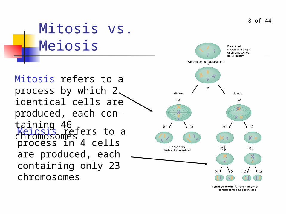
Mitosis refers to a process by which 2 identical cells are produced, each con-taining 46 chromosomesMeiosis refers to a process in 4 cells are produced, each containing only 23 chromosomes

9 of 44Mitosis vs. Meiosis, continued Mitosis is the process by which all cells
in the body replicate, except for production of sperm and ova.
Meiosis is the process by which parent cells divide into the gametes (sperm and ova)
Part 2A: Mitosis

11 of 44
MitosisMitosis is the division of a cell's nucleus. Along with cytokinesis (the division of the rest of a cell), mitosis results in a parent cell dividing into two daughter cells. The genetic information within each of these daughter cells is identical. In the illustration, only 4 of the 46 chromosomes are shown.
from: http://www.pbs.org/wgbh/nova/baby/divide.html#
12 of 44
Replication of Chromosomes
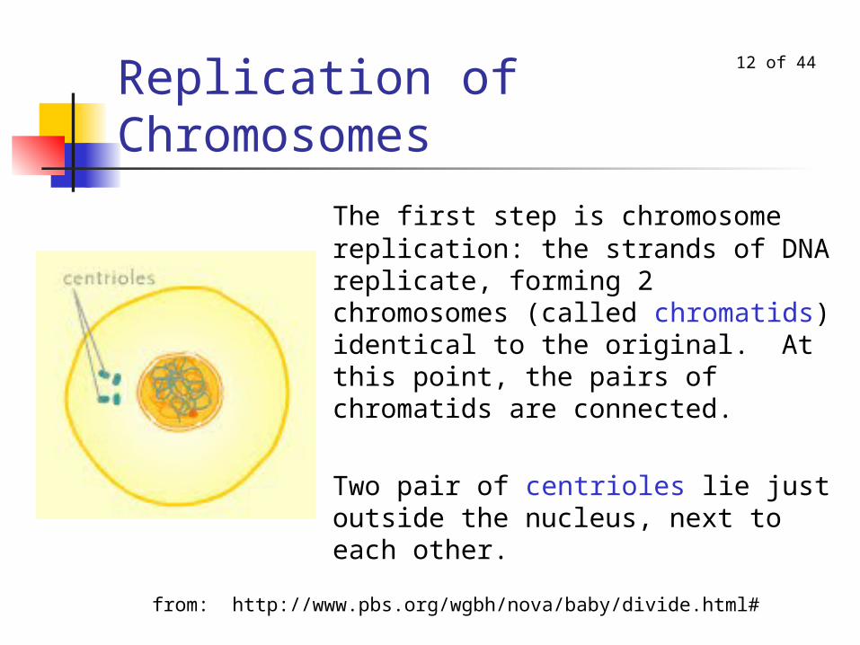
The first step is chromosome replication: the strands of DNA replicate, forming 2 chromosomes (called chromatids) identical to the original. At this point, the pairs of chromatids are connected.
Two pair of centrioles lie just outside the nucleus, next to each other.
from: http://www.pbs.org/wgbh/nova/baby/divide.html#

13 of 44
DNA: deoxyribonucleic acid
DNA is shaped like a twisted ladder. The sides of the ladder are made of sugar & phosphate molecules.
The rungs of the ladder are made of 4 bases: thymine, adenine, cytosine, and guanine. Each rung is made of a pair of bases: CG, GC, AT or TA

14 of 44
Replication of DNA
Because cytosine only bonds with guanine and thymine only bonds with adenine, the two new strands of DNA are exact duplicates of the original strand.

15 of 44
Mitosis, continued
Each original chromosome is now a pair of chromatids.
The centrioles begin to grow fibers, called spindles. The centrioles move to opposite sides of the nucleus.
from: http://www.pbs.org/wgbh/nova/baby/divide.html#
16 of 44
Mitosis, continued
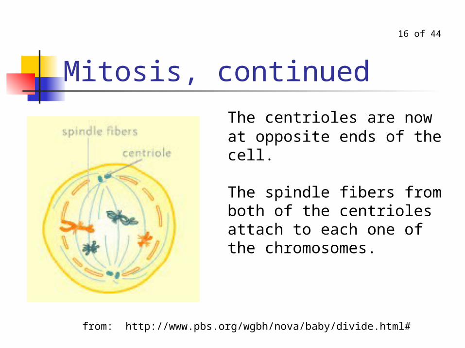
The centrioles are now at opposite ends of the cell.
The spindle fibers from both of the centrioles attach to each one of the chromosomes.
from: http://www.pbs.org/wgbh/nova/baby/divide.html#

17 of 44
Mitosis, continued
The chromosomes line up on the metaphase plate, an imaginary line that divides the cell in two.
Also, the fibers begin to tug each chromosome toward opposite ends of the cell.
from: http://www.pbs.org/wgbh/nova/baby/divide.html#
18 of 44
Mitosis, continued
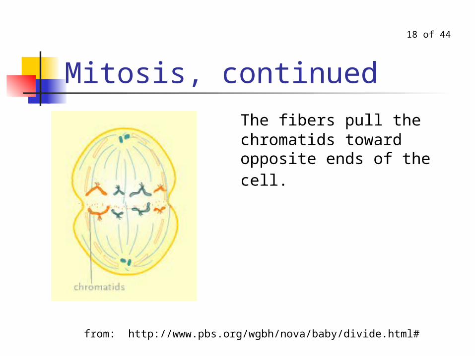
The fibers pull the chromatids toward opposite ends of the cell.
from: http://www.pbs.org/wgbh/nova/baby/divide.html#

19 of 44
Mitosis, continuedThe chromatids (now also considered chromosomes) arrive at the opposite ends of the cell, and new nuclear membranes form.
Mitosis, which describes only the division of the nucleus, is now complete.
from: http://www.pbs.org/wgbh/nova/baby/divide.html#

20 of 44
Mitosis: End of Cytokinesis
The rest of the cell divides.
Cytokinesis, the division of the cell's cytoplasm, is now complete.
from: http://www.pbs.org/wgbh/nova/baby/divide.html#

Part 2B: Meiosis

22 of 44
Meiosis Process by which gametes (ova and sperm) are
formed. The parent cell is a diploid cell, containing 46
chromosomes The gametes are haploid cells, each containing 23
chromosomes, one from each pair. If the number of chromosomes in the gametes was not
reduced by half, each sperm and egg would have 46 chromosomes, and after joining, the fertilized egg would have 92 chromosomes!

23 of 44
Cross-Over
During meiosis, the x-shaped chromosomes line up and intermix, yielding a novel genetic product.
Thus, the chromosomes of a child are not exact duplicates of the parents’ chromosomes.

24 of 44
Meiosis: Two PhasesIn meiosis, each cell replicates twice, resulting in 4 gametes from each parent cell.
In males all four sperm survive.
In females only one of the four ova survive.
`
25 of 44
Chromosomes Pair Up
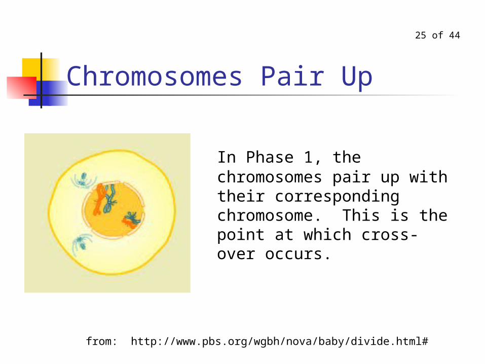
In Phase 1, the chromosomes pair up with their corresponding chromosome. This is the point at which cross-over occurs.
from: http://www.pbs.org/wgbh/nova/baby/divide.html#

26 of 44Chromosome Pairs Split Up
The spindle fibers from the centriole at one end of the nucleus attach to one chromosome in each of the 23 pairs.
The spindle fibers from the centriole at the other end of the nucleus attach to the other chromosome in each pair.
from: http://www.pbs.org/wgbh/nova/baby/divide.html#

27 of 44
End of Phase 1As in mitosis, the chromosomes arrive at opposite ends of the cell, and new nuclear membranes form.
The cell shown here is a male's sperm cell. With meiosis in a female, most of the cell's cytoplasm will be concentrated in one of the two emerging cells, resulting in one large cell and one small cell. The large cell will go on to divide again; the small cell will degenerate.
from: http://www.pbs.org/wgbh/nova/baby/divide.html#
28 of 44
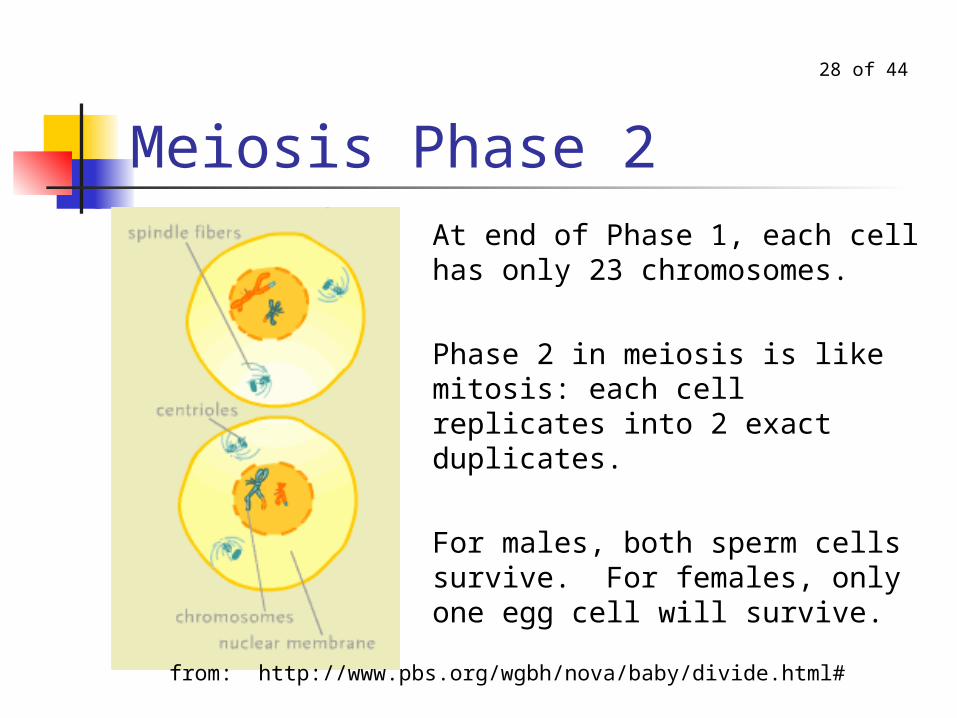
Meiosis Phase 2At end of Phase 1, each cell has only 23 chromosomes.
Phase 2 in meiosis is like mitosis: each cell replicates into 2 exact duplicates.
For males, both sperm cells survive. For females, only one egg cell will survive.
from: http://www.pbs.org/wgbh/nova/baby/divide.html#

Part 3: Chromosomal Abnormalities
30 of 44Chromosomal Abnormalities Abnormal number of one of the chromosomes May occur at time of cell division or may occur
later, due to spontaneous splitting and replication of a chromosome.
May occur in either the ovum or sperm
Three possible abnormalities: Having an extra chromosome Missing a chromosome Missing part of a chromosome
31 of 44
Types of Abnormalities Autosomal Abnormality: Having an
abnormal number of one of the autosomes.
Sex Chromosomal Abnormality: Having an abnormal number of one of the sex chromosomes.

32 of 44
Autosomal AbnormalitiesAutosomal Abnormality: Disorder caused by having too many or too few of one of the autosomes.
Three Examples: Down’s Syndrome 1 in 700 births Monosomy 21 extremely rare Cri du Chat Syndrome 1 in 50,000 births

33 of 44Trisomy 21 (Down’s Syndrome) Have three 21st chromosomes. Characteristics:
oriental-shaped eyes broad face thick tongue small, low-set ears flat nose respiratory
problems & heart defects mental retardation.
34 of 44
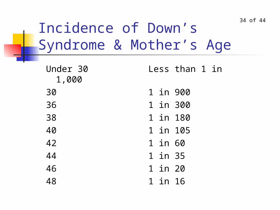
Incidence of Down’s Syndrome & Mother’s Age
Under 30 Less than 1 in 1,00030 1 in 90036 1 in 30038 1 in 18040 1 in 10542 1 in 6044 1 in 3546 1 in 2048 1 in 16
35 of 44Monosomy 21 (Antimongoloid) Have only one 21st chromosome Characteristics:
narrow face large ears broad prominent nasal bridge mental retardation
Only monosomics that survive until birth are Monosomy 21 and Monosomy 22, the smallest chromosomes. They both have severe retardation.

36 of 44
Cri du Chat Syndrome Missing part of an arm of the 5th chromosome Characteristics:
catlike cry as newborn low birth weight and slow growth small head with wideset eyes partial webbing of fingers/toes mental retardation

37 of 44Sex Chromosomal Abnormalities
Sex Chromosomal Abnormality: abnormality in number of sex chromosomes.
Most common types: Phenotypically female
XO Turner’s Syndrome 1 in 2500 XXX Trisomy X Syndrome 1 in 1000
Phenotypically male XXY Klinefelter’s Syndrome 1 in 600 XYY Jacob’s Syndrome 1 in 1000

38 of 44
Turner’s Syndrome Missing one of the X chromosomes. Physically female, but does not develop at
puberty. Characteristics:
short stature lack of ovarian development; infertility may have webbed neck, arms that turn out
slightly at the elbow, low hairline in the back of the head
may have easier time with verbal learning than with math and spatial learning.
characteristically more feminine than average girl

39 of 44
Trisomy X Most common X chromosome abnormality. Have three X chromosomes. Characteristics:
taller than average for girls may have speech and language delays and
mental retardation. some have infertility problems. Many Trisomic X girls have no symptoms

40 of 44
Klinefelter’s Syndrome Physically male, but does not mature at puberty. In addition to XY, have extra X chromosome. Characteristics:
Do not develop testes, thus infertile Generally very placid; can be shy Has delays in motor development and language
development; latter causes problems in school
41 of 44
Jacob’s Syndrome In addition to XY, have extra Y chromosome Characteristics:
taller than average male otherwise, no physical symptoms have tendency to be more active than normal usually have some mental retardation some suggestion that XYY males have higher
than normal levels of aggression

42 of 44
Number of X Chromosomes and Femininity
chromosomes
femininity height
Turner’sX high
shorter than
average
Normal XX average average
Trisomy X XXXlow
(tomboys)taller than average

43 of 44
Number of Y Chromosomes and Aggression
chromosomes
aggression height
Klinefelter’s XXY low
shorter than
average
Normal XY average average
Jacob’s XYY hightaller than average
44 of 44
Summary: Chromosomal Abnormalities
Abnormalities are caused by either having too many or too few chromosomes.
Caused before conception (usually at time that parent cell divides into gametes)
Reason for occurrence not known. Each chromosomal abnormality is multi-
symptomatic: Each chromosome contains many genes for many traits Having three chromosomes means having 3 genes for
each of those traits Having one chromosome means having 1 gene for
each trait on that chromosome

The End