chronic glp-1 receptor activation by exendin-4 induces - diabetes
TRANSCRIPT
Chronic GLP-1 Receptor Activation by Exendin-4 InducesExpansion of Pancreatic Duct Glands in Rats andAccelerates Formation of Dysplastic Lesions and ChronicPancreatitis in the KrasG12D Mouse ModelBelinda Gier,
1Aleksey V. Matveyenko,
1David Kirakossian,
1David Dawson,
2,3Sarah M. Dry,
2,3and
Peter C. Butler1,3
Pancreatic duct glands (PDGs) have been hypothesized to give riseto pancreatic intraepithelial neoplasia (PanIN). Treatment with theglucagon-like peptide (GLP)-1 analog, exendin-4, for 12 weeksinduced the expansion of PDGs with mucinous metaplasia andcolumnar cell atypia resembling low-grade PanIN in rats. In thepancreata of Pdx1-Cre; LSL-KrasG12D mice, exendin-4 led to ac-celeration of the disruption of exocrine architecture and chronicpancreatitis with mucinous metaplasia and increased formationof murine PanIN lesions. PDGs and PanIN lesions in rodent andhuman pancreata express the GLP-1 receptor. Exendin-4 inducedproproliferative signaling pathways in human pancreatic duct cells,cAMP–protein kinase A and mitogen-activated protein kinase phos-phorylation of cAMP-responsive element binding protein, and in-creased cyclin D1 expression. These GLP-1 effects were morepronounced in the presence of an activating mutation of Kras andwere inhibited by metformin. These data reveal that GLP-1 mimetictherapy may induce focal proliferation in the exocrine pancreasand, in the context of exocrine dysplasia, may accelerate formationof neoplastic PanIN lesions and exacerbate chronic pancreatitis.
Glucagon-like peptide (GLP)-1 is a proglucagon-derived peptide secreted by gut endocrine cells(L cells) in response to meal ingestion (1). TheGLP-1 receptor (GLP-1R) is a G-protein–coupled
receptor that is expressed in pancreatic islets and exocrineduct cells (2,3). The increased GLP-1 released after mealingestion amplifies postprandial nutrient-driven insulinsecretion, the so-called incretin effect (4). Based on thisproperty, GLP-1R activation became an attractive thera-peutic target for type 2 diabetes mellitus (T2DM).
To overcome the short half-life of circulating GLP-1 thatis rapidly degraded by dipeptidyl peptidase (DPP)-4 (5), twostrategies have been used in drug development. Oral DPP-4small molecule inhibitors, such as sitagliptin, prolong thehalf-life of endogenously secreted GLP-1 (6). Alternatively,GLP-1R peptide agonists given by injection, such as
exenatide (7) and liraglutide (8), are resistant to DPP-4degradation.
Pancreatitis emerged as an unexpected side effect ofGLP-1–based therapy in case reports (9,10), and in the U.S.Food and Drug Administration adverse-event reportsliraglutide and sitagliptin showed a signal of pancreatitis(11–13), although analysis of insurance claims recordshave been reported to show no association between GLP-1–based therapy and pancreatitis (14). Because the humanpancreas is inaccessible in treated patients, the questionas to whether GLP-1 mimetic therapy acts on the exocrinepancreas has been a subject of animal-based studies.
Pancreatic duct cell proliferation increased transientlywith a GLP-1 infusion in Wistar rats (15). Sprague-Dawleyrats treated with exendin-4 for 12 weeks developed low-grade chronic pancreatitis (16). Furthermore, DPP-4 in-hibition with sitagliptin for 12 weeks was associated withincreased pancreatic duct cell replication and acinar-to-ductal metaplasia and, in 1 of 10 rats, chronic pancreatitis(3). However, GLP-1–based therapy also has been reportedto not exacerbate chemically induced pancreatitis in mice(17). Also, exenatide was reported to have no effect onductal turnover in mice or rats, as well as to have a ben-eficial action in chemically induced pancreatitis (18).
Pancreatic duct glands (PDGs), under conditions ofchronic injury, such as chemically induced pancreatitis,may give rise to lesions resembling pancreatic intraepithelialneoplasia (PanIN) (19). To date, there is no information onthe actions of GLP-1–based therapy on PDGs or the de-velopment of PanIN in pancreata predisposed to dyspla-sia. Here, we sought to address the following questions.First, does chronic activation of GLP-1Rs by exendin-4lead to proliferation of the PDGs? Second, is GLP-1Rexpression present in PDGs and PanIN-like dysplasticlesions? Third, does chronic activation of GLP-1Rs alterthe phenotype of Pdx1-Cre; LSL-KrasG12D (Pdx1-Kras)mice?
RESEARCH DESIGN AND METHODS
Rodent studies. All animal studies were approved by the animal use and carecommittee at the University of California Los Angeles (UCLA). Animals werehoused individually in a 12-h light/dark cycle and were weighed weekly toadjust drug doses. Blood glucose and food intakeweremonitored on a biweeklybasis.Sprague-Dawley rats treated with exendin-4. To establish the actions ofGLP-1R activation in the exocrine pancreas, we treated 10 male Sprague-Dawley rats (Charles River Laboratories, Wilmington, MA) with daily injectionsof 10 mg/kg body wt exendin-4 (ChemPep, Miami, FL) administered by sub-cutaneous injection for 12 weeks starting at 10 weeks of age (20). Animalswere fed chow (Teklad; Harlan Laboratories, Madison, WI) ad libitum. A totalof 15 control rats received daily saline injections. We did not identify PDGs in
From the 1Larry L. Hillblom Islet Research Center, University of California LosAngeles (UCLA), David Geffen School of Medicine, Los Angeles, California;the 2Department of Pathology and Laboratory Medicine, UCLA, David GeffenSchool of Medicine, Los Angeles, California; and the 3Jonsson Comprehen-sive Cancer Center, UCLA, David Geffen School of Medicine, Los Angeles,California.
Corresponding author: Belinda Gier, [email protected] 8 August 2011 and accepted 16 November 2011.DOI: 10.2337/db11-1109This article contains Supplementary Data online at http://diabetes
.diabetesjournals.org/lookup/suppl/doi:10.2337/db11-1109/-/DC1.� 2012 by the American Diabetes Association. Readers may use this article as
long as the work is properly cited, the use is educational and not for profit,and the work is not altered. See http://creativecommons.org/licenses/by-nc-nd/3.0/ for details.
diabetes.diabetesjournals.org DIABETES 1
ORIGINAL ARTICLE
Diabetes Publish Ahead of Print, published online January 20, 2012
5 of 15 controls; therefore, these 5 rats were not included in subsequentanalyses. PDGs were identified in all treated rats.Pdx1-Kras mice treated with exendin-4. To investigate the effect ofchronic GLP-1 mimetic treatment on pancreatic cancer precursor lesions, theconditional KrasG12D from Hingorani et al. (21) was used. Experimental ani-mals were generated by crossing Pdx1-Cre with LSL-KrasG12D mice on a C57/BL6 background (both gifts of Guido Eibl, UCLA). Mice (6 weeks old) werefed an AIN-76A–based diet (Research Diets, New Brunswick, NJ) ad libitumfor 12 weeks, during which either saline (n = 7) or exendin-4 (5 nmol/kg bodywt) (n = 5) was injected subcutaneously daily.Pancreas fixation, embedding, and sectioning
Rat pancreas. After the rats were killed, the rat pancreata were rapidlydissected and then divided into two portions (the head and body of the pancreasand the tail of the pancreas). These were fixed in 4% paraformaldehydeovernight at 4°C and embedded in paraffin, oriented flat to permit subsequentmicroscopic sections to be made through the longitudinal plane of the pancreas.The block containing the head and body of the pancreas was sectioned at 4-mmintervals to obtain a minimum of 40 sections through the longitudinal plane ofthe pancreas. A minimum of 10 serial sections were obtained per block from thetail of the pancreas.Mouse pancreas. Successful excision-recombination events were confirmedby genotyping analysis (Transnetyx, Cordova, TN). Paraformaldehyde-fixed,paraffin-embedded pancreatic sections (4 mm) of whole pancreas were stainedas below.Human pancreas. Paraffin-embedded tissue blocks of nonneoplastic humanpancreata adjacent to surgically resected pancreatic adenocarcinoma wereselected from 10 case subjects from the UCLA pathology archives. All slides andtissue blocks were retrieved after institutional review board approval (no. 11-001724).Pancreas histology and stains. Tissue sections from rats and mice weredeparaffinized in toluene and rehydrated in an ethanol gradient. First, sectionswere stained in Harris hematoxylin solution (HHS16; Sigma) and eosin Y so-lution (HT110132; Sigma) to evaluate general histology. PDGs were definedbased on the previously described criteria (19) (Fig. 1). Sections were stainedby Alcian blue (Electron Microscopy Sciences) and p-aminosalicylic acid(PAS) (Sigma).
For immunhistochemical-immunofluorescent staining, antigen retrieval wasperformed via microwave heating in citrate buffer (H-3300; Vector, Burlingame,CA), and slides were blocked in Tris-buffered saline (3% bovine serum albumin,0.2% TX-100, and 2% bovine serum) for 1 h. The following primary antibodieswere used for the 12-h incubation (4°C): ductal cell marker cytokeratin (mouseanti-pancytokeratin, 1:50 [Sigma] or rat anti-cytokeratin19/TROMAIII, 1:100[Hybridoma Bank, University of Iowa, Iowa City, IA]); acinar cell markeramylase (rabbit anti-amylase, 1:300; Abcam); proliferation marker anti–Ki-67(1:50; Dako, Carpinteria, CA); GLP-1R (rabbit anti-human GLP-1R, NLS1206,1:100; Novus Biologicals, Littleton, CO); and pancreatic and duodenalhomeobox-1 (Pdx-1) (rabbit anti–Pdx-1, 1:500; b-Cell Biology Consortium,Nashville, TN). Validation of the GLP-1R antisera, NLS1206, was publishedpreviously (22). Secondary antibodies labeled with Cy3 and fluorescein iso-thiocyanate were obtained from The Jackson Laboratories (West Grove, PA)and used at dilutions of 1:100 for the 1-h incubation at room temperature.
For immunohistochemical staining, endogenous peroxidase activity wasquenched with 10% methanol, 10% H202 in Tris-buffered saline, followed byincubation with anti–Ki-67 (dilution 1:100; Dako) for 12 h (4°C). Subsequentdetection was performed with Envision+ anti-rabbit horseradish peroxidase(Dako), with 39,39-diaminobenzidine used as the chromogen and hematoxylinas the counterstain.
Likewise, sections of human pancreata were stained with hematoxylin andeosin, Alcian blue, and PAS in order to permit the identification of PDGs andPanIN lesions. Sections adjacent to those with PDGs and PanIN lesions alsowere stained by immunofluorescence for cytokeratin and for GLP-1R (the sameantibodies and dilution as above).Morphometric analysis of the pancreatic duct gland compartment in
rats. The slide with the greatest number of PDGs per animal was selected forquantitative analysis. Slides were digitally scanned in the UCLA TranslationalPathology Core Laboratory using an Aperio ScanScope XT (Aperio Technol-ogies, Vista, CA). Quantitative analysis was performed using Aperio Scanscopesoftware. The length of the large duct associated with PDGs and the meancross-sectional area per PDGweremeasured in each case. PDGs in the exendin-4–treated rats often showed complex epithelial architecture, including cribiformpatterns and pseudopapillae, consistent with epithelial proliferation. The ductscontaining this complex PDG epithelial architecture appeared more dilated. Toquantify if ductal dilation was present, we measured the longest axis of the largeducts present (duct length) and the inner circumference of the duct lumen. Thisallowed us to compute a ratio (inner duct luminal circumference to duct length)for each animal.
Histological analysis of chronic pancreatitis and murine PanIN (mPanIN)
lesions in Pdx1-Kras mice. Full histologic cross-sections of each pancreaswere stained with hematoxylin and eosin for histopathologic examination by twosubspecialty gastrointestinal pathologists (D.D. and S.M.D.) blinded to treatmentconditions. Chronic pancreatitis was graded using a semiquantitative scoringsystem, as previously described (23), with slight modification. Chronic pan-creatitis was given an index score (0–12) reflecting the sum of scores foracinar loss, lobular inflammation, and fibrosis. Acinar loss was based on thepercentage loss across the entire cross-section and graded as 0, absent;1, 1–25%; 2, 26–50%; 3, 51–75%; and 4, .75%. Inflammation was based on theaverage number of lobular inflammatory cells per 403 high power field (HPF)(as counted in 10 nonoverlapping HPFs) and graded as 0, absent; 1, 1–30 cells; 2,31–50 cells; 3, 51–100 cells; and 4, .100 cells. Fibrosis was based on the cu-mulative area of stromal fibrosis across the entire pancreas and graded as 0,absent; 1, 1–5%; 2, 6–10%; 3, 11–20%; and 4, .20% fibrosis. Duct profiles wereevaluated according to established consensus guidelines for the histologicevaluation of mPanIN (24) and quantified as previously described (25). All ductprofiles in one full pancreas cross-section were evaluated to determine therelative proportions of nondysplastic (normal, reactive, and metaplastic) ductsand each category of mPanIN lesion. The proportion of each mPanIN lesion tothe overall number of duct profiles was recorded for each animal.Duct cell replication frequency. To determine the frequency of replication ofPDG cells and cells in the adjacent main ducts in the head of the pancreas inrats, we quantified the percentage of Ki-67–positive cells. Thus, the total numberof duct cells (the head of the pancreas) evaluated was 57,261 in exendin-4–treated rats and 61,298 in controls. We also evaluated the frequency of duct cellreplication in the sections of the tail of the pancreas immunostained for cyto-keratin and Ki-67. The total number of duct cells evaluated from the tail was24,483 in exendin-4–treated rats and 19,796 in controls.
Duct cell replication in pancreata from Pdx1-Kras mice also was analyzed byKi-67. The extensive acinar-to-ductal metaplasia and frequent dysplastic ductallesions in GLP-1–treated Pdx1-Kras mice precluded distinguishing replicationfrequency in the various component of the ductal compartment (PDGs andnormal and dysplastic ducts). Slides were analyzed using the Ariol SL-50 au-tomated slide scanner (Leica Microsystems) to quantitate the amount ofpositive staining for each area of interest containing only ducts and dysplasticductal tissue. A total number of 121,883 (control group) and 101,830 (exendin-4–treated group) cells were analyzed.GLP-1 actions on pancreatic duct cells. In vitro experiments were carriedout to investigate the effects of exendin-4 on human pancreatic duct epithelial(HPDE) cells (26,27). HPDE cells (kindly made available by Dr. Ming-SoundTsao, University of Toronto) were maintained in keratinocyte serum-freemedia supplemented with bovine pituitary extract and human epidermalgrowth factor (Invitrogen) at 37°C with 5% CO2. HPDE cells transfected withthe empty vector (pBabepuro) (HPDE-pBP) or with oncogenic pBP-Kras4BG12V
(HPDE-Kras) also were used to permit the assessment of GLP-1R activation inthe presence of an activating Kras mutation.
To assess the effect of exendin-4 (10 nmol/L) on the phosphorylation ofcAMP-responsive element binding (CREB) protein and the mitogen-activatedprotein kinases (MAPKs) extracellular signal–related kinase (ERK) 1/2, as wellas levels of the cyclin A and D1, 90,000 cells were seeded in six-well platescontaining complete medium. At ~70% confluence (day 3 after plating), cellswere rinsed with PBS and incubated in starvation medium (lacking bovinepituitary extract and human epidermal growth factor) for 24 h. HPDE cellscontaining either a control plasmid (pBabepuro; HPDE-pBP) or the mutatedoncogenic pBP-KrasG12V (28,29) were pretreated with 100 mmol/L metforminfor 30 min. For stimulation experiments, exendin-4 was added in fresh, pre-warmed starvation medium for the indicated time points. Stimulation then wasstopped by adding ice-cold PBS, and HPDE cells were lysed in lysis buffer (50mmol/L Hepes, 1% Nonidet P-40, 2 mmol/L Na3VO4, 100 mmol/L NaF, 10 mmol/LPyrPO4, 4 mmol/L EDTA, 1 mmol/L phenylmethylsulfonyl fluoride, 1 mg/mLleupeptin, and 1 mg/mL aprotinin). Protein samples were denatured by boilingat 95°C for 5 min, separated on 4–12% Bis-Tris NuPAGE gels (25–40 mg/lane;Invitrogen), and blotted onto a polyvinylidene fluoride membrane (Fluoro-Trans; Pall Life Sciences, Ann Arbor, MI). Membranes were probed with thefollowing primary antibodies (dilution for all 1:1,000): rabbit anti-CREB/pCREB; rabbit anti-ERK1/2/pERK1/2 (both Cell Signaling); rabbit anti–cyclin A;and rabbit anti–cyclin D1 (both Santa Cruz). After incubation with horseradishperoxidase–conjugated secondary antibody (1:5,000; The Jackson Laborato-ries), proteins were visualized using enhanced chemiluminescence (Millipore),and levels were quantified using Labworks software (UVP, Upland, CA).Analytical procedures. Plasma glucose concentrations were measured by theglucose oxidase method (YSI Glucose Analyzer, Yellow Springs, OH). Plasmalipase concentrations were measured by a colorimetric enzyme assay (BioAssaySystems, Hayward, CA).Statistical analysis. Statistical analysis was performed using the Studentt test or ANOVA, where appropriate (Statistica, version 6; Statsoft, Tulsa, OK).
GLP-1 ACTIONS ON THE EXOCRINE PANCREAS
2 DIABETES diabetes.diabetesjournals.org
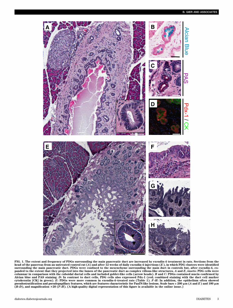
FIG. 1. The extent and frequency of PDGs surrounding the main pancreatic duct are increased by exendin-4 treatment in rats. Sections from thehead of the pancreas from an untreated control rat (A) and after 12 weeks of daily exendin-4 injections (E), in which PDG clusters were identifiedsurrounding the main pancreatic duct. PDGs were confined to the mesenchyme surrounding the main duct in controls but, after exendin-4, ex-panded to the extent that they projected into the lumen of the pancreatic duct as complex villous-like structures. A and E, insets: PDG cells werecolumnar in comparison with the cuboidal ductal cells and included goblet-like cells (arrow heads). B and C: PDGs contained mucin confirmed byAlcian blue and PAS staining. D: In contrast to duct cells, PDG cells also expressed Pdx-1 (red; combined staining with the duct cell markercytokeratin [CK] in green). E: PDGs were more common in exendin-4–treated rats (Table 1). F–H: In addition, the epithelium often showedpseudostratification and pseudopapillary features, which are features characteristic for PanIN-like lesions. Scale bars = 200 mm (A and E) and 100 mm(B–D), and magnification 320 (F–H). (A high-quality digital representation of this figure is available in the online issue.)
B. GIER AND ASSOCIATES
diabetes.diabetesjournals.org DIABETES 3
Data in graphs and tables are presented as means 6 SEM. Findings wereassumed statistically significant at P , 0.05.
RESULTS
Metabolic actions of exendin-4 in rats. Twelve weeksof daily exendin-4 injections had the anticipated effects ofdecreasing weight gain (66 6 8 vs. 164 6 5 g; P , 0.001exendin-4 vs. control) and blood glucose levels (99 6 2 vs.108 6 4 mg/dL; P , 0.01 exendin-4 vs. control). Asexpected, exendin-4 decreased daily food intake (153 6 5vs. 204 6 5 mg/day; P , 0.001 exendin-4 vs. control), butthe treated animals did not seem to be in any apparent painor distress (Supplementary Fig. 1).Effects of exendin-4 on exocrine pancreas in rats.Pancreas weight was comparable in the treated versuscontrol group (2.3 6 0.1 vs. 2.3 6 0.1 g; exendin-4 vs.control). However, relative to body weight, pancreaticweight in exendin-4–treated animals was increased (0.53 60.02 vs. 0.43 6 0.02; P , 0.01 exendin-4 vs. control)(Supplementary Fig. 1D).
There was no histological evidence of pancreatitis ineither the exendin-4 or control group. Consistent with this,lipase activity was not changed by exendin-4 (330 6 19 vs.299 6 11 units/L; exendin-4 vs. control) (SupplementaryFig. 1E). However, exendin-4 did induce a marked ex-pansion of the PDG compartment (Fig. 1 and Supplemen-tary Fig. 2). PDGs were identified, as previously described,as blind outpouchings from large pancreatic ducts presentin the mesenchyme surrounding the ducts. PDG cells werefurther distinguished from main duct cells by frequentlybeing columnar rather than cuboidal (Fig. 1A and E,insets) and mucin positive (Alcian blue and PAS stains).PDGs also expressed Pdx-1 (Figs. 1B–D). There was an~70% increase in the number of PDGs per unit of length ofthe main pancreatic duct following exendin-4 treatment(526 7 vs. 316 4 PDGs/mm main duct; P, 0.05 exendin-4vs. control) (Table 1). Moreover, the mean cross-sectionalarea of individual PDGs still confined within the mesen-chyme around the ducts was ~30% increased by exendin-4treatment (1,1846 102 vs. 9106 45 mm2; P, 0.05 exendin-4vs. control), a conservative estimate given that the expandedPDGs adopt a more coiled structure as previously de-scribed (19). The latter evaluation also likely underestimatesthe extent of the expansion of PDGs in exendin-4–treatedrats because in many cases the PDGs also expanded intothe duct lumen with a complex cribriforming and papillaryarchitecture. To account for this, we quantified the extentto which the main duct lumen was convoluted by anyintraluminal projection by computing the ratio of the cir-cumference of the inner duct lumen to the duct length,a metric that was ~35% increased in exendin-4–treatedanimals (5.0 6 0.2 vs. 3.7 6 0.3; P , 0.01 exendin-4 vs.control). With exendin-4, the epithelium also showedvariable nuclear pseudostratification and loss of polarity,as well as micropapillary architecture (Fig. 1F–H), his-tologic features that can be associated with PanIN lesionsand dysplasia when observed in human pancreas, althoughthe implications in rat pancreas are unknown. No car-cinoma was seen. Collectively, these findings confirm anincreased number of PDGs and expansion of the epithelialcell compartment of both PDGs and large ducts in re-sponse to exendin-4 treatment.
The frequency of PDG cell replication was fourfold in-creased in exendin-4 versus control rats (14.6 6 3.9 vs.3.8 6 0.9%; P , 0.05 exendin-4 vs. control) (Fig. 2). The
frequency of replication in the main pancreatic ducts wasmuch lower than that in the PDGs but still increasedtwofold by exendin-4 treatment (5.3 6 1.8 vs. 2.7 6 0.6%;P , 0.05 exendin-4 vs. control). In contrast, there was nostatistically increased frequency of duct cell replicationwith exendin-4 treatment in the small ducts of the tail ofthe pancreas (0.62 6 0.17 vs. 0.42 6 13%; P = 0.4 exendin-4vs. control).Actions of GLP-1 mimetic treatment on the exocrinepancreas in the Pdx1-Kras mutant mouse. In Pdx1-Krasmice, 12 weeks of exendin-4 treatment had no impact onbody weight (23.2 6 1.2 vs. 25.8 6 1.7 g), food intake(18.1 6 0.7 vs. 19.7 6 0.5 g per week), or blood glucoselevels (83.0 6 3.4 vs. 75.4 6 3.7 mg/dL) when comparedwith littermate control mice. However, GLP-1 mimetic treat-ment increased pancreatic weight (1.1 6 0.1 vs. 0.7 6 0.1 g;exendin-4 vs. control) (Supplementary Fig. 3).
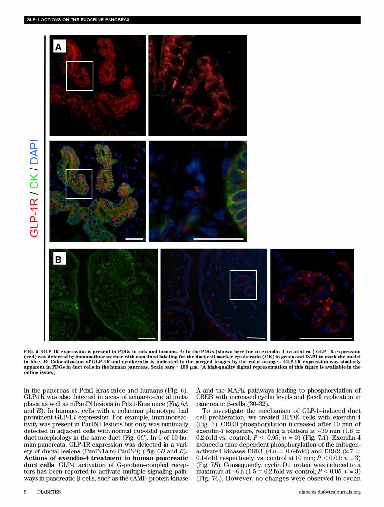
While overall lobular architecture was preserved in bothanimal groups, the exendin-4–treated animals demon-strated more extensive chronic pancreatitis with greaterloss of acini with replacement by reactive or metaplasticduct profiles (Fig. 3). The percentage of pancreas com-posed of acinar tissue was decreased by 61% by exendin-4treatment (13.0 6 13.5% vs. 33.66 14.6%; P , 0.05 exendin-4 vs. control). These changes were accompanied by in-creased inflammation, more extensive stromal fibrosis, andwidespread reactive and metaplastic changes, as de-termined by pancreatitis score (10.0 6 1.2 vs. 8.6 6 0.8;P , 0.05 exendin-4 vs. control). The plasma lipase activityalso was increased with exendin-4 (1,020 6 164 vs. 678 634 units/L; P , 0.05 exendin-4 vs. control) (SupplementaryFig. 3). In comparison to control animals, treated animalsshowed more extensive acinar-to-ductal metaplasia withreplacement of acini by ductules lined by mucin-producingcells primarily with small, round basally oriented nucleiwithout papillary features (mPanIN1). A minority of the ductprofiles demonstrated increased nuclear hyperchromasiaand pleomorphism with stratification and micropapillarychanges (mPanIN2 and mPanIN3) (Fig. 3). Moreover, GLP-1mimetic treatment induced increased duct cell proliferation(P , 0.05) in Pdx1-Kras mice when compared with controlanimals (Fig. 4).GLP-1R expression in PDGs and PanIN lesions. GLP-1R expression was readily detected in pancreatic b-cells inrat and human pancreas, serving as a positive control (datanot shown). GLP-1R expression also was present in PDG cellsin both rodent and in human pancreas (Fig. 5). GLP-1R ex-pression was not detected in pancreatic acinar cells. GLP-1Rexpression also was abundantly present in mPanIN lesions
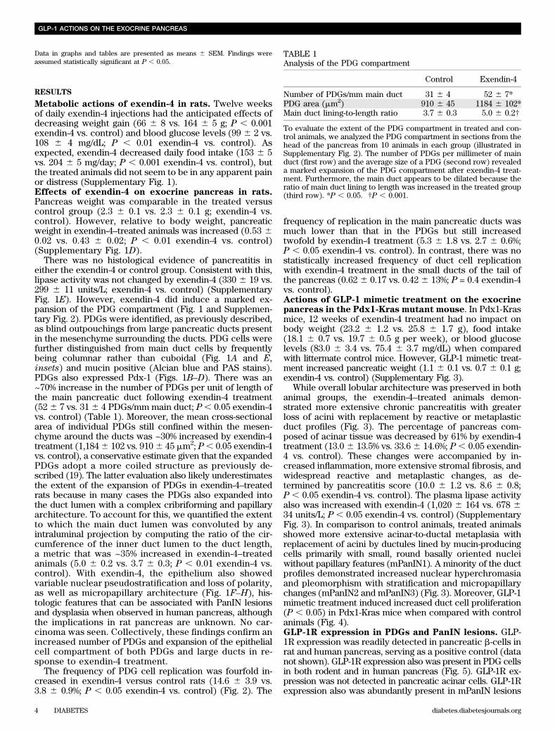
TABLE 1Analysis of the PDG compartment
Control Exendin-4
Number of PDGs/mm main duct 31 6 4 52 6 7*PDG area (mm2) 910 6 45 1184 6 102*Main duct lining-to-length ratio 3.7 6 0.3 5.0 6 0.2†
To evaluate the extent of the PDG compartment in treated and con-trol animals, we analyzed the PDG compartment in sections from thehead of the pancreas from 10 animals in each group (illustrated inSupplementary Fig. 2). The number of PDGs per millimeter of mainduct (first row) and the average size of a PDG (second row) revealeda marked expansion of the PDG compartment after exendin-4 treat-ment. Furthermore, the main duct appears to be dilated because theratio of main duct lining to length was increased in the treated group(third row). *P , 0.05. †P , 0.001.
GLP-1 ACTIONS ON THE EXOCRINE PANCREAS
4 DIABETES diabetes.diabetesjournals.org
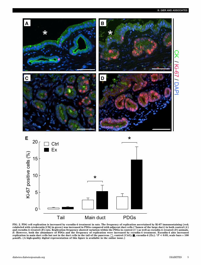
FIG. 2. PDG cell replication is increased by exendin-4 treatment in rats. The frequency of replication ascertained by Ki-67 immunostaining (red;colabeled with cytokeratin [CK] in green) was increased in PDGs compared with adjacent duct cells (*lumen of the large duct) in both control (A)and exendin-4–treated (B) rats. Replication frequency showed variation within the PDGs in control (C) as well as exendin-4–treated (D) animals.E: However, both the abundance of PDGs and the frequency of replication were increased by exendin-4 treatment. Exendin-4 also increasedreplication in main duct cells but not in the duct cells in the tail of the pancreas. □, control (Ctrl); ■, exendin-4 (Ex). *P < 0.05, scale bars = 100mmol/L. (A high-quality digital representation of this figure is available in the online issue.)
B. GIER AND ASSOCIATES
diabetes.diabetesjournals.org DIABETES 5
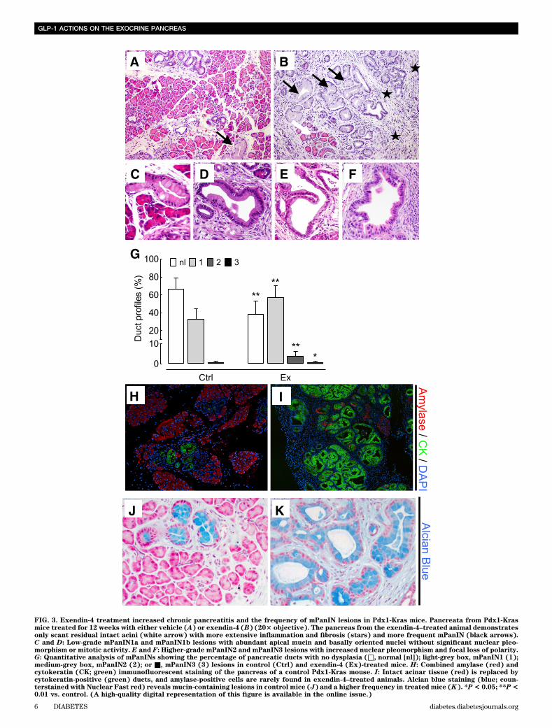
FIG. 3. Exendin-4 treatment increased chronic pancreatitis and the frequency of mPanIN lesions in Pdx1-Kras mice. Pancreata from Pdx1-Krasmice treated for 12 weeks with either vehicle (A) or exendin-4 (B) (203 objective). The pancreas from the exendin-4–treated animal demonstratesonly scant residual intact acini (white arrow) with more extensive inflammation and fibrosis (stars) and more frequent mPanIN (black arrows).C and D: Low-grade mPanIN1a and mPanIN1b lesions with abundant apical mucin and basally oriented nuclei without significant nuclear pleo-morphism or mitotic activity. E and F: Higher-grade mPanIN2 and mPanIN3 lesions with increased nuclear pleomorphism and focal loss of polarity.G: Quantitative analysis of mPanINs showing the percentage of pancreatic ducts with no dysplasia (□, normal [nl]); light-grey box, mPanIN1 (1);medium-grey box, mPanIN2 (2); or ■, mPanIN3 (3) lesions in control (Ctrl) and exendin-4 (Ex)-treated mice. H: Combined amylase (red) andcytokeratin (CK; green) immunofluorescent staining of the pancreas of a control Pdx1-Kras mouse. I: Intact acinar tissue (red) is replaced bycytokeratin-positive (green) ducts, and amylase-positive cells are rarely found in exendin-4–treated animals. Alcian blue staining (blue; coun-terstained with Nuclear Fast red) reveals mucin-containing lesions in control mice (J) and a higher frequency in treated mice (K). *P< 0.05; **P<0.01 vs. control. (A high-quality digital representation of this figure is available in the online issue.)
GLP-1 ACTIONS ON THE EXOCRINE PANCREAS
6 DIABETES diabetes.diabetesjournals.org
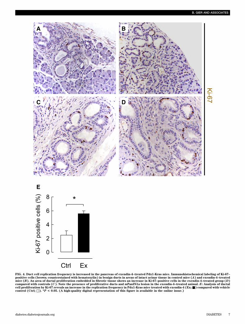
FIG. 4. Duct cell replication frequency is increased in the pancreas of exendin-4–treated Pdx1-Kras mice. Immunohistochemical labeling of Ki-67–positive cells (brown; counterstained with hematoxylin) in benign ducts in areas of intact acinar tissue in control mice (A) and exendin-4–treatedmice (B). An area of ductal proliferation embedded in fibrotic tissue shows an increase in Ki-67–positive cells in the exendin-4–treated group (D)compared with controls (C). Note the presence of proliferative ducts and mPanIN1a lesion in the exendin-4–treated animal. E: Analysis of ductalcell proliferation by Ki-67 reveals an increase in the replication frequency in Pdx1-Kras mice treated with exendin-4 (Ex;■) compared with vehiclecontrol (Ctrl; □). *P < 0.05. (A high-quality digital representation of this figure is available in the online issue.)
B. GIER AND ASSOCIATES
diabetes.diabetesjournals.org DIABETES 7
in the pancreas of Pdx1-Kras mice and humans (Fig. 6).GLP-1R was also detected in areas of acinar-to-ductal meta-plasia as well as mPanIN lesions in Pdx1-Kras mice (Fig. 6Aand B). In humans, cells with a columnar phenotype hadprominent GLP-1R expression. For example, immunoreac-tivity was present in PanIN1 lesions but only was minimallydetected in adjacent cells with normal cuboidal pancreaticduct morphology in the same duct (Fig. 6C). In 6 of 10 hu-man pancreata, GLP-1R expression was detected in a vari-ety of ductal lesions (PanIN1a to PanIN3) (Fig. 6D and E).Actions of exendin-4 treatment in human pancreaticduct cells. GLP-1 activation of G-protein–coupled recep-tors has been reported to activate multiple signaling path-ways in pancreatic b-cells, such as the cAMP–protein kinase
A and the MAPK pathways leading to phosphorylation ofCREB with increased cyclin levels and b-cell replication inpancreatic b-cells (30–32).
To investigate the mechanism of GLP-1–induced ductcell proliferation, we treated HPDE cells with exendin-4(Fig. 7). CREB phosphorylation increased after 10 min ofexendin-4 exposure, reaching a plateau at ~30 min (1.8 60.2-fold vs. control; P , 0.05; n = 3) (Fig. 7A). Exendin-4induced a time-dependent phosphorylation of the mitogen-activated kinases ERK1 (4.8 6 0.6-fold) and ERK2 (2.7 60.1-fold, respectively, vs. control at 10 min; P , 0.01; n = 3)(Fig. 7B). Consequently, cyclin D1 protein was induced to amaximum at ~6 h (1.56 0.2-fold vs. control; P, 0.05; n = 3)(Fig. 7C). However, no changes were observed in cyclin
FIG. 5. GLP-1R expression is present in PDGs in rats and humans. A: In the PDGs (shown here for an exendin-4–treated rat) GLP-1R expression(red) was detected by immunofluorescence with combined labeling for the duct cell marker cytokeratin (CK) in green and DAPI to mark the nucleiin blue. B: Colocalization of GLP-1R and cytokeratin is indicated in the merged images by the color orange . GLP-1R expression was similarlyapparent in PDGs in duct cells in the human pancreas. Scale bars = 100 mm. (A high-quality digital representation of this figure is available in theonline issue.)
GLP-1 ACTIONS ON THE EXOCRINE PANCREAS
8 DIABETES diabetes.diabetesjournals.org
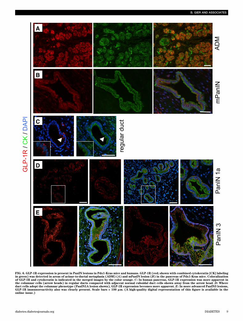
FIG. 6. GLP-1R expression is present in PanIN lesions in Pdx1-Kras mice and humans. GLP-1R (red; shown with combined cytokeratin [CK] labelingin green) was detected in areas of acinar-to-ductal metaplasia (ADM) (A) and mPanIN lesion (B) in the pancreas of Pdx1-Kras mice. Colocalizationof GLP-1R and cytokeratin is indicated in the merged images by the color orange. C: In human pancreas, GLP-1R expression was more apparent inthe columnar cells (arrow heads) in regular ducts compared with adjacent normal cuboidal duct cells shown away from the arrow head. D: Whereduct cells adopt the columnar phenotype (PanIN1A lesion shown), GLP-1R expression becomes more apparent. E: In more advanced PanIN3 lesions,GLP-1R immunoreactivity also was clearly present. Scale bars = 100 mm. (A high-quality digital representation of this figure is available in theonline issue.)
B. GIER AND ASSOCIATES
diabetes.diabetesjournals.org DIABETES 9

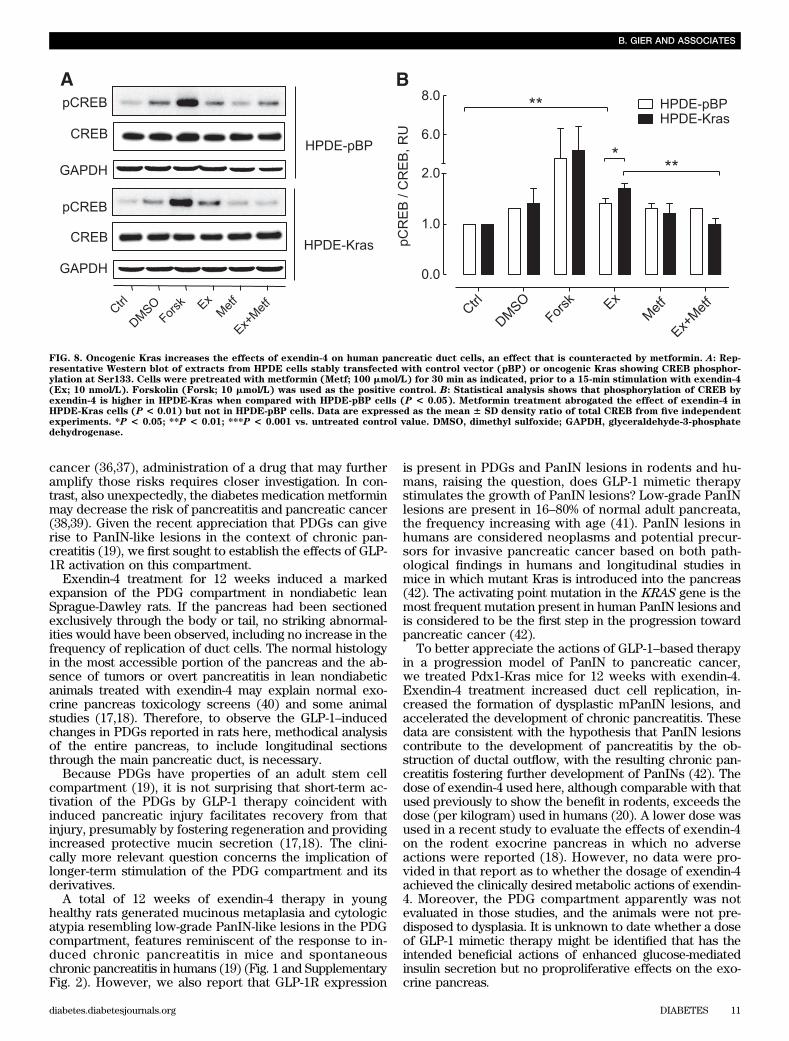
A levels (Fig. 7D). We also investigated the actions ofexendin-4 with or without metformin in the presence of theactivating Kras mutation in HPDE cells. Exendin-4 inducedCREB phosphorylation in control (pBP) cells (1.4 6 0.1-fold pCREB/CREB vs. control; P , 0.01; n = 3), an effectthat was more pronounced in the presence of mutant Kras(1.7 6 0.1-fold vs. control; P , 0.001; n = 3), and this effectwas abrogated by metformin pretreatment (1.0 6 0.1-foldvs. control; n = 3; P , 0.01 vs. exendin-4 treatment alone)(Fig. 8).
DISCUSSION
The possibility that GLP-1 mimetic therapy might inducesustained proliferative changes in the exocrine pancreasis of concern because therapy for T2DM may be admin-istered for decades (33,34). An increased reported ad-verse event rate in the U.S. Food and Drug Administrationadverse-event reporting system for pancreatitis and pan-creatic cancer in patients treated with GLP-1–based ther-apy underscores this concern (35). Because T2DM withobesity is a risk factor for pancreatitis and pancreatic
FIG. 7. Exendin-4 actions on human pancreatic duct cells. A and B: Time-course experiments of CREB (A) and ERK1/2 (B) phosphorylation inHPDE cells treated with exendin-4 (10 nmol/L) for 0–30 min as indicated. GAPDH, glyceraldehyde-3-phosphate dehydrogenase. Representativeexamples of Western blot experiments are shown in the top panels and the corresponding analysis in the bottom panels. C and D: Effect of long-term (0–9 h) stimulation on cyclin D1 (C) and cyclin A (D) protein levels. Data are expressed as the mean6 SD density ratio of total CREB, ERK1/2 (A and B), as well as glyceraldehyde-3-phosphate dehydrogenase (GAPDH) (C and D) from 3 to 5 independent experiments. *P < 0.05; **P <0.01; ***P < 0.001 vs. untreated control value.
GLP-1 ACTIONS ON THE EXOCRINE PANCREAS
10 DIABETES diabetes.diabetesjournals.org
cancer (36,37), administration of a drug that may furtheramplify those risks requires closer investigation. In con-trast, also unexpectedly, the diabetes medication metforminmay decrease the risk of pancreatitis and pancreatic cancer(38,39). Given the recent appreciation that PDGs can giverise to PanIN-like lesions in the context of chronic pan-creatitis (19), we first sought to establish the effects of GLP-1R activation on this compartment.
Exendin-4 treatment for 12 weeks induced a markedexpansion of the PDG compartment in nondiabetic leanSprague-Dawley rats. If the pancreas had been sectionedexclusively through the body or tail, no striking abnormal-ities would have been observed, including no increase in thefrequency of replication of duct cells. The normal histologyin the most accessible portion of the pancreas and the ab-sence of tumors or overt pancreatitis in lean nondiabeticanimals treated with exendin-4 may explain normal exo-crine pancreas toxicology screens (40) and some animalstudies (17,18). Therefore, to observe the GLP-1–inducedchanges in PDGs reported in rats here, methodical analysisof the entire pancreas, to include longitudinal sectionsthrough the main pancreatic duct, is necessary.
Because PDGs have properties of an adult stem cellcompartment (19), it is not surprising that short-term ac-tivation of the PDGs by GLP-1 therapy coincident withinduced pancreatic injury facilitates recovery from thatinjury, presumably by fostering regeneration and providingincreased protective mucin secretion (17,18). The clini-cally more relevant question concerns the implication oflonger-term stimulation of the PDG compartment and itsderivatives.
A total of 12 weeks of exendin-4 therapy in younghealthy rats generated mucinous metaplasia and cytologicatypia resembling low-grade PanIN-like lesions in the PDGcompartment, features reminiscent of the response to in-duced chronic pancreatitis in mice and spontaneouschronic pancreatitis in humans (19) (Fig. 1 and SupplementaryFig. 2). However, we also report that GLP-1R expression
is present in PDGs and PanIN lesions in rodents and hu-mans, raising the question, does GLP-1 mimetic therapystimulates the growth of PanIN lesions? Low-grade PanINlesions are present in 16–80% of normal adult pancreata,the frequency increasing with age (41). PanIN lesions inhumans are considered neoplasms and potential precur-sors for invasive pancreatic cancer based on both path-ological findings in humans and longitudinal studies inmice in which mutant Kras is introduced into the pancreas(42). The activating point mutation in the KRAS gene is themost frequent mutation present in human PanIN lesions andis considered to be the first step in the progression towardpancreatic cancer (42).
To better appreciate the actions of GLP-1–based therapyin a progression model of PanIN to pancreatic cancer,we treated Pdx1-Kras mice for 12 weeks with exendin-4.Exendin-4 treatment increased duct cell replication, in-creased the formation of dysplastic mPanIN lesions, andaccelerated the development of chronic pancreatitis. Thesedata are consistent with the hypothesis that PanIN lesionscontribute to the development of pancreatitis by the ob-struction of ductal outflow, with the resulting chronic pan-creatitis fostering further development of PanINs (42). Thedose of exendin-4 used here, although comparable with thatused previously to show the benefit in rodents, exceeds thedose (per kilogram) used in humans (20). A lower dose wasused in a recent study to evaluate the effects of exendin-4on the rodent exocrine pancreas in which no adverseactions were reported (18). However, no data were pro-vided in that report as to whether the dosage of exendin-4achieved the clinically desired metabolic actions of exendin-4. Moreover, the PDG compartment apparently was notevaluated in those studies, and the animals were not pre-disposed to dysplasia. It is unknown to date whether a doseof GLP-1 mimetic therapy might be identified that has theintended beneficial actions of enhanced glucose-mediatedinsulin secretion but no proproliferative effects on the exo-crine pancreas.
FIG. 8. Oncogenic Kras increases the effects of exendin-4 on human pancreatic duct cells, an effect that is counteracted by metformin. A: Rep-resentative Western blot of extracts from HPDE cells stably transfected with control vector (pBP) or oncogenic Kras showing CREB phosphor-ylation at Ser133. Cells were pretreated with metformin (Metf; 100 mmol/L) for 30 min as indicated, prior to a 15-min stimulation with exendin-4(Ex; 10 nmol/L). Forskolin (Forsk; 10 mmol/L) was used as the positive control. B: Statistical analysis shows that phosphorylation of CREB byexendin-4 is higher in HPDE-Kras when compared with HPDE-pBP cells (P < 0.05). Metformin treatment abrogated the effect of exendin-4 inHPDE-Kras cells (P < 0.01) but not in HPDE-pBP cells. Data are expressed as the mean 6 SD density ratio of total CREB from five independentexperiments. *P < 0.05; **P < 0.01; ***P < 0.001 vs. untreated control value. DMSO, dimethyl sulfoxide; GAPDH, glyceraldehyde-3-phosphatedehydrogenase.
B. GIER AND ASSOCIATES
diabetes.diabetesjournals.org DIABETES 11
Evaluation of the proliferative actions of GLP-1 in theexocrine pancreas in humans is not technically feasible.Therefore, we examined the actions of exendin-4 on hu-man pancreatic ductal epithelial cells in vitro. These invitro studies on the actions of GLP-1R activation in pan-creatic duct cells revealed a proproliferative action me-diated through the activation of MAPK pathways andphosphorylation of CREB, which was even more apparentin the setting of an activating Kras mutation and inhibited bythe actions of metformin. This provides a mechanistic basisfor the association of metformin treatment with decreasedrisk for pancreatitis and pancreatic cancer in individualswith T2DM (38,39). It is also consistent with a previousrodent study in which metformin attenuated the pro-liferative actions of the DPP-4 inhibitor sitagliptin on thepancreatic ductal tree (3).
In conclusion, we report that treatment of rats for 12weeks with exendin-4 induced a marked expansion ofPDGs through the mechanism of enhanced PDG cell rep-lication. Moreover, we report that the PDGs in rats andhumans express GLP-1Rs and that these also are abun-dantly expressed in PanIN lesions in human pancreas. GLP-1treatment advances the rate of formation of dysplasticmPanIN lesions and chronic pancreatitis in a mouse modelprone to the development of pancreatic ductal adenocarci-noma. Finally, we report that treatment of human pancreaticduct cells with the GLP-1 analog exendin-4 induces propro-liferative signaling pathways, an effect that is inhibited bymetformin. Collectively, these studies imply that GLP-1–induced proliferation within the exocrine pancreas is focaland may accelerate the development of dysplastic lesionswhen present.
ACKNOWLEDGMENTS
This work was supported by the National Institute of Di-abetes and Digestive and Kidney Diseases, National In-stitutes of Health, Grant DK-077967 and the Larry HillblomFoundation.
No potential conflicts of interest relevant to this articlewere reported.
B.G. performed the studies and assisted in the studydesign and interpretation and writing of the manuscript.A.V.M. assisted in executing the study and study interpre-tation. D.K. assisted in performing the studies and studyinterpretation. D.D. and S.M.D. assisted in evaluating thehistology, interpreting the study findings, and preparing themanuscript. P.C.B. contributed to the study design, studyinterpretation, and preparation of the manuscript and is theguarantor of this work and, as such, had full access to allthe data in the study and takes responsibility for the in-tegrity of the data and the accuracy of the data analysis.
The authors appreciate the technical assistance of BonnieYeh and Rosibel Hernandez and the editorial assistance ofBonnie Lui, from the Hillblom Islet Research Center atUCLA. They also acknowledge the provision of the Pdx1-Kras mouse model by Guido Eibl from the UCLA Centerfor Excellence in Pancreatic Diseases.
REFERENCES
1. Ebert R, Creutzfeldt W. Gastrointestinal peptides and insulin secretion.Diabetes Metab Rev 1987;3:1–26
2. Xu G, Kaneto H, Lopez-Avalos MD, Weir GC, Bonner-Weir S. GLP-1/exendin-4facilitates beta-cell neogenesis in rat and human pancreatic ducts. DiabetesRes Clin Pract 2006;73:107–110
3. Matveyenko AV, Dry S, Cox HI, et al. Beneficial endocrine but adverseexocrine effects of sitagliptin in the human islet amyloid polypeptide
transgenic rat model of type 2 diabetes: interactions with metformin. Di-abetes 2009;58:1604–1615
4. Holst JJ, Vilsbøll T, Deacon CF. The incretin system and its role in type 2diabetes mellitus. Mol Cell Endocrinol 2009;297:127–136
5. Deacon CF, Holst JJ. Dipeptidyl peptidase IV inhibition as an approach tothe treatment and prevention of type 2 diabetes: a historical perspective.Biochem Biophys Res Commun 2002;294:1–4
6. Aschner P, Kipnes MS, Lunceford JK, Sanchez M, Mickel C, Williams-Herman DE; Sitagliptin Study 021 Group. Effect of the dipeptidyl peptidase-4inhibitor sitagliptin as monotherapy on glycemic control in patients with type2 diabetes. Diabetes Care 2006;29:2632–2637
7. Fineman MS, Bicsak TA, Shen LZ, et al. Effect on glycemic control of ex-enatide (synthetic exendin-4) additive to existing metformin and/or sulfo-nylurea treatment in patients with type 2 diabetes. Diabetes Care 2003;26:2370–2377
8. Pratley RE, Nauck M, Bailey T, et al.; 1860-LIRA-DPP-4 Study Group.Liraglutide versus sitagliptin for patients with type 2 diabetes who did nothave adequate glycaemic control with metformin: a 26-week, randomised,parallel-group, open-label trial. Lancet 2010;375:1447–1456
9. Ahmad SR, Swann, J . Exenatide and rare adverse events. N Engl J Med2008;358:1970–1971; discussion 1971–1972
10. Ayoub WA, Kumar AA, Naguib HS, Taylor HC. Exenatide-induced acutepancreatitis. Endocr Pract 2010;16:80–83
11. Parks M, Rosebraugh C. Weighing risks and benefits of liraglutide: theFDA’s review of a new antidiabetic therapy. N Engl J Med 2010;362:774–777
12. Information for healthcare professionals: exenatide (marketed as Byetta):8/2008 Update Available from http://www.fda.gov/Drugs/DrugSafety/PostmarketDrugSafetyInformationforPatientsandProviders/ucm124713.htm.Accessed December 21, 2011.
13. U.S. Food and Drug Administration, Department of Health and HumanServices. Sitagliptin (marketed as Januvia and Janumet): acute pancre-atitis. 2009. Available from http://www.fda.gov/Safety/MedWatch/SafetyInformation/SafetyAlertsforHumanMedicalProducts/ucm183800.htm.Accessed December 21, 2011.
14. Dore DD, Bloomgren GL, Wenten M, et al. A cohort study of acute pan-creatitis in relation to exenatide use. Diabetes Obes Metab 2011;13:559–566
15. Perfetti R, Zhou J, Doyle ME, Egan JM. Glucagon-like peptide-1 inducescell proliferation and pancreatic-duodenum homeobox-1 expression andincreases endocrine cell mass in the pancreas of old, glucose-intolerantrats. Endocrinology 2000;141:4600–4605
16. Nachnani JS, Bulchandani DG, Nookala A, et al. Biochemical and histo-logical effects of exendin-4 (exenatide) on the rat pancreas. Diabetologia2009;53:153–159
17. Koehler JA, Baggio LL, Lamont BJ, Ali S, Drucker DJ. Glucagon-like peptide-1receptor activation modulates pancreatitis-associated gene expression butdoes not modify the susceptibility to experimental pancreatitis in mice.Diabetes 2009;58:2148–2161
18. Tatarkiewicz K, Smith PA, Sablan EJ, et al. Exenatide does not evokepancreatitis and attenuates chemically-induced pancreatitis in normal anddiabetic rodents. Am J Physiol Endocrinol Metab 2010;299:E1076–E1086
19. Strobel O, Rosow DE, Rakhlin EY, et al. Pancreatic duct glands are distinctductal compartments that react to chronic injury and mediate Shh-inducedmetaplasia. Gastroenterology 2010;138:1166–1177
20. Young AA, Gedulin BR, Bhavsar S, et al. Glucose-lowering and insulin-sensitizing actions of exendin-4: studies in obese diabetic (ob/ob, db/db)mice, diabetic fatty Zucker rats, and diabetic rhesus monkeys (Macacamulatta). Diabetes 1999;48:1026–1034
21. Hingorani SR, Petricoin EF, Maitra A, et al. Preinvasive and invasive ductalpancreatic cancer and its early detection in the mouse. Cancer Cell 2003;4:437–450
22. Gier B, Butler PC, Lai CK, et al. Glucagon Like Peptide-1 Receptor Ex-pression in the Human Thyroid Gland. J Clin Endocrinol Metab. 2011 Oct26. [Epub ahead of print]
23. Bai H., Li H., Zhang W, et al. Inhibition of chronic pancreatitis and pan-creatic intraepithelial neoplasia (PanIN) by capsaicin in LSL-KrasG12D/Pdx1-Cre mice. Carcinogenesis 2011;32:1689–1696
24. Hruban RH, Adsay NV, Albores-Saavedra J, et al. Pathology of geneticallyengineered mouse models of pancreatic exocrine cancer: consensus re-port and recommendations. Cancer Res 2006;66:95–106
25. Fendrich V, Chen NM, Neef M, et al. The angiotensin-I-converting enzymeinhibitor enalapril and aspirin delay progression of pancreatic intra-epithelial neoplasia and cancer formation in a genetically engineeredmouse model of pancreatic cancer. Gut 2010;59:630–637
26. Furukawa T, Duguid WP, Rosenberg L, Viallet J, Galloway DA, Tsao MS.Long-term culture and immortalization of epithelial cells from normal adult
GLP-1 ACTIONS ON THE EXOCRINE PANCREAS
12 DIABETES diabetes.diabetesjournals.org
human pancreatic ducts transfected by the E6E7 gene of human papillomavirus 16. Am J Pathol 1996;148:1763–1770
27. Ouyang H, Mou Lj, Luk C, et al. Immortal human pancreatic duct epithelialcell lines with near normal genotype and phenotype. Am J Pathol 2000;157:1623–1631
28. Qian J, Niu J, Li M, Chiao PJ, Tsao MS. In vitro modeling of human pan-creatic duct epithelial cell transformation defines gene expression changesinduced by K-ras oncogenic activation in pancreatic carcinogenesis. CancerRes 2005;65:5045–5053
29. Matsuo Y, Campbell PM, Brekken RA, et al. K-Ras promotes angiogenesismediated by immortalized human pancreatic epithelial cells through mitogen-activated protein kinase signaling pathways. Mol Cancer Res 2009;7:799–808
30. Jhala US, Canettieri G, Screaton RA, et al. cAMP promotes pancreatic beta-cellsurvival via CREB-mediated induction of IRS2. Genes Dev 2003;17:1575–1580
31. Arnette D, Gibson TB, Lawrence MC, et al. Regulation of ERK1 and ERK2by glucose and peptide hormones in pancreatic beta cells. J Biol Chem2003;278:32517–32525
32. Friedrichsen BN, Neubauer N, Lee YC, et al. Stimulation of pancreaticbeta-cell replication by incretins involves transcriptional induction of cy-clin D1 via multiple signalling pathways. J Endocrinol 2006;188:481–492
33. Butler PC, Dry S, Elashoff R. GLP-1-based therapy for diabetes: what youdo not know can hurt you. Diabetes Care 2010;33:453–455
34. Butler AE, Galasso R, Matveyenko A, Rizza RA, Dry S, Butler PC. Pancreaticduct replication is increased with obesity and type 2 diabetes in humans.Diabetologia 2010;53:21–26
35. Elashoff M, Matveyenko AV, Gier B, Elashoff R, Butler PC. Pancreatitis,pancreatic, and thyroid cancer with glucagon-like peptide-1-based thera-pies. Gastroenterology 2011;141:150–156
36. Noel RA, Braun DK, Patterson RE, Bloomgren GL. Increased risk of acutepancreatitis and biliary disease observed in patients with type 2 diabetes:a retrospective cohort study. Diabetes Care 2009;32:834–838
37. Gumbs AA. Obesity, pancreatitis, and pancreatic cancer. Obes Surg 2008;18:1183–1187
38. Li D, Yeung SC, Hassan MM, Konopleva M, Abbruzzese JL. Antidiabetictherapies affect risk of pancreatic cancer. Gastroenterology 2009;137:482–488
39. Currie CJ, Poole CD, Gale EA. The influence of glucose-lowering therapieson cancer risk in type 2 diabetes. Diabetologia 2009;52:1766–1777
40. Engel SS, Williams-Herman DE, Golm GT, et al. Sitagliptin: review ofpreclinical and clinical data regarding incidence of pancreatitis. Int J ClinPract 2010;64:984–990
41. Sipos B, Frank S, Gress T, Hahn S, Klöppel G. Pancreatic intraepithelialneoplasia revisited and updated. Pancreatology 2009;9:45–54
42. Hruban RH, Maitra A, Goggins M. Update on pancreatic intraepithelialneoplasia. Int J Clin Exp Pathol 2008;1:306–316
B. GIER AND ASSOCIATES
diabetes.diabetesjournals.org DIABETES 13