chronic stable angina
TRANSCRIPT

MANAGEMENT OFCHRONIC STABLE ANGINA
DR Suman Mitra
CHAIRPERSON- Dr DipankarGhosh Dastidar

Angina pectoris has been coined from the Greek word “ankhon” and the Latin word “pectus” meaning ‘strangling’ and ‘chest’ respectively.
Typical angina is defined as substernal chest discomfort with a characteristic quality and duration that is(1),provoked by exertion or emotional stress(2) and relieved by rest or nitroglycerin(3).
“Stability” usually implies that “there is no substantial change in symptoms over several weeks(60 days).”
Also includes the stable and often asymptomatic phases following an ACS.
SUPPLY
DEMAND
Reversible episodes of demand and supply mismatch.

Gradation of angina as per ccs scale:

Ischemia cascade:

TREATMENT BACKBONE
1. PATIENT EDUCATION
2. RATIONAL MEDICAL THERAPY
3.REVASCULARISATION
CONSIDERATION
PROPER FOLLOW UP

BASIC BACKBONE TO DIAGNOSIS:
BASIC TESTING:
• Biochemical tests- complete hemogram,lipidprofile(including LDL levels),thyroid profile,LFT,diabetic screening,creatinekinase(in patients on statins).
• Baseline ECG:
• For future comparisons.
• May help diagnosing vasospasm when taken at the time of pain.
• Detection of dyanamic ST segment changes.
• Other inherent abnormalities(lbbb,rbbb,lvh)

• Resting Echocardiography:
• To asses cardiac structure and function.
• May detect rwma even in presence of normal LV function which increases likelihood of CAD.
• May help ruling out AS,HCM as alternative causes of symptoms.

Essential decision making steps:
Pre test probability
Non invasive tests in
intermediate group
Initiation of OMT and
further risk stratification
Pre test probability determination(clinical)
Influenced maximally by age,gender,nature of symptoms
Achievement of management target

Essential non invasive modalities:
• Stress ECG:
• Simplest,elementary,most usefull.
• Performs best in a patient where the pre test probability is around 15-65%.
• Main diagnostic criteria-“horizontal/downsloping ST-segentdepression>0.1mV,persisting for at least 0.06-0.08 after qrs completion in one or more leads.”
• Mean sensitivity 68%,mean specificity 77%.(only valid when the baseline ecg was normal).
• Exercise tsting usually terminated after 85% of age-predicted maximum heart rate is reached.

• Stress Echocardiography(exercise>pharmacolgical stress)- asseseschanges in myocardial thickening compared to baseline.
• Overall sensitivity around 85%.
• Myocardial perfusion imaging:
• SPECT & PET.
• Tc99 is the most commonly used tracer.
• PET is qualitywise better while SPECT is more readily available.

Non invasive tests for coronary anatomy:
Computed tomography
Calcium scoring
CCTA
MRCA

INVASIVE PROCEDURE:
INVASIVE CORONARY ANGIOGRAPHY:
Usually rarely required for establishing the diagnosis in patients of chronic stable angina.
However,it may be indicated in:
o Patients who cannot undergo stress imaging techniques.
o Patients with typical angina and lvef<50%.
o In special professions like pilots for regulatory issues.
o For revascularization issues after risk stratification by non invasive means.
o In patients with very high PTP and clinical constellation suggesting high risk,it can be used as a first line of investigation.

SUSPECTED ISCHEMIC HEART DISEASE(or recent change in the clinical status in a patient
with known IHD)
Ruling out intermediate/high risk UA
Comprehensive clinical assesment of risk,comorbidities,health
status,cardiac medical associated conditions
Recent exercise/cardiac imaging study
Contraindications to stress testing
Previous history of coronary revascularization
resting ecg interpretation
MPI/ECHO without exercise.
PATIENT ABLE TO EXERCISE
Intermediate/high likelihood
PHARM STRESS MPI/ECHO
PHARM STRESS CMR/CC
TA

Interpretable baseline ECG
LOW LIKELIHOOD INTERME
DIATE
INTERMEDIATE TO
HIGH
STANDARD
EXERCISE ECG
STANDARD EXERCISE
ECG
MPI/ECHO with
E/ PHARM
CMR
IF SUGGESTIVE OF HIGH RISK LESION
MEDICAL THERAPY WITH REGULAR MONITORING
MEDICAL THERAPY INITIATION ALONG WITH
REVASCULARIZATION COUNCELLING TO IMPROVE
SURVIVAL

Known patient of stable IHD
Exercising ability? Interpretable
resting ecg?
MPI/Echo with
exerciseOr
Pharmstress CMR
Standard exercise test or MPI/Echo with
exercise
Not able to exercise
Pharm stress MPI/Echo
Pharmstress
CMR/CCTA
DO THE TESTS REVEAL EVIDENCE OF HIGH RISK CORONARY LESIONS?
Consider revascularization to
improve survival
Observe response to medical therapy
Consider revascularisation
monitoring

Known case of stable IHD
Irrespective of ability to exercise
LBBB on ECG
MPI/Echo with exercise
Known stenosis of unclear significance
Intermediate result from functional
testing
PharmMPI/Echo/CMR/CCTA
CCTA
DO THE RESULTS SUGGEST ANY HIGH RISK CORONARY LESION?

Patient Education:
Individualistaion of education plans to optimise care and well being.
Education plan
Medical adherence
Explanation of medical
management
Comprehensive review of therapeutic options
Physical activity encouragement
Self monitoring & adversity awareness

Medical therapy
Risk factor modification
Prevention of adverse
outcomes(MI/DEATH)
Relief of symptoms

OPTIMAL MEDICAL THERAPY
RISK FACTOR MODIFICATION
LIPID MANAGEMENTBLOOD PRESSUREDIABETES MELLITUSBODY WEIGHTPHYSICAL ACTIVITYSMOKINGPSYCHOLOGICAL FACTORS
PREVENTION OF ADVERSE OUTCOMES
ANTI PLATELATE THERAPY
BETA BLOCKERS
RAAS BLOCKADE
THERAPY
INFLUENZA VACCINATION
SYMPTOM CONTROL
ANTI ISCHEMIC MEDICATIONS
BETA BLOCKERS CALCIUM CHANNEL BLOCKERSNITROGLYCERINRANOLAZINEIVABRADINENICORANDILTRIMETAZIDINE

Risk factor modification:
Lipid management: With established CAD,reduction of LDL cholesterol irrespective of pretreatment levels, with statins.
Aggressive management recommended with target levels<70mg/dl.
In CKD stage 3/4/5- treat by reno protective statins.
Aggressive therapy also results in some amount of plaque regression as shown by IVUS.
Diabetes management: Target hbA1c levels < 7.(individualised approach). Target blood pressure is <140/85mm hg.
An ACEI or ARB should always be included in therapy considering the reno protective effects.

Risk factor modification:
Hypertension: Target B.P<140/90 mm hg.(elevated B.P is an independent risk factor for CAD as well as CVA,heart failure and renal failure).
Diet:
saturated fatty acids<10% of total energy intake.
Trans fatty acids<1%.
<5gms per day.
30-45gms of fibre per day.
200 gms of fruits and vegetable each.
Mediterranian diet closely resembles this.

Risk factor modification:
Physical exercise: isometric exercises are contrindicated.
Aerobic/isotonic exercise with the goal of achieving a sustained heart rate of about 70-85% of predicted heart rate atleast 3-4 times a week.(30 mins per session)
Definte mortality reduction value in all category of patients viz prior h/o MI,CABG,PCI or chronic stable angina.

ADVERSE EVENT PREVENTION
Aspirin (75mg) is the recommended therapy in chronic stable angina
(unless C/I). It reduces death,MI,stroke not only in high risk patients
but also in stable angina patients without previous h/o MI.
Clopidogrel is used as a second line drug in case of aspirin allergy or
C/I or adverse reactions.
Dual therapy with aspirin(75mg) and Clopidogrel(75mg) indicated
only in certain high risk patients.
DAPT- 1 month in BMS implant.
DAPT- At least 12 months in DES.
Long duration DAPT only in high risk groups (CHARISMA Trial).

ADVERSE EVENT PREVENTION…contd
Beta blockers- At least 3 yrs( if h/o MI or ACS,with
normal left ventricular function.
To be used in all patients with LV dysfunctin(EF<40%)
With heart failure or prior MI.
(carvedilol,metoprolol,bisoprolol reduce mortality)
RAAS BLOCKADE- ACEI indicated in all patients with
stable disease having comorbidities in the form of DM,
hypertension,LVEF<40% or CKD(unless contraindicated).

Symptom control and relief: anti-anginals
BETA BLOCKERS
CALCIUM CHANNEL BLOCKERS
NITROGLYCERIN
RANOLAZINE
IVABRADINE
NICORANDIL
TRIMETAZIDINE

Beta Blockers:
• Reduces rate,contractility,Atrioventricular
conduction,ectopic activity.
• May increase perfusion in ischemic areas by increasing
diastolic time and increasing vascular resistance in non-
ischemic areas.
• Prognostic benefit in patients post MI or heart failure.
• 30% risk reduction of death and adverse CV outcome in
post MI patients.
• Clearly effective in controlling exercise induced
angina,improving exercise capacity and limiting both
symptomatic/asymptomatic episodes.

Beta Blockers….contd
• Usually chosen based upon cardioselectivity,lipid
solubility,mode of excreation and dosing frequency.
• Atenolol,metoprolol,nebivolol,bisoprolol most widely
used.(doses?)
• Use metoprolol/carvedilol in renal compromise.
• Avoid combination with non dihydropyridine calcium
channel blockers.

Role of nitrates:
Nitrates act as arterial and veno dialators and decrease preload,which forms basis of symptomatic relief in patients.
Redistribute blood to the ischemic subendocardium.
Sublingual nitroglycerin (0.3-0.6mg) every 5 mins till subsidence of pain or maximum dose of 1.2 mg has been taken within 15 minutes.
Nitroglycerin spray actually acts even more rapidly.
Can be used prophylactically.
Isosorbide dinitrate(5mg sublingual) helps abort an attack for 1 hour(longer duration protection) but its onset > nitroglycerin.(owing to hepatic conversion).

Longer acting nitrates act as 2nd line to Beta Blockers in
symptom control.
Ineffective if used over longer periods(nitrate free interval
warranted).
Single dose>multiple dosing(Trial proven).
Single/dual dosing of mononitrates are adequate to provide
good antianginal coverage.
Tolerance is the main headache.
Never combine with PDE5 INHIBITORS!
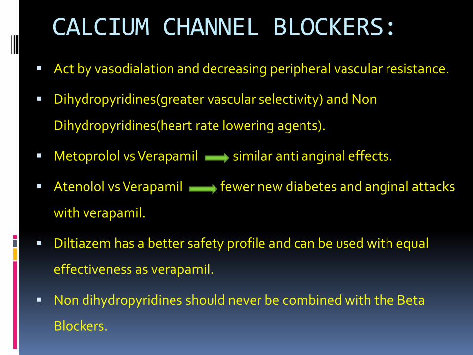
CALCIUM CHANNEL BLOCKERS:
Act by vasodialation and decreasing peripheral vascular resistance.
Dihydropyridines(greater vascular selectivity) and Non
Dihydropyridines(heart rate lowering agents).
Metoprolol vs Verapamil similar anti anginal effects.
Atenolol vs Verapamil fewer new diabetes and anginal attacks
with verapamil.
Diltiazem has a better safety profile and can be used with equal
effectiveness as verapamil.
Non dihydropyridines should never be combined with the Beta
Blockers.

CALCIUM CHANNEL BLOCKERS….contd
Dihydropyridines
Long acting Nifedepine
•Powerfull vasodialtor
•Few side effects
•ACTION Trial proved it
to be safe in stable CAD
and it reduces the need
for angiography and CV
interventions.
Amlodepine
•Effective once-a-day anti-anginal and anti-hypertensive due to its long half life and tolerability.•Amlodepine>atenolol in reduction of exercise induced angina.•In a 24 month trial,in a patient of CAD with normal blood pressure,amlodepine reduces risk of CV events.

Symptom control and relief:…(CONTD)
Ranolazine- usually a 2nd line anti anginal agent that can be combined with Beta
blockers as and when required.(ADD ON THERAPY in patients of stable angina inadequately
controlled on the 1st line agents).
Acts by inhibiting late sodium entry selectively into cytosol and hence prevents sodium-
dependent calcium accumulation.
Has anti-ischemic and metabolic properties,reduces diastolic stiffness,improves diastolic
flow, reduces frequency of anginal attacks(proved in the MERLIN Trial),increases exercise
tolerance, time to ST changes on treadmill tests.
TERISA study proved the utility of adding this agent to other well established anti anginals
in patients with elevted HbA1c levels.
Usual dose of 500-2000mg/day(without changes in BP/H.R.
QT prolongation may be hazardous.

Newer agents:
IVABRADINE:
Heart rate lowering agent acting through selective inhibition of sinus node pacemaking currents without any effects on BP or inotropism.(it reduces myocardial o2 demands).
EMA approved for use in patients inadequately controlled or intolerant to Beta Blocker therapy.
Acts best at heart rates>60/min.
Ivabradine=atenolon=amlodepine(in patients with CAD).
Adding ivabradine at a dose of 7.5 mg twice daily to atenololtherapy gave better control of heart rate and angina control.
The BEAUTIFUL Trial on ivabradine proved its efficacy in reducing composite endpoint of death, MI and hospitalisation due to MI in patients with angina and heart rates>70/min.

Nicorandil:
Can be used both for long term treatment as well as prevention of angina.
May be added after Beta Blockers and CCBs.
Opens ATP-sensitive potassium channels in vascular smooth muscles and dialates the epicardial blood vessels.
In the IONA(impact of nicorandil on angina)study it significantly reduced adverse CV events.
Long term use may lead to plaque stabilisation in patients with chronic stable angina.

Trimetazidine- anti-ischemic metabolic modulator drug.
Has got non-mechanical anti-ischemic properties.
No change in heart rate or rate-pressure product at rest or at
peak exercise.
Usual dose of 35 mg twice daily.
Contraindicated in parkinsonism and motion disorders.
Has positive effect on HbA1c and glycemic control.
Has not been evaluated yet on large scale studies in patients
with SCAD.

Persistant symptoms despite OMT
Consideration of revascularisation to improve symptoms
Potential for revascularisation
After assesingcomorbidities
and patient preferences
Coronary angiography
“heart team”opinion
Lesions correlated with evidence of ischemia
CABG/PCI along with ongoing medications
medical therapy with carefull monitoring
Depending on definite factors

High risk lesions on non invasive testing
Assesment of comorbidities and
patient preferences
Potential revasularisationwarranted
Angiography performed
Positive “heart team” opinion about anatomy /clinical factors
Optimal revascularisation
procedure determination
AnatomyPatient preferencesClinical factorsLocal resources
Continue ongoing medical therapy
Continue ongoing medical therapy
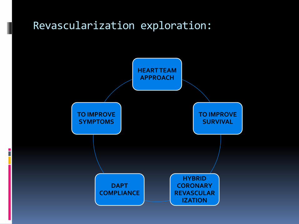
Revascularization exploration:
HEART TEAM APPROACH
TO IMPROVE SURVIVAL
HYBRID CORONARY
REVASCULARIZATION
DAPT COMPLIANCE
TO IMPROVE SYMPTOMS

Revascularization exploration….CONTD
CLASS 1 Recommendations to improve prognosis/survival:
“Heart team” approach for unprotected left main disease,in diabetics, in multivesseldisease(2/3 vessel disease),comorbidities.
Left main coronary stenosis>50%
Proximal LAD stenosis>50%
2/3 vessel disease with impaired LV function/heart failure.
>50% stenosis in single remaining vessel
Proven large area of ishchemia(>10% of LV).
:

WHERE NOT TO DO?
PCI should not be done in stable patients with
significant(>50% stenosis) of unprotected left main coronary
artery who have unfavourable anatomy for PCI or are good
CABG candidates.
PCI/CABG not to be done with sole intent to improve survival
in patients with SIHD with:
1 or more coronary stenosis,not anatomically/functionally
significant(<70% in a non-left main coronary artery
stenosis,FFR>0.8,No/mild ischemia on non invasive testing)
Involve only left circumflex or right coronary artery or subtend
a small portion of viable myocardium.

Revascularization exploration….CONTD
CLASS 1 Recommendations for improving symptoms:
All class 1 Recommendations for survival improvement,PLUS,
Any significant stenosis with limiting symptoms or symptoms not controlled with OMT.
Multivessel disease(2/3) with features of CHF or compromised LV is not a CLASS 1 recommendation in symptom improvement.

Where not to do?
In patients who do not meet anatomic(>50% of
left main coronary artery stenosis or >70% of non
left main coronary artery stenosis) or
physiological(eg- FFR criteria) for revasculariztion.
PCI with stenting(BMS/DES) should not be done
in a patient who is not likely to comply to the
DAPT for the necessary duration depending on
the type of stent employed.

OMT
CABGPCI
DILEMMA PERSONIFIED?????????????
Who answers??
Trials?

OMT vs CABG:
CABG>OMT(survival advantage) at 3 years in left main/3 vessel CAD.(Veterans affairs cooperative study,CASS,European coronary surgery study).
CABG>OMT (in terms of less subsequent MI,need for revascularization,cardiacdeath)in a 10yr follow up.(MASS2 Trial).
BARI 2D Trial ( though excluded patients with left main LAD,Included very small fraction with prox left main LAD,or with LVEF<50%) could not show superiority of OMT+CABG>OMT alone.

OMT vs PCI:Survival advantage of PCI over OMT could
not be demonstrated.( BARI 2D and COURAGE Trial)- 2 MOST contemporary trials). (????)
RCTs failed to show that PCI reduces risk of death or MI in patients without recent ACS!
TO summarise,PCI compared to OMT,
• Reduces angina incidence
• Dose not improve survival
• May increase short term MI risk
• Doesn’t lower long term MI risk.

Potential causes of lack of benefit of PCI over OMT:
RELATIVELY BENIGN
PROGNOSIS OF SIHD
OMT IMPROVES ENDOTHELIAL
FUNCTION AND PLAQUE STABILITY
FUTURE CULPRIT LESIONS ARE MOSTLY NON OBSTRUCTIVE
PERIPROCEDURAL MI WITH PCI
EEFECT OF COLLATERALS

CABG vs PCI : SYNTAX(SYNergy between PCI with TAXus
and cardiac surgery) has been a landmark trial comparing efficacy of CABG with PCI.
Also led to fomulation of SYNTAX SCORE which acts as a surrogate marker for extent of CAD and its complexity.(based on location of lesion,its complexity and severity).
SYNTAX SCORE>32 depicts high risk critical lesion.
SYNTAX STUDY randomly assigned around 1800 patients to DES PCI vs CABG.

CABG vs PCI….contd..
At 3 years, the rate of MACE(death,MI,stroke,repeat revascularization) and repeat revascularization were significantly higher in the PCI group!
The rates of death and stroke were similar,but MI and repeat revascularization were significantly lower in CABG group.
In patients with intermediate(22-32) or high(>32) SYNTAX scores,the MACE was increased in patients undergoing PCI.
Outcomes comparable in relatively uncomplicated CAD whereas CABG>PCI(EFFICACY) in cases of complex and diffuse CAD.

Follow up of patients:
At least periodic follow up anually to asses clinical
function,symptoms,surveillance for heart failure and arrythmias.
Monitoring of cardiac risk factors and ensuring adherence to
recommended lifestyle changes and medical therapy.
Standard exercise ECG recommended in know patients of stable disease
who have new/worsening symptoms not consistent with UA,and have
modest physical activity,no comorbidity and interpretable ECG.(Go for
exercise nuclear MPI/ECHO if ECG cant be interpreted at baseline).
Patients with worsening symptoms but incapable of exercise are advised
pharmacological stress imaging with MPI/ECHO or CMR(2nd option).

Take Home Message:
Patient education,knowledge,awareness and rationality on the part
of the physician are essential for management of chronic stable
angina.
Risk stratification is essential with either non invasive stress testing
or imaging studies to formulate management plan.
OMT forms a cornerstone of therapy.
OMT refractory stable angina warrants evaluation by invasive
angiography and formulation of revascularization plans.
Revascularization procedures have their own set of pros and cons.
Pragmatic selection of procedure is the key to succsfull treatment
………thank you


