c)ila.ilsl.br/pdfs/v33n3pt1a04.pdf · and testes. ~ome animals were given total body irradiation...
TRANSCRIPT

' \ "I I R\' n IO\' ",1. .J()nt~ .\1. OF l . n·kO~\' \ 'O lllIllC 3~, ~ lImh('r ~ I I/jilIn' ;11 1' ,.' , " ,
1'~XP1~Rr:MI'~NTAL l NF I':CTJO~ OW '1' Hl': C;OLD .I·~N nA~ISTI·~ f{ \VlrrH 1IJYCOBACTElU Uill LEPR.AEI
1\1. F. R \VATlm~/ 11. . .:-\. ~I.B. , 1f.R.C.P. AND JANET S. :F'. NIVEN, l\I.D., F'.C.PATH.
lYational I n.qtitnte for M edical R e.qem·r/, Mill Hill, London, N .W.'7, El1fj /al1d
Adler C) wa s the fir s t to attempt the transmi ss ioll of human lC' ]1-rosy to the golden hams ter (C1'icei'/,!s nl/Hllu s ). H e claimed that hacterial multipli cation occurred within 6 weeks of implanting lepromatous ti ssue. Burnet (8. n), and Burnet and .Jadfal'd CO) al so r eported successful transmission. 1 n contra st, however , only ve ry cautions cla ims were madC' by Doull and l\l egrail eH); Ca rpenter and Naylor-Foote (II) have been unahl e to r epeat their initial snccC'ss ; a nd negativC' rc sults have heen r eported by Duhoi s and Gavrilof (17), Oteiza and Blanco ( ~7), and Mal'choux et aZ. el ). 1'10l'eove r , the la st named authors drew attention to the ability of living and dead leprosy bacilli to r emain fo r long periods in the a nimal body and argued that the finding of such bacilli seve ra l months aftC'r inoculati on wa s not in itself evidencC' of progressive infection .
.In 19;)6, Binford (4) inocul ated 100 hal1l stC' rs with ~m spen s i on s of' bacilli prepared from chill ed 01' frozen human leprosy ti ssues . B ecause in man the leprosy bacillus appears to ha ve a predi lection for the cooler area s of the body, the hamster s were inoculated in the ear s a nd t estes. ~ome animal s were g iven total body irradiation and/or cortisone in a n attempt to lower resistance to experimellta l infectiOll. Histiocytic g ranulomata were found in 22 of the 85 hamster s examin ed; intraneural acid-fast hacilli were present in lesi01l s ill 3 ears . Control animals given heat-kill ed mate rial d id not devC'lop lesiom;. Hamste r to ham ster pa ssage wa s :; uccC' ssful, but it wa s s uhsequentl y discove red that the lesions w(>re du e to a cultivable m~rcobacterium (r..6), con sid ered b~r Shepard to belong to the g roup of nonphotochromogens C) . Further human to hamster experiments have been commenced hy Binford (5.6 ) and the finding of microscopic lesion s conta ining many well stained, llollcultivable, intracellular, and sometimes intra-1I emal, acid-fa st hacilli in a number of animal s ha s been r epol'tC'd hriefl y (1).
Following' Binford's earlie r claims, Convit and hi s colleagnes (1 :1 , 14) inoculated approximately 600 hamster s with f r esh suspension s prepared from biopsies from patients suffering from differ ent types of leprosy. Initially, of 26 strains used, 2 only, both derived from "borderline" patients , produced localized lesion s in ham ster ear s ; these contained la rge numbers of acid-fast bacilli. Each strain has bC'en passaged se riall y, with increa singly hort('1' incuhation periods, to
lReceived for publication Murch 29, 1965. :!Porlll crly of the Rcsea reh Uni t, S lIngei Buloh Lcprosn riulll, S ungri BlIloh, ?lInlnysin.

208 I/l Ul'IUltirma7 ./o/(l'lI ol of L f'ZJ }'osy 1965
cars, to. tes, fooL pads alld clH' l'k pOllclws. Neither s train has beon isolated on Loewonstcin-J ensen modium , although, more recently, a mycobacterium has been cultivated from les ions in hamster s that had been inoculated with a sll spension derived from human lepromatous tissue (15). On the other hand, Shepard eo, 31 ), who also inoculated a similar number of hamster s in the ears and testes, was able to identify acid-fast bacilli in very f ew animals and then only in small number s.
In an attempt to confirm Binford's earliest r eport (3), and on th e advice of R. .T. VV. R ees, a se ries of experiment. was started in 1959 using the gold en ham ster; these have now r eached the fifth passao'e. vVe present here a detailed account of the human to hamster and the :first passage hamster to hamster experiments.3
MATERIALS AND METHODS
I. H UMAN TO HAMsn~R EXPElUMENTS
Soul'ce and lJl'epal'ation of inocuZmn.-Ear trimmings or skin biopsies were removed on separatc occasions f1'oll1 :3 untreated leprolli atous or bOl'derline lepromatous p ati ents (A, B and C) and wcre immediately processed. The epidermis of each sp ecimen was removed, and the tissue was cut finely with scissol's, ground, suspended in sterile normal &Itlfae, and centrif uged at 1,000 l'.p.m. for 5 minutes. The supernatant below the lipid layer was removed, and stored at 4~ o C until a haci llary count for lJf. Zepme had been performed. The suspensions were inocul ated into hamsters within 5-24 honrs after tak-illg the lepromatous tissues. ,
Counts of the suspensions were made by a modification of the method of H art li nd Rees (18), in which the bacilli , including those in groups, were counted individually. The results were as follows : .
Suspension A-2.1 X 101 bacilli/mI. Suspension B--4.1 X 10⁸ bacilli/ m!. Suspension C-6.9 X 101 bacilli/Ill!' The morphology of suspension A was not noted. Suspension B contained 42 p el'
rent normal and 58 pel' cent degenerate organisms as judged by electron microscopy (22). Suspension C could not be f reed sufficientl y of ti ssue dehri s for electron microscopy, although a smear showed that 51 per cent of bacilli were solid-staining. Aliquots f rom each of the three suspensions were heated in a water bath at 70 °C for an hour to suppl y heat-killed control inocula,
I noculation.- Fi fty- three male hamsters, 4-5 months old, weighing 80-140 gm., were inoculated in three batches, 10 receiving suspension A, 14 suspension B, and 29 suspension C. Twenty-one animals received approximately :300 R whole body irradiation before inoculation, but, as there is no evidence that their resistance to M. lepl'ae was altered, no separate analysis will be presented.
The animals were anesthetized with sodium pentobarbitone given intraperitoneally. Each halllstCl' acted fl S its own control, living susp ension being inoculated into the left ea r and left testis, and equal volumes of the corresponding heat-killed susp ension into the right ear and right testis. Approximately 0.1 ml. of suspension was inoculated between the epidermis and thc cartilage of the upper surface of each ear; a conspicuous loca lized bleb was always produced, which soon disappeared. A dose of 0.2 ml. was injected into the substance of each testis. Th e dosage for each site was therefore in the range of 10L 101 bacilli. Tuberculin syringes with 1 Clll. , 27 gauge platinum needles were used, the needle being fl amed between inoculations. Thereafter the hamsters were kept in a nonairconditioned animal house with average da y and night temperatures of :32° and 24° C.
3A prelimin al'Y com muni cation W flS g iven at the VJJIth Intern ational Congress of L eprology, Rio de J anei ro, September 1963.

l\'o/r'1'.\· (r· .\'il ·r'l/: 7l1 Jrr/io1l of r:oldclI 7fo/JI .~t(/' lI'itli 11. ]rpl'f\.r 200
1'; (,(lIl1ill(l/iol/,. - Both trsfrs wrl'e rX: llllillrd blldel'iologi(',dly 01' hi stolor;i,·al l.,· ; hoth ('a J'~ 1'1'0111 23 ani ilia I" \1'1'1'1' I'xa Illi ned h i"tolo:;ri<'a II)' .
(a) lJlI('/('I'i%gi('.- (l) Lri't jrstis: EII('h lC'ft testis \l'a,.; l'('IIIOV('<1 aseptitHlly and plaeed in It g lass hO\l\Ogr\lizer. Oue pel' Cl'llt albUillill s:i1ille was Hdded, 1 ml. to testes weighillg less tlHIII 0.5 g \ll. , alld ].5 \III . to thos(' (lVI'\.' 0.5 gll1. The tl'stis was homogellized and centl'i fuged, nnd till' SUpel'lllltHllt rellloved a nd kept at 4* oC. A Ziehl-Xeelsenslainerl SllleHI' of the supel'llatn nt was cxaillined, Hnd f l'Oll1 Hil I1ss:Jy of 100 oi l illlIllCI's ion firld s, it wns esti mated that yiclds of appl'oxill1atl'ly 1 X 10.1 baeilli wPl'e detpctcd. POIlI' or the positive suspcnsions \l'CI'C also su(,(·essfuil y ('OUlltt'<1. (2) Right testis : An illlPI'Css ioll SllIear was made as :l l'outinr. ] II SOllie east's sus pensions \\'erc al so pl'rpl1l'rd :llId SlII rl1 l';:; rX:IIllincd.
( h) 11 is/(,/ogic.- Ti ssurs \\'cl'e fixed fol' 6 houl's ill 11 fl'cshly prcpnrcd mixture of satllrated IlIereul'ic chloride (06 nil. ) lind glHl'ial aept ic a(·id (± ml.) , and thcn tl'ansfe rred to 70 pel' ccnt al cohol. Thc cars were cut on a linr parllilel with the long axis into 3-;) nal'row sliecs, depcnding on the sizc, and thc tpstes \I'I';'e eut into 2 or 3 picees. Aftcl' embedding in panlffln wax, the spce illl cns werc cut in seetions at 4!J., ndjacent sections being sta ined by the Zichl-Xcelscn method, hcmatoxylin nnd eosin , and the periodic Hl'id-Sehiff (P AS) proccdure. Turnbull's method for il'on-contnining pigment a lso was llsed occa sionally. In nil instaners in which no mycobac.tcria WCl'e detcctrd in spc('imen~
inoculated with via hi e mycohacteria, further sections wel'C cxa lllinpd nnd a s illlilHr search \\'ns enrrird out if any unnsual histologic findings, su<,h ns focal ncrumulations o[ 1llononu('lral' cr ll s, wel'C ohscrved in specimens ino('ulated with inactivatcd lIIyrobactcl'ia.
II. F IRST l'ASSACE, .HA:\lSTEH TO HAl\ISTI·; n I';XPI.;RTM I, KTS
SOllrN rwd }Jl'ep(ll'll/io'l1 of inoclI/IIIII.- Left tcsticulnl' snspensions preparrd from o pl'imary inoculation hamste l's, nil of whirh conta ined solid-sta ining mycobacteria, werc usrd. Details of the suspensions al'e g iven in Table 1. ~o lIcid-fHst Ol'ganisllls wel'e isolated on Loewenstein-.J ensen medium f ro ll1 the suspension usrd for nnimal pnssllgc to inoculate halllsters P2-P5 and P12-P19. Fncilities for cultul'ing the suspensions ll sed fo r the inoculati on of halliste rs P6-Pl1 wcre not lIvn ila bl e,
IlIotillation.- The tc,.;ticular suspensions listrd in Tahle 1 wcre used to inoculate 18 ha mstp !,s, 6 weeks to -I- months old. Xo Hnimlll was il'radinted. As thr ·volume of testicular susrension was in evc ry cnse "pry snlHll «1.5 mI.) no heat-killrd controls were used. Testes nn(1 ears were again chosen as inoculation sites, the em's I'ereiving 0.1 ml. of suspension, and testcs 0.1 0 1' 0.2 ml. One an imal wns inocul lited in both testes, and a seco nd (No. Pll) in both tcstes and one ea l'; all othrl' fil'st pllssnge hamstel's \\'el'e inoculated in one cal' and onc testis.
TABLE 1.- D etails of 8HSjJen.,,·ons obtained j'1'OIn the left testes o( hamste1's killed 19-.'22 months after inoclilil /ion 't('i ll~ 811spensions of ~'fycohartel'iunl leprae 111'e}Jrl1'ed [1'om
human ti~sue .
P]'imnry I
First pasSlige ino<'ulntion Pcrcentage of hamster no.
hnmstp]' J noculull1 g iven Bllciliary sol icl-s tain ing inoculated with numhC'r (No. of ha(·i lli) yield ba cilli suspension
2 ±.2 X lOG X.C: 20 P3 5 -1- .2 X lOG N.C." (2/4) P2
16 8.3 X ]07 N.C: (2/2) P4 20 8.:3 X 10⁷ ~.C. · 71 P5 29 1.4 X 10⁷ 5.4 X ]04 (1/2) P8,P9,PI0 :10 1.4 X 10⁷ 3.0 X lOG 18 P12,P13,P14,P15 :~2 ].4 X 107 3.1 X lOG 27 P6,P7 45 ].4 X 107 3.0 X lOG 18 P] 6,P17,P18,P19 ;')3 1.4 X 10⁷ N.C: 6 Pll
"N.C.- No connt mad e.

:]00 11l / f'I' II(I/io//(I/ ./ 0 111'11(1/ of TJ 'PI'OR!J
/';, /'1/ 1/1 i ll(/I ifJII,- TllI' allilll:ll s \\' ( '1'(' killf'd :I \'t(,1' "17-1!) 1I101lths, (' x('('pl \'0)' "11( ' h:llll sh ' l' t1I:1t dif'rI at 1.') I11mdh ~, The inoc'lIl:1 t ion s it l's 1' 1'0111 !l halll sh' l's \\'''I'e ('xalll ill('(l hi stolo;,!:il'ally, as in till' 111I1I1 <1n to hallls tt ' l' ('xp('l'inH'lIt. Ears !l lld tl's tl'S \'1'0111 the othe r !) :lnilll:ll..; w(,l'e ~l' nt on \l't't i('e hy ail' ['I'Olll tilt' SU lI g'e i Buloh Ll'PI'()~lI l'iUlIl to the :\atioll:i1 Insti tute ['01' :\J edi('al Hf's(,Hl'ch . S uspensions \I'(,I'C PI't'p:ll'l'd f l'olll t h .. testes hy ('sscntiall .v t hf' ~!l llie t l'chniC' as Iwfol'r . I':a('h ra l' wa s shfl ved with a 1':17.01' hlad r, (, li t lillI'l .\' with s('i sso l's an d g' l'oulld with s tf'l'i lr salld and f1 prst lr in a 11l0r!':lI '. A nH'lhlll'Pt\ volunlP ( n sll 11 Il~' .f nil .) o f 1 pel' ('e ll t Il lhUillill sa lille Wfl S nddpd s lowly tn 111akr H ('l'lHll' snspension , whirh \\'lI S tiwn ('pntl'i['ngrd, nlld thr 1'l'sldting' SnlWl'1 lllt ll nt wa s I'x:l1llint'C] in Slllra l' pl'l'P:1l'll tioll :llId ('onlltpd fol' lwid-fast h:ll·i" i.
H !-::; U I : r :-i
I. I 'H I i\ I A H Y I ~OtTLAT I ():-; -- ll lTl\ I AN TO II A~I:-iTlm
]~'(f,.s.-~Ia cro sco pi c ex amination: Clinical C'x amination of the can.; wa s nC'ga t ive; 110 al'C'a 0(' thi ckC'lIing, 1I0dniatioll 01' alter a tion of pigm ent wa s drtrctrd . .As it " 'a s p lalln('(l to attempt passage only from NI l'S showing macroscopic ahnorma lity, 110 sns pf'm.; ioll s ,,"er e prrp:HC' d , a 11(1 no 1mctC' l'ial co unts \\ '(, 1'(' IlIade.
Hi stologic examination: ' I' he hCllll str l' ral' is cO\'(,I'C'd hy th in sf]na mou s C' pithC' liuIlI , ;)-+ crJJ s th ick, the stratum 111cidum a nd stratum g1'anulosum being poorly ]'('PI'('s(,lltp(l and melallill pigmrllt 1wing absent in the ha sa l lave r. lTair folli clC's, 11101'(' numC'I'OLlS 011 th C' dorsal than 0 11 tl1(' v(,lltral' aspC'ct, are fa irl y ulliforml y distrihuted, and sebaceous g lands Op<'11 illto the lmn r ll of the folliclr ahout half way f rom its base to its ext C' ]']]al aperture; sw('a t glands are ])I'C'sell t only at the ba se of the ea r . 'I'he ca rtilage is of h ya lill e type aJld is defined hy a moderately clC'll se p C' ri chondrium cOlltainillg elongated cell s. TIlt' continuity of th e cartilage is intC' lTupt C' (1 occasionall y by thin septa of conll ecti ve t issue. L ying parallel a,ll(l close to the ca rtilage on its ventral a spect arC' thin hundl es of skeletal muscle; on the (101'sa1 aspect, skeletal mu scle is found close to the ear attachment. The r emainder of th e ear cons ist s of conn ective ti ssue in which lie blood vessels, lymphati c chann els and nerves. Melanin-containing cell s arc a conspicuous feature of the conn ective tissue, and are more num erous on the dorsal than the ventra l a spect. They have a st riking dendritic appearance, which is du e to their elongated branching processes . It has not been possible to ddC'rmine whether they a. re chromatophores (cell s that have phagocytosC'd melanin produced hy melanobla sts) or are true m elan oblasts. In thi s communi cation, the tC'l'm melanophorr is used. Tissue mast cells al so are occasionally found.
Twenty-three pairs of ea r s from inoculated hamster s we1'(, examin ed at period s of 5 to 22 month s. III hamste r 49, kill ed 15 months after inoculation , the car s wer e ul cerated and showed extC'n s ive suhacute inflammatory change. No mycobacte ria wer e found and this anima l is omitted from the analys is. In ham stel' 40, killed 22 months after inoculation, both ear s showed an unusual pattern of hi stologi c change and widespread cl is trihlltion of l'lC id -fast hact('ria; it will be cir ~c ri1)('d separately .

WUI (.' 1'8 d; SiveH: J nfec livn vf (JvldcH }fulI/st a wi th M . l l' p n l l' HOt
In the remaill illg 21 an i rna Is, j Il t raccll u la r aci~ -fa s t bacte ria, morphologically identical with 111.. lepro e, wer e found in the ca rs inoculated with viable organi sms in 16 animals. In hamster 41, killed j months after inoculatioll, 3 mononuclea r cell s were found in the right ear , illocu lated with inactivated mycobacteria, each containing one we11-Rta ined aciu-fa st organism; non e wa s found in the left car, which had heen infected with viable mycobacteria , a nd considerahle search of the ri ght ca r fail ed to r ev('a l any more organisms. In hamster 23, kill ed after 22 months, mycobacteri a were readi ly fouml in the left ('£1 1' , and in th (' right ea r a small g roup of cells with in a nerve wa s fou nd to cOlltain solid-Rta ining mycohacte ria. No add iti onal foci of mycobact(' ria -containing cell s were found ill an~r of the additional section s examined. An analogous s ituati on wa s found in hamster 4;). 'In th(' ri ght ('a I', 3 mycobacte ria were found in one mononuclear cell. T~3xt (, lI s i v(' search of f nrtlwr sect ioll s fai led to r eveal any more infected cell s. As in ham st('l' 23, the l('ft ea r, inocnlatw l with viahle micr oorgalli sms, show ('d typical acid-fa st organisms in c('ll s of varions kinds (rPable 2) . 'I' h(' s(' w(' r(' th(' onl y in stanc(':o; in which acid-fa st organi sms morpholog'icall y r ('s('mhling M. 1el)1"(I('. w(' r (' dC't ('ct('d in s it('s inject('d wi th inacti va t('d microorgani sms.
As already statNl, in the 21 hamster (,<1 1'S ahout to be descrihed, no
'J' .~BI , I<: 2.- lJistriIJ//t iun 0/ (f cin-f(/st 0 '1".9 (/ n is III .' ill ea 'r .~ inucllio/ N/ lfi/Ii rillb le M. leprll e.
Hailister Ep IIF Mel ::\[ i\[11 X P C E l' l11llll b e r
1 + + + + + + + 14 :1 + + + +- + + 10 4 + + 12 9 + + 12
10 10 2:-3' + + + 22 27 + 20 29 + + 21 ,W + + + 22 :~2 + + 22 :i:) 22 .n" :i 42 + + :i 4:1 + + + + 22 45" + + + + + 22 4() + + 22 47 22 4H + + + + + + 22 :")2 + + + 21 :"):i -I- 22 ;) -1 22 Ep = Epi(]{'I" lllis ; HF = Ha il' fo llie l .. ; Mel = ?If l' lall ophorl's; M = M Ollolll \(· lp:I r [-p li s
Mu = Muse lc ; N = Nc n 'C ; P = P l'l'icho ndl"ium ; C = Cart il ng'c; E = E ndothelium; T = Tilll c ill mouth s after inoClllat ioll.
'M~' coiJactc ria fOllud i n right car. See t ext.

302 111/ l'l' lI atio llu/ JOll l' lI alo[ Lepl'osy 1!JG5
macro::;cojJic challg-ef; were obse rved alld g' rO f;S llIi croscopic les ioll s were absent. "As compared with the ea rs inoculated with inactivated mycobacteria, however, there was a gene ral increase in ce llularity, involving especially the fibrocyte s and pigment-containing cell s, and occasionall y the squamous epithelium was hype rplastic. Focal accumulation s of mononuclear cell s between epiderm is a nd cartilage were seen fairly frequently, and occasionally the re wa s lUI in creased cellularity within a nerve. A slight degree of fibro f; if; of skeleta l mu scle fiber s close to the p erichondrium was sometime f; se<'n and occa sion a ll y slight irr~gularity of the cartilage rods. Lymphocytic or plasma cell infiltrat ion was never found.
Tn 16 left ears f rom hamsters injected with viahle mycobacte ria, intracellular acid-fast mi croorganisms were found in nerves, pe richon drial cells, cartilage cells , skeletal muscle, endothelial cell s, squamou s epithelium and hair follicle cells, and melanophores. Tt mu st be emphasized, however, that in all in s tances , the di st rihution of the mycobacteria was focal and irregular. Frequently, furth er explorl'l tion of a pos itive sampl e failed to r eveal more infected cell., and, conv erscl~' , a search in other area s sometimes disclo:-;ccl intrncellull'll" mycohacteria ill a sp<'cim en con .. idel'ed initiall y to he negative. rl~he res ults arc summ a rized in r:rabl e 2.
Net·ves.- Tn g'eneral, it wa s poss ible to 1)(' certain that the mycobacte ria were intracellular. r:l~hey were sometim es found in cell s of t.he epineurium but more frequently in cells in clo 'e r elation to the axons. By the methods used it was not poss ible to differentiate betweell SChWalln cell and endon eurial cell s. No ev idence wa s found to sugges t that the microorga nisms were present within the axon s (Figs. 1 and 2).
P erichoncZ'riu1I1.--This wa s an <,q ually common s ite and easy to exam in e at high magnifications. 'l'hin elongated ce ll s, ly ing parallel with and close to the ca rtilage rods, were oftell s tuffed with mycohacte ria; they wcrr most numerou s on the inn er aspect of the pet'ichondrial membrane (Fig. 10).
Car·tilag e.-Tn only 2 in stances were mycohacte ria found 1n ca r tilage cell s (Figs. 8 and 9). Tn both spec im ens, the infected chondrocytes were small er than usual and it seems possihle eithe r that the cells ha.d become paras itized at a sta ,!2;c when they were becoming'
FIG. J . Seg nl en t of :1 n e rve s ho wing intrncytoplaslIlic myeob<l cte rin. Hnlll s ter 1. X1600. J~ I G , :!. Oblique scc ti on of a llBl'\·C. Mycobacteria a re pl'csent a lso ill pe ri ne uri n I cp lI s.
]( amst CI' 3 . X2400. FIG, 3. C ross scct ion of n s ke lehll IIIlI s~ l c fiber. Two lIIycohnctl' rin oce ul' within thc
lIlusc le s ubs tance. J-T a In s t e r 48. X2600, FIG, 4. Ob li que sect ion of s ke l.chl l mll sc le fi be r s llowing <' IIIII1PS of III.'"co hn f'tpria witl,in
t he musc le substan cc. "Myofib rils :11'(' vi,ihlc. H :J lI lstN 4R. X ~.j OO. FIG, 5, ~-r.\'cobaet('ria are pn's('"t ill s:lI'f'Olr' lIllll fl Ct,lIs t,lo spl .'" nppli ed to a lI111s('l (' fibpr
11 H III ster .j 8. X 1700. FIG. (j , ]\UIIIC I'O US 1ll.\"Co h:lctc ri" are P I't'S(: lIt lI"it l,i ll n s kl' lt'ta l Illlls(' le fihe l' e n t in l Oll .
g-itudinal sec tioll , Hams t e r 48 . X 1700, A ll th e sect ioll s wcrc sta ill cd by the Zi c hl -~cclsc lI Ill c tlrot1 Hll tl coullt c rsta ill ct1 \vitll
hpmatoxy lill .

l\'al c/'S (t, Xil' ell: /li fe c/ io lt of (; old{ 1I ll a lHs/ ( 1' Ici /It 211. h' pl'u( ' 303
tl'HIl Sforlll r d from ('llOlJ(lroblm-ds illto c:11OIldl 'o(' ,\' tl's 01' thal dllrill g' J'l'
pail' followillg dmllag'1' al illoclllalioll , 01' ill ci(lPlllal dalllage dlll'ill g 11ll' ohsl'l'nliioll I)('riod, illf(' ('lpd ]H'riellOlldrilll ('('lI s had IwcolllP trHIl SfOl'lll l'd illto eholltiro('yl(' s, \\' illl lhi s l'x(,pplioll, no nbllol'llHllily wa s foulld in 11w ('nrliln g'l' , (' \ ' (' 11 ill s IH'('inH' ll s in \yhi('h fairly llUllH' rOll S I)(' ri('holltirinl ('pll s \\'l' )'(' \'ollnd 10 1)(\ inf('(' l p d,
3

IlIl cl'llUliol/lIl .Jollrllol of Leprosy
S kel etal 'lnuscl e.- ln the -:I- animals ill which m ycobacte ria wore found in s keletal muscle, microorganisms W('1'e fOl1l1d also withill cell s in n er ves. Where the muscle fiber s were cut in c ross-section , the l1I.vcobacteria occurred either s ingly or in closely packed g roups, sometim es within a vacuole of the mu scle substance 01' ill close r01ati01l to a mu sc le cell nucleus (Fig. 3). fn long itudinal or oblique section, they appeared to lie irregularl y parall e l to alld within the muscle fiber (I~'igs . 4, 6 aJl(] 7). 'Mycobacte ri a were found also ill sarcolemma ce ll s closely applied to the basement memhran e of the muscle fih e r (Fig. ;j). R~cent e lect ron microscopy of ca rdiac muscle hy Lallnigan e") ha s showll that cell s c losely applied to th e sa rco lernma have proc0sses that imlellt the muscle s ubstance. A s imilar pattern may exi st ill skeleta l l1Iuscle, alld microorgan isms apparently withill the muscle fiber ma~r in fact 1)(' ly ing withill such a c0 il process. gvidence of mu scle damage, s uch a s multinucleated pla smodial formation, was never found, but myofib ril s wer0 less eOlls pi cuou s in those containing mycobacte ria. Damage to the tissues of th0 0Hr at illoculation can neve r h0 0xclucled and thus tho poss ihility that the myco bacte ria gained aCC0SS to the muscle ce ll s following' inoculation injury and r emain 0d i'n sit?/ whil e r egenerat ion took place, must also he kept in mind . A focal ill cr ease of mOllonuclea r ce ll s and fibrocytes wa s som ctimes foulld between individual muscle fib 0rs.
M onol1uciea'f cells.- I twas rathe r su rpri s ing' that tl1<:' incidence of mycobacte ria within individual mononuclea r cells or in coll ection s of' mOJlonuclcHr cell s was of a lowe r order than in 11<:'1'VeS, or p erichondrial cell s ('rabl c 2 ). Tn no ins tance did the numher s pr0sent within ce ll s approach those foulld in murin e lep rosy orin lepromatou s leprosy in man (Fig. 11). Occasionall y, focal collect ion s of cells W0rc found, of cha racte ris tic ova lor olonga t0d shapc and eccen trica lIy placcd nuclei, but containing onl.v very fine g ranular browllish material; sometimes these cell s gave a faillt diffuse react ioll for iron. 'Such area s ha vc always been examinod with cspcc ial ca re, alld occasionall~' a s ingle so lid-sta ining mycobacte rium 01' OllC 01' two il'l' eg- ularl~r s taill ing microorganisms havc heell found lying alllOllg the granules. Attention is drawn to this findjllg' , which ma y bc of s ignifica ncc in r c lation to the lif0 histor~r of M. 1('; /l1'(( e ill ham stc r cell s.
R,Jider1llis (lnd hoir fo llicl e epith eliI/11I.- ln 2 animal s (ham stc rs 1 ami :-l) isolatcd or small g roups of mycohacteria \\' c r c found in the
FI(; . i. Ob lique sect ion of a mu sc le libe l' showing H gl'oup of mycobnc tl'l'ia in :I "a cuole :just within til e SH I'colellllnn. Hall. s tCI· 48. X :3 000.
FIG. 8. Cholldl'ocy tc just withill th e pe l'i cholld "iu lll s tuffed with 1I1 ,'·coiJactc l'ia . 11 :1 IllSt[' I' 48. X]OOO.
FlO . 9. Focus of slIIall "hOlllil'ocy tes contain illg gl'oups of .uyco hnetl'l'ia . Halll:-;tc l' 5:2. XIOOO.
Pili, 10. Pl'ri(·holllil'i:Ji " .. II (',,"hi"ing (' IiIIlIJlS of 1lI ,' ·foh:H·tpl'i:J. ol"('Jll'ing thp IlJlrlp." . Ifn ll 's te l' 48. X 1700.
I~IG. II. GI'OUp of 1l101l0nllt· lp: IJ' ,·{' lI s with a lJlIlItl:.tllt ",,·topla " IlI , ('o nt:lilling" l1IycolJf1rl el'i a. H allistc l' 4:3. I~ OO.
All th e ~l' ..tioll s Wl'I'C sbill ctJ ],." th e Zil'ld · :'\ t'l'I"' 1I Il l(:t hod all d CO lllltl'l'st:.JiIl Cd with l, c lll atoxy Ij II.
11'111/1'-' ((, '\"il 'llI: III /(l'i illil 11/ (,'"lillli 1/11 ill ," I I' lI' i lll :'II , II'P!' ill ' :\0:)
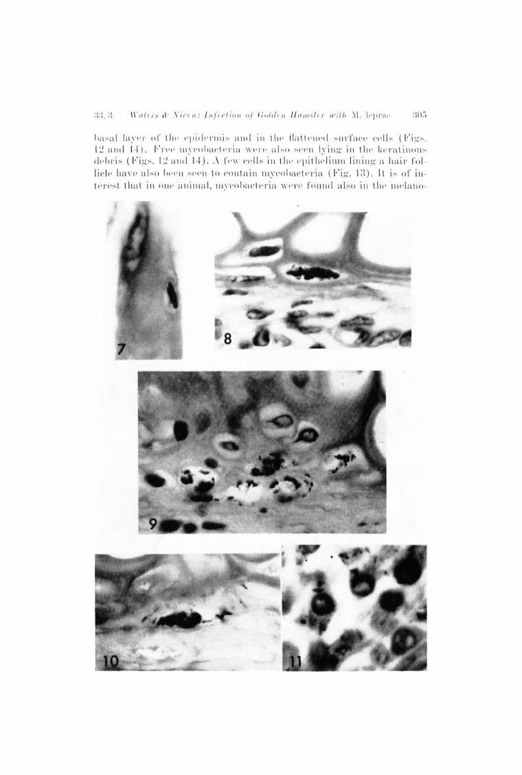
h:l ~ ;11 1:I,n'\' 01' t III' \'pidl'l'1I li ~ ;11It! ill 1 II\' 11:11 1 ('II('d ~ II\'I':I(,\' ('I''' ~ (I " i g~, U :llId 1-1- ). /·' \'1 '1' IlIy('oh:ll' (\'I'i:l " ' 1'\'1 ' :l1 ~ () ~ I '(' II I."ill g ill 111 1, k('ralill()l1 ~
d('h\'i~ (11'ig~. I:! :I lid 1-1-) .. \ 1'1'''' l'l' '' ~ ill (Il l' I'pitlll' Iill1l1 lilli1lg:l IIai\' l'ol li('11' 11<\\' (' al~o hPI'1i ~I'(' 11 10 cOlltai1l llI."eohael('ria ( I·'ig. I:{). /1 i~ of ill -1I'I'('~1 lhal ill 011(' allinlal , llI,YC'o)mclpl'ia \\"('l'(' fOll1ld al ~ o in 111(' Illl' /;1lI0 -

::Jon 1 III NU III i Oll o l .j 01/1'1/ 0 [ of Td'7Jl'osy 100:')
T A III , ," ?,- /Jisl I' ,:t)ll1 ion of (fr id-j'ast () I'{/(f II; ,' 11/ 8 In ell l'S oj' l s i, 7 1 I1 S,' (/,r;(~ ha ll/ st el's!
11 a IlIs t !' I'
I E p IJJ<' ,\I el .\1,
"l"" hel' Jill ~ I' l' 1 1~ 'l'
- -----P2 + + + + + + 18 P4 18 P7 + + + + + + + 17 P10 + + + + + 17 P1 3 18 PI 5 + + ] 8 P'I7 + + + 18 J?19 + + + 18
----"See 'J' :d) le 3 for kcy.
phores (~" i gs . 12 and] 3), ner ves, skeletal muscle, mOl1 onuclea e cells an(l peri chondrium. Although ordinarily the epidermi s is only 2-4 cell s thick, hyperplastic ar eas sometim es occur in which the usual features of thick mammalian epidermi s, stratum spinosum and st ra tum gl'anulosum , are fo und . 'rhi s change has not been found to have an y relationship to tIl(' occurrence of mycobacte ria in epidermal cell s, :lnd is con sidered to he the result of incidental trauma.
Melanophores.- l'l'l elanin-conta ining cell s a re a f ('atul'e of the hamster ea r and are frequ ently increased in number, especially on the dorsal asp('ct. They always maintain the typical dendritic appearance, however. In 3 specim ens many were found to contain typical acid-fast organisms, often situated within the long-branching processes (Figs. 12 and ] 3). Sometimes long processes containing mycobacteria wer e seen in close r elationship to the basal layer of the epidermis. In gell eral th e pigment content tended to be diminishC'f1 wh en the microorganisms were numerous.
Vas cular endotheliwn.- Tn hamsters 43 and 4:), in which mycobacteria were found fairl y easily in several sites, a single solid-staining microorganism was found within an endothelial cell of a vascular capill a ry. Tn 2 first passage animal s a similar obser vation was made (rl'abl e 3).
Tn hamster 40, the tis. 'ues of both right and left ea r s wer e indistinguishabl e, showin g extensive infiltration with mononuclear cell s ani! cell s of epithelioid type, many of which were stuffed with acid-fa st mi croorganisms. Organisms wert' abundant al so in c('lI s within nt' rves and in the epineurium, in muscle, perichondrium, and melanophores ,
}'IG . l ~. Squa mous epi thelium and subepitheli a l dermi s with " dend ri t ic" melanophol'Cs. close to a hnil' fo llicle. Mycobfl cte l'i a a re p rese nt in th l' epi t hclial cell s fi nd fI clump is y is ib le in tI lE' ke rat in on t lll~ surfn ee. Mycobacte ria a re a lso pl'CSl' nt in t he dcndriti c pl' ocesse~ of th c melnnoph o l·es. Hfl mstel' 1. X I600,
F IG. ] 3. i'>ec tion through th e base of n ha ir fo ll icle un it showing mycobncteria in t he epi t helial lining cells . Th ey 31'e prcscnt a lso in t he proces~es of mela noph ol'Cs . H a mst er l. X ] 600.
FIG. 14. Th in squnmous epi theli um ,,' ith intl'ncytop lnsmi c lll ,YcobnetCl'i n nnll m)'eobacter in ly in g f ree in t he kerat in ous debris. H a mstcl' 3. X 3000.
A ll t he sections we re stn in e,1 by t il e Ziehl ·N eels(' n metho,l a ntI cOllll t el sta ined with hcmatoxylin.

II ' (//Irs d' Yil' l'lI: [1I.(ll'Iioll III nO/rllll f{((III"/II' lI'illl ~1. Il 'pnlt' :l07

308 ill/ (,.j'Iwtiullui JUHI'?'Ialo! JJ('JlI'U,~.!J l!J(iG
ill thl' wall s alld elldothelium of blood vcsi-wl" and lymphatic challn els, and ill cp ide rmal and hail' fol licle ce ll s . ']'hey we re a lso foulld abu ll dalltl.\· ill cxt race llular spaces alld thc IUlllilla of hlood vesspls, a lld \\' e re ver~' lIumerou s amollg desq uamatcd epidermal ce ll s. ~I orphologica lly the mic roorgallisms \\'cre long'c r and thinllcr thall ill. le}Jrae as id entifi ecl in the other allimals of this st ud~· . r['he s ignifica nce of these obsprvntions will he di scussed latcr .
'/' esl es.- Thc ep ididymi s was g01l 01'tdl.v ill c lud('d in tlw sa lllpl0" takc ll fo r hi sto logic surve,v. ~o ulILHmal fcahlr0" ar0 prcscn/- ill th c testis of , the ham stc r. ']lh 0 illtc rst itial ce ll s of L(',vdig appcar in g0 n0ra l to 1)(' ]c>ss numcrous than in the mousc 01' rat , and as age advances, the tcstcs hccome atrophic <\lid spe rmatozoa an' lIO longcr produced. At th e sanw timc, the epith elial cc ll s and the int0rstitial cc ll s come to cO lltain va ry ing amounts of acid-fast g rallular piglll cllt, thc grallull's varyillgin siz;e from 2-3fL to just withill thc limit's of rcso lution; these grallLdes also givc a PAS-pos it ive I'eact ioll and thc ccll s somctimcs show a wcak rcaction for iron. rl'h is mah'l'ial ca n thus hc distinguished hi stochemicall y as wcll as morphologicall y f rom mycohactc ria in tissne sectioll s. Furthcrmore, the matcrial wa s cqually ahundant ill testcs inocula/w i withinactivatcd mycohactcria. III monolluclcar ccll s in the s tromH of the tcstcs, vc ry finc ac id -fast matcrial wa s somctimcs oh"r rvrd ill thr ma/-rr ia l rXHm in rd from Hllinll-tls in ocu latcd with viahlc l1I~'co hactc l'ia and to a lcsscr cxtcnt in allima ls with inactivated m ycobacteria (sec mononuclea r cc lls, pa~'c 30+). In oli ly one in sta nce, however , ham ster ;;2, were typical mycobacteria found in the testis inj ected with v iable orgallisms. 'J'hese occul'rrcl ill a localizcd coll ection of som ewhat elongated monolluclea r cells just within the capsule of the atrophied test is. rl1hcy showed unifol 'm sta i11ing, alld no dcgenerate forms we re detected. Furthe r examillatioll of th e testis fai led to reveal any other foci of acid-fast hactcria.
Bacteriologic ex a lIIinatioll .-Bactc ri ologic cxaminatioll wa s madp Oil lD pairs of tcstcs from animals killed 19 to 22 months after primary inoculation. All right tcstcs wer e n egativc, cxcrpt that, in one smeal' ollly, a sillgle irrcgularly sta inillg' and th cr cfo )'c presumahly dead, acid fast bacillus \\'as seen. Ninc of thc 19 lrft trsticu lar suspens ions COII
tai11cd solid a s wcll as il'rcgul arl~r stain ing aci(l-fast bacilli (rrahl e 1) . COlmts \\"c rr nllHl c of 4 of thcsc. rl'h c highcst ~r irld , howcvcr , wa s olll y 3.1 X 101i hacilli f),om a tcstis origillall~r inoculatNl with 1.4 X 10⁷ 0 1'gallisms (i.e., the yipld was ollly 22 pe l' CCllt of the origill al inoculu lll) a lid in eve ry case a fa 11 in hacilla ly popu la tioll had occu l'rrd. Mo]'c ovcr, in t he 3 suspens iolls with the highrst y ield s, only 27, 18 and 18 p('r c0nt of thc organisms \\'('rr sol id -sta ining . rl'h0sr ]'csults s llggrst IlHlt altlJough \'iahl r- Iooking ill. l PjJ1'(I C l1Ia~' f)'{'qlH' lItly rcmaill for nWlly m011ths ill hamstc r tes tes, the lHHnhc]'s gl'aduall~r dimillish. No mycobacteria were isolated from those suspensioll S culturcd on LoewcHstein-J-ensen medium.

Ira/ I'I 's cf· Xi" I' II: IU(I' l'fio li of (fold !' )] J((llml !'l' 'll'UII 1\f. lrprar
Ih:; l ui U.f),i(' e.nl/lI'i'l/(ll io 'II.- Th e illoculat.cd ca rs alld tes tes frOlll each of 8 animalH of the first passage sc ri cs \\'c re examined histologica ll y, the teHtcs ollly f rom one animal. In no in stance were ull equivoca l m?cobacteria found ill any of the 10 tcstes examin cd .
I n cont ra s t, howcvc r, l1Iycohactc ri a WC!'e found ill 6 out of the 8 ham stc r ca rs exa mill ed. Since the? wC'I'e ohscrved in s ites s imila r to those involved ill the prima ry Inoculatioll sc ri cs, it is Ullll ccessar~T to repeat thc deta il cd desc ription givcn ea rli er (pages 300-302). rl'he di str ihu tion ill illdi vidual hamste rs is g ivcll ill '1'ahle 3. 'l' hc ill cidcncl' ill sq ua mou s ep ithelium was highcr tha n ill the primary sc ri es, ill ;) out of the 8 ea rs examinec\, and in 2 of th esc (ham sters Pl0 and P19) , very scanty ext racellular mycohacteria \\'cre found amollg thc llOll nu cleated super fi cial squames.
B(f(·t e'rioloFlic eX(f111il1o t';011.-rl'es t cs : Snspensions w('r(' prepared from the left tcstes of 9 an imals, a llci t he one illoculatec\ ri gh t testis. ,\"hen mycobacte ri a w(' rc seell , the material wa s cultivated on Loewell s(-eill -.) enSCll mcdium. I;: ight tcsticula I' suspensions wcre Il egative. rl' h (' llillth contained 4 solid and 2 irregularl y sta ining hacilli in 100fielcls; the bacilli resemhled M . l epm e morphologica ll y, no g rowth was ohtaillecl on Loewell ste in -Jell sen med ium at 37 ° and 34°C, and hy analogy with the human to ham ster experiments it is considered probable that the few bacilli ha rvested were slowly decreasing survivo rs of th(' inoculum. The tenth one, from the left tcstis of hamstel'Pll, whose right testis also wa s inoculated, contain cd num erous acid-fast orgall isms. The yield was 2.3 X 10⁷ hacilli wi th 63 per cent solid-staining, but the bacilli wC' re shorte r ~\lld stouter than M. l ep1"a ej no growth wa s obta ined on Loewell stein-J ensen medium. H a mste rs (second passage) and mice have been inoculated with the two positive te sticular suspensions.
Ears : Homogenates of 9 ea rs from 0 animals cOlltained acid-fast bacilli morphologically resembling M . leprae. Counts on 4 confirmed a ten-fo ld increase. However, the y ield never exceeded lOG (Table 4). It has not proved possible to g row the mycobacteria on culture at 37° and 34°C. 'rhe 9 suspensioll s wer e used fo r further (second) passage to ham ste rs (ea rs alld foot pads), and mousc foot pads have also heen inoculated.
DISCUSSION
Currentl y, two ma in types of infection have been described follow ing the inoculati on of il l. l epm e into experimental animal s. Chatterjee C~), Convit Ca. 14 . , ,, ), a nd Bergc-l e) r eported the production of relatively ma ssive infection s, conta inin g ve ry numerous mycobacteria. On the other hand, Shepard e9 • a,) has obtained only limited multiplication ill the mouse foot pad, with a ce iling yield of ]06_107 bacilli. His work has been confirmed hy Hees e~) a nd by Jall ssens and Pattyn ('''). We ourselves initialJ~T modelled our experiments on those of

310 1?1/ f'1'1!rtlirmol J Ollrnol of l~f''P l'M'!J
'1' 111\1. 1<: -l .- IJ (· l lIi/~ oI " 1I"J)(' nsiu n~ ob l atllf(Z lrom I!ar.' III iir"t 1)(/"1'i(/ [j1' !lItlll sl crs Joillcll J I'- !!) lIIun/li i! aller !uo(' l1/al/:'JI! oj' i! 1I " Jlelt"iol/.s oj' ;\ r.H:ubll(·lt- riulIl leprlll' J)rcjlur l'll f ro/!!
/tam" ' I'1' 11',,11'8.
First passage Ill ueultllil g ivt'll ha ills ter lIulllber ( ~o . of bacilli )
P i) X. C." P6 iU X LO'-' PH :U X I(Fl P9 ii.-l X 10:l PIl N.C." PI~ 1.0 X 10" PI 4 :\.0 X 10" P1 () :"1.0 X 10 ~
PI 8 :\.0 X 10·'
"N.C.- No co un t made. "Too fe w organisms for nccurate count. '"Too mu ch deh ris fo r accurate coun t.
- -- - -Percelltage uf
Ba r illary y ield sulid-staining bacilli
~.4 X uri -1-7 -I-.!J X 10(; :'16 - " H() - I . (:~ ;:l) H.:l X IO ~ 7~
:l.H X IOH (i!J :U:; X 10(; 70 - . -I-H G.O X "10(; -1-2
Binford e· 4) (see page 298-299 ) , and used large in ocul a in th e fir st in stallce, examin illg the cars regularly for ma croscopi c lesion s ; as non e developed, the primar ), inoculation ea rs were examined histologically onl y, not bacteriologicall y. H owever, those hacte rial counts that were nndertaken, in hoth the human to hamster and the fi l' st pas age animals, have given yields not exceeding lOG bacilli. In addition, when the inoculating dose wa s less than lOG (in the fir st passage), multiplication to thi s level was detected. Ther efore,with inocula of the order of 10°-107 bacilli there was no detectable multiplication, but in ear s with smaller inocula, limited multiplication occurred. This limited multiplication has been amply confirmed by further pa ssage, as will be reported; the importance of including bacterial counts in such experiments i evident. Moreove r, the histologic findings, which were similar in the ears of primary inoculation and of first passage hamster s, are compatible with those of Shepard, if allow3llCe be made for the differences in the animal species used, and the tissues examined. Ther efor e we conclude that the type of infection we have obtained in the hamster 1S analogous to that described by Shepard in the mouse.
The histologic study has r evealed that a wide variety of tissues may be involved, and it is al so of value, with certain r eservation s, in r egard to the findin gs in human disease. Large granulomatous collection s of cells packed with mycobacteri a were absent, but the involvement of nerves has an analogy with human leprosy. So far as we know, the involvement of perichondrial cells in nasal leprosy in man ha s not been studied; and since in our material acid-fast bacilli have been very f requently found in peri chondrium, it would be of interest to examin e nasal cartilaginou ' hiopsies from eady, untreated, lepromatous cases. The involvement of melanophores and the variation in pigment content is of interest both in connection with the human di sease and aloin view of the hypothetic relationship of the e cells to the Schwann cells of dermal nerves. Quite fr equently, moreover, as

lrat e 'I' .~ d'; N i l!(, lI : 1'II j'ecf1'f)?1 oj' Gold (' n JIrrm st ('l' u'ith 1\IL \cp l'ae 311
al read y Itl cnLi oll ed, ill section s in which these cell s and squamou~ epithelium were seen to be iltvol ved, elongated dendritic processes containing' mycobacte ri a \\' ere found in cLose I' elatioll ship to the basal layer of the epid ermi s. I t is possible therefor e that the bacilli present in th e epid ermis were derived from melanophores ; this would account for their relatively frequ ent occurrence in the form er site in hamster s. In contl'a s t, in volvement of squamous epithelium in man is r egarded gen erall y as extremely ra re. H owever, Muir and Chatterji e~) have reported on a huma ll lepromatous pa tient whose non ulceratecl skill showed all ext£'l1 s ive invasi.on by Hcid-fast organ isms of sq uamou s epitheli al cells. Jnc1eed, ~ Iuir ea) has suggested subsequently that leprosy ba cilli may not infrequ ently be present in the epidel'mi.s of pati ents with ea rl y lepromato us di sease, with apparently normal skin. LOne of us (J.S.l<'.N.) has observed mycobacteria in the epithelium of a hair foll icle in lout of 4 biopsies from lepromatous cases.] The involvement of skeletal muscle is a phenomenon that we hope to investigate furth er. At pl'esent, as in th e 2 in stances of chondrocyte involvement, we fee l that it may be associated with trauma at inoculation or during the observation period. The fortunate observation of ba cilli in vascular endothelium is of interer-; t, in view of the we11-establi shed r eports of the OCCUl'rence of M. leprae in the blood in active lep romatou s disease . '\' he testis :findings wer e less interesting. 'While we would emphasize that M. l epme, as shown by our results, must have remained viable in the hamster testis, we have no evidence of multiplication. The hamster ear is without doubt a more useful collection of tissues for experim ental leprosy.
,\~he v0ry small numbers of bacilli detected in the 3 positive right car s of the primary inoculation series may be explain ed by the knowll per sistence of mycobacteria , dead 0]' alive, in animal ti ssues. The findings in ham ster 40, however, cannot be thus expla in ed. H ere, although the left ear received untreated suspensions and the right ear heattreated inoculum, the lesion s on both sides were similar, and contained large numbers of acid-fast bacilli. Each tuberculin syringe was used to inoculate the corresponding side of 10 animals, the platinum needle being flam ed between injections. But in no other animal inoculated from the same syringe as hamster 40 wer e similar lesions produced. \Ve therefor e consider that in this animal the infection had either occurred naturally in th e hamster, or had been introduced accid entall y a t the time of inoculation, presumably from the skin surface. rl'hese suggestions would al so account for th e unidentifi ed mycobacte rium obtain ed from the left testis of hamster PH, for its right testi s, inoculated at the sam e time with the same suspension, using the . ame though flam ed needle, and syringe, was negative. Nishimura and his coll eagu es e"· 26 ) have recently r eported on the spontan eous occurrence of mycobacteria in h ealthy mice, and hamster s. It is therefore essential that all positive suspension s should be cultured . HowQver, many of Nishimura' s isolates failed to produce growth in vitro, in this way

:J 12 ] %5
re~emhling out' own unid entifi ed m.vcobactcriulll. \r e sugg(\st the l't'fore that a careful study of the bacterial morphology ~ hould al so be made in eve ry case, with note of hoth the ~ ize and shape, of the bacilli, and th e p ercelltage of solid-stainillg' forms. All our isolates of (presumed) 1H. l eprae contain ed a definite and usually relatively high percentage of irregul a rl y staining bacilli, s imilar to the bacilli in human untreated di sea se (32). A very high percentage of solid-staining hacilli cou ld well a rou se s llspicion of a Ill. lppm c II1111'i lllll type of illtercurrent illfect ioll .
'Ii he illad equac ies of ou r study are readi Iy a ppa rl'nt, the most obvious heing the failure to correlate, in the sallle animal, the hi stologi c findillg' s with bactcrial CO Ull tS and cultural methods. Because the bacte rial multiplicatioll is limited and the distribution of the mycohacteria is focal a lld irregular (see page 302), it is not practicahle to use part of the same ea r for histologic eXHmination alld 'part for a hacterial count and culture. Ho\\'evel', a practical compromise ill future studies would be to take all aliquot for cul ture from <tll~' specimen illtend ed for hi sto logy, the remainde r beillg ('xmn inl'din complete seria l sectioll by appropriate method s. It is on ly by full utilization , alld carl'ful integration, of bacteriologic and hi stologic methods that claim s to the Sllccessful tran smission of hUllIall lepros)T to c~pl'rinl('ntal animals can be substantiated.
SUMMARY
Forty-eight golden ham ste rs , illoculated ill the left cal' alld left testi s with livillg suspensioll s of Ill. l epf(te and in the ri ght ca l' awl right test is with heat-killed suspensioll s, were maintained for 5-22 months . Sixteen of the 23 left ears examilled hi stologically sho\V('d typical intrace llular acid-fa st microorg'ani sms ill a variety of cell types. However , of 28 pairs of te~tes examill ed histologicall~r, ill only Oll e left testis were illtracellular mycobacteria found. Bacteriologically, acidfa s t bacilli were recovered from suspell siollS prepared from 9 left testes Ollt of 19 pairs of testes examilled, and these pos itive suspens ion s wer e used to attempt pa ssage to ] 8 hamsters . After 18 months, 6 of 8 fir st pa ssage ears examill ed histologically showed illtracellular mycobacter ia ill s ites similar to those found in the primary inoculation animal so Homogenates from 9 other ears contained acid-fast bacilli, and counts on 4 confirmed a ten-fold increa se, although the yi eld never exceeded 106 bacilli. It is concluded that a limited multiplication type of infection has been achieved in the ham ster ear, but not in the t estis, analogous to that described by Shepard in the mou se footpad.
Hl-::-:i U MEN
Durante 5 a 22 Illeses, f uerou mantenidos cual'enta y ocho "golden hnlllsters," los que fueron inocullldos en la oloeja izquierda y t('~ tfculo izquierdo con suspens iones de M, Zepme vivos y en la oreja derecha y testfculo derecho con suspensiones lI111ertos al enlor, De las

Ira/ el'S d" l\"ivc lI: lllj'ec lion oj' Golden Halllst cr ,vith M. lep!'(le 313
23 ol'cjm; izquiel'das eX3 111inadas hi ::; tologi callll'ntl- di e;', y scis nlostt':lrOIl tipicos mi crool'gan is illos acido-al cohol- l'csis tentcs intl'ace lularcs cn un variedad de tipos celulares. S ill clllbal'g o, dc los 28 pares de tes ticulos ('xalllinados histologiclllllente, solamente en un testlculo izquiel'do se encontral'on lI\i cobacterias intl'acelulares. Bacteriolog ica.mcnte, baeilos acido-alcohol-l'es istentes f ueron recobrados de suspensiones pl'epal'adas de 9 testfculos izquierdos de los 19 pares examinados, y estels suspensiones positivas f Uel'on usadas en un intento de pasaje a 18 hamsters, Despues de 18 lI\eses, 6 de los 8 primeros pasajes en orcjas eXH lllinados histo log icHinente Illos tl'al'on IlIi eohact(,l'ias intmcelulal'es en lugares s imilares a l!(jllCllos eneontrados en los anillla les de p i'in lern illoculaeion . HOlllogenatos de ot r:Js 9 or('jas contuvieron baci los acido-al cohol- resistentes, y rpcuentos en 4 confirlllaron un HUlllellto de ] 0 veees, aunque la produeeion nunea exeedi o los lOG baeilos. Se eonelu,ve qu e un tipo de infeceion lIIultipli eada lilllitada ha s ido eonseguida en la oreja del hallister, pero no en los t('sticulos, 'lIIalogo al desCI'ipto pOl' Shepard en la p lanta del raton.
Quarantc-huit haillsters do)'(~s ont He inocul e::; avec des sus pensions vivantcs de M, 71'1'1'11 1: dan i'oreill(' et dans Ie tes ti cule ga uche, et avee des suspensions tuees pal' la chu l('ur dans i'oreille et II' testicu ll' droit. Ces HnilllHux ont ete obscrves durant 5 It. 22 Ill ois. Sei;',e des 23 oreilles ga u('h e~ qui ont ete exai'llinees histologiquelllent ont 1Il0ntr e des Illi cro-o rg'anis llies acido-J'esistants intrl1cellulaires typiques dans des cellul es de types val'ies. Toutefoi s, pal'lni Ips 28 paires de testicul es, des III,Ycobactel'ies intrHcellulaires n'ont pu Ptrc recueillis que d'un seulf' te~ ti cule gau che. Au point de vue bacteriologique, des ha(,illrs lH'ido-resistants ont ete re('ueillis or susprnsions preparees II partil' de 9 tes ticul es g'a nriJ es, pn l'llli II'S 19 pail'rs exa lninef's. ('ps susppnsiolls positives ont ete utili see,' chez ] 8 hallls ters afin de tenter leur passage. A pres 18 mois, 6 des 8 oreill es ayant ete soumises It. un preilliri' passage et exa illiners histolog'iquement ont Ill ontre drs mycobacteries intra crllulaires ('II des endroits selllblahlf's ii. cellx ohserves che;', les anilliaux de primo-inoculation. Les suspensions hOlllogenei sees de 9 autl'f'S orei ll es contenaient des bacilles Hcidores istants, et chez 4 la numeration a revele une augnl entati on dans la proportion de 1 It 10, encore que Ie nOlllbre de bacill es recuei lli s n'a it jallwis depn sse un lIIilli on. On en conclut qu'une infection avec lIIultiplication lillli tee, 11l1a log'ue :1 celi e decl'ite pilI' S hepa rd dans la sole plantaire de 111. souris, a ete obtenue dans l' OI'eill e du haillster, IIlais· non dan, Ie testicul e.
A f'I'lIoII'it' d,Ql'II/I"IlI''' .- A II work pf'l'fol'lllf'd at Sung'ri Buloh Lep rosariulII was supportcd hy the Ministry of Hea lth, Federation of Ma laysia. We wish to thank ~Ir.
F. J. Higginson, Miss K Brodaty, and Inche Mohd. Bahi for skilled technical assist}1 1H'f', :MI'. M. R. Young for the photomicrogl'aphs, Miss Lyen, General Hospital , Kuahl LUlllpur for g iving thr whol e hody il'l'adiation, nno Dr . .T. H. 8. Ppttit fo r his kind (·ooperation . Onr of us O LF.R .W.) wishrs to thank Dr. R .. /. \Y. R res fo r hi s advice a nd ('nrOllrn gf'ItIf'1t t,
HEFJ':H)o;)\ (' J·:S
t. .\IJU: H.~ . TI!()('ulation of hlll1ll1n If'prosy into 8.vrifln haillstf' rs . Lnncet 2 (19:37) 71-!-7 1 ;).
2. Ih:llw,: I. , :\1. Infiuf'IH'f' of variOllS PI'o-oxidant nutritionnl rOIHlition. on g rowth ill I'il''' of' M. /t' J)I'/I P. Lf' pl'Os,Y Hf'v. 30 ( I!lii!l) li'i:1-1iiR.
:1. BI!O'OHIl. C. H. T1i"tio(,,Yti(' g'r:lllu lolll:ltOl!S 1I1.n'ohaC'tt'rial I('sioll;:; produ('f'd ill the g'oldf'lI hlllllst(,I' (('r i ('(' I II.'1 (//I/'(1ll1s) inoculnted with hUlllan leprosy- neg-ntive )'esnits ill ten ('xpf'rilllents l ls ing other :InilllHls. Trans. Vnth Internat. Cong r. 1.f'prol. Tok .,·o (1!)58); Tokyo ( Tof u Kyoklli) (1959) pp. 59-66; (!l~o Internllt, J . Lepro.·'y 26 (1958) 318-32-"'.

314 l ntc1'1wtio1w/ ./oU1'1wl of L f']J1'OSY ]965
4. BIN FO IW, C. H. II istioeytic gr<tn ul ollltltous Illyeobaeteriul les ioll s pl'odu(,l'd ill the golden hallls ter (CI'icetus aWl'attLs) inoculated with hUlllan leprosY-lI egative results in experin;ents using other anilllals. Lab. Invest. 8 (1950) 901-93 .. 1-,
5. BIN FOllD, C. H . The problem of transllli ssion of M . lepl'ae to a nilliais. 'l'rll.lls. Leonard 'W ood ;\Ielllorial-J ohns Hopkins Ulliversity S)'lIIposiulll on Rrsl'}lI'ch in Lepros.\', Baitilllorr, Md., 1\ la)' 8-10, 1961, pp. 235-24 .. 1"
6. BIN~'OI!IJ, C. 1-1 . Studies Oll a III.v('()bactl' riullI obtained frOlIl til(' goldcll halllstC'r (C"icetlls (tm'atu-,) after illo('ulation with leprolllatous tissue. Llh. IlIvrst. 11 (1963) 942-05;"}.
7. Bl~I" OI!l), C. n. a lld \IAI)ISO~ , R .. :\1. The tnlnSllli ssioll of' ilf. /epl'lI e to anilllals; nrl'vC' Il'siolls in the golden haltlster. Prese nted at \ ' Inth Intel'J1at. Cong- I' . !'rpl'o!., Hio dC' .Janriro, Srptc lllhrr 1963. A /i stmet in Tn tl'l'IIa t . . J. Leprosy 31 (H)6:l) 5:l:l .
8. BU H~ ET, E . I lIo('ulation positivI' dr In li'pl'e hUJllnine nu h:lll1 stCI'. C(lIIIPt. I·Plld . Acad . d. Re. 207 (1938) 690-69:2 .
9. BUHNI';T, E. Tn o('ulation positive ell' In lepre hUlllainc au h:lIl1stel'j illo('ulation IIPglltive 11 divers a utres rongcurs. Arch. Inst. Pasteur e1e Tunis 27 (19:'18) :-l27-3~0.
10. BU RN~~'I', g . and J ,\IWARD, H. Translili ssion de la lepre hunHline au halll ster pal' voie dig·cstive. Bu l!. Acad. de :\[eel. (Paris) 122 (1939) 383-389.
11. CARPE~TER, C. \L and XAYLOR-Foo'l'E, A. 'W, C. The hnrteri olog,v of leprosy. Tn Leprosy in Theol'Y and Practice, ed ited hy H. G. Cochralle. Bristol, John W ri gh t a nd Sons, Ltd. 1st eel. 1959, pp. 15-18.
12. CHA'Jvl'EH.JEP., K . R. Experilllental transllliss ion of hUlIlan l('pros,v infertion to It selected, laboratory bred hybrid black IIl0use. Illtel'llat. .1. L('prosy 26 (Hl58) 195-204.
] 3. COXV IT, J. Discu~s ion of recent work in Venezuela. Tr;lIls. Leonard \ \ ' ood l\Ielllol'i<1I-Johns Hopkins Univers ity SymposiullI on Rrs('arclr in Leprosy, Baltimore, \[d., \Jay 8-10, 1961, pp. 258-259.
14. CONV I'1', ,1., LAPEX'l'A , P., IL l.i KEVICH, A. and hIAED,\, T. Expel'illlcntal inoculation of hum<1n leprosy in laboratory animal s. I. Clinical, bacteriol ogic, and hi stopathologic stud~' . Intel'llat..T. Leprosy 30 ( 1962) 239-25:3 .
]5. CONV IT, J., LAP ENTA P ., ILUKE\,lCfI , A. a nd I~rA~; D'-\ , T. Experilllental illoculation of hUlllan leprosy in laboratory nnimals, III. Intel'l1nt. J. Leprosy 32 (1064) ] 36-140.
]6. Dou!.L, .T. 1\. and ?"[EGRA II" E . Inoculation of hUllllm lep rosy into the Syrian hamster. Internat. J. Leprosy 7 (1939) 509-512.
17. DUBOIS, A. and GA\'HILOF, 'vY. E ssais d'inoculation dp la lepre hUlllaine au hamster non s plenectomise. Arch. Inst. Pnsteur de Tunis 29 (1940) 170-173 j also Ann. Soc. beIge Illed. trop. 21 (1941) 189-194.
18. HART, P. D'ARCY and REES, R. J. '\T. gf/'ect of III;J Cl'oc.\'clon in acute and chrolli c pulmonary tuberculoll s infection in mice as shown by viable and total bacterial COllnts. British J. Expel'. Path. 41 (1960) 414-421.
19. JANSS ~::-<S, P. G. and PATTY N, S. R. Experiences with mOll~e inoculation of lepros.\' bacilli originating from the Congo. Prrsented at VJIlth Tntrl'llat. Co ngr. Lepro!. Rio de Janeiro, September 1963. AIJ"tl'u('/ ill InternaL. J. Leprosy 31 (1963) 522.
20. LA~XIGAN, R , Personal cOlllmunication.
21. MARCII OUX, E., CHOR INIO, V., C lr.\BAUD .. \. and Tlss~~u r", .T. E ssai s negatifs de In transmiss ioll de la lepre humain r au hamster dl' S,vrie, r l'icr/,ll s om·o/,us. Ann. Inst. Pasteur (Paris) 68 (J 942) 99-105.
22. \[CFADZEAK, .T .. \.. a nd VALE:-<TT:-<E, R. C. 'rh e rxaminntion nnd thc rletcl'lninatioll of the viability of Mycobactel"ium lepl'ae by electron microscopy. Leprosy He\'. 31 (1960) 6-11.
23. :MUlll, E. Leprosy. Suppl. to Caribbean \Ied. J. 3 (1941) 1-25.

'Iralas cf' .Vi vcJI: ll/fcction of Go/dell IIamstc/' wit h 1\1. lepme 315
::!4. MUJR, K and C J/ ATn~R.rr, S. N. The infect ion or stmtified epitheliulll in leprosy. Indian . .T . ~led. R es. 19 (1932) 1163-1164.
25. N ISHl lIIURA, S ., KAWAG UCHI, Y., KOHSAKA, K. and MOHr, T . Conta mination of hea lthy mi ce with murine leprosy- like acid-fast bacillus . La Lepro 33 (196J) 245-256.
26. N1SllUWR1I, S ., KOHSAKA, K . and K AWAGU CHI , Y. Murine lepr osy-like acid-fast bacilli found in healthy m ice and hamster . La Lepro 33 (1964) 109-110.
27. O'fEIZA, A. and LEON BLA2\ CO, F. ] ntentos de tn1nSllli s ion expe rill lCnta l de lepra hUlll a na a l hallls ters s iri o (Cric etus aa!'atas ). T~rans . Vth Internat. Congo Leprol., H avana, 1948; Havana (Editori a l Ccnit) 1949, pp. 526-528.
28. RICES, R .T . ' V. Limited mu ltipli cat ion of ac id -fast bacilli ill the footpa ds of lIli ce inoculated with My co bactel'l:lflll rCprfW. Briti sh J. ]<;x per. Path. 4S (1964) 207-218.
29. SIH~ l'AHD, C. C. A cid -fa s t ba (·illi in nasH l excretions of leprosy, and r esults of ill oculation of mi ce. A nll'ri can J . l lyg . 71 (1960 ) 147-157.
30. S HBPAHD, C. C. ))£I; OIl.""'io'l/, of th e problem of trllnsmiss ion of l1T. rep rae to a nimals, by C. H. B ill ford. TnlllS. Leo nard W ood :\lelllOrial-Johns Hopkins U niversity SY III posiurn on R esea rch in Leprosy, Baltilllore, Md., May 8-10, 1961, pp. 245-246.
31. SHEPAHO, C. C. :Multiplication of Mycobactel'i/('/Ib le}J rae in the footpad of the mouse. Intel'l1at. J. Leprosy 30 (1962) 291-306.
32. vYA'l' ERS, M. F. R and REF~S, R. J . VY. Cha ngrs in the morph ology of l11ycobactej'iuln lepl'ae in patients under tl'eatJlIent. Internat. J . Lepr osy 30 (1962) 266-277.