circadian disruption alters gut barrier integrity via a ß
TRANSCRIPT

1
Circadian disruption alters gut barrier integrity via a ß-catenin-MMP-related pathway
Sung Yong Eum¶, Nicolette Schurhoff¶, Gretchen Wolff, and Michal Toborek*
University of Miami Miller School of Medicine, Department of Biochemistry and Molecular
Biology, Miami, FL, USA.
¶These authors contributed equally to this work
Short title: Circadian disruption alters gut barrier integrity
* Corresponding Author:
Michal Toborek, MD, Ph.D. ([email protected]) University of Miami Miller School of
Medicine, Department of Biochemistry and Molecular Biology, Suite 528, 1011 NW 15th Street,
Miami, FL 33136, USA, Phone: 243-0236, Fax: 305-243-3955
2
ABSTRACT
We evaluated the mechanistic link between circadian rhythms and gut barrier permeability. Mice
were subjected to either constant 24-hour light (LL) or 12-hour light/dark cycles (LD). Mice housed
in LL experienced a significant increase in gut barrier permeability that was associated with
dysregulated ß-catenin expression and altered expression of tight junction (TJ) proteins. Silencing
of ß-catenin resulted in disruption of barrier function in SW480 cells, with ß-catenin appearing to
an upstream regulator of Bmal1 and Clock. In addition, ß-catenin silencing downregulated ZO-1
and occludin TJ proteins with only limited or no changes at their mRNA levels, suggesting post
transcriptional regulation. Indeed, silencing of ß-catenin significantly upregulated expression of
matrix metallopeptidase (MMP)-2 and MMP-9, and blocking MMP-2/9 activity attenuated
epithelial disruption induced by ß-catenin silencing. These results indicate the regulatory role of
circadian disruption on gut barrier integrity and the associations between TJ proteins and
circadian rhythms, while demonstrating the regulatory role of ß-catenin in this process.
Keywords: circadian rhythm disruption; intestinal barrier integrity; ß-catenin; tight junction
proteins; circadian clock genes; circadian rhythm molecules
3
INTRODUCTION
With rising disruption of light-dark cycles in human populations due to night-shift work, excessive
use of artificial light, and/or travel across different time zones, there is increasing concern for the
effects of circadian rhythm misalignment on susceptibility to disease. Circadian rhythms act as an
~24hour pacemaker for the human body and its individual cells. The “master clock”, so named
for its direct synchronization to light and its influence over all other peripheral clocks throughout
the body, is located in the suprachiasmatic nucleus (SCN) of the hypothalamus 1. Although the
circadian rhythm runs autonomously in each cell, they can also be influenced by different
environmental cues such as light and food intake 2. As a whole, circadian rhythms regulate daily
cycles such as sleep and wake, hunger, body temperature, and hormone secretion in order to
anticipate the body’s daily needs 3. At a molecular level, clock genes and clock-controlled genes
regulate the cell cycle through various cellular functions such as cell replication, apoptosis, and
DNA repair. These molecular clocks are regulated by core clock genes such as Bmal1 and Clock
that control the expression of proteins that undergo a vast variety of biochemical reactions. When
circadian rhythms are disrupted by chemical or environmental interference, processes regulating
homeostasis are altered, contributing to the development of various diseases, such as
gastrointestinal diseases, sleep disorders, metabolic syndromes, and cancer formation 4.
Mechanistically, circadian rhythm alterations have been linked to the ß-catenin/WNT-signaling
pathway. For example, circadian disruption accelerates tumor growth through ß-catenin/WNT-
signaling pathway 5. Positive and negative regulators of the ß-catenin/WNT-signaling pathway
also show high incidences of circadian rhythmicity 6.
The gut barrier is a multi-layer system composed of tight junction (TJ) proteins such as zonula
occludens (ZO)-1, tricellulin, and occludin that regulate its functions 7. TJ proteins are responsible
for managing the permeability of the gut barrier by providing a selective seal between the
neighboring epithelial cell lining 8. The outer layer acts as a selectively permeable barrier between
4
the gut and the surrounding tissues. The inner layer discriminates against pathogens and
regulates immune responses 9. Several circadian rhythm-related diseases show signs of gut
barrier leakiness, suggesting that circadian rhythms may be involved in controlling gut barrier
permeability 10,11,12. Various circadian clock genes and proteins, including Bmal1, PER 1/2/3, and
Clock, are expressed in the gastrointestinal tract and have been linked to gut functions such as
digestion, epithelial renewal, and absorption 13,14,15. In addition, we 16 and others 17 have reported
that circadian rhythm disruption alters gut microbiota towards an increase in pro-inflammatory
intestinal bacterial abundance, a decrease in anti-inflammatory intestinal bacterial abundances,
and impeded intestinal barrier function 18. In addition, quorum sensing molecules produced by
bacteria can disrupt epithelial barrier integrity 19. At the same time, the intestinal microbiota
undergoing diurnal compositional and functional oscillations may also influence host circadian
activity 20, indicating that gut microbiota can also signal back to the circadian clock 21. Circadian
rhythms also effectively control local and systemic metabolic processes and inflammation
responses; further implicating alterations of gut permeability with alterations of circadian rhythms
18,22–25.
The exact mechanistic interrelationships between the circadian rhythm and gut barrier integrity
are not fully understood. Therefore, the aim of the present study is to examine the link between
circadian rhythm disruption and alterations of gut barrier permeability. Our data indicate that loss
of barrier function as the result of circadian disruption is mediated, at least in part, by ß-catenin-
induced modulation of circadian clock gene expression and upregulation of matrix
metallopeptidase (MMP)2/9 leading to a reduction in TJ protein expression.

5
RESULTS
Disruption of circadian rhythms in mice by constant light.
Mice were subjected to constant 24h light (LL) for 4 weeks, with the control group maintained
under 12-hour light/dark cycles (LD). Voluntary wheel running was measured to evaluate
behavioral circadian rhythms at the beginning of the study (baseline) and after two weeks of
exposure to normal light or constant light. All mice showed regular running rhythms at baseline
and following two weeks in normal light (LD) (Figure 1A). Following two weeks of disrupted light
(LL), mice displayed consolidated voluntary running rhythms; however, the time of activity onset
was altered (Figures 1A and 1B). The phase angle is a circadian parameter that describes the
difference in hours between the time of lights-off (or former lights-off for LL 2nd week) which was
18:00 (zeitgeber time [ZT] 12), and the time of activity onset; corresponding to uninterrupted
activity on the wheel. In the LL group at the 2nd week, activity was significantly shifted by ~30 min
(Figure 1B).
In addition to wheel running, body temperature was measured as a second method to evaluate
circadian rhythmicity. Following two weeks of normal light mice showed a regular pattern of body
temperature, elevated during the night/active time (18:00 [ZT12] until 06:00 [ZT0]) and decreased
during the day/resting time (Figure 1C). Mice exposed to circadian disruption displayed an
arrhythmic pattern in body temperature throughout the day (Figure 1D).
Body mass was measured at baseline and then once a week for 4 weeks. Mice maintained in
normal light exhibited a typical, moderate increase in body weight. On the other hand, mice
subjected to circadian disruption showed significantly increased body mass compared with mice
exposed to normal light for 4 weeks (Figure 1E).
6
Circadian rhythm disruption represses the expression of β-catenin and circadian clock
genes in the gut.
We next analyzed the impact of circadian disruption on mRNA (Figure 2A) and protein expression
(Figures 2B-2C) of circadian clock molecules, such as Clock and Bmal1, in control mice and mice
with disrupted circadian cycles. These experiments included also β-catenin expression as the
WNT/β-catenin pathway was demonstrated to be under a strong circadian control 26. Alterations
of circadian rhythms by LL significantly reduced intestinal Clock and Bmal1 expression at both
mRNA levels (by ~70% and 50%, respectively) and protein levels (by ~55% and 65%,
respectively) (Figures 2A-2C). Interestingly, we also detected a significant downregulation of ß-
catenin by 65% at the mRNA level and by 35% at the protein levels in the LL group as compared
to LD controls.
Circadian rhythm disruption alters intestinal permeability and TJ protein expression.
Dysfunction of intestinal integrity may be one of the main outcomes of circadian rhythm disruption.
Therefore, intestinal permeability was evaluated in the ileum and colon segments in mice
maintained under normal and constant light conditions. The analyses were performed ex vivo by
perfusion of ileum or colon sections of the intestine with FITC-dextran 4 kDa. The circadian rhythm
disrupted group (LL) experienced a significant increase in intestinal permeability compared to LD
controls both in the ileum (Figure 3A) and the colon (Figure 3B). Changes in permeability
measures were more pronounced and consistent in the colon sections, therefore colon cells were
employed in subsequent in vitro experiments in Figures 5-7.
Alterations in intestinal permeability were accompanied by a significant downregulation of mRNA
levels of TJ genes, namely, ZO-1, occludin, and tricellulin (p=0.015, p=0.023, and p=0.007,
respectively) (Figure 4A). The loss of tricellulin is important because tricellulin can normally
replace occludin in cell junctions 27. We next analyzed TJ protein expression by immunoblotting

7
(Figure 4B, representative blots, Figure 4C, quantified data). Consistent with the results on
mRNA expression, disruption of circadian rhythms markedly downregulated protein levels of all
TJ molecules evaluated in the present study by ~50% with at the significance level of p<0.032 for
ZO-1, p<0.0001 for occludin, and p<0.0004 for tricellulin.
The expression of the clock molecules is controlled by β-catenin.
In order to investigate a potential correlation between β-catenin and clock gene expression, we
returned to in vitro studies based on human colon SW480 cells. β-catenin was silenced using
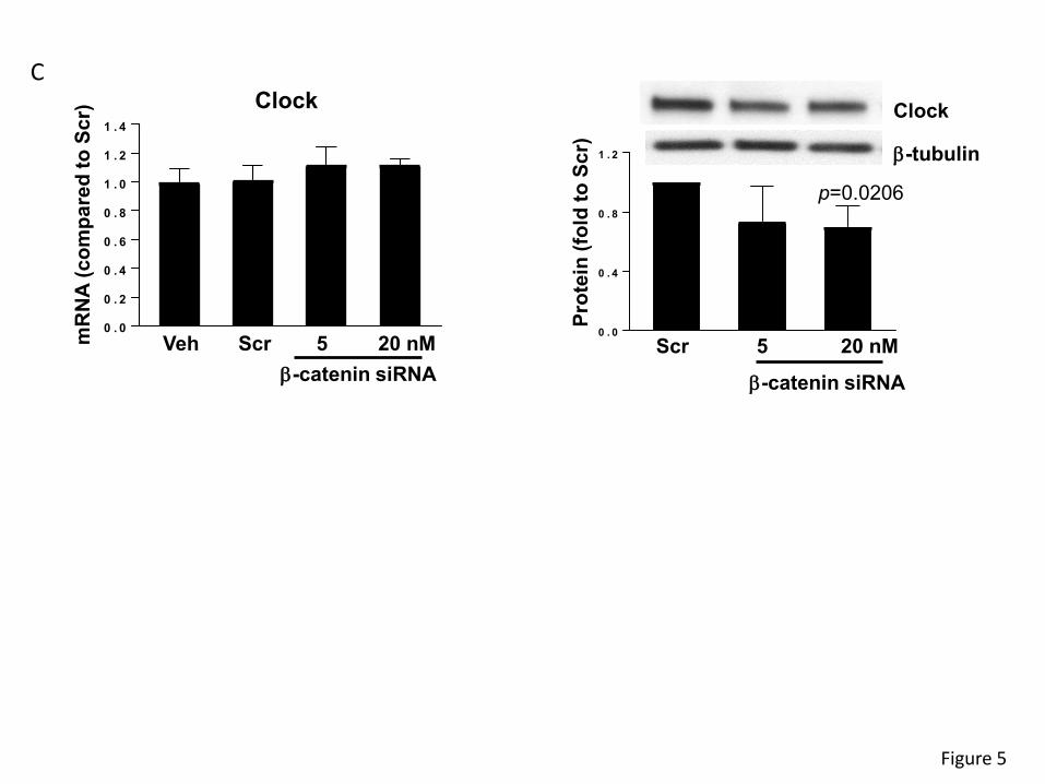
specific siRNA, followed by analysis of clock genes mRNA and protein expression (Figure 5).
Silencing β-catenin decreased its mRNA levels by ~80% and protein expression by ~30% (Figure
5A, left and right panel, respectively). Interestingly, β-catenin silencing diminished Bmal1 mRNA
and protein levels by ~40% (Figure 5B). Silencing of β-catenin also effectively decreased the
clock protein level by ~40% (Figure 5C, right panel). Overall, these results suggest that β-catenin
is upstream from the clock genes and proteins and may modulate their expression in intestinal
cells.
Silencing of circadian clock genes increases epithelial permeability.
To evaluate the impact of clock genes on epithelial barrier function, β-catenin, Clock, or Bmal1
expression was silenced in SW480 cells with specific siRNAs, followed by measuring permeability
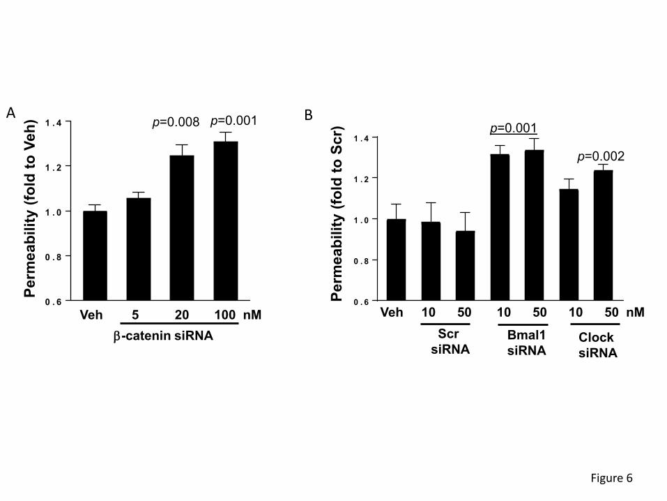
for FITC-dextran 20 kDa in the Transwell system. Permeability across SW480 monolayers was
significantly increased by 24% and 27% upon silencing with ß-catenin siRNA at 20 and 100 nM,
respectively (Figure 6A). In addition, silencing of Bmal1 with specific siRNA at 10 and 50 nM
increased permeability by ~28%. Silencing the Clock gene with 10 nM siRNA was ineffective,
however, silencing with 50 nM significantly increased permeability by 19% (Figure 6B).
8
Taking into consideration the substantial impact of β-catenin on the regulation of epithelial barrier
function and the role of β-catenin in circadian rhythm regulation, we next analyzed the
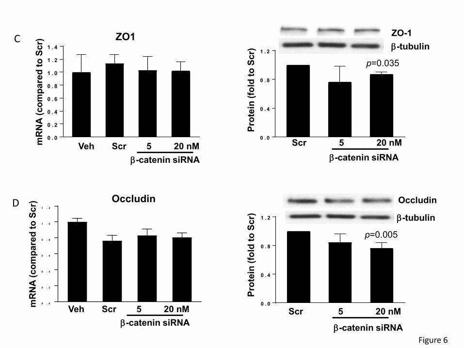
consequence of β-catenin knockdown on TJ protein expression. Silencing of β-catenin with 20nM
of specific siRNA did not alter ZO-1 mRNA levels; however, it decreased its protein levels (Figure
6C). Similarly, β-catenin silencing did not affect occludin gene expression. While there was a
~20% reduction in occludin mRNA expression as compared vehicle, similar downregulation of
occludin mRNA was observed in cells transfected with scrambled siRNA, suggesting non-specific
responses. As seen with ZO-1, silencing of β-catenin with 20 nM siRNA significantly decreased
occludin protein expression (Figure 6D).
β-catenin regulates epithelial barrier function via upregulation of MMPs.
A decrease in TJ protein expression without changes on mRNA levels suggests post-
transcriptional modification. Therefore, we evaluated a possible involvement of MMPs, which can
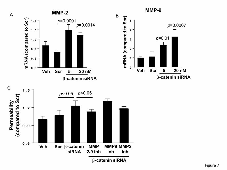
modulate permeability by degradation of TJ proteins 28. Transfection with β-catenin siRNA
significantly upregulated the mRNA expression of MMP-2 and MMP-9 (p < 0.01) (Figures 7A and
7B, respectively). In addition, silencing of β-catenin significantly increased permeability across
epithelial monolayers created by SW480 cells. In order to determine if increased production of
MMPs may be involved in this effect, cells were transfected with β-catenin siRNA and co-treated
with MMP-2 or MMP-9 inhibitors. Specific inhibitors for MMP-2 or MMP-9 individually did not affect
β-catenin siRNA-induced elevation of epithelial permeability. However, inhibition with a dual-
action blocker of both MMP-2 and MMP-9 attenuated disruption of permeability induced by β-
catenin siRNA (Figures 7C), suggesting that MMPs may modulate the impact of β-catenin on
epithelial barrier function in the context of circadian disruption.
9
DISCUSSION
Circadian rhythms are critically important to maintain normal gut function, to provide intestinal
barrier function, to engage bacterial dissemination, and to regulate inflammation, which when
disrupted can have several pathophysiological consequences 29,30,31,32,33. The exact mechanisms
linking circadian rhythm disruption and gut barrier integrity are not yet fully understood. Detailed
assessment of the gut barrier’s TJ proteins and their interactions with core clock genes provides
a better understanding of the relationship between circadian rhythm disruption and gut barrier
integrity. Identification of the mechanisms of such interactions is crucial for facilitation of treatment
in diseases that are influenced by circadian disruptions.
Molecular clocks regulate the 24-hour period of the circadian rhythm, which is driven by a
transcriptional–translational autoregulatory feedback loop. The core members of the mammalian
molecular clock are transcription factors Bmal1 and Clock that bind to the E-box promoter and
activate the transcription and translation of PER and CRY genes 34,35,36. The proteins of PER and
CRY dimerize to form a feedback inhibition that halts the initial transcription of Bmal1 and Clock
37,38,39,40,41. The molecular clock is also regulated by posttranslational modifications such as the
degradation of PER and CRY proteins via targeting by SCF ubiquitin ligases 42,43,44. The
degradation of PER and CRY proteins breaks the feedback inhibition and allows Bmal1 and Clock
to act as transcription factors thereby continuing the cycle. Other
posttranscriptional/posttranslational modifications include sirtuin 1 (Sirt1), nuclear receptors
retinoic acid-related orphan receptor alpha (Rora), and reverse erythroblastosis virus alpha
(Reverba) 45,46,18,47. The involvement of Sirt1 may link alterations of circadian regulation with
inflammatory responses. Indeed, Sirt1 is a ubiquitously expressed deacylase that regulates,
among others, acetylation and thus activation status of nuclear factor-κB (NF-κB), a potent
stimulator of inflammation 48. Moreover, Sirt1 activation remains under a partial control of cellular

10
occludin levels 49. The importance of these reactions stem from the fact that integrity of tissue and
cellular barriers are susceptible to altered cellular redox status 50.
The gut barrier is vital for maintaining homeostasis and performing functions such as providing a
selective barrier, degrading pathogens and antigens, preventing bacterial adhesion and
colonization, and initiating an immune response 51,52. Circadian rhythms within the gut anticipate
these daily needs by signaling to tight junction proteins and other basal membrane proteins in the
matrix to aid in intestinal absorption, produce antimicrobial substances, gastric acid, pancreatic
juice, and secrete biliary fluids 52,53. When circadian rhythms are disrupted, signaling pathways
become dysregulated and gut barrier integrity is compromised. Our experimental findings align
with observations demonstrating that disruption of normal light cycles in mice results in increased
gut barrier permeability (Figure 3) 54. It was also observed that LL shifted control-fed mice had
similar levels of intestinal hyperpermeability as LD non-shifted alcohol-fed mice 54. This suggests
that the disruption of the circadian rhythm via light cycle shifts has comparable results on gut
barrier permeability as chronic alcohol consumption. According to epidemiological data and
comparative studies, several gastrointestinal diseases such as chronic inflammatory bowel
disease, gastroesophageal reflux disease, irritable bowel syndrome, and peptic ulcers are more
common in shift workers with disrupted circadian rhythms 55,56,57,58.
In the present study, circadian rhythm disruption through light cycle manipulation led to
dysregulation of ß-catenin and circadian rhythm molecules Bmal1 and Clock as well as disrupted
TJ protein expression of occludin and ZO-1 at both the mRNA and protein levels (Figures 2 and
4). These results suggest that the circadian rhythm may regulate gut barrier integrity via a ß-
catenin pathway. From a transcriptional standpoint, our findings showed that siRNA-mediated
knockdown of β-catenin expression resulted in significant downregulation of Bmal1 at both mRNA
and protein levels and of Clock at protein levels (Figure 5B and 5C). Interestingly, it was

11
demonstrated that Bmal1 and Clock act as transcription factors that bind directly to the E-box
elements of occludin promoters and induce transcription in both mice and human small intestinal
epithelial cells 30. Furthermore, other studies have found that mRNA levels of occludin show time-
dependent variation in wild type mice, while a lack of such oscillation accompanied by an increase
in gut barrier permeability has been observed in circadian rhythm disrupted mice (ClockΔ19/Δ19)
30,54.
Circadian regulation of other barriers, such as in the inner blood-retina barrier (iBRB), has also
been demonstrated. For example, TJ protein claudin-5 was found to be under control of the core
clock in studies in which RNAi-mediated knockdown of Bmal1 affected the integrity of the iBRB
59. Additional observations indicated that mice were more resistant to Salmonella infection and
disruption of gut barrier permeability when ß-catenin expression was modified to be constitutively
active in intestinal endothelial cells, altering occludin and ZO-1 expression 60. The findings of the
present study are in line with these results indicating that ß-catenin has a regulatory effect on gut
barrier integrity. Indeed, silencing ß-catenin resulted in an increase in gut barrier permeability
(Figure 6A). Permeability of the gut barrier also increased when Bmal1 and Clock were silenced,
further demonstrating a link between circadian rhythm and gut barrier integrity (Figure 6B).
However, only protein levels of ZO-1 and occludin (Figure 6C and 6D) were found to significantly
decreased with ß-catenin knockdown, suggesting that expression of these TJ proteins is
regulated via post-translational mechanisms.
We further investigated which post-translational mechanisms may be involved and found that ß-
catenin knockdown led to higher mRNA expression levels of MMP-2 and MMP-9 as well as an
increase in gut barrier permeability (Figures 7). MMPs are endopeptidases that regulate
extracellular matrix homeostasis 61. MMP-2/9 specifically are gelatinases that activate pro-
inflammatory agents and mediate TJ protein degradation 62,63. Our observations are in agreement

12
with the involvement of MMPs in the regulation of tissue barrier integrity as shown previously by
us 64 and others 65. ß-catenin has been linked to MMP-2/9 in epithelial cells via negative regulation
as inhibition of Wnt/ß-catenin signaling led to increases in EMMPRIN, an extracellular MMP
inducer, and MMP-2/9. In addition, activation of Wnt/ß-catenin signaling resulted in large
decreases in EMMPRIN and MMP-2/9 66. While ß-catenin appears to control the integrity of
epithelial monolayers via MMP2/9 activity, alternative molecular pathways have also been
suggested to maintain gut barrier integrity, such as sonic hedgehog (SHH) signaling and
endothelial to mesenchymal transition (EndoMT) signaling 60.
Conclusion. Circadian rhythm disruption induced by light cycle manipulation resulted in
decreased gut barrier integrity concurrent with dysregulation of TJ proteins. Silencing of ß-catenin
and core clock genes such as Bmal1 and Clock increased gut barrier permeability, implicating
essential clock involvement in gut barrier regulation. Interestingly, ß-catenin appears to be a
master regulator of this process by acting upstream from Bmal1 and Clock and altering expression
of MMP2/9 (Figure 8).
13
METHODS
Animal Housing
Male C57BL/6 mice (Jackson Labs, Bar Harbor, ME), 12 weeks of age, were allowed to
acclimatize to the animal facility for four weeks with free access to food and water. Mice were
randomly assigned to normal lighting (light/dark, LD) and constant lighting (light/light, LL)
conditions for four weeks. Throughout the study, all mice had unlimited access to food and water.
Mice were euthanized with carbon dioxide followed by decapitation and collection of small and
large intestine. Ethics Statement: All animal procedures were approved by the University of
Miami Institutional Animal Care and Use Committee and performed in accordance with National
Institutes of Health (NIH) the American Association for Accreditation of Animal Care (AAALAC)
guidelines and regulations. Moreover, the study was carried out in compliance with the ARRIVE
guidelines.
Circadian Manipulation and circadian rhythm measurements
The circadian disruption protocol, constant light for 4 weeks, was intended to disrupt normal
circadian rhythms in a similar manner that could be observed in persons in intensive care units
living under constant illumination. The objective was to create an environment with no light
entraining cues (disruption) compared with normal day/night light cycling experienced by the
control mice in 12-hour light/12-hour dark. The illuminance was measured in and around the
cages where animals were being housed. The same lighting source (fluorescent lights on ceiling)
was used in both LD and LL rooms. Light was measured using a light meter (VWR International,
Radnor, PA) in and around the cages ranged from 400-600lx. The lighting schedule for LD mice
was 12 hours of light followed by 12 hours of darkness, lights on at 6AM (ZT0) and lights off at
6PM (ZT12), Eastern Standard Time.
14
Mice were initially housed under normal cycling light (LD) as described above. For four days they
were allowed to acclimate to single housing, diet, and environment, then baseline circadian
rhythms were measured by collecting rectal body temperature every 4 hours for 24 hours.
Following collection of body temperature, mice were singly housed in running wheels for 48 hours
to monitor locomotor behavioral rhythms. After 48 hours mice were randomized to LD or LL. Mice
were maintained in these rooms for two weeks at the end of which, circadian rhythms were
measured a second time by collecting rectal body temperature using a rectal thermometer (Acorn
series, OAKTON Instruments, Vernon Hills, IL). Body temperature was measured at the same
time points for both groups, 16:00 (ZT10), 20:00 (ZT14), 24:00 (ZT18), 04:00 (ZT22), 08:00 (ZT2),
and 12:00 (ZT6), Eastern Standard Time.
Voluntary running was measured twice during the experiment (at baseline and after 2 weeks of
circadian disruption). Activity rhythms were monitored using plastic cages measuring 30.5 X 15.2
X 12.7 centimeters containing a running wheel (Coulbourn Instruments, Whitehall, PA). During
the time in wheel cages mice had voluntary access to the running wheel as well as food and
water. Wheel revolutions were counted on an attached computer using Clocklab software
(Actimetrics, Wilmette, IL). Mice had no prior exposure to the running wheels before the
experiment began, and running was voluntary.
Ex vivo and in vitro intestinal permeability
Intestinal permeability was measured ex vivo in isolated ileum and colon segments. Briefly, 6-cm
segments of the ileum and the colon were removed, rinsed with ice-cold PBS, filled with 700 µl
DPBS containing 2 mg/ml FITC-dextran 4 kDa and ligated at both ends. The filled intestine
segments were incubated in DMEM containing 1% FBS. The sacs were removed after 45
minutes. The amount of FITC-dextran that transversed the intestine was quantified by
fluorescence plate reader at Ex 485 nm and Em 530 nm. In addition, ileum and colon segments
15
were sectioned and stained for nuclei (DAPI). Fluorescent images visualizing FITC-dextran 4 kDa
and nuclei were taken using confocal microscopy.
SW480 cells were seeded on collagen type I-coated Transwell polyester filters (12-mm diameter,
0.4 µm pore size, Corning Costar), transfected with β-catenin, Bmal1, Clock, or scrambled (scr)
siRNA. In selected experiments, MMP inhibitors were added to both the lower and the upper
compartments of the Transwell system. Then, 0.5 ml of FITC-dextran 20 (FD-20, 1 mg/ml in KRG
solution) was loaded into the upper chamber, the system was allowed to incubate for 60 min at
37 oC in humidified atmosphere (5% CO2), and the assay was stopped by removing the upper
chambers. Aliquots (100 µl) from the lower chambers were transferred to new wells of 96-well
fluorescence plate, and the fluorescence intensity of FITC-dextran was determined with a
microplate spectrofluorometer (Molecular Devices SPECTRA-Max Gemini EM) using 490 nm and
520 nm as excitation and emission wavelengths, respectively. Relative permeability was
expressed by the ratio of FD-20 transported into the lower chamber compared to control groups.
All assays were performed at least in quadruplicate.
Cell culture, treatment, and transfections with small interfering RNA
SW480, epithelial colonic cancer cells, were purchased from the American Type Culture
Collection (ATCC, Manassas, VA) and cultured in ATCC-formulated Leibovitz's L-15 Medium
(Catalog #30-2008). In selective experiments, cells were pretreated for 0.5-1 h with
pharmacological inhibitors of MMPs, including MMP-2/MMP-9 Inhibitor I, MMP-9 Inhibitor II,
MMP-2 inhibitor III (Millipore Sigma) at 20 µM.
SW480 cells were transfected with control siRNA or targeted siRNA at indicated concentrations
using Lipofectamine 2000 (ThermoFisher) in OptiMEM I medium (Invitrogen, Carlsbad, CA). Cells

16
were incubated with transfection mixtures for 6-20 h and allowed to recover in complete medium
for 48 h before the assays.
Real-Time RT-PCR
Total RNA was isolated and purified using RNeasy Mini Kit (Qiagen) according to the protocol of
the manufacturer. Then, 1 µg of total RNA was reverse transcribed at 25 °C for 15 min, 42 °C for
45 min and 99 °C for 5 min in 20 µl of 5 mM MgCl2, 10 mM Tris-HCl, pH 9.0, 50 mM KCl, 0.1%
Triton X-100, 1 mM dNTP, 1 unit/µl of recombinant RNasin ribonuclease inhibitor, 15 units/µg of
AMV reverse transcriptase, and 0.5 µg of random hexamers. For quantitative PCR, amplifications
of individual genes were performed on ABI PRISM® 7000 Sequence Detection System (Applied
Biosystems, Foster City, CA) using TaqMan® Universal PCR Master Mix, gene-specific TaqMan
PCR probes and primers, and a standard thermal cycler protocol (50 °C for 2 min before the first
cycle, 95 °C for 15 sec and 60 °C for 1 min, repeated 45 times). The primers and probes were
obtained from Applied Biosystems. The threshold cycle (CT) from each well was determined using
ABI Prism 7000 SDS software. Relative quantification, which represents the change in gene
expression from real-time quantitative PCR experiments between treated and control groups, was
calculated by the comparative CT method as described earlier 67. The data were analyzed using
equation 2–ΔΔCT, where ΔΔCT = [CT of target gene - CT of housekeeping gene] treated group – [CT of
target gene - CT of housekeeping gene] untreated control group. For the treated samples, evaluation of 2–
ΔΔCT represents the fold change in gene expression, normalized to a housekeeping gene (β-actin)
and relative to the untreated control.
Immunoblotting
Protein expression levels of β-catenin, Bmal1 and Clock, ZO-1, occludin and tricellulin were
assessed in the intestinal epithelial cell-enriched fractions of the isolated colon or in SW480 cells

17
by immunoblotting. All primary antibodies were purchased from ThermoFisher, and HRP-
conjugated secondary antibodies were obtained from Santa Cruz Biotechnology (Santa Cruz,
CA). Briefly, gut homogenates or treated cells were lysed with RIPA lysis buffer (1.0% Nonidet P-
40, 0.5% deoxycholic acid, 0.2% SDS, 40 mM Tris-HCl [pH 7.6], 1 mM EDTA, 1 mM EGTA, 10
mM MgCl2, 150 mM NaCl, 1 mM Na3VO4, 1 mM NaF, 1 × EDTA-free protease inhibitor cocktail
[Roche Applied Science], and 1 mM phenylmethylsulfonyl fluoride) for the total cell extract. Protein
concentration was determined using BCA protein assay kit (Thermo Scientific, Rockford, IL).
Then, 10 µg protein of cell lysates was electrophoresed on SDS-polyacrylamide gels, transferred
to a polyvinylidene fluoride (PVDF) membrane, blocked with 3% BSA in PBS-T (0.1% Tween-20)
solution and incubated with the primary antibodies overnight at 4°C. After incubation with the
secondary antibody for 2 h, immunoblots were visualized using the ECL detection system
(Amersham Biosciences). GAPDH or actin was determined as the loading control. The band
density was measured using Image J software (NIH).
Statistical analysis
Data were statistically analyzed using one-way ANOVA, followed by Tukey’s multiple
comparisons test. Statistical probability of p<0.05 was considered significant. Results are
expressed as means ± S.D. All experiments were repeated at least 3 times.
Data availability
All source data supporting the findings of this manuscript are available from the corresponding
authors upon request.

18
REFERENCES
1. Asher, G. & Sassone-Corsi, P. Time for food: The intimate interplay between nutrition,
metabolism, and the circadian clock. Cell 161, 84–92 (2015).
2. Huang, Y.-J., Pai, Y.-C. & Yu, L. C.-H. Host-Microbiota Interaction and Intestinal Epithelial
Functions under Circadian Control: Implications in Colitis and Metabolic Disorders. Chin.
J. Physiol. 61, 325–340 (2018).
3. Konturek, P. C., Brzozowski, T. & Konturek, S. J. Gut clock: Implication of circadian
rhythms in the gastointestinal tract. Journal of Physiology and Pharmacology (2011).
4. Bechtold, D. A., Gibbs, J. E. & Loudon, A. S. I. Circadian dysfunction in disease. Trends
in Pharmacological Sciences 31, 191–198 (2010).
5. Yasuniwa, Y. et al. Circadian disruption accelerates tumor growth and
angio/stromagenesis through a wnt signaling pathway. PLoS One 5, 15330 (2010).
6. Soták, M., Sumová, A. & Pácha, J. Cross-talk between the circadian clock and the cell
cycle in cancer. Ann. Med. 46, 221–232 (2014).
7. Zhao, X. et al. The In Vitro Protective Role of Bovine Lactoferrin on Intestinal Epithelial
Barrier. Molecules 24, 148 (2019).
8. Turner, J. R. Molecular basis of epithelial barrier regulation: From basic mechanisms to
clinical application. Am. J. Pathol. 169, 1901–1909 (2006).
9. Viggiano, D. et al. Gut barrier in health and disease. 1077–1085 (2015).
10. Maury, E., Ramsey, K. M. & Bass, J. Circadian Rhythms and Metabolic Syndrome. Circ.
Res. 106, 447–462 (2010).
11. Portaluppi, F. et al. Circadian rhythms and cardiovascular health. Sleep Medicine
Reviews 16, 151–166 (2012).
12. Takeda, N. & Maemura, K. Circadian clock and cardiovascular disease. Journal of
Cardiology 57, 249–256 (2011).
13. Sládek, M. et al. Insight Into the Circadian Clock Within Rat Colonic Epithelial Cells.
19
Gastroenterology 133, 1240–1249 (2007).
14. Hoogerwerf, W. A. et al. Clock Gene Expression in the Murine Gastrointestinal Tract:
Endogenous Rhythmicity and Effects of a Feeding Regimen. Gastroenterology 133,
1250–1260 (2007).
15. Froy, O. & Chapnik, N. Circadian oscillation of innate immunity components in mouse
small intestine. Mol. Immunol. 44, 1954–1960 (2007).
16. Deaver, J. A., Eum, S. Y. & Toborek, M. Circadian disruption changes gut microbiome
taxa and functional gene composition. Front. Microbiol. 9, (2018).
17. Teichman, E. M., O’Riordan, K. J., Gahan, C. G. M., Dinan, T. G. & Cryan, J. F. When
Rhythms Meet the Blues: Circadian Interactions with the Microbiota-Gut-Brain Axis. Cell
Metabolism 31, 448–471 (2020).
18. Voigt, R. M., Forsyth, C. B., Green, S. J., Engen, P. A. & Keshavarzian, A. Circadian
Rhythm and the Gut Microbiome. in International Review of Neurobiology (2016).
doi:10.1016/bs.irn.2016.07.002
19. Eum, S. Y., Jaraki, D., Bertrand, L., András, I. E. & Toborek, M. Disruption of epithelial
barrier by quorum-sensing N-3-(oxododecanoyl)-homoserine lactone is mediated by
matrix metalloproteinases. Am. J. Physiol. - Gastrointest. Liver Physiol. 306, (2014).
20. Thaiss, C. A. et al. Microbiota Diurnal Rhythmicity Programs Host Transcriptome
Oscillations. Cell 167, 1495-1510.e12 (2016).
21. Mu, C., Yang, Y. & Zhu, W. Gut microbiota: The brain peacekeeper. Frontiers in
Microbiology 7, (2016).
22. Asher, G. & Schibler, U. Crosstalk between components of circadian and metabolic
cycles in mammals. Cell Metabolism 13, 125–137 (2011).
23. Eckel-Mahan, K. & Sassone-Corsi, P. Metabolism and the circadian clock converge.
Physiol. Rev. 93, 107–135 (2013).
24. Green, C. B., Takahashi, J. S. & Bass, J. The Meter of Metabolism. Cell 134, 728–742

20
(2008).
25. Fonken, L. K., Weil, Z. M. & Nelson, R. J. Mice exposed to dim light at night exaggerate
inflammatory responses to lipopolysaccharide. Brain. Behav. Immun. 34, 159–163
(2013).
26. Matsu-ura, T. et al. Intercellular Coupling of the Cell Cycle and Circadian Clock in Adult
Stem Cell Culture. Mol. Cell 64, 900–912 (2016).
27. Junichi, I., Hiroyuki, S., Sachiko, T., Mikio, F. & Shoichiro, T. Loss of Occludin Affects
Tricellular Localization of Tricellulin. Mol. Biol. Cell 19, 4687–4693 (2008).
28. Huang, W., Eum, S. Y., András, I. E., Hennig, B. & Toborek, M. PPARα and PPARγ
attenuate HIV‐induced dysrégulation of tight junction proteins by modulations of matrix
metalloproteinase and proteasome activities. FASEB J. 23, 1596–1606 (2009).
29. Voigt, R. M. et al. The Circadian Clock Mutation Promotes Intestinal Dysbiosis. Alcohol.
Clin. Exp. Res. 40, 335–347 (2016).
30. Oh-oka, K. et al. Expressions of tight junction proteins occludin and claudin-1 Are under
the circadian control in the mouse large intestine: Implications in intestinal permeability
and susceptibility to colitis. PLoS One 9, (2014).
31. Voigt, R. M., Forsyth, C. B. & Keshavarzian, A. Circadian rhythms: a regulator of
gastrointestinal health and dysfunction. Expert Review of Gastroenterology and
Hepatology 13, 411–424 (2019).
32. Pagel, R. et al. Circadian rhythm disruption impairs tissue homeostasis and exacerbates
chronic inflammation in the intestine. FASEB J. 31, 4707–4719 (2017).
33. Swanson, G. R. et al. Night workers with circadian misalignment are susceptible to
alcohol-induced intestinal hyperpermeability with social drinking. Am. J. Physiol. -
Gastrointest. Liver Physiol. 311, G192–G201 (2016).
34. Bunger, M. K. et al. Mop3 Is an Essential Component of the Master Circadian Pacemaker
in Mammals. Cell 103, 1009–1017 (2000).

21
35. Mohawk, J. A., Green, C. B. & Takahashi, J. S. Central and Peripheral Circadian Clocks
in Mammals. Annu. Rev. Neurosci. 35, 445–462 (2012).
36. Schibler, U. The daily rhythms of genes, cells and organs. EMBO Rep. 6, S9–S13 (2005).
37. Van Der Horst, G. T. J. et al. Mammalian Cry1 and Cry2 are essential for maintenance of
circadian rhythms. Nature 398, 627–630 (1999).
38. Reppert, S. M. & Weaver, D. R. Coordination of circadian clocks in mammals. Nature
418, 935–941 (2002).
39. Lee, C., Etchegaray, J. P., Cagampang, F. R. A., Loudon, A. S. I. & Reppert, S. M.
Posttranslational mechanisms regulate the mammalian circadian clock. Cell 107, 855–
867 (2001).
40. Yamamoto, Y., Yagita, K. & Okamura, H. Role of Cyclic mPer2 Expression in the
Mammalian Cellular Clock. Mol. Cell. Biol. 25, 1912–1921 (2005).
41. Gallego, M. & Virshup, D. M. Post-translational modifications regulate the ticking of the
circadian clock. Nat. Rev. Mol. Cell Biol. 8, 139–48 (2007).
42. Shirogane, T., Jin, J., Ang, X. L. & Harper, J. W. SCFβ-TRCP controls Clock-dependent
transcription via casein kinase 1-dependent degradation of the mammalian period-1
(Per1) protein. J. Biol. Chem. 280, 26863–26872 (2005).
43. Eide, E. J. et al. Control of Mammalian Circadian Rhythm by CKI -Regulated
Proteasome-Mediated PER2 Degradation. Mol. Cell. Biol. 25, 2795–2807 (2005).
44. Siepka, S. M. et al. Circadian Mutant Overtime Reveals F-box Protein FBXL3 Regulation
of Cryptochrome and Period Gene Expression. Cell 129, 1011–1023 (2007).
45. Bass, J. & Takahashi, J. S. Circadian integration of metabolism and energetics. Science
330, 1349–1354 (2010).
46. Grimaldi, B., Nakahata, Y., Kaluzova, M., Masubuchi, S. & Sassone-Corsi, P. Chromatin
remodeling, metabolism and circadian clocks: The interplay of CLOCK and SIRT1.
International Journal of Biochemistry and Cell Biology 41, 81–86 (2009).
22
47. Yang, X. et al. β-catenin induces β-TrCP-mediated PER2 degradation altering circadian
clock gene expression in intestinal mucosa of ApcMin/+ mice. J. Biochem. 145, 289–297
(2009).
48. Singh, V. & Ubaid, S. Role of Silent Information Regulator 1 (SIRT1) in Regulating
Oxidative Stress and Inflammation. Inflammation 43, 1589–1598 (2020).
49. Castro, V. et al. Occludin controls HIV transcription in brain pericytes via regulation of
SIRT-1 activation. FASEB J. 30, 1234–1246 (2016).
50. Toborek, M., Barger, S. W., Mattson, M. P., McClain, C. J. & Hennig, B. Role of
glutathione redox cycle in TNF-α-mediated endothelial cell dysfunction. Atherosclerosis
117, 179–188 (1995).
51. Camilleri, M., Madsen, K., Spiller, R., Van Meerveld, B. G. & Verne, G. N. Intestinal
barrier function in health and gastrointestinal disease. Neurogastroenterology and Motility
24, 503–512 (2012).
52. Keita, Å. V. & Söderholm, J. D. The intestinal barrier and its regulation by neuroimmune
factors. Neurogastroenterol. Motil. 22, 718–733 (2010).
53. Vaughn, B., Rotolo, S. & Roth, H. Circadian rhythm and sleep influences on digestive
physiology and disorders. ChronoPhysiology Ther. 4, 67 (2014).
54. Summa, K. C. et al. Disruption of the Circadian Clock in Mice Increases Intestinal
Permeability and Promotes Alcohol-Induced Hepatic Pathology and Inflammation. PLoS
One (2013). doi:10.1371/journal.pone.0067102
55. Chung, T. H., Lee, J. & Kim, M. C. Impact of night-shift work on the prevalence of erosive
esophagitis in shipyard male workers. Int. Arch. Occup. Environ. Health 89, 961–966
(2016).
56. Kim, H. I. et al. Impact of shiftwork on irritable bowel syndrome and functional dyspepsia.
J. Korean Med. Sci. 28, 431–437 (2013).
57. Segawa, K. et al. Peptic ulcer is prevalent among shift workers. Dig. Dis. Sci. 32, 449–
23
453 (1987).
58. Knutsson, A. & Bøggild, H. Gastrointestinal disorders among shift workers. Scand. J.
Work. Environ. Heal. 36, 85–95 (2010).
59. Hudson, N. et al. Dysregulated claudin-5 cycling in the inner retina causes retinal pigment
epithelial cell atrophy. JCI Insight 4, 1–13 (2019).
60. Spadoni, I. et al. Gene expression profile of endothelial cells during perturbation of the
gut vascular barrier. Science (80-. ). 350, 830–834 (2015).
61. Ravi, A., Garg, P. & Sitaraman, S. V. Matrix metalloproteinases in inflammatory bowel
disease: Boon or a bane? Inflammatory Bowel Diseases 13, 97–107 (2007).
62. Kurzepa, J., Kurzepa, J., Golab, P., Czerska, S. & Bielewicz, J. The significance of matrix
metalloproteinase (MMP)-2 and MMP-9 in the ischemic stroke. International Journal of
Neuroscience 124, 707–716 (2014).
63. Feng, S. et al. Matrix metalloproteinase-2 and -9 secreted by leukemic cells increase the
permeability of blood-brain barrier by disrupting tight junction proteins. PLoS One 6,
(2011).
64. Eum, S. Y., Jaraki, D., András, I. E. & Toborek, M. Lipid rafts regulate PCB153-induced
disruption of occludin and brain endothelial barrier function through protein phosphatase
2A and matrix metalloproteinase-2. Toxicol. Appl. Pharmacol. 287, 258–266 (2015).
65. Rempe, R. G., Hartz, A. M. S. & Bauer, B. Matrix metalloproteinases in the brain and
blood-brain barrier: Versatile breakers and makers. Journal of Cerebral Blood Flow and
Metabolism 36, 1481–1507 (2016).
66. Liu, X., Zhang, Z., Pan, S., Shang, S. & Li, C. Interaction between the Wnt/β-catenin
signaling pathway and the EMMPRIN/MMP-2, 9 route in periodontitis. J. Periodontal Res.
53, 842–852 (2018).
67. Livak, K. J. & Schmittgen, T. D. Analysis of relative gene expression data using real-time
quantitative PCR and the 2-ΔΔCT method. Methods 25, 402–408 (2001).
24
ACKNOWLEDGEMENTS
Supported by the National Institutes of Health (NIH), grants MH072567, HL126559, DA044579,
DA039576, DA050528, and the University of Miami internal funds. The content is solely the
responsibility of the authors and does not necessarily represent the official views of the National
Institutes of Health.
AUTHOR CONTRIBUTIONS SY, GW, and MT designed the experiments, analyzed data, and assembled figures. SY and GW
performed the experiments. NS analyzed data and wrote the manuscript. MT provided the
funding. All authors reviewed the manuscript.
COMPETING INTERESTS The authors report no competing interests.

25
FIGURE LEGENDS
Figure 1: Four weeks of constant light disrupts circadian rhythms in mice. (A) Summary
actograms at baseline (before light manipulation) and two weeks after 12-hour light/dark (LD) or
circadian disruption (constant light, LL). Shaded areas, 18:00 (ZT12) to 6:00 (ZT0), indicate time
of lights-off, or former lights-off for LL 2nd week. Numbers along the x-axis indicate hours of the
day. Black areas are wheel revolutions binned each minute and reported here as the average for
n=19 mice per group. (B) Arrhythmicity of exercised mice maintained in LL as demonstrated by
phase angle assessment. Phase angle is the difference in hours between the time of lights-off (or
former lights-off for LL 2nd week), which was 18:00 (ZT12), and the time of activity onset;
corresponding to uninterrupted activity on the wheel. In the LL group at the 2nd week, activity was
significantly shifted ~30 min before lights-off. (C-D) Body temperature rhythm measured every 4
hours over 24 hours in LD (C) and LL (D) mice on the 2nd week. (E) An increase in body mass in
mice maintained in LL as compared to LD. *p<0.05 compared with LD. Values are mean ± SEM.
Figure 2. Expression of circadian clock molecules in the gut of the circadian rhythm
disrupted mice. The mRNA and protein expression of circadian clock molecules, β-catenin,
Bmal1, and Clock were measured in the intestinal epithelial cell-enriched fractions of the colon of
mice exposed to 12-hour light/dark cycling (LD) or constant 24-h light (LL) for 4 weeks. (A) mRNA
expression of β-catenin, Bmal1 and Clock was assessed using RT-PCR. (B-C) Protein expression
levels from the same mice as (A). Protein expression level of β-catenin, Bmal1 and Clock was
measured by immunoblotting (B) and band intensity was quantified by densitometric analysis
using Image J program (C). Values are mean ± SEM; n=4-5.
26
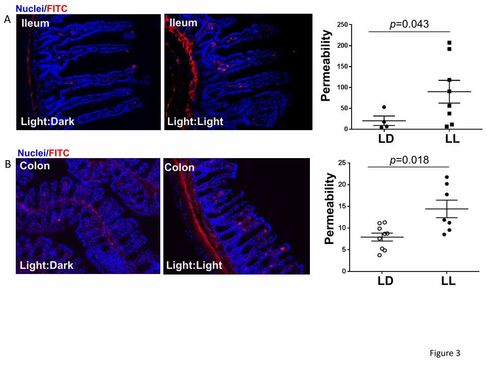
Figure 3. Circadian rhythm disruption alters ileum and colon permeability. Mice were treated
as in Figures 1 and 2. (A) Left panel, representative images of ex vivo permeability to FITC-
dextran 4 kDa through the ileum segment in mice maintained under 12-hour light/dark (LD) or
constant light (LL) for 4 weeks. (B) Ex vivo permeability in the colon in the same mice as in (A).
Left panels, gut sections stained for nuclei (blue) and FITC (red). Right panels, quantitative data
from ex vivo permeability measurements. Values are mean ± SEM; n=4-9.
Figure 4. Alterations of tight junction protein expression in the colon of mice subjected to
circadian rhythm disruption. The expression of tight junction molecules was measured in the
intestinal epithelial cell-enriched fractions of the colon of mice exposed to 12-hour light/dark
cycling (LD) or constant 24-h light (LL) for 4 weeks. (A) mRNA expression of ZO-1, occludin, and
tricellulin as analyzed by real-time PCR. (B-D) Protein expression levels from the same mice as
(A). Protein expression of ZO-1, occludin and tricellulin was assessed by immunoblotting (B) and
band intensity was quantified by densitometric analysis using Image J program (C). Values are
mean ± SEM; n=4-5.
Figure 5. Silencing of β-catenin alters mRNA and protein expression of circadian clock
molecules. SW480 cells grown on the 12 well cell culture plates were transfected with β-catenin-
specific siRNA at the indicated concentration or with non-specific, scrambled siRNA. mRNA (left
panels) and protein (right panels) expression of β-catenin (A), Bmal1 (B), and Clock(C) were
assayed and quantified. Blots illustrate representative data. Band intensity was assessed by
densitometric analysis using Image J program. Values (means ± SD) are expressed as fold
change compared with Scr; n = 3-6.

27
Figure 6. Silencing of circadian clock molecules increases paracellular permeability
across epithelial monolayers. SW480 cells grown on the apical side of wells (0.4-µm pore size,
12-mm diameter) of Transwell inserts were transfected with siRNA of β-catenin (A), Bmal1, or
Clock (B) at the indicated concentrations. Control cultures were either treated with vehicle or
transfected with non-specific, scrambled (Scr) siRNA. Epithelial permeability was determined by
measuring paracellular passage of FITC-dextran 20 kDa from the apical side to the basolateral
side across SW480 cell layers. Values (means ± SD) are expressed as fold change compared
with Veh or Scr. Values are mean ± SEM; n=5-6. (C-D) Silencing of β-catenin alters expression
of tight junction proteins without affecting mRNA levels. SW480 cells grown on the 12 well cell
culture plates were transfected with β-catenin-specific siRNA at the indicated concentration or
with non-specific, scrambled siRNA. mRNA (left panels) or protein (right panels) expression of
ZO-1 (C) and occludin (D) were assayed and quantified. Blots illustrate representative data. Band
intensity was assessed by densitometric analysis using Image J program. Values (means ± SD)
are expressed as fold change compared with Scr; n = 3-6.
Figure 7. β-catenin-induced alterations of paracellular permeability is mediated by MMPs.
SW480 cells were transfected with β-catenin-specific siRNA at the indicated concentrations or
with non-specific, scrambled siRNA, followed by mRNA assessment by RT-PCR of MMP-2 (A)
and MMP-9 (B). Values (means ± SD) are expressed as fold change compared with Scr; n = 5-
6. (C) SW480 cells grown on the apical side of wells (0.4-µm pore size, 12-mm diameter) of
Transwell system were transfected with β-catenin siRNA or scrambled siRNA (both at 20 nM).
Additional cultures were treated with pharmacological inhibitors of MMPs, namely, MMP-2/MMP-
9 Inhibitor I, MMP-9 Inhibitor II, or MMP-2 inhibitor III all at 20 µM. Epithelial permeability was
determined by measuring paracellular passage of FITC-dextran 20 from the apical side to the
basolateral side across SW480 cell layers. Values (means ± SD) are expressed as fold change
28
compared to Scr; n = 5. (D) Schematic diagram of the major findings of the present manuscript.
ß-catenin appears to be a master regulator of epithelial barrier integrity in circadian rhythm
disruption by transcriptional and translational modulation of TJ proteins, such as occludin and ZO-
1.
Figure 8. Schematic diagram of the major findings of the present manuscript. ß-catenin
appears to be a master regulator of epithelial barrier integrity in circadian rhythm disruption by
transcriptional and translational modulation of TJ proteins, such as occludin and ZO-1.

A B
C D
Figure 1
16 20 24 4 8 12 16 (hour)
Tem
pera
ture
(C)
16 20 24 4 8 12 16 (hour)
Tem
pera
ture
(C)
E
Bod
y w
eigh
t (g)
0 1 2 3 4 Time (week)
LD LL LD LLBaseline Week 2
*
Hou
rsp<0.05

Figure 2
LD LL
β-Catenin
Bmal1Clock
Actin
0 . 0
0 . 5
1 . 0
1 . 5
L D
L L
β-Catenin Bmal1 ClockProt
ein
(fold
com
pare
d to
LD
)
p=0.008 p=0.001 p=0.002
A
B
C
0 . 0
0 . 5
1 . 0
1 . 5
2 . 0
2 . 5
L D
L L
β-Catenin Bmal1 Clock
mR
NA
(fold
com
pare
d to
LD
)p=0.027 p=0.078 p=0.028
Colon
Light:Dark
Colon
Light:Light
Ileum Ileum
Light:Dark Light:Light
Figure 3
Perm
eabi
lity
LD LL
p=0.043
Perm
eabi
lity
LD LL
p=0.018
A
B
Nuclei/FITC
Nuclei/FITC

0 . 0
0 . 5
1 . 0
1 . 5
2 . 0
L DL L
Figure 4
LD LL
TricellulinOccludin
Actin
ZO-1
ZO-1 Occludin Tricellulin
p=0.015 p=0.023 p=0.007m
RN
A (fo
ld c
ompa
red
to L
D)A
B
0 . 0
0 . 5
1 . 0
1 . 5
2 . 0
L DL L
ZO-1 Occludin Tricellulin
p=0.032 p=0.0001 p=0.0004
Prot
ein
(fold
com
pare
d to
LD
)C

0 . 0
0 . 4
0 . 8
1 . 2
0 . 0
0 . 4
0 . 8
1 . 2
0 . 0
0 . 4
0 . 8
1 . 2
0 . 0
0 . 4
0 . 8
1 . 2
Figure 5
A β-Cateninβ-tubulin
Bmal1β-tubulin
B
mR
NA
(com
pare
d to
Scr
)
Veh Scr 5 20 nM
β-catenin
β-catenin siRNA
p=0.0001
mR
NA
(com
pare
d to
Scr
) Bmal1
p=0.001
Veh Scr 5 20 nM β-catenin siRNA
p=0.0095 p=0.0007
Scr 5 20 nM β-catenin siRNA
Prot
ein
(fold
to S
cr)
Scr 5 20 nM β-catenin siRNA
Prot
ein
(fold
to S
cr)
p=0.0434 p=0.0001
0 . 0
0 . 4
0 . 8
1 . 2 β-tubulin
Clock
C
Figure 5
mR
NA
(com
pare
d to
Scr
) Clock
0 . 0
0 . 2
0 . 4
0 . 6
0 . 8
1 . 0
1 . 2
1 . 4
Veh Scr 5 20 nM β-catenin siRNA
Prot
ein
(fold
to S
cr)
Scr 5 20 nM
β-catenin siRNA
p=0.0206
0 . 6
0 . 8
1 . 0
1 . 2
1 . 4
Veh 10 50 10 50 10 50 nM
Figure 6
Veh 5 20 100 nM0 . 6
0 . 8
1 . 0
1 . 2
1 . 4
β-catenin siRNA
p=0.008 p=0.001
Perm
eabi
lity
(fold
to V
eh)
ScrsiRNA
Bmal1siRNA
Clock siRNA
Perm
eabi
lity
(fold
to S
cr) p=0.001
p=0.002
A B
Occludin
0 . 0
0 . 4
0 . 8
1 . 2
0 . 0
0 . 4
0 . 8
1 . 2
Prot
ein
(fold
to S
cr)
Figure 6
C ZO-1β-tubulin
β-tubulin
mR
NA
(com
pare
d to
Scr
) ZO1
0 . 0
0 . 2
0 . 4
0 . 6
0 . 8
1 . 0
1 . 2
mR
NA
(com
pare
d to
Scr
) OccludinD
0 . 0
0 . 2
0 . 4
0 . 6
0 . 8
1 . 0
1 . 2
1 . 4
Veh Scr 5 20 nM β-catenin siRNA
Veh Scr 5 20 nM β-catenin siRNA
p=0.035
Scr 5 20 nM β-catenin siRNA
Prot
ein
(fold
to S
cr)
Scr 5 20 nM
β-catenin siRNA
p=0.005
0 . 6
0 . 9
1 . 2
1 . 5
Figure 7
Perm
eabi
lity
(com
pare
d to
Scr
)A B
C
mR
NA
(com
pare
d to
Scr
)
Veh Scr 5 20 nM β-catenin siRNA
p=0.0014p=0.0001
0 . 3
0 . 6
0 . 9
1 . 2
1 . 5
1 . 8
MMP-2 MMP-9
0
1
2
3
4
5
mR
NA
(com
pare
d to
Scr
)
Veh Scr 5 20 nM β-catenin siRNA
p=0.01
p=0.0007
Veh Scr MMP 2/9 inh
MMP9 inh
MMP2 inh
p˂0.05 p˂0.05
β-catenin siRNA
β-catenin siRNA
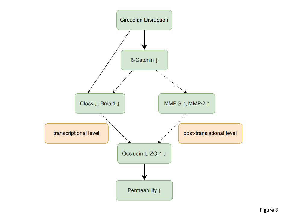
Figure 8