cirrhosis dr akhondei. cirrhosis cirrhosis is a pathologically defined entity that is associated...
TRANSCRIPT

CIRRHOSISDr akhondei

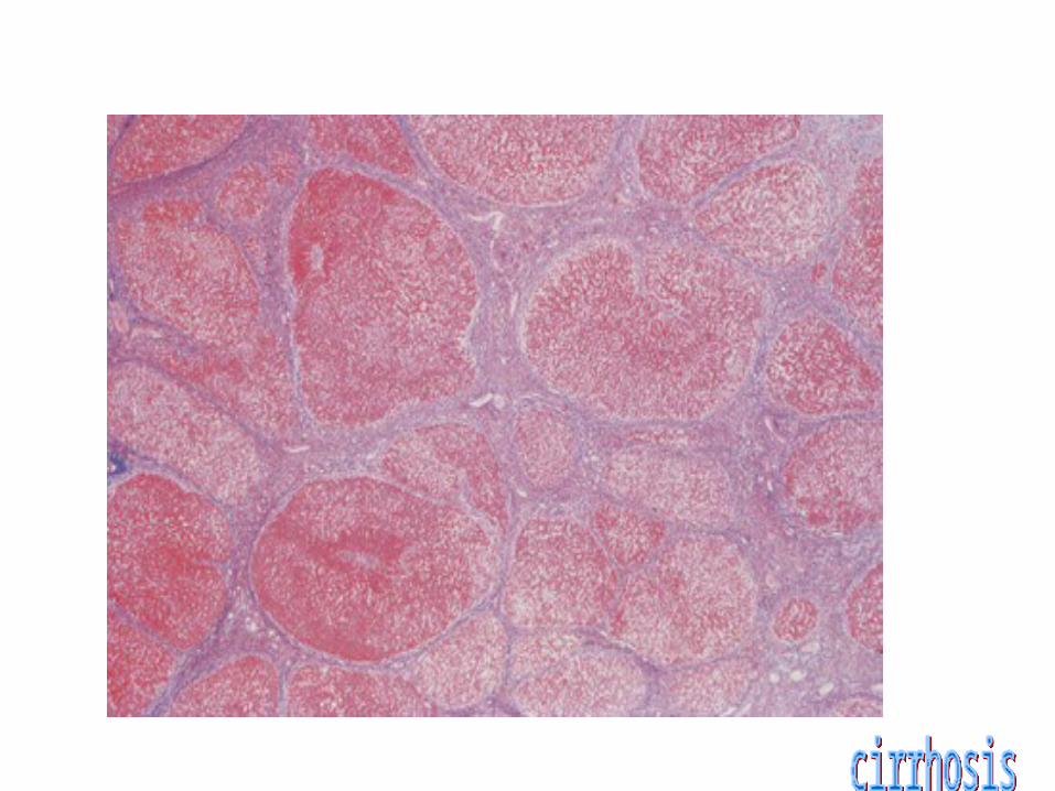
cirrhosis
Cirrhosis is a pathologically defined entity that is associated with a spectrum of characteristic clininical manifestation
1-Irreversible chronic injury of the hepatic parenchyma
2-Extensive fibrosis 3-formation of regenerative
nodules
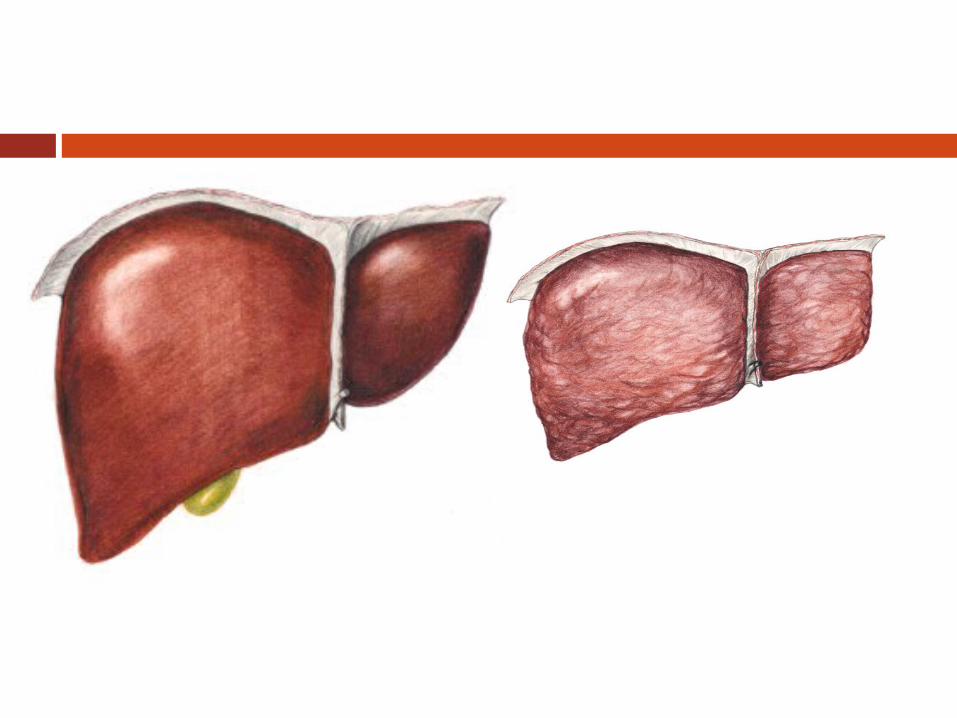


Normal liver


Cirrhosis of the liver is the third leading cause of death in people between the ages of 25 and 65 years, exceeded only by cardiovascular disease and cancer.
The cost of cirrhosis in terms of human suffering, financial burden, and loss of productive life is devastating.

Patients with cirrhosis may present in a variety of ways.
Variceal bleeding New onset of ascites Hepatomegaly and/or splenomegaly

Patients with cirrhosis may present in a variety of ways.
Incidental Discovery Of Abnormal Laboratory Tests
Serum Aminotransferases, Hypoalbuminemia, Prolonged Prothrombin Time, Hrombocytopenia)

Patients with cirrhosis may present in a variety of ways.
Detection of the peripheral stigmata ofchronic liver disease such as
Jaundice, Dupuytren's contracture, Manifestations related to hyperestrogenemia
such as o Spider angiomata, o Palmar erythema,o Gynecomastia, and testicular atrophy which
may also be related to low testosterone concentrations

Patients with cirrhosis may present in a variety of ways.
hepatic encephalopathy or one or more complications or systemic manifestations of hepatocellular carcinoma
Some patients never come to clinical attention. In older reviews, cirrhosis was diagnosed at autopsy in up to 30 to 40 percent of patients

Cirrhosis represents the terminal stage of a number of chronic liver diseases including those caused by
excessive ethanol consumption Viral hepatitis Drugs and toxins, Vascular Autoimmune Metabolic disorders Cryptogenic. In some cases, no cause can be determined and the cirrhosis is

Early cirrhosis may be asymptomatic and undetectable except by biopsy.
However, by the time cirrhosis becomes clinically apparent, hepatic functional reserve is markedly impaired.
If liver injury cannot be arrested, the prognosis without intervention is poor
The onset of complications (eg, ascites, encephalopathy, variceal
hemorrhage) indicates a more advanced stage of disease and a poorer prognosis, with median survival of about 5 years or less.

CIRRHOSIS & PORTAL HYPERTENSION

CIRRHOSIS
Term was 1st coined by Laennec in 1826
Many definitions but common theme is injury, repair, regeneration and scarring
NOT a localized process; involves entire liver
Primary histologic features:1. Marked fibrosis2. Destruction of vascular & biliary elements3. Regeneration4. Nodule formation
Cirrhosis: Pathophysiology
Primary event is injury to hepatocellular elements
Initiates inflammatory response with cytokine release->toxic substances
Destruction of hepatocytes, bile duct cells, vascular endothelial cells
Repair thru cellular proliferation and regeneration
Formation of fibrous scar

Cirrhosis: Pathophysiology
Primary cell responsible for fibrosis is stellate cell
Become activated in response to injury and lead to ed expression of fibril-forming collagen
Above process is influenced by Kupffer cells which activate stellate cells by eliciting production of cytokines
Sinusoidal fenestrations are obliterated because of ed collagen and EC matrix synthesis

Cirrhosis: Pathophysiology
Prevents normal flow of nutrients to hepatocytes and increases vascular resistance
Initially, fibrosis may be reversible if inciting events are removed
With sustained injury, process of fibrosis becomes irreversible and leads to cirrhosis

Causes of Cirrhosis
Alcohol Viral hepatitis Biliary obstruction Veno-occlusive disease Hemochromatosis Wilson’s disease Autommune Drugs and toxins Metabolic diseases Idiopathic

Classification of Cirrhosis
WHO divided cirrhosis into 3 categories based on morphological characteristics of the hepatic nodules
1. Micronodular2. Macronodular3. Mixed

Micronodular Cirrhosis
Nodules are <3 mm in diameter Relatively uniform in size Distributed throughout the liver Rarely contain portal tracts or efferent
veins Liver is of uniform size or mildly
enlarged Reflect relatively early disease

Macronodular & Mixed Cirrhosis Nodules are >3 mm in diameter and vary
considerably in size Usually contain portal tracts and efferent
veins Liver is usually normal or reduced in size Mixed pattern if both type of nodules are
present in equal proportions

Cirrhosis - Alcohol
Also known as Laennec’s cirrhosis >50% of pts. with alcoholic cirrhosis die
within 4 yrs of diagnosis Develops in only 10% to 30% of heavy
drinkers Morphologically, micronodular pattern Multifactorial - genetic, nutritional, drug
use and viral

Cirrhosis - Alcohol
Fatty liver, alcoholic hepatitis Histology - megamitochondria, Mallory
bodies, inflammation, necrosis, fibrosis Key mediator is acetaldehyde (ADH), the
product of alcohol metabolism by alcohol dehydrogenase
ADH directly activates stellate cells, inhibits DNA repair and damage microtubules

NAFLD/NASH
Nonalcoholic Fatty Liver Disease and Steatohepatitis
Becoming more common Infiltration of the liver with fat ±
inflammation Pathologically similar to alcoholic liver but
in absence of alcohol Associated with obesity, hyperlipidemia,
NIDDM,

Viral Hepatitis
Most common cause of cirrhosis worldwide (>50% of cases)
Incidence of cirrhosis in Hepatitis B pts. is 1% and 10% in Hepatitis C pts.
Incidence increases to 70-80% in HBV +ve pts. who are superinfected with HDV

DIAGNOSIS
Can be asymptomatic for decades History Physical findings: Hepatomegaly,
jaundice, ascites, spider angioma, splenomegaly, palmar erythema, fetor hepaticus, purpura etc.
Elevated LFTs, thrombocytopenia,

DIAGNOSIS
Definitive diagnosis is by biopsy or gross inspection of liver
Noninvasive methods include US, CT scan, MRI
Indirect evidence - esophageal varices seen during endoscopy

Manifestations of Cirrhosis
Hepatorenal syndrome Hepatic encephalopathy Portal hypertension Water retention Hematologic Hepatocellular carcinoma

Portal Hypertension (PH)
Portal vein pressure above the normal range of 5 to 8 mm Hg
Portal vein - Hepatic vein pressure gradient greater than 5 mm Hg (>12 clinically significant)
Represents an increase of the hydrostatic pressure within the portal vein or its tributaries

Pathophysiology of PH
Cirrhosis results in scarring (perisinusoidal deposition of collagen)
Scarring narrows and compresses hepatic sinusoids (fibrosis)
Progressive increase in resistance to portal venous blood flow results in PH
Portal vein thrombosis, or hepatic venous obstruction also cause PH by increasing the resistance to portal blood flow

Pathophysiology of PH
As pressure increases, blood flow decreases and the pressure in the portal system is transmitted to its branches
Results in dilation of venous tributaries Increased blood flow through collaterals
and subsequently increased venous return cause an increase in cardiac output and total blood volume and a decrease in systemic vascular resistance
With progression of disease, blood pressure usually falls

Portal Vein Collaterals Coronary vein and short gastric veins ->
veins of the lesser curve of the stomach and the esophagus, leading to the formation of varices
Inferior mesenteric vein -> rectal branches which, when distended, form hemorrhoids
Umbilical vein ->epigastric venous system around the umbilicus (caput medusae)
Retroperitoneal collaterals ->gastrointestinal veins through the bare areas of the liver


Etiology of PH
Causes of PH can be divided into1. Pre-hepatic
2. Intra-hepatic
3. Post-hepatic

Pre-hepatic PH
Caused by obstruction to blood flow at the level of portal vein
Examples: congenital atresia, extrinsic compression, schistosomiasis, portal, superior mesenteric, or splenic vein thrombosis

Post-hepatic
Caused by obstruction to blood flow at the level of hepatic vein
Examples: Budd-Chiari syndrome, chronic heart failure, constrictive pericarditis, vena cava webs

Budd-Chiari Syndrome
Caused by hepatic venous obstruction At the level of the inferior vena cava, the
hepatic veins, or the central veins within the liver itself
result of congenital webs (in Africa and Asia), acute or chronic thrombosis (in the West), and malignancy

Budd-Chiari Syndrome
Acute symptoms include hepatomegaly, RUQ abdominal pain, nausea, vomiting, ascites
Chronic form present with the sequelae of cirrhosis and portal hypertension, including variceal bleeding, ascites, spontaneous bacterial peritonitis, fatigue, and encephalopathy
Diagnosis is most often made by US evaluation of the liver and its vasculature
Cross-sectional imaging using contrast-enhanced CT or MRI

Budd-Chiari Syndrome
Gold standard for the diagnosis has been angiography
Management has traditionally been surgical intervention (surgical decompression with a side-to-side portosystemic shunt)
Minimally invasive treatment using TIPS may be first-line therapy now
Response rates to medical therapy are poor

Portal Vein Thrombosis
Most common cause in children (fewer than 10% of adult pts.)
Normal liver function and not as susceptible to the development of complications, such as encephalopathy
Diagnosis by sonography, CT and MRI Often, the initial manifestation of portal
vein thrombosis is variceal bleeding in a noncirrhotic patient with normal liver function

Portal Vein Thrombosis - Causes Umbilical vein infection (the most common
cause in children) Coagulopathies (protein C and
antithrombin III deficiency), Hepatic malignancy, myeloproliferative
disorders Inflammatory bowel disease pancreatitis trauma Most cases in adults are idiopathic
Portal Vein Thrombosis
Therapeutic options are esophageal variceal ligation and sclerotherapy
Distal splenorenal shunt Rex shunt in patients whose intrahepatic
portal vein is patent (most commonly children)

Splenic Vein Thrombosis
Most often caused by disorders of the pancreas (acute and chronic pancreatitis, trauma, pancreatic malignancy, and pseudocysts)
Related to the location of the splenic vein Gastric varices are present in 80% of
patients Occurs in the setting of normal liver function Readily cured with splenectomy (variceal
hemorrhage), although observation for asymptomatic patients is acceptable.
Complications of PH
GI bleeding due to gastric and esophageal varices
Ascites Hepatic encephalopathy

Varices
Most life threatening complication is bleeding from esophageal varices
Distal 5 cm of esophagus Usually the portal vein-hepatic vein
pressure gradient >12 mm Hg Bleeding occurs in 25-35% of pts. With
varices and risk is highest in 1st yr.

Prevention of Varices
Primary prophylaxis: prevent 1st episode of bleeding
Secondary prophylaxis: prevent recurrent episodes of bleeding
Include control of underlying cause of cirrhosis and pharmacological, surgical interventions to lower portal pressure
Prevention of Varices
Beta blockade: Beta blockade (Nadolol, Propranolol)
Nitrates:Organic nitrates Surgery: No longer performed*
Endoscopy: Sclerotherapy (no longer used*) and variceal ligation
* Refers to primary prophylaxis

Treatment of Varices
Initial Management:1. Airway control2. Hemodynamic monitoring3. Placement of large bore IV lines4. Full lab investigation (Hct, Coags, LFTs,)5. Administration of blood products6. ICU monitoring

Pharmacologic Treatment of Varices Decreases the rate of bleeding Enhances the endoscopic ability to
visualize the site of bleeding Vasopressin - potent splanchnic
vasoconstrictor; decreases portal venous blood flow and pressure
Somatostatin: decrease splanchnic blood flow indirectly; fewer side effects
Octreotide: Initial drug of choice for acute variceal bleeding

Endoscopic Therapy for Varices Endoscopic Sclerotherapy: complications
occur in 10-30% and include fever, retrosternal chest pain, dysphagia, perforation
Endoscopic variceal ligation: becoming the initial intervention of choice; success rates range from 80-100%

Balloon Tamponade
Sengstaken-Blakemore tube Minnesota tube Alternative therapy for pts. who fail
pharmacologic or endoscopic therapy Complications: aspiration, perforation,
necrosis Limited to 24 hrs; works in 70-80%

TIPS
Transjugular inrahepatic portasystemic shunt
1st line treatment for bleeding esophageal varices when earlier-mentioned methods fail
Performed in IR Success rates 90-100% Significant complication is hepatic
encephalopathy

Surgical Intervention
Liver transplantation: only definitive procedure for PH caused by cirrhosis
Shunts Totally diverting (end-side portacaval) Partially diverting (side-side portacaval) Selective (distal splenorenal shunt)
Devascularization

Severe complications
such as spontaneous bacterial peritonitis or ascites that is refractory to diuretic therapy, occur in the most advanced disease and are associated with a median life expectancy of less than 1 year.

Cirrhosis, even if early and stable, predisposes to development of primary hepatocellular carcinoma in as many as 3% of patients per year.

Until recently, little could be done to alter the prognosis of patients with cirrhosis and most died of complications. Today, the outlook has begun to change because of advances in orthotopic liver transplantation and development of therapies to prevent and treat complications.

Biologic Basis of hepatic fibrosis
Hepatic fibrosis is a wound-healing
response The same cell type produce hepatic
fibrosis regardless of the underling cause.
hepatic fibrosis follows chronic,but not self-limited,injury.
Fibrosis occurs earliest in regions where injury is most severe.

Antifibrotic therapies :Rationale and specific Agent
The paradigm of satellite cell activation provides an important framework to define potential sites of antifibrotic therapy follows
1.Cure the primary disease to prevent injury.
2.Reduce inflammation or the host response to avoid stimulating satellite cells.
3.Directly down –regulate stellate cell activation

Antifibrotic therapies :Rationale and specific Agent
4.Neutralize the proliferative,fibrogenic,contractile,and proinflammatory responses of stellate cells.
5.Stimulate apoptosis of stellate cells. 6.Increase the degradation of scar
martix,either by stimulating cells that produce matrix proteases,down-regulating their inhibitors ,or directly administering matrix proteases.

DIAGNOSTIC APPROACH
Unless the diagnosis is already established, specific serologic tests and often a liver biopsy are required to establish the cause of the cirrhosis .

DIAGNOSTIC APPROACH
In addition, patients should undergo laboratory testing to document the severity of the disease and assessment of whether ascites or hepatic encephalopathy is present ..
DIAGNOSTIC APPROACH
Obtaining this information helps to determine prognosis, the possibility of specific therapy, and the possible necessity for screening family members for inherited diseases or chronic viral hepatitis

specific therapy may be indicated
Antiviral therapy for chronic hepatitis C and B,
Corticosteroids (with or without immunosuppressive therapy) for autoimmune hepatitis,
Phlebotomy for hereditary hemochromatosis,
Penicillamine for Wilson's disease.

In some cases, even hepatic fibrosis can be reversed; examples include abstinence from alcohol in alcoholic liver disease and immunosuppressive therapy in autoimmune hepatitis

30 - 50 Years30 - 50 Years
Progression of Hepatitis B Infection
Short-termShort-termInfectionInfection
Long-termLong-termHepatitisHepatitis
CirrhosisCirrhosis LiverLiverCancerCancer
DeathDeath
Long-termLong-termCarrierCarrier
ResolutionResolution
CirrhosisCirrhosis
ResolutionResolution
DeathDeath
SilentSilentCirrhosisCirrhosis

initial testing should consist of the following
Measurement of serum aminotransferases, bilirubin, alkaline phosphatase, albumin, creatinine, and sodium (otherwise unexplained hyponatremia is a marker of severe disease), the blood urea nitrogen, platelet count, and prothrombin time
.


initial testing should consist of the following
• Abdominal ultrasonography to assess the portal circulation. In one study, the severity of liver disease correlated with portal vein blood velocity as determined by ultrasound

Portal hypertension is the most common and lethal complication of chronic liver disease
Esophageal varices and hemorrhage ascites renal dysfunction hypersplenism portal-systemic encephalopathy
P H.. PPG>5mmHg

Hepatocellular carcinoma The issue of periodic surveillance of
such patients with serum alpha-fetoprotein (AFP) measurements (values above 20 µg/L being abnormal) and ultrasound examinations remains a contentious issue from the viewpoint of cost-effectiveness since an improvement in survival has not yet been demonstrate
All patients with cirrhosis should be evaluated for the presence of varices because of the beneficial effect of prophylaxis with propranolol or nadolol

Patients with most forms of cirrhosis, particularly due to hepatitis B and C, hereditary hemochromatosis, and alcoholic liver disease, are at high risk (1 to 6 percent per year) for the development of hepatocellular carcinoma
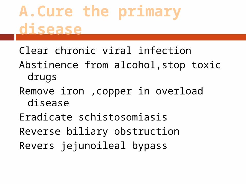
A.Cure the primary diseaseClear chronic viral infectionAbstinence from alcohol,stop toxic drugsRemove iron ,copper in overload diseaseEradicate schistosomiasisReverse biliary obstructionRevers jejunoileal bypass


Nonalcoholic steatohepatitis NASH
NASH is a disorder diagnosed by liver biopsy. The biopsy findings are indistinguishable from those of alcoholic hepatitis described above but the patient lacks a history of significant alcohol consumption. The liver disease is stable in most patients but a minority progress to cirrhosis

CIRRHOSIS & PORTAL HYPERTENSION
SURGICAL COMPLICATIONS
Turner Lisle
Resident Teaching Conference
May 2008

Cirrhosis
End result of anything that causes hepatocellular
injury Toxins, viruses, prolonged cholestasis,
autoimmunity, metabolic disorders
Pathologic response is uniform Hepatocellular necrosis Fibrosis Nodular regeneration

Cirrhosis - why do we care
Hepatic failure Portal hypertension - variceal bleeding
Ischemia & autoimmune factors thoughtto play a role, although mechanism is notclear

Anatomy
Dual blood supply Hepatic arterial Portal venous
Total hepatic blood flow ~1500 mL/min or 25% of cardiac output
Portal vein 2/3 of total hepatic blood flow
Hepatic artery >1/2 of O2

Portal Hypertension
Usually due to increased portal venous resistance
Classification based on site of increased resistance Prehepatic Intrahepatic

Prehepatic Portal HTN
Portal vein thrombosis most common Isolated splenic vein thrombosis
Usually caused by pancreatic inflammation or neoplasm
Results in gastrosplenic HTN with ensuing formation of gastric varices
Normal superior mesenteric & portal vein pressure
Easily reversed by splenectomy alone

Intrahepatic Portal HTN
Often a combination of pre-, intra-, or post-sinusoidal
Presinusoidal Schistosomiasis Nonalcoholic cirrhosis
Sinusoidal EtOH
Postsinusoidal - overall rare EtOH Budd-Chiari syndrome (hepatic vein thrombosis) Constrictive pericarditis Heart failure

Portal Hypertension
Portal pressure > 5 mmHg Portal pressure > 8 mmHg needed for
collateralization

Collateralization

Evaluation of the Cirrhotic
1) Dx of underlying liver disease2) Estimation of fxn hepatic reserve3) Definition of portal venous anatomy4) Hepatic hemodynamic evaluation5) If present, identification of site of UGI
bleeding
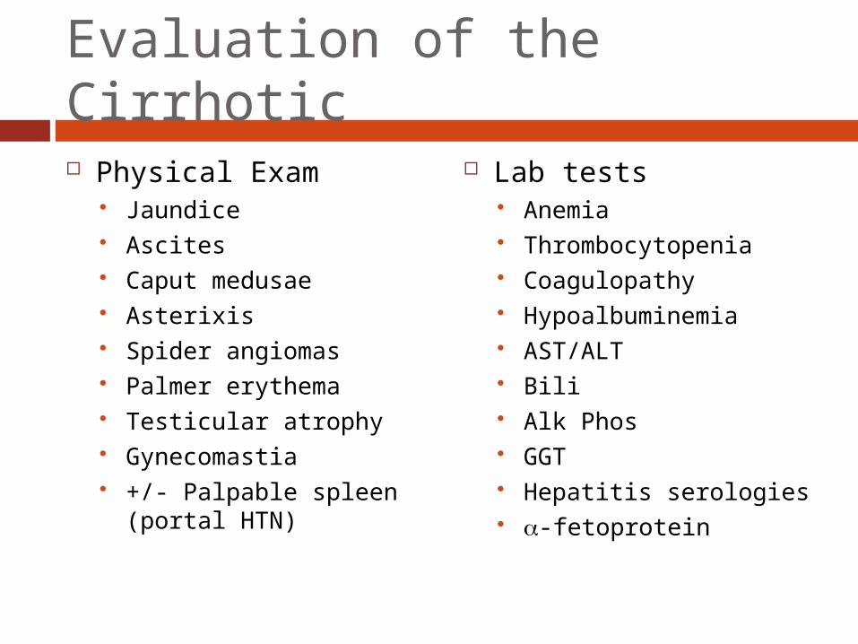
Evaluation of the Cirrhotic
Physical Exam Jaundice Ascites Caput medusae Asterixis Spider angiomas Palmer erythema Testicular atrophy Gynecomastia +/- Palpable spleen
(portal HTN)
Lab tests Anemia Thrombocytopenia Coagulopathy Hypoalbuminemia AST/ALT Bili Alk Phos GGT Hepatitis serologies -fetoprotein

Liver biopsy
Cause of cirrhosis Activity of liver disease Approaches
Percutaneous (avoid with ascites or INR) Transjugular venous Laparoscopic Open

Hepatic Functional Reserve
CHILD-PUGH Encephalopathy grade Amount of ascites Bilirubin Albumin PT (INR)
Operative mortality A (5%) B (10-15%) C (25%)
Subjective……
MELD Bilirubin INR Creatinine Cr Max = 4.0 mg/dL On dialysis → calculate
with CR = 4.0 mg/dL
Calculators on the web

MELD 5-201% increase in mortality with each integer rise inMELD score
MELD >20Additional 2% increase in mortality with each integerrise in MELD score

Diagnosis of bleeding
NGT decompression & lavage Endoscopy - key procedure for UGIB UGIB with portal HTN
90% due to varices 10% other causes (Mallory-Weiss tears, ulcers,
etc) Majority are esophagogastric varices Isolated gastric varices
Likely splenic vein thrombosis
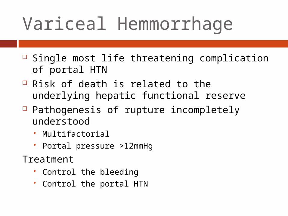
Variceal Hemmorrhage
Single most life threatening complication of portal HTN
Risk of death is related to the underlying hepatic functional reserve
Pathogenesis of rupture incompletely understood Multifactorial Portal pressure >12mmHg
Treatment Control the bleeding Control the portal HTN

Treatment of acute variceal bleeding
Emergency tx should be non-operative Resuscitation first Endoscopy
Successful in >85% Pharmacotherapy
Vasopressin +/- NTG Somatostatin/Octreotide (fewer complications)
Balloon tamponade (rare) TIPS

Endoscopic treatment
Most common treatment modality for acute bleeding as well as prevention
Sclerosis Ligation Complications
Esophageal ulceration Perforation Worsening hemorrhage Aspiration
Failure after two unsuccessful attempts
•Equally efficacious (80-90%)

•Potentially lethal complications
•as pharmacotherapy and endoscopy
•Has been shown to be as effective
•-esophageal perforation•-ischemic necrosis of esophagus
•May be life saving
•Deflation followed by
•high re-bleeding rate
•Sengstaken-Blakemore tube

TIPS
Portal decompression without surgery (nonselective shunt)
Not recommended as initial therapy for acute variceal bleeding
Preferred tx when pharmacotherapy and endoscopic means have failed
Best when used as a bridge to transplant Problems include shunt stenosis/occlusion,
encephalopathy (50% at 1-year)
•Transjugular Intrahepatic Portosystemic Shunt

Emergency surgery Indications
Failure of acute endoscopic tx Failure of TIPS placement Hemorrhage from gastric varices
Esophageal transection rapid/simple Portacaval shunt or splenorenal shunt Operative mortality >25% in most series

Prevention of recurrent hemorrhage
Likelihood of repeat episodes >70% Goals
Prevention of recurrent bleeding Maintenance of satisfactory hepatic function
Options Pharmacotherapy (-blockers +/- nitrates) Chronic endoscopy (successful in 2/3) TIPS (less bleeding, more encephalopathy) Operative shunts Transplantation

Portosystemic shunts
Most effective means of preventing recurrent bleeding in patients with portal hypertension
Problems Encephalopathy Accelerated hepatic failure
Types Nonselective - End-to-end/side portocaval Selective - Distal splenorenal Partial - Small diameter PTFE

Nonshunt procedures
Objectives Ablation of varices Extensive interruption of collateral vessels
Options Transection and reanastamosis of the distal
esophagus Extensive esophagogastric devascularization +
esophageal transection and splenectomy
Rebleeding rates similar to endoscopicexperience


Ascites Usually an indicator of advanced cirrhosis Initiated by altered hepatic and splanchnic
hemodynamics Intravascular volume deficit resulting in
increased aldosterone….. Central goal is to achieve a NEGATIVE
sodium balance

Ascites treatment
Dietary restriction Dietary restriction + Diuretics
spironolactone +/- lasix 5-10% are refractory
Intermittent paracentesis TIPS Peritoneovenous “Denver” shunt (rarely used)
SBP PMN >250/mm3 or positive culture
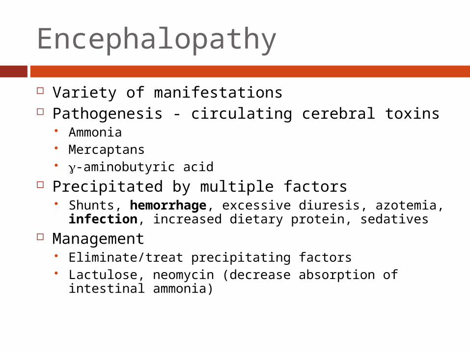
Encephalopathy
Variety of manifestations Pathogenesis - circulating cerebral toxins
Ammonia Mercaptans -aminobutyric acid
Precipitated by multiple factors Shunts, hemorrhage, excessive diuresis, azotemia,
infection, increased dietary protein, sedatives Management
Eliminate/treat precipitating factors Lactulose, neomycin (decrease absorption of intestinal
ammonia)
Questions
