citrus canker ref 10
TRANSCRIPT

J. Appl. Hort.,5(1):52-60, January-June, 2003
Citrus canker – A review
A.K. Das
National Research Centre for Citrus, Amravati Road, PO Box 464, Nagpur-440 010, Maharashtra, India.
AbstractOf all the agricultural pests and diseases that threaten citrus crops, citrus canker is one of the most devastating. The disease,caused by the bacterium Xanthomonas axonopodis pv. citri, occurs in large areas of the world's citrus growing countries includingIndia. At least 3 distinct forms or types of citrus canker are recognized. Among these, Asiatic form (Canker A) is the most destructiveand affects most of the major citrus cultivars. Severe infection of the disease produces a variety of effects including defoliation,dieback, severely blemished fruit, reduced fruit quality and premature fruit drop. Warm, humid, cloudy climate, along with heavyrainfall and strong wind promotes the disease. Control of canker in countries or regions where the disease is not present includequarantine or regulatory programme to prohibit introduction of infected citrus plant material and fruit, as well as continuous and strictsurveying in the field and the immediate destruction of infected trees. In countries where canker is present, integrated systems ofcompatible cultural practices and phytosanitary measures consisting of resistant hosts, removal of inoculum sources, properlydesigned windbreak systems, timely application of protective copper-containing and/or antibiotic sprays are generally the mosteffective means of disease management. This paper reviews the current state of knowledge and understanding on pathogens andstrains associated with the disease and their identification, host-pathogen interaction, molecular mechanism of pathogenicity,epidemiological aspects and management practices.
Key words : Citrus canker, Xanthomonas axonopodis pv. citri, pathogen, strain, epidemiology, disease management
States region of USA in 1915. The Gulf States outbreak is believedto have resulted from a shipment of infected nursery stock fromAsia (Dopson, 1964). The disease also appeared earlier this centuryin South America (Rossetti, 1977), South Africa (Doidge, 1916)and Australia (Garnsey et al, 1979). The disease was reportedlyeliminated in these countries as well as the Gulf States throughnursery and orchard inspections, quarantines, and the on-siteburning of infected trees. Subsequent epidemics have occurred inArgentina, Australia, Brazil, Oman, Saudi Arabia, Reunion Island,the USA, and Uruguay. In some locations, eradication efforts havebeen attempted and failed. In others, active eradication campaignscontinue (Florida, Uruguay, Brazil) (Schubert and Miller, 2000).
In India, citrus occupies third position among fruits after mangoand banana and canker is one of the major constraints of itscultivation. Citrus canker was first reported from Punjab (Luthraand Sattar, 1942; Bedi, 1961). Its occurrence was further recordedin Tamil Nadu (Ramakrishnan, 1954) , Andhra Pradesh (GovindaRao,1954), Karnataka (Venkatakrishnaiah,1957; Aiyappa,1958),Rajasthan (Prasad,1959), Madhya Pradesh (Parsai,1959), Assam(Chowdhury,1951) and Uttar Pradesh (Nirvan, 1960). Several othershave reported the incidence of canker on the acid lime and othervarieties of citrus. Further, the disease appear as a serious problemwhereever acid lime (C. aurantifolia) is grown on a large andcommercial scale (e.g., Akola region in central India, Nellore andPeriyakulum regions in southern India and Khera region of westernIndia) and has become a permanent major problem to the citrusgrowers of this country. Recently canker has been detected in kinnowmandarin nursery in the state of Punjab (Anonymous, 2000).
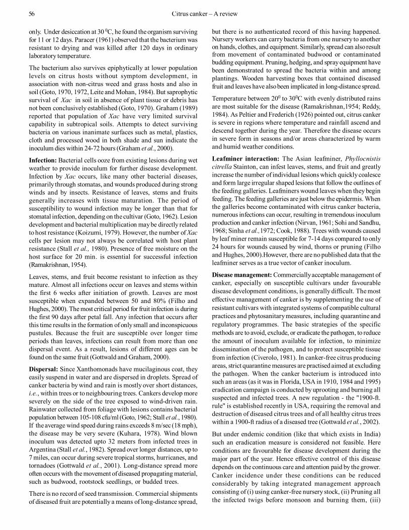
Distribution and economic importance: In spite of the heightenedregulations imposed by many countries to prevent introduction,
Citrus canker is one of the most feared of citrus diseases, affectingall types of important citrus crops. The disease causes extensivedamage to citrus and severity of this infection varies with differentspecies and varieties and the prevailing climatic conditions. Thedisease is endemic in India, Japan and other South- East Asiancountries, from where it has spread to all other citrus producingcontinents except Europe. Generally canker does not occur inarid citrus growing areas and has been eradicated from someareas. However, widespread occurrence of the disease in manyareas is a continuous threat to citriculture especially in canker-free areas. Intensive research on citrus canker is being carriedout throughout the world which has been reviewed by Rossetti(1977), Civerolo (1981,1984), Chand and Pal (1982), Schoulties etal. (1987), Stall and Civerolo (1991) and Goto (1992). However, allthese reviews are either brief, restricted to one country, or bynow out of date. This review aims to present an overview ofcitrus canker worldwide with special reference to India.
Origin and history: The geographical origin of citrus canker is amatter of controversy. Lee (1918) reported that it may have arisenin southern China, and he assumed Fortunella hindsii to be thewild host plant. However, Fawcett and Jenkins (1933) reportedthat citrus canker originated in India and Java, rather than inother regions of the Orient, because they detected canker lesionson the oldest citrus herbaria kept at the Herbaria of the RoyalBotanic Gardens in Kew, England (i.e., Citrus medica collectedfrom India in 1827-1831 and C.aurantifolia from Indonesia in1842-1844). These findings suggest the origin of disease in thetropical areas of Asia, such as South China, Indonesia, and India,where Citrus species are presumed to have originated and tohave been distributed to other citrus- growing areas in the formof budwood. Citrus canker was described afterwards in the Gulf

the disease continues to increase its geographic range. Citruscanker presently occurs in over thirty countries in Asia, the Pacificand Indian Ocean islands, South America, and the SoutheasternUSA (Fig. 1).
The economic importance of citrus canker can be analyzed fromseveral different points of view. Loss assessment has not beendetermined clearly, as in the case of diseases of annual crops.When citrus infection occurs in the early growing stage, thefruits crack or become malformed as they grow, and the heavilyinfected ones fall prematurely. Light infection in later growthstages may cause only scattered canker lesions on the surface offruits but makes fresh fruits unacceptable for market. The severityof fruit infection usually parallels that of foliage infection. Eightyto ninety percent of fruit infection is not uncommon in susceptiblecitrus trees that have already sustained severe foliage infection.Such heavy foliage infection often causes severe defoliation,leaving only bare twigs (Goto,1992). In Argentina, for example,83-97% of the fruit of grapefruit trees were diseased in unsprayedplots during 1979-1980 and in the same plots, upto 88% of theleaves were infected (Stall and Seymour, 1983).
Worldwide, millions of dollars are spent annually on prevention,quarantines, eradication programs, and disease control.Undoubtedly, the most serious consequence of citrus cankerinfestation is the impact on commerce resulting from restrictionsto interstate and international transport and sale of fruit originatingfrom infested areas. The disease has been studied in greaterdetail in the U.S. where it caused very serious damage, so muchso that millions of canker affected trees were cut and burnt. InFlorida, for example, during the year 1915-33, nearly 2,57,000orchard trees and 3,000,000 nursery plants were destroyed at acost of over $ 6 million and again during the year 1984-86, nearly20 million citrus nursery plants were destroyed at a cost of over$ 25 million (Schoulties et al., 1987). Presently over $ 12 millionper year and over 600 personnel are dedicated to this programme.
and severe form of the disease. The disease is endemic throughoutIndia, Pakistan, the islands of Indian Ocean, South-East Asia,China and Japan. Cancrosis B (canker B or false canker), causedby X. axonopodis pv. aurantifolii (Hasse) Gabriel Vauterin is aserious problem on lemons in Argentina, Paraguay and Uraguay.Mexican lime, sour orange, and pummelo are also susceptible.Cancrosis B causes canker-type lesions on fruit, leaves, and twigsthat are similar to but smaller than those produced by the A form.In culture, cancrosis B bacteria grow more slowly than canker Abacteria on nutrient agar, and a specific medium containingsucrose, peptone, salts, and purified agar has been developedfor this form. Cancrosis B isolates can be differentiatedserologically from the canker A bacteria but not from CancrosisC isolates. Cancrosis C, also caused by X. axonopodis pv.aurantifolii, has been isolated from Mexican lime in Brazil.Symptoms are the same as those of canker A. In 1984, a newxanthomonad disease of citrus was discovered in Florida nurseries.The causal bacterium is shown to have no relationship to theexisting two pathovars of Xanthomonas axonopodis (causingcanker A, B and C) and named as Xanthomonas axonopodis pv.citrumelo (Hasse) Gabriel Vauterin (earlier called group E canker orcanker E). The disease is most commonly referred to as citrusbacterial spot (CBS). At present CBS is only known from Florida,where it appears to be restricted entirely to nurseries (Gottwaldand Graham, 2000). The differential characteristics of the threeforms of citrus canker and CBS are given in Table 1.
Other forms of citrus canker have also been reported. For example,canker D, sometimes called citrus bacteriosis, was reported in theColima area of Mexico in 1980s (Rodriquez et al.,1985) but later itwas found to be caused by Alternaria limicola. An isolate ofXanthomonas was discovered in Oman in 1986 that producedcanker A-like lesions only on Mexican lime. Similar isolates(known as A*) have been found in Saudi Arabia, Iran, and India(Verniere et al.,1998). Another atypical form of canker A bacteria,
Table 1. Comparison of three different forms of citrus canker and citrus bacterial spot (CBS) of citrusCharacteristics Citrus Canker Citrus bacterial spot (CBS)Canker form A B CPathogen X. axonopodis pv. citri X. axonopodis pv. aurantifolii X. axonopodis pv.aurantifolii X. axonopodis pv. citrumeloDistribution Asia, Africa,South Argentina, Paraguay, Uruguay Brazil, Mexico America (Florida)
AmericaOceaniaHost range Wide Limited Limited WideMajor host plant Citrus spp. Lemon Mexican lime Citrus spp. (nursery)Symptoms Spongy erupted at first; corky rough lesions with a raised, greasy margin Flat or sunken lesion; extreme
later; water-soaked appearance water soakingModified from Goto (1992)
In spite of this effort, the disease continues to spread in theMiami area of Florida,USA (Schubert and Miller, 2000) andhence some researchers, growers and residents are disputingthe concept and feasibility of eradication.
Forms: There are three different forms of citrus canker diseasecaused by various pathovars and variants of the bacteriumXanthomonas axonopodis Starr and Garces emend. Vauterinet al.(1995). Differentiation of these forms is mainly based ongeographical distribution and host range of the pathogen(Stall and Seymour, 1983). The Asiatic form of canker (cankerA, cancrosis A or true canker), caused by X. axonopodis pv.citri (Hasse) Vauterin (Xac) is the most common, widespread
Fig. 1. Distribution map of citrus canker (Xanthomonas axonopodis pv. citri)
Citrus canker – A review 53

which has high levels of resistance to penicillin related antibiotics,has been described from Reunion and surrounding islands in theIndian Ocean (Gottwald and Graham, 2000).
Symptoms: The diseased plants are characterized by theoccurrence of conspicuous raised necrotic lesions that developon leaves, twigs and fruits. Lesions can be detected by drawingthe fingers over the surface of infected tissues. On leaves, firstappearance is as oily looking, 2-10 mm circular spots, usually onthe abaxial surface (reflecting stomatal entry following raindispersal). Lesions are often similarly sized. Later, both epidermalsurfaces may become ruptured by tissue hyperplasia induced bythe pathogen. On leaves, stems, thorns and fruit, circular lesionsbecome raised and blister-like, growing into white or yellowspongy pustules. These pustules then darken and thicken into alight tan to brown corky canker, which is rough to the touch.Often a water- soaked margin develops around the necrotic tissueand is easily viewed with transmitted light. On stems, pustulesmay coalesce to split the epidermis along the stem length, andoccasionally girdling of young stems may occur. Older lesionson leaves and fruit tend to have more elevated margins and are attimes surrounded by a yellow chlorotic halo (that may disappearas canker lesions age) and a sunken center. Sunken centers areespecially noticeable on fruits, but the lesions do not penetratefar into the rind thereby not affecting internal quality. Severeinfection results in defoliation, die-back, deformation of fruit andpremature fruit drop (Rossetti, 1977; Civerolo, 1981; Chand andPal, 1982; Stall and Seymour, 1983). Canker causes fruit lossesranging from premature fruit drop due to abscission to nonmarketable quality due to lesions. Disease of the fruit is probablythe most economically important damage since fruits with cankerlesion are not acceptable for fresh market and fetch very littleprice.
An essential diagnostic symptom of the disease is citrus tissuehyperplasia (excessive mitotic cell divisions), resulting in cankers(Gabriel et al., 2000). Canker symptoms on leaves and fruit can bereadily obtained by artificial inoculations. If cankers are notpresent on leaves, stems and fruit of mature trees, or if leaves andfruit of susceptible Citrus species do not develop cankersfollowing artificial inoculation, a diagnosis of citrus canker is notindicated. Occurrence of lesions is seasonal, coinciding with
periods of heavy rainfall, high temperatures and growth flushes.
Host range and varietal susceptibility: Civerolo (1984) lists anumber of plants in the family rutaceae other than Citrus andPoncirus that can serve as hosts of Xac under experimentalconditions or heavy disease pressure in nature. Amongcommercial citrus varieties and rootstocks, Asiatic citrus cankeris most severe on grapefruit (C. paradisi), limes (C. aurantifolia,C. limettioides), trifoliate orange (Poncirus trifoliata) and theirhybrids because of their high susceptibility (Table 2).
In India, citrus canker is reported to be relatively more on acidlime and less commonly on mandarin and sweet orange(Ramakrishnan, 1954). According to Aiyappa (1958) all thecultivated varieties of citrus and some wild species in Karnatakaare suspectible to canker possibly due to heavy rainfall, highhumidity and low temperature. Prasad (1959) from Rajasthan madesimilar observations. The descending order of susceptibility incitrus species is Kaghzi Lime, grape fruit, Karnakhata and sweetoranges (Nirvan, 1961). Mandarins and lemons are resistant andKumquats are commercially immune under conditions existing inUttar Pradesh. Jain (1959) reported that different varieties of sweetlime, grape fruit and sweet orange were infected almost to sameextent in Himachal Pradesh. According to Naik (1949) acid limes,some varieties of lemon, sweet orange and grapefruit were verysusceptible to canker, while Nepali oblong and round seedlesslemons were highly resistant. Mundkur (1961) observed no infectionin sweet orange and pummelo but Jambheri, sour orange and Kaghzilime were very susceptible.
Host- pathogen interaction: Citrus canker research has beenprimarily oriented toward the ecological behaviour of the causalbacterium. Studies from physiological and biochemical standpointare therefore very limited.
Xac produces abundant extracellular polysaccharides (EPS), bothin culture media and in host tissues. The bacterial cells in cankerlesions are embedded in a dense matrix of EPS and are dispersed,together with EPS, by rain splash. The EPS molecules exhibitgreat protective effects against the 'dilution effect' in water anddesiccation in air, providing benefits for the bacterial ecology(Goto, 1985). After entering the intercellular space (throughstomata or wounds) they adhere to the host cell walls through aninteraction between bacterial EPS and citrus agglutinins
Table 2. Susceptibility of several citrus varieties and rootstocks to Xanthomonasaxonopodis pv. citriHighly Susceptible Moderately SusceptibleCitrus paradisi Macf., grapefruit C. sinensis (L.) Osbeck, sweet orangeC. aurantifolia (Christ.) Swingle, acid lime C. aurantium L., sour orangeC. limettioides Tan., Palestine sweet lime C. limon (L.) Burm., lemonPoncirus trifoliata (L.) Raf., trifoliate orange C. tangelo J. Ingram & H.E. Moore,
tangeloModerately Resistant Highly ResistantC. reticulata Blanco, mandarin, tangerine C. medica L., citronC. maxima (Burm.) Merr., pummelo Citrofortunella microcarpa (Bunge)
Wijnands, calamondinC. aurantifolia (Christ.) Swingle, Person Fortunella spp., kumquator Tahiti limeRecently, it was reported that goat weed (Ageratum conyzoides L.) could serve as a hostof Xac. This plant is common in citrus orchards in the state of Assam in India (Kalita et al.1997). This represents the only report of a non-Rutaceous host of Xac.
(Takahashi and Doke, 1984). Ethylene production bycitrus leaves inoculated with Xac and increasedconcentration of indole acetic acid (IAA) in the Xacinoculated leaves have also been reported (Goto etal., 1979a).
Padmanabhan et al. (1973) studied the physiologyof canker infected citrus leaves with special referenceto halo formation, and reported that halo zonerespired more than the cankered tissue. Catalaseactivity was very high in the halo region. Bothperoxidase and ascorbic acid-oxidase activityincreased in canker as well as in halo regions. Theyagain recorded a descrease in chlorophyll a, b,carotene and xanthophyll contents in the canker, haloand pre-halo regions of the citrus leaves infected bycanker-inducing bacterium. Photosynthesis wasimpaired in the infected regions while starch contentwas not affected in the halo regions (Padmanabhan
54 Citrus canker – A review

et al., 1974). Total sugar content decreased in all the infectedregions. Kishore and Chand (1972, 1975) carried out biochemicalanalysis of healthy and canker infected leaves and reported thatamino acid content decreased in infected leaves. They also noticedmore total phenols in resistant C. reticulata than in susceptibleC. aurantifolia.
Pathogen biology
Pathogens and strains: Based on currently available information,at least three pathovars (sometimes called strains) ofXanthomonas axonopodis have been recognized. Thesepathovars are distinguished from one another by geographicaldistribution and by different pathogenicity to members of genusCitrus.The pathogen for canker A was first identified anddescribed as Pseudomonas citri by Hasse (1915). Bacterialnomenclature has undergone many changes since then and thecausal bacterium is now known as Xanthomonas axonopodispv. citri (Hasse) Vauterin [Syns. X. citri (Hasse) Dowson and X.campestris pv. citri (Hasse) Dye] (Dye et al., 1980; Vauterin etal.,1995). The pathogen for canker B and C and other relatedstrains associated with the disease have already been discussed(see Forms).
The bacterium (Xac) is rod-shaped measuring 1.5-2.0 x 0.5-0.75µm, Gram-negative, and has a single polar flagellum. Growth isobligately aerobic. Colonies on culture media are usually yellowas a result of xanthomonadin pigment production. When glucoseor other sugars are added to the culture medium, colonies becomevery mucoid due to the production of an extracellularpolysaccaride slime. The optimum temperature range for growthis 28 to 30° C (82 to 860 F), and the maximum temperature range forgrowth is 35 to 390 C (95 to 1020 F). Bacterial cells are positive forhydrolysis of starch, aesculin, casein, liquefaction of gelatin,and production of tyrosinase, catalase, reducing substance fromsucrose, and hydrogen sulfide. The bacterium is negative fornitrate reduction, indole production and for methyl red test (Chandand Pal, 1982; Goto, 1992).
Goto (1969) in Japan, differentiated 300 isolates of X. citri into 5strains by their ability to oxidise mannitol and lactose, and byrapidity of breakdown of mannose. In Argentina, two biotypeswere distinguished among 65 isolates of Xac based on growthon media with carbohydrates, acid production in litmus milk andcolony appearance in wakimoto’s medium (Falico de Alcaraz,1980). Goto et al. (1980) distingushed canker A strain from the Bstrain by bactoriophage sensitivity test. A strains are susceptibleto lysis by phage CP 1 or CP 2 while B strains are susceptible tolysis by CP 3. Civerolo and Fan (1982) successfully employedELISA to identify the different strains of Xac. Alverez et al. (1991)produced monoclonal antibodies for A, B and C-form pathogensand noticed that canker A MAb did not react with strainsassociated with other forms of citrus canker (B,C).
In India, occurrence of strains (pathotypes) of the pathogen hasbeen reported by Rangaswami and Soumini (1957) and Hamlin(1967). Khan and Hingorani (1970) grouped 15 isolates of thepathogens into 3 strains by their reaction on Murraya exotica.Kishore and Chand (1972) studied the reaction of eight isolateson C. aurantifolia, C. sinensis and C. jambhiri and showed thepresence of more than one strain of the pathogens in Harayana.Similarly Prasad et al. (1978) and Buragohain and Chand (1991)
also observed strain variation in Xac. Recently Das (2002) reportedthe existence of pathogenic variability within the 'A' strain ofXac.
Pathogen and strain identification: Because symptoms aregenerally similar, identification and separation of cankerpathogens and strains are based on cultural and physiologicalcharacteristics (Schaad,1988), bacteriophage sensitivity (Goto etal., 1980; Civerolo, 1984), serology (Alvarez et al., 1991), plasmidfingerprints (Pruvost et al., 1992), DNA- DNA homology (Egel etal.,1991) and by various RFLP (restriction fragment lengthpolymorphism) and PCR (polymerase chain reaction) analyses(Gabriel et al., 1988; Hartung and Civerolo, 1989; Gillings et al.,1995; Hartung et al., 1996; Miyoshi et al., 1998; Cubero andGraham, 2002). When the DNA-based assays are unavailable,strains of Xac can be distinguished from other pathovars byinfecting a panel of susceptible and resistant citrus hosts or as abioassay on detached-leaves or leaf-disks (Gottwald et al., 1993).Such pathogenecity test is an essential component in diagnosticprogrammes for regulation of citrus canker diseases (Schubert etal., 2001).
Pathogenecity: Identical symptoms induced by twotaxonomically distinct groups of strains (canker A and B) areindicative of a common pathogenicity factor. Gene pthA isessential for Xac to elicit cankers on citrus, and pthA confers thisability to various X. axonopodis strains (for example, pathovarsalfalfae and citrumelo) ( Swarup et al.,1991; Swarup et al.,1992).Functionally homologous genes (pthB and pthC) have also beenidentified and cloned from X. axonopodis pv. aurantifoliipathotype B and pathotype C, respectively (Gabriel et al., 2000).Both pthB and pthC are essential for X. axonopodis pv. aurantifoliipathotypes B and C, respectively, to cause cankers on citrus,and pthB and pthC confer this ability to various X. axonopodisstrains. All three genes are therefore functionally interchangeable,and these genes may have been transferred horizontally onplasmids between Xac and X. axonopodis pv. aurantifolii strains.Genes pthA, pthB and pthC are all members of an avirulence /pathogenicity gene family widely distributed in the genusXanthomonas (Swarup et al.,1992; De Feyter et al.,1993). GenespthA, pthB and pthC, when transferred into Xac, X. axonopodispv. aurantifolii or X. axonopodis pv. citrumelo, confer ability toelicit hyperplasia (cell divisions or cankers) on all citrus speciesin the normal host range of the recipient strain. Mutations ofgenes encoding either the protein injection system of thepathogen (a type III secretion system encoded by hrp genes) orthe effector molecule, pth A/B/C, abolish pathogenicity of cankerbacteria (Gabriel et al., 2000).
Disease cycle and epidemiology
Survival: Xac survives primarily in naturally occurring lesions.Cankerous leaves, twigs and branches constitute the main sourceof inoculum. Since affected leaves drop early, they may not serveas the main source of inoculum (Nirvan, 1963), but Rao andHingorani (1963) found that the bacterium survives upto 6 monthsin the infected leaves. The disease is carried from season toseason mainly in the cankers on twigs and branches. Thepathogen can survive in diseased twigs upto 76 months(Chakravarti et al, 1966). Vasudeva (1958) found that the organismsurvived in the infected leaves for more than six months, in thesterilized soils for 52 days and in the unsterilized soils for 9 days
Citrus canker – A review 55
only. Under desiccation at 30 0C, he found the organism survivingfor 11 or 12 days. Paracer (1961) observed that the bacterium wasresistant to drying and was killed after 120 days in ordinarylaboratory temperature.
The bacterium also survives epiphytically at lower populationlevels on citrus hosts without symptom development, inassociation with non-citrus weed and grass hosts and also insoil (Goto, 1970, 1972, Leite and Mohan, 1984). But saprophyticsurvival of Xac in soil in absence of plant tissue or debris hasnot been conclusively established (Goto, 1970). Graham (1989)reported that population of Xac have very limited survivalcapability in subtropical soils. Attempts to detect survivingbacteria on various inanimate surfaces such as metal, plastics,cloth and processed wood in both shade and sun indicate theinoculum dies within 24-72 hours (Graham et al., 2000).
Infection: Bacterial cells ooze from existing lesions during wetweather to provide inoculum for further disease development.Infection by Xac occurs, like many other bacterial diseases,primarily through stomatas, and wounds produced during strongwinds and by insects. Resistance of leaves, stems and fruitsgenerally increases with tissue maturation. The period ofsusceptibility to wound infection may be longer than that forstomatal infection, depending on the cultivar (Goto, 1962). Lesiondevelopment and bacterial multiplication may be directly relatedto host resistance (Koizumi, 1979). However, the number of Xaccells per lesion may not always be correlated with host plantresistance (Stall et al., 1980). Presence of free moisture on thehost surface for 20 min. is essential for successful infection(Ramakrishnan, 1954).
Leaves, stems, and fruit become resistant to infection as theymature. Almost all infections occur on leaves and stems withinthe first 6 weeks after initiation of growth. Leaves are mostsusceptible when expanded between 50 and 80% (Filho andHughes, 2000). The most critical period for fruit infection is duringthe first 90 days after petal fall. Any infection that occurs afterthis time results in the formation of only small and inconspicuouspustules. Because the fruit are susceptible over longer timeperiods than leaves, infections can result from more than onedispersal event. As a result, lesions of different ages can befound on the same fruit (Gottwald and Graham, 2000).
Dispersal: Since Xanthomonads have mucilaginous coat, theyeasily suspend in water and are dispersed in droplets. Spread ofcanker bacteria by wind and rain is mostly over short distances,i.e., within trees or to neighbouring trees. Cankers develop moreseverely on the side of the tree exposed to wind-driven rain.Rainwater collected from foliage with lesions contains bacterialpopulation between 105-108 cfu/ml (Goto, 1962; Stall et al., 1980).If the average wind speed during rains exceeds 8 m/sec (18 mph),the disease may be very severe (Kuhara, 1978). Wind blowninoculum was detected upto 32 meters from infected trees inArgentina (Stall et al., 1982). Spread over longer distances, up to7 miles, can occur during severe tropical storms, hurricanes, andtornadoes (Gottwald et al., 2001). Long-distance spread moreoften occurs with the movement of diseased propagating material,such as budwood, rootstock seedlings, or budded trees.
There is no record of seed transmission. Commercial shipmentsof diseased fruit are potentially a means of long-distance spread,
but there is no authenticated record of this having happened.Nursery workers can carry bacteria from one nursery to anotheron hands, clothes, and equipment. Similarly, spread can also resultfrom movement of contaminated budwood or contaminatedbudding equipment. Pruning, hedging, and spray equipment havebeen demonstrated to spread the bacteria within and amongplantings. Wooden harvesting boxes that contained diseasedfruit and leaves have also been implicated in long-distance spread.
Temperature between 200 to 300C with evenly distributed rainsare most suitable for the disease (Ramakrishnan,1954; Reddy,1984). As Peltier and Frederich (1926) pointed out, citrus cankeris severe in regions where temperature and rainfall ascend anddescend together during the year. Therefore the disease occursin severe form in seasons and/or areas characterized by warmand humid weather conditions.
Leafminer interaction: The Asian leafminer, Phyllocnistiscitrella Stainton, can infest leaves, stems, and fruit and greatlyincrease the number of individual lesions which quickly coalesceand form large irregular shaped lesions that follow the outlines ofthe feeding galleries. Leafminers wound leaves when they beginfeeding. The feeding galleries are just below the epidermis. Whenthe galleries become contaminated with citrus canker bacteria,numerous infections can occur, resulting in tremendous inoculumproduction and canker infection (Nirvan, 1961; Sohi and Sandhu,1968; Sinha et al.,1972; Cook, 1988). Trees with wounds causedby leaf miner remain susceptible for 7-14 days compared to only24 hours for wounds caused by wind, thorns or pruning (Filhoand Hughes, 2000).However, there are no published data that theleafminer serves as a true vector of canker inoculum.
Disease management: Commercially acceptable management ofcanker, especially on susceptible cultivars under favourabledisease development conditions, is generally difficult. The mosteffective management of canker is by supplementing the use ofresistant cultivars with integrated systems of compatible culturalpractices and phytosanitary measures, including quarantine andregulatory programmes. The basic strategies of the specificmethods are to avoid, exclude, or eradicate the pathogen, to reducethe amount of inoculum available for infection, to minimizedissemination of the pathogen, and to protect susceptible tissuefrom infection (Civerolo, 1981). In canker-free citrus producingareas, strict quarantine measures are practised aimed at excludingthe pathogen. When the canker bacterium is introduced intosuch an areas (as it was in Florida, USA in 1910, 1984 and 1995)eradication campaign is conducted by uprooting and burning allsuspected and infected trees. A new regulation - the "1900-ft.rule" is established recently in USA, requiring the removal anddestruction of diseased citrus trees and of all healthy citrus treeswithin a 1900-ft radius of a diseased tree (Gottwald et al., 2002).
But under endemic condition (like that which exists in India)such an eradication measure is considered not feasible. Hereconditions are favourable for disease development during themajor part of the year. Hence effective control of this diseasedepends on the continuous care and attention paid by the grower.Canker incidence under these conditions can be reducedconsiderably by taking integrated management approachconsisting of (i) using canker-free nursery stock, (ii) Pruning allthe infected twigs before monsoon and burning them, (iii)
56 Citrus canker – A review

periodical spraying of suitable copper-based bactericides (toreduce inoculum build-up on new flushes and to protectexpanding fruit surfaces from infection) alongwith an insecticide(to control insect injury), (iv) taking some precautions to reducethe risk of spread of disease in orchards and nurseries and (v) byevolving canker-resistant varieties suited to local environmentalconditions (Das and Singh,1999, 2001).
Fawcett (1936), Naik (1949), Cheema et al. (1954), Ramakrishnan(1954), Govinda Rao (1954), Prasad (1959) and Paracer (1961)recommended pruning of infected twigs before the onset ofmonsoon and spraying of 1% Bordeaux mixture at periodicalintervals for an effective control of the disease. Patel and Desai(1970) reported that pruning of affected twigs every year duringNov-Dec and 3 to 4 sprays of Bordeaux mixture (1%) in a yearcould reduce the disease. Two prunings alongwith 4 sprays of5000 ppm copper oxychloride or 1% Bordeaux mixture is reportedto be effective against the disease (Kishun and Chand, 1987).Other chemicals found effective against the canker were perenox(Chowdhury,1951), Ultrasulphur (Nirvan, 1961), mixture of sodiumarsenate and copper sulphate (Patel and Padhya, 1964), Blitoxand nickel chloride (Ram et al.,1972). According to Rangaswamiet al. (1959), 500-1000 ppm streptomycin sulphate was effectivewhen sprayed with 1% glycerine on acid lime. Six sprays of 1000ppm streptomycin sulphate along with two prunings reduced thecanker in acid lime (Balaraman and Purushotman, 1981). Othereffective antibiotics were Agrimycin (Sawant et al., 1985),Streptocycline (Mathur et al., 1973) and Streptocycline incombination with Bordeaux mixture (Krishna and Nema, 1983).Kale et al. (1988), in field trials with 7 different chemicals, foundthat the best control of Xac was given by Paushamycin + Blitoxfollowed by Bordeaux mixture. Application of neem cake solutionon the foliage reduced the canker in nurseries (Dakshinamurthiand Rao, 1959; Reddy and Rao, 1960). Kale et al. (1994) suggestedthat for better control of canker, spraying of streptocycline +Copper oxychloride (0.1%) should preferably be done at 7 daysor 15 days interval. Integrated application of pruning of infectedtwigs, Copper oxychloride (0.3%), streptocycline (100ppm) andneem cake suspension was found very effective in controllingthe disease (Das and Singh, 2000). Canker incidence can also bereduced by periodic spraying of insecticides to control of leafminer damage to newly unfolded leaves, as such damage facilitatescitrus canker infection.
Control measures developed in Japan include windbreaks(Koizumi et al., 1996) or pruning of diseased summer and autumnshoots, forecasting and chemical sprays. Six or seven sprays ofcopper are necessary to protect new growth from infection(Kuhara, 1978). In China control measure consists of sprayingcopper ammonium WC during summer and autumn months(Chen,1998). Gottwald and Timmer (1995) reported the efficacy ofwind- breaks in reducing the spread of citrus canker in Argentina.McGuire (1988) evaluated 13 bactericidal chemicals over 3 seasonson 3 citrus species to determine their ability to control canker. Infield trials conducted in Argentina, he noticed copper ammoniumcarbonate with 8% metallic copper was consistently superior toother products in controling Xac. In another field test on maturegrapefruit trees, three applications per seasons of copperammonium carbonate (CAC) or copper hydroxide + maneb were
observed to reduce lesions numbers on fruit but not on leaves(Timmer, 1988). Where copper resistance was foundrecommendation include addition of mancozeb to the coppersprays (Canteros, 2000). When canker occurred in the USA, theemphasis was on eradication, and other measures for control ofcanker were not adequately researched (Stall and Civerolo,1991).However, recently in Florida, USA, some induced systemicresistance (ISR) compounds (e.g. Messenger, Nutri-phite, Oxycomand FNX-100) are under evaluation for their potential to controlcanker A using citrus bacterial spot on swingle citrumelo as asurrogate pathosystem (Graham et al., 2000).
In India, where canker disease has established since a long periodit was suggested that resistant varieties and species should begrown (Mundkur, 1961). Here canker infestation is relatively moreon acid lime and less common on mandarin and sweet orange.Kumquat (Fortunella spp.) and Hazara Narangi (C. microcarpa)are commonly grown in India for ornamental purpose and theseare found resistant to canker. C. latifolia was also found to beresistant to the disease (Kishun and Chand, 1987). Althoughseveral acid lime selection/clone or hybrids have been claimedeither as resistant or tolerant from different regions e.g. RHR-L-49 (Sai Sarbati) (Desai et al.,1999), Tenali (Madhavi et al., 2000),ALH-77 (lime x lemon hybrid) (Prasad et al., 1997), these need tobe tested through multilocational trials.
Studies on biological control of citrus canker are still in apreliminary stage. Some strains of bacteria viz., Pseudomonassyringae, Erwinia herbicola, Bacillus subtilis and Pseudomonasfluorescence isolated from citrus phylloplane were reported tobe antagonistic in vitro to the canker pathogen (Ota, 1983; Gotoet al., 1979b; Kalita et al., 1996; Unnimalai and Gnanamanickam,1984). However, it seems difficult to find antagonistic bacteriathat reside stably on smooth surfaces of mature citrus leaves.
Future prospects: Citrus canker continues to be the cause ofworldwide concern as a potentially hazardous threat to citriculture.There is a wide range of physiological, biochemical, serological,molecular and pathogenic variation among strains of bacteriaassociated with citrus canker. Moreover new strains areoriginating regularly as a result of mutation. A betterunderstanding of the pathogenic specialization and properidentification of Xac strains are needed. This could be importantalso for breeding new canker resistant cultivars. The developmentof effective chemicals for control of citrus canker has been longclaimed by citrus growers and pathologists. However, theseefforts have actually been unsuccessful, as has been the casewith other plant bacterial diseases in general. Most chemicalswith great effectiveness in vitro do not necessarily showsatisfactory effects. The gaps found between effectiveness invitro and in situ may stem in part from the mode of bacterialinfection. Under rainy conditions, some bacterial cells mayachieve direct access to the front cavity of stomata or to woundswithout being exposed to the protective chemicals left on the leafsurface. Therefore, for development of effective bactericides,emphasis must be placed on the effectiveness of chemicalsreaching at least to the depth of the stomatal cavity. Recent findingshave demonstrated that the plants usually carry the internalresident microbes (endophyte) in vascular systems. There is a
Citrus canker – A review 57

substantial possibility that an antagonistic microbe may be foundamong these endophytes which will be useful in biological controlof citrus canker. Fresh approaches are also to be made to developenvironmentally safe methods to combat this bacterium viz. searchfor its resistance in wild citrus and its relatives in orchards andforests of the endemic areas and application of biotechnology orgenetic engineering utilizing the knowledge on its molecularmechanism of pathogenicity.
ReferencesAiyappa, K.M. 1958. Citrus canker - Xanthomonas citri (Hasse)
Dowson. Mysore Agric. J., 13: 164-167.Anonymous, 2000. Proceedings of the group discussion of the All India
Coordinated Research project and ICAR ad hoc schemes on tropicalfruits. 5-8 Jan 2000,Rahuri. Tech. Doc. No. 72, p. 31.
Alvarez, A.M., A.A. Benedict, C.Y. Mizumoto, L.W. Pollard and E.L.Civerolo, 1991. Analysis of Xanthomonas campestris pv. citri andX.c. citrumelo with monoclonal antibodies. Phytopathology, 81: 857-865.
Balaraman, K. and R. Purushotman, 1981. Control of citrus canker onacid lime. South Indian Hort., 29: 175-177.
Bedi, K.S. 1961. Some important observations on the citrus canker inPunjab. Punjab Hort. J., 2: 89-91.
Buragohain, V.P. and J.N. Chand, 1991. Variation among the isolates ofXanthomonas campestris pv. citri in Haryana. Indian J. Mycol. Pl.Pathol., 21: 106.
Canteros, B.I. 2000. Citrus canker in Argentina - control, eradicationand current management. Proc. Intn. Citrus canker Res. Workshop.June 20-22, 2000, Ft. Pierce, Florida, pp. 10-11.
Chand, J.N. and V. Pal, 1982. Citrus canker in India and its management.In : Problems of citrus diseases in India (S.P. Raychaudhuri and Y.S.Ahlawat, Eds.). Surabhi Printers and Publishers, New Delhi. pp.21-26.
Chakravarti, B.P., S. Porwal and M. Rangarajan, 1966. Studies on citruscanker in Rajasthan. I. Disease incidence and survival of the Pathogen.Labdev J. Sci. Tech., 4: 262-265.
Cheema, G.S., S.S. Bhat and K.C. Naik, 1954. Commercial fruits ofIndia. Macmillan and Co., Bombay, p. 422.
Chen, Zhisheng, 1998. Control of canker of citrus with copper-ammonium WC. J. Zhejiang Fores. College, 15(1): 108-110.
Chowdhury, S. 1951. Citrus Canker in Assam. Pl. Prot. Bull., 3: 78-79.Civerolo, E.L. 1981. Citrus bacterial canker disease : An overview. Proc.
Intn. Soc. Citric., 1: 390-394.Civerolo, E.L. and F. Fan, 1982. Xanthomonas campestris pv. citri
detection and identification by enzyme-linked immunosorbentassay. Plant Dis., 66: 231-226.
Civerolo, E.L. 1984. Bacterial canker disease of citrus. J. Rio GrandeValley Hortic. Soc., 37: 127-146.
Cook, A.A. 1988. Association of citrus canker pustules with leaf minertunnels in North Yemen. Plant Dis., 72: 546.
Cubero, J. and J. H. Graham, 2002. Genetic relationship among worldwidestrains of Xanthomonas causing canker in citrus species and designof new primers for their identification by PCR. Appl. Environ.Microbiol., 68:1257-1264.
Dakshinamurthi, V. and D.K. Rao, 1959. Preliminary studies on the controlof citrus Canker on acid lime. Andhra Agric. J., 6: 145-148.
Das, A.K. and Shyam Singh, 2000. Management of Acid lime canker byusing chemicals with compatible cultural practices. Hi-tech CitrusManagement – Proc. Intn. Symp. Citric., Nov. 23-27, 1999, Nagpur,Maharashtra (S.P. Ghosh and Shyam Singh, Eds.) pp. 1054-1056.
Das, A.K. 2002. Pathogenic variability in Xanthomonas axonopodis pv.citri, causal agent of citrus canker. J. Mycol.Pl. Pathol. (In Press).
Das, A.K. and Shyam Singh, 1999. Management of Bacterial Canker inAcid lime. Intensive Agriculture, 36(11-12): 28-29.
Das, A.K. and Shyam Singh, 2001. Managing citrus bacterial diseases inthe state of Maharashtra. Indian Hort., 46(2): 11-13.
De Feyter, R., Y. Yang and D.W. Gabriel, 1993. Gene-for-genesinteractions between cotton R genes and Xanthomonas campestrispv. malvacearum avr genes. Mol.Plant-Micr. Interact., 6: 225-237.
Desai, V.T., S.A. Ranpise, C.V. Pujari and S.B. Raijadhav, 1999."Saisarbati" promising acid lime cultivar for western Maharashtra.Proc. Natl. Symp. Citric., Nov. 17-19, 1997, Nagpur, Maharashtra.pp. 38-41.
Doidge, E.M. 1916. Citrus canker in South Africa. South African FruitGrower, August issue.
Dopson, R.N. 1964. The eradication of citrus canker. Plant Dis. Reptr.,48: 30-31.
Dye, D.W., J.F. Bradbury, M.Goto, A.C Hayword, R.A. Lelliot andM.N. Schroth, 1980. International standards for naming pathoversfor phytopathogenic bacteria and a list of pathover names andpathotype strains. Rev. Plant Pathol., 53: 153-168.
Egel, D. S., J. H. Graham and R. E. Stall, 1991. Genomic relatedness ofXanthomonas campestris strains causing diseases of citrus. Appl.Environ. Microbiol., 57:2724-2730.
Falico de Alcaraz, L. 1980. Variability in Xanthomonas citri (Hasse)Dow. Fitopathologia, 15: 7-12.
Fawcett, H.S. 1936. Citrus diseases and their control. McGraw-HillBook Co. Inc., New York, p. 656.
Fawcett, H.S. and A.E. Jenkins, 1933. Records of citrus Canker fromherbarium specimens of the genus Citrus in England and the UnitedStates. Phytopathology, 23: 820-824.
Filho, A. B. and G. Hughes, 2000. Citrus canker epidemiology -methodologies and approaches. Proc. Intn. Citrus canker Res.Workshop, June20-22, 2000, Ft. Pierce, Florida, pp. 24-25.
Gabriel, D.W., G.E. Hunter, J.W. Miller, and G.R. Lazo, 1988. Clonalpopulation structure of Xanthomonas campestris and genetic diversityamong citrus canker strains. Mol. Plant Micr. Intereact., 1:59-65.
Gabriel, D.W., M.T. Kingsley, J.E. Hunter and T.R. Gottwald, 1989.Reinstatement of Xanthomonas citri (ex Hasse) and X. phaseoli (ex.Smith) and reclassification of all X. campestris pv. citri strains. Intn.J. Syst. Bacteriol., 39: 14-22.
Gabriel, D.W., Y.P. Duane and C. Ramadugu, 2000. The molecularmechanism of citrus canker pathogenicity and a gene engineeringapproach to control. Intn. Soc. Citriculture Cong., Dec. 3-7, Orlando,Florida (Abst.), p. 51.
Garnsey, S.M., E.P. Ducharme, J.W. Lightfied, C.P. Seymour and J.T.Griffiths, 1979. Citrus canker. Citrus Industry, 60: 5-6, 8, 10, 13.
Gillings, M.R., P.C. Fahy, P. Broadbent and D. Barnes, 1995. Rapididentification of a second outbreak of Asiatic citrus canker in theNorthern Territory using the polymerase chain reaction and genomicfingerprinting. Australasian Pl. Pathol., 24: 104-111.
Goto, M. 1962. Studies on citrus canker. I. Bull. Fac. Agric. ShizuokaUniv. Itwada, Japan, 12 : 3-72. (in Japanese with English summary).
Goto, M. 1969. Studies on citrus canker in Japan. Proc. 1st Intn. CitrusSymp., Vol. 3, 1251-1252.
Goto, M. 1970. Studies on citrus canker III. Survival of Xanthomonascitri (Hasse) Dowson in soils and on the surface of weeds. Bull.Fac. Agric. Shizouka Univ., 20: 21-29.
Goto, M. 1972. Survival of Xanthomonas citri in the bark tissues ofcitrus trees. Can. J. Bot., 50: 2629-2635.
Goto, M., Y. Yaguchi and H. Hyodo, 1979a. Ethylene production incitrus leaves infected with Xanthomonas citri and its relation todefoliation. Physiol. Plant Pathol., 16: 343-350.
Goto, M., Y. Tadanchi and N. Okabe, 1979b. Interaction betweenXanthomonas citri and Erwinia herbicola in vitro and in vivo. Ann.Phytopathol. Soc. Japan. 45 : 618-624.
58 Citrus canker – A review

Goto, M., A. Toyoshima and M.A. Messina, 1980. A comparativestudy of the strains of Xanthomonas campestris pv. citri isolatesfrom citrus canker in Japan and cancrosis B in Argentina. Ann.Phytopathol. Soc. Japan. 46: 329-338.
Goto, M. 1985. The role of extracellular polysaccharides of Xanthomonascampestris pv. citri in dissemination and infection: A review.Abstracts on Fallen Leaf Conference on the Genus Xanthomonas.Sept. 20-23, p. 15.
Goto, M. 1992. Citrus canker. In: Plant diseases of internationalimportance. Vol. III (J. Kumar, H.S. Chaube, U.S. Singh and A.N.Mukhopadhyay, Eds.) Prentice- Hall, Englewood Cliff, NJ. pp.170-208.
Gottwald, T.R., J.H. Graham, E.L. Civerolo, H.C. Barret and C.J. Hearn,1993. Differential host range reaction of citrus and citrus relativesto citrus canker and citrus bacterial spot determined by leafmesophyll susceptiblity. Plant Dis.,77: 1004-1009.
Gottwald, T.R. and L.W. Timmer, 1995. The efficiency of windbreaksin reducing the spread of citrus canker caused by Xanthomonascampestris pv. citri. Trop. Agriculture, 72: 194-201.
Gottwald, T.R. and J.H. Graham, 2000. Canker. In: Compendium ofcitrus diseases, 2nd edn. (L.W. Timmer, S.M. Garnsey and J.H.Graham, Eds.) APS Press, pp. 5-8.
Gottwald, T.R., G. Hughes, J.H. Graham, X. Sun and T. Riley, 2001.The citrus canker epidemic in Florida: The scientific basis ofregulatory eradication policy for an invasive species. Phytopathology,91: 30-34.
Govinda Rao, P. 1954. Citrus diseases and their control in Andhra State.Andhra Agric. J., 1: 187-192.
Graham, J.H. 1989. Population dynamics and survival of Xanthomonascampestris in soil in citrus nurseries in Maryland and Argentina.Plant Dis., 73: 423-427.
Graham, J.H., T.R. Gottwald, T.D. Riley, J. Cubero and D.L. Drouillard,2000. Survival of Xanthomonas campestris pv. citri (Xcc) on varioussurfaces and chemical control of Asiatic citrus canker (ACC). Proc.Intn. Citrus canker Res.Workshop. June 20-22, 2000, Ft. Pierce,Florida, p.7.
Hamlin, S.A. 1967. Studies on occurrence of pathotypes in Xanthomonascitri (Hasse) Dowson. Punjab Hort. J., 7: 90-93.
Hartung, J.S. and E.L. Civerolo, 1989. Restriction fragment lengthpolymorphism distinguish Xanthomonas campestris strains isolatedfrom Florida citrus nurseries from X. c. pv. citri. Phytopathology, 79:793-799.
Hartung, J.S., O.P. Pruvost, I. Villenmot and A.M. Alvarez, 1996. Rapidand sensitive colorimetric detection of Xanthomonas axonopodispv. citri by immunocapture and a nested polymerase chain reactionassay. Phytopathology, 86: 95-101.
Hasse, C.H. 1915. Pseudomonas citri - the cause of citrus canker. J.Agric. Res., 4: 97-100.
Jain, S.S. 1959. Citrus canker. Proc. Seminar on Diseases of HorticulturalPlants, Simla. pp. 104-77.
Kale, K.B., S.O. Kolte and N.L. Peshney, 1994. Economics of chemicalcontrol of citrus canker caused by Xanthomonas campestris pv citriunder field conditions. Indian Phytopath., 47: 253-255.
Kale, K.B., J.G. Raut and G.B. Ohekar, 1988. Efficacy of fungicides andantibiotics against acid lime canker. Pesticides, 22(1): 26-27.
Kalita, P., L.C. Bora and K.N. Bhagabati, 1996. Phylloplane microfloraof citrus and their role in management of citrus canker. IndianPhytopath., 49: 234-237.
Kalita, P., L.C. Bora and K.N. Bhagabati, 1997. Goat weed - a host ofcitrus canker (Xanthomonas campestris pv. citri). J. Mycol. Pl.Pathol., 27: 96-97.
Khan, L.D. and M.K. Hingorani, 1970. Strain studies on Xanthomonascitri (Hasse) Dowson . J. Hort. Sci., 45: 15-17.
Kishore, V. and J.N. Chand, 1972. Citrus Canker in Haryana. HaryanaAgric. Univ. J. Res., 27: 124-127.
Kishore, V. and J.N. Chand, 1975. Resistance of citrus to citrus cankercaused by Xanthomonas citri - analysis of phenols and sugars. IndianPhytopath., 28: 46-50.
Kishun, R. and J.N. Chand, 1987. Studies on germplasm resistance andchemical control of citrus canker. Indian J. Hort., 44: 126-132.
Koizumi, M. 1979. Ultrastructural changes in susceptible and resistantplants of citrus following artificial isolation with Xanthomonas citri(Hasse) Dowson. Ann. Phytopothol. Soc. Japan, 45: 635-644.
Koizumi, M., E. Kimijima, T. Tsukamoto, M. Togawa and S. Masui.,1996. Dispersion of citrus canker bacteria in droplets and preventionwith windbreaks. Proc. Intn. Soc. Citric., 1: 340-344.
Krishna, A. and A.G. Nema, 1983. Evaluation of chemicals for thecontrol of citrus canker. Indian Phytopath., 36: 348-350.
Kuhara, S. 1978. Present epidemic status and control of citrus cankerdisease Xanthomonas citri (Hasse) Dow. in Japan. Rev. Plant Prot.Res.,11: 132-142.
Lee, H.A. 1918. Further data on the susceptiblity of rutaceous plants tocitrus canker. J. Agr. Res., 15: 661- 665.
Leite, R.P. and S.K. Mohan, 1984. Survival of Xanthomonas campestrispv. citri (Hasse) Dye in soil and in association with some gramineousplants. Proc. Intn. Soc. Citric., 2: 365-368.
Luthra, J.C. and A. Sattar, 1942. Citrus canker and its control in Punjab.Punjab Fruit J.,6(1): 179-182.
Madhavi, M., K.V. Seshadri, G. Subbi Reddy, M.R.S. Reddy, K. Gopaland R. Rao, 2000. Tenali acid lime – a high yielding canker resistantacid lime clone. Hi-tech Citrus Management – Proc. Intn. Symp.Citriculture, Nov. 23-27, 1999, Nagpur, Maharashtra. (S.P.Ghoshand Shyam Singh, Eds.), pp. 977-981.
Mathur. A.S., I. Irulappan and R.B. Godhar 1973. Efficacy of differentfungicides and antibiotics in the control of citrus canker caused byXanthomonas citri (Hasse) Dowson. Mysore Agric. J., 60: 626.
McGuire, R.G. 1988. Evaluation of bactericidal chemicals for control ofXanthomonas on citrus. Plant Dis., 72: 1016-1020.
Miyoshi, T., H. Sawada, Y.S. Tachibana and I. Matsuda, 1998. Detectionof Xanthomonas campestris pv. citri by PCR using primers fromthe spacer region below the 16 S and 23 S r RNA genes. Ann.Phytopathol. Soc. Japan, 64: 249-254.
Mundkur, B.B. 1961. Fungi and Plant Disease. Macmillan and Co.Ltd., New York., p. 246.
Naik, K.C. 1949. South Indian Fruits and Their culture. Varadacharyand Co. Madras, p. 335.
Nirvan, R.S. 1960. Effect of antibiotic sprays on citrus canker. Hort.Adv., 4: 155-160.
Nirvan, R.S. 1961. Citrus canker and its control . Hort. Adv., 5: 171-175.Nivran, R.S. 1963. Citrus canker and its control. Gardening, 4(11): 52-
58.Ota, T. 1983. Interaction in vitro and in vivo between Xanthomonas
campestris pv. citri and antagonistic Pseudomonas sp. Ann. PhytopathSoc. Japan, 49: 308.
Padmanabhan, D., P. Vidhyasekaran and C. K. S. Rajagopalan, 1973.Physiology of citrus leaves infected by Xanthomonas citri (Hasse)Dowson with special reference to halo formation : respiration andoxidative enzymes. Indian J. Expt. Biology, 11(4) : 359-361.
Padmanabhan, D., P. Vidhyasekaran and C.K.S. Rajagopalan, 1974.Changes in photosynthesis and carbohydrates content in cankerand halo regions in Xanthomonas citri infected citrus leaves. IndianPhytopath., 27: 215-217.
Paracer, C.S. 1961. Some important diseases of fruit trees. PunjabHort. J. ,1(1): 45-47.
Parsai, P.S. 1959. Citrus canker. Proc. Seminar on Diseases ofHorticultural Plants. Simla, pp. 91-95.
Patel, M.K. and A.C. Padhya, 1964. Sodium arsenite, copper sulphatespray for the control of citrus canker. Curr. Sci., 33: 87-88.
Patel. R.S. and M.V. Desai, 1970. Control of citrus canker. Indian J.Hort., 27: 93-98.
Citrus canker – A review 59

Peltier, G.L. and W.J. Frederich, 1926. Effects of weather on the worlddistribution and prevalence of citrus canker and citrus scab. J. Agr.Res., 32: 147- 164.
Prasad, N. 1959. Citrus canker. Proc. Seminar on Disease of HorticulturalPlants, Simla, pp. 87-88.
Prasad, M.V.R., G.J. Moses and G.S. Reddy, 1978. Variability inXanthomonas citri, the incitant of citrus canker. Indian Phytopath.,31: 227-229.
Prasad, M.B.N.V., R. Singh, A. Rekha and R. Chand, 1997. Evaluationof lemon cultivars and acid lime x lemon hybrids for resistance toXanthomonas axonopodis pv. citri. Scientia Hort., 71: 367-272.
Pruvost, O., J.S. Hartung, E.L. Civerolo, C. Dubois and X. Perrier,1992. Plasmid DNA fingerprints distinguish pathotypes ofXanthomonas campestris pv. citri, the causal agent of citrus bacterialcanker disease. Phytopathology, 82: 485-490.
Ram, G., R.S. Nirvan, and, M.L. Saxena, 1972. Control of citrus canker.Prog. Hort., 12: 240-243.
Ramakrishnan, T.S. 1954. Common diseases of citrus in Madras state.Govt. of Madras publication.
Rangaswami, G., R.R. Rao and A.R. Lakshaman, 1959. Studies oncontrol of citrus canker with streptomycin. Phytopathology, 49:224-226.
Rangaswami, G. and R.C.K. Soumini, 1957. Disease of citrus canker inMadras State. Indian Hort., 5: 50-57.
Rao, Y.P. and M.K. Hingorani, 1963. Survival of Xanthomonas citri (Hasse)Dowson in leaves and soil. Indian Phytopath., 16: 362-364.
Reddy, B.C. 1984. Incidence of bacterial canker of citrus in relation toweather. Geobios New Reports, 3: 39-41.
Reddy, G.S. and A.P. Rao, 1960. Control of canker in citrus nurseries.Andhra Agric. J., 7(3): 11-13.
Rodriquez, G.S., J.G. Garza- Lopez, J.J. Stapleton and E.L. Civerolo,1985.Citrus bacteriosis in Mexico. Plant Dis., 69: 808-810.
Rossetti, V. 1977. Citrus canker in Latin America : A review. Proc. Int.Soc. Citric., 3: 918-924.
Sawant, D.M., A.G Ghawte, J.V. Jadhav and K.G. Chaudhari, 1985.Control of citrus canker in acid lime. Maharashtra J. Hort., 2: 55-58.
Schaad, N.W. 1988. Laboratory Guide for Indentification of PlantPathogenic Bacteria. APS Press, St. Paul, Minnesota.
Schoulties, C.L., E.L.Civerolo, J.W. Miller and R.E. Stall, 1987. Citruscanker in Florida. Plant Dis., 71: 388-395.
Schubert, T.S. and J.W. Miller, 2000. Bacterial citrus canker. Gainesville,Florida, FDACS, Division of plant industry, 6 fold.
Schubert, T.S., S.A. Rizvi, X. Sun, T.R. Gottwald, J.H. Graham andW.N. Dixon, 2001. Meeting the challenge of eradicating citrus cankerin Florida-Again. Plant Dis., 85: 340-356.
Sinha, M.K., R.C. Batra and D.K. Uppal, 1972. Role of citrus leaf-miner (Phyllocnistis citrella Stainton) on the prevalence and severityof citrus canker (Xanthomonas citri (Hasse) Dowson). Madras Agric.J., 59: 240-245.
Sohi, G.S. and M.S. Sandhu, 1968. Relationship between citrus leafminer (Phyllocnistis citrella Stainton) injury and citrus canker(Xanthomnas citri (Hasse) Dowson) incidence on citrus leaves. J.Res. Punjab Agriculture University (Ludhiana), 5: 66-69.
Stall, R.E., J.W. Miller, G.M. Marco, and B.I. Canteros, 1980. Populationdynamics of Xanthomonas citri causing cancrosis of citrus inArgentina. Proc. Fla. State Hort. Soc., 93: 10-14.
Stall, R.E., G.M. Marco and B.I. Canteros, 1982. Importance ofmesophyll in mature-leaf resistance to cancrosis of citrus.Phytopathology, 72: 1097-1100.
Stall, R.E. and E.L. Civerolo, 1991. Research relating to the recent outbreakof citus canker in Florida. Ann. Rev. Phytopathol., 29: 399-420.
Stall, R.E. and C.P. Seymour, 1983. Canker, a threat to citrus in theGulf- Coast States. Plant Dis., 67: 581-585.
Swarup, S., R. De Feyter, R.H. Brlansky and D.W. Gabriel, 1991. Apathogenicity locus from Xanthomonas citri enables strains fromseveral pathovars of X. campestris to elicit canker-like lesions oncitrus. Phytopathology, 81: 802-809.
Swarup, S., Y. Yang, M.T. Kingsley and D.W. Gabriel, 1992. AXanthomonas citri pathogenicity gene, pthA, pleiotropically encodesgratuitous avirulence on nonhosts. Mol. Plant Micr. Intereact., 5:204-213.
Takahashi, T. and N. Doke, 1984. A role of extracellular polysaccharidesof Xanthomonas campestris pv. citri in bacterial adhesion to citrusleaf tissues in preinfectious stage. Ann. Phytopath. Soc. Japan,50:565-573.
Timmer, L.W. 1988. Evaluation of bactericides for control of citruscanker in Argentina. Proc. Fl. State Hort. Soc., 101 : 6-9.
Unnamalai, N. and S.S. Gnanamanikam, 1984. Pseudomonas flurenscenceis an antogonist to Xanthomonas citri, the incitant of citrus canker.Curr. Sci., 53 :703-704.
Vasudeva, R.S. 1958. Sci. Rep. Indian Agric. Res. Inst., New Delhi,1956-57. p. 93.
Vauterin, L., B. Hoste, K. Kersters and J. Swings, 1995. Reclassificationof Xanthomonas. Intn. J. Systematic Bacteriol., 45: 472-489.
Venkatakrishnaiah, N.S. 1957. Canker disease of sour lime and its control.J. Mysore Hort. Sci., 2(2, 3): 40-44.
Verniere, C., J.S. Hartung, O.P. Pruvost, E.L. CIverolo, A.M. Alvarez,P. Maestri and J. Luisetti, 1998. Characterization of phenotipicallydistinct strains of Xanthomonas axonopodis pv. citri fromSouthwest Asia. Euro. J. Pl. Pathol., 104: 477-487.
60 Citrus canker – A review