clarifying tetrapod embryogenesis, a physicist's point of view
TRANSCRIPT

Eur. Phys. J. Appl. Phys. 45, 30101 (2009)DOI: 10.1051/epjap/2009033 THE EUROPEAN
PHYSICAL JOURNALAPPLIED PHYSICS
Clarifying tetrapod embryogenesis, a physicist’s point of view
V. Fleurya
CNRS/Institut de Physique de Rennes, Universite de Rennes 1, 35042 Rennes, France
Received: 10 June 2008 / Accepted: 26 January 2009Published online: 11 March 2009 – c© EDP Sciences
Abstract. The origin of tetrapods is a complex question that webs together genetic, paleontological, de-velopmental and physical facts. Basically, the development of embryos is described by a complex mix ofmechanical movements and biochemical inductions of genetic origin. It is difficult to sort out in this sci-entific question what are the fundamental features imposed by conservation laws of physics, and by forceequilibria, and what can be ascribed to successive, very specific, stop-and-go inductions of genetic nature.A posteriori, evolution selects the parameters of this process as found in the observed species. Whetherthere is a general law to animal formation seems out of the question. However, several concepts developedin biology, like the concept of “organizer” seem questionable from a physics point of view, since the entiredeformation and force field should be the “organizer” of development, and one can hardly ascribe such arole to a single small area of the embryo body. In the same spirit, the concept of “positional information”encapsulated in concentration of chemicals seems questionable since the deformation and force fields inembryonic tissues are tensors. Finally, the concept of a development organized in space along three or-thogonal (“Cartesian”) axes associated to chemical gradients seems also questionable, since early embryodevelopment is driven by complex vortex fields, with hyperbolic trajectories which span the entire embryo.Such hyperbolic trajectories are best understood by a description in terms of dipolar components of themorphogenetic forces, whose projections along orthogonal axes have no specific meaning except as a math-ematical tool. I review here the present state of description of several aspects of tetrapods morphogenesisand evolution, from the point of view of physics. It is getting clear that several basic features of tetrapodsbody are a direct consequences of fundamental laws of physics. Several lines of work reviewed here showthat the topology of the tetrapods may be directly related to the structure of the earliest movements inembryos. The bio-mechanical approach leads to important consequences for the constraints on evolution ofthe craniates. Such consequences have received a controversial welcome in the last decade, although theymay encapsulate the true origin of craniates, esp. simians, and eventually homo.
PACS. 87.19.lx Development and growth – 87.18.-h Biological complexity – 68.15.+e Liquid thin films
1 Introduction
There is no need to explain that animal embryos, espe-cially vertebrates, are material “things”. As such theydevelop following the laws of condensed matter physics,especially Newton’s law for deformations (elasticity laws)and deformation rates (viscous materials). A complete de-scription of animal body formation should end by findingthe entire morphogenetic field. An animal Ai should bewritten in terms of its fertilized ovocyte Oi and the dis-placement rate vector ui:
Ai(x, y, z, T ) = Oi (x, y, z) +∫ T
0
ui(x, y, z, t)dt.
a Present address: Laboratoire Matiere et Systemes Com-plexes, Universite de Paris VII-Denis Diderot, 75013 Paris,France.e-mail: [email protected]
One may expect the field ui (x, y, z, t) to contain all thephysical parameters of genetic origin. Understanding thedevelopment of an animal requires then to find the dis-placement rate field, as a function of all chemicals in theproblem, which may seem a formidable task. In order to doso, a classical approach in physics consists in finding therelationship between deformation rates, stresses and vol-ume forces, and encapsulate this relationship into a con-stitutive equation for the said material.
The genetic approach is completely different. It is as-sumed that genes, being the true parameters of the devel-opmental process, determine completely animal traits, andthat finding the entire set of genes playing a role in theproblem, and the way they influence each other, shouldclarify the problem of animal development. Since genesmay be altered or duplicated during cell division mishaps(leading eventually to new species), and since modifica-tion of genes may modify the complex set of inductive ac-tions, a precise description of body formation may require
Article published by EDP Sciences

The European Physical Journal Applied Physics
a fantastic analysis of lots of genetically chained reactions.In this spirit, there would exist a few master genes whichorganize body axis, like bicoid or nanos, and homeoticHox genes [1], which influence other genes which are found“downstream”, and determine the exact shape of localizedbody parts by chemical modification of proteins (eitherconcentrations, timing or interactions). Also, due to hi-erarchical construction of the genetic reaction networks,there would exist specific robust genetic regulatory ker-nels, which are less likely to evolve during evolution [2,3].
Understanding gross morphological traits would con-sist in understanding the interplay of the main mastergenes, before going into more detailed features (e.g.: hav-ing feathers or hairs, hooves or nails, etc.). A considerableactivity exists in this field of developmental biology, espe-cially in relation to the fly model drosophila, but it is get-ting increasingly evident that fly morphogenesis is quitedifferent from vertebrate morphogenesis.
In terms of animal description, paleontology lies in be-tween physics and genetics. Indeed, in many cases pale-ontologists are able to describe the transition from oneform to another, by analyzing in detail shifts, or drifts, inbody parts, e.g. neck or tail elongation [4], or transitionfrom plantigrades (animals walking on the sole, like hu-mans) to digitigrades (animals walking on the fingers, likedogs) [5] (Fig. 1), but it is very rare that direct geneticdata may have been preserved in fossils, except the latestones [6].
Therefore, genetic analysis of developmental shifts oftraits between extinct species is difficult. Shifts of traits asobserved on species are ascribed to selection of improvedtraits, in terms of adaptation, but traits should be ableto shift, in the first place. In this rationale, traits appearfirst in a small group of animals, possibly one single in-dividual, and then spread by Darwinian selection of thefittest, with possible subtleties like accelerated selectionin geographically isolated groups of individuals (punctu-ated equilibrium, etc.) [7]. There exist bitter controversiesabout the dynamics of natural selection [8,9]. However, thepaleontological description of how traits actually appear,before being selected, is interesting from the point of viewof physics: anatomical traits seem to “derive” or “drift”from previous traits. Folds seem to “extend”, joints “ro-tate”, body parts “expand”, vestigial organs progressively“reduce”. This wording assumes implicitly some mechani-cal feature, and the existence of dimensionless traits whichcan be scaled up or down, wound and rewound, elongatedor stretched arbitrarily, in a space of latent forms.
It has long been recognized, for example in the clas-sical observations of D’Arcy Thomson [10] (Fig. 2), thatanimal forms may often be obtained one from each otherby seemingly simple distortion maps, i.e., by a simple inte-gral over an abstract space-time “on paper” of a physicaldeformation rate field. This is to say that, although twoanimal species are formed of different individuals, whichdeveloped in different eggs or wombs at different times,it seems that one can construct an abstract geometricalspace in which species forms can be deformed into one an-
Fig. 1. Transition from digitigrades to unguligrades1. Theplate represents the bone structure of animals as found in thefossil record, with transitional forms from plantigrades to dig-itigrades and unguligrades (From Ref. [5]). The fossil recordsshows animal species with progressive limb extension, these ex-tensions correlate with reduction of lateral fingers. The lateralfingers may remain as vestigial, almost useless phalanges.
other by formal spatio-temporal mechanical fields in thespace of shapes.
Therefore, there is a possibility that animal formationmay be understood on the basis of physical principles inthe following way: genetic parameters, and their inductiverelationships set a number of mechanical properties, suchas material constants, instability parameters and volumeforces, then the integral during development in each in-dividual of the deformation rate tensor gives the animalshape at birth. A modified individual shape correspondsto a different set of input genetic parameters, and selec-tion acts a posteriori on the modified shape to stabilizethe species and maintain it over a significantly long time,such that the species may be present in the fossil recordat all. Consider two animals belonging to different species,
1 It is interesting to note that there exists even in humana condition called “equine walk”, by which children, and evenadults have a digitigrade walk. In some instances this conditionis related to tendon shortening. In other cases, no particularpathology is found, and these patients just walk like that.
30101-p2

V. Fleury: Clarifying tetrapod embryogenesis
Fig. 2. Anamorphosis maps proposed by d’Arcy Thomson. From On growth and form [10]. D’Arcy Thomson had the intuitionthat different animal forms could be related by simple macroscopic deformations acting on the same qualitative pattern (statedotherwise, a “dimensionless” archetypic animal can be mapped onto other species by affine transformations).
if they are formed by the same developmental process, al-though with different biomechanical parameters, they willstill be related by apparent simple deformations, since thedevelopmental process has a dimensionless form, rescaledby the said parameters.
A natural question arises then, whether this processof animal formation can be described by a general law ofcondensed matter physics, with a given set of parameters,such that general scaling laws may be found resting onfew parameters, and if yes, how many such parameters?At present, the count of main master genes is 39 [11].However, it is obvious from simple inspection that limbsmay extend during evolution, although retaining the gen-eral “scaffold” of the limb. This is indeed exactly whatwas recognized by Charles Darwin [12]. Especially, he ex-pected the forelimb of the mouse to become the wing ofthe bat, “by extending the bones, but without changingin any way the scaffold” [13]. Nowadays, gene promotershave been found in the bat which, once transferred to themouse, are indeed able to extend the mouse limb by, say,6% [14]. The authors in reference [14] consider that theexistence of such a promoter of the gene (Prx1 ) is a proofof the Darwinian theory of natural selection. However, itis an extraordinary fact, unexplained by Darwin, that thelimb of a mouse can extend directly, in a straightforwardmanner, by modifying a single gene promoter. When Dar-win states that limbs extend by “conserving the scaffold”,he means that all the other erroneous plans that may haveemerged were put off by selection. Out of context, theoriginal sentence is quite ambiguous2. For Darwin, thestraightforward character of the wing evolution is an il-lusion due to disappearance by selection of all the otheroptions. Darwinian evolution is not just a matter of slowchanges of body forms, it is a matter of slow changes, se-
2 “In changes of this nature, there will be little or no ten-dency to modify the original pattern, or to transpose parts.The bones of a limb might be shortened and widened to anyextent, and become gradually enveloped in thick membrane, soas to serve as a fin; or a webbed foot might have all its bones,or certain bones, lengthened to any extent, and the membraneconnecting them increased to any extent, so as to serve as awing”. (ibid. Chap. XIII).
lected among an infinite set of possible, but not selected,shapes. More specifically, Darwin does not ascribe the con-servation of the “scaffold” to some fundamental physicalconstraint, but to the fact that the evolution is incremen-tal. His statements on this issue are unclear, because hestresses properties of species, as they actually exist afterselection, without addressing the morphogenetic processitself, which occurs in embryos3. In the same spirit, whenpaleontologists show how bowing prehominians progres-sively stand more upwards [15], they do not expect thistendency to be a straightforward possibility but rather theretrospective illusion due to disappearance of all alterna-tives. Actually, the existence of a bat gene that, trans-ferred to mice, extends their limbs, is not a true proofof Darwinian evolution, as long as all the other possibleoutcomes, which were not selected, are not shown. Un-fortunately, since aberrant mutations do not form speciesit is hard to tell whether the gene associated to the limbelongation was actually selected among many others, orwhether limbs cannot do anything else but elongate orshorten.
In particular, when Charles Darwin states explicitly(ibid. Chap. XIII) that limb bones can be stretched orflattened in arbitrary proportions, he fails to recognizetwo important facts. First, that this corresponds to affinetransformations only, and, in the absence of morpho-genetic constraints, there is no a priori reason to excludeother deformation modes, and, more importantly, thatflattening in one direction can be correlated to extension inthe other by mechanical laws, thereby reducing the num-ber of parameters in the problem. Also, it is an obviousfact that limbs can be stretched in enormous proportions,while no animal has a femur with a diameter as large asa bat wing is long. In addition to size change, the num-ber and distribution of bones inside limbs may be modified
3 In Darwin’s book, physical morphogenesis is sometimesevoked as the source of “variability”. When searching for thecauses of morphogenetic “variability”, Darwin in a few casescomes to the conclusion that physical forces, and especiallypressure gradients, may drive morphogenetic variability, andinternal shape modifications. When this happens, he states,natural selection plays no role in the observed form.
30101-p3

The European Physical Journal Applied Physics
with subtle mode-locking effects, such as reduction of pha-lanx count. Such correlations may not be identified as asimple consequence of limb extension, but be ascribed tocomplex genetic inductions, or to selection pressure.
It comes as a new feature of animal morphogenesis,that the animal parts may be physical entities which maybe elongated or shortened, by direct modification of genes,without changing at all the pattern, in its dimensionlessform. The origin of this phenomenon lies in the existence ofstreamlines inside the embryo body, these streamlines con-fine cell paths, and hence animal modification by modifi-cation of cellular forces have to follow the streamlines [16].This suggests that, although complex 3D patterns, the an-imal body parts, and possibly the entire animal, may bephysical objects deformable by the action of very few, andin some areas of the body, only one degree of freedom, ontowhich a considerable number of genes are mapped. Thisis apparent in the existence of mutations which cause forexample dwarfism [17], or gigantism [18]. It is also appar-ent in the fact that, when a genetic cause is searched fora given morphological abnormality or pathology, it is veryrare that a single molecular defect is identified: severalmolecular causes induce similar morphogenetic effects. Inthe same spirit, also, evolutionary convergence, i.e. mor-phological homologies between distant species, are not as-cribed to induction of identical pathways in different gen-era, but to common macroscopic morphogenetic processes,induced by completely different means.
How all this may be possible is related to the fact thatanimal formation is a matter of physical deformation of anearly fertilized egg, which is basically formless (round),and the deformation modes of a round material into anelongated one (for the case of bilaterians) have a finitetypology.
In the sequel, I shall review in more detail the ge-netic description of formation of animal bodies, startingby the fly, and then exposing the case of the vertebrates(Sects. 2 and 3). Then, I shall review the paleontologicaldata (Sect. 4) dealing with the appearance of the verte-brate body plan. I will stress on facts more relevant for ourpurpose, which is to highlight how biomechanical fields oftissue deformation and of cell orientation may constrainbody shapes and evolution.
However, the genetic and paleontological issues do notallow one to truly understand how an animal is actuallyconstructed. Therefore, I next review the developmentaldata about the actual formation of the vertebrate bodyplan (Sect. 5), with a focus on embryo movements, and onthe “limb field”. This leads us to a description of the em-bryo morphogenetic movements, which appear quite sim-ple, actually. These movements are so simple that, inde-pendently of the genetic substrate, physics can already sayenough about this phenomenon to make a few qualitative,but important, predictions about the body organization,the number and positioning of tetrapods limbs, and thetendencies of the morphogenetic dynamics4. The physical
4 It is generally thought in developmental biology that theredoes not exist a unified description of vertebrate development,and that different animals, for example, chickens, frogs and pri-
description is thus treated in part 6, for the general as-pects, and 7 for a more detailed application to tetrapodembryogenesis. Section 8 is a short section devoted to thequestion of segmentation. Section 9 discusses the conse-quences of tissue expansion in the case of the craniates.
In conclusion (Sect. 10), and returning to current con-troversies in paleontology, I show that a very simple sce-nario of body plan formation emerges, which may explain,on mathematical grounds, the origin of these animals, andwhich sheds some light on the constraints of Darwinianevolution by natural selection. The formation of this bodyplan can be qualitatively boiled down to a simple hyper-bolic flow as I have proposed in references [16,119]. Thisflow generates a “generic” or “archetypic” tetrapod. Ac-tually, such an archetype is explicitly invoked by CharlesDarwin. In the Origin of species, Charles Darwin explainsthat all animals are very much alike, and that he expectsall animal forms to derive from a small set of about 4 or“at most” 5 typical plans, called “archetypes”5 or “pro-totypes”6, (ibid. Chap. XIV). According to Darwin, theapparent variety of forms is obtained, as for the bat wing,by extensions of a general pattern (ibid. Chap. VI, thedescription made by Darwin of the possible transforma-tions is what are called affine transformations). There-fore, he writes, for each body plan (about 4 of them),of an “archetypic animal”. But, if such is the case, theremust exist one simplest description, among all instances ofanimal development, which encapsulates qualitatively allfeatures of the archetype. The mathematical descriptionof such an archetype, which will be given in the end of thisreview is a qualitative description of the other instances.
While I present in this review a mathematical descrip-tion which may be called a personal point of view, thedescription of embryo movements in terms of a hyperbolic
mates, have quite different early stages of development. From aphysicist’s point of view, this is not accurate, and even mislead-ing. It is clear that, apart from minor issues which do not affectthe topology of the problem, these developments are actuallyvery much similar if not identical. Two apparently important,but minor differences are, first, the fact that chicken embryosform from a flat disk, while frogs or mammals form from around mass of cells. But in fact, these animals all form froma central part of that mass of cell, which departs only slightlyfrom flatness, and although some embryos may be more curvedthan others at start, due to the underlying geometry, the topol-ogy of the problem is the same. Second, many species grow bydigesting the protein rich content of the egg, therefore growingsignificantly as they develop (e.g. chicken embryos digestingthe yolk), some others, like the frogs, have less vitellus, andwill form at roughly constant volume, the cells getting smallerand smaller as they divide. Again, the topology of the prob-lem is not affected by this issue, first because dilation effectsare second order effects as compared to morphogenesis (as wellknown from simple inspection of child growth), and second be-cause the truly morphogenetic events are quite rapid (of theorder of a day).
5 “Archetype” is even an entry in the glossary of the secondedition of Darwin’s opus.
6 And plants plans may be “even fewer” than 5. (ibid.Chap. XIV).
30101-p4

V. Fleury: Clarifying tetrapod embryogenesis
flow has been reached independently by others, especially,to my knowledge, by Cornelis Weijer of the University ofDundee, and his co-workers. Although there may be a de-bate over the exact role of advective7 transport vs. chemo-tactism, there is a general agreement about the existenceof a hyperbolic (saddle) point.
The novelty, as regards the ideas of Darwin, is twofold:first the archetype plan corresponds to a simple physi-cal symmetry breaking occurring in the ovocyte which isscaled “upwards” to the final animal shape by a hydro-dynamic (“emergent”), flow field, and, second, the actualanimals are obtained from the archetype by multiple ex-tensions along one dimensional growth fields, such as dig-ital rays, which are fixed by the anisotropies of physicalfields. By following blindly the fantastically advanced in-tuition that animal bones could more easily be stretchedor flattened (affine transformations), than bent or twisted,Charles Darwin leapfrogged the deep physical question ofwhy should development be so much constrained. In ef-fect, Charles Darwin in the same opus, states at severalplaces that the possibilities of morphological adaptation“have no limits”, which is contradictory with the observedrestricted set of affine skeletal extensions. However, suchaffine transformations occur only after the hyperbolic flowhas established the global pattern.
The novelty as regards D’Arcy Thomson is thatD’Arcy considered mostly these affine transformations,say between one fish specie, and another one, and didnot propose a deformation field for the appearance of thearchetype itself; however, there is no way an affine trans-formation can transform a round blastula into anythingresembling a fish, or a mouse. The transformation thatforms an archetypic fish, from a round blastula is a vortexflow, with a hyperbolic singularity, as has been demon-strated recently.
2 The invertebrate morphogenesis.
2.1 The fly bauplan8
The initial “formless” animal is either round, as in verte-brates, or oblate (cigar-like), as in the fruit fly (and also inC. Elegans). This to say that the fly ovocyte (syncitium),is roughly a one dimensional tube. In the fly ovocyte, earlygradients of maternal proteins exist (bicoid, nanos) whichset an antero-posterior9 gradient, however it is not clear
7 Transport by convection, without active feedback forces.8 A long tradition of german embryology has established the
word “bauplan” or “blueprint” for the body plans. For some,this word is dangerous because it gives the impression of anarchitect predetermining body shapes. However, if global an-imal patterns (such as bilateral, radiate, etc.) are determinedby general principles of symmetry, then the word “bauplan”can be used safely.
9 Axis in the embryos are defined as antero-posterior (fromhead to tail), dorso-ventral (from back to belly), left-right, andproximo-distal (from near the body and extending away fromit). Rostro-caudal is also used instead of antero-posterior.
Fig. 3. (Color online) Three consecutive, very early, stagesof the fly development. The images, from top to bottom showprogressive expression of “morphogenetic” genes which estab-lish the prepattern of the fly. Top: early gradient of maternalproteins (bicoid gradient, Irionn and St Johnson, Nature 2007)Middle: expression of Pair-rule genes induced by the early ma-ternal genes (not shown even-skipped and frizzled genes alsoexpressed at that stage). Bottom Hox genes induced shortlyafterwards. At this stage, complex surface movements start.Description of techniques that generated the bottom picturecan be found at McGinnis Lab: http://supefly.ucsd.edu/
labs/mcginnis.
how this gradient is actually “read” [19]. The fact that thisgradient is a simple 1D monotonous decrease may be as-cribed both to the fact that the high concentration sourceof (say) bicoid is positioned at one end, and to the fact thatthe ovocyte is quite elongated. It is known that, in the fly,these early maternal gradients are related to the forma-tion of regular stripes of expression. Let us note that thegradient of bicoid is not found in all insects, and that someother gene has to take over this role in other species [20].Specific genes, gap genes (there exist 4 such gap genes)“switch on” and interact along the body, in response togiven levels of the said molecular gradient (Fig. 3), form-ing regular stripes which pre-pattern the embryo of fly atthe syncitium stage (when it is formed of a single largecigar-like bag). Next, pair rule genes are expressed in al-ternating stripes. Gap and pair rule genes interact witheach other in complex genetic networks which are still de-bated [21].
These chemical inductions do not rule out a role for ge-ometrical constraints and physical forces: for example, leftand right appendages are indeed totally different (mirrorsymmetry), and this may be tracked back to the mere fact
30101-p5

The European Physical Journal Applied Physics
Fig. 4. (Color online) How “morphogenetic genes” are distributed along the body axis. Left: collinearity of genes along thegenome (inside each cell) and their spatial expression inside the multicellular tissue. From Arizona University Biology LearningCenter, Educational course 1. Right, genetic homology between an arthropod (fly), a primitive chordate (Amphioxus), anda mammal (mouse), showing the homologies, and inferred genetic duplications which occurred during evolution. From theseanalyses it is inferred that there must have existed a common ancestor with a more restricted set of such genes. (from Garcia-Fernandez, Holland, Nature 370, 563 (1994)).
that the curvature of the fly egg is opposite on each side.In the same spirit, inside each stripe, cells are seen toalign by local rearrangements. The stripes of expressioncorrespond to actual cellular lines, which are congruentwith concentration level lines. Current genetic networksdo not take into account this cellular reorganization, likelylinked to compression forces. Gap and pair-rule genes in-duce by genetic reactions, expression of homeobox genes,which “pattern” the animal, during complex gastrulationmotions (germ band extension) which form furrows alongthe body (visible in Fig. 3, bottom), appendages form fromcellular discs which nucleate along the sides of the embry-onic fly at boundaries between segments.
2.2 The homeobox genes
Genetic analysis of the fruit fly, and its mutants, hasshown that a number of genes (homeobox genes) are ex-pressed from head-to-tail, in an order which is collinearinside the insect body, with the order along the genome.In the fly, these genes form a single cluster of 8 genes lo-cated on the same chromosome, and “collinearity” meansthat genes are expressed one after the other in space, inthe same order which they have along the genome (Fig. 4).This means physically that there is a spatial arrow atthe scale of an organ or body part, which is somehowconverted into a spatial arrow at the scale of the DNA,and/or vice-versa. It is assumed that levels of chemicals,in gradients, induce the next genetic expression by thresh-old/network of reactions [22], or that timely inductionsalong the genome are transformed into spatial cues bygrowth; however, it has also been shown that mechani-cal forces are able to induce morphogenetic events in thefly [23], therefore, a mechanical gradient such as a staticgradient of pressure or of tension may as well explain thespatial expression of given genes, which would be associ-ated, for example, to mechano-sensitive proteins, like ionicchannels [24]. There exists a model of collinearity based
on the pulling action of cells upon Hox clusters which isclaimed to recover most features of collinearity experimen-tally observed [25].
The “morphogenetic” genes are called homeoboxgenes, because they contain a domain (a “box”) whichcodes for a protein which binds to DNA. This domain isa 183 bp DNA sequence that yields a 61 amino acid pro-tein [26]. Therefore, these genes code for proteins10 whichinteract with other genes downstream along chains of ge-netic inductions. Each gene is expressed sequentially inturn along the body axis of the fly, overlapping the pre-vious gene expressions present in previous stripe. There-fore the body plan instructions are spatially segmented,and different biochemical pathways will be expressed ineach stripe, downstream of the homeobox genes, expressedin an increasing number from head to tail. These path-ways will “induce” either legs, antennas, halteres, or wingsalong the body by nucleation of appendage primordia atintersections of genetic boundaries. However, wings andhalteres are more dorsal (hence the wings “on the back”),while antennas and legs are more ventral (hence legs hang-ing below the abdomen). Antennas can be genetically “up-graded” into legs, and halteres into wings, but more ven-tral appendages like legs cannot be morphed into moredorsal appendages, because the difference is not only adifference of genetic expressions: there is also a topolog-ical difference (what matters is both the nature of theproteins, and the spatial organization of the primordia).
Since gene expressions overlap in subsequent segments,the segment forms are obtained one after the other byadding new genetic expressions in a region physicallydownstream to the ones in the previous stripes. A sim-ple proof of that, relies in mutations which are able tomodify in a deterministic and spectacular way the ani-mal: the mutation Antennapedia [27] is able to generatea fly with legs instead of antennas. Ultrabithoraxs [28] isa repressor of wing formation, in the next segment after
10 Called transcription factors.
30101-p6
V. Fleury: Clarifying tetrapod embryogenesis
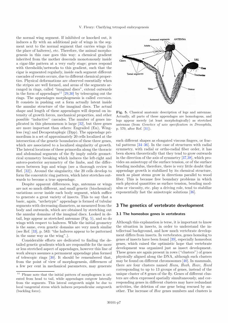
the normal wing segment. If inhibited or knocked out, itinduces a fly with an additional pair of wings in the seg-ment next to the normal segment that carries wings (inthe place of halteres), etc. Therefore, the animal morpho-genesis in this case goes this way: a chemical gradientinherited from the mother descends monotonously insidea cigar-like pattern at a very early stage; genes respondwith thresholds/networks to this gradient, such that thecigar is segmented regularly, inside each segment differentcascades of events occurs, due to different chemical proper-ties. Physical deformations are observed essentially whenthe stripes are well formed, and areas of the segments ar-ranged in rings, called “imaginal discs”, extend outwardsin the form of appendages11 [29,30] by telescoping out therings. The appendages morphogenesis is called eversion.It consists in pushing out a form actually latent insidethe annular structure of the imaginal discs. The actualshape and length of these appendages will depend on in-tensity of growth forces, mechanical properties, and otherpossible “inductive” cascades. The number of genes im-plicated in this phenomenon is large [32], but three genesare more important than others: Engrailed (En), Wing-less (wg) and Decapentaplegic (Dpp). The appendage pri-mordium is a set of approximately 20 cells localized at theintersection of the genetic boundaries of these three genes,which are associated to a localized singularity of growth.The lateral locations of these primordia along the thoracicand abdominal segments of the fly imply subtle geomet-rical symmetry breaking which induces the left-right andantero-posterior asymmetry of the limbs, and the differ-ences between legs and wings (see a thorough review inRef. [32]). Around the singularity, the 20 cells develop toform the concentric ring pattern, which later stretches out-wards to become a true appendage.
Despite apparent differences, legs, antennas or wingsare not so much different, and small genetic (biochemical)variations occur inside each body segment, which sufficeto generate a great variety of insects. This to say that abasic, again, “archetypic” appendage is formed of tubularsegments with decreasing diameters, as measured from thebody and outwards, which are obtained by stretching outthe annular domains of the imaginal discs. Looked in de-tail, legs appear as stretched antennas (Fig. 5), and so dowings with respect to halteres. While the initial geometryis the same, even genetic domains are very much similar(see Ref. [33], p. 583: “the halteres appear to be patternedin the same way as the wing”.).
Considerable efforts are dedicated to finding the de-tailed genetic gradients which are responsible for the moreor less stretched aspect of appendages, however this line ofwork always assumes a permanent appendage plan formedof telescopic rings [30]. It should be remembered that,from the point of view of morphogenesis, differences ofa few per cent in mechanical parameters, may generate
11 Please note that the initial pattern of morphogenes is ori-ented from head to tail, while appendages outgrow laterallyfrom the segments. This lateral outgrowth might be due tolocal tangential stress which induces perpendicular outgrowth(buckling force).
Fig. 5. Classical anatomic description of legs and antennas.Actually, all parts of these appendages are homologous, andlegs appear merely (at least morphologically) as stretchedantennas (from Genetics of axis specification in Drosophila,p. 570, after Ref. [31]).
such different shapes as elongated viscous fingers, or frac-tal patterns [34–36]. In the case of structures with radialsymmetry, with radial or ortho-radial fiber order, it hasbeen shown theoretically that they tend to grow outwardsin the direction of the axis of symmetry [37,38], which pro-vides an anisotropy of the surface tension, or of the surfacebending modulus, therefore, there is very little doubt thatappendage growth is stabilized by its chemical structure,much as plant stems grow in directions parallel to woodfiber. This is because free boundary problems in whichsuch physical quantities as surface tension, bending mod-ulus or viscosity, etc. play a driving role, tend to stabilizeexponentially fast the anisotropic solutions [36].
3 The genetics of vertebrate development
3.1 The homeobox genes in vertebrates
Although this explanation is terse, it is important to knowthe situation in insects, in order to understand the in-tellectual background, and how much vertebrate develop-ment differs from insects. In vertebrates, genes homolog togenes of insects have been found [39], especially homeoboxgenes, which raised the optimistic hope that vertebratedevelopment was organized just as insect development.These genes are again present in rows (“clusters”) of genesphysically aligned along the DNA, although such clustersmay be found on different chromosomes [40]. In mammals,there are four clusters named Hoxa, Hoxb, Hoxc, Hoxdcorresponding to up to 13 groups of genes, instead of theunique cluster of 8 genes of the fly. Genes of different clus-ters are often expressed spatially simultaneously, and cor-responding genes in different clusters may have redundantactivities, the deletion of one gene being rescued by an-other. The increase of Hox genes numbers and clusters is
30101-p7

The European Physical Journal Applied Physics
due to genetic duplications during evolution. This rendersthe notion of collinearity “more complex” in vertebrates.However, it should be noted that the general bauplan ofvertebrates is universal (head, body tail, and limbs side-ways), despite considerable variability of gene clusters andgene counts, between say, fish, and humans. There seemsto be something deeper in the global plan.
The Hox genes certainly play an important role in ver-tebrate development, since knock-out experiments (whichconsist in generating mutants which lack a given gene),show morphological modifications (“phenotypes”). Rulesfor the functioning of these genes have been establishedsuch that: they are expressed in collinear order along thetrunk, and more genes are expressed in the more posteriorregions. For example, mice having shorter body and longertail may be generated by affecting the gene Hoxa2 [41].In the same spirit, a modification of the gene Hoxd3 in-duces an anatomical modification of vertebrae in the re-gion of the neck [42]. But it is clear that “homeotic” trans-formations in vertebrates are much more modest than inflies [43]. Homeotic transformations are body form changesdue to deletion or misexpression of Hox genes. There doesnot exist mutants having hands instead of jaws, or feet in-stead of hands, or skulls instead of phalanges. There seemsto be something qualitatively different between the seg-mentation process in flies, and in vertebrates, which canbe tracked back to the fact that arthropods segment veryearly, such that downstream genetic pathways are more lo-calized than in vertebrates. In vertebrates, segmentationoccurs during the early morphogenetic movements whichform the body plan, but somewhat after the start of themotion, during folding. The segmentation in vertebrates isa uniform instability wave which propagates in an alreadyformed embryo. Hence a vertebrate can be described as“an animal body with segments inside” (the bones), andnot as “a segmented animal” like arthropods. Hox genesmodulate the identity of segments, they do no create them.
In vertebrates, at most, modest shifts of body parts,or growth of extra ribs can be “homeotically” induced bychange in Hox genes. These shifts seem to follow some ex-isting stream or path. For example: it is possible to shiftthe beginning of the tail, but more anterior or more poste-rior to the tail direction, not to the neck, or to the wrist,it is possible to shift the limb position by one vertebraalong the thorax, but not to the forehead, it is possible togenerate a mouse with longer or shorter limbs, not with anectopic skull in the hand region, etc. Generally, homeotictransformations tend to shift the form, and produce “pos-teriorisation” or “anteriorisation” of forms. This is due tothe collinearity of the genes expression with spatial order.Why this is so will become clear below.
However, it is no at all true that there is, in vertebrates,such a strict collinearity between the genetic organizationin the nuclear DNA, and the organization of the bodyplan. Limbs form by growth of limb buds away from thebody axis. In the case of insects, the body axis is quite welldefined, and so is the notion of “lateral outgrowth”, or of“away” from the body axis. In the case of vertebrates,there exists rotational engulfments of the tissue (as we
shall see in Sect. 5, such that the body axis is not a relevantconcept in areas of strong bending or twist.
Moreover, by definition, DNA sequences are linear, andif collinearity between DNA sequences and body form ex-ists as a concept, then nothing can molecularly specifylateral “branchings”, or outgrowths. Lateral outgrowthsrequire a change in growth direction. But such a change isnot present as a specific instruction in the genome. Thisis to say that, while the subsequent genes in the Hox clus-ters may be correlated to specific forms observed in thestripes (where such or such genes are expressed), there isno interruption of the cluster of genes, by any gene whichwould serve to instruct a right turn to the growth process.The symmetry breaking which orients growth sideways isa physical, implicit one12, this is to say that it is an in-stability of the growth process itself, an anisotropy or atensorial feature.
In addition, it is known that genetic expressions in limbbuds make use of similar genes in the hindlimb, and fore-limb, and in the tail, namely genes Hoxa, Hoxd 9 to 13 areused for all these 3 body appendages of vertebrates [44],in a proximo-distal order.
Therefore, while it is indeed true that vertebrate limbsare “some sort of a tail” growing sideways, the points ofoutgrowth of the limbs do not correspond to a sequentialorder of genes, in specific segments or boundary intersec-tions that would induce specific appendages, as in flies13.The tail is not located at a specific segment, nor are thelimbs, the ears, the jaw or whatever. It may be possiblethat collinear sequences of homeobox genes are re-usedobliquely along the body in order to generate the limbs,after having been used along the body to generate head-to-tail pattern, but this concept does not explain why it isso. Moreover, recent data show that the distal part of thelimb expresses Hox genes actually in a reverse collinear-ity pattern [45]. Detailed analysis of mutants shows that“Unlike the situation in the trunk, and in contrast to whatwas originally expected, loss-of-function phenotypes can-not be interpreted as classical homeotic transformations.”(Zakany and Duboule [44]). These authors conclude thatthe outcome of Hox genes function is “non specific”, “flex-ible”, and related to “context”. This is to say that bound-ary conditions, and previous history will be a concern forthe actual outcome.
If genes are induced sequentially by automatic in-ductions, linked possibly to gradients of some trigger-ing molecule, the existence of reverse orders of expres-sion imply a complex spatio-temporal organization of thegenetic expressions. The collinearity observed along thetrunk might be an erroneous concept, which appeared im-portant only because the physical object “trunk” is almost1 dimensional. It is important to note that the concept ofcollinearity requires a 1D physical substrate, but the originof the 1 dimensionality is completely different in an insect
12 As is, also, the point of intersection of the domains of En,Wg, and Dpp from which insect appendages emanate.13 Also, while the appendage primordium in insects is a verylocalized cluster of approx. 20 cells, in vertebrates, the limbbud encompasses thousands of cells.
30101-p8

V. Fleury: Clarifying tetrapod embryogenesis
Fig. 6. Left, genetic collinearity between clusters of genes and limb development, as deduced from knock-out mutants (fromRef. [60]). Right, more detailed 2D genetic expression in the limb bud. As the limb extends, the gene expressions forms specificareas, although they are not strictly correlated to hand patterns. As time passes by these domain of expressions become morefuzzy.
(elongated syncitium) and in a vertebrate (formation ofmedian folds, see Sects. 6 and 7, especially Fig. 40). Atpresent, the formation of a vertebrate body is grossly de-scribed by a collinear expression of Hox genes from headto tail, a collinear expression of genes from shoulder towrist, and a reversed collinear expression of genes in theautopod (“the hand”). However, how all this transformsinto morphogenetic fields which generate bone, tendons,joints, etc. is not clearly established, but assumed to belargely related to early embryo movements [46] (Burke andNowicki state that: “It seems likely that the establishmentof Hox expression patterns, and the positioning of cellpopulations are related, thus insuring harmony betweenanterior-posterior and medial-lateral patterning axes” ).
3.2 Limb patterning in tetrapods
In order to attempt to bring vertebrate development to-wards the description of insect development, a number ofgenes have been pointed out whose role seems somewhatanalogue to what is observed in insects. The position oflimb outgrowth has been correlated to the levels of Hoxc6,Hoc8, Hoxb5, Hoxb6, Hoxb9, Hoxc9, Hoxd9 genes [44,47].It appears that hindlimb and forelimb seem to protrudeout at boundaries of expression of Hoxc6 gene, or Hoxc8gene. It has also been shown that snakes express Hoxc6gene in a much more expanded area as compared to othertetrapods (Fig. 7), thus correlating to the position of ves-tigial limbs in these animals [48]. Also, homeobox geneshave been related to the presence or absence of ribs inrelation to vertebrae (which suggests that actually, insectappendages are homologues of ribs, and not of vertebratelimbs, which are something else). However, as stated byRallis et al. “neither gene deletion nor gene misexpres-sion experiments have provided direct evidence for a roleof Hox genes in limb positioning” [49]. Limb positionsseem to be related to the thoraco-sacral junction, and tothe thoraco-cervical junction, in all animals. Therefore,the outgrowth of limbs seem geometrically linked to these
Fig. 7. (Color online) Domain of expression of Hoxc6, ina chicken, left, and in a snake, right. The evolution fromtetrapods to snakes seem to imply an extension of the do-main of Hoxc6, rostrally, and caudally. Photo by McOmberand Burke.
anatomical regions, by the morphogenetic process, and theHox genes domains of expression may be more a conse-quence, than a cause of limb positioning. From a physicspoint of view, it seems that limbs do not form “at” thethoraco-sacral and “at” the thoraco-cervical junction, bysome molecular coincidence, but that these junctions cometogether with the limbs in the morphogenetic process. Thepelvis, for example has a large and complex shape span-ning many vertebrae; it shows a clear vortex pattern bothin the global shape and locally in the trabecular bone ori-entation. It is not a shape with a defined “axial” form. Inaddition, it has a concavity in the area of the leg joint as-sociated to antagonist growth of the cartilage cups of thepelvis and of the femur (please refer to a general anatomytextbook). Clearly the pelvis shape is linked to the en-tire, extended, physical field of morphogenesis, and notto a simple inductive axial (“collinear”) event that would“trigger a pelvis” in a definite, strictly instructed, shape.It is extremely unlikely that a “pelvis” could be induced
30101-p9

The European Physical Journal Applied Physics
Fig. 8. Direct observation of limb development. Formation ofthe limbs starts by a lateral plate (limb field) out of whichlittle paddles extend, to become progressively true limbs. Atthe moment of formation of the limb paddle, the segmentationwave has descended along the body backbone, generating thepresumptive vertebrae. The wavelength of these segments seemto influence the wavelength of segments in the hand (digitalrays, which become true fingers after cellular death in betweenfinger bones), because the number of somites facing the limbbud is always close to the finger count. A direct count of thesegments shows here that there are indeed about 5 segments infront of the paddle, before it extends. From Martin, UniversityCollege, London, UK.
anywhere else by a genetic induction. Also, in many ani-mals the curve of the pelvis continues itself in the shouldergirdle; the hindlimb grows out at a specific node of thepelvis, and not truly “along the trunk”, etc. If the con-cept of genetic induction were true, one would expect tosee mutants with a pelvis without limbs at any given levelof the thorax, and, anywhere else, limbs without pelvis orshoulders, which is certainly not the case.
Whatsoever, these facts do not fit into an antero-posterior gradient of anything, especially since the do-mains of expression of Hoxc6 shift both towards the head,and towards the tail, when going from tetrapods to snakes.Therefore in vertebrates the concept of antero-posteriorgradient is not related to the correct positioning of limbs.
The further patterning of the limbs remain largelymysterious. It has long been known that limbs emanatefrom “lateral” plates of the embryo, which constitute a“limb field”. The limbs grow outwards in the shape ofpaddles, and eventually limbs (Fig. 8).
There exists a considerable literature dedicated tothe morphological commitment of limb buds, as a func-tion of grafts, ectopic14 expressions of molecules, geneticknock outs15 or mutants, etc. Molecular studies distin-guish the role of transcription factors, which bind to DNA(homeodomain genes), and of signalling molecules, such
14 Ectopic grafts or expressions, consist in positioning a graftor a molecular source in an aberrant place, and watch the de-velopmental outcome. For example, ectopic limbs can be ob-tained in aberrant places, by putting ectopically a bead of somechemical under the ectoderm, in that aberrant place.15 To knock out genes consist in generating an animal in whichall alleles of a gene are absent. A mouse ko for a given genewill show traits related to the absence of that gene.
as growth factors, which maintain proliferation, etc., butwithout affecting the DNA (Fig. 9).
It is especially clear that fibroblasts growth factors16(FGFs) produced by mesodermal cells are involved in limboutgrowth expansion, since implants of beads soaked inFGF1, FGF2, FGF4 or other chemicals generate ectopic(supernumerary) limbs [51], and mutants lacking FGF10have no limbs (in addition to having no lungs [52], but itshould be noted that they do have pelvis and scapula rudi-ments). Also, double deletion of FGF4 and FGF8 annihi-lates limb growth, in addition to many other abnormali-ties [53]. Annihilating FGF8 expression only, has no effecton limb pattern, while double deletion of FGF4 and FGF8,impairs limb growth, but does not prevent the formationof the initial bud, which is positioned at the correct place.This shows that the relationship between growth factorsand the final pattern is somewhat puzzling, there seemsto be strong “redundancies”, and “regulatory loops”, al-though the rationale may be otherwise: as long as quanti-tative levels of growth factors are not related to physicalparameters of growth, it is hard to tell why a limb growsor not. It seems that the topology of the pattern is actuallynot related to the growth factor: either the pattern grows(because cells divide actively), or it does not, but if it does,it grows with a topology independent of the growth factor,which would be coherent with the idea that growth factorsare not morphogenes, like Hox genes. Nevertheless, cellu-lar proliferation induced by FGF growth factors seems thecrucial developmental engine of the buds, to form limbs,this is why FGF’s are considered as the “controllers” ofthe proximo-distal axis of growth.
Now what starts this proliferation, and why there, andhow is the limb type determined? Genetic studies seemto imply that it is the role of transcription factors suchas the Hox genes to modulate the magnitude of thesegrowth factor along the flanks, by the dosing or timingof many molecules downstream. Considering the patternof expression of Hox genes [47,48], and loss-of-functionphenotypes [44] it is unlikely that Hox genes provide aspatial code or a mosaic for the limb pattern, but rathersubtle quantitative gradients of parameters of some phys-ical process of patterning.
The exact positioning of the limb, and limb type hasbeen related to the transcription repressors of the Tboxfamily [54,55]. It has been shown that it is possible to shiftslightly the position of outgrowth of the limb by repress-ing or activating Tbx3 [49], but shifting a limb is not thesame thing as inducing a limb17. Negative misexpression
16 Fibroblast growth factors are molecules which (amongother roles) maintain the proliferative activity of fibroblasts.There exist about 23 members of this family, with similar se-quence. There exist 4 receptors of these growth factors. Fibrob-lasts are one of the major cell types. They provide the produc-tion of collagen and related supporting molecules. Hence FGFsand their receptors play a key role in many aspects of embryodevelopment, angiogenesis, etc.17 When limb shift is obtained in a biochemical assays, it isgenerally accompanied by a strong flexion of the embryo body.Experiments in which the contralateral side is not modified
30101-p10

V. Fleury: Clarifying tetrapod embryogenesis
Fig. 9. (Color online) Stainings of several molecules in the early chick embryo. Left transcription factors, right signallingmolecules. (From Ref. [49], and references therein). Please note the strong Tbx4 and Tbx5 stainings in resp. hindlimb, andforelimb. The Tbx4 staining is clearly linked to the mesoderm engulfment, and not to some linear gradient: the crescent shapeof the deep blue Tbx4 staining follows the rotational pattern of cell flows in this area (more in Sects. 5–7). Color stainingsshould be taken with some care, as the stainings follow actual twists of the tissue.
of Tbx5 [56] leads to a massive reduction of lateral plate inthe forelimb area, and complete absence of the forelimb inthe newborn, Figure 10. But still, there is a lateral plate.
The origin of identity of the limbs remain mysteri-ous: early hopes [50] suggested that limb type was re-lated to the pattern of Tbx4 and Tbx5 genes, since thesewere specifically present in resp. hindlimb, and forelimb ofchicken (see Fig. 9h, 9i). Also Pitx1 seems to be specificof hindlimb, and may play a role in hindlimb identity vs.forelimb. Loss of function of Tbx5 in the forelimb leadsto complete absence of forelimb as shown in Figure 10.When expressed in place of Tbx5, Tbx4 was able to rescuethe formation of the forelimb [57]. But surprisingly, thelimb was of a normal forelimb type. Unfortunately Tbx5and Tbx4 were found in limbs of both types altogetherin the newt model [58], which sheds doubt on the rela-tionship between Tbx and limb identity. Electroporationexperiments, by which Tbx5 is expressed in legs are some-what controversial: the leg has only 3 digits, like a wing,but the phalange count is not correct (“misexpression ofTbx5 in the leg can induce wing-like skeletal patterns, al-beit partially ” Takeuchi et al. [59]). We all know thatchicken legs carry scales on the skin of anckles and digits,while chicken wings carry feathers: scale to feathers con-version on the legs can indeed be obtained genetically [58].The wing in which Tbx4 is expressed (while Tbx5 is thenormal Tbx gene there) looks qualitatively like a leg: thefingers of the wing separate, as in a normal leg, and thefingers have claws. The success rate of these experiments
and serves as control shows that shift of limb position comestogether with a global deformation of the entire embryo side,generally more elongated in the direction of the shift [46,51].
is very low (∼10%), and puzzling for physicists. Especiallyone would expect electroporation experiments of Tbx5 ina right leg bud to give either 50% of left wings and 50%of right wings, if limb identity is only defined by Tbx5,or 100% of right wings, if the limb identity is defined byTbx5 and the context. Still 90% of right legs are actuallyobtained18.
While there is indeed an impact on skeletal elementsof the normal limbs of misexpression of genes, the extralimbs formed by expressing ectopically Tbx5 or Tbx4 in awrong area are generally not normal. Again, there seems tobe “something else” in the spatio-temporal outgrowth oflimbs than just the expression of transcription factors, andartefacts are not excluded. Especially, while the presenceof Tbx5 is necessary for limb outgrowth, what determinesthe localization of Tbx5 and the gradients that create thebias in limb axis?
In the same spirit, the identity of the ectopic limbsgenerated with soaked beads is not clearly related tothe growth factor inserted ectopically, but more likely tothe embedding tissue (the posterior or anterior nature ofthe limb depending on the position along the flanks), andthe way it is disrupted by the insertion of the bead (espe-cially in terms of symmetry breaking of the bud orienta-tion; insertion of beads leads to complex digits distribu-tions). It is rarely clear what exactly ectopic expressionsof signalling molecules prove about the limbs in the nor-mal case: in the normal case there are no beads, and it isproven that several molecules used historically to induce
18 According to the authors, Tbx5 is actually quitemechanosensitive (Ogura, Personnal communication, and inpreparation).
30101-p11

The European Physical Journal Applied Physics
Fig. 10. (Color online) Typical genetic assays on limb devel-opment. A mutant is generated which lacks a given gene, hereTbx5. A massive reduction of the lateral plate (region of thepresumptive limb) is obtained in the mutant mice defectivefor Tbx5. As a result, the mice have no forelimbs. From ref-erence [56]. (By the way, one may remark that the number ofsomites in front of the early limb field is of the order of 5).
ectopic limbs [51], are just not candidates for normal limboutgrowth initiation.
The pattern of fingers itself has been related to molec-ular gradients in the limb paddle. Schematically, the limbpaddle seems to be chemically organized in the follow-ing way. First, there exists a pool of a signalling moleculeSonic Hedgehog (SHH) [60–62] located at the frontier be-tween the limb bud and the flank in an area called Zoneof Polarizing Activity [62]. The gradient of SHH, and thetime of exposure to SHH seems to be related to the iden-tity of the observed fingers. Briefly stated, the gradient ofSHH inside the hand, in the thumb-to-auricular directionseems to play an important role in the difference in shapeof these fingers. But this is not true of all fingers. Nested
expressions of Hoxd genes [63], and reverse collinear ex-pressions of genes in the “hand” play also a role, but theoverall regulation of these genes is not understood. Whilegradients of molecules may be deterministically correlatedto finger patterns, how the molecules actually pattern thefingers is not understood. For example, placing a source ofSHH opposite to the normal source of SHH, mimicking asecond zone of polarising activity, induces a forelimb withmirror distribution of fingers [64]. The activity in this fieldof developmental biology consists in finding the proximo-distal19, antero-posterior, and dorso-ventral gradients ofmolecules which would be responsible for the 3D spatialorganization of the growth process (and their temporalexpressions) SHH and hox genes are candidates for “con-trolling” the antero-posterior gradient, but a considerablenumber of other genes have been implicated, whose activ-ity is related. Especially Hoxb8 seems to induce Shh whichinduces Fgf8. Wnt and Fgf are related, Gli3 and Shh arerelated, Shh induces the cartilage forming pathway viaGremlin (Bone morphogenetic protein, BMP), etc. Thesemolecules interact by positive and negative feedback loopswhich are rarely understood in detail.
Now, the proximo-distal axis of the paddle is said tobe “controlled” by an area of the limb edge, known asapical ectodermal ridge (AER). This area has been shownfor long to play an important role [47,50,61] in order tomaintain the proximo-distal arrow of growth. This area isa narrow thickening located along the distal edge of thelimb paddle, which seems to feed the tissue in between thetip of the limb, and the flank of the required growth fac-tors by an interplay of Fgf and Wnt genes, but again it isnot clear whether these growth factors actually have a pat-terning role. The edge itself seems to be the line of sutureof the endoderm and the ectoderm (see Fig. 1 in Ref. [49]),and the fingers form actually away from this thickening,more inwards the paddle, and towards the flanks.
Many experiments, most of which consisting in sever-ing parts of the limbs, or transplanting parts, lead to longlists of results (mirror fingers, reduced distal elements,etc.), which remain controversial [65]. It is clear that thepotential to form complete limbs or additional fingers iswidespread along the body, with even tail cells being ableto form digits if transplanted at the proper place, etc.
The apical ectodermal ridge, generally thought to beso important for limb development, was even found tobe totally absent in some animals (the so-called directfrogs [66], which are frogs which form legs normally, with-out passing through the stage of a limbless tadpole).
Finger types are certainly related to the distal expres-sion of Hox genes, but not their number. The formationof bony structures in the hand and limb seem more of abiomechanical wave of material instability (denser tissueforming the cartilaginous pattern of bones [67] by a pro-cess of self-enhanced attraction of fibroblasts), indepen-dent of the Hox genes, and Shh is an important parame-ter in this process. This is also true of vertebrae segments.This is apparent in the fact that many extent species,
19 Proximal = close to the body, distal = more away from thebody.
30101-p12

V. Fleury: Clarifying tetrapod embryogenesis
or mutants, have repeated sets of identical fingers [68,69],sometimes largely exceeding the available number of Hoxgenes. Therefore there is something quite simple in thegeneration of rows of radiating digital rays, which spanthe limb paddle like an instability wave20, much like theformation of vertebrae along the spine [70]. Hox genesmodulate the final shape of these domains of instability,but with a moderate impact. It seems that Hox genescome into play “too late”, when the digital rays are al-ready established. If Shh is completely suppressed, limbbuds still form fairly normally, but aberrant limbs extend,which may have for example a single finger [71]. The en-tire shh-/- animal (animal in which both maternal andpaternal alleles of the gene shh are knocked out) body ishowever completely aberrant.
Now, about the dorso-ventral “gradient”. In generalterms, the body exhibits a dorsal side and a ventral side,inherited from the existence of slightly different cellulartypes on the endoderm and the ectoderm. This dorso-ventral polarity is also present in the hands, with an ob-vious palmar and a dorsal side. Bio-chemically, this dif-ference seems to be related to the genetic pathway of en1and wnt-7a [62], and analogous genes may be at play inthe dorso-ventral polarity of insect wings [33]. But the ex-pression of specific genetic pathways is related actually tothe geometry of the limb bud at the moment of formation:if the limb bud forms from endoderm and ectoderm21, ithas dorso-ventral polarity (like a normal hand), if a limbbud forms more proximally by a fold of ectoderm only, thelimb paddle has double dorsal polarity [62]. Therefore, thedorso-ventral polarity is a matter of gradient of molecules,but the boundary conditions for these molecules are geo-metrically carried by the respective layers of cells. Froma geometrical point of view, the formation of the limbbud appears as a physical process, which does not requirespecific cell sheets, but it happens that ectoderm and en-doderm have originally a molecular difference, and thissuffices to induce a dorso-ventral “gradient” across thedorso-ventral direction.
The genetic descriptions of limb patterning get pro-gressively more and more complex and controversial, andthe view of how the vertebrate body is actually orga-nized gets progressively more cloudy. The gradients ofmolecules forming an orthogonal system of coordinates donot seem to suffice to describe cartilage patterns inside ahand. Since such a representation is systematically used,I, too, have used it above to describe hand formation.However, when described in detail, the pattern of bonesin the “wrist” of a mouse seems actually to have under-
20 The digital rays, regularly spanning the limb paddle, arecalled the “digital arch”.21 The initial formless embryo (the “blastula”) is composedof one layer (the epiblast). A second layer, the hypoblast nu-cleates underneath the epiblast, therefore the hypoblast em-anates from the epiblast. During morphogenesis, a third layerappears by invagination, during gastrulation, of the first twolayers inside themselves. In the literature, as the body forms,new names are ascribed to the layers: ectoderm (skin on top),endoderm (layer below), and mesoderm (in between).
gone a flexion, or rotation, as compared to a fish fin22 (thesturgeon) [73], which cannot be described by orthogonalgradients.
Still, a primitive limb is such a simple thing: a roundpaddle with rays. A number of facts explain this darkness.First of all, genetic inductions always need a previous ge-netic induction, so that it is never clear what is the chainof inductive events to take into account, and this is all themore true as the movements which advect the patterns ofexpressions are not taken into account. The second fact isexperimental: molecular experiments consist generally inexpressing in an aberrant concentration, and in an aber-rant place, a molecule normally present in the physiologi-cal case. It is therefore not at all clear how the experimentshould be interpreted, if one does not know how the saidmolecule is produced in the physiological situation. Forexample, if it is quite simple to generate a supernumerarylimb with a bead soaked in retinoic acid, does it meanthat it would be very easy for nature to produce such asupernumerary limb? Or does it mean that it would beextremely difficult, because nature is unable to providea soaked bead? Stated otherwise, what is the equivalentof the bead soaked in retinoic acid, in the physiologicalcase? How does nature manage to create such a startingpoint for a limb? If localisation of any molecule, possiblyFGF10 or Tbx5, is necessary to generate a limb, how is itlocalised, in the natural case, and why is the limb induc-tion possible with so many molecules? More specifically, ifthe entire flanks between tail to shoulder is competent togive limbs, why do we have only four? If the word “geneticinduction” has a meaning, why “induction” of a limb is notpossible ahead of the shoulder and caudal to the pelvis,with the same “inductive” molecules that “induce” a limbin between shoulder and pelvis? [51].
Another problem lies in the spatio-temporal aspect ofthe development. It has been stated that “One of the mostmysterious features of morphogenesis is that structuresform themselves as they grow” [32]. In the same spirit,recent analyses in terms of gene regulatory networks haveraised some surprise, since the same genes regulatory net-works seem already present 570 My ago in cnidarians (jellyfish, etc. [72]); therefore if the same genetic networks arepresent, either some minor molecular subtleties, down-stream of the main genetic kernels, make the differencebetween animal bauplan (say hydra and rabbit), or it is amatter of spatio-temporal organization (different symme-try breaking) of the same genes with non-chemical bound-ary effects and wavelength locking.
Most, if not all, present molecular descriptions assumean existing area, for example a limb bud, or a body axis,and try to organize the chemical fields inside or along it.However, it is clear from a physical point of view, thatbody, organ, or appendage development is a free boundary
22 As will be discussed in Sections 5–7, the affine stretch ofthe bones (seen for example in the bats forelimb) is possibleon digital rays which have a roughly 1-D morphotype. In areaswhere tissue rotations occur at early stages of development,the coupling between bone segmentation and tissue movementsleads to less obvious bone configurations.
30101-p13

The European Physical Journal Applied Physics
Fig. 11. (Color online) Top (from Grotewold, Ruther, The EMBO Journal 21, 966 (2002)), staining against BMP4 in a wildtype mouse, and in an shh-/- mouse. The expression of BMP4 is localized in the axillary region, which is likely to undergoa higher stress. Bottom, one example of genetic description of the limb bud organization, with the regulatory pathways inwhich SHH is involved (from Tickle, Nature reviews, Molecular Cell Biology 7, 45–53 (2006)). The limb bud is described ashaving a Zone of polarizing activity (controlled by a spatial map of SHH, located in the axillary area, here in red), from whichSHH diffuses, influencing the expression of Gli, and other genes. Sonic Hedgehog, has been shown to be correlated with theAntero-posterior differences in 3 fingers only. Current views do not clearly explain how individual fingers are patterned.
problem, and one has to treat the displacement vector ateach point, without a priori on the possible outcome, andincluding physical forces. Indeed, the collinearity conceptthat prevails in developmental biology already assumes anoriented 1D growth pattern over which stripes of geneticexpressions form (see for example Fig. 6 left). The de-scription of growth by zebra-stripes of genetic expressionsalready supposes that these stripes decorate an existing“ribbon”, hence they make the implicit assumption thatgrowth is 1D, as does Darwin with bat limb formation.This is explicitly stated, although en passant, in reputedwork, such as Duboule’s [73], for this author, both thesuccession of Hox genes along the trunk, and the geneticpattern of hands are “unidirectional”. Such an oriented1D growth has to be explained by the physics of the pro-cess23. In addition, when growth occurs sideways, almostperpendicularly, an anisotropic feature, or an instabilityexplaining lateral outgrowth has to be invoked, in order toexplain physically the change in growth direction. For ex-ample: when the segments of the spine form, cells presentinside the segmentation furrow tend to escape the line by
23 In the same spirit, the differences in appendages forms ofthe fly is finely studied by marking gene expressions on therings of the imaginal discs. However, by assuming an alreadyexisting imaginal disc, these studies only address minor differ-ences inside a predetermined pattern of appendage whose basicplan is a set of telescopic tubes.
migrating away exactly perpendicularly to the segment.Therefore, the formation of the segmentation line perpen-dicular to one axis, induces a migration perpendicular tothe axis, without a specific molecular instruction to doso, this might possibly be an explanation of the tendencyof appendages to grow outwards at “boundaries” betweendomains of genetic expressions.
Moreover, purely geometrical effects play an impor-tant role: the shape of the SHH and of the BMP gradientaround the limb bud, and its temporal action, is appar-ently linked to the antero-posterior pattern of the fin-gers [62]. However the spatial organization of these geneproducts is associated to a strong bending of the bound-ary condition in the axillary region, which is naturally amore stressed area (Fig. 11). Therefore it seems not suffi-cient to describe chemical actions in terms of orthogonalgradients. In the case of shh, not all fingers have a shaperelated to shh, therefore, although shh may modulate fin-ger shape24, it is not responsible for the existence of fingersper se (mouse without shh at all still form one finger, al-most normal [71]). This shows that, in general terms, aconceptual framework allowing one to relate genetic ex-periments to physics of pattern formation is still lacking.
Understanding limb growth requires to couple mathe-matically the chemical inductions with the spatio-temporal
24 The spatial gradients are important but also the total timeof exposure to the molecule.
30101-p14

V. Fleury: Clarifying tetrapod embryogenesis
Fig. 12. (Color online) A typical description of limb bud formation from lateral plate mesoderm. From Martin Cohn [75]. Thefigure represents 3 stages of limb bud outgrowth along the flanks. Only the right half of the body is schematized, and only thepart of the body located schematically between pelvis and shoulders. The little squares numbered from 15 to 30 represent thesomites, which are precursors of vertebrate (actually there is a shift of 1/2 of a wavelength between vertebrae and somites).The protruding features with a purple edge are the nascent limb buds. Such descriptions always assume an existing lateralplate (here represented by the blue areas of the body at the stage represented to the left). Ab initio (left), zones precursor oflimbs exist in the form of “lateral plate mesoderm” already present. Interestingly for physicists, such an image shows a forwarddisplacement of the limb bud boundary (purple), as a free boundary growth, but it is not said what determines the displacementfield. Much like fly appendages, the description already assumes the existence of an area committed to become a limb.
field of growth, including such mechanical features as ten-sion along curved surfaces and internal stresses, sincecompletely different gradient maps can be obtained, indifferent boundary and force conditions, especially if me-chanical feedback regulates tissue growth [74].
Another problem lies in experimental skill. It seemsfrom oral interview of scientists in limb developmental bi-ology, that, in order to produce extra limbs or extra dig-its, beads soaked in chemicals have to be put at specifictimes, and specific places, in an area called “limb field”.Especially, aberrant digit distributions can be obtainedby grafting pieces of limb buds onto other limb buds, butat very early stages where digits are supposed not to bepresent at all, therefore even the timing of digit appear-ance is not truly understood.
In the same spirit, there is no way an extra limbcan be generated on the head or the chest, by the samesoaked beads which so easily give an extra limb along thelimb field. Therefore, the interpretation of the inductiveevents is incomplete, and requires a more detailed (spatio-temporal) understanding of what the limb field actually is.
3.3 The limb field
True as well as extra limbs actually originate in the lateralmesoderm25 of the flanks, in an area called “limb field” or“lateral plate” or “limb plate” [75]. While preparing thisreview, I found it impossible to find a description of wherethe lateral plates come from. All existing work assumes an25 The early embryo is composed of 3 cell layers, the upper celllayer called ectoderm, the inner cell layer called mesoderm, andthe lower cell layer called endoderm. During morphogenesis,complex motions tend to reshuffle and fold the layers.
already formed lateral plate for the extension of the limb(Fig. 12), or for the early expression of limb markers (suchas Tbx5). Therefore, all existing work in limb formationdoes not actually address the problem of limb origin, butrather, fine adjustments inside an area already committedto give limbs.
In a 2000 article [75] we can read: “However, controlof limb development is apparently situated in the lateralplate mesoderm, and has been experimentally shown tobe independent of an axial Hox code” but nothing is saidabout the origin of the lateral plate itself.
In a 2002 review, the origin of the lateral plate waspresented in the following way [76]: “Moving backwardsin developmental time raises the questions of how bud-ding of the limb is first initiated and what normallyserves to restrict the positions of the limb buds alongthe rostral-caudal axis of the body. Molecular experi-ments in the chick suggest that an intricate dance be-tween FGF and WNT signaling is involved in limb budinitiation (Kawakami et al. [77]). A series of sequential sig-nals are passed between WNT and FGF in a wave acrossthe medial to lateral aspect of the body (somite, interme-diate mesoderm, lateral plate mesoderm, ectoderm). Inthis dance the partners are exchanged while the overallmelody remains the same. In the presumptive forelimb re-gion, Wnt2b becomes restricted along the rostral-caudalregion to the intermediate and lateral plate mesoderm.It is not yet known what genes are involved in definingthe rostral-caudal domain of Wnt expression. Presumablyaxial patterning determinants are important, and thesecould include the Hox genes as mutation of Hoxb5 leadsto a rostral shift of the forelimb field (Rancourt et al.,1995 [78]).”
30101-p15

The European Physical Journal Applied Physics
However, Kawakami’s article deals with Fgf-Wnt sig-naling in already existing lateral plates, or in regenerativetissue. Rancourt’s article deals also with an already ex-isting lateral plate. In a 2003 article entitled “Tbx5 is es-sential for forelimb bud initiation following patterning ofthe limb field in the mouse embryo” [79] one finds an im-portant triggering effect of Tbx5 upon limb outgrowth,but again inside an existing lateral plate. In this arti-cle, it is stated that: “Limb formation occurs as a resultof interplay between fibroblast growth factor (FGF) andWnt signaling. What initiates these signaling cascades andthus limb bud outgrowth at defined locations along theantero-posterior axis of the embryo is not known.” Andalso: “The definition of the fields of Lateral Plate Meso-derm that will give rise to limb buds occur possibly as aresult of patterning by Hox gene activity along the APaxis of the vertebrate embryo (Cohn et al., 1997; Cohnand Tickle, 1999 [51]; Popperl et al., 2000 [80]; Rancourtet al., 1995 [78])” However, Popperl et al.’s article dealsmostly with the role of lazarus in the cranial (hindbrainand anterior trunk) compartment, (especially it shows howpbx genes participate in the regulatory loop of hox genesneeded to specify rhombomere boundaries in the hind-brain) and when coming to limbs gives the same evidencethat Tbx5 triggers forelimb outgrowth, since lzr-/- ze-brafish do not show fin rudiments at all, and no Tbx5staining. There is no rationale about limb positioning,apart from a correlation between absence of lazarus, andabsence of fins. Especially, it is quite clear that the finrudiments appear as round balls on the side of the flanks(Fig. 6. I in Ref. [80]). No mechanistic explanation is givenof how a collinear series of genes along the neural crestmight induce lateral formation of roundish masses of cells(the fin precursor). Popperl’s et al. article deals with dele-tion of hoxb-5 and hoxb-6 genes, and shows only a verymodest shift of the shoulder girdle of about one or atmost two vertebrae, when hoxb-5 genes are deleted andno mechanistic explanation. It is difficult to understandfrom this work in what respect hoxb-5 might be relatedto limb “positioning”, since when totally absent, there isa normal limb, which is “positioned” by something, andthe position is almost normal, except that it is slightlyshifted from the wild type. Cohn’s et al. 1997 shows theopposite of what is claimed (if hox genes play a regula-tory role in the lateral plate, it is independent of the APpolarity), and Cohn and Tickle’s 1999 article deals withlimblessness in snakes. While this last article may give aclue of why snakes do not have limbs, it does not give ahint of why, in tetrapods, limbs would appear where theyappear (stated otherwise, they correlate limb position anddomains of hox genes, but what serves to position the do-main of expression, in the first place?).
An other article shows a correlation between Hox9genes and limb positioning, but there is no mechanisticrelationship between limb positions and Hox9 genes ex-pression [81]. To make a long story short, in a 2006 re-view [82] we still read: “The first problem in human limbdevelopment is not outgrowth but locating the point ofoutgrowth along the body wall. Little is known about the
Fig. 13. (Color online) The earliest known ancestor of thevertebrates, the bilateran, segmented Pikaia 550 My ( c©MaryParrish/The Smithsonian Institution).
exact mechanism, but globally acting genes such as Hox9and Tbx4 seem to be crucial for hind limb placement.They are expressed in the lateral plate mesoderm beforeoutgrowth.” A sentence which again already supposes theexistence of lateral mesoderm.
The review presented here highlights that inductivecascades of unidirectional gradients of scalar quantitiescannot explain morphogenesis. In effect, if morphogenesiswere organized by successive discrete inductions by feed-back loops along the slopes of scalar gradients of mor-phogenes, the animal shapes would be possibly arbitrary,without geometrical constraints, and animal forms wouldtruly be random. But, as has been recognized for long,and quantified more accurately lately, the body axis andlimb fields are actually organized by a hydrodynamic-likeflow, in which a segmentation wave propagates. Such thatbeing a forelimb or being a hindlimb, is actually a matterof geometry of a deformation field advected by a flow (SeeSect. 7), which serves as boundary condition for geneticexpressions.
4 The paleontological point of view
4.1 Tetrapod origin
If we turn to paleontology, we find a description oftetrapods appearance into three main steps. Appearanceof chordates, segmentation of lateral fins, appearance oftetrapods. The chordates are the class of animals compris-ing vertebrates and a small number of jaw-less, vertebrate-less fish like the amphioxus (lancelet)26 and the lam-prey27 [83]. The first fish-looking, but jaw-less chordates,date back to the Cambrian era, about 540 millions yearsago. At the Cambrian explosion a number of complex bau-plan of multicellular organisms appeared very rapidly [84],among which the first chordates, presumably Cathaymirusdiadexus [85] or Pikaia gracilens [86] are our ancestor(Fig. 13).
26 A cephalochordate with a notochord but no clear vertebraesegments.27 A fish with a circular plunger-mouth, and vertebrate-likesegments.
30101-p16

V. Fleury: Clarifying tetrapod embryogenesis
Fig. 14. (Color online) The first true chordate, Haikuichtys(470 My).
As a transition towards true vertebrates in the fos-sil record, appears haikuichtys [87] (or MyllokunmigiaFengjiaoa) (470 My), of the lamprey family (Fig. 14).
From then on true fishes are present in the fossil record.Early fishes seemed to have fin folds running along thebody like curtains. Although there also exist specimens offishes with rudimentary paddles below the jaw, as earlyas approx. 400 My, Ateleaspis tesselatta [88].
Somehow, the elongated fins present along the body“became segmented”, restricted to some areas, and thelateral fins became recognizable as pectoral and pelvic fins.These fins were essentially paddles with rays fanning awayfrom the body, as observed on so many common fish. Onefinds then along the fish body: the caudal fin, the anal fin,and the dorsal fin. Simple inspection of these appendagesshow that they seem to be continuations of existing folds,which may be more or less elongated. The dorsal fins seemto drift caudally during development, as the tail extends.
Progressively during evolution, some families of fishdisappeared, other appeared. Fish body became separatedinto several forms, bony fishes and non-bony fishes, ag-naths (fish without jaws) and gnathostome (with jaws).Among the non-bony fishes, one finds the class of shark-like fishes, which include flat fishes like the torpedo andray-fish (class Chondrichthyes, 370 My). The usual bonyfishes, said Osteichthyes appear 395 My ago. There existthree kinds of bony fishes, the ones with ray-finned pad-dles, known as teleosts. They form the largest and mostcommon group of present fishes. The second group is thelungfishes. The other group, from which tetrapods descendare the Sarcopterygian fishes. Please refer to a generaltextbook on vertebrate evolution for more details.
The Sarcopterygian fishes had well developed lateralfins, with a paddle located away from the body at theapex of a fleshy limb (“lobbed fin”). Around 350 My fromnow, a group known as the Crossopterygian (among whichthe celebrated Latimera chalumnae, known also as coela-canth) evolved limbs, which allowed them to conquer theland, as amphibians. Actually, it is well admitted thatlimbs appeared before fish were able to conquer the land,
Fig. 15. (Color online) The first known tetrapod, Tiktaalik,c©Arthur Weasley 2007.
Fig. 16. (Color online) The first known tetrapod having anautopod (a “hand”) instead of a lobbed fin, Acanthostega,c©Arthur Weasley 2007.
and that early tetrapods had an amphibian life in swamps.This is a likely feature of Darwinian evolution in some in-stances: useful traits in a new niche, have first to appearin another niche, where they may not be as useful (seeexplanation by Darwin on this point in the chapter IV ofhis opus). There still exists a fish with well enough de-veloped “limbs” to be able to run, quite rapidly, on theland (goby fish (Proterorhinus) [89]), and there also existfishes with lobbed fins that fortuitously “walk” the bot-tom of the sea. However, these fishes are known not to berelated to the ancestors of tetrapods in any straightfor-ward manner [90]. Whatsoever, the first known tetrapodis an elongated animal somewhat crocodile-looking, Tik-taalik roseae [91] (Fig. 15), dating back 370 My. Theseearly tetrapods have well formed complex limbs appar-ently almost “right away”. The number of “iterations”,i.e., the number of significant mutations altering the bau-plan between a fish and a tetrapod may as well be verysmall, a fact confirmed by recent genetic analysis (it hasbeen shown quite recently, conforming common beliefs upto this date, that the distal rays of the fins – the fin blade– are genetically akin to fingers [92]).
From there on, progressive evolution has slightly mod-ified the tetrapods, of course, but over a fixed plan (tail,limbs, head, backbone). The first known tetrapod to havefingers is Acanthosthega [93] (Fig. 16), which had eightfingers.
4.2 About pentadactily
The early tetrapods had a count of digital rays or of truefingers much higher than later tetrapods [68], and a pro-gressive reduction in finger count was observed, until thepattern was stabilized to the well known pentadactylouspattern, composed of five fingers, which is almost univer-sal in present tetrapods. The origin of pentadactily is not
30101-p17

The European Physical Journal Applied Physics
Fig. 17. (Color online) Reprinted from reference [69]. A mutant mouse (gli3-/-) having 9 digits in its digital arch (“M” in theoriginal caption). The hand shape reflects the shape of the early limb bud (“H”), and the number of digital rays equates (inthe background) the number of somites at the connection between flanks and bud. The figure to the right shows an overlap ofthe hand, at the stage “M” of the growth, over the initial limb bud along the flank, showing that the digital arch spans exactlythe early form of the limb bud (image obtained by centering, scaling and averaging M and H), with the distal-most phalangesfollowing exactly the apical ectodermal ridge, where nails form. The convergent nature of the rays in the wrist area creates acomplex morphogenetic pattern for the wrist. In addition, the set of carpals on which the fingers form seems to be bent byprevious morphogenetic movements [73].
known, unless related to complex genetic inductions innested form; especially, pentadactily has been related tothe expression of Hoxd genes [91]. These are expressed inthe caudal part of the embryo, including hindlimb terri-tory and “surprisingly” also in the forelimb. As stated byTabin, there may be 5 “addresses” for finger type, in theform of available Hoxd genes. However, these genes mayspecify fingers identity, but not finger count. From a pointof view of evolution, it is assumed that a limb with fivedigits is easier to flex [94]. It is clear that finger count isnot a matter of each finger being individually fixed, sincedeletion of a single gene is able to jump the finger countfrom 5 to approximately 10 in a mutant mouse [69]. It ismore likely that the finger count is a matter of an instabil-ity wavelength, as first suggested by Shubin et al. [68], andthat modifications of physical parameters in the processof cartilage condensation [67] will generate more or lessfingers, with possible period doublings as for the Gli3 -/-mouse28. This kind of period doubling is also present infish fins, for example Figure 20 shows a fish stained againsthox genes present in limbs of tetrapods. These genes arepresent in the fins. However, a careful count of rays in thefins, shows that they correspond to a period doubling withrespect to the vertebrae (seen also in the most commonfish).
Direct inspection of the limb paddle (Fig. 8) shows thatit finds itself in front of five vertebrae precursors (somites),at the moment of outgrowth, therefore, the digit countmay be just a matter of mode locking of finger count ontovertebrae wavelength, in the early limb field, which ex-tends in the form of the well known “digital arch”. Thedigital arch is the arch-like structure formed by the fin-gers, located distal to the carpal bones (to which fingersarticulate), this “arch” spans the fingers and the tissuein between (the tissue that exists prior to cellular death
28 This is also apparent in the fact that massive elongation ofthe limbs is associated to reduction of finger numbers, often byloss of vestigial fingers on the sides of a central unique finger(unguligrades, see Fig. 1).
that frees the individual fingers in humans). If one lookscarefully at the phalanx segmentation, and at the troughsbetween fingers, one recognizes a striking regular pattern,which has an arch-like shape (Fig. 17). For example theforelimb pattern of the gli3-/- mouse which has 9 fingers isthat of a regular fan of digital rays forming an arch. Thisarch overlaps exactly the shape of the early limb bud, atthe moment when it outgrows from the flanks, and thisnumber corresponds to the number of vertebrae precur-sors located parallel to it.
This is also apparent in the fact that massive elon-gation of the limbs is associated to reduction of fingernumbers, often by loss of vestigial fingers on the sides ofa central unique finger (unguligrades, see Fig. 1).
Therefore, understanding the distribution of bones inthe body, especially in the limbs, is a matter of couplingthe formation of the body, with its limb fields, to the seg-mentation wave which propagates the formation of carti-lage condensations. As this wave descends along the bodyto form the successive vertebrae, it interacts sideways withthe limb field, at a very early stage, to induce the patternof bones of the limbs, anal fin, dorsal fin, etc., in relationto collinear expressions of Hox genes which gently modifythe shape of each ray, which are otherwise all identical atstart. This is probably the meaning of the sentence above“In this dance the partners are exchanged while the overallmelody remains the same”.
4.3 Fold extension
All fin-like appendages, be it anal, pelvic, pectoral or dor-sal seem to be progressive extensions of superficial folds.Therefore, it seems that folds have a natural tendency togrow away as if being “a fold” would suffice to induceeasily more development. The fold geometry implies greatshear on cells, therefore stimulating growth factors. Thisis because along a fold, the deformation field is in trac-tion in the convexity and in compression in the concav-ity. Cells located right at the fold edge are quite sheared.
30101-p18

V. Fleury: Clarifying tetrapod embryogenesis
Fig. 18. SEM photography showing the “pocket” like fold ofthe chorion which is bound to shroud the entire embryo body(CAF = chorio amniotic fold; NF = neural fold). This pocketforms by a forward dive of the embryo brain folds From ref-erence [96]. See also database GEISHA [97] neuropillin2, fgfr2stainings and others.
The Apical ectodermal ridge is, in this respect, a fold. Itis well known that compression and stretch forces tend tostimulate growth factors and cell migration (see Sect. 6),and have a considerable structural and dynamical impacton fibroblasts ([95], and references therein). This might bethe natural explanation of the tendency of fold edges togrow out, in the shape of fins, ear lobes, trunks, tails, etc.
The first example of large fold which has an acceler-ated growth is actually the chorio-amniotic fold of the am-niotes29. The amniotic sac is a pouch which surrounds cer-tain vertebrates during development (the amniotes). Thispouch forms by rapid growth of an edge fold located justahead of the head, and caudal of the tail [96]. This pouchstarts as a pocket-like fold, which eventually shrouds theentire animal body (Fig. 18).
Physically, it seems to me that as the extremities of theembryo move more asunder, they tend to fold the surfaceon which they grow, as the embryo tip so to speak “dives”and curls downwards. As soon as it is formed, the lip of thefold undergoes a tremendously rapid growth (a few hours),
29 Amniotes are vertebrates which developed an amniotic sacaround the embryo, thus providing a protected liquid envi-ronment for the development of the embryo (reptiles, birds,mammals are amniotes), the amphibians are anamniotes.
Fig. 19. (Color online) Torpedo fish c© DK Images.
until it surrounds the entire embryo. The final point ofclosure is located above the navel area.
As a consequence, the transition from anamniotes toamniotes may be a matter of a one-or-nothing physicalevent: either the fold forms, and it grows quickly untilcompletion of the amniotic sac, or it does not. In thisspirit, evolutionary “bifurcations” correspond actually tobifurcations in the usual physical sense.
The origin of the amniotic sac may be tracked backto the fact that the embryo has a “downwards”, ventral,curl30 so that it tends to fold its substrate. The fact thatamniotes are characterize by having an amniotic sac, anddevelopmental differences in the head (more pronouncedskull and orbits) makes sense: a more forward dive ofthe head rudiments give more likely an amniotic sac, andlarger head features. The chorio-amniotic fold is analogousto a fin fold, except that the topology of the edge is a circlewhich closes up progressively like an iris, to form a hollowbag, and not a flat veil. I believe that a genetic analysisof the amniotic edge would give similar genetic inductionsas a tail (see data base GEISHA [97]).
Nevertheless, when folds growth is prolonged by evo-lution, it is so in the direction of the existing fold. Statedotherwise, if the appearance of a fold corresponds to cross-ing of a generic biomechanical threshold for folding, onemay expect that the fold is itself stable in an open set ofparameters, such that, “conserving the scaffold”, as statedby Darwin, a fold may elongate or shorten during evolu-tion. This is to say that the more or less elongated aspectof rays in fins is somewhat predictable, it suffices to pushforward the pattern of fish bones in the rays. This is alsotrue of other edge-like organs, such as ears, nostrils or lips,which are well known to be observed in nature with almost“arbitrary” lengths, a word often used by Darwin.
30 We shall see below that the curl (Sects. 6 to 9) is a physicalconsequence of vorticity.
30101-p19

The European Physical Journal Applied Physics
Fig. 20. Ateleaspis tesselatta headshield and pectoral fins in dorsal and ventral views (from Ritchie 1967). Early fish, likeAteleaspis tessellata have limb paddles below a head which has a typical inverted horseshoe shape. The limb paddles emanatefrom a bent area forming axils. As explained below, this body organization is typical of a trail, as formed by the visco-elasticdeformation of the early blastula.
In the case of the pectoral and pelvic fins, they arebelieved to originate from a uniform fin present along thebody; in the centre of this fin, an area was interruptedor somehow inhibited. As a central portion of the lateralfin disappeared, the limbs appeared as fin folds restrictedto a more anterior and a more posterior part, but ini-tially connected. This interruption is visible in torpenidfish (Fig. 19) which, although they do not have actuallimbs, have lateral fins which are interrupted, in the shapeof an “8-shape”. This is also true of the dorsal fin.
One may imagine that a similar interruption occurredin early fish, that led to more restricted fin-folds, givingrise to actual limbs, instead of the large veil of torpenids.How this happened is not known, but it may imply a de-formation field leading to a constriction in the centre ofthe animal, and an expansion in the anterior and poste-rior parts. As stated by Tabin [93] “As fins evolved, thefin field contracted so that the posterior Hox genes areno longer expressed in a strictly contiguous pattern in thehind limb and the flank.” In the same spirit, extra limbexperiments lead Mikiko et al. [65] to write “the potentialto form limbs is much more widespread throughout thebody. This may be a legacy from ancestral vertebratesfrom which present day vertebrates evolved.” This is tosay that it is not the regulatory loops of the genetic ex-pressions which actually induced the splitting of the fininto limbs, but some “contraction”, which remains to beunderstood. With the same genes, a contracted veil gives
two limb areas, forelimb, and hindlimb, but the rest of theflank could as well have produced a limb.
One also finds in the fossil record fish with reasonablywell formed limb buds, which hardly evoke an interruptedlateral fin [88]. It seems as though some twist or engulf-ment occurred in the area of the axils (Fig. 20), the buditself rotating towards the median line, while the head-shield rotates away from the median axis in the form of atrail.
4.4 Limb extension
Genetic analysis, in relation to evolutionary issues, showsthat actually, genes for limbs and for tails are similar, andmany are identical ([98] and references above31, Ref. [60]).Also, genes which serve to form the true limb skeleton, areactually present in fish fins, in which such skeletal elementsare absent [98] (Fig. 21).
Therefore, the situation in this field is not quite clear.It seems again that the same genes, already present priorto limb appearance, may serve to make a tail, a fin, or alimb, depending on where they are expressed, i.e., depend-ing on the local geometrical context, and physical twistswhich occurred before genetic expression, in addition to
31 “Not only are the same Hox genes expressed in both devel-oping appendages but they are expressed in identical spatialand temporal patterns” in reference [85].
30101-p20

V. Fleury: Clarifying tetrapod embryogenesis
Fig. 21. (Color online) Left. Fish stained against Hox genes [98], present during limb development. Such genes are found infins. Count of wavelength in the area of the fin rays (right) shows a period doubling between rays in fin folds and vertebratecount along the dorsal axis. (original legend: Underside and side views of a juvenile specimen of the North American paddlefish,Polyodon spathula, stained for developing cartilage. Photo by Davis).
chemical differences. This reminds of the fact that cnidari-ans and vertebrates have similar gene regulatory networks.The inductive cascade is not just a mere nuclear inductionfollowing strictly the blueprint of some Hox genes. The dy-namic, spatio-temporal features are crucial. It is easy toimagine that the same genes may grow a tail, along themedian axis, because of absolute symmetry, while theywill grow a limb sideways, because of the broken symme-try. There is no doubt that exactly the same genes maygive totally different physical realizations, depending onboundary conditions, since a left and a right limb are to-tally different, but made with the same genes. In this view,the left and right limb are the chiral outcome of the sameprocess which, when non-chiral, gives a different “limb”,which is called “a tail”.
Later during evolution limbs disappeared from sometetrapod lineages which became aped, such as snakes.Snakes follow lizards, in evolution [48,99]. However, manysnakes still have vestigial limbs (boa, python [49]). A fewyears ago a fossil was found of a snake Eupodophis de-scouensi with extremely small vestigial hindlimbs [100].Therefore it seems that the natural pattern of “tetrapod”limbs is 0, 2 or 4 limbs. When there are 4 limbs, 2 ofthem are found above the navel, two below. Hindlimbsare generally larger, suggesting a natural tendency of bodymorphogenesis towards bipedalism, which is actually quitefrequent, and always on hindlimbs. Hindlimbs form firstduring development of most tetrapods, and they are likelyto be last to disappear, as known in snakes. The fact thathindlimbs form first and are thus often larger, may be as-cribed to the narrow spacing in the caudal part of theearly embryo (more below).
Many animals without limbs have limbs at an embry-onic stage, just as we humans have a tail at embryonicstage. For example, hindlimbs seem absent of cetaceans,but the disappearance of the hindlimbs in these genera isin fact a progressive absorption of the hindlimbs by thegrowing body at late stages of gestation; cetaceans havehindlimbs at an early embryonic stage. It is even a clas-sical observation that dolphins and whales, which do not
normally have pelvic fins, will be spuriously found withperfectly well developed pelvic fins [101]. In humans, hav-ing a well formed tail, sometimes as long as thirty cen-timetres, and mobile, is a very rare, but well documentedcondition [102]. These tails may be formed of soft tissueonly, or may contain vertebrae. Needless to say such an ap-pendage is generally severed by medical doctors at birth,although there is no obvious reason to do so, except hu-man prejudice.
Therefore, in tetrapods, limbs are “always there”,topologically present, and so is the tail, and they mayor may not grow out, depending on the correct supply ofmany candidate triggering molecules, they are “latent”.However, if limbs do grow, they only grow in the area ofthe normal limbs of tetrapods, and nowhere else, with pos-sible modest shifts along the body, just like a tail growsor not, but always in the area of the tail, and nowhereelse. We shall see in Setion 7 why this is topologicallyfixed. (NB: I do not discuss in this review all the unfortu-nate teratologies seen in pediatrics which would deservea review in themselves. Especially, the Siamese twins aregenerally a consequence of two embryos progressively fus-ing. Several famous cases of newborns with, for exampleup to six hindlimbs, are actually cases of co-joined twinsin which the entire body of one of the twins was absorbedby the other – a situation known as foetus in foetu, (seethe recent famous case of Lakshmi, the Indian baby girlborn with four legs and four arms [103]).
4.5 Limb flexion
An additional fact concerns the flexion of the limbs. Inall vertebrates, hindlimb and forelimb flex in the oppositeway (legs flex downwards after the knee, arms flex up-wards after the elbow) [95,104]. It is assumed that geneticdifferences between the set of genes at the hindlimb, andat the forelimb must have evolved from an initial identicalset which allowed this specific difference to be obtained bya progressive drift after duplication [105]. Some attempts
30101-p21

The European Physical Journal Applied Physics
Fig. 22. (Color online) Lizards, and especially some shingel-backs, have almost perfectly symmetrical limbs. In tetrapods,if a left arm is considered as “left handed”, then the left leghandedness, is actually a right handedness. It may be possiblethat in addition to the exact left-right symmetry, there existsan antero-posterior partial mirror symmetry, around the navel.Limb position would be decided by a mirror symmetry roundthe navel instead of an antero-posterior gradient of chemicals.
have been made to modify limb flexion by genetic means,with only one case of significant success.
The wrist flexion may be very modestly modified [105]by genetic transfections by Pitx1 (which is normallypresent only in hindlimbs), but the origin of limb flexionat the knee or elbow joint remains somewhat mysterious.The concept that gene duplication was followed by a slowdrift of genes allowing the direction of flexion to radicallychange may as well be erroneous. Although it is believedthat genes must have evolved away to reach the oppo-site flexion, it must be observed that ancient animals, likelizards or crocodiles, have much more symmetrical limbs(with complete opposite flexions) than say, humans, whilethe opposite would be expected (Fig. 22).
As stated in introduction, left arm, and right arm haveopposite flexion, in terms of chirality, although they aremade with identical genes. The difference between the flex-ions of left arm and right arm are not ascribed to slow ge-netic drifts, but to a symmetry of the boundary conditions.Therefore, it may be possible that the difference in flexionof legs and arms may be a consequence of boundary con-ditions, instead of a specific set of genes. The same genes,when “duplicated” from the tail, towards the hindlimbfield, and forelimb field would “automatically” give limbswith opposite flexions in these areas [16].
It should be insisted, here, on an ambiguity about“limb identity”. Being a forelimb or a hindlimb definesthe “limb identity”. However, some animals have very dif-ferent limbs, in addition to a modified flexure, betweenhindlimbs and forelimbs. Especially birds have wings in-stead of legs as forelimbs. It has been proposed that somegenes esp. Pitx1 was a “determinant” of hindlimb iden-tity [106] since that gene is not expressed in forelimbs.Strong statements are found in the literature such as“Pitx1 determines the morphology of muscle, tendon, andbones of the hindlimb”. However, when inspected in detail,this sort of work shows generally that the limb flexion, thisis to say, the over all chirality of the limb, is independenton such genes. These genes seem to determine more the
“type”, in the sense of legs vs. wings (although expressionof such genes in the “wrong limb” may modify the shape ofbones). Indeed, DeLaurier et al. [106] were able to generateforelimb bones resembling hindlimb bones (e.g. elbow jointlooking like knee joint), but without affecting the directionof flexion of the elbow. In reference [105] the authors haveshown that it was possible to somewhat alter the develop-ment of the avian wing (forelimb) to make it ressemblantto a hindlimb, but again, when looked in detail, this sort ofwork shows that the wing may be reverted to “a leg”, butnot a hind leg, a foreleg. In two articles which appearedback to back [59,107], experiments of transfection of fore-limb buds and hindlimb buds with Pitx1, Tbx5 and Tbx4show generally alteration of legs towards a wing type, andvice-versa, but the limb flexion is not altered in any ofthe data presented in reference [106], and possibly only ina few percent of the experiments shown in reference [59](see Fig. 1b). Moreover, in reference [107], when a wingis induced in the leg area, it is not a normal limb, but awing with complete mirror symmetry. This is to say thata wing can be indeed obtained, which has all the flexionsof a leg, but it is not that the left wing is flexed in theother direction. The significance of the reversed flexion ofthe hindlimb in 10% of the experiments reported in refer-ence [59] is unclear since the electroporation experimentused to insert Tbx5 in the hindplate prior to hindlimbgrowth has a polarity in itself. If this experiment wouldbe confirmed, it would be an uncommon case of a chirality,directly induced by a scalar non-chiral field. This wouldsuggest that Tbx5 codes for a chiral molecule.
As far as the author can judge, there is no way achicken wing may be exactly transformed into a chickenleg genetically, and vice versa. It seems that, indeed, thegross pattern of the limb is actually a matter of geomet-rical context, and not of genes.
The limb identity has also been correlated to collinearexpression of Hox genes from proximal to distal, however,as stated by Zakany and Duboule [44]: “Only the removalof either HoxA or HoxD leads to a detectable phenotype,which is not drastic and mostly affects the digital plate.However, the combined deletion of both HoxA and HoxDleads to an early arrest of limb growth, pointing to a largefunctional redundancy between these two clusters”. Thisis to say that entire clusters of Hox genes, containing upto 11 genes, which are normally present during limb de-velopment can be annihilated, with very, very, minor ef-fects on limb shape (see Fig. 1 in Ref. [44]), correspond-ing to an interruption of the one-directional growth, atsome point along the growth, which acts as cursor. Thissuggests again that limb development may just be “some-thing else”, than a genetic induction. As a concluding re-mark, let us note that despite a tremendous and constantattack on all nuts and bolts of the genetic constructionof vertebrates, an animal having regularly well positionedectopic limbs (say a vertebrate hexapod or octopod) wasnever obtained32. More specifically, the announcement ofa mutation that is able to generate additional limbs showsthat such limbs are actually associated to a duplication of
32 Of course, absence of evidence is not evidence of absence.
30101-p22

V. Fleury: Clarifying tetrapod embryogenesis
the entire body axis, with the corresponding movementsof engulfment and segmentation which create the normallimb plates [108].
5 Developmental description of the formationof the vertebrate bauplan
5.1 Description of gastrulation motions
Understanding vertebrate development requires to under-stand the spatio-temporal aspect of this phenomenon.This is almost absent from the genetic inductions descrip-tion, and absent from the paleontological approach. There-fore one has to go into the description made by develop-mental biologists. A typical vertebrate development, suchas development of avian or human embryos is describedin embryology textbooks in the following way. The ma-ternal ovocyte, although spherical, is not completely ho-mogeneous, it has a bilateral symmetry breaking. Whenfertilized by a spermatozoid, some rapid rearrangementof the cytoplasm occurs. Specifically, the cytoplasm ro-tates slightly in the direction of the point of entry of thespermatozoid [109,110], (“cortical rotation, . . . somethingpurely physical and macroscopic is involved in this stage ofdevelopment [109]”), such that the ovocyte acquires alsoan antero-posterior symmetry breaking Although impor-tant and interesting, we shall not go into the details ofthe fertilization process, and of the earliest events. As thefirst few hours pass by, the ovocyte divides, and becomesa roundish mass of cells called “morula” [111]. It is al-ready clear that the early cleavages respect basic symme-try principles, and that there exist important mechanicalaspects to cell walls equilibria. Especially, due to the ar-row of time, not all cleavages are identical, thus creatinga physical polarity in the early morula. More specifically,the morula inherits from the earlier cortical rotation agradient of cell sizes, the cells being smaller towards thecaudal pole. Now, the growth of the morula progressivelytransforms it into a larger mass of cells of a few tens ofthousands of cells, which is “formless”, i.e. either discoidalfor animal eggs which have yolk (megalolecithal eggs), orround for the general case (amniotes or anamniotes). Thisformless embryo is called blastula.
At an early stage, the cells of the embryo (blastomeres)can be separated and they will each generate a completeanimal33. This was shown in historical experiments byDriesch [112] (on the urchin) and Chabry [113] on thefrog. Therefore, the morphogenetic instructions seem tobe positioned at later stages, in the blastula.
When the blastula reaches about 20000 cells, a mas-sive reorganization called gastrulation [114] occurs, afterwhich the embryo is truly called an embryo, and it has arecognizable shape (a head rudiment, a tail rudiment, abody axis, limb rudiments, eye cups, etc.). During gastru-lation, and after, splitting the embryo in several parts isout of the question: a pattern is formed inside the embryowhich is no longer plastic, except for some intriguing facts33 Not true in fish.
A
B
Fig. 23. (A) Situation in the human blastula, at the momentwhen the primitive streak forms (left) and then starts to recede(right). To the left the early blastula, with the primitive streakmoving forward inside the blastula. Meanwhile, the blastuladeforms to become elongated. Then the apex of the primitivestreak recedes, while a fold appears immediately afterwardsahead of it, and the blastula elongates even more caudally. (B)actual images in the avian embryo (from database GEISHA).
(e.g.: nose bud, if transferred to the flank, will induce alimb, reviewed in [62]34). The motion that establishes thebody plan from a formless disc is very rapid (about 12 hin the chicken), it occurs when the embryo is very small(about 4 mm), it encompasses the entire blastula, and af-ter that event, a topologically complete, although not fin-ished animal is formed. The reorganization is so complexthat it is very difficult to follow where exactly each cellgoes. Developmental biologists build up fate maps35 whichare quite complex, and clearly non-connected. A numberof wrong views have spread due to difficulty of doing suchfate maps [115,116]. Retrospectively, as we shall see thatthe body organization is a matter of cross-product of arapid flow by a buckling folding, it is natural that con-structing fate maps is a difficult and awkward task. Aftergastrulation, the embryo is complete in that all parts ex-ist rudimentary, although each part is far from finishedyet. The process may be called top-down: the gross planis established first, then the details in each corner.
34 When an otic vesicle or a nose bud is transplanted to theflank, it triggers growth of a limb. This is ascribed to the factthat they produce signalling molecules of the FGF family. How-ever, neither triggers a limb in the head area, therefore, thereis “something else”.35 “Fate maps are the projection of advanced developmentalstages of an organism back to an earlier stage” [102].
30101-p23

The European Physical Journal Applied Physics
Fig. 24. Continuation of the growth from Figure 23. Hensen’s node keeps on receding southwards. Folds form just aboveHensen’s node, as this one recedes. As the folds get more marked, the entire blastula adopts a “Figure 8” shape and the embryostarts to exhibit a mirror symmetry around the navel region. The folds in the anterior region are the neural plate, the folds inthe posterior region form the tail bud.
In more detail, the gastrulation proceeds this way:the flat roughly discoidal bi-layered sheet (called blastula)undergoes a motion towards the south of the disk. As itdoes, it deforms to become more elongated caudally, anda furrow appears which progresses up to a point. This fur-row is called “the primitive streak” (PS) (Fig. 23). Theapex is called Hensen’s Node (HN) [114,117].
A large part of the disk ingresses in the furrow, andstarts to migrate underneath in opposite direction. Thetissue that has ingressed forms the yolk-sac36 (YS), andother extra-embryonic organs, and also internal layers ofthe embryo (mesoderm essentially). At “some moment”the apex of the furrow starts to recede, and the embryobody will now truly be formed. Since the body features“appear” as Hensen’s node recedes, it is believed thatthis structure (Hensen’s node HN) generates the entirebody plan. So that HN is called the embryo “organizer”.In effect, while HN slowly moves southbound of the disk,bumps and folds appear on top of the disk which will formthe neural plate, neural crest, eye rudiments, tail bud, limbbuds, etc. (Fig. 24). A number of misconceptions havespread about the “organizers”. In an historically impor-tant experiment Speeman and Mangold [118] transferred afrog organizer onto another blastula, and got almost mir-ror image embryos (two tadpoles co-joined by the belly).However, reinterpretation of the experiment shows appar-ently that if the organizer alone is transplanted, abnor-mal tissues are formed, without head nor tail. In order toget a more or less complete animal, a rather large patchof all three germ layers have to be transplanted, whichrenders this experiment almost meaningless (reviewed inRef. [114]).
36 The yolk-sac is an organ which “digests” the yolk in orderto feed the embryo. In the avian egg, it is the main extra-embryonic organ. In mammals, the yolk-sac may become aplacenta, or the placenta may be formed from any other extra-embryonic organ. From the point of view of morphogenesis,the yolk-sac and placenta are actually parts of the embryo.The complex motions of gastrulation so to speak segregate theearly mass of cells into a disc growing away (the yolk-sac) anfolds converging towards the median axis (the embryo). Howthis is achieved is explained below. At birth, the remainingconnection between the two is interrupted (navel).
The propagation of Hensen’s Node across the presump-tive embryo body is a deformation wave; it is not a trans-lation of a physical object: HN is “made” of cells whichcome in and move out of it perpetually [116]. Hensen’sNode “regenerates” itself spontaneously when it is ex-cised [117]. The assays consisting in excising Hensen’snode show clearly that the “regeneration” of Hensen’snode is a progressive viscous closure (healing) of a largecrack made in the embryo by the excision.
5.2 Brief summary of cell movements during tetrapodformation
If we follow from a physical point of view the embryo for-mation, it seems that the mechanism of animal formationis the following:
The animal is first round. It has retained from the cor-tical rotation a specific area located caudally, which hasattracting properties. The disc contracts around this areaand a soft “crack-like” furrow propagates like an involut-ing avalanche37, with a special region at the apex, similarto the apex of a crack (Hensen’s node), and then recedes.By avalanche, it is meant that cells located at Hensen’sNode, which start to dive and involute “downwards”, (in-stead of crawling in plane) induce this same behaviour onthe cells located somewhat ahead of the current position ofHensen’s Node, but which are still in-plane. This is howthe deformation wave, which Hensen’s node actually is,propagates, giving the impression of a line of “infolding”which advances across the blastula, following a straightline which is akin to a soft crack (the correspondence withthe crack is linked to the existence of force thresholds forthe propagation kinetics: in a true crack above a threshold,the material ruptures, so the crack apex advances, here thecells contract and buckle inwards so that Hensen’s nodemoves forward).
A large part of the embryo ingresses in this soft crackwhile it is open. The cells which have involuted and in-gressed migrate away and form the yolk-sac. Then the
37 By this it is meant that a straight and double line of for-ward dive of the tissue propagates linearly from the presump-tive tail to head.
30101-p24

V. Fleury: Clarifying tetrapod embryogenesis
motion of the crack reverts, and cell ingression is nowconcomitant with crack recession and closure.
Crack recession and closure is followed immediately inspace and time by embryo formation (folds). The foldswhich form truly the animal are formed from cells thatkeep on converging. Cells that migrate away are lost fromthe embryo, and form the extra-embryonic organs. Thearea of the contraction (the most contracted area) is un-stable and generates the first vertebrate segments aroundthe navel area, which trigger a segmentation wave acrossthe embryo folds (the somites). As the folds located ros-trally expand they remain open and become a head. Thecaudal fold closes up and becomes a tail, the closing ofthe fold is complete up to the cervical vertebrae. As thesegmentation wave enters different areas of the folds, itgenerates first vertebrae then limbs and ribs, etc., by whatmight as well be the same segmentation process.
5.3 Deformation and cell movements during tetrapodformation
As it recedes, the line over which the crack (“PrimitiveStreak”) was before, is folded into an embryo shape. As itfinally arrives in the caudal region, Hensen’s node progres-sively vanishes completely as if it were “healed”, and newfolds form above it which will be the caudal bud, and othercaudal parts. While this entire process occurs, the initialdisc is deformed into some sort of a regular “8-shape” orkeyhole shape, centred on the presumptive navel. This 8-shape is in fact more slender and elongated in the medianpart, than far away [119].
The movements of the embryo may seem complex atfirst glance [120], and seem to require an exquisite bio-chemical balance, which may not be so. The cell flow andreorganisation seem to comprise different components, likepassive advection [121], cell shape change and interca-lation [122,123], which are ascribed to different geneticpathways [119,124]; but these may as well be differentoutcomes of the same cell flow, the physical forces act-ing as epigenetic cues. Indeed, it is well known in foam(say soap bubbles) dynamics that foam walls may be ad-vected or may reorganize, depending on local stress con-ditions [125]. Intercalation is equivalent to foam wall re-organization under compression, the cell tesselation beingakin to a distribution of foam walls.
Cells seem first to flow towards the median axis with noapparent objective. Once the central fold starts to form,cells intercalate along the median axis. This is to say that,in addition to slowly drifting towards the median axis,they tend to rearrange to pile up in an orderly fashionalong the presumptive backbone. Therefore, the formationof the embryo body seems a matter of a 2D flow, whichbecomes 3D by folding, and inside which cells reorganiseto adopt regular textural orientations, i.e., the cells have amajor axis of orientation, and cell-to-cell orientation corre-lates over considerable distances which parallel body plandirections, as obvious eventually in muscle, tendon, andcartilage orientations. The cell orientation is congruentwith the macromolecular (mostly collagen fibers) orien-tation of the extra-cellular matrix, as observed directly
in vivo by fluorescent techniques [126] (see also discussionof cartilage order in Sect. 9). Cells seem to simply alignin the local stretch, as they are known to do in stretchforces, or in uniaxial shear [127,128].
The initial flow of cells exhibits large vor-tices [121,129–131], which imply that descriptionsbased on diffusion of chemicals are not correct, at leastnot complete. In particular, any organized early antero-posterior gradient dating from the ovocyte or the morulais advected and stirred in the pattern of eddies, therefore,there is nothing like a “pre-pattern” of the animal at theblastula stage, and in particular, the limb positions arecertainly not fixed by early gradients of chemicals. An-other problem lies in the fact that cells on the ectodermand cells from the same pool which have invaginatedinside the furrow (while Hensen’s Node travels throughthe blastula), migrate in opposite directions, a fact whichis explained currently by chemical gradients of differentchemicals acting with opposite affinities: cells would beattracted by a chemo-attractant up to the median axis,then repelled by a chemorepellent [132]. The trouble isthat cells are still attracted on one side, while others arealready repelled on the other side. Many cells remain inthe median area to form the body of the embryo. The factthat cells invaginate and migrate away renders the inter-pretation of molecular stainings extremely tricky. Manyearly stainings as seen for example in data base GEISHA(stages 1–5) are just impossible to interpret if one doesnot know that the stained cells correspond to both cells inthe ectoderm moving in the distal-to-proximal directionand cells underneath in the ingressed mesoderm movingin the opposite direction, both following large vortices.
The cells which have ingressed and move away from thePS although they ingress along a line, will eventually forma perfectly circular piece of tissue (the yolk-sac), com-pletely featureless (except for the vasculature, of course,Fig. 25), whose speed of growth is much larger by theedge than by the median axis, while the opposite wouldbe expected for a chemorepellent diffusing away from themedian axis (personal observation).
It is a priori stunning to see a piece of embryo, whichhas undergone invagination, migration, etc. form a com-pletely circular, biological “body” larger than the embryoitself, devoid of any organ, and hence in a state of completeabsence of morphogenetic instructions of any sort. Fromthe point of view of logic, the yolk-sac is just the simplestanimal, associated to a radiating topology: a round andstable vascularised piece of tissue.
In addition, the primitive streak, with HN at theapex, first grows in the posterior to anterior direction (inFig. 23A, for example), then reverts to grow in an ante-rior to posterior direction (in Fig. 24 left). If HN is follow-ing chemical gradients, how can one explain the reversalof the gradient across the embryo body, in a very rapidtime? [133] Not even mentioning that cells in Hensen’snode are perpetually coming in and going out (it is a de-formation wave).
30101-p25

The European Physical Journal Applied Physics
A B
Fig. 25. (Color online) (A) A typical chicken embryo at 3 days of development showing the large featureless disk (the yolk-sac) formed by the cells which have ingressed into the primitive streak. In the centre, the embryo formed by the folds (FromRef. [151]). (B) Origin of the yolk-sac, explained schematically (let us insist that it is a continuous flow). Cells in the embryo(which are on top) and cells in the yolk-sac (which are underneath) migrate in opposite direction. Initially, cells convergetowards the south of the embryo, then they invaginate, and move away. In Figure 25B right, the embryo finds itself in a stateof “convergence” while the yolk-sac expands away.
5.4 Significance of the hyperbolic point
The early stages of vertebrate formation seem to pro-gressively boil down to the following scenario. There ex-ists a specific area in the bottom of the blastula, inher-ited from cortical rotation, and called Koller’s38 sickle,which attracts neighboring cells39. The area which firstattracts the cells has been shown to be marked by thegene nodal [134,135], and inhibition of nodal prevents for-mation of primitive streaks. Therefore the localized spot ofthis protein is candidate to be the early triggering event,although it may be only a remnant of some other previ-ous event. As cells neighboring the nodal spot strive toconverge towards this point, they generate a cellular flowwhich self-organizes into vortices40 (adding deformationsand conformational changes to the problem). It has longbeen known that there were two large vortices inside theblastula disc [129], Figure 26.
However, for some epistemological reason, scholars ne-glected to observe that there were actually four revolu-
38 Or Rauber’s sickle, depending on authors we shall now oncall it KR sickle or KRS.39 Cortical rotation is an entire subject in itself. Cortical ro-tation, as created by the impact of the spermatozoid, amountsto a shift of the outer shell of the cell, with respect to the innercytosol by a rotation of magnitude dα, if we assume that theovocyte chemical content was at spatio-temporal equilibriumwith respect to all cortical fluxes, cortical rotation creates animbalance of fluxes proportional to grad(C).dα of any concen-tration map. Therefore, one may wonder whether the attrac-tive character of the embryo “organizers”, may be a remoteconsequence of these imbalances trying to restore the earlierequilibrium, and hence provoking a global attraction towardsthe opposite point as the point of entry of the spermatozoid.40 Stainings against fgf8, wnt, nodal,, etc. as displayed in thedata base GEISHA (http://geisha.arizona.edu/geisha/)stage 3 of development, or in the original papers, clearly showthat the dynamic pattern of these genes is an advection of aninitial spot.
Fig. 26. The existence of vortices in the blastula has beenknown for long. Wetzel, R. Untersuchungen am Huhnchen. DieEntwicklung des Keims wahrend der ersten beiden Bruttage.Arch. Entw. Mech. Org. 119, 188–321 (1929).
tions, and not two. Indeed, it is only recently that it hasbeen realized that there exists a saddle-point, and thatthe cellular flows revolve away from this saddle-point in ahyperbolic fashion [121,131], as they collide in their marchtowards the nodal spot which is tself advected by the cellu-lar flow (Fig. 27). While there exist two large and completevortices spanning the main part of the blastula, “south-bound” of the KRS, the cells revolve in the opposite direc-tion, forming two other segments of vortices, difficult toimage, especially considering that the flow is never quitein steady state, but which do exist.
This flow makes clear that a vector, or tensor field the-ory of development of vertebrates has to be put forward.The pattern of revolution shows the typical behaviour ofa continuous material, be it a viscous or plastic mate-rial. In such a case, physical motion of one area, associ-ated to a velocity V , implies a coordinated velocity in the
30101-p26

V. Fleury: Clarifying tetrapod embryogenesis
Fig. 27. (Color online) The vortices are now described by a “flow”. These plates, from reference [121], show cell tracking duringgastrulation movements in the chicken blastula. In the left plate, the blastula occupies almost the entire frame (3 mm). To the leftof both plates, just below the red square, a furrow starts faintly to appear. The red square shows the position of the saddle-point,which coincides with the intersection of the KRS with the presumptive antero-posterior axis. The regularity and smoothness ofthe flow leads experts to consider it as hydrodynamic-like ([121] “cell flow patterns very much resemble flow patterns in a fluid,where viscosity controls the interactions and flow properties.”). Right, tracking of velocity vectors (the nail representing thevelocity vector has a length proportional to velocity modulus, it is oriented with the head upstream). These vectors correspondto the displacement over 40 min of cells. The magnitude of the velocities are typically of 0.5 microns per second.
neighbourhood, by mass conservation law. If the materialis assumed incompressible41, the conservation of matter ateach point implies a differential relationship of the formdiv(V ) = 0, which relates spatially the motions. Statedotherwise, a single cell moving across the material has tomove apart the material ahead of it, in order to move for-ward. This creates an interaction between each cell and itsurroundings, that can be treated by continuum mechan-ics theory.
Especially, in the present case where the blastula ma-terial is seen to form a saddle-point, it should be insistedthat it is very difficult to generate and stabilize a saddle-point with only chemotactism towards chemical gradients,because potential flows (of the form grad(C)) do not ad-mit circular trajectories in the absence of discontinuities.Stated otherwise, when a loop is observed in the cellu-lar traffic, and if one assumes that cells go up concen-tration gradients, then following the loop, one returns atthe same point after completion of the loop, although thispoint is now supposed to be located at a higher concen-tration value since the gradient was followed constantlyuphill. In order to generate loops one needs (at least) twosources, one positive and one negative, such that a dipo-lar flow is generated. In this case, the discontinuity occursacross the dipole, when jumping from the negative sourceto the positive one, while the field far away has a pat-tern of loops. Conversely, vortices are classical solutionsof physical vector fields, such as hydrodynamic flows, andare easily obtained as a consequence of volume forces.
A side result of this analysis is that the physical dif-ference between arthropods and vertebrates is that say,the gradient of bicoid in arthropods is static, (as it gen-erates the segments), while the gradient of nodal in ver-tebrates induces immediately a flow which patterns the
41 Blastulas are composed of 95% water.
embryo with a chord, limb fields, etc. prior to segmenta-tion. Intuitively, it is clear that time shifts between thesegmentation process and the movements may be a causeof animal evolution [135].
6 Simple mathematical integrationof movements of interacting cells
6.1 The cell traction field
We now progressively proceed to the physics of the prob-lem. Two complementary views of the problem exist atpresent. Yang and co-workers have proposed as hypothe-sis that the cellular flows were generated by pools of FGF8and FGF4 acting as negative and positive feedbacks on cellmigration [132]. However, it is not clear whether the con-centration levels used to asses chemotactic properties arerelated to physiological concentrations at the moment ofinitial cell flow. In addition, while there does exist a pool ofFGF8 in the area of the saddle point, at start of the flow,many other molecules have the same initial concentrationmap (see below), and they follow the same dynamics, asthe flow itself advects the pools of chemicals [132]: whenthe FGF8 pattern is followed dynamically, it seems to bemerely advected by the flow, and not to create it, this isalso true of many other genetic expressions. In additionthere is a logical inconsistency in ascribing the flow tostatic chemical gradients. However, the observation of asaddle-point implies the existence of at least 4 such pools,and not two, and these are not present initially, to ourknowledge.
The other view is that the movement of the cells isintrinsically dipolar, and, taking into account the initialcondition, the flow will be trapped in a generic move-ment, with self-organizing properties. In this view, the
30101-p27

The European Physical Journal Applied Physics
low Reynolds blastula flow, acting on an initial symme-try breaking, scales up the “cross pattern” latent in theblastula, and inherited from early cell divisions. This scal-ing up is an instance of the principle of Curie, by whichthe final pattern formed by a dynamic process, reflects thebroken symmetry of the initial conditions. The flow, withits typical vortices, is an emergent property of the fieldequations, and is not anywhere “coded” as such.
The saddle-point, as observed in the blastulas, is as-sociated to a hyperbolic flow, and it can be obtained bythe collision head on of 2 jet-flows [131], parallel to theKRS, but with opposite orientations, which would havethe cortical rotation line (line of first cleavage), and thesperm entry point (second cleavage line) as remote origin.By generating a pattern of cell walls in the form of a cross,the initial cell divisions induce a preferred path (the inte-gral of filopodia forces is biased); further durotaxis wouldorient cell traction forces in a pattern having the samesymmetry, which would itself be scaled up by the flowfield into a dipolar flow. Each jet requires one dipole offlow, and the saddle-point is actually a quadrupolar flow,and not a dipolar flow.
Since a vortex flow has obviously to be invoked.Cui and co-workers suggested a Rayleigh-Benard42 typevortex flow induced by a stable gradient of polarisingmolecules [121], however it has not been shown at atechnical level whether this possibility stands. The au-thors underline the importance of hydrodynamics in theestablishment of the flow. One cannot simply considerthat cells follow chemical gradients with a speed v =gradC1 + gradC2 + gradC3 + gradC4, because this formof motion does not comply with Newton’s law. In effect,Newton’s law fixes relationships between forces and ac-celerations. If one writes ex nihilo a relationship of theform v = gradC of cells moving inside a blastula, onethen assumes implicitly a viscous behaviour, because onlythe force of cells may be related to gradC, not the abso-lute velocity. However, in such a case, a viscous behaviourimplies drag on the surrounding material, which is absentfrom the ansatz v = gradC. Such an ansatz does not treatthe advection of the pools of concentration by the bulkflow, and it does not treat the interaction between cells.It has been observed that the original pool of FGF8 is ad-vected by the flow [134], and also that when an extra poolof FGF8 is positioned in the yolk-sac, cell motion is biasedtowards the pool of FGF8, but the entire pool of FGF8is itself advected away by the cell flow ([132], Fig. 5D),which is typical of a viscous material flowing away underthe action of a volume force (otherwise one would expecta convergent flow toward a static pool).
However, the existence of a saddle-point implies thatthe vortices are not vortices corresponding to the onset ofa R-B-like instability. In R-B instability, a steady pattern
42 Rayleigh-Benard instability is observed when a liquid con-fined between two plates is heated from below. As the warmliquid rises, competition between viscous effects and buoyancygenerates a pattern of rolls. In the Rayleigh-Benard instabil-ity, the flow revolves from bottom to top, and back. The fluidspeed is zero at the plates surface, and the plates are static.
of eddies forms which revolve from one end of the gradi-ent of temperature to the other end (the cold liquid fallsdown towards the warm plate, and then raises). The ex-istence of a saddle point inside the blastula would implyquite non-linear vortices which is unlikely (no spontaneoussymmetry breaking at such low Reynolds), or two R-Bcells rocked at 90◦ and facing each other. In addition, theform of the embryo is a matter of transport into the flow ofthe pattern itself, while in R-B convection, the boundariesare immobile. A way to understand this approach consistsin considering the FGFs pools as dynamically driving theboundary of the blastula during the revolution. Numericalsolutions based on large scale computation of cellular be-haviours are starting to emerge [137,138], however, whendealing with very large number of cells, a continuum me-chanics approach becomes possible.
In this spirit, a simple hydrodynamic analysis of theproblem was put forward in reference [131]. In a differentapproach I have proposed to treat directly the influenceof mechanics and of cell-to-cell interaction, in this prob-lem. It is getting increasingly evident that mechanical fac-tors play an important role in embryo development [139].The recent progress should not shadow the remarkable pi-oneering work of Jacobson and Gordon [140], who wereable already in 1976 to produce simple numerical sim-ulations of convergent extension (“morphodynamics”) ofembryo body based on only a few cell behaviours, suchas shrinkage and extension, although several aspects arenot accurate due to absence of ingression in the model,to the fact that the notochord is put by hand, and thatdipoles are not introduced in the model. It seems to theauthor, that the mechanical approach to embryo develop-ment has been overtaken by genetics, probably becausetensorial analysis is intrinsically difficult, and because itwas believed that biology followed specific laws, almostbeyond physics; there is no a priori reason to assume thatshapes form by following static gradients of scalars, build-ing up fields of “information position”.
Physics plays a role in development through severalaspects: first of all any push, swelling, fold, etc. that maybe observed in biology has to comply with Newton’s law.In a given material, a constitutive equation relates stressesto deformations or deformation rates so that biochemistrymay already play an important role by modifying visco-elastic parameters of the living material (for example mak-ing it more or less viscous), or by modifying the kind ofphysical behaviour followed by the material (for exam-ple, a given material may be homogeneous, and follow ahydrodynamic-like equation, or be oriented, and follow aliquid-crystal type of equation).
It has been shown recently, in many instances,that genetic expressions are induced by mechanicalforces [23,141–144], and even that maps of geneticinductions may in some if not all cases, be viewedas stress maps [142]. This is especially true of FGF10map. Also, mechanics orients actually the cellular divi-sion [145], modifies and is modified by cell adhesion [146],may regulate the interstitial volume of water [147,148],thus regulating the effect of any soluble growth factor.
30101-p28
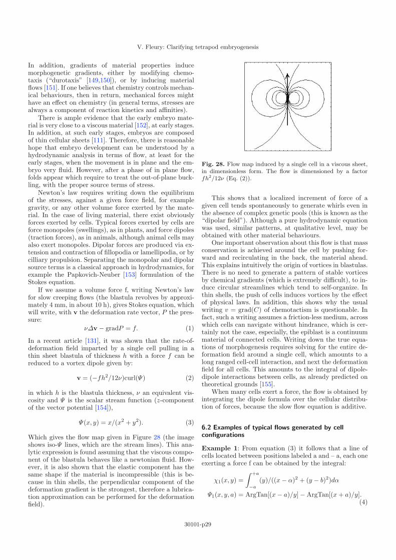
V. Fleury: Clarifying tetrapod embryogenesis
In addition, gradients of material properties inducemorphogenetic gradients, either by modifying chemo-taxis (“durotaxis” [149,150]), or by inducing materialflows [151]. If one believes that chemistry controls mechan-ical behaviours, then in return, mechanical forces mighthave an effect on chemistry (in general terms, stresses arealways a component of reaction kinetics and affinities).
There is ample evidence that the early embryo mate-rial is very close to a viscous material [152], at early stages.In addition, at such early stages, embryos are composedof thin cellular sheets [111]. Therefore, there is reasonablehope that embryo development can be understood by ahydrodynamic analysis in terms of flow, at least for theearly stages, when the movement is in plane and the em-bryo very fluid. However, after a phase of in plane flow,folds appear which require to treat the out-of-plane buck-ling, with the proper source terms of stress.
Newton’s law requires writing down the equilibriumof the stresses, against a given force field, for examplegravity, or any other volume force exerted by the mate-rial. In the case of living material, there exist obviouslyforces exerted by cells. Typical forces exerted by cells areforce monopoles (swellings), as in plants, and force dipoles(traction forces), as in animals, although animal cells mayalso exert monopoles. Dipolar forces are produced via ex-tension and contraction of fillopodia or lamellipodia, or bycilliary propulsion. Separating the monopolar and dipolarsource terms is a classical approach in hydrodynamics, forexample the Papkovich-Neuber [153] formulation of theStokes equation.
If we assume a volume force f, writing Newton’s lawfor slow creeping flows (the blastula revolves by approxi-mately 4 mm, in about 10 h), gives Stokes equation, whichwill write, with v the deformation rate vector, P the pres-sure:
νΔv − gradP = f. (1)
In a recent article [131], it was shown that the rate-of-deformation field imparted by a single cell pulling in athin sheet blastula of thickness h with a force f can bereduced to a vortex dipole given by:
v = (−fh2/12ν)curl(Ψ) (2)
in which h is the blastula thickness, ν an equivalent vis-cosity and Ψ is the scalar stream function (z-componentof the vector potential [154]),
Ψ(x, y) = x/(x2 + y2). (3)
Which gives the flow map given in Figure 28 (the imageshows iso-Ψ lines, which are the stream lines). This ana-lytic expression is found assuming that the viscous compo-nent of the blastula behaves like a newtonian fluid. How-ever, it is also shown that the elastic component has thesame shape if the material is incompressible (this is be-cause in thin shells, the perpendicular component of thedeformation gradient is the strongest, therefore a lubrica-tion approximation can be performed for the deformationfield).
Fig. 28. Flow map induced by a single cell in a viscous sheet,in dimensionless form. The flow is dimensioned by a factorfh2/12ν (Eq. (2)).
This shows that a localized increment of force of agiven cell tends spontaneously to generate whirls even inthe absence of complex genetic pools (this is known as the“dipolar field”). Although a pure hydrodynamic equationwas used, similar patterns, at qualitative level, may beobtained with other material behaviours.
One important observation about this flow is that massconservation is achieved around the cell by pushing for-ward and recirculating in the back, the material ahead.This explains intuitively the origin of vortices in blastulas.There is no need to generate a pattern of stable vorticesby chemical gradients (which is extremely difficult), to in-duce circular streamlines which tend to self-organize. Inthin shells, the push of cells induces vortices by the effectof physical laws. In addition, this shows why the usualwriting v = grad(C) of chemotactism is questionable. Infact, such a writing assumes a friction-less medium, acrosswhich cells can navigate without hindrance, which is cer-tainly not the case, especially, the epiblast is a continuummaterial of connected cells. Writing down the true equa-tions of morphogenesis requires solving for the entire de-formation field around a single cell, which amounts to along ranged cell-cell interaction, and next the deformationfield for all cells. This amounts to the integral of dipole-dipole interactions between cells, as already predicted ontheoretical grounds [155].
When many cells exert a force, the flow is obtained byintegrating the dipole formula over the cellular distribu-tion of forces, because the slow flow equation is additive.
6.2 Examples of typical flows generated by cellconfigurations
Example 1: From equation (3) it follows that a line ofcells located between positions labeled a and – a, each oneexerting a force f can be obtained by the integral:
χ1(x, y) =∫ +a
−a
(y)/((x − α)2 + (y − b)2)dα
Ψ1(x, y, a) = ArgTan[(x − a)/y] − ArgTan[(x + a)/y].(4)
30101-p29

The European Physical Journal Applied Physics
A B
C D
Fig. 29. (A) The flow map exerted by a line of cells alignedalong the same line, each one pulling with the same force (hereb = 0, and a = 1/6). The figure shows the levels of the streamfunction Ψ (with a linear shaded grayscale between levels, indimensionless form. The actual scale is defined by the viscosityof the material, the magnitude of the pulling force, and theblastula thickness). The stream function iso-values curves arelocally tangent to v. (B) The flow map exerted by a segmentof cells, each one pulling parallel to the horizontal-axis, butthemselves piled up along the vertical axis, with the same force.(C) The flow map exerted by two segments of cells exertinga contraction towards a hyperbolic point. (D) The flow mapas it should be viewed on the blastula at the beginning ofgastrulation.
Which gives the flow map given in the Figure 29A.Example 2: from equation (3) it follows that a segmentof parallel cells pulling forward exerts a force obtained bythe integral:
Ψ2(x, y, c) =∫ +c
−c
(y − α)/((x − a)2 + (y − α)2)dα
= (1/2)Log((x2 + (y − c)2)/x2 + y2))−(1/2)Log((x2 + (y + c)2)/x2 + y2)). (5)
Which gives the flow map given in Figure 29B.Another solution exists, which is a solid body revo-
lution around a point. Indeed, a straightforward calcula-tion shows that the stream function for a uniform volumeforce oriented along the orthoradial vector, in polar coor-dinates, gives a circular vortex with an orthoradial speedwhich varies linearly with the distance to the center, like amerry-go-round (solid-body revolution). This type of re-volving vortex is observed for cilliary flows of epithelialcells (Roberto Camassa, personal communication), whenthe epithelial cells are attached bio-chemically to a sub-strate. In this case, the epithelial cells are immobile, andthe culture medium above them revolves with a viscousvortex which has a solid-body revolution pattern.
7 Application to embryo morphogenesis
7.1 Simplification of generic embryo movements
The movements observed during gastrulation may lookvery complex, and this is why the role of many molecu-lar expressions are investigated. At present, the back andforth motions of cells in Hensen’s node, and the back andforth motion of cells towards the primitive streak andaway, are “explained” by chemical gradients acting oneway, or the other way around, at given times. It is how-ever not certain that it is necessary to invoke first the ex-istence of a chemo-attractant along the body axis for cellsof the epiblast, in order to explain their motion towardsthe AP axis, and then a chemo-repellent, which presum-ably starts to act as soon as the cells dive forward, in orderto explain why the mesoderm migrates away from the APaxis, immediately after having migrated towards it. Oneimportant unsolved question is how evolution could haveselected independently a chemical process of opening of astreak and another chemical process for closing of the samestreak, while forming for supposedly many generations aviable animal with an open streak. It is more logical thatthe two phenomena are correlated enough, that in somerange of parameters, they will occur “automatically” i.e.the opening is necessarily followed by closure.
Since the entire geometry of the blastula and theboundary conditions of the cellular flows change as theprimitive streak opens and cells invaginate, one must alsoaddress the impact of these phenomena on the directionsof the flows. One finds a reasonable explanation of gastru-lation movements, when taking into account the changein boundary conditions during the cellular flows.
As stated above (Sect. 5), gastrulation starts by thepulling action inside a sector of cells [122,130,156]. Thecells may have been prepared in this state by earlierevents, what matters is that they start off with a specificorientation. The cells may actually flow, in which casethey exert a pulling action, or they may change shape,in which case they exert a small quadrupole (they pull inone direction, and push in the other). It makes little dif-ference, from the point of view of physics, and at this levelof description, because of linearity of the solutions, withrespect to the sources of force.
Since the initial cortical rotation is a physical processthat creates a mismatch of all chemicals between the cy-tosol and the cortex, and since the area of KRS is verythin, we may assume that the cells are all identical insidethis sector of cells, and find the flow map inside the epi-blast, by positioning two solutions of equation (4) headto head (Figs. 29C, 29D). The corresponding stream func-tion is:
Ψ3 = ArcTan[(x − a)/(y)] − ArcTan[(x)/(y)]−(ArcTan[(x)/(y)] − ArcTan[(x + a)/(y)]). (6)
This implies the existence of a high stress point located atthe edge of the segment and advected towards the cross-ing of the sector and the AP axis, this point is apparent
30101-p30

V. Fleury: Clarifying tetrapod embryogenesis
in Figures 29C,29D as the hyperbolic point (flow is con-vergent in one direction, divergent in the other). Moreprecisely, the flow exhibits a hyperbolic pattern aroundthe neutral point (saddle-point). This is associated to hy-perbolic lines which correspond to stream function of theform Ψ(x, y) = kxy (Ψ = cte defines the streamlines).Around this point, the hyperbolic flow can be linearizedsuch that the x and y components write:
vx = −kx (7a)vy = ky (7b)
with k a dimensioning constant. The velocities are lin-ear. In case of an inertial flow, the pressure (stress) isfound by Bernouilli’s law, by writing P = P0/ρ − v2/2which gives a maximum pressure at the saddle point(v = (0, 0)). For a viscous flow following a Poiseuille pro-file v = −h2/12νgrad(P ), where h is the layer thicknessand ν the viscosity; the solution for the pressure is givenby
P = P0 + (6ν/h2)(kx2 − ky2). (8)
Which shows that the flow arrives along the x axis from aregion of high stress (high x2), and flows away in the y di-rection towards regions of low stress (low – y2). Therefore,the area around the saddle point is at high stress, and thestress decreases in a saddle form away from it anteriorly,and posteriorly.
It is known that polarised cell flow can be inducedby a mechanical stress [163]. Therefore if there exists athreshold of stress such that cells contract to invaginate(some sort of escape of the stress), this threshold will firstbe reached at the most compressed point. From there,cells will start to invaginate in an avalanche mechanism,and a line of epiboly will propagate like a crack towardsthe caudal pole and the anterior pole, as observed. Thiscrack is what was described above as the primitive streak(PS), Figure 23. However, since the pulling sector is al-ready caudal, the propagation is mostly conspicuous inthe anterior direction, starting from the posterior hyper-bolic point (Fig. 23 left). It has been stated for long thatthe primitive streak propagates in the posterior to ante-rior direction [111]. In my opinion, this is actually wrong(personal observation). The primitive streak propagatesin both directions, from the intersection of Koler-Rauber’ssickle with the median axis.
Now, as the PS propagates, it enters regions where thestress in the tissue is smaller, because of the recircula-tion of the flow (the term −ky2 in equation (8) brings anegative component).
As a consequence, the streak will propagate up to apoint where the sum of the contractile stress exerted bythe PS, and the surrounding stress linked to the KRS addup to a stress which finds itself below the threshold forepiboly. There, the furrow should end, and this is the pointof arrest of Hensen’s node (HN). The Primitive Streakhas a length L(PS) at which the stress term exerted bythe cells along the PS cancels the stress created by theKRS. Quantitatively, one can estimate this distance sinceknowing f1 the individual force exerted by cells inside the
contractile sector, and f2 the force of the epibolizing cells,the distance is such that L(PS) = L(KRS)f1/f2. Thereis little doubt that the pulling or contracting force of cellsshould be of comparable magnitudes, so that the lengthof the primitive streak is of the same order of magnitudeas that of the KRS, hence the body axis occupies a largeportion of the blastula.
Now, as cells ingress inside the (presumptive) embryo,the traction force at the epiboly adds a flow componentalong the furrow which is given by equation (5), with thecorresponding length c = L(PS)/2. Indeed, we assumethat cells which start to epibolize exert a traction on theepiblast fixed by internal dynamics, and independent ofpossible gradients. This amounts to supposing that cellsare in a bistable state: either they fold inwards or theydon’t, but if they do, they do so with equal force, simplybecause the cell genetic content is supposedly the same.Therefore, when the primitive streak exists, the flow fieldis as in equation (5), and the cells progressively invagi-nate along the streak, as schematized in Figure 25B right.However, as cells invaginate, make a U turn, and start tomigrate in opposite direction, they form the yolk-sac (YS).
Cells in the YS migrate away from the streak, under-neath with a similar flow as equation (4). It has beenshown recently that the flow of cells in a field like equa-tion (4), generates automatically a round YS around theembryo (exact solution of the cell transport) when no spe-cific morphogenic instruction of any sort is assumed [119].
7.2 Coupling chemical gradients,mechanotransduction, and hydrodynamics
It is not established whether the flow pattern organizesitself in response to chemical gradients present in the en-tire blastula, which fine tune the vector field at each spot.At present, the most reasonable origin of the entire move-ment, is the force term generated inside this narrow area(Koller’s sickle) [157]. Which in turn, and together withstress forces, induces such growth factors as FGF8, andtranscription factors as Brachiury (Tbox). Once “trig-gered”, the flow continues and self-organizes. However,pools of chemicals do exist later during embryo move-ments, which have chemotactic properties, and the en-tire morphogenesis may become a complex mix of chemo-tactism and physical flow, with possible gradients of celladhesiveness. Biologists sometimes attempt to distinguishbulk flow and cell migration, although bulk migration isinduced by cell force (hence individual migration) [158].Moreover, this is done by separating the motion of theextra-cellular matrix from the motion of the cells. It mustbe observed that both form a physical layer but only themoving cells exert forces. Therefore, by the principle ofaction and reaction, the extra-cellular matrix bears anopposite force as that of cells, therefore both the mo-tion of cells, and the antagonist motion of the ECM havea hydrodynamic-like “bulk” component, and one cannotstate that the ECM flow is the bulk flow and the chemotac-tism the difference between the total flow and the extra-cellular matrix flow.
30101-p31

The European Physical Journal Applied Physics
Fig. 30. While the furrow nucleates at the most stressed point it will stop when the stress reverts below the threshold forepiboly. This will occur as the furrow enters regions of opposite stress in the anterior part. In the figure above, right, we showthe stress state in the region of nucleation of the streak, and the stress state, in the region of arrest. The lines represent streamlines. The flow is organized from a high pressure point located in the area of the presumptive navel, towards a low pressurepoint located in the anterior area.
The weakness of the chemotactic hypothesis is thatit requires a fine tuning of molecular pathways, and anexplanation of the origin of each pool of chemicals, spatio-temporally, which is still lacking, not even mentioning thephylogeny of the entire flow pattern.
This is to say that the inductive view finds itself in anever ending regression, trying to find a prepattern, whichactually is permanently advected by the flow (what biolo-gists call “translocation” or “dynamic expression”). Thishypothesis considers the fine tuning of all elements in theproblem as crucial to the appearance of the animals, up tothe point that it is generally believed that animals couldas well not have appeared, and that random events playeda great role. I believe this is a misleading view. If animalsform from an active material which produces under stressthe growth factors congruently with the morphogeneticfield, the fine tuning of the chemistry is less essential:many aspects are absorbed into scaling parameters.
The problem with the hydrodynamic hypothesis is thatit seems uncorrelated to the presence of chemicals, exceptfor the initial condition, although it is well known that themorphogenesis can be completely disrupted by brute forceadministration of many chemicals [159,160]. One interestof the hydrodynamic hypothesis is that direct inspection ofcellular flows show that the morphogenesis of the embryopattern is qualitatively complete after less than one cel-lular revolution. This is to say that, contrary to commonknowledge, global morphogenesis is very rapid (betweenone and two days in the chicken), and does not requiremany iterations of stop-and-go discrete inductions43 (basi-cally: a horizontal bar is advected and flattened to becomea vertical one, the process requires 1/4th of a revolution).
A way of reconciling the two approaches consists inwriting the constitutive equation as (assuming that cells
43 Anecdotally, the best physical analogue of formation of avertebrate embryo is observed during Chinese shrimp chip fry-ing. Chinese shrimp chips are delivered with a saddle point.When fried, they undergo considerable deformation. The formobtained transiently is very much similar to an embryo at thegastrula stage, just after formation of the neural crest.
align in the main shear force)
νΔv − gradP = fv/||v||. (9)
And add the chemotactants as volume forces of the formgrad(C). However, it has been shown that many growthfactors especially that of the FGF family are in fact regu-lated linearly by pressure [142]. In this case, grad(C) canbe written as proportional to grad(P ).
grad(C) = αgrad(P ) (10)
and the constitutive equation becomes of the form:
νΔv − (1 + α)gradP = fv/||v||. (11)
This allows to couple the chemotactism and the volumeforce exerted by cells, into an equation of the Stokes formwith rescaled parameters. This is a closed equation. Itprovides a model system, in which the chemotactants areproduced dynamically by the flow itself. In this view, cellsgenerate by stress the growth factors which are their ownincentive for the continuation of their movement, and cellsuse the established pattern to generate the orientation ofthe physical force. This situation is known to occur in lungor kidney development, and it is known that FGF are cru-cial growth factors for both limbs and lungs [52], and thisis also true of Tbx5 for lungs44 [161]. Therefore, if the be-haviour of fgf ’s with respect to force is the same in limbmesoderm, and in lung mesoderm, then an equation likeequation (11) provides a simple constitutive equation forthe viscous living sheet, assuming that biochemical pro-duction is in steady state with respect to the flow. As aconsequence, the overall apparent motion of cells follow-ing growth factors and pulling themselves forward in the44 One should observe that FGF10, Tbx4 and Tbx5 areall important in limb and lung development. However, over-expression of Tbx5 in the lung has a similar effect as an in-crease in pressure (acceleration of branch growth). It may bepossible that the same pathways are used in limb and lungdevelopment, although the topological difference between limband lung is that the order of lung cell sheets is “inside out” ascompared to a limb.
30101-p32

V. Fleury: Clarifying tetrapod embryogenesis
direction of the flow, is a Stokes flow of the cells in a lessviscous material, but with the same force oriented alongthe local existing streamline. Although the true constitu-tive equation might be formally more complex, it does notrender the phenomenon itself more complex.
As explained above, the flow is a quadrupolar hyper-bolic flow. It has been shown that such a flow transformsthe blastula, and everything inside, into elongated “8”shapes [16,131]. This explains very well the overall form ofthe early embryo, and also, of several pools of chemicals,whose form can be understood on the basis of a lagrangiandescription of the flow (see Sect. 7). Especially, stainingsagainst FGF8, which is so important for development, atearly stages, have shown that there exists a localized spotof FGF8 around the contraction centre, which progres-sively becomes a slender 8-shaped zone of expression [162],Figure 31. (See also other gene stainings, especially wnt,in GEISHA database, or in Ref. [162]).
This fits perfectly well with a biomechanical advectionin a hyperbolic flow, and shows how chemical gradientsare reshuffled by the stream. This is actually perma-nently read in developmental articles, where genetic ex-pressions are constantly described as “dynamic” (passim).A static diffusible chemical/inductive explanation requiresa formidable analysis of chemical reactions, to explain thedynamical change of a circular spot into an elongated baror an elongated “8-shape”.
This field of research will probably progress by find-ing a more realistic equation for the chemotactic fields (ifany), or by numerical integration of advection-diffusionequations for the growth factors. It is important in thiscontext to measure locally the mechanical properties ofthe tissue, and correlate genetic expressions dynamically,to the stress fields. From the point of view of regenera-tive medicine, it is important to know whether the properboundary conditions for a limb bud could be artificiallyreproduced by some artefact. While it is unlikely that anentire limb or hand could be properly generated (the jointorganization will not be fit), it is reasonable to imaginethat an isolated limb paddle of arbitrary length could bereconstructed, to taylor at will rows of digital rays, or evenrows of fingers.
7.3 Significance of the closure of the primitive streak
As the YS expands away, it adds a stress which contributesnegatively to the stress of the PS and the stress of theKRS. Therefore, the primitive streak, which is an openfurrow of epiboly, progressively recedes, as increases thetotal stress the cells (located at the epiboly line) haveto fight to succeed to invaginate. Stated otherwise, theflow linked to the KRS, plus the flow linked to the YSprogressively shuts the primitive streak like a zipper, asobserved. At the apex of the zipper, the stress is equal tothe threshold for cell epiboly. Therefore, Hensen’s node in-deed moves forward during the “crack opening” and thenrecedes during the “crack closure”, but this back and forthmotion of the HN might simply correspond, as observed,
to a deformation wave in the area of the apex of the crack,controlled by the genetics of cell traction force.
Now, as the PS closes, cells which move towards the APaxis start to intercalate. This word, in biology, means thedeformation of cells by which they move their neighbour-cells sideways to intercalate between them. Although theremay exist chemical incentives to intercalation [120,166], anatural explanation is that cells moving towards the APaxis, as they do, under the action of the epiboly traction,will intercalate if they are hindered in their epiboly mo-tion. In simple words, intercalation would be just the samething as an epiboly or a cell migration, it depends only onthe stress context (epigenetic cue) of the given cells whichtry to move forward. On mathematical grounds, an in-tercalation along a line such as the AP line is given bythe mathematical solution of equation (4) (rocked to thevertical, and with a minus sign), if we assume that cellsat the intercalation line all behave identically, which is acrude assumption, however, this assumption is justified bythe fact that the hyperbolic flow acts as a massive stretchalong the median axis of the initial spot of FGF8, there-fore, we may expect the possible inhomogeneities to besmoothed away.
In other words: intercalation of a cell causes an incre-ment of stretch or extension along the AP axis, as ob-served, and the integral of these individual stretches canbe modelled by a flow. The parallel intercalation of allcells generates a large quadrupole along the body, centredon the neutral point of stretch (presumptive navel).
So, along the body axis where the primitive streak isalready closed, the tissue feels a deformation field whichis the sum of a stress due to cellular divergence from thePS which tends to orient the flow away from the PS, andan extension flow along the body axis oriented away fromthe navel due to cellular intercalation. This flow patternwrites (again with the assumptions that cells along themidline behave mechanically in the same way):
Ψ1(x, y)+Ψ2(x, y) = α{(1/2)[Log((x2+(y − c)2)/x2+y2))
− (1/2)Log((x2 + (y + d)2)/x2 + y2))]}+ β{ArgTan[(x − f)/(y)]− 2ArgTan[x/y]+ArgTan[(x+g)/(y)]}.
(12)
Or, in a more pictorial representation (Fig. 32):where α and β are ad hoc parameters, of comparable val-ues related to the magnitude of the force terms, and possi-ble gradients, controlled of course by genetic data. Becauseof symmetry around the initial nucleation point, the flowsare centred around the presumptive navel, where a sin-gular point exists. c, d are parameters of the contractionsegment and f, g parameters of the extension segment,characterizing the length of the streak, and of the epibolyline. We assume that these are in proportion of the cellularforces, and the number of pulling cells.
Now, such figures as the embryo shape represented inFigures 23, 24 become quite clear. As the stress on topof the epiblast is released, the primitive streak recedes(crack closure). However, as more cells intercalate, the
30101-p33

The European Physical Journal Applied Physics
Fig. 31. (Color online) These images show several stages, at shortly different times, of concentrations of genes, as stained ondifferent embryos, forming a stroboscopy of the developmental process. During primitive streak (p.s.) formation, the patternof expression of Fgf8 (top) transforms from a circular spot around the contraction area, into a large “8-shape” centred on thepresumptive navel area, between stage 2 and 3 (Hamilton & Hamburger stages of embryo development) from reference [163]. Asimilar advection pattern is obtained for other genes such as Wnt8c and chordin (middle) or Clef1 or Gata5, which transformfrom a horizontal “bar” or “oval” to a vertical “bar” [162,165]; see also many early stainings of the database GEISHA. All genesactually seem to follow an advection into a hyperbolic flow (bottom, in a lagrangian view, the hyperbolic flow is elongational,and transforms affinely a spot into an elongated bar positioned along the median axis).
30101-p34

V. Fleury: Clarifying tetrapod embryogenesis
α. + β. Fig. 32. A simplified picture of the rate-of-deformation field in the embryo is given by the sum of a contraction, and anexpansion along the body axis, which describes a convergent-extension with identical cells (Eq. (12)). (The lines represent iso-Ψvalues of the stream function, the graded areas the gradient between values, in dimensionless form. Such parameters as forcemagnitude, viscosity, and thickness of the blastula will scale the problem).
stress, which is strictly neutral at the moment of perfectclosure of the primitive streak, becomes compressive per-pendicularly to the AP axis. Hence the tissue buckles andfolds. Therefore, it is completely natural that the primi-tive streak recession (descent of the zipper, neutral stress)is followed immediately in space and in time by forma-tion of folds (the neural crest, positive stress), giving anerroneous impression that the tip of the streak (Hensen’sNode) is “inducing” the embryo body as it descends.
In addition, the entire embryo tissue is transportedin the intercalation flow and produces naturally the large“8” around the embryo folds (Fig. 33). Moreover, the ex-istence of a limited extension in the anterior part of theinitial primitive streak, implies a finite length for the inter-calation parameter f , while the primitive streak extendsall the way down inside the caudal area. If we advecta blastula-like loop of tissue in the “extension” (α = 0,β = 1), we immediately see that the loop flattens in thedirection of the navel, while the re-circulation of the flowlines induces a progressive closure of the “neural crest”in the middle of the body around the navel, with a de-scent of the closure of the neural crest towards the tailbud, while a furrow remains wide open in the “head” re-gion. This may explain why a head exists eventually onthe anterior side, while the neural crest closes completelyat the tail side, a fact which has no explanation otherwise.Also, since the tail part is closer to the caudal edge of theblastula, the winding of the epiblast towards the bodyis more rapid in the caudal area, hence explaining whyhindlimbs will form before forelimbs. Molecular stainingswill be advected in this flow, as quite clear in Figure 9, orin database GEISHA.
All this is a consequence of hydrodynamic conservationlaws in a viscous sheet. In addition, the neural folds moveforward and push sideways the ocular regions, in a ham-mer or horseshoe shape formed by some sort of a “trail” ofthe fold deformed in the given flow, as classically known indevelopmental biology (See data base GEISHA especially
Hamilton & Hamburger stages 5). It is known from graftexperiments that the entire area of the flow trail in thehead region can potentially give retina tissue [167]. Theformation of the eye is a matter of progressive physical,lateral evagination of optic vesicles. Due to the horseshoeshape of the trail, eye precursors drift and get positionedin the actual eye cup [168]. However, zebrafish mutantswhich have a much reduced trail, and a head which pro-trudes more ahead than sideways are cyclopic, they havea single eye ahead [169], this is ascribed to the fact thatthe eye spacing is spatio-temporally decreased by modi-fications of genes (kny and Tri) which affect the hyper-bolic flow. When the size of the “forehead” is too small,a bifurcation from a “peanut shape” eye to a single cen-tred eye occurs such that a single eye is formed on theforehead. This situation is quite reminiscent of the limbdevelopment: as for limbs, an entire field is “competent”for development of some parts (eyes, limbs) but these willonly grow out at a specific localized areas, which are con-comitant with curved bumps (see Sect. 7.4) defined bygeometrical and physical constraints.
This suggests that the bilateral symmetry of the eyes,and of the face is a hydrodynamic consequence of the flowmagnitude which generates a wide horseshoe form of thepresumptive head region. In more evolved vertebrates thehorseshoe head so to speak “inflates” above the spine togive craniates. More primitive animals like Tiktaalik havea flat horseshoe head shape.
To end this section, let us note that the global hy-perbolic flow flattens the tissue along the AP axis in theregion of the presumptive limbs (quite visible in Fig. 22,in the region of the hindlimb), while the tail apex movesmore caudally along the AP axis, and progressively closes,creating 4 windings away from the presumptive navel, 2above and 2 below. Therefore, the formation of the bodyplan may be a direct, and quite simple, consequence ofthe flow pattern, with several biomechanical parametersof genetic origin, and many events described in biology by
30101-p35

The European Physical Journal Applied Physics
Fig. 33. Transport of a circular pattern in a generic “extension flow”, with a navel positioned arbitrarily towards the south ofthe initial blastula. One sees the progressive collision of the circle along the presumptive neural crest. The first region of theembryo to close is the central area (around the navel) Because of the caudal position of the Koller-Rauber’s sickle, the tail fold(tail neuropore) closes completely as it extends caudally, while the head fold remains wide open. This is apparent here in thatthe keyhole or “8 shape” of the second snapshot, is elongated so much in the AP directions that the bottom circle of the eightvanishes progressively, but not the top circle.
different morphogenetical stages may actually be just theconsequence of the transport in the flow. What the cal-culation shows, is that apparently very complex motions,can boil down to very natural hydrodynamic flows. Thehardly intuitive character of hyperbolic flows may let onethink that embryogenesis is very complex. Integrating intime the trajectories in a non trivial, but simple hyperbolicflow, suggests that, although the true equations may beslightly more complex technically, the phenomenon itselfis deceptively simple.
7.4 Discussion of he biomechanical origin of limbs
As explained above, the positioning of the limb is still anopen problem, from the developmental biology point ofview. We understand that the origin of lateral plates (the“limb field”) is crucial, since these will define the posi-tioning of the normal limbs, and even many details suchas orientation of the limbs, if not their kind (forelimb orhindlimb). The “commitment” of the limb may well berelated to the exact cellular distribution in the cellularfield existing around and inside the lateral plate, prior tothe outgrowth of the limb bud. Tetrapods are defined bythe existence of four lateral plates, which eventually de-velop into different limbs, these plates find themselves ad-vected naturally into their own “field” which participatesin anisotropies, inductions, cellular traffic, etc. of the limbpaddle. Understanding the overall orientation should bepossible by addressing the deformation field during mor-phogenesis. It must be observed that the orientation ofthe tissue (the mean direction of cells and fiber bundles)at very early stages is an important component of morpho-genesis [126,170–172]. Stopak et al. [170] show the crucialfact that “. . . within developing tissues, patterns of forcesexist that are capable of physically rearranging collagenand determining its long-range order”. Rotation of pieceof tissue disrupt completely the growth, therefore “posi-tion information” [171] is actually not a scalar quantity,but more a vector or a tensor. When limbs are producedectopically with a localized spot of growth factors, it isgenerally aberrant, although it “looks like” a limb (one ex-
Fig. 34. (Color online) Ectopic limb, created in between fore-limb and hindlimb by a soaked bead, shows a straight pattern(no flexion at the “knee-elbow” joint.). From reference [77].
ample of ectopic limb growth along the flanks of a chickenin Fig. 34).
Let us return to the establishment of the limb posi-tion. A typical Carnegie stage 14 human embryo showsclearly its limb buds, 2 forelimbs, 2 hindlimbs (Fig. 35A).If one winds backwards the development of the limb,e.g., hindlimb, one finds that it originates in a roundishtissue thickening located just below the presumptivenavel area, for the hindlimb, and above for the forelimb(Figs. 35B, 35C, 35D its a chick). If one winds again to-wards the second day of development (of a chick embryo)one finds that the lateral plate originates in the rotationalengulfment of the epiblast45 towards the median antero-posterior axis, while the axis itself extends caudally duringthe convergent-extension46 (Figs. 35D, 35E, 35F). There-fore, the rationale for lateral plate formation is that of awinding of the epiblast towards the body axis, while thebody axis extends caudally (Fig. 36).
A typical image indicating how lateral plates may be“deposited” is found in [66]. Figure 37 shows the limb budsalong the AP axis of a direct frog embryo (a frog whichdevelops its legs in the egg, not at a tadpole stage). Itshows that the lateral plates find themselves in the con-tinuity of a line winding caudally, and rostrally, arounda neutral point located half way between forelimbs and45 In the early blastula, at a stage when the embryo is a flatdisc, the top foil is called epiblast, the bottom foil is calledendoblast.46 This is the name of the hyperbolic flow in biology.
30101-p36

V. Fleury: Clarifying tetrapod embryogenesis
A B C
D E
F
Fig. 35. (Color online) (A) typical Carnegie stage 14 embryo (human, Dr Mark Hill website, UNSW medical school) showingthe limb buds. (B) Typical chicken embryo (stage HH19, ULB Louis de Voos web site) showing the bumps on the flanks (earlylateral plates). (C) typical chicken embryo, showing the lateral thickenings forming the lateral plates (Geisha database). In (D)stage 15 HH, the thickenings are only faintly visible. In (E) (courtesy Alia Al-Kilani), they almost vanish, but they appearas a winding towards the posterior, or anterior part, the presumptive navel being in the middle. In (F) the winding in thepresumptive caudal plates appears more clearly. In this image shadowgraph surface imaging was used to show that there is anuplift of the epiblast in the presumptive hip-region, which accompanies the rotational engulfment of the epiblast towards themedian axis.
30101-p37

The European Physical Journal Applied Physics
Fig. 36. (Color online) Left, description of the process of tetrapod development (chicken). Right the flow in the early blastula(AP axis is horizontal). The lateral plate originates in the winding of the epiblast tissue converging towards the body axis, whilethe body axis extends in the antero-posterio direction. The body axis extends caudally, in the tail region, and anteriorily in thehead region, thus creating a strong winding of the lateral plate mesoderm, and related ectoderm folds away from a neutral pointwhich is the presumptive navel (from Ref. [173]). Many molecular stainings in the caudal area just stain the actual geometry ofthe engulfments (see for example Tbx4 in Fig. 9 above).
Fig. 37. Left, from reference [66], limb positioning in a direct frog. Middle and right, explanation of the anlagen 48of limbbuds, as a consequence of the vortex motion in the blastula. The epiblast lifts up as a consequence of von Karman bucklingunder the hyperbolic stress [179].
hindlimbs. This situation is the one generated by the epi-blast winding during gastrulation [16], in this view, theorigin of the lateral plates is entirely within the motion ofthe cellular flow which self-organizes in the shape of twovortex dipoles colliding along the median axis, as a contin-uation of the hyperbolic flow (Fig. 37 right) and lifting upthe tissue in the areas of strong winding towards the me-dian axis, by von Karman buckling. Meantime, the wind-ing pushes the head and tail so much forward that theydive in a curled manner, thus inducing the ectoderm tofold in a pocket shape that generates the chorio-amnioticfold. The collision of such vortex dipoles is quite appar-ent on films of the early moving gastrula. Such a movieis visible on Keller’s website (shown also at http://www.gastrulation.orgmovie 15-3), and on youtube (keywordgastrulation).
Therefore the 2 + 2 = 4 distribution of limbs intetrapods seems to be a direct consequence of the typi-
cal hyperbolic nature of the flow around the navel region,and so is the transition from anamniotes to amniotes. Inaddition, the overall orientation of the cellular fields inthe area of the limbs may be a consequence of the flowwinding.
If we assume that the limb bud will emerge from thecentral part of the winding, as it collides along the flanks,we see that the more proximal part will form out of themore bent parts of the lateral plate. As a consequence,the differential outgrowth of the lateral plate will progres-sively “blow up” or “reveal” the structure of the initialwinding. This is why the limb bud of the hindlimb willbe formed of elements impinging on the pelvis upwards,and then descending towards the paddle, while the limbbud of the forelimb will be formed proximally by a firstsegment descending from the scapula, and this descend-ing element will next rotate upwards towards the forelimbpaddle. Pelvis and scapula will retain the general outlook
30101-p38

V. Fleury: Clarifying tetrapod embryogenesis
of the motion. A process of internal segmentation whichwe shall not discuss here, creates the bony structures, buton an overall limb form which is not itself segmented.
It is therefore clear that different parts of the embryoarrive where they have to by the flow, and that the flowhas to be calculated by the laws of physics. The wind-ings and folds of the early embryo have a direct impact onfurther growth, by fixing the boundary conditions and theinitial cell orientations in all organ rudiments prepared forfurther growth, hence providing this coherent textural pat-tern associated to muscle, tendons, ligaments orientation,etc. which has been remarkably imaged by Kardon [172].Therefore, there are no physical grounds in saying that asingle restricted area such as Hensen’s node is the “orga-nizer” of the entire process. For example: the closure ofthe neural crest at the tail, the opening of the neural crestin the cephalic region, and the position of the limb budsare linked to the way the flow field recirculates, Also itis quite clear why ectopic limbs are generally not normal,since they are not positioned by exactly the same gentleconvective pattern.
More specifically, it has been shown that the lateralplates protrude out during rotation and convergence ofthe epiblast towards the median axis. Mechanical mea-surements on the lateral plates show that they correspondto low stress areas, as expected for bent areas [173,174],such that cells in the limb field actually have a special“physical” phenotype. Therefore, in addition to possiblechemical gradients which are stretched and reshuffled bythe flow (advection-diffusion), such as Chordin, Hoxc6,Fgf8, Gata, etc. the physical force that winds the tissue inthe areas of the lateral plates lifts them up by mechanicalforce, preparing a possible outgrowth, if growth factors areproduced continuously. If they are not, limb growth canbe interrupted at any time, giving the tremendous list ofexperimental assays, which show any type of limb, from asimple skin fold remaining along the body, to well formedlimbs. However, if growth of the animal does continue, af-ter the collision and winding, the first area to grow willbe the limb buds of the lateral plates. These may inheritfrom early mode lockings the aspect of a digital arch, withregular rays.
8 About segmentation
While the embryo body forms, it also segments in whatwill progressively become cartilage condensations andbony elements. Indeed, it is often stated that a subtleinterplay of chemical waves generates from the base ofthe neck down towards the tail the body segments of thevertebrates (early precursors of vertebrae, ribs, etc.). Aconsiderable work is dedicated to the study and analy-sis of the genetic determinants of this genetic clock [175].There exists a possible erroneous concept conveyed by thegenetic clocks which are believed to generate these bodysegments in vertebrates. In effect, it is true that this seg-mentation wave descends from the presumptive neck downalong the spine. However, there is ample Scanning Elec-tron Microscopy evidence that, in fact, the body is already
Fig. 38. Explanation of the global orientation of the limbs,in addition to their positioning. The hyperbolic flow windsthe lateral mesoderm, and flattens the folds along the flanks.Progressive outgrowth of the most protruding part of the lat-eral plate will progressively reveal the winding pattern, andform limbs having the anatomically observed orientation (el-bows flex rostrally, knees flex caudally). The number of digitswill be locked onto the somite (vertebrae) number, at start ofoutgrowth. Typical pelvis shape is the result of the global flow.
segmented into somitomeres49, which are precursors of thesegments, at the stage when the segmentation wave pro-ceeds [76,176]. This segmentation wave seems only to am-plify a pre-existing segmentation of the body, in somit-omeres. The somitomeres are lenticular, stellar, foci ofmesodermal cells which appear at regular intervals fromhead to tail, as the primitive streak closes [176]. Theyare positioned along the folds of the embryo body. Ex-perts in the field ascribe this regular segmentation of thefold to the shear occurring in the area of the tip of theprimitive streak [177,178], and personal communicationwith Prof. Jacobson]. It was once thought that Hensen’sNode was organizing the somitomeres pattern as it re-cedes. However, it was shown that HN is not actuallynecessary for formation of somitomeres [177]. In addition,when the embryo is split, the somitomeres form every-where at the same time, and not in a sequential fashionfollowing HN [178], thus suggesting a direct effect of shearalong the fold (some sort of buckling). Such a segmenta-tion process may be described by buckling equations of thevon Karman type [179], acting on a sandwich of ectodermand mesoderm. Such bilayers are intrinsically unstable,and buckle above a threshold of instability. Such models
49 Precursors of somites, themselves precursors of vertebrae(one vertebrae forms out of 2 somite halves).
30101-p39

The European Physical Journal Applied Physics
have been introduced recently for epidermal ridges forma-tion [180].
9 Consequences for evolutionof the craniates
In this section, I will simplify the hydrodynamic problem,and show how basic physics constrains the developmentalpattern of the early embryo. This developmental descrip-tion leads to several important evolutionary consequences,since all animal species emanate from individual animal,hence from possible embryonic modifications.
First, the description by a saddle-point given by refer-ences [121] and [131] can be approximated by a hyperbolicflow around the presumptive navel. While the limbs are as-sociated to the areas of stronger winding, in the area ofthe saddle-point itself, the stream function has a hyper-bolic shape hence a linearized form k.xy [173]. This showsthat the displacement rate has opposite values around thesaddle-point. In this situation, although two-dimensional,the flow is controlled by one degree of freedom only. Thisis to say that the topology of the problem, coupled to theconservation law, forces the flow pattern as much as if itwere inside a tube. The family of hyperbolic flows col-lapses onto a master flow map, with a prefactor k. Thismeans that during a given developmental process, eithera tetrapod is formed, in which case the entire genetic ac-tivity is projected onto a given value of k, or somethingelse is formed. As the flow pattern is transformed into ananimal by the folding process, we see that the final shapeof the animal will be related to the magnitude of k, whichis proportional to speed of cellular flows.
Simple mathematics shows that around the saddle-point, the deformation is purely elongational (in a la-grangian view where we follow the fate of a piece of vol-ume): because of the linear form of the components of thedisplacement rate, a square centred on the saddle pointis deformed continuously into a more and more elongatedrectangle (Fig. 39), although each point follows a hyper-bolic line.
This gives a direct clue of why almost all molecularstainings at this stage correspond to progressive elonga-tion of pools of chemical along the presumptive body. Thisalso explains why the body of a given animal has an elon-gated shape centred on the navel, since making an an-imal consists in elongating or shortening the body awayfrom the navel, while thinning down the animal in the left-right direction. The transition between lizards and snakes,or carp fish and anguids becomes a simple one-degree-of-freedom problem, and all genetic parameters are mappedat first order onto the value of this parameter, which actsas a cursor along the degree of freedom. Of course, themapping onto a given value may correspond to entirely dif-ferent genes, thereby favoring “evolutionary convergence”.
One experimental clue of this phenomenon is observedby the phenotype of the chato knock-out embryo [181].When the gene chato is knocked-out, a less elongated,more “squarish” embryo is obtained. This does not mean
Fig. 39. A hyperbolic flow is elongational. Although all trajec-tories follow hyperbolas, the vector field components are linearin x and y. Therefore, a given shape, say a square, is contin-uously deformed into an elongated rectangle, with increasedaspect ratio. This explains simply why body formation is amatter of a circle becoming an elongated form. It gives a clueof how lizards become snakes, the bodies of the species “down-stream” correspondig to a stroboscopy of a dimensionless dy-namic flow, by modification of the prefactors.
that there exists a gene of “squareness” of animals, it sug-gests that the development is a physical process with itsown dynamics anyway, with prefactors such that whenthese are modified, shapes are obtained along the familyof possible shapes within the dynamics. The descriptionof the transition between lizards and snakes seem to fitsuch a concept (especially considering the elongation ofthe pelvis in snakes which have vestigial limbs [49]); theobservation by the authors that the body of snakes “re-sembles a stretched thorax” of a lizard is natural withinthe physical explanation. This process of hyperbolic flowmay also contribute to explaining the shift of Hox genedomains between species, such as the 7 vertebrae shift ofthe thoracic segments of chicken, as compared to mice [11].
In addition, the hyperbolic form of the stream func-tion is associated to a pressure map in the shape of a sad-dle. Pressure is high along, and far away of the left-rightaxis, and descends away from the saddle point along thebody axis, which may be a clue of why genetic expressions(Fgf8, Hoxc6, Nodal, Chordin, Goosecoid, Shh, etc.) havethe same geometrical profile (and not an antero-posteriorgradient).
Now, as we observe that the flow around the saddle-point gives a one parameter family of streamlines, we cancontinue the rationale. Actually, this hyperbolic flow isformed by the collision of vortices. Simple flow dynam-ics tells that flow singularities tend to collide and moveaway like particles [16,182]. Indeed, when vortices are sin-gular solutions of a hydrodynamic equation, with a corepositioned at a given point Mi, introducing several suchsolutions into the hydrodynamic equation and expandingthe dynamics in terms of the positions Mi shows that atfirst order, the transient flow dynamics amounts to a flowof each singular vortex, defined by the velocity dMi/dtin the flow created by all the others. This classical andspectacular property is an “emergent” property of field
30101-p40

V. Fleury: Clarifying tetrapod embryogenesis
Fig. 40. Image of the early body fold showing to the left the open folds of the presumptive head. To the right, the tail fold isalready closed. From Meier and Packard, reference [96].
equations. In presence of singular solutions, the flow ofeach singularity is found by advecting the singularity inthe flow created by the others. In the present case, on eachside of the saddle point, vortex dynamics tend to move thecores of the vortices away, as they collide towards the me-dian body axis. This explains simply why the body tendsto stretch and thin down along the head-tail axis, whilethe limbs get closer to the body; by the same token pelvisand shoulders tend to rotate. Also, the growth is not fromhead to tail, but tail-bound and head-bound, away fromthe navel.
This early flow orients further embryonic growth. Itis a simple fact that the tail may be viewed as a one di-mensional entity. It is less simple to observe that, in thedirection of the limbs, and of the head, a similar reduc-tion of the problem occurs. If indeed the flow dynamicsselects the points of outgrowth, at the moment of out-growth, the limb is a 3d entity in interaction with theflanks. At that moment complex mode-lockings may oc-cur, such as pentadactily. However, as the limb extends,cellular flows become well organized with a liquid-crystallike behaviour [172], i.e., there is an established cellularorientation n with cell-to-cell correlations. This correla-tion is likely associated to a physical interaction betweenbiopolymers (hence energy terms which give a penalty todiv (n) terms). In this instance, the limb extension will beconstrained by the oriented phase of the tissue, and thisis amplified by the fact that cells tend to follow, by duro-taxis, the underlying collagen bundles of the extra-cellularmatrix [126].
It is a classical observation in paleontology thattetrapods are first plantigrades, then digitigrades, thenunguligrades. It seems that, regardless of chronology, inthe space of forms, these anatomical drifts correspond infirst approximation to a progressive extension of a fixed“scaffold”, as stated by Darwin, accompanied by a reduc-tion of digits which may remain as vestigial digits on thesides of the hooves of unguligrades.
The one dimensional character of the cell flows duringearly morphogenetic movements is passed to the cartilagepattern, and further to the bones [184]. Deep cartilage ori-entation is perpendicular to the subchondral bone direc-tion, and becomes parallel to joint surface, under mechan-
ical forces of reorientation [185]. The orientational natureof the cartilage is quite important for the proper exten-sion of the bones [172,187]. Genes which affect the correctalignment of cartilage material induce very serious boneabnormalities, dwarfism, and severe developmental condi-tions [186]. The bat gene promoter mentioned in introduc-tion (non coding zone of the Prx1 gene) which promoteslonger forelimbs in mice is actually a promoter of cartilagegrowth. In this view, cartilage “plan” is controlled by thetexture (the scaffold of collagen constraints cells, such thattraction forces can change magnitude but not orientation),and modifying the magnitude of growth factors stretchesthe limb in the direction of the existing texture, whichgives to Darwinian evolution a deterministic flavour (an-imals seem to follow “tendencies”, and similar forms arere-iterativey obtained at different times, or on differentcontinents), as observed by Darwin himself. In this spirit,making a proper animal is a matter of cartilage texture.
Cartilage being very well organized, it constraints fur-ther developments along the orientational arrow of thetexture, itself inherited from gastrulation motion and seg-mentation. Such textural constraint is also known to posi-tion blood vessels along dorsal segments, muscle fibers, orskin [131,148,171,188,189]50. This may be, indeed, a quitesimple explanation of the “scaffold conservation”: it wouldbe a consequence of the constraint by the physical process.Any other explanation, such as assuming a wide variety ofpossible genetic inductions, and then supposing that mostof them were put off by selection, requires to understandwhy all the other body plans were not evolutionary fit.Stated otherwise, it is actually not physically possible fora limb to evolve a vertebra-like or a skull-like bone at thewrist, or a set of ribs instead of the fingers, although allthese bones are fundamentally made with the same genes.
It can be said that the hyperbolic flow prepares theyoung embryo in a state where cells of each part are de-posited with a specific orientation, along specific areas forfurther development. Much like a starfish, the vertebrate
50 In this elegant work, Krenn and Wachtler rotated smallsquare pieces of avian mesenchyme, they observed a very sig-nificant disruption of cell migration for HH stages later than24.
30101-p41

The European Physical Journal Applied Physics
Fig. 41. The neural plate region will face a recirculation ofthe fold (a “trail”), such that there will be a kink in the foldcontour. The image to the left is a simple calculation of kinkformation in a circular form advected in a typical convergentextension pattern (already visible above in Fig. 33). The for-mation of this kink in the presumptive head area is a physicalconsequence of the deformation field.
is also a star, but a six-armed star, one arm being the tail,4 other arms being the limbs, and the last arm being thehead. If we now turn to the head, we see that the situationin this area is more complex, in that instead of closing up,the folds induced by the hyperbolic flow remain open.
However, as these folds grow and blow up, the problembecomes also a one parameter problem, among a family ofdimensionless vector fields. Indeed, if a developmental pro-cess of any embryo has reached the situation in which thefolds of the neural crest are formed, then cells find them-selves along the folds of the neural crest in the cephalicregion oriented perpendicular to the folds along the neuralplate fold edges, as classically observed ([176,178,188] andreferences therein). Due to the dynamics of the flow field,the neural plate finds itself spontaneously closed at thenotochord, in the region of the presumptive neck (Fig. 40,to the left), at the upper point where the somite region ofthe back meets the cephalic open folds, and it finds itselfprogressively facing an angular reentrance in the region ofthe presumptive eyes (Fig. 41).
In this situation, cells find themselves along the neuralcrest folds oriented upwards in a situation of upspring ofa viscous flow analogous to the one given in equation (5)(Fig. 29B), but this time along the AP median axis, ori-ented vertically out-of-plane. More specifically, due to theexistence of the singular point at one end of the notopore,and in the eye region (the kink shown in Fig. 41), the flowwinds around these two singularities, forming a vortex ringwhich injects the cephalic tissue upwards (Fig. 42).
Therefore the flow of the neural plate amounts to acell injection around two pivots which tend to generatea roundish boundary that winds forward (above the eye)and backwards (in the presumptive neck region). Suppose,now, that the offsprings of an individual undergo somemolecular mutation changing the biomechanical proper-ties of the cells. Either the cells lose their oriented field, inwhich case the pattern of growth is completely disrupted,or they do retain this orientational aspect along the neu-ral plate, and they arrive along the edge of the cephaliccrest in the situation of a vortex ring injected upwards,anyway. Therefore, all mutations that do not affect theorientational aspect of cells along the crest provide an ini-
Fig. 42. In the region of the presumptive brain, the tissueis injected with a flow pattern which is analogous to a vortexring, and it pivots around two singularities which are the baseof the neck, and the eye cups.
tial condition for the cephalic formation which is topologi-cally the same in terms of vector field. However, inside thistopological situation, many parameters can be modified,especially, the thickness of the epiblast, the viscosity of thetissue, the time scale of the process, etc. Still, all these pa-rameters will only affect the prefactors of the vector field,not its topology, which explains why such events as headformation may occur at completely different speeds, butstill form similar shapes.
More precisely, the skull starts in embryos by minutevesicles of approx. 1 millimeter, and end in the humanskull, by a cranium of diameter 25 cms (Fig. 43). On thebrain vesicle, the orientation of the stress field appearsclearly through the mean path taken by blood vessels(Fig. 43 left). The situation encountered in the head canbe reproduced in the yolk-sac, (which is completely cir-cular) by welding an obstacle that constricts one half ofthe yolk-sac. Then the yolk-sac starts to rotate around theedges of the obstacle (Fig. 43). Instead of growing round,the yolk-sac pivots around the edges of the paper. In ahead, the obstacles are the closure of the notopore, in thedorsal area of the neck, and the eye orbits.
In the morphogenetic space, no other degree of free-dom than a dilation of the radius is observed. Craniumnever give way to hands or feet, or ribs. All tetrapods(craniates51) have a cranium, in between small and large,but it is always a cranium. The dilation of the brain vesi-cle is again a one parameter family of shapes, which scalesa dimensionless dilation process.
In the end, the cephalic vesicle will wind anywayupwards and around the pivots, but more, or less, de-pending on the exact value of the parameters. There-fore, the possible cephalic forms finally observed willbe related by the winding of the flow around thepivots, as discovered observationally by Dambricourt-Malasse [190,191] by careful analysis of the rotationof the sphenoid bones in the human lineage (Fig. 44).
51 Tetrapods without a skull have a flat head in a shape of ahorse-shoe. Craniates have a head with a round cavity passingover the eyes.
30101-p42

V. Fleury: Clarifying tetrapod embryogenesis
Fig. 43. (Color online) Comparison of a small brain vesicle in an early chicken embryo and a skull of and adult homo habilis.Left, a very young avian embryo, at approx. 5 days of development, close view of the brain vesicle (diameter 3 mm). The patternof capillaries in the early embryo head orients itself in the shear force [148]. Right, a drawing of an homo habilis skull, with apattern of presumptive stream lines superimposed. The head material rotates around the ear orifice (roughly), and above theeye orbits. The pattern of vessels in the brain is similar to the pattern of vessels in the yolk-sac, except that it is 3D [148].While the yolk-sac expands radially in a discoidal fashion (see Fig. 25), the brain vesicle expands in a spherical fashion. Thesituation in which the eyes and the ear orifice (closure of notopore) act as obstacles for skull expansion can be reproduced byplacing and obstacle around one half of a yolk-sac. The yolk-sac starts to pivot around the obstacles (bottom).
In the head, indeed, it is observed that the floor of thehead (sphenoid bone) flexes, with a rotation of the en-tire face, which winds and flattens clockwise with respectto the floor of the head, while the rest of the head (halfof the brain, cerebellum) rotates anti clockwise and windstowards the neck. These motions correspond to a dipolarflow of the head tissue with an injection located at thefloor of the head, and a vortex ring winding towards andabove the eyes, and towards and above the ear duct. Thismotion generates a larger brain, enlarging both towardsthe dorsal part of the neck (atlas bone), and towards andabove the eye orbits.
However, the winding of the tissue in the early neu-ral plate implies a pulling action on the rest of the blas-tula, by conservation laws of physics, therefore, the wind-ing upwards of the cephalic region has a pulling effect onthe surrounding tissue which forms the neck, the mouth,etc. We see that, as observed by Dambricourt-Malasse,and confirmed by a simple analysis of deformation fields,the deformation of the blastula, will, in cascade, imply aglobal deformation of all parts of the final head which isobtained. If we turn to the case of a specific craniate, like
the hominids, we see that, say, a homo habilis skull willevolve following a winding around two pivots which arefor one located around the ear orifice, and the other inthe region of the eye orbits (Fig. 45).
If we run the winding of the flow around the pivotseither one way or the other, we find a deformation of thehead, which is a consequence of the deeply rooted de-formation of the blastula. We may speculate that thereexists a dynamic phenomenon, inside the embryos, cor-responding to a master flow map, which is revealed inthe fossils as an apparent dynamical phenomenon throughsubsequent species shapes, because the genetic param-eters stroboscope shapes by arresting the developmen-tal phenomenon at different times. The flow dynamicstransforms one form obtained in one species onto a formobtained in another species, as they should be relatedfollowing the dimensionless morphogenetic field, and asannounced in introduction (and so well exemplified byD’Arcy-Thomson, and pioneered, at a technical level, byJacobson and Gordon [140]). In the case of hominids,this deformation leads either to an enhanced prognathismor a reduced prognathism, associated to a skull volume
30101-p43

The European Physical Journal Applied Physics
A
BFig. 44. The paleontological data reveal a progressive dipolar rotation of the head. The floor of the brain is engulfed upwardsand the angle at the summit of a triangle passing by the atlas, in the neck region, and by the eyes (shown here in (B)) tends tobecome more acute. This is to say that the segment eye/brain-floor, and the segment brain-floor/atlas rotate counter-clockwise,and clockwise, with respect to the summit (with the head orientation shown in (B)). As this occurs, the brain volume increases,while the face recedes (less pronounced prognathism).This is the sign of a dipolar flow. As this dynamic pattern is observedduring evolution, it seems to imply the existence of a deterministic, dynamic, process of humanisation in the space of shapes(Dambricourt-Malasse [190]).
increase or decrease. The jaw narrows or widens as it ispulled up or pushed down. This, again, fits the experi-mental observations and model of Dambricourt-Malasse,which she termed “inside story”, as opposed to “east-sidestory”, the scenario of emergence of humans in Africa, eastof the Rift Valley. The phrase “inside story” has in fact astrict scientific meaning: there exists a dimensionless vec-tor field, for the development of the skull, which is truly“inside” the developmental process, that is, in an abstractmathematical space.
Now, we see that in terms of selective advantage, theconservation laws of physics relate an increase in brain sizewith a decrease in jaw size. Therefore, if it is favourable totrade a mandibular advantage for a cognitive advantage,the forward motion of the vortex winding will be selected,as observed.
10 Conclusion
Reviewing the paleontological, genetic, developmental andphysical data, shows that a dynamic picture of embryodevelopment, resting on fundamental laws of physics, canbe proposed. In this view, development consists in a con-tinuum deformation of an initial formless animal which
progressively changes shape by scaling up an initial sym-metry breaking. Starting from such a symmetry breaking,a uniform behaviour of cells (constitutive equation) suf-fices to induce a deterministic asymptotic form, by lowReynolds flow. The dimensionless flow forms a generallaw, whose parameters have a genetic origin. Modifyingthe parameters shifts the animal forms along morphologi-cal diagrams which follow the streamlines of the flow. Al-though several aspects (phaners, metabolism, cognition,etc.) add to the problem lots of features which we havenot addressed, there may exist a simple biomechanical ra-tionale that explains in detail the global pattern of embryostructure in vertebrates. The formation of a streak, the in-vagination of the yolk-sac, the back and forth motion ofHensen’s node, the formation of a body which has globallythe shape of “an 8”, the lateral position of the limb fields,are all very complex and important developmental ques-tions, which in fact may be reduced to simple hyperbolicviscous flows in the embryonic sheets. These flows requirean initial symmetry breaking and possibly genetic pools ofmolecules which overlap mechanical fields. The rationaleof animal formation is that of a cellular flow which runsaway by producing its own factors of self-organization.
In terms of complex fluid dynamics, much remains tobe understood in the coupling of biochemicals with the
30101-p44

V. Fleury: Clarifying tetrapod embryogenesis
(a)
(b)
Fig. 45. (a) Super-imposition of the expected flow and of thecorresponding pivots in the skull (one at the base of the neck,near the ear orifice, one in the ocular area). (b) Speculativeevolution of the human skull following the model presentedhere, in a typical winding flow. Starting from a reference homohabilis (in the middle). (b, left) Retro-evolution, (b, right), evo-lution towards the future. One may obtain a more simian-likeskull by winding the vortices “backwards”, or a more humanskull by winding the vortices “forward”. More human skullsare characterized by a less pronounced prognathism, a brainwhich descends towards the neck, and a jaw that is stretchedvertically.
true deformation field, but whatever the future progress,it appears that the formation of a vertebrate body may betopologically much simpler than expected. There exists asaddle-point in the early pattern of tissue movements. Asconfirmed by experimental an theoretical work, the over-all pattern-formation of the embryo seems to be drivenby a generic hyperbolic attractor, acting on a visco-elasticmaterial. The formation of the vertebrate body comprisesthree steps: planar hyperbolic flow (low Reynolds), out-of-plane buckling of a thin viscous sheet (quite reducednumber of folds, about 2), propagation inside of a segmen-tation wave (with mode-lockings between parts). Duringthese phenomena, the texture aligns in the main shear52,and orients tissue development. Although a difficult math-ematical challenge, the phenomenon in itself is simple, andit seems to explain the strong tendency of bone “scaffolds”to simply stretch during evolution, except in areas of vor-ticity. The parameters of such a process are in a very smallnumber.
52 Because embryos are formed of thin cellular sheets, themain shear is oriented in the direction of the cellular flow andacting on the in plane facets of an elementary volume.
If one considers the fate of the ingressing mesoderm,the flow pattern “induces” naturally the formation ofa progressive circular yolk-sac, with cells that migratearound the anterior and posterior parts of the blastula.A circular animal is the simplest animal, for cells flowingaway. It has been shown recently that Stokes flow can de-scribe this circular organ [151]. It seems that an embryo isnothing but the same tissue, but in a state of convergence,instead of divergence. From a mathematical point of view,the yolk-sac is a hyperbolic inversion of the embryo, withrespect to the navel. In alternative views, cells of the epi-blast which epibolize, start immediately to migrate awayfrom the antero-posterior axis by a chemical coincidence.In existing concepts, one needs a chemoattractant towardsthe body axis for the initial epiblast flow, which turns in-stantaneously into a chemorepellent, as soon as cells diveand make a U turn. While this may be the case, thereremains to explain the origin of all the coincidences of thechemical expressions leading to cellular flows, invagina-tion, U turn, etc. if not by the stress field (the morpho-genetic field itself). In addition, not all cells migrate away,therefore there remains to explain why some cells residein the notochord, while others migrate away, etc., whichleads to apparent inconsistencies.
The presumptive body axis derives from an initiallyhorizontal (Left Right) sickle, formed by cortical rota-tion, which is transformed into a straight vertical (An-tero Posterior) line by a hyperbolic flow. As shown above,considering a uniform cellular behaviour along the epi-boly line, and a uniform cellular behaviour along the in-tercalation line suffices to generate an entire body planand a yolk-sac, very much similar to what is observed,at very early stages of embryo development. This sug-gests that it is in fact very simple to generate dynamicallyan “archetypic tetrapod”, as coined by Darwin, and thatcascades of genetic inductions with stringent chemotacticpatterns might not be required. This is deeply rooted inthe hydrodynamical field, which consists actually in writ-ing the interaction between cells in a continuum medium.In Darwin’ s book, it appears in chapter V that whenthere exists interactions between parts, the observed mor-phologies should be “independent of utility, and thereforeof natural selection”, but actually, the cellular interactionbetween cells is already an example of such physical corre-lations, governed by physical laws, and the global patternis an emergent consequence of this interaction.
Very fine analysis of cellular motions have shown thatif, indeed, a true difference can be made between advectiveflows and chemotactic navigation, these two componentsshould anyway be of the same order of magnitude [158].It is quite possible that one large share of the chemotac-tic component be induced by force gradients, as these arealways present, and known to induce expressions of im-portant chemotactic molecules.
A possibility arises, therefore, that embryogenesis beorganized by a flow field of cells which exhibit a quiterestricted set of behaviours. These behaviours, some-times interpreted as completely different features, may beviewed as different outcomes of the same cause (physical
30101-p45

The European Physical Journal Applied Physics
stress acting as epigenetic cue). In addition, the patternis constrained by the initial topology, such that inside anopen set of parameters, genetic influences are shifts overa deep physical process, which is generic. The flow takesits parameters in the biochemical content of the embryonictissue, which is related to all genetic expressions. But oncethe vector field is established, the parameters of the fieldappear as prefactors of a dimensionless problem, and thefurther evolution of the animal consists essentially of aflow in a field whose field lines confine cells along deter-ministic trajectories.
It is possible that the same rotational flow generatesthe lateral plates, the neural plates, the yolk-sac and navel,the amniotic sac, etc., by the same token, thus reshufflinga round blastula into a tetrapod, even surrounded by abag. Especially, the topological description by a hyper-bolic flow challenges the accepted view of genetic dupli-cation of collinear clusters of homeobox genes. It may bepossible that the 4-fold distribution of limbs in tetrapodsbe not a matter of chance, but a topological consequenceof the symmetry breaking. That would naturally explainthe observed chirality of limbs.
Ectopic expressions and the like, however spectacu-lar they may be, often correspond to artificial situationswhich cannot spontaneously exist in a true embryo, sincethe localisation of FGFs, RA, Tbox or other moleculeshave to comply with the previous spatio-temporal or-ganization of the cellular flow anyway, itself controlledby cell-cell interaction via the continuum material whichthe blastula is. The concept that morphogenesis is “con-strained” by physics is inappropriate, since all deforma-tion and flows, be it normal or aberrant, have to abide bythe laws of physics (especially here, mechanical interac-tion and mass conservation). Of course the parameters ofthe process have a genetic origin.
Now, turning to evolution, we see that indeed this sit-uation renders the evolution predictable, to some extent.Why is it so? For three reasons. First of all, once the cellsare polarized, the type of flow is generic, and after ar-rival at the limb field, the tail, the neural plate or otherparts, in a parallel position, they inject the tissue in anorganized fashion anyway, or if they don’t, the pattern isgrossly aberrant [16]. Secondly, a given flow implies inter-nal correlations between morphogeness of different parts,independent of the context. In the case of the head, thevorticity of the flow relates the expansion of one area tothe crushing of another, therefore, independently of thebiotope, there is already a correlation between the de-velopment of embryo parts, a fact well known of CharlesDarwin (ibid. Chap. VIII, paragraph “Correlative vari-ations”, and also Chap. XIII paragraph “Morphology”,Darwin ascribes correlations in forms between plant or-gans to pressure gradients, he even suggests that intra-chorionic pressure may have an epigenetic effect on headsize in humans), and sadly overlooked. In the case of theskull, the expansion of the cranium is related to the de-crease of the jaw (and also to the transition from anam-niotes to amniotes).
If the expansion of one part is favourable, even takinginto account the crushing of the other, it is so in an openset of variables. Therefore, either the shape (e.g. skull sizevs. jaw size) has reached an optimum, or, if it has not,the evolution will proceed following a motion which con-tinues along the same vortex flow, since the parametersaffect the prefactors, not the topology of the flow. In thisset of parameters, the evolution is either finished, or pre-dictable. What do I mean by that? If we consider, say, theproblem of head evolution, or more simply, the problemof limb evolution, one may wish to plot on a chart thefitness of the successive animals as observed in the fossilrecord. For example limb length of equids, or skull sizeof primates, and find that, following Darwinian concepts,the fitness of the species increases, as a function of varia-tion of the said trait. However, in this view, such a chartdoes not represent the time stroboscopy of any dynamicalprocess, because the points along the chart correspond tothe subset of selected animals, among all random devel-opmental variations which gave animals, most of whichwere not selected. Finding a dynamics in such a chartis an a posteriori, retrospective, illusion. This is why, inclassical Darwinian views, even if a series of points maybe phenomenologically traced, they do not correspond toany predetermined developmental path; and they cannotbe extrapolated in any way. However, the situation is en-tirely different if there is a master vector field of defor-mation, on which the genetic variants are all projected.In this case, the lengths of the limbs are the outcomesof a dynamical system in the embryo, and discontinuousvariations of the genetic parameters map discontinuouslythe animal forms onto closely related animals with longeror shorter limbs (or smaller or longer jaws, etc.). Whilethe genetic parameters may have discontinuities, the spaceof forms is itself continuous, and there exists a dynamicmapping among forms. However, if within a given envi-ronment, increasing the length of a limb was an evolu-tionary advantage, the process will deterministically con-tinue, discontinuously (stochastically), as long as geneticvariants are provided. But, in the specific case of the skull,a decrease in jaw size, is related by conservation laws toan increase of skull size, and vice-versa, by the vorticityof the stream-lines in the area of the neural crest. As aconsequence, if the fitness of the animal is a monotonouspositive function of jaw size (having a bigger jaw is anadvantage), and a positive monotonous function of brainsize (having a larger brain is an advantage), then the totalfitness of “the head” is a matter of difference of an advan-tage minus an advantage, which are both correlated bythe same morphogenetic dynamics. Needles to say, havingno jaw, or no brain is detrimental. Therefore, if animalshaving larger brains are locally favoured, as observed inthe fossil record in the last 3My, it means that the fit-ness associated to the difference in advantages of the twotraits is a monotonous function of the flow winding, andtherefore either genetic variants progressing upwards onthe head scale will still be favoured, or a local maximumis reached, by which evolution is arrested, in so far as theenvironment is not modified (in this case the brain of that
30101-p46

V. Fleury: Clarifying tetrapod embryogenesis
specie is big enough, considering that the specie cannotafford having even smaller jaws).
This gives a strict scientific meaning to the observationby Darwin that there existed “mysterious laws of corre-lation” (ibid. Chap. VIII) in animal morphological evolu-tion. Such correlations, happening so to speak “by the in-side” of the developmental mechanics, are not sensitive toenvironmental changes, and they imply deterministic ef-fects in evolution. Darwin himself, in his celebrated book,states that, when morphological variability is under thecontrol of physical forces of morphogenesis, natural selec-tion plays no role (Chap. IV).
One third and important reason that makes animalevolution somewhat predictable lies in the fact that thegeneric patterns are controlled by very few degrees of free-dom, possibly only one (magnitude of the hyperbolic flow).Although such a parameter may vary randomly duringevolution, a set of morphogenetic flows, which depend ononly one degree of freedom, will be recurrent. Indeed, intraditional Darwinian evolution, shapes traits appear ran-domly. While this may be true in some respect (geneticmutations), randomness does not have the same conse-quences in a system with a high number of degrees offreedom, or only one. If a system has only one degreeof freedom, random variations of the parameters (geno-types), map the system onto forms which are along thesame path, back and forth (phenotypes). Stated mathe-matically, the mapping of genes onto forms is not bijective,not an isomorphism, but a projection onto a low dimen-sional variety.
If arrested at a given form, the next forms are upstreamor downstream the existing flow lines, and these are re-peatedly explored by modifications of the parameters. Forexample, animals with short or long legs, evolve from an-imals with long or short legs, and vice-versa. Legs cannotdo anything else but elongate or shorten. If, at a givenmoment, having longer legs is favourable, then longer legsare selected. If they are not, longer legs will await a morefavourable opportunity, but anyway, longer legs will al-ways eventually recur. This is also true for necks, fortails, for skull volume, and even for head to pelvis dis-tance, around a hyperbolic point. This is not contradictorywith the genetic description in terms of Hox genes. Actu-ally, the collinearity concept, introduced in genetic studies,makes the implicit assumption of a one dimensional geom-etry along the body axis (stated otherwise, it assumes thatsomething like an axis exists for the body, without explain-ing how that axis is constructed during development). Thereiteration of collinearities in appendages is rendered pos-sible by the existence of twists and bendings of a physicalprocess which produces lateral evaginations, folds, plates,etc. which locally break the symmetry of the 1D part.Each appendage has grossly a 1D character, and most ofthe morphogenetic-developmental-evolutionary difficultiesarise in the area of the engulfments (hips and shoulder gir-dles, wrist bones) where the body parts are more 3D.
A possibility therefore appears, that the apparance oftetrapods be generic, and that it follows a general law withfew degrees of freedom, although it has lots of genetic pa-
rameters. In which case, Darwinian evolution plays with avery restricted set of shapes, with stringent internal (phys-ical) correlations, and the known body forms might beunavoidable, in the long run.
Glossary
Advection. Transport of molecules may occur by diffu-sion, by active molecular transport, or be a mere transportby the tissue as it flows, like a log in a river. In this case,one speaks of advection by the flow.Anlagen. A german word for the way biological struc-tures, tissues, or bio-molecular fields are deposited andlaid down.Antero-posterior. From head to tail.Apical ectodermal ridge. AER: a narrow thickeninglocated along the edge of the limb bud, which is consideredto drive the growth of the limb. It is recognizable on hands,as it spans eventually the area of the nails.Autopod. The more distal segment of the limb (the handin the case of humans).Blastula. Name of the developing animal, at a stagewhere it is composed of a round mass of cells (from a fewtens, to a few tens of thousands). At the blastula stage theliving mass of cells is either a disc (as in birds) or round,as in mammals and amphibians. However, only a fractionof this mass of cells will truly form the embryo. A largepart of the mass of cell will serve to form extra-embryonicorgans such as the yolk-sac and the placenta. At the blas-tula stage, the mass of cells is generally composed of twolayers of cells, partially delaminated.Bauplan. A german word for the “blueprint” of the an-imals bodies. Whether this actually exists is debated. Ineffect, embryonic movements have a finite typology, there-fore each generic attractor of the movement may corre-spond to a class of “blueprints” (for example: radiates orbilaterans), without understating any creator or “archi-tect” for these blueprints.Buckling. A non-linear physical phenomenon by whichplates or sheets form stable wavy profiles under the actionof in-plane stresses. Buckling is the natural explanation ofmost if not all biological folds, like brain convolutions,fingerprints, villi, neural crests, etc.Chemotactism (chemotaxy). Tendency for cells tomove up gradients of some chemical (signalling molecule).Chemotactism is related to a bias in filiopodia polymer-ization, in the presence of molecular gradients.Cortical rotation. At the instant of fertilization, thespermatozoid triggers a calcium wave inside the ovocytewhich provokes a rotation of the internal chemical contentof the egg with respect to the outer shell (cortex). This iscalled cortical rotation. It is a physical phenomenon, whichadvects the entire content of the egg in a angular motionof approximately 30◦, in the direction of the point of entryof the spermatozoid. This creates a symmetry-breaking inthe egg, which acts on all constituents. The first cleavageline passes through the plane of asymmetry created bycortical rotation. The second cleavage is orthogonal to the
30101-p47
The European Physical Journal Applied Physics
first. This induces a global symmetry breaking forming across pattern, with a central point located caudally.Collagen. A macromolecule which forms fibers and bun-dles of fibers in the body. It is the most important struc-tural constituent of the body, forming up to 80% of thedry weight of tissues. All tissues contain collagen in an in-creasing concentration in the order: skin, muscle, tendon,ligament, cartilage, bone. The web of collagen may serveas guiding cue for cell migration.Contralateral. The opposite side of an embryo. Quiteoften, developmental biologists do an experiment on oneside only of an embryo, the other side serving as control.Cytosol. The internal content of the cell, in between themembrane and the nucleus.Deformation. The deformation is a tensor obtainedfrom the displacement vector εi, by forming the terms1/2(∂iεj +∂jεi). Deformed objects may exert forces (elas-tic forces), elastic objects displaced as a whole do not exertforces.Digital arch. The set of all fingers plus the tissue in be-tween, which form an arch which articulates to the carpalbones. The distribution of bones inside the digital arch isvery simple, while the set of bones in the wrist is morecomplex.Dimensionless equation. All dynamic equations, inparticular the ones which govern the development offorms have a general “dimensionless” mathematical writ-ing, which contains dimensioning constants. For example,gravitation law varies like 1/square(distance) in dimen-sionless form, but there exists a weak dimensioning fac-tor, called gravitational constant. In the case of fluid dy-namics, the viscosity, the magnitude of the driving forceand the size of the sample are general dimensioning pa-rameters. The same dynamics, with different dimensioningconstants may lead to similar patterns, albeit at differentspatial or temporal scales.Displacement. The vector εj defined at each point bythe local displacement, in a deformed configuration, withrespect to a reference configuration (generally a stress freestate). The final form during a dynamical process is ob-tained by the time integral of the displacement field. Thedisplacement field is obtained from the equilibrium equa-tion (Newton’s law of dynamics).Direct frog. Frogs which form normal legs, directly afterthe gastrulation motions, inside their egg. This is unlikenormal frogs, which form legs after hatching, at the tad-pole stage. Direct frogs have limb buds, bud do not showan Apical Ectodermal Ridge.Distal. Located afar from the body, or from some centerof growth: for example, the hand is more distal than theelbow, the marginal vein is more distal than the navel.Actually, the word “distal” is ill defined. From a physicist’spoint of view, it seems that a proper definition of “distal”would rather be, downstream the pressure gradient.Dorsal. Related to the back. One should be aware thatactually, vertebrates embryos form with the dorsal sideupwards inside the egg. Te dorsal side of the blastula is theectoblast, the ventral side the endoblast. The embryo isformed from folds of the upper tissue layers of the blastula,
which close up on the ventral side (hence the presence ofscar-lines on many individuals in the area of the belly).Ectoderm. The upper layer of the embryonic sheets, atthe stage where the embryo is composed of 3 layers (aftergastrulation).Ectopic. Ectopic grafts or expressions, consist in posi-tioning a graft or a molecular source in an aberrant place,and watch the developmental outcome. For example, ec-topic limbs can be obtained in aberrant places, by puttingectopically a bead of some chemical under the ectoderm,in that aberrant place.Electroporation. A technique used to insert moleculesinside embryonic tissues. Electrodes are used to generatelocal electric fields which render cell membrane porous;by these means signalling molecules or transcription fac-tors can be introduced at ectopic places of the embryo.Electroporation experiments have a very low success rate,although they give spectacular results.Embryo. Name of the developing animal, after themorula, blastula, and gastrula stages.Endoderm. Lower embryonic sheet, at the stage whenthe embryo is composed of 3 layers (after gastrulation).Engulfment. Embryogenesis of vertebrates truly startswhen the number of cells reaches a value of approx. 50 000.At this moment a complex vortex motion occurs. Duringthis motion, the upper layer of cells, called epiblast rotatesand dives underneath. During this motion the epiblast istwisted by a combination of rotation towards the medianaxis, vertical U turn, and rotation away from the medianaxis, underneath. All this is often summarized as an “en-gulfment”.Epiblast. The upper layer of the presumptive embry-onic sheets, at the stage where the presumptive embryois composed of 2 layers (before gastrulation, at the blas-tula stage).Epiboly. Movement of downwards curl and involution ofthe tissue layers during gastrulation. During the epiboly,the cells invaginate through a “hole” in the embryo up-per layers. This hole is linear in the chicken, and calledprimitive streak.Extra-cellular matrix. Material outside the cells, form-ing a net of macromolecules which serves as supportingmedium for cell motion, and which is also an essentialelement of tissue stiffness, and orientational pattern.Hox genes. Genes implicated in the actual forms of an-imal features. These genes are transcription factors, theycode for proteins which interact with the DNA, thereforemodifying the set of cellular actions.Fibroblast growth factor. Fibroblast growth factorsare molecules which (among other roles) maintain theproliferative activity of fibroblasts. There exist about 23members of this family, with similar sequence. There exist4 receptors of these growth factors. Fibroblasts are one ofthe major cell types. They provide the production of col-lagen and related supporting molecules. Hence FGFs andtheir receptors play a key role in many aspects of embryodevelopment, angiogenesis, etc. FGFs also have chemotac-tant properties.
30101-p48

V. Fleury: Clarifying tetrapod embryogenesis
Gastrulation. Complex movements by which a “form-less” animal, acquires a recognizable shape. The move-ments start at the blastula stage, when the embryo is“formless”, i.e. essentially round. These motions may in-clude cellular flow and mechanical stretch and folding. Theformation of the animal body is not a matter of a “prepat-tern” being drawn inside the ovocyte, and passed to theinitially formless mass of cell. The formation of an animalis a combination of vortex flow and physical deformationof a flat layer.Halteres. Set of appendages of the insects which looklike a dwarf wing. These appendages serve to control thebalance during flight.Hensen’s node. A small area located at the apex of theprimitive streak which is considered as the “organizer”of embryo development. From a physicist’s point of viewthis area is the front of a deformation wave, and not an“object” in itself.Homeotic transformation. A transformation of theactual body of an animal, with respect to the normalbody, obtained by knocking out, or over-expressing a givenhomeobox gene (in the entire animal body, not at an ec-topic place). In many cases, homeotic transformations cor-respond to shifts along the body, in the direction of thecolinearity axis.Imaginal discs. Discs of cells (containing a few tensof thousands cells) from which insect appendages form.These groups of cells form at the boundaries of insect seg-ments. They contain at first only a clutch of cells, whichform a round mass, and progressively grows to form con-centric rings. This rings form appendages by telescopingthemselves outwards under the action of growth forces.Invagination. A process of involution of tissue sheets bywhich the topology of a tissue sheet is changed from asheet to a “sock”.Knock out. To knock out genes consists in generating ananimal in which all alleles of a gene are absent. A mouseko for a given gene will show traits related to the absenceof that gene in the entire animal, and for the entire devel-opmental process (unlike ectopic expressions, which showeffects localized in time and space).KRS: Koller Rauber Sickle. During avian and mammalmorphogenesis, a region forms in the caudal part of theblastula which has the shape of a sickle (a crescent), andin which many molecular stainings are denser. This areaseems to generate the vortex flow which is observed dur-ing gastrulation. The origin of this sickle may be trackedback to cell division anisotropies which accumulate a gra-dient of cells sizes forming a boundary along the sickle.This gradient may itself be tracked back to the early cellcleavage pattern which is asymmetrical (the cleavage linedoes not pass strictly through the middle of the first cells).This asymmetry is itself induced by the entry of the sper-matozoid. The KRS is found roughly at the opposite ofthe entry point of the spermatozoid.Limb plate. The area along the flanks from which limbsemanate. The limb plate is composed of ectoderm, meso-derm, and endoderm. It may have a complex structure,inherited from the embryo gastrulation movements.
Lateral plate. The entire flanks are “competent” to givelimbs, from the pelvic area, up to the shoulder girdle area.This area is called lateral plate. It may correspond to thearea where some fish have long fins. Somehow, the lateralplate gets segmented into limb plates.Maternal proteins. The ovocyte is produced by a fe-male. The proteins inside the ovocyte (or the insect synci-tium), which are produced by the DNA of the mother aresaid to be maternal. Later during development, the em-bryo will produce its own proteins, with its own RNA.Mesoderm. The middle layer of cells of an embryo, aftergastrulation.Morula. Very early stage of development at which thenumber of cells is less than about a hundred. At this stage,the mass of cells looks like a raspberry (morula in latin).Neural crest. The folds of the embryo located along thebackbone.Poiseuille (Jean-Louis Marie). French medical doctor(1797–1869), who studied blood circulation, and who in-troduced the viscous flow in tubes, or between plates, theso-called “Poiseuille flow”.Poiseuille flow. The Poiseuille flow is a viscous flow be-tween plates with a small spacing or inside a tube of smalldiameter. In such a geometry a lubrication approxima-tion is done: fluid laminae are supposed to be parallel tothe direction of the tube or plate. The velocity profile isfound by considering the shear between these laminae asthe most important. In this case, each lamina has a speedV , V = 0 on the surfaces and is maximal in the centerof the tube, or half-way between plates, and the profile ofvelocities is parabolic. This is a very good approximationfor viscous fluids in thin films or capillary tubes, which arein contact with a more viscous fluid or a solid surface. Itis used also to model flow in porous media. The Poiseuilleprofile is also used for open channel flows, and for elasticsolids pushed between thin plates.Potential vector. 2D flows are by definition “in plane”.In such a case, the velocity vector can be analytically ob-tained from a scalar function, which is the third compo-nent (out-of-plane) of a so-called potential vector which isperpendicular to the plane of the flow. The in-plane ve-locity is the curl of the out-of-plane potential vector. Theformalism is analogous to the electromagnetism.Proximal. Opposite to distal (see distal).Koller’s or Rauber’s sickle. See KRS.Regulatory region. While genes code for proteins whichmay have a direct structural role (like collagen), otherparts of the DNA may have a regulatory role for the genes.Modifying the regulatory region can affect the levels ofexpression of genes, without modifying the chemicals for-mulae of proteins.Scaffold. Term used constantly by Darwin to describethe pattern of bones of animals. For Darwin, animals areobtained from each other, without “changing the scaffold”(e.g.: the bat forelimb has bones homologous to the mouseforelimb, but 500 times greater).Rostro-caudal. From head to tail, generally synonym ofantero-posterior, for animals which have a tail.
30101-p49

The European Physical Journal Applied Physics
Segmentation. A physico-chemical process by which uni-form bodies acquire a segmented pattern (for example: for-mation of vertebrae along the backbone). The mechanismof segmentation is still debated. It contains molecular os-cillations in the pattern of expression of growth factors,and physical movements of cells which orient themselvesto form denser clusters separated by what will eventuallybe the segmentation furrow.Sickle. Crescent area located at the caudal pole of theblastula which has attractive properties for cells. Thiscrescent is inherited from the initial cortical rotation ofthe ovocyte, which is “blown up” by further cell divisions.Signaling molecule. A molecule which activates or in-hibits cellular behaviours directly (not by changing theDNA expression pattern).Somites. Segments along the dorsal area which are pre-cursors of the vertebrae and other dorsal structures. Thereis a mismatch of 1/2 wavelength between the somites andthe final vertebrae distribution.Sonic Hedge Hog. A gene coding for a protein whichis a crucial signalling molecule, which plays a role in thepattern of fingers (among others).Sphenoid bone. An important bone located at the baseof the human head, which has been used as “tracer” of themorphogenetic movements by Dambricourt-Malasse. Thisbone shows a clear flexion during evolution, associatedto a more pronounced forward winding of the head. Thiswinding implies a contraction of the face, associated to adilation of the cranium, hence a deterministic dynamicalpattern of head formation (“inside story”).Stokes flow. Limiting case of the hydrodynamic equa-tions, in which the inertial term is neglected, and only theviscous term is retained. It corresponds generally to veryslow flows, in narrow regions. In general terms, solutionsto the Stokes flow breakdown at some point where the con-ditions of the approximation is not satisfied. For example,in the case of vortices, the Stokes equations gives an in-finite value of the speed of revolution in the core, whichis non-physical (Stokes paradox). In the core of a vortex,the Stokes approximation breaks down, and actually, thecore revolves as a solid rotation.Stream function. A scalar function whose iso-valueslines correspond to streamlines. It is the out of plane com-ponent of the potential vector.Stress. A physical tensor, each term corresponding to theforce exerted per unit surface on an elementary volume ofmatter, as considered in all its direction.Stylopod. The segment of the limb between the shoulderand the elbow.Syncitium. In insects, the cell nucleus of the early fer-tilized ovocyte will divide while the membrane will not.A large oval bag forms, containing many cell nuclei whichare regularly organized (in stripes), although there is onlyone single cell membrane encapsulating all the nuclei (upto hundreds).Texture. Internal correlations of a material, generallyorientational correlations due to the existence of specificdirections of growth which create a well recognizable ten-dency; the texture, defined as the orientation order pa-
rameter is a quantitative notion. In the case of polycrys-tals, the texture is formed by the direction of growth ofcolumnar single crystals which share a common axis ofgrowth. In the case of biological tissue, the texture is re-lated to the orientational order of macromolecules, suchthat growth is parallel to the local director of the macro-molecules, and segmentation tends to be perpendicular orparallel to this director, hence forming regular patterns ofsegmented rays.Transcription factor. A factor which modifies the pat-tern of expression of proteins of a cell, by binding to itsDNA.Tetrapod. Animal having four limbs (humans, horses,cats). Fish are not considered as tetrapods although theygenerally have 2 pelvic and two pectoral fins. Also, thereexists a continuum of shapes between fish (sarcopterygianfish) and true tetrapods.Variability. Word used by Charles Darwin often as a syn-onym for physical and chemical patterns of morphogene-sis.Ventral. Opposite to the dorsal side. The ventral side ofthe hand is the palmar side.von Karman equation. When a flat plate is subjectedto an in-plane stress, it tends to buckle, this is to say todeform in the perpendicular direction. The origin of thisdeformation is in the combination of in plane tensions, andout-of plane bending. Out-of-plane bending is associatedto a force momentum, while in plane stress is associatedto an elongation. The von Karman equation is the equilib-rium equation which allows one to find the buckled statefrom the material constants and the compressive stresses.Buckling is invoked in biological folding for brain convo-lution development, fingerprints, neural crest formation,limb buds, intestine villi, etc.Yolk-sac. An extra embryonic organ which has a circu-lar shape. The yolk-sac is very well vascularized and con-nected to the embryo by a navel. The navel is formed ac-tually of 2 halves, one left and one right half. In birds andreptiles, the yolk-sac serves to digest the yolk and bringback the fatty acids towards the embryo. In mammals,the yolk-sac often contributes to the placenta, by stickingitself to the mother uterus, instead of resting on a yolk.The lower part of the yolk-sac in contact with the yolk,has regular villosities, quite reminiscent of the villositiesof the placenta in contact with the uterus.Zeugopod. The second segment of the limb in tetrapods,from the elbow to the wrist.
References
1. V.E. Prince, Dev. Biol. 249, 1 (2002)2. E.H. Davidson, D.H. Erwin, Science 311, 796 (2006)3. V.F. Hinman, E.H. Davidson, PNAS 104, 19404
(2007)4. A.C. McPherron, A.M. Lawler, S.-J. Lee, Nat. Genet.
22, 260 (1999)5. A.S. Romer, Vertebrate Paleontology (The University
of Chicago Press, Chicago, 1966)
30101-p50

V. Fleury: Clarifying tetrapod embryogenesis
6. M. Krings, A. Stone, R.W. Schmitz, H. Krainitzki,M. Stoneking, S. Paabo, Cell 90, 19 (1997)
7. N. Eldredge, S.J. Gould, phyletic gradualism inModels in Paleobiology, edited by T.J.M. Schopf(Freeman Cooper, San Francisco, 1972), pp. 82–115,reprinted in N. Eldredge, Time frames (PrincetonUniv. Press., Princeton, 1985)
8. R. Dawkins, The Blind Watchmaker (W.W. Norton& Company, Inc., New York, 1996)
9. M. Dennet, Darwin’s Dangerous Idea: Evolution andthe Meanings of Life (Simon and Schuster, 1995)
10. D’Arcy Wentworth Thomson, On growth and Form(Cambridge University Press, Cambridge, 1917)
11. S.J. Gaunt, Int. J. Dev. Biol. 44, 109 (2000)12. C. Darwin, On the origin of species (Murray, London,
1859)13. C. Darwin, On the origin of species (Murray, London,
1859), Chap. XIII. All further quotations of Darwin’swork will appear in the text as (ibid., 1859), Chap. X
14. C.J. Cretekos, Y. Wang, E.D. Green, NISCComparative Sequencing Program, J.F. Martin, J.J.Rasweiler IV, R.R. Behringer, Genes Dev. 22, 141(2008)
15. L’origine de l’Humanite, edited by Y. Coppens, P.Picq (Fayard, Paris, 2000)
16. V. Fleury, Revue des Questions Scientifiques 177,235 (2006)
17. G.A. Bellus, T.W. Hefferon, R.O. de Luna, J.T.Hecht, W.A. Horton, M. Machado, I. Kaitila, I.McIntosh, C.A. Francomano, Am. J. Hum. Genet.56, 368 (1995)
18. R.J. Ferry Jr., M. Shim, topic Gigantism andAcromegaly e-medicine, July 2007, http://www.emedicine.com/ped/topic2634.htm
19. H. Lodish, A.B. Lawrence, S. Zipursky, P.Matsudaira, D. Baltimore, J. Darnell, Molecular CellBiology (W.H. Freeman and Company, New York,2000), Chap. 14
20. P.K. Dearden, M.J. Wilson, L. Sablan, P.W.Osborne, M. Havler, E. McNaughton, K. Kimura,N.V. Milshina, M. Hasselmann, T. Gempe, M.Schioett, S.J. Brown, C.G. Elsik, P.W.H. Holland, T.Kadowaki, M. Beye, Genome Res. 16, 1376 (2006),doi:10.1101/gr.5108606
21. L. Sanchez, D. Thieffry, J. Theor. Biol. 224, 517(2003)
22. A central concept in developmental biology is that ofa morphogen, a substance that specifies cell identityas a function of its concentration. A continuous gra-dient of morphogen concentration can elicit a set ofunique cellular responses at a finite number of thresh-old concentrations: above the threshold, one responseis elicited; below it, cells respond differently. In ref-erence [19], Chap. 14
23. E. Farge, Curr. Biol. 13, 1365 (2003)24. E. Honore, F. Maingret, M. Lazdunski, A.J. Patel
EMBO J. 21, 12 (2002)25. S. Papageorgiou, Int. J. Dev. Biol. 50, 301 (2006)26. W.J. Gehring, in Hox Gene Expression, edited by
Dr. S. Papageorgiou (Landes Bioscience and SpringerScience + Business Media, 2007), Chap. 1
27. G. Struhl, Nature 292, 635 (1981)28. P.H. Lawrence, The making of a fly (Blackwell
Publishing, Oxford, 1992)29. L.I. Held Jr., Imaginal Discs, Developmental and
Cell Biology Series (Cambridge University Press,Cambridge, 2005)
30. V. French, P.J. Bryant, S.V. Bryant, Science 193,969 (1976)
31. J.H. Poslethwait, H. Schneiderman, Dev. Biol. 25,606 (1971)
32. N. Serrano, P.H. O’Farrell, Curr. Biol. 7, R186 (1997)33. J.A. Williams, S.W. Padock, S.B. Caroll,
Development 117, 571 (1993), see also the in-teractive fly website: http://www.sdbonline.org/fly/aimain/1aahome.htm
34. Branching in Nature, edited by V. Fleury, M.Leonetti, J.-F. Gouyet (EDP Sciences, Les Ulis, 2000)
35. T. Vicsek, Fractal Growth phenomena (WorldScientific, Singapore, 2000)
36. P. Pelce, Theory of growth and form in condensedmatter Physics (Lavoisier, Paris, 2008)
37. V. Fleury, T. Watanabe, C.R. Biologies 327, 663(2004)
38. J. Dumais, S.L. Shaw, C.R. Steele, S.R. Long, P.M.Ray, Int. J. Dev. Biol. 50, 209 (2006)
39. W. McGinnis, R.L. Garber, J. Wirz, A. Kuroiwa, W.Gehring, Cell 37, 403 (1984)
40. D. Duboule, Development (Suppl.) 120, 135 (1994)41. D. Duboule, P. Dolle, EMBO J. 8, 1497 (1989)42. B.G. Condie, M.R. Capecchi, Development 119, 579
(1993)43. J. Charite, Cell 8, 589 (1994)44. J. Zakany, D. Duboule, Curr. Opin. Genet. Dev. 17,
359 (2007)45. F. Spitz, F. Gonzalez, D. Duboule, Cell 113, 405
(2003)46. A.C. Burke, J.L. Nowicki, Am. Zool. 41, 687 (2001)47. M. Coates, M. Cohn, BioEssays 20, 371 (1998)48. M.J. Cohn, C. Tickle, Nature 399, 474 (1999);
M. Cohn, K. Patel, R. Krumlauf, D.G. Wilkinson,J.D.W. Clarke, C. Tickle, Nature 387, 97 (1997)
49. R.C. Rallis, J.D. Buono, M.P.O. Logan, Development132, 1961 (2005)
50. J.W.R. Schwabe, C. Rodriguez-Esteban, J.C.Izpisua-Belmonte, Trends Gen. 14, 229 (1998)
51. M.J. Cohn, J.C. Izpisua-Belmonte, H. Abud, J.K.Heath, C. Tickle, Cell 80, 739 (1995)
52. K. Sekine, H. Ohuchi, M. Fujiwara, M. Yamasaki,T. Yoshizawa, T. Sato, N. Yagishita, D. Matsui, Y.Koga, N. Itoh, S. Kato, Nat. Genet. 21, 138 (1999)
53. A.M. Boulet, A.M. Moon, B.R. Arenkiel, M.R.Capecchi, Dev. Biol. 273, 361 (2004)
54. D.L. Chapman, N. Garvey, S. Hancock, M. Alexiou,S.I. Agulnik, J. Thomas, R.J. Bollag, L.M. Silver,V.E. Papaioannou, Dev. Dyn. 206, 379 (2000)
55. J.J. Gibson-Brown, S.I. Agulnik, D.L. Chapman, M.Alexiou, N. Garvey, L.M. Silver, V.E. Papaioannou,Mech. Dev. 56, 93 (1996)
56. C. Rallis, B.G. Bruneau, J.D. Buono, C.E. Seidman,J.G. Seidman, S. Nissim, C.J. Tabin, M.P.O. Logan,Development 130, 2741 (2003)
30101-p51

The European Physical Journal Applied Physics
57. C. Minguillon, J.D. Buono, M.P. Logan, Dev. Cell 8,75 (2005)
58. P. Khan, B. Linkhart, H.-G. Simon, Dev. Biol. 250,383 (2002)
59. J.K. Takeuchi, K. Koshiba-Takeuchi, K. Matsumoto,A. Vogel-Hopker, M. Naitoh-Matsuo, K. Ogura, N.Takahashi, K. Yasuda, T. Ogura, Nature 398, 810(1999)
60. J. Zakany, D. Duboule, Cell Tissue Res. 296, 19(1999)
61. R.D. Riddle, R.L. Johnson, E. Laufer, C. Tabin, Cell75, 1401 (1993)
62. E. McGlinn, C.J. Tabin, Curr. Opin. Genet. Dev. 16,426 (2006)
63. D. Brian, P. Harfe, J. Scherz, S. Nissim, H. Tian, A.P.McMahon, C. J. Tabin, Cell 118, 20 (2004)
64. J.W. Saunders, M.T. Gasseling, in Epithelial-Mesenchymal Interactions, edited by R.E.Billingham, R. Fleichmajer (Williams and Wilkins,1968), pp. 78–97
65. T. Mikiko, M.J. Cohn, P. Ashby, M. Davey, P.Martin, C. Tickle, Development 127, 4011 (2000)
66. M.K. Richardson, T.F. Carl, J. Hanken, R.P. Elinson,C. Cope, P. Bagley, J. Anat. 192, 379 (1998)
67. G.F. Oster, J.D. Murray, P.K. Maini, J. Embryol.Exp. Morph. 89, 93 (1985)
68. N.H. Shubin, P. Alberch, in Evolutionary Biology,edited by M.K. Hecht, B. Wallace, G.I. Prance(Plenum Press, New York, 1986), pp. 319–387
69. S. Rushikesh, M.F. Bastida, M. Ros, Dev. Biol. 310,430 (2007)
70. J. Dubrulle, O. Pourquie, Curr. Opin. Genet. Dev.12, 519 (2002)
71. C. Chiang, Y. Litingtung, M.P. Harris, B.K. Simandl,Y. Li, P.A. Beachy, J.F. Fallon, Dev. Biol. 236, 421(2001)
72. D. Chourrout, F. Delsuc, P. Chourrout, R.B.Edvardsen, F. Rentzsch, E. Renfer, M.F. Jensen, B.Zhu, P. de Jong, R.E. Steele, U. Technau, Nature442, 684 (2006)
73. D. Duboule, Science 266, 575 (1994)74. B.I. Shraiman, PNAS 102, 8 (2005)75. M.J. Cohn, Nature 406, 953 (2000)76. L. Niswander, Int. J. Dev. Biol. 46, 877 (2002)77. Y. Kawakami, J. Capdevila, D. Buscher, T. Itoh, N.J.
Rodriguez-Esteban, J.C. Izpisua-Belmonte, Cell 104,891 (2004)
78. D.E. Rancourt, T. Tsuzuki, M.R. Capecchi, GenesDev. 9, 108 (1995)
79. P. Agarwal, J.N. Wylie, O. Arkhito, C. Li, C. Deng,R. Grosschedle, B.G. Bruneau, Development 130,623 (2003)
80. H. Popperl, H. Rikhof, H. Chang, P. Haffter, C.B.Kimmel, C.B. Moens, Mol. Cell 6, 255 (2000)
81. M.J. Cohn, K. Patel, R. Krumlauf, D.G. Wlkinson,J.D. Clarke, C. Tickle, Nature 387, 97 (1997)
82. E. Mooney, Lower Limb Embryology Review (2006),emedicine; http://www.emedicine.com/plastic/topic215-htm
83. K. Kardong, Vertebrates: Comparative anatomy,function, evolution, 2nd edn. (McGraw-Hill, 1998)
84. S.C. Morris, Proc. Natl. Acad. Sci. USA 97, 4426(2000)
85. D. Shu, X.-L. Zhang, S.C. Morris, Nature 384, 157(1996)
86. S.C. Morris, The Crucible of Creation: The BurgessShale and the Rise of Animals (Oxford UniversityPress, New York, 1998)
87. D.-G. Shu, H.-L. Luo, S.C. Morris, X.-L. Zhang, S.-X. Hu, L. Chen, J. Han, M. Zhu, Y. Li, L.-Z. Chen,Nature 402, 42 (1999)
88. A. Ritchie, Zool. J. Linn. Soc. 47, 69 (1967)89. C.E. Thacker, Mol. Phyl. Evol. 26, 354 (2003)90. P.J. Miller, J. Fish Biol. 5, 353 (2006)91. E.B. Daeschler, N.H. Shubin, F.A. Jenkins Jr.,
Nature 440, 757 (2006)92. D. Ahn, R.K. Ho, Dev. Biol. 322, 220 (2008)93. C. Tabin, Development 116, 289 (1992)94. J.A. Clarck, M.I. Coates, Bull. Mus. Nat. d’Hist.
Nat., Paris, 4e serie 17, 357 (1995)95. H.M. Langevin, K.N. Storch, M.J. Cipolla, S.L.
White, T.R. Buttolph, D.J. Taatjes, Histochem. CellBiol. 125, 487 (2006)
96. S. Meier, D. Packard, Dev. Biol. 102, 309 (1984)97. One finds many molecular stainings of the chorio-
amniotic fold in the data base GEISHA (Neuropillin,Fgfr2, etc.), http://geisha.arizona.edu/geisha
98. M.C. Davis, R.D. Dahn, N.H. Shubin, Nature 447,473 (2007)
99. T.J. Sanger, J. Jeremy, J. Gibson-Brown, Evolution58, 2103 (2004)
100. J.C. Rage, F. Escuillie, C.R. Acad. Sci. Paris EarthSci. 330, 513 (2000)
101. H. Amasaki, H. Ishikawa, M. Daigo, Anat. Anz. 169,145 (1989)
102. P. Falzoni, R. Boldorini, M. Zilioli, G. Sorrentino, Z.Minerva Ped. 47, 489 (1995)
103. http://www.cbc.ca/health/story/2007/11/06/india-eight-limbs.html
104. J.S. Rackoff, The origin of the tetrapod limband the ancestry of vertebrates, in The TerrestrialEnvironment and the Origin of Land Vertebrates,edited by A.L. Pachen (Academic Press, London,1980)
105. M. Logan, C.J. Tabin, Science 283, 1736 (1999)106. A. DeLaurier, R. Schweitzer, M. Logan, Dev. Biol.
299, 22 (2006)107. C. Rodriguez-Esteban, T. Tsukui, S. Yonei, J.
Magallon, K. Tamura, J.C.I. Belmonte, Nature 398,814 (1999)
108. X. Liao, M.D. Collins, Dev. Dyn. 237, 1553 (2008)109. J.C. Gerhardt, M. Danilchik, T. Doniach, S. Roberts,
B. Rowning, R. Stewart, Development 107, 37 (1989)110. C. Nouri, R. Luppes, A.E.P. Veldman,
J.A. Tuszynski, R. Gordon, Phys. Biol. 5, 1(2008)
111. S. Gilbert, Developmental Biology (SinauerAssociates, MA, 2000)
30101-p52

V. Fleury: Clarifying tetrapod embryogenesis
112. H. Driesch, in Foundations of ExperimentalEmbryology, edited by B.H. Willier, J.M.Oppenheimer (Hafner, New York, 1892)
113. L. Chabry, Embryologie normale et teratologique desAscidies, edited by F. Alcan (Paris, 1887)
114. Gastrulation, edited by C. Stern (Cold Spring HarborLaboratory Press, New York, 2004)
115. M.C. Lane, M.D. Sheets, Dev. Biol. 296, 12 (2006)116. W. Kimura, S. Yasugi, C.D. Stern, K. Fukuda, Dev.
Biol. 289, 283 (2006)117. K. Joubin, C.D. Stern, Cell 98, 559 (1999)118. O. Spemann, H. Mangold, Entwicklungsmech.
Org. 100, 599 (1924); Translated into English byV. Hamburger, in: Foundations of ExperimentalEmbryology, edited by B.H. Willier, J.M.Oppenheimer (Prentice Hall International,Englewood Cliffs, NJ, 1924)
119. V. Fleury, A. Al-Kilani, M. Unbekandt, T.-H.Nguyen, Organogenesis 3, 49 (2007)
120. R. Keller, Science 298, 1950 (2002)121. C. Cui, X. Yang, M. Chuai, J.A. Glazier, C.J. Weijer,
Dev. Biol. 284, 37 (2005)122. R. Keller, J. Shih, A. Sater, Dev. Dyn. 193, 199
(1992)123. R. Keller, L. Davidson, A. Edlund, T. Elul, M. Ezin,
D. Shook, P. Skoglund, Philos. Trans. R. Soc. Lond.Biol. Sci. 355, 897 (2000)
124. C.D. Stern, O. Voiculescu, F. Bertocchini, I.Skromne, R.E. Keller, Nature 306, 318 (2007)
125. N. Rivier, M.F. Miri, C. Oguey, Coll. Surf. A 263,39 (2005)
126. C. Odin, T. Guilbert, A. Al-Kilani, O.P. Boryskina,V. Fleury, Y. Le Grand, Opt. Expr. 16, 16151 (2008)
127. B. Wojciak-Stothard, A.J. Ridley, J. Cell Biol. 161,429 (2003)
128. S. Munevar, Mechanics of Fibroblast Migration: aDissertation, UMMass Medical school (printed byscholarship@UMMS 2003), and references therein
129. R. Wetzel, Arch. Entw. Mech. Org. 119, 188 (1929)130. M. Callebaut, E. Van Nueten, H. Bortier, F.
Harrisson, J. Morphol. 255, 315 (2003)131. V. Fleury, Organogenesis 2, 6 (2005)132. X. Yang, D. Dormann, A.E. Munsterberg, C.J.
Weijer, Dev. Cell 3, 425 (2002)133. J. Painter, O.K. Maini, Bull. Math. Biol. 62, 501
(2000)134. S.C. Chapman, F.R. Schubert, G.C. Schoenwolf, A.
Lumsden, Dev. Biol. 245, 187 (2002)135. N.H. Patel, Science 266, 581 (1994)136. F. Bertocchini, C.D. Stern, Dev. Cell 3, 735 (2002)137. F. Graner, J.A. Glazier, Phys. Rev. Lett. 69, 2013
(1992)138. M. Chuai, C.J. Weijer, Curr. Top. Dev. Biol. 81, 135
(2008)139. Int. J. Dev. Dyn., Special issue Developmental
Morphodynamics 50, (2006)140. A.G. Jacobson, R. Gordon, J. Exp. Zool. 197, 2
(1976)141. F. LeNoble, D. Moyon, L. Pardanaud, L. Yuan, V.
Djonov, R. Mattheijssen, C. Breant, V. Fleury, A.Eichmann, Development 131, 361 (2004)
142. M. Unbekandt, P.M.D. Moral, F. Sala, S. Bellusci, D.Warburton, V. Fleury, Mech. Dev. 125, 314 (2008)
143. A.M. Malek, G.H. Gibbons, V.J. Dzau, S. Izumo, J.Clin. Invest. 92, 2013 (1993)
144. C.S. Muratore, H.T. Nguyen, M.M. Ziegler, J.M.Wilson, J. Pediatr. Surg. 35, 906 (2000)
145. M. Thery, A. Jimenez-Dalmaroni, V. Racine, M.Bornens, F. Julicher, Nature 447, 7143 (2007)
146. P. Roy, Z. Rajfur, P. Pomorski, K. Jacobson, Nat.Cell Biol. 4, E91 (2002)
147. D.J. Tschumperlin, G. Dai, I.V. Maly, T. Kikuchi, L.Laiho, A.K. Mc Vittie, K.J. Haley, C.M. Lilly, P.T.So, D.A. Lauffenburger et al., Nature 429, 83 (2004)
148. A. Al-Kilani, S. Lorthois, T.-H. Nguyen, F. Le Noble,A. Cornelissen, M. Unbekandt, O. Boryskina, L.Leroy, V. Fleury, Phys. Rev. E 77, 1 (2008)
149. C.-M. Lo, H.-B. Wang, M. Dembo, Y.-L. Wang,Biophys. J. 79, 144 (2000)
150. K.A. Lazopoulos, D. Stamenovic, J. Biomech. 41,1289 (2008)
151. T.-H. Nguyen, A. Eichmann, F. Lenoble, V. Fleury,Phys. Rev. E 73, 061907 (2006)
152. G. Forgacs, R.A. Foty, Y. Shafrir, M.S. Steinberg,Biophys. J. 74, 2227 (1998)
153. K. Aderogba, J. Eng. Math. 10, 143 (1976), and ref-erences therein
154. E. Guyon, J.-P. Hulin, L. Petit, C.M. Mitescu,Physical Hydrodynamics (Oxford University press,Oxford, 2001)
155. U.S. Schwarz, S.A. Safran, Phys. Rev. Lett. 88,048102 (2002)
156. R. Keller, L. Davidson, in Gastrulation from Cellsto Embryos, edited by C. Stern (Cold Spring HarborPress, New York, 2004)
157. C. Niehrs, H. Steinbeisser, E.M. De Robertis, Science263, 817 (1994)
158. E.A. Zamir, A. Czirok, C. Cui, C.D. Little, B.J.Rongish, Proc. Natl. Acad. Sci. USA 103, 52 (2006)
159. M. Chuai , W. Zeng, X. Yang, V. Boychenko, J.A.Glazier, C.J. Weijer, Dev. Biol. 296, 137 (2006)
160. C.-H. Lee, B.M. Gumbiner, Dev. Biol. 171, 363(1995)
161. J.-I. Sakiyama, A. Yamagishi, A. Kuroiwa,Development 130, 1225 (2003)
162. T. Mikawa, A. Poh, K. Kelly, Y. Ishii, D. Reese, Dev.Dyn. 229, 422 (2004)
163. A.B. Verkhovski, T.M. Svitkina, G.G. Borisy, Curr.Biol. 9, 11 (1999) (in this remarkable piece of work,the authors show that it suffices to gently push a cellon one side to generate a massive directional motionof the cell, this means that cells react to a presuregradient by a directional move oriented by the gradi-ent)
164. I. Skromme, C. Stern, Development 128, 2915 (2001)165. D.L. Chapman, N. Garvey, S. Hancock, M. Alexiou,
S.I. Agulnik, J. Thomas, R.J. Bollag, L.M. Silver,V.E. Papaioannou, Dev. Dyn. 206, 379 (1996)
166. J.-A. Montero, C.-P. Heisenberg, Trends Cell Biol.14, 620 (2004)
167. H.B. Adelmann, Q. Rev. Biol. 11, 284 (1936)
30101-p53

The European Physical Journal Applied Physics
168. M. Saha, M. Servetnick, R. Grainger, Curr. Opin.Genet. Dev. 2, 582 (1992)
169. F. Marlow, F. Zwartkruis, J. Malicki, S.C.F.Neuhauss, L. Abbas, M. Weaver, W. Driever, L.Solnica-Krezel, Dev. Biol. 203, 382 (1998)
170. D. Stopak, N.K. Wessels, A.K. Harris, Proc. Natl.Acad. Sci. USA 82, 2804 (1985)
171. V. Krenn, F. Wachtler, Anat. Embryol. 181, 453(1990)
172. G. Kardon, Development 125, 4019 (1998)173. O.P. Boryskina, V. Fleury, abstract of the VII
Kharkiv Young Scientist conference, UkraineNational Academy of Science, Institute of radio-physics and Electronics, 2008; O.P. Boryskina, Y.Le Grand, C. Odin, V. Fleury, J. Eur. Microw.Association 4, 255 (2008)
174. B.J. Damon, V. Mezentseva, J.S. Kumaratilake, G.Forgacs, S.A. Newman, Dev. Biol. 321, 319 (2008)
175. O. Pourquie, Ann. Rev. Cell Dev. Biol. 17, 311(2001)
176. A.G. Jacobson, in The origin and fate of somites,edited by E.J. Sanders (IOS Press, Amsterdam, 2001)
177. B.H. Lipton, A.G. Jacobson, Dev. Biol. 38, 91 (1974)178. S. Meier, A.G. Jacobson, J. Exp. Zool. 219, 217
(1982)179. P.L. Gould, Analysis of Plates and Shells (Prentice
Hall, 1999)180. M. Kucken, Forensic Sci. Int. 171, 85 (2007)
181. M.J. Garcia-Garcia, J.T. Eggenschwiler, T. Caspary,H.L. Alcorn, M.R. Wyler, D. Huangfu, A.S.Rakeman, J.D. Lee, E.H. Feinberg, J.R. Timmer,K.V. Anderson, Proc. Natl. Acad. Sci. USA 102,5913 (2005)
182. K. Shariff, A.A. Leonard, Annu. Rev. Fluid. Mech.24, 235 (1992)
183. H. Mae-Wan, J. Haffegee, R. Newton, Z. Yu-ming,J.S. Bolton, S. Ross, Bioelectrochem. Bioenerg. 41,81 (1996)
184. J.W. Freeman, F.H. Silver, Connect. Tissue Res.456, 107 (2005)
185. W. Wilson, N.J. Driessen, C.C. van Donkelaar, K.Ito, Osteoarthritis Cartilage 14, 1196 (2006)
186. G. Karsenty, E.F. Wagner, Dev. Cell 2, 389 (2002),and references therein
187. R. Amprino, Anatom. Embryol. 172, 1 (1985)188. S. Meier, J. Embryol. Exp. Morph. 55, 291 (1980)189. M.A. England, in Somites in developing embryos,
edited by R. Bellairs, D.A. Ede, J.W. Walsh (PlenumPress, New York, 1986)
190. A. Dambricourt-Malasse, C.R. Palevol. 5, 109 (2006)191. A. Dambricourt-Malasse, M.-J. Deshayes, F. Cahiers
d’anthropologie et de biometrie humaine 23, 1 (2005)
Note from the publisher: Figures 1, 3–24, 31, 34, 40, 44:All rights reserved.
To access this journal online:www.edpsciences.org
30101-p54