class ii composite resin restorations: faster, easier ... · provides clinical knowledge of a...
TRANSCRIPT
Class II composite resin restorations: faster, easier, predictableR. D. Jackson1
restorative procedure. For the newly qualified practitioner, rubber dam placement is likely the first step deleted from the restorative procedure as it is learned that skipping its use shortens the appointment time without seemingly affecting results.
Although amalgam has served dentistry well for a very long time, it is not a restora-tive material without drawbacks. Amalgam undergoes constant corrosion in the mouth, does not strengthen teeth, requires additional tooth structure removal for retention, and is not aesthetic. In addition, in spite of extensive evidence to the contrary, the potential effect of released mercury on patient health remains con-troversial.2–12 More recently, the environmental impact of waste mercury has added another dimension of concern over the use of amalgam in dentistry.13 This has led to limitations of use or even a ban of its use in some countries.14–16 In 2013 a global treaty was signed by 128 countries calling for the promotion of cost and clinically effective mercury-free fillings and, among other provisions, encourages professional organisations and dental schools to educate and train on the use of mercury-free alternatives.17 Currently, the obvious alternative material to use as a direct
Introduction
Silver amalgam has been used in dentistry to restore posterior teeth for well over a century.1 Preparing posterior teeth to receive amalgam fillings has universally been one of the first surgical procedures students perform in dental schools and the material has been the mainstay of services provided to patients well into the 1990s. The reasons for its popularity are many but primarily include: simplicity, predictabil-ity, longevity and low cost. Indeed, it is one of the most forgiving restorative materials in dentistry and its use is quickly and easily learned. Although isolation with a rubber dam is recommended and taught in dental schools, for the inexperienced, its placement can sometimes be the most difficult part of the
Composite resin continues to displace amalgam as the preferred direct restorative material in developed countries. Even
though composite materials have evolved to include nanoparticles with high physical properties and low shrinkage stress,
dentists have been challenged to efficiently create quality, long lasting, predictable restorations. Unlike amalgam, composite
resin cannot be condensed making the establishment of a predictable, proper contact more difficult. In addition, composite
requires an understanding of adhesives and an appreciation for their exacting application. These facts combined with the
precise adaptation and light-curing of multiple layers makes placement of quality Class II composite restorations tedious and
time-consuming. For private practicing dentists, it can also have an effect on economic productivity. Clinicians have always
wanted an easier, efficient placement technique for posterior composite restorations that rivals that for amalgam. It appears
that advances in instrumentation, materials and technology have finally delivered it.
restorative in posterior teeth is composite resin.18
This adhesively bonded material seals teeth, rein-forces teeth, is more conservative since it does not require mechanical retention or specific prepa-ration geometry, and satisfies patient desires for a natural looking restoration.19–26 Also, today’s restorative composite resins are highly advanced materials with high micro and nano filler content which optimises high physical properties and increased wear resistance, a necessity for durable function over time.27–30 In fact, composite resin is already a significantly, if not dominantly, used restorative material for posterior teeth, par-ticularly in developed countries.31–37 Its current popularity is confirmed by the fact that, in 2010, for dentists in the United States, placement of composite resin restorations exceeded amalgam fillings by a ratio of 2:1 and 1/3 of dentists reported not using amalgam at all.38,39 However, the transition for dentists from using amalgam to using composite resin hasn’t been easy and dentists sometimes find posterior composite resin restorations, particularly of the Class II type, challenging to place. Therefore, the contra-diction is that even though posterior composite resin restorations have become mainstream in many countries, dentists complain that placing
1Fellow, American Academy of Cosmetic Dentistry, Madison, Wisconsin; Diplomate, American Board of Aesthetic Dentistry, Columbus, Ohio; Private Practice, Middleburg, Virginia, USA Correspondence to: R. D. Jackson Email: [email protected]
Refereed Paper. Accepted 7 March 2016 DOI: 10.1038/sj.bdj.2016.856©British Dental Journal 2016; 221: 623-631
Explains how ongoing advances in instrumentation, materials and technology have solved the major challenges for placing Class II composite resin restorations.
Informs the reader of the current research in new bulk fill composites, validating their use and efficacy.
Provides clinical knowledge of a specific technique for placing a Class II composite restoration by viewing a case report.
In briefIn brief
BRITISH DENTAL JOURNAL | VOLUME 221 NO. 10 | NOVEMBER 18 2016 623
PRACTICEBDJ Aesthetic Dentistry Series
© 2016
British
Dental
Association.
All
rights
reserved.
them is exacting, tedious, time consuming and not always predictable.40 The critique often cited is ‘technique sensitive’.
The problems
The placement of a successful Class II composite resin restoration can be compared to the con-struction of a three legged stool. To function, all three legs have to be made correctly, that is, be exactly the same length, located in the right position and strongly attached to the stool. In the case of an economical and successfully placed Class II composite resin restoration, the three challenges (legs of the stool) are: 1) achievement of a predictable contact; 2) no or minor post-operative sensitivity of short duration; and 3) access to a simplified, faster and easier placement technique that delivers a consistent high quality result.
ContactThe contact problem stems from the fact that, unlike amalgam, composite resin cannot be condensed. To overcome this, dentists have resorted to using the wedge not only to seal the gingival margin in Class II preparations but to separate the teeth by pushing it hard into the embrasure space. This is arbitrary because the dentist can’t tell if the teeth have truly separated enough to account for the thickness of the matrix band. Given the number of Class II restorations dentists place in a day, an occa-sional restoration with a light or no contact is an inevitable result. Lack of predictability alone heightens the stress for the clinician. The stress increases significantly in those instances where, at the end of the procedure, flossing reveals a light or no contact. The restoration will then have to be replaced or modified at considerable cost to the dentist and inconven-ience to the patient.
SensitivityThe first rule in dentistry is to not hurt the patient, or at the very least, to take steps to minimise discomfort. It is common for amalgam restorations to be sensitive to cold for several days following placement. However, post-operative sensitivity, particularly to chewing, for weeks or even months,41 has been an infrequent, but persistent, and unpredicta-ble problem for clinicians following placement of posterior composite restorations.
Time and effortSuccessful composite resin restorations require significantly more time and effort to place than amalgam restorations. The procedure includes: careful isolation, proper placement and light curing of the adhesive; placement and light curing of a low viscosity liner for intimate adaptation to the pupal and gingival floors as well as into irregularities and undercuts; and then placement, adaptation and curing of multiple 2 mm layers of composite resin with the final layer requiring sculpting, finishing and polishing.42 The procedure begs for simplifica-tion. Finally, in circumstances where the fees are controlled by a third party, the profitability can be less for composite restorations as compared
to amalgam restorations because of the addi-tional time required to perform them.29
Solutions
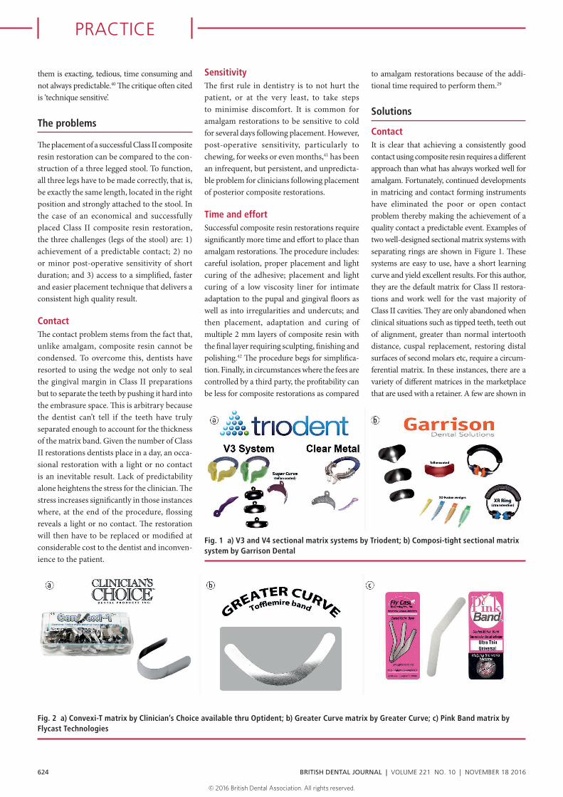
ContactIt is clear that achieving a consistently good contact using composite resin requires a different approach than what has always worked well for amalgam. Fortunately, continued developments in matricing and contact forming instruments have eliminated the poor or open contact problem thereby making the achievement of a quality contact a predictable event. Examples of two well-designed sectional matrix systems with separating rings are shown in Figure 1. These systems are easy to use, have a short learning curve and yield excellent results. For this author, they are the default matrix for Class II restora-tions and work well for the vast majority of Class II cavities. They are only abandoned when clinical situations such as tipped teeth, teeth out of alignment, greater than normal intertooth distance, cuspal replacement, restoring distal surfaces of second molars etc, require a circum-ferential matrix. In these instances, there are a variety of different matrices in the marketplace that are used with a retainer. A few are shown in
Fig. 2 a) Convexi-T matrix by Clinician’s Choice available thru Optident; b) Greater Curve matrix by Greater Curve; c) Pink Band matrix by Flycast Technologies
Fig. 1 a) V3 and V4 sectional matrix systems by Triodent; b) Composi-tight sectional matrix system by Garrison Dental
624 BRITISH DENTAL JOURNAL | VOLUME 221 NO. 10 | NOVEMBER 18 2016
PRACTICE
© 2016
British
Dental
Association.
All
rights
reserved. ©
2016
British
Dental
Association.
All
rights
reserved.

Figure 2. When a circumferential matrix is used, the wedge is inserted only to seal the gingival margin in the same manner as when placing amalgam. Instead of trying to separate the teeth with the wedge, contact forming instruments are used to achieve a contact. The instruments shown in Figure 3 are simple to use and yield consistently good contacts. Although what has been mentioned above is the author’s approach to creating predictable quality contacts in Class II composite resin restorations, there are other systems of various designs available as well.
SensitivityA properly placed adhesive seals the dentine so most composite resin restorations will have no post-operative sensitivity at all. It is not unusual for some restored teeth to have a transient sensi-tivity to cold for a few days. This can result from brief pulpal hyperaemia because of presenting caries and/or trauma of the surgical procedure. However, what has plagued many dentists are patients reporting persistent pain on chewing. For this symptom, it is important to distinguish the type of post-operative sensitivity the patient is experiencing. If the restoration is high, the pain occurs every time the patient chews on the tooth. The patient quickly accommodates by chewing only on the opposite side. If irreversible pulpitis is not present, marking and adjusting the occlusion eliminates the pain. What has frustrated dentists is when patients state the pain is intermittent, occurs only when they ‘hit a certain spot’ and continues even when repeated adjustments result in light or no opposing contact. In this author’s opinion, the cause of sensitivity in these instances is almost always lack of dentine seal.43–47 In the past, this type of problem has been ascribed to high shrinkage stress of the composite resin on cavity walls or the use of the phosphoric acid in the etch and rinse approach to adhesion. Shrinkage stress of the composite resin is an unlikely cause when proper incremental placement technique
is used and given that the higher filler content of contemporary composite resins has reduced volumetric shrinkage and resulting stress.48 With regards to the use of phosphoric acid to condition dentine, controlled clinical trials have repeat-edly shown little or no post-operative sensitivity whether the etch and rinse or a self-etch adhesive technique is used.43,49 Furthermore, anecdotal reports show the incidence of post-operative sensitivity declines as dentists master the more exacting technique required when using etch and rinse adhesives. For dentists who still have the occasional patient experiencing the intermit-tent post-operative sensitivity described above, switching to a self-etch adhesive along with selective etching the occlusal enamel with phos-phoric acid, should solve the problem without compromising either the enamel or dentine bond.44 Because contamination can be a cause of postoperative sensitivity as well as early failure of the restoration, it is important to note that isolation throughout the adhesive process and composite placement is mandatory. Although others may use a different approach, in this author’s opinion, a well-placed rubber dam is still the optimal method for obtaining and maintain-ing the required isolation throughout the entire adhesive and composite placement procedure.
Working with a trained assistant, using a few simple, well designed clamps and practice makes the placement process very efficient (Fig. 4). The additional up front time involved in placement is more than made up by elimination of the cheek, tongue, gingiva and saliva from the operating field along with improved vision for the operating team.
Time and effortEven with a consistent contact and elimina-tion of post-operative sensitivity, dentists are still left with the considerable time and effort needed to insert, carefully adapt and then cure multiple layers of composite resin. In recent years manufacturers have introduced new ‘bulk-fill’ composite resins that allow dentists to place restorations in fewer increments. These materials fall into two categories: low viscosity (flowable) and high viscosity (sculptable). One unique system, SonicFill (Kerr) is a high viscosity composite resin, but, upon activation with a sonic handpiece, becomes a low viscosity material during the insertion phase. Manufacturers have altered the chemistry (for example, Tetric EvoCeram Bulk Fill [Ivoclar Vivadent -Schaan, Lichtenstein]), or polymer structure (for example, SDR Flow [Dentsply – York, Pennsylvania]), or
Fig. 3 a) Contact Pro2 by CEJ Dental; b) Trimax by ADdent; c) Preform by Garrison Dental
Fig. 4 Rubber dam clamps by Dentsply
BRITISH DENTAL JOURNAL | VOLUME 221 NO. 10 | NOVEMBER 18 2016 625
PRACTICE
© 2016
British
Dental
Association.
All
rights
reserved. ©
2016
British
Dental
Association.
All
rights
reserved.

polymerisation kinetics (for example, SonicFill [Kerr - Orange, California]) of these advanced materials to control shrinkage stress and allow a high depth of cure up to 4 or 5 mm (Table 1).50–53
Low viscosity materials act as a flowable base. They adapt very well to cavity walls and into undercuts and can be placed up to 4 mm in thickness. Dual cure flowables will cure at any depth. Since they are not sculptable and generally have low wear resistance to occlusal forces, these materials need to be covered or capped by one or two increments of a high viscosity material in all but very small cavities.54 It should to be noted that some of these materials are very translucent, a fact that needs to be considered when covering preparations having dark internal stains. For cli-nicians, gaining adaptation with a single 4 mm base shortens placement time and effort.
The new high viscosity bulk-fill materials are shaded, sculptable, and, due to high filler content, have high strength and wear resistance. However, because adapting these materials intimately to pulpal floors, irregular preparations and undercuts can be challenging, lining the cavity with a low viscosity liner before placing the high viscosity material seems prudent.55–57 Once again, placing these materials in up to 4 mm increments saves time and effort for the clinician.
SonicFill (Kerr) seems to be in a class by itself. Due to its high filler content, this material is clas-sified as a high viscosity material in the ‘bulk-fill’
market. Its uniqueness lies in the fact that it is activated and injected into the cavity using a sonic handpiece. High frequency vibration of this material significantly lowers the viscosity, vibrates it into intimate contact with cavity walls and rapidly fills most cavities in seconds. No cavity liner is required for adaptation although the clinician can choose to apply one if desired. A depth of cure up to 5 mm means the majority of cavities can be filled in a single increment.52,58–61
Although a specific handpiece is required, this system of composite resin placement seems to take the bulk fill concept one step further in speed and simplicity.
Case reportA patient presented with a leaking amalgam filling in a lower premolar that required replace-ment (Fig. 5). The patient was anaesthetised and a rubber dam with wax floss ligature was placed. After removing the amalgam, any base or liner and all caries, a sectional matrix (Triodent – Kati Kati, New Zealand) was placed. An appropriate sized wedge was inserted to seal the gingival margin. The stabilising/separating ring with V shaped tines was placed to adapt the matrix and create slight separation of the teeth (Fig. 6). Burnishing the matrix against the adjacent tooth established the size and location of the contact. The cavity measures 5 mm in depth and thus can be restored in a single increment of
SonicFill composite resin (Fig. 7). The adhesive was placed and cured according to the manu-facturer’s directions (Fig. 8). The handpiece has settings from 1 to 5 (slow to fast) to determine the extrusion rate desired for the composite resin material (Fig. 9). The extrusion rate should not be controlled through the rheostat. Complete activa-tion of the rheostat is necessary for the material to achieve full liquefaction.52 A setting of 4 was chosen to fill this cavity. The tip is placed near the gingival floor of the proximal box (Fig. 10). It is withdrawn as the cavity is filled but kept in contact with the material at all times (Fig. 11). The cavity is filled in a few seconds (Fig. 12). A round end condenser or silicone tipped instrument is used to compress the material and simultane-ously wipe away excess (Fig. 13). The material retains the activation energy for several minutes before returning back to a high viscosity state.
Fig. 5 Premolar with failing amalgam and recurrent caries
Table 1 ”Bulk-Fill” Composite Resin Materials
Materials Composite type Depth of cure* Needs enamel replacement layer**
Low viscosity liner recommended
Low viscosity
SureFil SDR Flow† (Dentsply/Caulk) Flowable 4 mm Yes No
Tetric EvoFlow Bulk Fill† (Ivoclar Vivadent) Flowable 4 mm Yes No
Beautiful-Bulk Flowable† (Shofu) Flowable 4 mm Yes No
X-tra Base† (Voco) Flowable 4 mm Yes No
Venus Bulk Flow† (Heraeus Kulzer) Flowable 4 mm Yes No
Filtek Flow Bulk Fill† (3M/Espe) Flowable 4 mm Yes No
HyperFil† (Parkell) Flowable Infinite‡ Yes No
Fill Up† (Coltene) Flowable Infinite‡ Yes No
BulkEZ† (Danville) Flowable Infinite‡ Yes No
High viscosity
Tetric EvoCeram Bulk Fill (Ivoclar Vivadent) Sculptable 4 mm No Yes
X-tra Fil (Voco) Sculptable 4 mm No Yes
Beautiful-Bulk Restorative (Shofu) Sculptable 4 mm No Yes
Filtek Bulk Fill Posterior Restorative (3M/Espe) Sculptable 4 mm No Yes
SonicFill§ (Kerr) Sculptable 5 mm No No
*Manufacturer’s data. **Unless non-occlusal load bearing. †Dentine replacement (base). ‡Dual Cure. §Sonic delivery.
626 BRITISH DENTAL JOURNAL | VOLUME 221 NO. 10 | NOVEMBER 18 2016
PRACTICE
© 2016
British
Dental
Association.
All
rights
reserved. ©
2016
British
Dental
Association.
All
rights
reserved.

The liquefaction renders the material soft at this stage but it does not slump when sculpted and is non-sticky (Fig. 14). Using a light emitting diode curing light with an output above 1,000 mW/cm2, the material is cured from the occlusal for
10 seconds (Fig. 15). Upon removal of the matrix assembly and wedge, a sharp number 12 blade on a scalpel handle is used to trim away any excess from the proximal margins (Fig. 16). It is the author’s preferred technique to retain any bonded
occlusal flash and merely feather it with a 7,404 or 7,406 carbide finishing bur (Fig. 17). The cure is completed by applying ten additional seconds to
Fig. 7 Cavity depth from marginal ridge to gingival margin is slightly less than 5 mm
Fig. 10 The tip is placed into the proximal box just above the gingival margin
Fig. 13 An instrument is used to compress the material and wipe away the excess
Fig. 16 At this stage excess flash is removed easily
Fig. 6 Sectional matrix, wedge and stablilising/separating ring is placed. Contact is burnished
Fig. 9 SonicFill handpiece with composite resin filled tip attached. The numbers at the base of the handpiece regulate the extrusion speed of the material
Fig. 12 The tip is moved in a straight line motion across the preparation filling the cavity. Waving the tip around could cause voids
Fig. 15 A ten second cure from the occlusal surface is performed
Fig. 8 The adhesive is placed and light cured
Fig. 11 The tip is withdrawn as the material fills the box. It is always kept in contact with the material
Fig. 14 Because the material retains the sonic energy for several minutes, it will feel soft but will not slump. Lack of stickiness allows quick and easy sculpting of anatomy
Fig. 17 Because curing is incomplete, occlusal flash can be feathered with a carbide bur. Using diamonds risks removing enamel, damaging margins and altering the occlusion
BRITISH DENTAL JOURNAL | VOLUME 221 NO. 10 | NOVEMBER 18 2016 627
PRACTICE
© 2016
British
Dental
Association.
All
rights
reserved. ©
2016
British
Dental
Association.
All
rights
reserved.
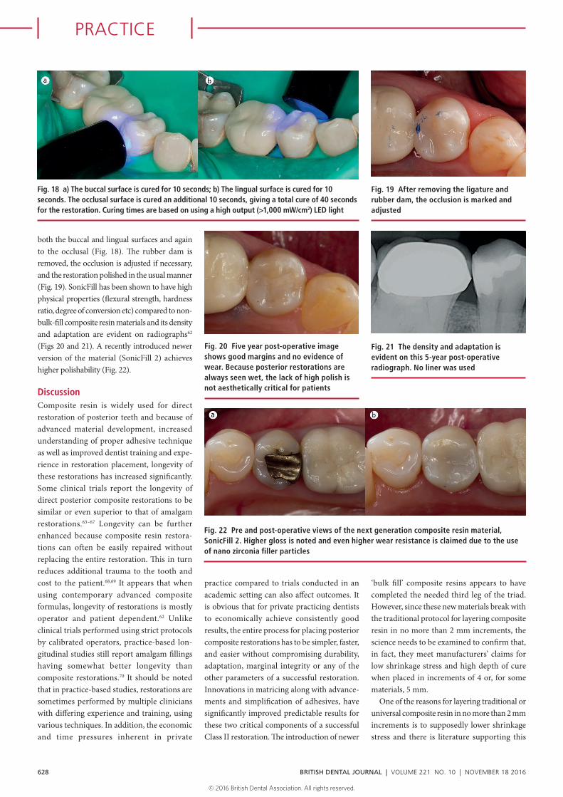
both the buccal and lingual surfaces and again to the occlusal (Fig. 18). The rubber dam is removed, the occlusion is adjusted if necessary, and the restoration polished in the usual manner (Fig. 19). SonicFill has been shown to have high physical properties (flexural strength, hardness ratio, degree of conversion etc) compared to non-bulk-fill composite resin materials and its density and adaptation are evident on radiographs62 (Figs 20 and 21). A recently introduced newer version of the material (SonicFill 2) achieves higher polishability (Fig. 22).
DiscussionComposite resin is widely used for direct restoration of posterior teeth and because of advanced material development, increased understanding of proper adhesive technique as well as improved dentist training and expe-rience in restoration placement, longevity of these restorations has increased significantly. Some clinical trials report the longevity of direct posterior composite restorations to be similar or even superior to that of amalgam restorations.63–67 Longevity can be further enhanced because composite resin restora-tions can often be easily repaired without replacing the entire restoration. This in turn reduces additional trauma to the tooth and cost to the patient.68,69 It appears that when using contemporary advanced composite formulas, longevity of restorations is mostly operator and patient dependent.62 Unlike clinical trials performed using strict protocols by calibrated operators, practice-based lon-gitudinal studies still report amalgam fillings having somewhat better longevity than composite restorations.70 It should be noted that in practice-based studies, restorations are sometimes performed by multiple clinicians with differing experience and training, using various techniques. In addition, the economic and time pressures inherent in private
practice compared to trials conducted in an academic setting can also affect outcomes. It is obvious that for private practicing dentists to economically achieve consistently good results, the entire process for placing posterior composite restorations has to be simpler, faster, and easier without compromising durability, adaptation, marginal integrity or any of the other parameters of a successful restoration. Innovations in matricing along with advance-ments and simplification of adhesives, have significantly improved predictable results for these two critical components of a successful Class II restoration. The introduction of newer
‘bulk fill’ composite resins appears to have completed the needed third leg of the triad. However, since these new materials break with the traditional protocol for layering composite resin in no more than 2 mm increments, the science needs to be examined to confirm that, in fact, they meet manufacturers’ claims for low shrinkage stress and high depth of cure when placed in increments of 4 or, for some materials, 5 mm.
One of the reasons for layering traditional or universal composite resin in no more than 2 mm increments is to supposedly lower shrinkage stress and there is literature supporting this
Fig. 18 a) The buccal surface is cured for 10 seconds; b) The lingual surface is cured for 10 seconds. The occlusal surface is cured an additional 10 seconds, giving a total cure of 40 seconds for the restoration. Curing times are based on using a high output (>1,000 mW/cm2) LED light
Fig. 19 After removing the ligature and rubber dam, the occlusion is marked and adjusted
Fig. 21 The density and adaptation is evident on this 5-year post-operative radiograph. No liner was used
Fig. 20 Five year post-operative image shows good margins and no evidence of wear. Because posterior restorations are always seen wet, the lack of high polish is not aesthetically critical for patients
Fig. 22 Pre and post-operative views of the next generation composite resin material, SonicFill 2. Higher gloss is noted and even higher wear resistance is claimed due to the use of nano zirconia filler particles
628 BRITISH DENTAL JOURNAL | VOLUME 221 NO. 10 | NOVEMBER 18 2016
PRACTICE
© 2016
British
Dental
Association.
All
rights
reserved. ©
2016
British
Dental
Association.
All
rights
reserved.
technique.30,71 However, there is also research questioning the value of incremental placement in reducing shrinkage stress and at least one investigation concluded this technique actually increases it.72–77 Nevertheless, placing traditional restorative composites in 2 mm layers may still be required for purposes of good adaptation and to achieve adequate depth of cure. The current literature reporting on shrinkage stress for bulk-fill materials predominately shows these materials at 4 or 5 mm thicknesses to have similar or lower values when compared to 2 mm thicknesses of universal composites.78–81 In a comprehensive evaluation of bulk-fill versus traditional multi-increment composite resins carried out at the American Dental Association laboratory, shrinkage stress values for 9 out of 11 bulk fill materials tested were not significantly different than the two conven-tional composite resin controls. The value for X-trabase (Voco- Indian Land, South Carolina, USA) was significantly higher and SonicFill (Kerr) was significantly lower than all the other materials tested.48 Another in vitro investigation examining gap formation at dentine margins of Class II restorations showed similar results for Tetric EvoCeram Bulk Fill (Ivoclar Vivadent - Schaan, Lichtenstein), SonicFill (Kerr) and SDR Flow (Dentsply) compared to a conventional layered composite (Tetric EvoCeram (Ivoclar Vivadent). The two low viscosity bulk-fills, x-trabase (Voco) and Venus Bulk Fill (Hereaus Kulzer – South Bend, Indiana, USA), showed larger gaps.82 Whether this difference would be clinically significant is not known. With minor difference for some products, in vitro studies on marginal adaptation/marginal integrity show similar results for bulk fill products (high and low viscosity) when compared to traditional layered controls.83–87 Overall, the data for the bulk fill materials seems to support manu-facturers’ claims that the modifications made in these materials have succeeded in control-ling shrinkage stress to nearly the same or lesser amount than experienced with current conventional composites. Finally, in a recent investigation comparing an incrementally placed composite restoration (Point 4, Kerr) to a sonicated bulk fill (SonicFill, Kerr) restoration, the sonicated bulk fill composite resin showed significantly fewer voids. 88
There are two common methods used to measure depth of cure. An unsophisticated, but simple method that can be carried out by any dentist is the International Standards Organisation (ISO) #4049:2009.89 This method cures a column of composite in a metal mould
from the top and scrapes away the soft, uncured composite from the bottom until reaching hard cured material. The depth of cure is then defined by dividing the remaining length of hard composite by two. Since this method does not actually measure polymerisation (carbon con-version) at any given depth, it is an approxima-tion at best. Its underestimation of depth of cure was pointed out in an investigation by Tiba and colleagues into depth of cure for several bulk-fill materials and was presented at the International Association of Dental Research (IADR) meeting in 2013. The authors concluded their study with the following statement: ‘This study shows some limitations of ISO 4049 for testing depth of cure in relation to the more important hardness ratio for bulk fill composite materials.’90
Other investigations have also questioned the testing protocol and clinical relevancy of the ISO 4049 testing method.61,91 A second method used by many investigators for measuring depth of cure and referred to in the Tiba statement above, uses microhard-ness testing. It defines the depth of cure as the distance from the top of the cured column of composite to a point where the microhardness value is at least 80% of the top surface value.92
Hardness has been shown to correlate to polymerisation (80% bottom-to-top hardness equals 90% carbon conversion).93 Since it indirectly measures actual polymerisation, the microhardness method for determining depth of cure seems more accurate and clinically relevant. When applying the ‘scrape test’ to various bulk fill materials, some have failed to meet manufacturers’ claims except when tested using actual teeth instead of a metal mould.61 However, investigations using the more valid microhardness test have consistently shown bulk fill materials meeting or exceeding manufacturers’ specifications.48,80,82,90,,94–100 The manufacturer of SonicFill notes that measure-ments for shrinkage stress and depth of cure should be done on the activated material rather than the static material to accurately reflect the true values that would occur in clinical use. 101
Since bulk fill composite resin materials are so new, long term clinical trials are lacking. Short term clinical trials, published and unpublished are just beginning to appear. Early data shows bulk fill materials performing clinically similar compared to 2 mm layered materials. 102–104 Nevertheless, surveys show high early acceptance and strong growth in use among dentists in the marketplace.105,106
Given the number of posterior composite res-torations dentists place in practice, this growth
would seem unlikely if these new materials and technologies weren’t performing successfully.
Along with a more efficient placement technique, bulk fill materials may also reduce operator error. A university dental school study showed that new graduates achieved better margins and less gaps between layers using bulk fill techniques compared to conventional layered techniques.107 This fact confirms that easier, simpler techniques lead to consistently better results.
Conclusion
The development of innovative sectional matrix systems and simplified universal adhesives, with or without selective etch, along with the advent of bulk fill composites, would seem to be a significant turning point in posterior direct restorative dentistry. This combination creates a streamlined, straight forward, faster, more efficient and economi-cal placement technique with less effort than previous methods. Practicing dentists have always desired a less labour intensive clinical protocol for placing posterior composite resin restorations – one that was as easy and timely as amalgam. It appears it has finally arrived.
Competing interests Dr Ron Jackson discloses that he acted as a consultant in the development of SonicFill™ and retains a financial interest.
1. Black G V. Pathology of the hard tissue of the teeth. Operative Dentistry, Vol. 1 Chicago: Medico-Dental. Publishing Company, 1908.
2. Vimy M J, Boyd N D, Hopper D E, Lorscheider F L. Glomerular Filtration impairment by mercury released from dental ‘silver’ fillings in sheep. Physiologist 1990; 33: 651.
3. Enwonwu C O. Potential health hazard of use of mercury in dentistry: critical review of the literature. Environ Res 1987; 42: 257–274.
4. Mackert J R, Berglund A. Mercury exposure from dental amalgam fillings: absorbed dose and the potential for adverse health effects. Crit Rev Oral Biol Med 1997; 8: 410–436.
5. Bjorkman L, Paedersen N L, Lichtenstein P. Physical and mental health related to dental amalgam fillings in Swedish twins. Community Dent Oral Epidemiol 1996; 24: 260–270.
6. Mackert J R Jr., Leffell M S, Wagner D A, Powell B J. Lym-phocyte levels in subjects with and without amalgam restorations. JADA 1991; 122: 49–53.
7. Koral S M. Mercury from Dental Amalgam: Exposure and Risk Assessment. Compend Contin Educ Dent 2013; 34: 138–146.
8. ADA Council on Scientific Affairs. Dental Amalgam: Update on Safety Concerns. J Am Dent Assoc 1998; 129: 494–503.
9. Ucar Y, Brantley W A. Biocompatability of dental amal-gams. Int J Dent 2011; 981595.
10. Scientific Committee on Emerging and Newly Identified Health Risks, European Commission. Are dental fillings safe? Safety of dental amalgam and alternative dental restoration materials. European Commission. Available online at http://ec.europa.eu/health/scientific_com-mittees/docs/citizens_dental_filling_en.pdf (accessed October 2016).
BRITISH DENTAL JOURNAL | VOLUME 221 NO. 10 | NOVEMBER 18 2016 629
PRACTICE
© 2016
British
Dental
Association.
All
rights
reserved. ©
2016
British
Dental
Association.
All
rights
reserved.

11. Homme K G, Kern J K, Haley B E et al. New Science challenges old notion that mercury dental amalgam is safe. Biometals 2014; 27: 19–24.
12. Mutter J. Is dental amalgam safe for humans? The opinion of the scientific committee of the European Commission. J Occu Med Toxi 2011; 6: 2.
13. BIO Intelligence Service. Study on the potential for reducing mercury pollution from dental amalgam and batteries. Final report prepared for the European Commission – DG ENV. 2012. Available online at http://ec.europa.eu/environment/chemicals/mercury/pdf/final_report_110712.pdf (accessed October 2016).
14. Norwegian Ministry of the Environment and Interna-tional Development, 2008.
15. Danish Ministry for Health, 2008.16. Swedish Ministry of Health and Social Affairs, 2008.17. Minamata Convention on Mercury. January 19 2013.
Geneva, Switzerland.18. Lynch C D, Opdam N J, Hickel R et.al. Guidance on Pos-
terior Resin Composites: Academy of Operative Dentistry – European Section. J Dent 2014; 42: 377–383.
19. Coelho-De-Souza F H, Camacho G B, Demarco F F, Powers J M. Fracture resistance and gap formation of MOD restorations: influence of restorative technique, bevel preparation and water storage. Oper Dent 2008; 33: 37–43.
20. Liberman R, Ben-Amar A, Gontar G, Hirsh A. The effect of posterior composite restorations on the resistance of cavity walls to vertically applied occlusal loads, J Oral Rehab 1990; 17: 99–105.
21. McCullock A J, Smith B G M. In Vitro Studies of cusp reinforcement with adhesive restorative material. Brit Dent J 1986; 161: 450–452.
22. Fissore B, Nicholls J, Youdelis R. Load fatigue of teeth restored by a dentin bonding agent and a posterior composite resin. J Pros Dent 1991 65: 80–85.
23. Eakle W S, Fracture Resistance of teeth restored with Class II bonded composite resin. J Dent Res 1986; 65: 149–152.
24. Macpherson L C, Smith B G N. Reinforcement of weak-ened cusps by adhesive restorative materials: an in vitro study. Brit Dent J 1995 178: 341–344.
25. Lynch C D, Frazier K B, McConnel R J et al. Minimally invasive management of dental caries. J Am Dent Assoc 2011; 142: 612–620.
26. de Freitas C R, Miranda MI, de Andrade M R et al. Resis-tance to maxillary premolar fractures after restoration of class II preparations with resin composite or ceromer. Quint Int 2002; 33: 589–594.
27. Klapdohr S, Moszner N. New inorganic components for dental filling composites. Monatshefte fur Chemie 2005; 136: 21–45.
28. Leprince J G, Palin W M, Hadis M A, Devaux J, Leloup G. Progress in dimethacrylate-based dental composite technology and curing efficiency. Dent Mater 2013; 29: 139–156.
29. Christensen G J. Class II Resins: Nanofill Brands as Group Show Best Performance Yet. Clinician’s Report 2014: 7.
30. Ferracane J L. Resin composite-state of the art. Dent Mater 2011; 27: 29–38.
31. Sunnegardh-Gronberg K, van Dijken J W V, Funegardh U, Lindberg A, Nilsson M. Selection of dental materials and longevity of replaced restorations in Public Dental Health clinics in northern Sweden. J Dent 2009; 37: 673–678.
32. Mitchell R J, Koike M, Okabe T. Posterior amalgam restorations – usage, regulation and longevity. Dent Clin North Am 2007; 51: 573–589.
33. Opdam N J M, van de Sande F H, Bronkhorst E et al. Longevity of Posterior composite restorations: a sys-tematic review and meta-analysis. J Dent Res 2014; 93: 943–949.
34. Burke F J. Amalgam to tooth-coloured materials: implications for clinical practice and dental education. Governmental restrictions and amalgam usage survey results. J Dent 2004; 32: 343–350.
35. Lund A. In your dental practice, is dental amalgam still the restorative material of choice? J Am Dent Assoc 2002; 133: 1046.
36. Burke F J, McHugh S, Hall A C, Randall Rc, Widstrom E, Forss H. Amalgam and composite use in UK general dental practice in 2001. Br Dent J 2003; 194: 613–618.
37. Christensen G J, Child P L Jr. Has resin-based composite replaced amalgam? Dent Today 2010; 29: 108, 110.
38. Clinical Research Associates CRA Newsletter. Clinician’s Preferences 2001 #15, CRA Newsletter. 2001: 3.
39. Christensen G J. Should resin-based composite dominate restorative dentistry today? J Am Dent Assoc 2010; 141: 1490–1493.
40. Gilmour A S, Latif M, Addy L D, Lynch C D. Placement of posterior composite restorations in United Kingdom dental practices: techniques, problems, and attitudes. In Dent J 2009; 59: 148–154.
41. Briso A L F, Mestrener G, Delico R H, et.al. Clinical assessment of post-operative sensitivity in posterior composite restorations. Oper Dent 2007; 32: 421–426.
42. Burke F T J, Shortall A C C. Successful Restoration of load-bearing cavities in posterior teeth with direct-re-placement resin-based composite. Dent Update 2001; 388–398.
43. Swift E. Ask the Experts: Dentin/Enamel Bonding. J Esth Rest Dent 2010; 22: 352–353.
44. Van Meerbeek B, Yoshihara K, Yoshida Y, Mine A, De Munck J, Van Landuyt KL. State of the art self-etching adhesives. Dent Mater 2011; 27: 17–28.
45. Loguercio A et al. Effect of Solvent removal on adhesive properties of simplified etch-and-rinse systems and on bond strengths to dry and wet dentin. J Adhes Dent 2009; 11: 213–219.
46. Peumans M, Kanumilli P, DeMunck J et al. Clinical effec-tiveness of contemporary adhesives: a systematic review of current clinical trials. Dent Mater 2005; 21: 864–881.
47. Van Meerbeek B, DeMunck J, Yoshida Y et al. Adhesion to Enamel and Dentin: Current status and future chal-lenges. Oper Dent 2003; 28: 215–235.
48. Tiba A, Zeller G G, Estrich C G, Hong A. A laboratory evaluation of bulk-fill versus traditional multi-incre-ment-fill resin-based composites. J Am Dent Assoc 2013; 8: 13–17.
49. Perdigao J, Geraldeli S, Hodges J S. Total-etch versus self-etch adhesive effect on post-operative sensitivity. J Am Dent Assoc 2003; 134: 1621–1629.
50. Tetric EvoCeram Bulk Fill- Scientific Documentation. Ivoclar Vivadent.
51. SDR Flow – Scientific Documentation., Dentsply.52. SonicFill – Scientific Documentation. Kerr.53. Al-Ahdal K, Ilie N, Silikas N, Watts D C. Polymerization
kinetics and impact of post polymerization on the degree of conversion of bulk-fill resin-composite at clinically relevant depth. Dent Mater 2015; 31: 1207–1213.
54. Ilie N, Bucuta S, Draenert M. Bulk-fill resin-based composites: an in vitro assessment of their mechanical performance. Oper Dent 2013; 38: 618–625.
55. Agarwal R S, Hiremath H, Agarwal J, Garg A. Evaluation of cervical margin and internal adaptation using newer bulk fill composites: An in vitro study. J Conserv Dent 2015; 18: 56–61.
56. Opdam N J M, Roeters J J M, Peters T C R B, Burgersdijk R C W, Teunis M. Cavity wall adaptation and voids in adhesive Class I resin composite restorations. Dent Mater 1996; 12: 230–235.
57. Chuang S F, Jin Y T, Liu J K, Chang Ch, Shieh D B. Influ-ence of flowable composite lining thickness on Class II composite restorations. Oper Dent 2004; 29: 301–308.
58. Yapp R, Baumann A, Powers J M. Comparative curing and thermal properties of demi ultra LED curing light. Research report – number 58. Dent Advisor 2014. Available online at www.dentaladvisor.com/publications/research-reports/comparative-curing-and-thermal-properties-of-demi-ul-tra-led-curing-light.pdf (accessed July 2014).
59. Christensen G. Advantages and challenges of bulk-fill resins. Clin Rep 2012; 5: 1–2.
60. Hansen E K, Asmussen E. Visible-light curing units: correlation between depth of cure and distance between exit window and resin surface. Acta Odontol Scand 1997; 55: 162–166.
61. Tiba A, Vinh R, Estrich C. Clinically relevant measure-ments of depth of cure of bulk-fill composites. IADR March 2015, Boston, Massachusetts.
62. Leprince J G, Palin W M, Vanacker J, Sabbagh J, Devaux J, Leloup G. Physico-mechanical characteristics of commercially available bulk-fill composites. J Dent 2014; 42: 993–1000.
63. Demarco F F, Correa M B, Cenci M S, Moraes R R, Opdam N J. Longevity of posterior composite resto-rations: not only a matter of materials. Dent Mater 2012; 28: 87–101.
64. Heintze S D, Rousson V. Clinical effectiveness of direct Class II restorations – a meta-analysis. J Adhes Dent 2012; 14: 407–431.
65. Opdam N J, van de Dande F H, Bronkhorst E et al. Longevity of posterior composite restorations: a sys-tematic review and meta-analysis. J Dent Res 2014; 93: 943–949.
66. Opdam N J M, Bronkhorst E M, Loomans B A C, Huys-mans MC. 12-year survival of composite vs. amalgam restorations. J Dent Res 2010; 89: 1063–1067.
67. Cetin A R, Unlu N, Cobanoglu N. A five-year clinical evaluation of direct nanofilled and indirect composite resin restorations in posterior teeth. Oper Dent 2013; 38: E31-E41.
68. Fernandez E, Martin J, Vildosola P, Oiveira Junior O B et al. Can repair increase the longevity of composite resins? Results of a 10 year clinical trial. J Dent 2015; 43: 279–286.
69. Opdam N J M, Bronkhorst E M, Loomans B A C, Huys-mans M C. Longevity of repaired restorations: A practice based study. J Dent 2012; 40: 829–835.
70. Kopperud S E, Bjorg Tveit A, Gaarden T, Sandvik L, Espelid I. Longevity of posterior dental restorations and reasons for failure. Eur J Oral Sci 2012; 120: 539–548.
71. Park J, Chang J, Ferracane J L, Lee I B. How should com-posite be layered to reduce shrinkage stress incremental or bulk filling. Dent Mater 2008; 24: 1501–1505.
72. Versluis A, Douglas W H, Cross M, Sakaguchi R L. Does an incremental filling technique reduce polymerization shrinkage stress. J Dent Res 1996; 75: 871–878.
73. El-Badrawy W, Jafarbour S, Jazi H S, McComb D. Effect of Composite Insertion technique on cuspal deflection using in vitro simulation model. Oper Dent 2012; 37: 399–305.
74. Rees J S, Jagger D C, Williams D R, Brown G, Duguid W. A reappraisal of the incremental packing technique for light cured composite resins. J Oral Rehab 2011; 31: 81–84.
75. Campodonico C E, Tantbirojn D, Olin Ps, Versluis A. Cuspal deflection and depth of cure in resin-based composite restorations filled by using bulk, incremental and transtooth-illumination techniques. J Am Dent Assoc 2011; 142: 1176–1182.
76. Idriss S, Habib C, Abduljabbar T et al. Marginal adaptation of class II resin composite restorations using incremental and bulk placement techniques: an ESEM study. J Oral Rehabil 2003; 30: 1000–1007.
77. Braga R R, Ballester Ry, Ferracane J L. Factors involved in the development of polymerization shrinkage stress in resin-composites: a systematic review. Dent Mater 2005; 21: 962–970.
78. EL-Damanhoury H M, Platt J A. Polymerization Shrinkage Stress kinetics and related properties of bulk-fill resin composites. Oper Dent 2014; 39: 374–382.
79. Kim R J Y, Kim Y J, Choi N S, Lee I B. Polymerization shrinkage, modulus, and shrinkage stress related to tooth-restoration interfacial debonding in bulk-fill composites. J Dent 2015; 43: 430–439.
80. Olafsson V G, Effect of Composite Type and Placement Technique on Shrinkage Stress; IADR Boston Massachu-setts, March 2015. #2198.
81. Rosatto C M P, Bicalho A A, Veríssimo C et.al. Mechanical properties, shrinkage stress, cuspal strain and fracture resistance of molars restored with bulk-fill composites and incremental filling technique. J Dent 2015; 43: 1520- 1528.
82. Benetti A R, Havndrup-Paedersen C, Honoure D, Paedersen M K, Pallesen U. Bulk-Fill resin composites: polymerization contraction, depth of cure and gap formation. Oper Dent 2015; 40: 190–200.
83. Campos E A, Ardu S, Lefever D, Jasse F F, Bortolotto T, Krejci I. Marginal adaptation of class II cavities restored with bulk-fill composites. J Dent 2014; 42: 575–581.
84. Agarwal R S, Hiremath H, Agarwal J, Garg A. Evaluation of cervical marginal and internal adaptation using newer bulk fill composites: an in vitro study. J Conserv Dent 2015; 18: 56–61.
85. Orlowski M, Tarczydlo B, Chalas R. Evaluation of marginal integrity of four bulk-fill dental composite materials: in vitro study. Sci World Journal 2015; 2015: DOI: 10.1155/2015/701262
86. Furness A, Tadros M Y, Looney S W, Rueggeberg F A. Effect of bulk/incremental fill on internal gap formation of bulk-fill composites. J Dent 2014; 42: 439–449.
630 BRITISH DENTAL JOURNAL | VOLUME 221 NO. 10 | NOVEMBER 18 2016
PRACTICE
© 2016
British
Dental
Association.
All
rights
reserved. ©
2016
British
Dental
Association.
All
rights
reserved.

87. Haak R, Naeke T, Pfeffer M, et.al. Adaptation of High-Viscosity Bulk-fill Composites in Class II Cavities; IADR Boston Massachusetts, March 2015. #651.
88. Jarisch, J, Lien W, Guevara P H, Greenwood W J, Dunn W J. Microcomputed tomographic comparison of pos-terior composite resin restorative techniques: sonicated bulk fill versus incremental fill. Gen Dent 2016; 20–24.
89. International Standard ISO 4049: 2009. Dentistry-Poly-mer-based restorative materials. Geneva: ISO.
90. Tiba A, Hong A, Zeller G. Examining the Depth of Cure for Bulk Fill Composite Materials, IADR Seattle Washing-ton, March 2013. #2435.
91. Vogel K. Factors that Influence Depth of Cure Measure-ment According to ISO 4049, IADR September 2014 #502.
92. Watts D C, Amer O, Combe E C. Characteristics of visible-light activated composites. Brit Dent J 1984; 156: 209–215.
93. Bouschlicher M R, Rueggeberg F A, Wilson B M. Correla-tion of bottom-to-top surface microhardness and conver-sion ratios of various resin composite compositions. Oper Dent 2004; 29: 698–704.
94. Ilie N, Kefsler A, Durner J. Influence of various irradiation processes on the mechanical properties and polymeriza-
tion kinetics of bulk fill resin based composites. J Dent 2013; 41: 695–702.
95. Alshali R Z, Silikas N, Satterthwaite J D. Degree of conver-sion of bulk fill compared to conventional resin-composites at two time intervals. Dent Mater 2013; 29: e213–e217.
96. Czasch P, Ilie N. In vitro comparison of mechanical prop-erties and degree of cure of bulk fill composites. Clin Oral Investig 2013; 17: 227–235.
97. Bucuta S, Ilie N. Light transmittance and micro-mechanical properties of bulk fill vs. conventional resin based compos-ites. Clin Oral Investig 2014; 18: 1991–2000.
98. Zorzin J, Maier E, Harre S et al. Bulk-fill resin composites: Polymerization properties and extended light curing. Dent Mater 2015; 31: 293–301.
99. Alshali R Z, Salim N A, Satterthwaite J D, Silikas N. Post-ir-radiation hardness development, chemical softening, and thermal stability of bulk-fill and conventional resin-compos-ites. J Dent 2015; 43: 209–218.
100. Alrahlah A, Silikas, Watts D C. Post-cure depth of cure of bulk fill dental resin-composites. Dent Mater 2014; 30: 149–154.
101. Personal Communication. Dreschler U, Senior Research Scientist, Kavo Kerr. Orange, California.
102. van Dijken J W V, Pallesen U. Randomized 3-year clinical evaluation of class I and II posterior resin restorations placed with a bulk fill resin composite and a one-step self-etching adhesive. J Adhes Dent 2015; 17: 81–88.
103. Ernst C P. Random Split Mouth Trial. Dept Oper Dent Johannes Gutenberg University Mainz, Germany., (unpublished).
104. Geissberger M, Kachalia P. University of Pacific School of Dentistry June 2011, Clinical Evaluation of Restorations using a New Composite Material and Oscillating Handpiece and Comparing it with Traditional Composite Material and Placement Technique (unpublished).
105. Restorative Survey, Dentaltown March, 2015 pp. 58. Available online at www.dentaltown.com/dentaltown/article.aspx?i=385&aid=5269 (accessed November 2016).
106. Restorative Dental Survey. Posterior Composites, Dental Products Report. April 2015; 32– 33. http://www.dentalproductsreport.com/files/files/dpr0415_ezine.pdf (accessed November 2016).
107. McConnell R, Lappin M. Comparison of Composite Placement Techniques by Recently Qualified Dental Practitioners, IADR March 2015 Boston, Massachusetts.
BRITISH DENTAL JOURNAL | VOLUME 221 NO. 10 | NOVEMBER 18 2016 631
PRACTICE
© 2016
British
Dental
Association.
All
rights
reserved. ©
2016
British
Dental
Association.
All
rights
reserved.