class-specific interaction of profilin and adf … · class-specific interaction of profilin...
TRANSCRIPT

Class-Specific Interaction of Profilin and ADF Isovariants withActin in the Regulation of Plant Development W
Muthugapatti K. Kandasamy, Brunilıs Burgos-Rivera, Elizabeth C. McKinney, Daniel R. Ruzicka,and Richard B. Meagher1
Department of Genetics, Davison Life Sciences Building, University of Georgia, Athens, Georgia 30602
Two ancient and highly divergent actin-based cytoskeletal systems have evolved in angiosperms. Plant genomes encode
complex actin and actin binding protein (ABP) gene families, most of which are phylogenetically grouped into gene classes with
distinct vegetative or constitutive and reproductive expression patterns. In Arabidopsis thaliana, ectopic expression of high
levels of a reproductive class actin, ACT1, in vegetative tissues causes severe dwarfing of plants with aberrant organization of
most plant organs and cell types due to a severely altered actin cytoskeletal architecture. Overexpression of the vegetative
class actin ACT2 to similar levels, however, produces insignificant phenotypic changes. We proposed that the misexpression
of the pollen-specific ACT1 in vegetative cell types affects the dynamics of actin due to its inappropriate interaction with
endogenous vegetative ABPs. To examine the functionally distinct interactions among the major classes of actins and ABPs,
we ectopically coexpressed reproductive profilin (PRF4) or actin-depolymerizing factor (ADF) isovariants (e.g., ADF7) with
ACT1. Our results demonstrated that the coexpression of these reproductive, but not vegetative, ABP isovariants suppressed
the ectopic ACT1 expression phenotypes and restored wild-type stature and normal actin cytoskeletal architecture to the
double transgenic plants. Thus, the actins and ABPs appear to have evolved class-specific, protein–protein interactions that
are essential to the normal regulation of plant growth and development.
INTRODUCTION
In plant cells, the actin cytoskeleton undergoes dramatic reor-
ganization in response to external and internal cues and thus
regulates several vital cellular processes, including cytoplasmic
streaming, organelle movement and repositioning, establish-
ment of cell polarity, tip growth, cell division, expansion, and
differentiation, and reactions to pathogen attack, wounding, and
hormones (reviewed in Staiger, 2000; Wasteneys and Galway,
2003; Hussey et al., 2006; Staiger and Blanchoin, 2006). The
dynamic reorganization of actin is modulated by the specific
activity of a plethora of actin binding proteins (ABPs) that either
promote or inhibit actin polymerization. Actins and most ABPs
in plants are encoded by multigene families (Meagher and
Fechheimer, 2003). For example, the Arabidopsis thaliana ge-
nome contains eight actin genes that are grouped based on their
phylogeny and expression pattern into two major classes: veg-
etative and reproductive (Figure 1A). The vegetative class actin
genes are expressed constitutively in all vegetative organs and
cell types, whereas the reproductive class actin genes are
expressed predominantly in the mature pollen, growing pollen
tubes, and in some cases in ovules (e.g., ACT11; Meagher et al.,
1999b). The vegetative actins differ from the reproductive actins
by 4 to 7% at the amino acid sequence level, a difference similar
to or even greater than that among cytoplasmic and muscle ac-
tins of vertebrates. Moreover, these two ancient and differentially
expressed plant actin classes have not shared a common an-
cestral gene for at least 300 million years (McDowell et al., 1996;
Meagher et al., 2000). Profilins and actin-depolymerizing factors
(ADFs), the two most highly expressed ABPs in plants, also
contain ancient subclasses of genes that are differentially ex-
pressed in patterns clearly defined as vegetative or constitutive
and reproductive (Hussey et al., 2002; Kandasamy et al., 2002b).
Significantly, the two classes of actin-based cytoskeletal
systems appear to be functionally distinct in most angiosperms.
This supposition is supported by the presence in maize (Zea
mays) of two distinct classes of profilins that are different in their
biochemical properties. The vegetative (class II) profilins of maize
have higher affinity for poly-L-Pro, and they sequester more
monomeric actin and disrupt the actin cytoplasmic architecture
in live cells more rapidly than pollen-specific (class I) profilins
(Kovar et al., 2000). Moreover, our studies of ectopic expression
of a reproductive actin in vegetative tissues also suggest distinct
roles for vegetative and reproductive classes of actins in Arabi-
dopsis (Kandasamy et al., 2002a). We find that the misexpression
of the pollen-specific reproductive actin ACT1, but not the
overexpression of the vegetative actin ACT2, in vegetative tis-
sues is extremely toxic and results in the formation of severely
dwarfed plants with a highly altered organization of the actin
cytoskeleton. We hypothesized that the high level ectopic ex-
pression of pollen actin in vegetative tissues affected actin
dynamics, perhaps due to the weak interactions among the
reproductive actin and the endogenous vegetative ABPs, thus
1 Address correspondence to [email protected] author responsible for distribution of materials integral to thefindings presented in this article in accordance with the policy describedin the Instructions for Authors (www.plantphysiol.org) is: Richard B.Meagher ([email protected]).W Online version contains Web-only data.www.plantcell.org/cgi/doi/10.1105/tpc.107.052621
This article is published in The Plant Cell Online, The Plant Cell Preview Section, which publishes manuscripts accepted for publication after they
have been edited and the authors have corrected proofs, but before the final, complete issue is published online. Early posting of articles reduces
normal time to publication by several weeks.
The Plant Cell Preview, www.aspb.org ª 2007 American Society of Plant Biologists 1 of 16

leading to massive polymerization of actin into star- or sheet-like
structures. This hypothesis predicts the existence of essential
class-specific interactions among the actins and ABPs to prop-
erly streamline the formation of the actin microfilament arrays
necessary for normal development of different plant organs and
cell types. It appears that disruption in the balance between actin
and the coexpressed accessory proteins leads to deleterious
consequences for cell morphology and proliferation, resulting in
aberrant plant development.
In yeast, which has a single actin gene, overproduction of actin
is lethal. But when profilin is overexpressed together with actin,
the deleterious effects of high levels of actin are suppressed
(Magdolen et al., 1993). Profilin is a small (14 kD) actin mono-
mer binding protein that forms 1:1 complexes with monomeric
G-actin and buffers actin filament formation by sequestering
G-actin from the soluble pool and participating in the polymer-
ization of F-actin (Carlsson et al., 1977; Pollard and Cooper,
1986). To examine whether profilin can counteract the deleteri-
ous effects of overproduction of pollen-specific ACT1 in the
vegetative organs and to illustrate the existence of class-specific
interactions among actins and ABPs in vivo, we ectopically
coexpressed reproductive actin and different profilin isovariants
in the same transgenic plants. Here, we demonstrated that
simultaneous overproduction of reproductive actin and repro-
ductive profilin, but not reproductive actin and vegetative profilin,
almost fully suppressed the morphological and cytoskeletal
phenotypes of dwarf plants. Moreover, we have shown that the
ADFs, which bind both monomeric and filamentous actin and
thus regulate actin organization, also suppressed the ACT1 ec-
topic expression phenotypes in a class-specific manner. Only
the pollen/trichoblast-specific class of ADF genes, but not the
constitutive class of ADFs, significantly suppressed the ACT1-
induced morphological and cellular phenotypes. Because the
ADF proteins regulate actin dynamics by depolymerizing fila-
ments from their pointed ends and by severing actin filaments
and increasing the concentration of barbed ends for promotion
of actin polymerization (Carlier, 1998; Bamburg, 1999; Chen
et al., 2000), we assume that ectopically overproducing repro-
ductive actin and the appropriate class of ADFs might have led to
balanced, normal remodeling of actin filaments. Our results con-
firm the existence of functional specificity among the vegetative
Figure 1. Arabidopsis Actin, Profilin, and ADF Families.
(A) Actin tree showing two major classes of protein isovariants: vegeta-
tive and reproductive, which are encoded by three and five actin genes,
respectively.
(B) Profilin tree showing two ancient classes of protein isovariants,
constitutive and pollen specific, which are encoded by three and two
profilin genes, respectively.
(C) ADF tree containing 11 proteins that are grouped into two major
classes, constitutive and pollen/trichoblast specific, and encoded by
four subclasses of genes (I to IV). The asterisks represent various protein
isovariants that were overexpressed or misexpressed in this study.
Branches with bootstrap values >50% are indicated in these neighbor-
joining trees (see Methods).
(D) Various actin (1, A2P:A1; 2, A2P:A2), profilin (3, A2P:P4; 4, A2P:P1),
and ADF (5, A2P:ADF7; 6, A2P:ADF8; 7, A2P:ADF9) constructs used in
the ectopic expression and suppression studies. The ACT2 expression
cassette is shown in two white boxes [left box: promoter, 59 untranslated
region, leader exon, to AUG codon of second exon; right box: 39
untranslated region and poly(A) sequences]. The black boxes represent
cDNAs from reproductive class genes, and the gray boxes indicate
vegetative or constitutive cDNAs.
2 of 16 The Plant Cell
and reproductive classes of actins and ABPs and suggest that
class-specific interactions between these two classes of cyto-
skeletal proteins in vivo are essential for proper control of actin
assembly and normal development of plants.
RESULTS
Ectopic Expression of Reproductive ACT1 in Vegetative
Tissues Is Toxic to Plant Growth
Arabidopsis contains a modestly large actin gene family with
eight expressed genes: three vegetative (ACT2, ACT7, and
ACT8) and five reproductive (ACT1, ACT3, ACT4, ACT11, and
ACT12; Figure 1A). Among the five reproductive class genes,
ACT11 is expressed in both male and female reproductive
tissues, whereas the rest of the four genes are predominantly
expressed in mature pollen and growing pollen tubes. By con-
trast, the three vegetative class actin genes are expressed
constitutively and strongly in all vegetative organs and tissues.
Moreover, ACT7 is induced to higher levels in young and mer-
istematic tissues and in response to hormones (Meagher et al.,
1999b; Zimmermann et al., 2004). In addition to their differential
expression patterns, the two classes of actin genes are func-
tionally distinct. Ectopic expression of pollen-specific ACT1
under the control of vegetative ACT2 regulatory sequences (A2P:
A1) results in the production of dwarf transgenic plants (Figures
2A, 2D, and 3) with altered organ morphology and aberrant actin
cytoskeletal organization (Kandasamy et al., 2002a). In the dwarf
plants, A2P:A1 expression increased the total actin levels
three- to fourfold above the levels of endogenous vegetative
actin. By contrast, transgenic plants with low levels of ACT1 ex-
pression (less than onefold above endogenous vegetative actin)
have plant morphology very much similar to the wild type (data
not shown). Also, none of the normal-looking transgenic plants
contained higher levels of ACT1, and there was always a direct
correlation between the level of ACT1 expression and the extent
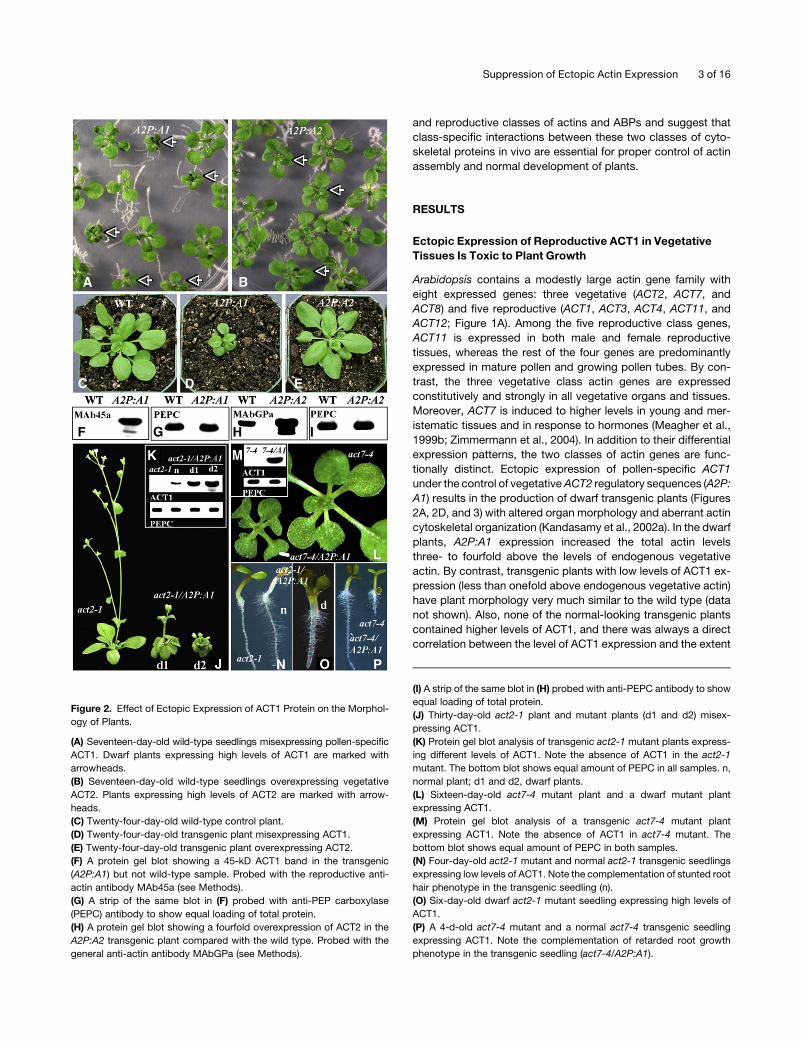
Figure 2. Effect of Ectopic Expression of ACT1 Protein on the Morphol-
ogy of Plants.
(A) Seventeen-day-old wild-type seedlings misexpressing pollen-specific
ACT1. Dwarf plants expressing high levels of ACT1 are marked with
arrowheads.
(B) Seventeen-day-old wild-type seedlings overexpressing vegetative
ACT2. Plants expressing high levels of ACT2 are marked with arrow-
heads.
(C) Twenty-four-day-old wild-type control plant.
(D) Twenty-four-day-old transgenic plant misexpressing ACT1.
(E) Twenty-four-day-old transgenic plant overexpressing ACT2.
(F) A protein gel blot showing a 45-kD ACT1 band in the transgenic
(A2P:A1) but not wild-type sample. Probed with the reproductive anti-
actin antibody MAb45a (see Methods).
(G) A strip of the same blot in (F) probed with anti-PEP carboxylase
(PEPC) antibody to show equal loading of total protein.
(H) A protein gel blot showing a fourfold overexpression of ACT2 in the
A2P:A2 transgenic plant compared with the wild type. Probed with the
general anti-actin antibody MAbGPa (see Methods).
(I) A strip of the same blot in (H) probed with anti-PEPC antibody to show
equal loading of total protein.
(J) Thirty-day-old act2-1 plant and mutant plants (d1 and d2) misex-
pressing ACT1.
(K) Protein gel blot analysis of transgenic act2-1 mutant plants express-
ing different levels of ACT1. Note the absence of ACT1 in the act2-1
mutant. The bottom blot shows equal amount of PEPC in all samples. n,
normal plant; d1 and d2, dwarf plants.
(L) Sixteen-day-old act7-4 mutant plant and a dwarf mutant plant
expressing ACT1.
(M) Protein gel blot analysis of a transgenic act7-4 mutant plant
expressing ACT1. Note the absence of ACT1 in act7-4 mutant. The
bottom blot shows equal amount of PEPC in both samples.
(N) Four-day-old act2-1 mutant and normal act2-1 transgenic seedlings
expressing low levels of ACT1. Note the complementation of stunted root
hair phenotype in the transgenic seedling (n).
(O) Six-day-old dwarf act2-1 mutant seedling expressing high levels of
ACT1.
(P) A 4-d-old act7-4 mutant and a normal act7-4 transgenic seedling
expressing ACT1. Note the complementation of retarded root growth
phenotype in the transgenic seedling (act7-4/A2P:A1).
Suppression of Ectopic Actin Expression 3 of 16
of defect in the morphology of the plant (Kandasamy et al.,
2002a). On the other hand, overexpression of ACT2 under its
own regulatory sequences (A2P:A2) increased the level of total
actin three- to fourfold (Figure 2H) but did not result in any
extreme dwarf phenotypes in the wild type (Figures 2B and 2E) or
the act2-1 mutant background (data not shown). A small per-
centage of plants (<2%) overexpressing high levels of ACT2 was
50% or more smaller than the wild type, whereas a higher
percentage (20%) of plants misexpressing similar levels of ACT1
was highly dwarf and abnormal (Figure 3). This suggests that
perhaps even higher levels ACT2 (severalfold overproduction)
may be necessary to reveal any aberrant phenotype.
To further demonstrate that the misexpression, but not over-
production, of actin caused aberrant architecture of plants, we
introduced ACT1 into mutant plants expressing significantly
reduced amounts of total actin. For example, transformation of
A2P:A1 (ACT1) into act2-1 mutant plants, which contain ;40%
reduced levels of total actin (data not shown), also produced
extremely small and highly aberrant transformants (Figure 2J).
Furthermore, the misexpression of ACT1 in the act2-1 mutants
affected the morphology of the plants in an ACT1 protein
concentration-dependant manner (Figure 2K). Transgenic mu-
tant plants expressing high levels of ACT1 were severely dwarfed
with highly curved, tiny leaves compared with the untransformed
act2-1 plants (Figures 2J and 2K). Also, they were fully sterile.
Even manual self-pollination or cross-pollination of stigmas of
these severely dwarf plants with wild-type pollen resulted in the
production of no siliques or seeds. The plants with slightly lower
levels of ACT1 than these extremely dwarf plants were small, but
they had mildly curved leaves (see d1 in Figures 2J and 2K), and
they were partially fertile. By contrast, plants with very low levels
of ACT1 (see n in Figure 2K) were normal like the wild type and
fully fertile (data not shown). However, both normal (Figure 2N,
right; Gilliland et al., 2002) and all dwarf transgenic plants (Figure
2O) revealed suppression of the stunted act2-1 mutant root hair
phenotype (Figure 2N, left). Moreover, the introduction of the
A2P:A1 transgene into act7-4 mutant plants that showed ;30%
reduction in the total protein levels (data not shown) also induced
a dwarf plant phenotype (Figures 2L and 2M). However, the
ectopic expression of ACT1 still suppressed the retarded root
growth phenotype of the act7-4 mutant plants (Figure 2P). Thus,
the ACT1 misexpression phenotype in wild-type plants is not due
to too much actin alone because ACT1 expression induced the
dwarf phenotype even in actin mutants that showed significant
reduction in the endogenous vegetative actin levels. The man-
ifestation of severely dwarf plant architecture is consequently
due to the overexpression of the inappropriate class of actin.
Also, the complementation of the vegetative actin phenotypes
revealed in a subset of cell types in actin mutants suggests that
ACT1 (also control ACT2; data not shown) overproduced in the
vegetative cells is functional and most likely folded correctly.
Heterologous Expression of Arabidopsis Reproductive and
Vegetative Actins in Yeast Affects Cell Growth and
Cytoskeletal Architecture
Because the formation of actin filaments is delicately balanced
by the interaction of actin (monomeric and filamentous) with a
complex system of accessory ABPs in all eukaryotic cells, we
presumed that the dwarf phenotype and aberrant cytoskeleton
described above are caused by the poor interaction of the
reproductive ACT1 with the endogenous vegetative ABPs ex-
pressed in the vegetative organs and cell types (Kandasamy
et al., 2002a). To examine the behavior of plant actins in a het-
erologous system, we coexpressed a reproductive actin isovar-
iant, ACT12, or a vegetative isovariant, ACT8, with yeast actin in
yeast cells. For ease of cloning, the reproductive ACT12 and
vegetative ACT8 genes were chosen for our studies with yeast
cells, instead of the homologous ACT1 and ACT2, respectively.
Our results showed that high levels of both classes of plant actin
isovariants were toxic to yeast cells, as evidenced by their slow
growth and altered cell morphology (large cell size) and cyto-
skeletal architecture. While trace amounts of ACT12 and ACT8
proteins were incorporated into F-actin patches and filaments,
the majority of plant actins were found in unusual rod-like
structures, thick cables and spots that are mostly found in the
mother cells and stained only with plant actin-specific antibody
(see Supplemental Figures 1B and 1C online) and not with
phalloidin (data not shown). However, the F-actin binding reagent
phalloidin stained actin patches heavily in the bud and actin
filaments mainly in the mother cell of actively growing wild-type
yeast cells (see Supplemental Figure 1A online). These results
suggest that plant actins were incorrectly organized into rods or
wrongly folded in yeast, perhaps because the fungal accessory
proteins did not effectively recognize plant actin sequences.
Ectopic Expression of Reproductive Profilin Does Not
Produce an Aberrant Phenotype
Arabidopsis encodes five highly divergent profilin isovariants that
are grouped based on their phylogeny and expression patterns
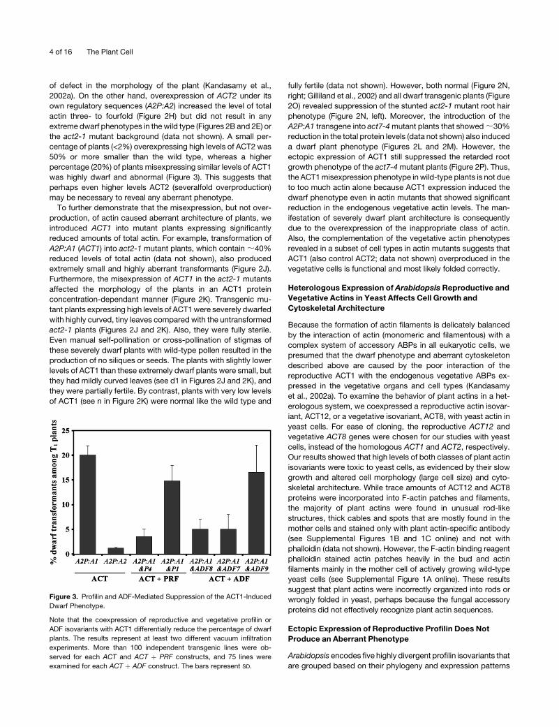
Figure 3. Profilin and ADF-Mediated Suppression of the ACT1-Induced
Dwarf Phenotype.
Note that the coexpression of reproductive and vegetative profilin or
ADF isovariants with ACT1 differentially reduce the percentage of dwarf
plants. The results represent at least two different vacuum infiltration
experiments. More than 100 independent transgenic lines were ob-
served for each ACT and ACT þ PRF constructs, and 75 lines were
examined for each ACT þ ADF construct. The bars represent SD.
4 of 16 The Plant Cell

into two ancient classes: constitutive (PRF1-3) and pollen spe-
cific (PRF4 and 5) (Kandasamy et al., 2002b; Figure 1B). To
examine whether misexpression of a pollen-specific profilin in
vegetative tissues, like reproductive ACT1, has any toxic effect
on plant growth, we ectopically expressed PRF4 under the
control of the ACT2 regulatory sequences (A2P:P4). Protein gel
blot analysis with a PRF4-specific monoclonal antibody revealed
no endogenous expression of PRF4 in the leaf tissue of wild-type
plants but showed various levels of PRF4 expression in different
(>50) A2P:P4 transgenic plants examined (Figure 4C). Even
plants containing very high levels of PRF4 in vegetative tissues
looked morphologically indistinguishable from wild-type plants
(Figures 4A and 4B). Immunolocalization confirmed the absence
of any PRF4 protein in the leaf cells of wild-type seedlings and a
high level of PRF4 expression in the transgenic plants, where it is
seen dispersed uniformly throughout the cytoplasm (Figures 4D
and 4E). Moreover, we constitutively overexpressed the vegeta-
tive PRF1 under the regulation of the ACT2 regulatory sequences
(A2P:P1). On protein gel blots, a PRF1-specific monoclonal
antibody detected a 14-kD faint band in the wild-type sample
and a strong PRF1 band in the A2P:P1 transgenic plants (Figure
4H). Immunolabeling also revealed a relatively higher intensity of
PRF1 staining in the transgenic leaf cells compared with the wild
type (Figures 4I and 4J). Quantification of PRF1 bands on the
protein gel blots of wild-type and dozens of transgenic plant leaf
samples suggested that even a three- to sixfold overproduction
of PRF1 in the transgenic plants did not produce any obvious
morphological abnormality (Figures 4F to 4H). Thus, both the
misexpression of PRF4 and overexpression of PRF1, even at
high levels, were not harmful to the growth of Arabidopsis plants.
These results were surprising because earlier studies of micro-
injection of purified recombinant birch pollen profilin into Trad-
escantia stamen hair cells showed drastic changes in cellular
organization and cessation of cytoplasmic streaming (Staiger
et al., 1994; Valster et al. 1997). These discrepancies may very
well be due to the significantly higher concentrations of profilin
injected into the stamen hair cells (i.e., ;5- to 15-fold increase in
cytoplasmic profilin concentration in interphase cells) compared
with only a two- to fourfold overproduction of total profilin
(endogenous profilin þ transgenic PRF1 or PRF4) achieved in
the transgenic Arabidopsis tissue.
High-Level Expression of Reproductive, but Not Vegetative,
Profilin Suppresses the ACT1 Misexpression Phenotype
In yeast, coexpression of the actin monomer binding protein
profilin had been shown to suppress the deleterious effects of the
overexpression of actin by reducing actin assembly (Magdolen
et al., 1993). So, to examine whether overproduction of profilin
can suppress the dwarf phenotype and the aberrant cytoskeletal
architecture of ACT1 misexpression in Arabidopsis, we coex-
pressed reproductive actin and profilin isovariants together in the
same vegetative organs using the ACT2 regulatory sequences.
First, we identified two A2P:P4 single insertion plant lines from
the various PRF4-misexpressing transgenic lines by scoring the
segregation ratios of antibiotic (hygromycin [Hyg]) resistant to
sensitive seedlings (3:1). These lines were also tested for high
levels of stable PRF4 protein expression in the vegetative tissue
Figure 4. Effect of Profilin Ectopic Expression and Overexpression on
Plant Growth.
(A) Three-week-old wild-type seedling.
(B) Three-week-old transgenic seedling misexpressing PRF4.
(C) Protein gel blot (top panel) showing expression of PRF4 in transgenic
(A2P:P4) but not wild-type leaf sample. Top blot reacted with
MAbPRF45. The bottom panel shows a Coomassie blue–stained dupli-
cate gel.
(D) Wild-type leaf cell labeled with MAbPRF45 showing no staining.
Nuclei stained with 49,6-diamidino-2-phenylindole (DAPI) are shown
in red.
(E) Transgenic (A2P:P4) leaf cell showing strong staining for PRF4.
Orange color indicates nucleus.
(F) Twenty-five-day-old wild-type plant.
(G) Twenty-five-day-old transgenic plant overexpressing PRF1 (A2P:P1).
(H) Protein gel blot (top panel) showing strong expression of PRF1 in
transgenic (A2P:P1) plant. Wild-type leaf sample shows a 14-kD faint
band. Top blot reacted with PRF1-specific MAbPRF1. The bottom panel
shows a Coomassie blue–stained duplicate gel.
(I) Wild-type leaf cell labeled with MAbPRF1 showing a weak staining.
Nucleus stained with DAPI is shown in red.
(J) Transgenic (A2P:P1) leaf cell showing a strong staining for PRF1.
Orange color indicates nucleus.
Bars ¼ 20 mm.
Suppression of Ectopic Actin Expression 5 of 16

by protein gel blot analysis. ACT1 (A2P:A1) was then introduced
separately into both of these PRF4-misexpressing transgenic
lines by Agrobacterium tumefaciens–mediated transformation.
More than 100 independent double transgenic lines (;50 for
each PRF4 line) were isolated based on antibiotic resistance
(Hygr for PRF4 and kanamycinr [Kan] for ACT1). Examination of
seedlings and adult plants revealed that <3% of these double
transformants expressing both ACT1 and PRF4 were smaller
than the wild type (50% or less the size), whereas 20% or more of
the plants misexpressing ACT1 alone were dwarf (Figure 3).
Protein gel blot analysis showed that several (;15%) of the
normal-looking A2P:A1 and P4 double transgenic plants con-
tained high levels of ACT1, similar to the dwarf A2P:A1 single
transformants (e.g., Figures 5A and 5C). The rest of the plants
(;85%) had only low-level expression of ACT1 and thus were
expected to be morphologically normal. However, probing of
duplicate blots using PRF4-specific monoclonal antibodies sug-
gested that all these double transgenic plants contained high
levels of PRF4 protein, whereas no PRF4 expression appeared in
the wild-type or A2P:A1 dwarf plants (Figure 5D). These results
indicate that coexpression of high levels of the reproductive
PRF4 in vegetative tissues suppressed the ACT1-induced dwarf
phenotype. In other words, although they contain high levels of
ACT1, this group of 15% or more plants looked similar in size
(>90% height) to the wild-type plants with large rosette leaves
and thick inflorescence stems (Figures 5A and 5B), and they were
fully fertile (data not shown). Next, we examined the actin
cytoskeletal architecture of these suppressed plants by immu-
nofluorescence microscopy analysis of leaf cells. Our qualitative
observations showed that the A2P:A1 and P4 plant cells (Figures
6E and 6F) had more intense actin staining compared with wild-
type cells (Figures 6A and 6B), but almost all of these cells lacked
the aberrant star- or sheet-like aggregates of actin typically
observed in the A2P:A1 plants (Figures 6C and 6D). Thus, the
coexpression of reproductive PRF4 significantly reduced the
abnormal actin structures induced by the misexpression of
reproductive ACT1.
In addition, we co-overproduced reproductive ACT1 and
vegetative PRF1, which is expressed constitutively in all organs,
including the pistil (Kandasamy et al., 2002b), in the vegetative
cell types using the ACT2 regulatory sequences. As described
above for the generation of A2P:A1 and P4 plants, we first iso-
lated two high-level PRF1 protein–overexpressing A2P:P1 lines
and then transformed them with A2P:A1. We produced >100
double transgenic plants and evaluated them for morphological,
molecular, and cellular phenotypes by observing the size and
fertility of the plants and by performing protein gel blot and
immunocytochemical analyses. Unlike the A2P:A1 and P4 dou-
ble transgenic plants, almost 15% of A2P:A1 and P1 plants still
Figure 5. Suppression of ACT1-Induced Dwarf Phenotype by Coex-
pression of Pollen-Specific PRF4.
(A) Approximately 7-week-old plants. A2P:A1, dwarf single transformant
misexpressing ACT1; A2P:A1&P4, a double transformant misexpressing
both ACT1 and PRF4 simultaneously. Note the suppression of dwarf
phenotype in the double transgenic plant.
(B) Rosette leaves of just-bolted wild-type and single (A2P:A1) and
double (A2P:A1&P4) transgenic plants.
(C) Protein gel blot analysis of ACT1 expression. Top panel shows a blot
probed with MAb45a for ACT1. Both the dwarf single transformant
misexpressing ACT1 and the normal double transgenic plant coexpress-
ing ACT1 and PRF4 have same levels of ACT1 protein. The wild type has
no detectable ACT1. Bottom panel shows a Coomassie blue–stained
duplicate gel.
(D) Protein gel blot analysis of PRF4 expression. Top panel shows a blot
probed with MAbPRF45 for PRF4 protein. Only the double transgenic
plant shows strong PRF4 band. Bottom panel is a Coomassie blue–
stained duplicate gel to show equal loading of total protein.
6 of 16 The Plant Cell
revealed the dwarf phenotype (Figure 3). They contained smaller
leaves and weaker inflorescence stems (Figures 7A and 7B) and
showed highly reduced fertility. Protein gel blot analysis revealed
that none of the large, normal-looking plants contained high
levels of ACT1. Furthermore, all the dwarf double (A2P:A1 and
P1) transgenic plants had approximately the same amount of
ACT1 as the dwarf, high-level ACT1 (A2P:A1) expressing plants
(Figure 7C). Probing of duplicate blots with a PRF1-specific
monoclonal antibody revealed that all the small plants contained
4 to 5 times more PRF1 protein compared with its levels in wild-
type or A2P:A1 dwarf single transformants (Figure 7D). Thus,
even with greatly increased levels of coexpression of PRF1, high
levels of ACT1 expression in shoots resulted in a dwarf pheno-
type. Our successful complementation studies of prf1 mutants
with the A2P:P1 transgene suggested that the overexpressed
PRF1 protein is functional in transgenic plants (data not shown).
Immunofluorescence microscopy analysis revealed that ;50%
of cells of the dwarf double A2P:A1 and P1 transformants still
contained aggregates of actin in the form of sheets (Figure 6G),
although the extent of aberration is slightly less severe compared
with the severely dwarfed A2P:A1 plants (Figures 6C and 6D).
These observations suggest that co-overproduction of vegeta-
tive profilin and reproductive actin did not fully suppress the
ACT1-induced cellular and morphological phenotypes. There
may be a marginal effect due to the coexpression of PRF1 protein
because we observed a small drop in the percentage of dwarf
A2P:A1 and P1 plants (15%) compared with A2P:A1 plants
(20%; Figure 3) and less severe cellular and morphological
phenotypes at least in some dwarf double transformants.
Class-Specific Suppression of ACT1-Induced Dwarf
Phenotype by ADF Isovariants
ADFs are another class of small (;17 kD) ABPs that regulate
actin dynamics by binding both monomeric and filamentous
actin in eukaryotic cells. In Arabidopsis, there are 11 diverse ADF
genes that are broadly grouped, based on their expression
pattern, into two major classes: constitutive (ADF1 to 6 and 9)
and pollen/trichoblast specific (ADF7, 8, 10, and 11). Based
on the phylogeny, they are further subdivided into four sub-
classes (Figure 1C; Ruzicka et al., 2007). To examine whether co-
overexpression of ADF protein isovariants with ACT1 protein
could suppress the dwarf phenotype manifested by ACT1 mis-
expression, we ectopically overproduced ACT1 with ADF7, ADF8,
or ADF9 in the vegetative tissue. ADF7 and 8 are closely related in
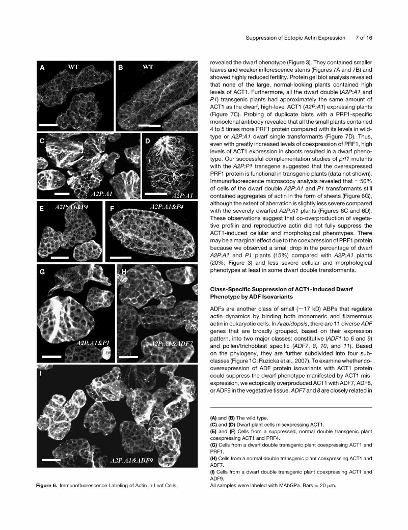
Figure 6. Immunofluorescence Labeling of Actin in Leaf Cells.
(A) and (B) The wild type.
(C) and (D) Dwarf plant cells misexpressing ACT1.
(E) and (F) Cells from a suppressed, normal double transgenic plant
coexpressing ACT1 and PRF4.
(G) Cells from a dwarf double transgenic plant coexpressing ACT1 and
PRF1.
(H) Cells from a normal double transgenic plant coexpressing ACT1 and
ADF7.
(I) Cells from a dwarf double transgenic plant coexpressing ACT1 and
ADF9.
All samples were labeled with MAbGPa. Bars ¼ 20 mm.
Suppression of Ectopic Actin Expression 7 of 16
sequence (78% identical at the amino acid sequence level) but
show completely different expression patterns. The ADF7 gene
is active predominantly in the mature pollen, as revealed by ADF7
promoter-b-glucuronidase fusion gene expression (Figure 8A),
whereas ADF8 is strongly expressed in the root trichoblast cells
and developed root hairs (Figure 8B). On the other hand, ADF9,
which is ;50% identical to ADF7 and ADF8 at the amino acid
sequence level, is constitutively expressed in all organs and cell
types, except pollen (Figure 8C). After examining several dozen
independent transgenic plants, we found that the expression of
any of these three ADFs alone under the control of ACT2
regulatory sequences had no harmful effect on the growth of
plants. Thus, the single transgenic plants either misexpressing
ADF7 (Figure 8D) or ADF8 (Figure 8F) or overexpressing ADF9
(Figure 9A) were indistinguishable from wild-type plants at the
seedling or adult stage of development.
When we coexpressed pollen-specific ADF7 and ACT1 by
transforming single insertion lines carrying A2P:ADF7 with the
A2P:A1 transgene, <5% of the double transgenic plants revealed
the dwarf phenotype in contrast with 20% or more dwarf trans-
genic plants carrying ACT1 alone (Figure 3). The rest of the plants
were morphologically identical to the wild-type plants or plants
carrying only ADF7 (Figure 8D). Protein gel blot analysis on those
normal-looking plants with ACT1-specific monoclonal anti-
bodies revealed that several (;15%) of them contained high
levels of the misexpressed ACT1 protein, similar to the single
A2P:A1 dwarf plants (Figures 8D and 8E). Moreover, those
double transformants with high levels of ACT1 all contained
high levels of ADF7 protein (Figure 8E, middle panel). Thus, the
ectopic expression of pollen-specific ADF7 protein suppressed
the toxic effect of ACT1 in plants containing high levels of this
reproductive actin protein. Interestingly, the closely related
trichoblast/root hair–specific ADF8 also showed an effect similar
to ADF7 when coexpressed along with ACT1 (Figures 3, 8F, and
8G). As a result, plants containing high levels of ACT1 in vege-
tative tissue have large rosette leaves and looked like normal
wild-type plants in the presence of ADF8 (Figure 8F). On the other
hand, when the vegetative ADF9 protein was coexpressed along
with the reproductive ACT1, this ADF isovariant had only mar-
ginal effects on the suppression of dwarf phenotype. Nearly 17%
of the double transformants containing A2P:A1 and A2P:ADF9
transgenes were dwarf compared with 5% or less dwarf plants in
those with A2P:A1 and A2P:ADF7 or A2P:ADF8 transgenes
(Figure 3). Because even high-level expression of ADF9, as
determined by quantitative RT-PCR analysis of the transcripts in
leaves (Figure 9C), could not suppress the toxic effect of ACT1,
those plants containing high levels of ACT1 in the vegetative
tissue were still very small compared with wild-type plants
(Figures 9A and 9B). The ADF isovariant-specific suppression
of the ACT1-induced dwarf phenotype is clearly evident in Figure
9D. Although they had about the same level of ACT1 protein
(Figure 9E), the adult plant expressing ADF7 (Figure 9D, left) was
almost twice the size of the plant expressing ADF9 (Figure 9D,
right). Immunocytochemical analysis suggested that comisex-
pression of ADF7 (Figure 6H) or ADF8 (data not shown) with ACT1
greatly reduced the aberrant actin cytoskeletal structures in most
(;95% normal) leaf cells, whereas co-overexpression ADF9 had
only a little effect because a lot of cells (40 to 50%) still contained
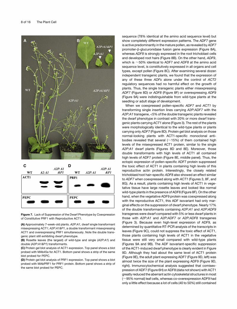
Figure 7. Lack of Suppression of the Dwarf Phenotype by Coexpression
of Constitutive PRF1 with Reproductive ACT1.
(A) Approximately 7-week-old plants. A2P:A1, dwarf single transformant
misexpressing ACT1; A2P:A1&P1, a double transformant misexpressing
ACT1 and overexpressing PRF1 simultaneously. Note the double trans-
genic plant still exhibiting dwarf phenotype.
(B) Rosette leaves (the largest) of wild-type and single (A2P:A1) and
double (A2P:A1&P1) transformants.
(C) Protein gel blot analysis of ACT1 expression. Top panel shows a blot
probed with MAb45a for ACT1. Bottom panel shows a strip of the same
blot probed for PEPC.
(D) Protein gel blot analysis of PRF1 expression. Top panel shows a blot
probed with MAbPRF1 for PRF1 protein. Bottom panel shows a strip of
the same blot probed for PEPC.
8 of 16 The Plant Cell

aggregates of actin bundles (Figure 6I). Even in ACT1 expressing
dwarf single transformants only 50 to 60% of leaf cells showed
obvious aberrant actin structures. Thus, the coexpression of
pollen/root hair-specific ADF7 or ADF8, but not the constitutive
ADF9, with the reproductive ACT1 considerably suppressed the
ACT1-induced aberrant plant and cellular phenotypes.
DISCUSSION
The Extent of Functional Redundancy in
Plant Actin Cytoskeleton
Complex multicellular organisms like plants contain gene fam-
ilies encoding actin and various ABPs that together modulate
the dynamics of the actin-based cytoskeleton (Meagher and
Fechheimer, 2003). For example, the Arabidopsis genome en-
codes eight actin, five profiling, and 11 ADF genes, which are
broadly grouped into constitutive or vegetative and reproductive
(pollen-specific) classes (Figure 1). There are at least 14 addi-
tional families of ABPs encoded by multiple genes, but they are
less well characterized in plants (Cvrckova, 2000; Meagher and
Fechheimer, 2003). The significance of our actin misexpression
studies must therefore be considered in light of the complex actin
cytoskeletal system. Molecular genetic analyses of null muta-
tions in many of the differentially expressed plant cytoskeletal
genes reveal only mild phenotypes that are restricted to a few cell
types. Even mutation in the strongly and constitutively expressed
ACT2 gene shows only a stunted root hair phenotype, and the
lack of this actin isovariant has minimal effects on the survival
and growth of individual plants (Gilliland et al., 2002; Ringli et al.,
2002; Nishimura et al., 2003). Moreover, the root hair development
phenotype of the act2-1 mutation can be complemented with
most other actin isovariants (M.K. Kandasamy, E.C. McKinney,
and R.B. Meagher, unpublished data), including the highly di-
vergent reproductive class actin ACT1 (Gilliland et al., 2002).
Similarly, mutations in the other vegetative actin gene, ACT7,
which is active in all young developing tissues and responds to
phytohormones, reveal retarded root growth and reduced ability
Figure 8. Suppression of ACT1-Induced Dwarf Phenotype by Coex-
pression of Pollen/Trichoblast-Specific ADFs.
(A) to (C) ADF promoter-b-glucuronidase fusion gene expression.
(A) Flower showing pollen-specific expression of ADF7.
(B) Two-day-old germinating seedling showing root hair–specific ex-
pression of ADF8.
(C) Two-week-old seedling showing constitutive expression of ADF9.
(D) Twenty-four-day-old wild-type plant, single transformants misex-
pressing ADF7 (A2P:ADF7) and ACT1 (A2P:A1), and a double transform-
ant (A2P:A1&ADF7) coexpressing ACT1 and ADF7.
(E) Protein gel blot analysis of ACT1 and ADF7 expression in leaves. Top
blot probed with reproductive actin-specific antibody MAb45a to show
ACT1 protein. The weak bottom bands represent breakdown products.
Middle blot from a duplicate gel is probed with MAbADF8, which
recognizes ADF7. Bottom blot is probed with anti-PEPC antibody to
show equal loading of proteins. Note the wild-type sample has no ACT1
and ADF7 proteins, the ADF7-misexpressing normal plant has no ACT1
protein, and the ACT1-misexpressing dwarf plant has no ADF7 protein.
(F) Twenty-four-day-old wild-type plant, single transformants misex-
pressing ADF8 (A2P:ADF8) and ACT1 (A2P:A1), and a double transform-
ant (A2P:A1&ADF8) coexpressing ACT1 and ADF8.
(G) Protein gel blot analysis of ACT1 and ADF8 expression in leaf
samples. Top blot probed with MAb45a to show ACT1 protein. The
breakdown products were not detectable in this blot. Middle blot from a
duplicate gel is probed with MAbADF8 to show ADF8 protein. Bottom
panel probed with anti-PEPC antibody.
Suppression of Ectopic Actin Expression 9 of 16

to form callus on hormone-containing medium as the only
phenotypes. Otherwise the ACT7-deficient plant morphology is
quite comparable to the wild type (Kandasamy et al., 2001;
Gilliland et al., 2003). However, mutations in both these actin
genes together (act2-1/act7-4) are very deleterious and result in
extremely dwarf plants whose viability could be rescued only by
growing them initially on medium containing sucrose as carbon
source (M.K. Kandasamy and R.B. Meagher, unpublished data).
Knockout mutations in individual reproductive class late-pollen
actin genes also reveal no detrimental effect on fertility and seed
set (Pawloski et al., 2006). Similarly, knockout or knockdown
mutations in several of the ABP genes, including profilins
(McKinney et al., 2001) and ADFs (D.R. Ruzicka and R.B.
Meagher, unpublished data), also showed only moderate to no
effect on the growth and overall architecture of plants. The weak
phenotypes of single mutants suggest that there is some degree
of functional redundancy among these cytoskeletal genes in
plants. However, in simple unicellular organisms such as yeast,
where there is only a single copy for most of these cytoskeletal
genes, null mutations in actin and some ABPs (e.g., cofilin and
profilin) are lethal or show severe deleterious phenotypes ap-
proaching lethality (Shortle et al., 1982; Haarer et al., 1993; Moon
et al., 1993).
Phylogenetic analysis suggests that the vegetative and repro-
ductive class plant actin genes have not shared a common
ancestor for 300 to 400 million years, and even the actin genes
encoding highly similar proteins within each class (e.g., ACT2
and 8; ACT1 and 3 with single amino acid differences) have
existed as duplications in plant genomes for approximately the
past 50 million years (McDowell et al., 1996; Meagher et al.,
2000). If vegetative and reproductive actin-based cytoskeletal
systems have been functioning independently in different organs
and tissues for this length of time, it might not be surprising to find
both novel and dysfunctional interactions among proteins from
these two different systems (Meagher et al., 1999a). Moreover,
the various plant actin proteins have an unusually large number
of nonconservative amino acid substitutions (6 to 10%), which
map to the surface of the molecule, in comparison to far fewer
total changes (3 to 7%) and only a few surface changes among
Figure 9. Effect of Coexpression of Reproductive ACT1 and Constitutive
ADF9 on Plant Development.
(A) Twenty-five-day-old wild-type plant, single transformants overex-
pressing ADF9 (A2P:ADF9) and misexpressing ACT1 (A2P:A1), and a
double transformant (A2P:A1&ADF9) coexpressing ACT1 and ADF9.
Note the dwarf stature of the double transformant.
(B) Protein gel blot analysis of ACT1 in leaf samples. Top blot probed with
MAb45a to show ACT1 protein. Bottom panel probed with anti-PEPC
antibody. Wild-type and the normal single transformant overexpressing
ADF9 contain no ACT1 protein.
(C) RT-PCR analysis of ADF9 expression. The single A2P:ADF9 trans-
formants and the double A2P:A1&ADF9 transgenic plants have >100
times the level of ADF9 transcripts than the wild type and plants
misexpressing ACT1. Because of lack of ADF9-specific antibody, the
levels of ADF9 expression are monitored here by RT-PCR analysis.
(D) Adult double transgenic plants coexpressing ACT1 and ADF7 (left)
and ACT1 and ADF9 (right).
(E) Protein gel blot showing the levels of ACT1 protein in the double
transgenic plants shown in (D). Probed with reproductive actin-specific
MAb45a.
10 of 16 The Plant Cell

the 500–million-year-old animal muscle and cytoplasmic actin
subclasses (Hightower and Meagher, 1986; McDowell et al.,
1996). All these amino acid substitutions in plant actins should
have significant effect on protein–protein interactions because
even the few changes in animal actin isovariants lead to differ-
ent physical properties in vitro (Garrels and Gibson, 1976). For
example, despite their relative similarity, the vertebrate non-
muscle and muscle actins display differential binding capacity
for the actin monomer binding proteins profilin and thymosin
(Larsson and Lindberg, 1988; Oshima et al., 1989; Weber et al.,
1992). Moreover, dominant-negative amino acid changes (e.g.,
V163L, V163M, and R183G) resulting from mutations in human
a-skeletal muscle actin cause nemaline myopathy and distinct
pathological phenotypes, such as formation of cytoplasmic
nemaline bodies and intranuclear rods (Ilkovski et al., 2004). A
single amino acid change (Ile-76 to Val-76) also rendered Dro-
sophila flight muscle actin inactive (Fyrberg et al., 1998). Fur-
thermore, the different classes of plant profilin and ADF protein
isovariants exhibit varying biochemical properties (Kovar et al.,
2000; Allwood et al., 2002). These observations combined with
differential gene regulation strongly favor the view that the dif-
ferent classes and subclasses of cytoskeletal protein isovariants
have functional relevance in plants, as in animals.
To understand the extent of functional redundancy and/or
specificity among the cytoskeletal proteins in plants, we have
ectopically expressed pollen-specific actin (ACT1) in the vege-
tative tissues of wild-type plants (Kandasamy et al., 2002a) and in
act2-1 and act7-4 mutants containing markedly reduced
amounts of total actin (this study). Misexpression of high levels
of ACT1 in vegetative tissues retarded the growth of both wild-
type and mutant plants and severely altered the architecture of
most plant organs. The overproduction of ACT1 in vegetative
cells, where there is no expression of reproductive accessory
proteins, resulted in the formation of sheet- or star-like aberrant
actin structures and thick transverse actin cables instead of the
longitudinal arrays of thin actin filaments seen in the wild type
(Kandasamy et al., 2002a). However, overexpression of ACT2 in
vegetative tissues had little effect on the morphology of the plant
or assembly of actin filaments. This clearly shows that the degree
of redundancy among more divergent actin isovariants in the
same plant is not high and that there are definitely functional
differences between the two different classes of actin isovar-
iants. Because of this functional specificity, the misexpressed
pollen actin might have interacted poorly with various ABPs
present in the vegetative cells, and this might have caused an
imbalance between these two groups of proteins and resulted
somehow in abnormal polymerization and arrangement of actin
filaments. Surprisingly, however, the misexpression of diverse
isovariants of two ABPs alone, profilins and ADFs, did not reveal
any harmful effects. We propose that in these plants the misex-
pressed reproductive ADFs and profilins might interact weakly
with the native vegetative actin and cause no toxicity to the
assembly of actin cytoskeleton and thus have little effect on the
morphology of the plant. The activity of native vegetative actin is
normally buffered by the endogenous profilins and ADFs and
myriad other ABPs. However, profilin microinjection studies have
shown drastic effects on the actin cytoskeleton and thereby the
streaming of cytoplasm in Tradescantia stamen hair cells (Staiger
et al., 1994; Valster et al., 1997). However, in transgenic plants,
we are not overexpressing PRF4 or PRF1 to the levels even one
half that of the microinjected recombinant profilin, and this may
be one of the reasons for not seeing any phenotype with profilin
overexpression. Also, an increase in cellular concentration of the
small molecular weight foreign (ectopically expressed) ABPs
(e.g., PRF4 and ADF7) may not be as toxic as the foreign
reproductive actin (ACT1) proteins that tend to aggregate or
polymerize abnormally in the absence of proper interacting
partner APBs, as further revealed by aberrant cytoskeleton
resulting from the heterologus expression of plant actins in yeast
cells.
Class-Specific Interaction among Actin and ABPs in Vivo
Our suppression data further support functional specificity and
class-specific interaction in vivo among the two major classes
of actin and two classes of ABPs. When we ectopically
co-overproduced reproductive actin ACT1 and reproductive
profilin PRF4 simultaneously in the same vegetative tissue, there
was almost full suppression of the toxic effect of misexpressed
ACT1. Thus, plants overproducing both ACT1 and PRF4 looked
quite similar to wild-type plants, and immunocytochemical stud-
ies revealed that the suppression worked by reducing abnormal
actin assembly (aggregation) because their cells contain a typ-
ical, but intensely staining, actin cytoskeleton. However, when
we overproduced reproductive ACT1 and vegetative profilin
PRF1, there was only a marginal effect on the suppression of
dwarf morphological phenotype, and the dwarf plant cells still
exhibited the aggregated actin cellular phenotype. This clearly
showed that there is preferential, class-specific interaction be-
tween the actin and profilin isovariants in plant cells, but it was
still rather surprising to see that overexpression of reproductive
class profilin alone was sufficient to compensate for the over-
production of ACT1. The actin assembly and disassembly in
eukaryotic cells generally involves profilin and a host of other
associated accessory proteins (Pollard et al., 2000; Paavilainen
et al., 2004; Staiger and Blanchoin, 2006). Because the mani-
festation of the dwarf phenotype was ACT1 concentration de-
pendent, even sequestering moderate amounts of overproduced
ACT1 monomers by PRF4 might be enough to bring the actin
concentration below the toxic threshold level and prevent actin
aggregation into sheet- or star-like structures.
Moreover, we also examined the effect of coexpressing three
different isovariants of ADF (ADF7, ADF8, and ADF9) on the toxic
effects of ACT1. The ADF protein is also a critical player in the
remodeling of the plant actin cytoskeleton (Maciver and Hussey,
2002). Similar to the results with profilin, only the pollen-specific
ADF7, but not the constitutive ADF9, was able to suppress the
dwarf phenotype of ACT1-misexpressing plants. Interestingly,
the trichoblast/root hair–specific ADF8 was also able to interact
in vivo with ACT1 and suppress the deleterious effects of its
misexpression. ADF8 is very closely related in amino acid se-
quence to the pollen-specific ADF7, compared with the consti-
tutive ADF9 (Maciver and Hussey, 2002; Feng et al., 2006). Also,
both pollen tubes and root hairs are fast, tip-growing cells
requiring highly dynamic, polarized actin cytoskeleton and tip-
directed vesicle trafficking modulated by actin (Hepler et al.,
Suppression of Ectopic Actin Expression 11 of 16
2001; Cole and Fowler, 2006; Samaj et al., 2006), and these
similar cell types may have some common components involved
in actin assembly. Therefore, the pollen-specific ADF7 and root
hair–specific ADF8 were able to interact with ACT1 in vivo and
suppress the dwarf phenotype.
The differential expression of ABPs may also explain why
ACT1 was able to complement the stunted root hair phenotype of
act2-1 mutants, when ectopically expressed using the ACT2 pro-
moter (Gilliland et al., 2002; this study). In the ACT1-misexpressing
act2-1 mutant plants, the root hair growth is restored, but the
organization of all other vegetative organs and cell types are
severely altered. Because root hairs endogenously express
ADF8, which was shown here to suppress the dwarf plant
phenotype when coexpressed along with ACT1, it is possible
that the endogenous root hair–specific ADFs (ADF8 and ADF11)
might interact properly with ACT1 and therefore might comple-
ment the act2-1 root hair phenotype. Moreover, ACT1 comple-
ments the retarded root growth phenotype of the act7-4 mutant.
From both these complementation studies, it is clear that ACT1 is
able to suppress a subset of vegetative actin mutant phenotypes
while adversely affecting the development and organization of
other organs. This may be due to the differential expression of
some of the ABPs that interact properly with ACT1 (e.g., root
trichoblast–specific ADF8 and 11) or may be due to the expres-
sion of constitutive ABPs in roots (e.g., ADF6, which is expressed
both in pollen, root, and other vegetative tissue) that may
moderately interact with the reproductive actins. In the aerial
vegetative organs, there is no expression of ADF8 or ADF11 or
enough expression of other ABPs with similar properties; hence,
the pollen-specific ACT1 may not be able to interact normally
with any of the endogenous APBs, resulting in aberrant cyto-
skeletal organization and development.
Actin Dynamics in ACT1-Misexpressed Dwarf and
Suppressed Normal Plants
Actin is generally organized in plant cells into arrays of thin cables
and fine filaments and baskets surrounding the nucleus and
chloroplasts (Kost et al., 1998; Kandasamy and Meagher, 1999;
Sheahan et al., 2004). The proper assembly and organization of
actin depends upon the expression of an appropriate mixture of
ABPs and signals mediated by various cellular signaling path-
ways involving molecules like the Rho family of GTPases
(Staiger, 2000; Valster et al., 2000; Vantard and Blanchoin,
2002). When the delicate balance between the various cytoskel-
etal components is impaired, there are unusual consequences
for actin assembly and cellular architecture, as observed in the
ACT1-misexpressed dwarf plants. Although we assume that
weak and/or inappropriate interactions of reproductive actin with
the endogenous vegetative ABPs in the cells might be the cause
for formation of abnormal star- or sheet-like actin structures, the
understanding of the exact biochemical mechanism behind this
unusual process requires further analysis. However, the genetic
suppression of the ACT1-induced dwarf phenotype and the
aberrant actin organization by the coexpression of reproductive
profilin or ADF isovariants supports the need for class-specific
interaction in vivo among actin and various ABP isovariants for
proper assembly of the actin cytoskeleton. In plant cells, as
reported for maize and Papaver pollen and tobacco suspension
cells where profilin is massively abundant, a remarkably small
percentage (5 to 10% and 1 to 2%, respectively) of total actin is
present in the filamentous form (Gibbon et al., 1999; Snowman
et al., 2002; Wang et al., 2005), and the large monomer pool is
predicted to be in a complex with profilin (Staiger and Blanchoin,
2006) and other ABPs. The massive overproduction of pollen
actin in the vegetative cells of dwarf plants might result in lack of
or insufficient levels of appropriate actin monomer sequestering
proteins like profilin. Moreover, poor interaction of endogenous
vegetative profilin and other ABPs with the pollen actin isovariant
might have left the cells with excess free actin monomers. As
observed in yeast mutants deficient in profilin or other ABPs,
where actin is deposited into bar-like structures (Magdolen et al.,
1993), the excess reproductive actin monomers in the dwarf
(A2P:A1) plant cells are arranged into star- or thick cable-like
structures (see the model in Supplemental Figure 2B online).
Similarly, the plant actins expressed in yeast cells were incor-
porated into rods, thick cables, and unusual patches, probably
because of their poor affinity for the yeast accessory proteins.
However, when we coexpressed reproductive actin and repro-
ductive profilin or ADF in the same plant cells (A2P:A1 and
A2P:ADF7 or PRF4), there appeared to be proper interaction
between the two cytoskeletal components. The reproductive
ABPs balance out the excess of reproductive actin monomers in
the cell; hence, there was normal F-actin polymerization, albeit
with more actin filaments (see Supplemental Figure 2C online).
We suggest that because of poor affinity, the endogenous or
coexpressed vegetative ABPs could not balance the concentra-
tion of excessive reproductive actin monomers (in A2P:A1 and
A2P:ADF9 or PRF1 double transformants) and resulted in plants
that were still dwarfed with abnormal actin structures (see
Supplemental Figure 2D online). In the control plants overex-
pressing vegetative actin alone (A2P:A2), the levels of endoge-
nous vegetative ABPs may be sufficient to buffer the excess actin
and regulate polymerization to form more actin filaments but
avoid formation of abnormal actin structures (see Supplemental
Figure 2E online).
Biochemical evidence for the differential binding of reproduc-
tive and vegetative ABPs with the two major classes of plant actin
would provide further support to our genetic suppression studies
and our model. In this regard, it is worth mentioning that Chris
Staiger’s group (Purdue University) has recently found marked
differences in the preference of the Arabidopsis vegetative PRF2
and reproductive PRF4 for plant and vertebrate actins. Specif-
ically, they discovered that PRF4 has ;3.5-fold higher affinity for
monomeric actin from plants than it does for vertebrate actin,
whereas PRF2 shows equal binding to both types of actin
monomers (F. Chaudhry, S. Huang, S. Kasina, and C.J. Staiger,
unpublished data). The vegetative PRF1 used in this study is the
closest homolog to PRF2. The findings from Staiger’s lab obvi-
ously lend support to our assumption that the reproductive
and the vegetative ABPs differentially interact with ACT1. More-
over, future cross-linking and immunoprecipitation studies with
isovariant-specific antibodies will elucidate the differential bind-
ing properties of various vegetative and reproductive cyto-
skeletal proteins in plants that are wild-type or have different
transgenic backgrounds. More efficient binding of ACT1 to PRF4
12 of 16 The Plant Cell
or ADF7 than ACT1 to PRF1 or ADF9 would explain why the
vegetative and reproductive ABPs differentially suppress the
ectopic expression phenotypes of pollen-specific ACT1 protein
in vegetative cell types. In summary, our data on the ectopic
expression of ACT1 and ABP isovariant-specific suppression of
the dwarf morphological and actin cellular phenotypes provide
strong evidence for the existence of functional differences
among the two classes of actin and ABP isovariants in vivo.
METHODS
Plant Material and Generation of Transgenic Plants
Wild-type (Columbia), mutant (act2-1 and act7-4), and transgenic Arabi-
dopsis thaliana plants were cultivated in growth chambers at 228C with
16 h of light and 8 h of dark periods. As described earlier (Gilliland et al.,
2002), act2-1 is a null mutant allele with a T-DNA insertion just five
nucleotides upstream of ATG in the vegetative class ACT2 gene. The
act7-4 null mutant allele has a T-DNA insertion in the leader intron of the
vegetative ACT7 gene (Gilliland et al., 2003). Seven different constructs
were made for this study of suppression of ACT1 ectopic expression
phenotypes (Figure1D): (1) A2P:A1, an actin misexpression construct that
contains a 1.1-kb full-length ACT1 cDNA inserted between a 1.3-kb
promoter and the terminator region of ACT2; (2) A2P:A2, a control actin
overexpression construct in which the ACT1 cDNA was replaced with a
1.1-kb full-length ACT2 cDNA. Similarly, different profilin and ADF full-
length cDNAs replaced ACT1 cDNA in the rest of the constructs: (3)
A2P:P4, a profilin misexpression construct with a 405-bp PRF4 cDNA; (4)
A2P:P1, a profilin overexpression construct containing a 396-bp PRF1
cDNA; (5) A2P:ADF7, an ADF misexpression construct with a 414-bp
ADF7 cDNA; (6) A2P:ADF8, another ADF misexpression construct with a
423-bp ADF8 cDNA; (7) A2P:ADF9, an ADF overexpression construct
with a 426-bp ADF9 cDNA. The various steps involved in cloning A2P:A1
and A2P:A2 constructs were described previously (Kandasamy et al.,
2002a).
All the ACT, PRF, and ADF cDNAs were PCR amplified from a mature
flower library in the plasmid vector pCDNAII (Invitrogen), except for ADF8,
which was amplified from a root cDNA library. The expression plasmids
were mobilized into the Agrobacterium tumefaciens strain C58C1 and
transformed into wild-type or mutant Arabidopsis plants by vacuum
infiltration. Transformants were selected by plating the seeds on medium
containing 35 mg/L Kan or 50 mg/L Hyg. For generating double trans-
genic plants, we first identified single insertion lines for different PRF and
ADF transgenes by scoring the segregation ratio of antibiotic resistant to
sensitive (3:1) plants in the T2 generation, and then we selected two plant
lines expressing high levels of the respective protein isovariants by
performing protein gel blot analyses. Finally, we transformed them with
A2P:A1. Phenotypic assessment of wild-type and antibiotic-resistant
single and double transgenic plants was made at different stages of
development. To determine the plant size, we measured the diameter of
the whole rosette at the time of bolting and the height of adult plants when
they were 7 to 8 weeks old.
Heterologus Expression of Plant Actins in Yeast
The vegetative ACT8 (U42007; An et al., 1996) and the pollen-specific
ACT12 (U27982; Huang et al., 1996) cDNAs were cloned into the yeast
expression plasmid p413-HIS3 with the TEF (translation elongation
factor) promoter. These constructs were transformed into the yeast strain
DDY384 (Drubin et al., 1993). Transformed yeast cells were plated on
minimal medium lacking Leu, uracil, and histitine to maintain the pDD41
and p413 plasmids, and glucose was used as a carbon source (Sachs
et al., 1987). Because the strain DDY384 contains a deletion of the only
essential yeast actin yACT1, it was complemented by the presence of the
plasmid pDD41 containing wild-type yACT1.
Antibodies
Two monoclonal antibodies were used to detect actin either by protein gel
blot analysis or by confocal immunofluorescence microscopy: (1)
MAbGPa, a general plant actin–specific antibody that detects equally
all eight expressed Arabidopsis actins (Kandasamy et al., 1999); and (2)
MAb45a, a reproductive actin-specific antibody that reacts uniformly with
actins ACT1, ACT3, ACT4, and ACT12 (Kandasamy et al., 1999). We used
two profilin monoclonal antibodies to detect profilin at the protein gel blot
or cellular level: (1) MAbPRF45 reacted with both pollen-specific profilin
isovariants PRF4 and PRF5; and (2) MAbPRF1 was specific to the
constitutive profilin isovariant PRF1 (Kandasamy et al., 2002b). ADF7
and ADF8 proteins were detected on protein gel blots with MAbADF8,
which was raised against ADF8 recombinant protein, but recognized all
four ADFs (ADF7, ADF10, ADF8, and ADF11) of the pollen/trichoblast-
specific subclasses (see Figure 1C; Ruzicka et al., 2007). An anti-PEP
carboxylase polyclonal antibody (Rockland) was used as control to
monitor variation in protein loading and uniform transfer during electro-
blotting.
Protein Gel Blot Analysis
For detection of actin on protein gel blots, protein samples from frozen
wild-type, mutant, and transgenic Arabidopsis leaves were prepared as
described previously (Kandasamy et al., 1999) using an extraction buffer
containing 25 mM Tris-HCl, pH 7.5, 10 mM NaCl, 10 mM MgCl2, 5 mM
EDTA, and complete protease inhibitor cocktail (one tablet/10 mL; Roche
Diagnostics). The protein extracted in the extraction buffer was precip-
itated with trichloroacetic acid, and the pellet was washed with cold
acetone, dried, and dissolved in solubilization buffer (11 mg Na2CO3 per
mL 100 mM DDT). The protein suspension was mixed with equal volume
of 23 sample buffer (Laemmli, 1970), boiled (5 min), and then loaded onto
SDS-PAGE gels. However, for profilin analysis, the frozen leaf samples
were extracted in extraction buffer, and after centrifugation the superna-
tant was directly mixed 1:1 with 23 sample buffer, boiled for 5 min, and
then loaded onto the gels. On the other hand, ADF was assayed by
directly extracting the frozen samples in 23 sample buffer. Different
extraction procedures were followed to optimize for maximum content of
each desired protein in a sample. Equal loading of proteins was moni-
tored by Coomassie Brilliant Blue staining of duplicate gels, and uniform
transfer of protein to the polyvinylidene fluoride membrane was deter-
mined by probing identical blots or strips of blots (>80 kD) with anti-PEP
carboxylase antibody. The protein bands that were detected using the
ECL kit (Amersham) were quantified using the NIH Sci Image program.
RT-PCR Analysis of ADF9 Expression
RNA was isolated from leaf tissues of wild-type and various transgenic
plants using the RNeasy plant mini kit (Qiagen), and it was treated with
RQ1 RNase-free DNase (Promega) before reverse transcription. Three
micrograms of treated RNA were added to RT reactions using the
Invitrogen SuperscriptIII first-strand synthesis kit with random hexamer
primers to make cDNA. Real-time PCR was used to analyze cDNA
populations using 18S primers (18S-RT2S, 59-GGGGGCAATCGTATTT-
CATA-39, and 18S-RT2A, 59-TTCGCAGTTGTTCGTCTTTC-39) as the en-
dogenous control. The primers used for ADF9 detection are ADF9totalS
(59-TGGTGTTCACTACGAGCTTCA-39) and ADF9totalA (59-GATAAAA-
TCCAGGACCGGG-39). Reactions were performed on an Applied Bio-
systems 7500 real-time PCR system using SYBR Green detection
chemistry (Applied Biosystems) as described previously (Deal et al.,
Suppression of Ectopic Actin Expression 13 of 16
2007). The 2�(ddCT) method (Livak and Schmittgen, 2001) of relative
quantification was used in all experiments.
Immunofluorescence Labeling and Confocal Microscopy of
Plant Cells
Cryofixation and freeze-substitution of leaves from young seedlings were
done as described elsewhere (Kandasamy et al., 2002a). In brief, the sam-
ples were rapidly frozen in liquid propane (�1808C), freeze-substituted in
acetone at �808C for 72 h, and then gradually brought to room temper-
ature over an 8-h period. After rehydration through a graded acetone
series, the seedlings were washed in PME (50 mM PIPES, pH 7.0, 5 mM
EGTA, 1 mM MgSO4, and 0.5% casein), permeabilized by treating with
1% Cellulysin (Calbiochem) and 0.1% Pectolyase (Sigma-Aldrich) in PME
containing protease inhibitors (complete mini EDTA-free protease inhib-
itor tablets) for 20 min, washed again in PME (5 min) and PBS (23 10 min),
and squashed onto chrom-alum and gelatin-coated slides. Following air-
drying, the leaf cells attached to the slides were further permeabilized in
0.5% Triton X-100 in PBS for 20 min and�208C methanol for 10 min. After
rinsing in PBS, the slides were blocked for 1 h in TBST-BSA-GS (10 mM
Tris-HCl, pH 7.5, 150 mM NaCl, 0.05% Tween 20, 2.5% BSA, and 10%
goat serum) and then incubated in the primary antibody diluted (5 mg/mL)
in TBST-BSA-GS. After overnight incubation, the slides were rinsed with
PBS, and then labeled for 3 h with fluorescein isothiocyanate–conjugated
anti-mouse secondary antibody (Sigma-Aldrich) at 1:100 dilution. The
slides were rinsed in PBS (33 10 min) and mounted with 80% glycerol in
PBS containing 1 mg/mL p-phenylenediamine (Sigma-Aldrich). The actin
microfilaments in the labeled cells were visualized with a Leica confocal
laser scanning microscope (TCS-SP2). For profilin, after labeling with
antibodies, the cells were stained with DAPI (Sigma-Aldrich) and ob-
served with a Leica fluorescence microscope.
Localization of Plant and Native Actin in Yeast
Wild-type and transformed yeast cells were fixed in 4% paraformalde-
hyde freshly prepared in PME containing the protease inhibitor cocktail
(Roche Diagnostics) for 1 h. After washing in PME containing 1.2 M
sorbitol, the fixed cells were permeabilized by treating with Zymolyase
(25 mg/mL; ICN) and Glusulase (55 mL/mL; NEN) for 30 min, washed again
in PME as above, and immobilized onto chrom-alum and gelatin-coated
slides. The cells were further permeabilized in cold methanol (6 min) and
acetone (30 s), air-dried, and immunolabeled with plant actin-specific
MAbGPa as described above for plant cells. For F-actin staining in wild-
type cells, the blocked slides were incubated with Texas Red–conjugated
phalloidin (0 to 25 mM; Molecular Probes) for 2 to 3 h, rinsed with PBS,
mounted with 80% glycerol in PBS containing 1 mg/mL p-phenylenedi-
amine, and observed with a Leica confocal microscope.
Sequence Comparisons and Phylograms
Sequences were aligned with ClustalW 1.82 (Higgins and Sharp, 1988)
using default settings, and phylogenies were constructed with PAUP 4.0
(Rogers and Swofford, 1999) using the neighbor-joining tree building
method. Bootstrap support values were based on 100 replicates using a
full heuristic search. Actin, ADF, or profilin sequences of Saccharomyces
cerevisiae and Chlamydomonas reinhardtii were used to root these trees.
Specific sequence identifiers are given in Supplemental Data Sets 1 (Ac-
tin), 2 (profilin), and 3 (ADF) online.
Accession Numbers
Sequence data from this article can be found in the Arabidopsis Genome
Initiative or GenBank/EMBL databases under the following accession
numbers: At ACT1, U39449 (At2g37620); At ACT2, U41998 (At3g18780);
At PRF1, U43322 (At2g19760); At PRF4, U43324 (At4g29340); At ADF7,
NM_118691 (At4g25590); At ADF8, NM_116293 (At4g00680); At ADF9,
NM_119663 (At4g34970). The Arabidopsis Genome Initiative locus num-
bers are given in parenthesis.
Supplemental Data
The following materials are available in the online version of this article.
Supplemental Figure 1. Actin Localization in Yeast Cells.
Supplemental Figure 2. Models for Class-Specific Interaction of PRF
and ADF Isovariants with Actin and Regulation of Actin Assembly.
Supplemental Data Set 1. Amino Acid Sequences Used in Con-
struction of the Actin Tree in Figure 1A.
Supplemental Data Set 2. Amino Acid Sequences Used in Con-
struction of the Profilin Tree in Figure 1B.
Supplemental Data Set 3. Amino Acid Sequences Used in Con-
struction of the ADF Tree in Figure 1C.
ACKNOWLEDGMENTS
We thank Roger Deal and Gay Gragson for critical reading of the
manuscript and the anonymous reviewers for their useful comments.
Confocal microscopy was conducted at the Center for Advanced
Ultrastructural Research at the University of Georgia. We thank Beth
Richardson for her help with rapid freezing of samples for actin local-
ization. This work was supported by the National Institutes of Health
(GM-36397).
Received May 2, 2007; revised September 6, 2007; accepted September
24, 2007; published October 12, 2007.
REFERENCES
Allwood, E.G., Anthony, R.G., Smertenko, A.P., Reichelt, S., Drobak,
B.K., Doonan, J.H., Weeds, A.G., and Hussey, P.J. (2002). Regu-
lation of the pollen-specific actin-depolymerizing factor LlADF1. Plant
Cell 14: 2915–2927.
An, Y.Q., McDowell, J.M., Huang, S., McKinney, E.C., Chambliss, S.,
and Meagher, R.B. (1996). Strong, constitutive expression of the
Arabidopsis ACT2/ACT8 actin subclass in vegetative tissues. Plant J.
10: 107–121.
Bamburg, J.R. (1999). Proteins of the ADF/cofilin family: Essential
regulators of actin dynamics. Annu. Rev. Cell Dev. Biol. 15: 185–230.
Carlier, M.F. (1998). Control of actin dynamics. Curr. Opin. Cell Biol. 10:
45–51.
Carlsson, L., Nystrom, L.-E., Sundkvist, I., Markey, F., and Lindberg,
U. (1977). Actin polymerizability is influenced by profilin, a low mo-
lecular weight protein in non-muscle cells. J. Mol. Biol. 115: 465–483.
Chen, H., Bernstein, B.W., and Bamburg, J.R. (2000). Regulating
actin-filament dynamics in vivo. Trends Biochem. Sci. 25: 19–23.
Cole, R.A., and Fowler, J.E. (2006). Polarized growth: Maintaining
focus on the tip. Curr. Opin. Plant Biol. 9: 579–588.
Cvrckova, F. (2000). Are plant formins integral membrane proteins?
Genome Biol. 1: Research001.1–001.7.
Deal, R.B., Topp, C.N., McKinney, E.C., and Meagher, R.B. (2007). Re-
pression of flowering in Arabidopsis requires activation of FLOWER-
ING LOCUS C expression by the histone variant H2A.Z. Plant Cell 19:
74–83.
14 of 16 The Plant Cell

Drubin, D.G., Jones, H.D., and Wertman, K.F. (1993). Actin structure
and function: Roles in mitochondrial organization and morphogenesis
in budding yeast and identification of the phalloidin-binding site. Mol.
Biol. Cell 4: 1277–1294.
Feng, Y., Liu, Q., and Xue, Q. (2006). Comparative study of rice and
Arabidopsis actin-depolymerizing factors gene families. J. Plant
Physiol. 163: 69–79.
Fyrberg, E.A., Fyrberg, C.C., Biggs, J.R., Saville, D., Beall, C.J., and
Ketchum, A. (1998). Functional nonequivalence of Drosophila actin
isoforms. Biochem. Genet. 36: 271–287.
Garrels, J.I., and Gibson, W. (1976). Identification and characterization
of multiple forms of actin. Cell 9: 793–805.
Gibbon, B.C., Kovar, D.R., and Staiger, C.J. (1999). Latrunculin B had
different effects on pollen germination and tube growth. Plant Cell 11:
2349–2363.
Gilliland, L.U., Kandasamy, M.K., Pawloski, L.C., and Meagher, R.B.
(2002). Both vegetative and reproductive actin isovariants comple-
ment the stunted root hair phenotype of the Arabidopsis act2–1 mu-
tation. Plant Physiol. 130: 2199–2209.
Gilliland, L.U., Pawloski, L.C., Kandasamy, M.K., and Meagher, R.B.
(2003). Arabidopsis actin gene ACT7 plays an essential role in germi-
nation and root growth. Plant J. 33: 319–328.
Haarer, B.K., Petzold, A.S., and Brown, S.S. (1993). Mutational anal-
ysis of yeast profilin. Mol. Cell. Biol. 13: 7864–7873.
Hepler, P.K., Vidali, L., and Cheung, A.Y. (2001). Polarized cell growth
in higher plants. Annu. Rev. Cell Dev. Biol. 17: 159–187.
Higgins, D.G., and Sharp, P.M. (1988). CLUSTAL: A package for
performing multiple sequence alignment on a microcomputer. Gene
73: 237–244.
Hightower, R.C., and Meagher, R.B. (1986). The molecular evolution of
actin. Genetics 114: 315–332.
Huang, S., An, Y.Q., McDowell, J.M., McKinney, E.C., and Meagher,
R.B. (1996). The Arabidopsis thaliana ACT4/ACT12 actin gene sub-
class is strongly expressed throughout pollen development. Plant J.
10: 189–202.
Hussey, P.J., Allwood, E.G., and Smertenko, A.P. (2002). Actin-
binding proteins in the Arabidopsis genome database: properties of
functionally distinct plant actin-depolymerizing factors/cofilins. Philos.
Trans. R. Soc. Lond. B Biol. Sci. 357: 791–798.
Hussey, P.J., Ketelaar, T., and Deeks, M.J. (2006). Control of the
actin cytoskeleton in plant cell growth. Annu. Rev. Plant Biol. 57:
109–125.
Ilkovski, B., Nowak, K.J., Domazetovska, A., Maxwell, A.L.,
Clement, S., Davies, K.E., Laing, N.G., North, K.N., and Cooper,
S.T. (2004). Evidence for a dominant-negative effect in ACTA1 nema-
line myopathy caused by abnormal folding, aggregation and altered
polymerization of mutant actin isoforms. Hum. Mol. Genet. 13:
1727–1743.
Kandasamy, M.K., Gilliland, L.U., McKinney, E.C., and Meagher,
R.B. (2001). One plant actin isovariant, ACT7, is induced by auxin and
required for normal callus formation. Plant Cell 13: 1541–1554.
Kandasamy, M.K., McKinney, E.C., and Meagher, R.B. (1999). The
late pollen-specific actins in angiosperms. Plant J. 18: 681–691.
Kandasamy, M.K., McKinney, E.C., and Meagher, R.B. (2002a).
Functional nonequivalency of actin isovariants in Arabidopsis. Mol.
Biol. Cell 13: 251–261.
Kandasamy, M.K., McKinney, E.C., and Meagher, R.B. (2002b). Plant
profilin isovariants are distinctly regulated in vegetative and repro-
ductive tissues. Cell Motil. Cytoskeleton 52: 22–32.
Kandasamy, M.K., and Meagher, R.B. (1999). Actin-organelle interac-
tion: Association with chloroplast in Arabidopsis leaf mesophyll cells.
Cell Motil. Cytoskeleton 44: 110–118.
Kost, B., Spielhofer, P., and Chua, N.H. (1998). A GFP-mouse talin
fusion protein labels plant actin filaments in vivo and visualizes the
actin cytoskeleton in growing pollen tubes. Plant J. 16: 393–401.
Kovar, D.R., Drobak, B.K., and Staiger, C.J. (2000). Maize profilin
isoforms are functionally distinct. Plant Cell 12: 583–598.
Laemmli, U.K. (1970). Cleavage of structural proteins during the as-
sembly of the head of bacteriophage T4. Nature 227: 680–685.
Larsson, H., and Lindberg, U. (1988). The effect of divalent cations on
the interaction between calf spleen profilin and different actins.
Biochim. Biophys. Acta 953: 95–105.
Livak, K.J., and Schmittgen, T.D. (2001). Analysis of relative gene
expression data using real-time quantitative PCR and the 2(-Delta
Delta C(T)) Method. Methods 25: 402–408.
Maciver, S.K., and Hussey, P.J. (2002). The ADF/cofilin family: Actin-
remodeling proteins. Genome Biol. 3: reviews3007.1–3007.12.
Magdolen, V., Drubin, D.G., Mages, G., and Bandlow, W. (1993). High
levels of profilin suppress the lethality caused by overproduction of
actin in yeast cells. FEBS Lett. 316: 41–47.
McDowell, J.M., Huang, S., McKinney, E.C., An, Y.Q., and Meagher,
R.B. (1996). Structure and evolution of the actin gene family in
Arabidopsis thaliana. Genetics 142: 587–602.
McKinney, E.C., Kandasamy, M.K., and Meagher, R.B. (2001). Small
changes in the regulation of one Arabidopsis profilin isovariant, PRF1,
alter seedling development. Plant Cell 13: 1179–1191.
Meagher, R.B., and Fechheimer, M. (2003). The Arabidopsis cytoskel-
etal genome. In The Arabidopsis Book, E.M. Meyerowitz and C.R.
Somerville, eds (Rockville, MD: American Society of Plant Biologist),
doi/10.1199/tab.0096, http://www.aspb.org/publications/arabidopsis/.
Meagher, R.B., McKinney, E.C., and Kandasamy, M.K. (1999a).
Isovariant dynamics expand and buffer the responses of complex
systems: The diverse plant actin gene family. Plant Cell 11: 995–1006.
Meagher, R.B., McKinney, E.C., and Kandasamy, M.K. (2000). The
significance of diversity in the plant actin gene family: Studies in
Arabidopsis. In Actin: A Dynamic Framework for Multiple Plant Cell
Functions, C.J. Staiger, F. Baluska, D. Volkmann, and P. Barlow, eds
(Dordrecht, The Netherlands: Klewer Academic Publishers), pp. 3–27.
Meagher, R.B., McKinney, E.C., and Vitale, A.V. (1999b). The evolu-
tion of new structures: Clues from plant cytoskeletal genes. Trends
Genet. 15: 278–284.
Moon, A.L., Janmey, P.A., Louie, K.A., and Drubin, D.G. (1993). Cofilin
is an essential component of the yeast cortical cytoskeleton. J. Cell
Biol. 120: 421–435.
Nishimura, T., Yokota, E., Wada, T., Shimmen, T., and Okada, K.
(2003). An Arabidopsis ACT2 dominant-negative mutation, which
disturbs F-actin polymerization, reveals its distinctive function in
root development. Plant Cell Physiol. 44: 1131–1140.
Oshima, S., Abe, H., and Obinata, T. (1989). Isolation of profilin from
embryonic chicken skeletal muscle and evaluation of its interaction
with different isoforms. J. Biol. Chem. 105: 855–857.
Paavilainen, V.O., Bertling, E., Falck, S., and Lappalainen, P. (2004).
Regulation of cytoskeletal dynamics by actin-monomer-binding pro-
teins. Trends Cell Biol. 14: 386–394.
Pawloski, L.C., Kandasamy, M.K., and Meagher, R.B. (2006). The late
pollen actins are essential for normal male and female development in
Arabidopsis. Plant Mol. Biol. 62: 881–896.
Pollard, T.D., Blanchoin, L., and Mullins, R.D. (2000). Molecular
mechanisms controlling actin filament dynamics in nonmuscle cells.
Annu. Rev. Biophys. Biomol. Struct. 29: 545–576.
Pollard, T.D., and Cooper, J.A. (1986). Actin and actin-binding pro-
teins. A critical evaluation of mechanisms and functions. Annu. Rev.
Biochem. 55: 987–1035.
Ringli, C., Baumberger, N., Diet, A., Frey, B., and Keller, B. (2002).
ACTIN2 is essential for bulge site selection and tip growth during root
hair development of Arabidopsis. Plant Physiol. 129: 1464–1472.
Suppression of Ectopic Actin Expression 15 of 16
Rogers, J.S., and Swofford, D.L. (1999). Multiple local maxima for
likelihoods of phylogenetic trees: a simulation study. Mol. Biol. Evol.
16: 1079–1085.
Ruzicka, D.R., Kandasamy, M.K., McKinney, E.C., Burgos-Rivera,
B., and Meagher, R.B. (September 18, 2007). The ancient subclasses
of Arabidopsis ACTIN DEPOLYMERIZING FACTOR genes exibit novel
and differential expression. Plant J. http://dx.doi.org/10.1111/j.1365-313x.
2007.03257.x.
Sachs, A.B., Davis, R.W., and Kornberg, R.D. (1987). A single domain
of yeast poly(A)-binding protein is necessary and sufficient for RNA
binding and cell viability. Mol. Cell. Biol. 7: 3268–3276.
Samaj, J., Muller, J., Beck, M., Bohm, N., and Menzel, D. (2006).
Vesicular trafficking, cytoskeleton and signalling in root hairs and
pollen tubes. Trends Plant Sci. 11: 594–600.
Sheahan, M.B., Staiger, C.J., Rose, R.J., and McCurdy, D.W. (2004).
A green fluorescent protein fusion to actin-binding domain 2 of
Arabidopsis fimbrin highlights new features of a dynamic actin cyto-
skeleton in live plant cells. Plant Physiol. 136: 3968–3978.
Shortle, D., Haber, J.E., and Botstein, D. (1982). Lethal disruption of
the yeast actin gene by integrative DNA transformation. Science 217:
371–373.
Snowman, B.N., Kovar, D.R., Shevchenko, G., Franklin-Tong, V.E.,
and Staiger, C.J. (2002). Signal-mediated depolymerization of actin in
pollen during the self-incompatibility response. Plant Cell 14: 2613–
2626.
Staiger, C.J. (2000). Signaling to the actin cytoskeleton in plants. Annu.
Rev. Plant Physiol. Plant Mol. Biol. 51: 257–288.
Staiger, C.J., and Blanchoin, L. (2006). Actin dynamics: Old friends
with new stories. Curr. Opin. Plant Biol. 9: 554–562.
Staiger, C.J., Yuan, M., Valenta, R., Shaw, P.J., Warn, R.M., and
Lloyd, C.W. (1994). Microinjected profilin affects cytoplasmic stream-
ing in plant cells by rapidly depolymerizing actin microfilaments. Curr.
Biol. 4: 215–219.
Valster, A.H., Hepler, P.K., and Chernoff, J. (2000). Plant GTPases:
The Rhos in bloom. Trends Cell Biol. 10: 141–146.
Valster, A.H., Pierson, E.S., Valenta, R., Hepler, P.K., and Emons,
A.M.C. (1997). Probing the plant actin cytoskeleton during cytokinesis
and interphase by profilin injection. Plant Cell 9: 1815–1824.
Vantard, M., and Blanchoin, L. (2002). Actin polymerization processes
in plant cells. Curr. Opin. Plant Biol. 5: 502–506.
Wang, H.Y., Yu, Y., Chen, Z.L., and Xia, G.X. (2005). Functional
characterization of Gossypium hirsutum profilin 1 gene (GhPFN1) in
tobacco suspension cells. Characterization of in vivo functions of a
cotton profilin gene. Planta 222: 594–603.
Wasteneys, G.O., and Galway, M.E. (2003). Remodeling the cytoskel-
eton for growth and form: An overview with some new views. Annu.
Rev. Plant Biol. 54: 691–722.
Weber, A., Nachmias, V.T., Pennise, C.R., Pring, M., and Safer, D.
(1992). Interaction of thymosin b4 with muscle and platelet actin:
Implications for actin sequestration in resting platelets. Biochemistry
31: 6179–6185.
Zimmermann, P., Hirsch-Hoffmann, M., Hennig, L., and Gruissem,
W. (2004). GENEVESTIGATOR. Arabidopsis microarray database and
analysis toolbox. Plant Physiol. 136: 2621–2632.
16 of 16 The Plant Cell

DOI 10.1105/tpc.107.052621; originally published online October 12, 2007;Plant Cell
Richard B. MeagherMuthugapatti K. Kandasamy, Brunilís Burgos-Rivera, Elizabeth C. McKinney, Daniel R. Ruzicka and
DevelopmentClass-Specific Interaction of Profilin and ADF Isovariants with Actin in the Regulation of Plant
This information is current as of August 1, 2018
Supplemental Data /content/suppl/2007/09/25/tpc.107.052621.DC1.html
Permissions https://www.copyright.com/ccc/openurl.do?sid=pd_hw1532298X&issn=1532298X&WT.mc_id=pd_hw1532298X
eTOCs http://www.plantcell.org/cgi/alerts/ctmain
Sign up for eTOCs at:
CiteTrack Alerts http://www.plantcell.org/cgi/alerts/ctmain
Sign up for CiteTrack Alerts at:
Subscription Information http://www.aspb.org/publications/subscriptions.cfm
is available at:Plant Physiology and The Plant CellSubscription Information for
ADVANCING THE SCIENCE OF PLANT BIOLOGY © American Society of Plant Biologists