class/course: m.sc. biotechnology subject : basic ... shukla model_answer...subject : basic...
TRANSCRIPT

Class/Course: M.Sc. BiotechnologySemester : II Subject : Basic Enzymology & Enzyme TechnologySub. Code : LBTM-203
Answer 1.1. b) Lyases2. a) Hydrolases3. d) The amino acids….4. d) the shape of the enzyme molecule is changed.5. d) They frequently show cooperative binding to S6. c) has a characteristic value for any set of enzyme and substrate7. a) Activation Energy8. d) Multiple binding sites for substrate for regulatory modifiers9.b) RNA Molecules10. Extremozymes
Answer 2. A purification table is used after a series of separation steps. This table, similar to the one you observed in the purification tutorial, is used to present the information about the yield and purity of the purification. There are only a couple of data needed to prepare the table:• Volume of each step of the purification, starting from the lysates, to the final pooled sample.• Total protein (mg) for each step.• Units (amount) of protein being purified (typically done using the enzyme activity units).Calculations –
1. Units – it is unit of enzyme activity and can be expressed as ‘U’ (International unit)or ‘Kat’ SI unit. It is a measure of enzymatic efficiency of the fraction.
2. Total Activity – this is a measure of how many units of enzyme you have in a sample. Simply multiply the activity in the sample by the total volume.
3. Specific Activity – specific activity is a way to measure how much of a measured enzyme there is with all of the other contaminating proteins. Divide the Activity by the mg of protein. The higher the value, the higher purity.
4. Fold Purification – Divide the specific activity of each fraction by the specific activity found in the lysates. This number changes depending on the enzyme you are working with. There is no good or bad value. However, this number along with the percent yield, indicates if a step was worthwhile or not. Poor fold purification with a low yield is a step to avoid in the future, while a high fold purification and high yield is a great thin.
5. Percent Yield – Calculate the percentage of the yield for each step using the total activity from the starting step.

Example (Purification Chart)S. No.
Steps Volume (ml)
Total activity (U)
Total protein(mg)
Specific activity (U/mg)
Yield (%)
Purification (fold)
1.
2.
3.4.5.
Crude (cell free extract)Ultra filtrationAmmonium-sulphate precipitation (30-70%)DialysisIon-exchangeGel filtration
500
10040301010
5100
40502425182414321006
2400
45020568.214.02.35
2.13
9.0011.8226.74102.28428.08
100
79.4047.5035.7628.0719.70
1.0
4.205.5612.5048.13201.45
Answer 3.
Enzyme activity is expressed in units. Enzyme units are expressed in two ways viz, as International init or as Si unit.
The International unit (U) of an enzyme is defined as the amount of enzyme that will convert 1µmol of substrate to product in one min. under defined conditions (generally 25 or 30°C and the optimum pH)
The SI unit of enzyme activity is defined as the amount of enzyme that will convert 1 mol of substrate to product in 1 second. It has the units of katal (kat)
1Kat = 6x10⁷U
1U = 1.7x10⁻⁸ kat
Specific activity is a way to measure how much of a measured enzyme there is with all of the other contaminating proteins. Divide the Activity by the mg of protein. The higher the value, the greater the purity.
Specific activity=Total units of enzyme in fractionTotal units of protein infraction2a
Turn Over number is equivalent to the number of substrate molecules converted to product in given unit of time on a single enzyme molecule when the enzyme is saturated with substrate. It is denoted as Kcat (first order rate constant. In simple words, it is the numbers of substrate molecules transformed per second per mole of enzyme.
Eg.
Enzyme Turn over number
Catalase 4x10⁷
Carbonic anhydratase 1x10⁶

Acetylcholine esterase 2.5x10⁴
Answer 4.The Lineweaver–Burk plot (or double reciprocal plot) is a graphical representation of the Lineweaver–Burk equation of enzyme kinetics, described by Hans Lineweaver and Dean Burk in 1934. The plot provides a useful graphical method for analysis of the Michaelis–Menten equation
Taking the reciprocal gives
The Lineweaver–Burk plot is widely used method to determine important terms in enzyme kinetics, such as Km and Vmax. The y-intercept of such a graph is equivalent to the inverse of Vmax; the x-intercept of the graph represents −1/Km. It also gives a quick, visual impression of the different forms of enzyme inhibition.
The double reciprocal plot distorts the error structure of the data, and it is therefore unreliable for the determination of enzyme kinetic parameters. Although it is still used for representation of kinetic data, non-linear regression or alternative linear forms of the Michaelis–Menten equation such as the Hanes-Woolf plot or Eadie–Hofstee plot are generally used for the calculation of parameters.
When used for determining the type of enzyme inhibition, the Lineweaver–Burk plot can distinguish competitive, non-competitive and uncompetitive inhibitors. Competitive inhibitors have the same y-intercept as uninhibited enzyme (since Vmax is unaffected by competitive inhibitors the inverse of Vmax also doesn't change) but there are different slopes and x-intercepts between the two data sets. Non-competitive inhibition produces plots with the same x-intercept as uninhibited enzyme (Km is unaffected) but different slopes and y-intercepts. Uncompetitive inhibition causes different intercepts on both the y- and x-axes but the same slope.
Eadie–Hofstee is a graphical representation of enzyme kinetics in which reaction rate is plotted as a function of the ratio between rate and substrate concentration:

Where, v represents reaction rate, Km is the Michaelis–Menten constant, [S] is the substrate concentration, and Vmax is the maximum reaction rate.
It can be derived from the Michaelis–Menten equation as follows:
invert and multiply with :
Rearrange:
Isolate v:
A plot of v against v/[S] will hence yield Vmax as the y-intercept, Vmax/Km as the x-intercept, and Km as the negative slope. Like other techniques that linearize the Michaelis–Menten equation, the Eadie-Hofstee plot was used historically for rapid identification of important kinetic terms like Km and Vmax, but has been superseded by nonlinear regression methods that are significantly more accurate and no longer computationally inaccessible. It is also more robust against error-prone data than the Lineweaver–Burk plot, particularly because it gives equal weight to data points in any range of substrate concentration or reaction rate. (The Lineweaver–Burk plot unevenly weights such points.) Both plots remain useful as a means to present data graphically.
One drawback from the Eadie–Hofstee approach is that neither ordinate nor abscissa represent independent variables: both are dependent on reaction rate. Thus any experimental error will be present in both axes. Also, experimental error or uncertainty will propagate unevenly and

become larger over the abscissa thereby giving more weight to smaller values of v/[S]. Therefore, the typical measure of goodness of fit for linear regression, the correlation coefficient R, is not applicable
In Michaelis_Menten plot the Km value can not be determined accurately because the Vmax is never attained and the observed Vmax is only apparent. Therefore, the linear plots are used routinely to determine the Km and Vmax accurately. The most popular linear plot is the Lineweaver-Burk plot also known as reciprocal plot..
Answer 5Multi-substrate reactions follow complex rate equations that describe how the substrates bind and in what sequence. The analysis of these reactions is much simpler if the concentration of substrate A is kept constant and substrate B varied. Under these conditions, the enzyme behaves just like a single-substrate enzyme and a plot of v by [S] gives apparent KM and Vmax constants for substrate B. If a set of these measurements is performed at different fixed concentrations of A, these data can be used to work out what the mechanism of the reaction is. For an enzyme that takes two substrates A and B and turns them into two products P and Q, there are two types of mechanism: ternary complex and ping–pong.Random-order mechanismA random-order mechanism is one in which any substrate can bind first to the enzyme and any product can leave first. It is a sequential mechanism and for a twosubstrate reaction involves the formation of a ternary complex (one involving enzyme and both substrates):
Compulsory-order mechanismA compulsory-order (or simply ordered) mechanism is a sequential mechanism where the order of binding to and leaving the enzyme is compulsory. For a twosubstrate reaction, a ternary complex will be involved. The precise order must be specified, .e.g
Answer 6Some enzymes need helpers or partners, and some don't. There are different types of enzyme helpers, too, with different enzymes requiring different helpers or different kinds of friends.

Cofactors-The first type of enzyme partner is a group called cofactors, or molecules that increase the rate of reaction or are required for enzyme function. Cofactors are not proteins but rather help proteins, such as enzymes, although they can also help non-enzyme proteins as well. Examples of cofactors include metal ions like iron and zinc.Coenzymes- A specific type of cofactor, coenzymes, are organic molecules that bind to enzymes and help them function. The key here is that they're organic. 'Organic' does not mean you'll find them in a special aisle in the grocery store. Rather, organic molecules are simply molecules that contain carbon. Coenzymes are not really enzymes. As the prefix 'co-' suggests, they work with enzymes. Many coenzymes are derived from vitamins.
Role of metal ions: These molecules often sit at the active site of an enzyme and aid in recognizing, attracting, or repulsing a substrate or product. Remember that a substrate is the molecule upon which an enzyme catalyzes a reaction. Coenzymes can also shuttle chemical groups from one enzyme to another enzyme. Coenzymes bind loosely to enzymes, while another group of cofactors do not.
More than a quarter of all known enzymes require the presence of metal atoms for full catalytic activity. Metal atoms usually exist as cations and often have more than one oxidation state, as with ferrous (Fe2*) and ferric (Fe3*1 iron. We have noted that this positive charge can stabilize transition-states by electrostatic interactions, giving one mechanism for catalysis by metals. However, irrespective of the oxidation state and charge carried, a metal ion can bind a particular number ofgroups (ligands) by accepting free electron pairs to form co-ordinate bonds in specific orientations Therefore, metal ions can be involved in enzyme catalysis in a variety of ways: they may accept or donate electrons to activate electrophiles or nucleophiles, even in neutral solution; they themselves may act as elechophiles; they may mask nucleophiles to prevent unwanted side reactions; they may bring together enzyme and substrate by means of co-ordinate bonds, possibly causing strain to the substrate in the process; they may hold reacting groups in the required three-dimensional orientation; or they may simply stabilize a catalytically-active conformation of the enzyme.
With metalloenzymes, the metal is tightly bound and retained by the en4me on purification. With metal-activated en4rmes, the binding is less tight and the purified enzymes may have to be activated by the addition of metal ions. There is no clear-cut division between the two groups. Albert Mildvan (1970) pointed out that temary complexes formed between an enzyme (E), metal ion (M) and substrate (S) may be enzyme bridge complexes (ME-S), substrate bridge complexes (E-S-IO or metal bridge complexes (E-M-S). Metalloen4rmes cannot form substrate bridge complexes, since the purified enzyme exists as E-M.
The involvement of metal ions in enzymes may be investigated by NMR, ESR and proton relaxation rate (PRR) enhancement techniques, as well as forming part of more general investigations of enzyme structure and function. Examples are given below.
Activation by alkali metal cations (Na* and K)
Alkali metal cations bind only weakly to forrn complexes with enzymes, but K+, the most abundant intracellular cation, is known to activate a greal many enzymes, particularly those catalysing phosphoryl transfer or elimination reactions.
It appears that the role of K* is largely to bind to negatively-charged groups on an inactivi form of the enzyme and cause a change in conformation to a more active form. However, in some

cases, it may also aid substrate binding. For example, muscle pyruvate kinase, a teffameric enzllne which catalyses the reactlon:
has a requirement for alkali metal cations and for Mn2* (or Mg'*), all of which bind in the region of the active site. Various studies indicated that the carboxyl group of PEP binds to the enzyme-bound K*, whereupon a conformational change takes place which facilitates the progress of the reaction via an E-Mn"-PEP complex. Note also that each sub-unit has an α/β panel domain, like TIMActivation by alkaline earth metal cations (Ca2+ and Mg2) Oxygen atoms are often involved in the bonds of both alkali metal and alkaline earth meial cations, bonds of the latter being relatively stronger. The divalent cations, C++ andMg2+, can form six co-ordinate bonds to produce octahedral complexes. Mg2+ is aicumulated by cells in exchange for transport of Ca2+ in the opposite direction. As might be expected, therefore, enzymes requiring Ca'* for activation are mainly extraceiular ones, e.g. the salivary and pancreatiJ a-amylases. The Ca2+upp.uir to play a role in maintaining the structure required for catalytic activity. A veriety of intracellular enzymes require Mgt* for activity and, in most cases, this requiiement can be replaced in vitro by one for Mn2+. Unlike Mg2*, Mn2*_ is paramagnetic, which enables the system to be more easily investigated. (Note that it4tr* -uy, in its own right, be a component of a metalloenzyme, e.g. arginase, where it stabilizes a reactive hydroxide ion, ensuring that an activated nucleophile is available for catalysis. In the rat liver enzyme, Mn'* is bound to histidine-l0l, aspartab-L24, and aspartate-232 in each of the three sub-units, and has a catalytic interaction with aspartate-I 28.)It has been shown that all possible types of ternary bridge complexes involving divalent cations can exist. Most kinases form E-S-M complexes, where S is the reacting nucleotide. Let us consider, as an example, the reaction catalysed by muscle creatine kinase: creatine + ATP ↔ ADP + phosphocreatine + H+ . The true substrate is in fact Mg+ ATP and the reaction proceeds via the formation of the complex creatine -E-ATP-Mg2+. Mildred cohn and colleagues (1971) showed that the divalent cation binds to the o- and p-phosphates of the nucleotide, but not to the terminal (γ) phosphate, which is transferred to creatine. The cation helps in the orientation of the complex, and may also assist in the breaking of the pyrophosphate bond by withdrawing electrons from the p-phosphate. The overall reaction Las a random-order rapid-equilibrium kinetic mechanism' and dead-end complexes may be formed. Identification of some of these, e.g. B-ADP-Mn2+helped in the elucidation of the mechanism of the reaction. In contrast, the reaction catalysed by pyruvate kinase involves a cyclic metal bridge complex.
Ans 7.
Allosteric enzymes - from the Greek allos for "other" and stereos for "shape" (or site) meaning "other site". These enzymes function through reversible, non-covalent binding of a regulatory metabolite at a site other than the catalytic, active site. When bound, these metabolites do not participate in catalysis
directly, but lead to conformational changes in one part of an enzyme that then affect the overall conformation of the active site (causing an increase or decrease in activity, hence these metabolites are termed allosteric activators or allosteric inhibitors).
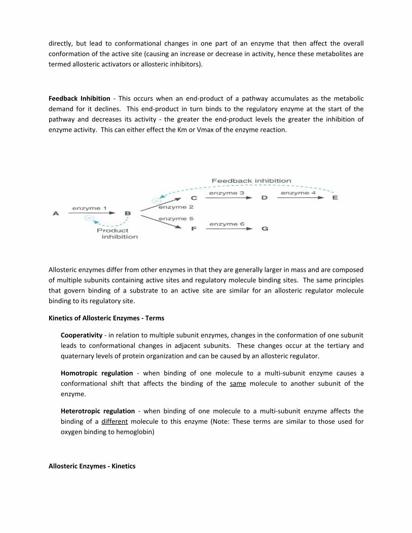
Feedback Inhibition - This occurs when an end-product of a pathway accumulates as the metabolic demand for it declines. This end-product in turn binds to the regulatory enzyme at the start of the pathway and decreases its activity - the greater the end-product levels the greater the inhibition of enzyme activity. This can either effect the Km or Vmax of the enzyme reaction.
Allosteric enzymes differ from other enzymes in that they are generally larger in mass and are composed of multiple subunits containing active sites and regulatory molecule binding sites. The same principles that govern binding of a substrate to an active site are similar for an allosteric regulator molecule binding to its regulatory site.
Kinetics of Allosteric Enzymes - Terms
Cooperativity - in relation to multiple subunit enzymes, changes in the conformation of one subunit leads to conformational changes in adjacent subunits. These changes occur at the tertiary and quaternary levels of protein organization and can be caused by an allosteric regulator.
Homotropic regulation - when binding of one molecule to a multi-subunit enzyme causes a conformational shift that affects the binding of the same molecule to another subunit of the enzyme.
Heterotropic regulation - when binding of one molecule to a multi-subunit enzyme affects the binding of a different molecule to this enzyme (Note: These terms are similar to those used for oxygen binding to hemoglobin)
Allosteric Enzymes - Kinetics

Allosteric enzymes do exhibit saturation kinetics at high [S], but they have a characteristic sigmoidal saturation curve rather than hyperbolic curve when vo is plotted versus [S] (analogous to the oxygen saturation curves of myoglobin vs. hemoglobin). Addition of an allosteric activator (+) tends to shift the curve to a more hyperbolic profile (more like Michaelis-Menten curves), while an allosteric inhibitor (-) will result in more pronounced sigmoidal curves. The sigmoidicity is thought to result from the cooperativity of structural changes between enzyme subunits (again similar to oxygen binding to hemoglobin).
Vo vs [S] for Allosteric Enzymes
Regulation of Enzyme Activity by Covalent Modifications
Another common regulatory mechanism is the reversible covalent modification of an enzyme. Phosphorylation, whereby a phosphate is transferred from an activated donor (usually ATP) to an amino acid on the regulatory enyme, is the most common example of this type of regulation. Frequently this phosphorylation occurs in response to some stimulus (like a hormone or growth factor) that will either activate or inactivate target enzymes via changes in Km or kcat.
Allosteric and Phosphorylation Regulation - Glycogen Phosphorylase

Allosteric regulation of substrate affinity;
Turn on or off by covalent modification of the enzyme; Turn on by regulated expression of gene (transcription) or of synthesis of enzyme (translation);Turn off by regulated destruction of the enzyme (the amino acids are recycled andused for other purposes). The first two effects tend to give immediate responses to changing conditions. The last
two methods provide longer term responses.
The enzyme aspartate transcarbamoylase (ATCase) is a classic case ofallosteric regulation of affinityATCase catalyzes an early step in synthesis of pyrimidines ATCase more steps
carbamoyl phosphate + aspartate → carbamoyl aspartate → → uridine, cytidine
ATCase is negatively regulated by CTP If CTP is present, there's no need to make more
This is an example of negative feedback control. ATCase is positively regulated by ATP If ATP is abundant and [ATP] > [CTP], it's time to make more pyrimidines to
match the availability of purines.
ATCase shows sigmoidal kinetics with respect to substrate aspartate

Allosteric kinetics is described in terms of a low affinity T-state and high affinity R-state When [Asp] is low, the enzyme is in T-state; as [Asp] increases, the enzyme switches to a
high affinity R-state. The enzyme is monitored in terms of the fraction of max rate of catalysis, vo/Vmax, which is essentially a measure of the occupancy of the catalytic site. With substrates only, the reaction rate follows sigmoidal curve C. When CTP binds (curve D), this favours the T-state, so the curve is shifted to the right (higher [Asp] is needed to get the enzyme going). When ATP binds (curve B), this favours the R-state, so the curve is shifted to the left (less [Asp] needed). If [ATP] is high enough (curve A), there is no T-state, and ATCase follows the hyperbolic curve of the pure R-state. For allosteric enzymes, the substrate concentration giving vo/Vmax = 0.5 is designated as K' or K0.5, and represents the apparent substrate affinity, just as P50 represents O2 affinity for hemoglobin. Although KM is derived in a similar manner, the term KM is not used for allosteric enzymes because they don't follow the Michaelis-Menten equation.
T - R switch induced by substrate occupancy is called the positive homotropic effect (the basic sigmoidal curve C above). Homotropic means literally "change induced by the same substance", positive because affinity increases with increasing [S]
ATP and CTP are allosteric effectors for ATCase T − R switch induced by binding ATP is
called a positive heterotropic effect, and shifts the curve left. Heterotropic means literally "change induced by the another substance" (other than a substrate).
R − T switch induced by binding CTP is called a negative heterotropic effect; and shifts the curve right. Negative because affinity decreases with increasing [CTP].
ATP and CTP are allosteric effectors, substances that do not participate directly in the catalytic reaction, but which modulate the allosteric behaviour of the enzyme to regulate the reaction.
When ATP and CTP are both present, they compete for the same binding site and the relative concentration of the two nucleotides will determine whether a positive or egative effect is observed. CTP has slightly more affinity, so [ATP] has to be significantly higher than [CTP] for a positive effect. Allosteric effectors are much more versatile than simple competitive or noncompetitive inhibitors. When substrate concentration [S] is close to the enzyme's K0.5, a positive effector can induce nearly full

activity, while a negative effector can virtually turn the enzyme off, exactly as desired for metabolic regulation.
Ans 7(a)
A ribozyme (from ribonucleic acid enzyme, also called RNA enzyme or catalytic RNA) is an RNA molecule that catalyzes a chemical reaction. Many natural ribozymes catalyze either the hydrolysis of one of their own phosphodiester bonds, or the hydrolysis of bonds in other RNAs, but they have also been found to catalyze the aminotransferase activity of the ribosome.
Ribozymes are antisense RNA molecules that have catalytic activity. They function by binding to the target RNA moiety through Watson-Crick base pairing and inactivate it by cleaving the phosphodiester backbone at a specific cutting site.
Five classes of ribozymes have been described based on their unique characters in the sequences as well as three-dimensional structures. They are denoted as (1) the Tetrahymena group I intron, (2) RNase P, (3) the hammerhead ribozyme, (4) the hairpin ribozyme, and (5) the hepatitis delta virus ribozyme. They may catalyze self-cleavage (intramolecular or "in-cis" catalysis) as well as the cleavage of external substrates (intermolecular or "in-trans" catalysis).
RNA can also participate in the intramolecular catalysis of the self-splicing and acts as a protein enzyme. The definition of biological enzyme was broadened since then, by recognizing the enzymatic function of some RNA molecules.The name of hammerhead ribozyme is given by the similarity between its secondary structure and the shape of a hammerhead. They are the best understood subcategory of all ribozymes. As well as other ribozymes, the hammerhead ribozyme is an antisense RNA.

Some of the ribonucleotides within the sequence selectively form Watson-Crick base pairs with others to form a stem, while the rest stay in single stranded state called loop. These loops and stems can be predicted at the secondary structure level using conformational energy analysis, such as RNA draw and mfold; and three dimensional structures were obtained mainly by X-ray crystallography.
Ribozymes are capable of catalyzing their own synthesis under very specific conditions, such as an RNA polymerase ribozyme. Mutagenesis and selection has been performed resulting in isolation of improved variants of the "Round-18" polymerase ribozyme from 2001. "B6.61" is able to add up to 20 nucleotides to a primer template in 24 hours, until it decomposes by hydrolysis of its phosphodiester bonds.
Some ribozymes may play an important role as therapeutic agents, as enzymes which tailor defined RNA sequences, as biosensors, and for applications in functional genomics and gene discovery.
The other important application of ribozymes is to develop new drugs and protocols for gene therapy. Ribozymes have been proposed as potentially efficacious therapeutics for treatment of a variety of infectious agents. Even targeting of prokaryotic pathogens is possible through the use of phage or other delivery systems. Vectors expressing ribozymes have been used for cleaving essential gene products and work as tentative therapeutics on diseases, such as HIV infection.
To date, the achievement of ribozymes for human clinical trials has centered on viral pathogens, and ribozymes appear to offer some unique advantages when compared to traditional antisense or protein expression strategies. By protecting cells against viral cytopathic effects, ribozymes may promote survival of populations of functional trransduced cells in vivo, leading to increased therapeutic effects over time.
By producing only therapeutic RNAs, ribozymes are likely to avoid entirely or at least largely (antibodies directed against small nuclear RNAs have been detected in several autoimmune disorders, Bernstein, 1990) the problems of immune eradication of transduced cells or production of autoimmune antibodies. For persistent agents, such as retrovirus infections like HIV-1, gene therapy would appear to offer and ideal route to permanent protection of cells. Two clinical trials with hammerhead ribozyme for treatment of HIV-1 have been advanced in US. These include a protocol for ex vivo transduction and infusion of autologous T-lymphocytes from infected individuals using murine vectors producing a hairpin ribozyme directed against the U5 leader sequence of HIV(Wong) and a protocol for the transduction and transplantation of CD34 peripheral blood-derived stem cells in HIV-1 infected individuals using murine vectors containing a hammerhead ribozyme targeting HIV tat RNA.

Ribozyme therapy may also be applied on DNA virus infections. By inhibiting expression of the early genes of many DNA viruses (e.g., herpes simplex virus), it should be possible to block virus replication and the production of progeny virions. This suggests a possible role of ribozymes as antineoplastic agents by targeting of the replication of oncogenic DNA viruses.
There are concerns about the potential toxicity of ribozyme therapy in human use. Similarly, concerns about the oncogenic potential of retroviral vectors have to be answered in extended human trials. Undoubtedly, ribozymes represent an unique therapeutic tools with some capabilities not possessed by traditional protein targeted strategy. The future of their application should be broad and exciting.
7(b) Abzymes:
Antibodies and enzymes share the ability to bind with compounds with great specificity and high affinity. This property has been exploited in the development of antibodies with catalytic activity. Antibodies have been 1st characterized as proteins produced by the IS for binding with molecules called antigens.One basic difference between antibodies and enzymes is that the former binds the complementary structure in its ground state , while enzymes bind in high energy state
In 1986, the 1st monoclonal catalytic antibodies termed abzymes against a chemically stable analog of the transition state of a reaction were obtained. Abzymes are catalytic antibodies having structural complementarity for the transition state of an enzyme catalyzed reaction. They bind strongly to the transition state with high association constant, enhancing the reaction rate. Abzymes reduce rotational entropy.
Abzymes are usually artificial constructs. They also obtained from human and animal serum. Found in normal humans and ii patients with autoimmune diseases. These are capable of hydrolyzing proteins, DNA, RNA, polysaccharides etc Natural abzymes with proteolytic activity are called Protabzymes .e.g.: hydrolysis of specific proteins in patients with autoimmune diseases such as bronchial Asthma ,multiple sclerosis. DNA hydrolyzing activity are called DNA abzymes. The pathogenic role of DNA abzymes is not quite clear. However they act as a powerful regulator of apoptosis.
Antibody molecules are produced by the immune system to bind and neutralize foreign substances called antigens. Foreign proteins of bacteria, viruses and some chemical molecules called haptens , act as antigens . Transition state analogs are molecules which are more stable than the transition state itself, but they mimic its 3D structure.
If injected into the blood stream of an animal, transition state analogs act as haptens and elicit antibody production. Abs are isolated from the serum of the animal and used as abzymes .Theoretically, if the Ab binds to a transition state molecule, it may be expected to catalyze a corresponding chemical reaction by forcing substrates into transition state geometry.
Hydrolysis of ester by abzymes: Ester forms a tetrahedral intermediate during hydrolysis. The phosphate analog of ester mimic this intermediate, used as antigen to elicit antibodies.These

antibodies recognize and bind to tetrahedral intermediate and stabilize it resulting in rate acceleration.
Biosynthesis of Heme: It involves introduction of Fe2+ into protophorphyrine by ferrochelatase. This process is called metallation. Metallation involves the distortation of pyrole ring by 36ºto create a bent transition state
Reactions catalyzed by Abzymes
1. Amide hydrolysis 2. Trans- Esterification3. photo cleavage4. Photodimerization5. Decarboxylation 6. Oxidation7. Cyclization8. Reduction of diketone 9. Hydrolysis of enol ethers
Applications
Synthesis of simple organic molecules Drug development Treat Cancer Treat allergy treat viral and bacterial infection