classification of invasive breast carcinoma...
TRANSCRIPT

CLASSIFICATION OF INVASIVE BREAST
CARCINOMA ACCORDING TO ST GALLEN
CLASSIFICATION 2011 WITH EMPHASIS ON Ki67
INDEX AMONG SABAHAN POPULATION
By
MUHD AFIF BIN MOHD YUSOF
Dissertation Submitted In Partial Fulfilment Of The
Requirements For The Degree Of Master Of Pathology
(Anatomic Pathology).
UNIVERSITI SAINS MALAYSIA
2016

ii
Acknowledgement
I would like to express my deepest gratitude to the following people who
have provided assistance and guidance in the undertaking of the research and
the writing of this dissertation and manuscript; To my supervisor and co-
supervisors who have been supportive mentors and provided guidance
throughout; Dr Anani Aila Mat Zin, Dr Ewe Seng Ch’ng, Dr Junalina Jaafar and
Dr Ahmad Toha Samsudin. To all the staffs and laboratory technicians of
Hospital Queen Elizabeth, Kota Kinabalu Sabah who have provided technical
laboratory assistance and assist in collecting the samples; particularly Mr Jefri,
Mr Gary, Mr Jaya, Ms Laura, Mr Federim, Mr Yusof, Ms Isnawati, Ms Ros and to
the; Lecturers of Department of Pathology, School of Medical Sciences, USM,
postgraduate students of Master of Pathology (Anatomic Pathology) and staff of
Department of Pathology, School of Medical Sciences, USM for their help,
guidance and support in this research. Special thanks also to my parents and
family members for their understanding support. With which it is more
meaningful.
Funding
This study was supported by Universiti Sains Malaysia Short Term Grant
(304/PPSP/61313059).
iii
Table of Contents
Acknowledgement ............................................................................................. ii
List of abbreviations ........................................................................................ vi
List of tables .................................................................................................... vii
List of figures .................................................................................................... ix
Abstrak ............................................................................................................... x
Abstract ............................................................................................................ xii
CHAPTER 1: INTRODUCTION ........................................................................... 1
1.1 Literature review ......................................................................................... 1
1.1.1 Stage .................................................................................................... 4
1.1.2 Histologic grade .................................................................................... 4
1.1.3 Lymph node involvement ..................................................................... 5
1.1.4 The St Gallen Classification ................................................................. 6
1.1.5 Hormonal receptors .............................................................................. 7
1.1.6 Luminal A subtype ................................................................................ 7
1.1.7 Luminal B subtype ................................................................................ 8
1.1.8 HER2 Overexpressed subtype ............................................................. 8
1.1.9 Triple negative subtype ........................................................................ 9
1.1.10 Ki67 .................................................................................................... 9
1.2 Rationale of the study .................................................................................. 11

iv
1.3 Objectives .................................................................................................... 11
CHAPTER 2: STUDY PROTOCOL .................................................................. 12
2.1 Study design and method ............................................................................ 12
2.2 Ethical approval letter (KKM) ....................................................................... 13
2.3 Ethical consideration ................................................................................... 14
CHAPTER 3: MANUSCRIPT ............................................................................ 15
3.1 Introduction .................................................................................................. 15
3.2 Materials and Methods ................................................................................ 17
3.2.1 Laboratory work .................................................................................... 18
3.2.2 Microscopic analysis ............................................................................. 19
3.2.3 Ki67 ....................................................................................................... 19
3.2.4 Statistical analysis ................................................................................. 20
3.3 Results ......................................................................................................... 21
3.3.1 Clinicopathological data of the study subjects ...................................... 21
3.2.2 Ki67 Proliferative Index and Luminal B Subtype ................................... 22
3.4 Discussion and limitation ............................................................................. 23
3.5 Conclusion ................................................................................................... 26
3.6 References .................................................................................................. 28
3.7 Tables and figures ....................................................................................... 31
3.8 Photomicrographs ....................................................................................... 48
CHAPTER 4: APPENDICES ............................................................................. 49
4.1 Elaboration of methodology and laboratory component .............................. 49
4.1.1 Study Method ........................................................................................ 49

v
4.1.2 H&E ....................................................................................................... 49
4.1.3 Immunohistochemistry ....................................................................... 50
4.1.4 Raw data acquisition and morphometry ............................................. 52
4.1.5 Microscopic analysis .......................................................................... 52
4.1.6 Ki67 .................................................................................................... 53
4.2 Selected journal format ............................................................................. 54
4.3 Additional figures ...................................................................................... 55
4.4 Additional photomicrographs .................................................................... 57
4.5 Grant approval ............................................................................................. 60
4.6 Evident of publication/ poster presentation .................................................. 62
CHAPTER 5: REFERENCES ........................................................................... 66
vi
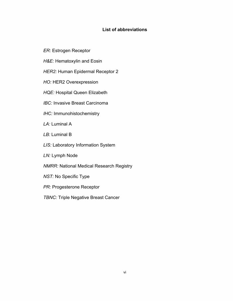
List of abbreviations
ER: Estrogen Receptor
H&E: Hematoxylin and Eosin
HER2: Human Epidermal Receptor 2
HO: HER2 Overexpression
HQE: Hospital Queen Elizabeth
IBC: Invasive Breast Carcinoma
IHC: Immunohistochemistry
LA: Luminal A
LB: Luminal B
LIS: Laboratory Information System
LN: Lymph Node
NMRR: National Medical Research Registry
NST: No Specific Type
PR: Progesterone Receptor
TBNC: Triple Negative Breast Cancer

vii
List of tables
Table 1: Histological grade in breast carcinoma (Elston-Ellis modification of
Scarff-Bloom-Richardson grading system) (Hoda et al., 2009)
Table 2: St Gallen classification of patients with primary breast cancer into risk
groups (1998) (Salisbur, 2002)
Table 3: Recommendations for adjuvant systemic therapy for node-negative
patients aged 70 years or less (Salisbur, 2002)
Table 4: Surrogate definitions of intrinsic subtypes of breast cancer (Goldhirsch
et al., 2011)
Table 5: Systemic treatment recommendations for subtypes (Goldhirsch et al.,
2011)
Table 6: Characteristics of the 158 cases of Invasive Breast Carcinoma Of No
Specific Type
Table 7: Age distribution of IBC NST (biopsy followed by mastectomy)

viii
Table 8: Cases of IBC NST according to hospitals/ districts (biopsy followed by
mastectomy)
Table 9: Prevalence of different subtypes of IBC NST (biopsy followed by
mastectomy)
Table 10: Comparison between prevalence of subtypes in this study with
prevalence in western population (Ki67 considered) (Komen, 2015).
Table 11: Prevalence of different subtypes before and after the application of
Ki67 (n=158)
Table 12: Prevalence of Luminal A and Luminal B before and after the
application of Ki67 and analysis with McNemar’s test (n=120)
Table 13: Comparison of grade, stage and lymph node metastasis of Luminal A
and Luminal B subtype with and without Ki67 analysed using simple logistic
regression test.

ix
List of figures
Figure 1: Classification of breast cancer subtypes according to IHC in correlation
with intrinsic gene expression microarray categorization (Onitilo et al., 2009)
Figure 2: Age distribution of Invasive Breast Carcinoma of No Specific Type
Figure 3: Cases of IBC NST according to hospitals/ districts (biopsy followed by
mastectomy)
Figure 4: Cases of IBC NST according to ethnicity (biopsy followed by
mastectomy)
Figure 5: Prevalence of different subtypes of IBC NST (biopsy followed by
mastectomy)
x
Abstrak
Latarbelakang: Kanser Payudara Tanpa Jenis Spesifik (IBC NST)
terbahagi kepada empat subtaip menggunakan ujian immunokimia tisu iaitu
Estrogen Receptor (ER), Progesterone Receptor (PR) dan Human Epidermal
Receptor 2 (HER2). Ia diklasifikasikan kepada Luminal A (LA), Luminal B (LB),
HER2 overexpressed (HO) dan triple negative (TN). Pengkelasan St. Gallen
2011 menyatakan penggunaan indeks proliferatif, Ki67 antibodi untuk
menentukan subtaip LB daripada LA. LB mempunyai prognosis yang lebih teruk
dan rawatan yang berbeza.
Objektif: Kajian ini bertujuan untuk menentukan HER2 negatif subtaip
Luminal B menurut Pengkelasan St Gallen 2011 menggunakan antigen Ki67
dan membandingkan ciri-ciri klinikopatologi bagi setiap subtaip luminal.
Kaedah: Kes-kes IBC NST yang mempunyai spesimen biopsi tisu yang
disusuli mastektomi diambil daripada arkib Hospital Queen Elizabeth di Kota
Kinabalu, Sabah. Biopsi tisu LA diuji menggunakan antibody terhadap Ki67.
Kes-kes LA dengan Ki67 ≥14% diklasifikasikan semula sebagai subtaip LB
manakala Ki 67< 14% kekal sebagai LA. Perbandingan setiap subtaip luminal
dengan tahap barah, saiz barah dan kewujudan metastasis ke noda limfa
seterusnya dibuat. Analisis univariasi menggunakan regresi logistik mudah
diadakan untuk menentukan peratusan ekspresi Ki67 dalam kes-kes IBC NST.
Ujian McNemar’s digunakan untuk analisis kategori berkembar. Semua

xi
pengiraan dibuat menggunakan SPSS versi 22 dan nilai-p <0.05 diset sebagai
signifikan.
Keputusan: LA adalah subtaip yang biasa (43%; 68/158), diikuti LB yang
lebih agresif (33%; 52/158). Hanya 37 daripada 68 kes luminal A diuji dengan
Ki67 disebabkan sampel yang terhad dan 43% (16/37) menunjukkan Ki67 ≥14%
(dikelaskan semula sebagai subtaip LB). Penggunaan Ki67 memberikan
perbezaan keputusan yang signifikan secara statistik (P<0.001; P<0.05). Walau
bagaimanapun, subtaip LB menunjukkan keputusan statistik yang tidak
signifikan apabila dibandingkan antara tahap, gred dan status noda limfa.
Kesimpulan: Klasifikasi barah payudara menurut Pengkelasan St Gallen
2011 menggunakan Ki67 antigen selain ER, PR and HER2 dalam mengenali
kes subtaip LB, yang mempunyai prognosis yang lebih teruk.

xii
Abstract
Background: Invasive Breast Carcinoma of No Specific Type (IBC NST)
is divided into four subtypes using Estrogen Receptor (ER), Progesterone
Receptor (PR) and Human Epidermal Receptor 2 (HER2) immunohistochemistry
markers. They are classified into Luminal A (LA), Luminal B (LB), HER2
Overexpressed (HO) and Triple Negative (TN) subtype. The St. Gallen 2011
Classification recognizes the use of Ki67 proliferative index to identify LB
subtype from LA group. LB has a worse prognosis and different approach of
treatment.
Objective: This study aimed to identify HER2 negative LB subtype
according to St Gallen Classification 2011 using Ki67 and to compare the
clinicopathological features of different subtypes.
Methods: Tissue biopsies of LA subtype were stained with antibody
towards Ki67. LA cases with Ki67 ≥14% were reclassified as LB subtype.
Luminal subtypes with corresponding stage (tumour size), histological grade and
lymph node metastases were compared. Univariate analysis using simple
logistic regression was performed to determine the percentage of Ki67
expression among all IBC NST cases. McNemar’s test was used for paired
categorical analysis. All calculations performed using SPSS version 22 and a p-
value of <0.05 was set to denote statistical significance.
Results: LA is the most common subtype (43%; 68/158), followed by LB
(33%; 52/158). Only 37 out of 68 cases were stained with Ki67 due to sample
limitations. From these LA cases, 43% (16/37) showed Ki67 ≥14% (reclassified

xiii
as LB subtype). There was significant result when using Ki67 (P<0.001;
P<0.05). However, LB subtype showed statistically insignificant result when
compared with between stage, grade and lymph node status.
Conclusion: The classification according to the St Gallen Classification
2011 utilized Ki67 marker in addition to ER, PR and HER2 in identifying luminal
B subtype cases, which have a worse prognosis.
1
CHAPTER 1: INTRODUCTION
1.1 Literature review
Breast cancer remains as the major cause of death for women in the
21st century and the most common female cancer worldwide (Peter Boyle,
2008). The aetiology and pathogenesis are diverse and there are still not well
understood (Abdulkareem, 2013). It is the number one cause of death due to
cancer in Malaysian women (Leong, 2007; Cheng Har Yip, 2006). The
incidence varies among ethnicities in Malaysia, mostly affecting Chinese
(Pathy et al., 2011). Malaysian women have a 1 in 20 chance of developing
breast cancer during their lifetime whereas in Sabah, where the population is
3.39 million with more than 30 ethnic groups, most cases presented with
advanced disease (Agarwal et al., 2007; Benjamin Dak Keung Leong, 2007;
Leong, 2007; Ibrahim et al., 2012; de Deus Moura et al., 2015). The
percentage of breast cancer detected at stage I and II was 58% (Goldhirsch
et al., 2011). To date, there was no specific study focusing on Invasive
Breast Carcinoma of No Specific Type (IBC NST) in Sabah (CH Yip, 2014).
The most significant classification of tumours of the breast was that
produced by the World Health Organization (Hoda et al., 2009; Tavassoéli,
2012; Sinn and Kreipe, 2013). IBC NST are tumours devoid of special
features thus the designation NST (Edwin R. Fisher, 1975). The special types
of invasive breast carcinoma includes tubular, cribriform, mucinous,
medullary, lobular, metaplastic, adenoid cystic and invasive secretory

2
carcinoma (Page, 2003; Alessandra Fabbri, 2008; Sinn and Kreipe, 2013).
IBC NST is further subclassified into molecular subtypes (Goldhirsch et al.,
2011; Malhotra et al., 2014). Different subtypes carry different prognosis and
different treatments (Ferguson et al., 2013). Most typing is possible by way of
studying the morphology on H&E staining whereby molecular study is the
more accurate way of subtyping albeit costly (Eroles et al., 2012).
Several studies have shown the heterogeneity of IBC NST and new
classifications were produced that can alter management and outcome of
patients (Goldhirsch et al., 2011; Hortobagyi, 2012a; Molloy et al., 2012). The
most popular method of subtyping is by surrogating using
immunohistochemistry staining (Cheang et al., 2009; Maisonneuve et al.,
2014). The importance is because of different response to therapy and the
availability of specific targeted treatment using hormonal and
chemotherapeutic agents (Lim and Winer, 2011; Boyle et al., 2013a).
Different subtypes also carry different prognosis in term of metastasis and
relapse (Guarneri et al., 2009; Kennecke et al., 2010; Voduc et al., 2010;
Nuria Ribelles, 2013).
There are 4 subtypes of IBC NST according to the St Gallen
classification 2011 (Goldhirsch et al., 2011) (Table 4). In previous
classification each subtypes are characterized by the expression of hormonal
receptors namely Estrogen Receptor (ER), Progesterone Receptor (PR) and
Human Epithelial Receptor (HER2) on the tumour cells (Figure 1) (Onitilo et
3
al., 2009). In addition to ER, PR and HER2, the classification in 2011
incorporates Ki67, a proliferative marker. Determining subtypes is important
because of different prognosis and treatment modalities (Chen et al., 2014;
Criscitiello et al., 2014). This study concentrates on the use of Ki-67 labelling
index, as the means of identifying Luminal B (LB) subtype of IBC NST. Cases
of Luminal A (LA) with Ki67 of 14% and more are classified as HER2
negative LB.
Determining luminal subtypes is a part of prognostic factor aimed to
foresee the outcome of patients irrespective of treatment. On the other hand,
predictive factors intend to assess the outcome of patients associated with
sensitivity or resistance to therapy. Thus, a predictive factor is also a
therapeutic target such as in the case of ER and HER2. Evaluation by
immunohistochemistry (IHC) of ER, PR, and HER2 status represent
consolidated and standardized prognostic factors. ER, PR and HER2 status
are also validated predictive markers. Other accepted prognostic markers are
represented by morphological findings such as tumour size (stage), grade,
lymphovascular invasion, lymph node status and Ki67 (Rampaul et al., 2001;
Faratian and Bartlett, 2008; Hoda et al., 2009; Song et al., 2011; Haroon et
al., 2013; Inic et al., 2014; Francisco Acevedo, 2015; Liu et al., 2015). There
is potential for Ki67 as a predictive factor as well, in the case where patients
with high Ki67 benefit from adjuvant chemotherapy (Criscitiello et al., 2014).
However there were contradicting study regarding this (Ferguson et al.,
2013).

4
1.1.1 Stage
Pathological staging of breast cancer takes into account the size of the
tumour, denoted as a capital T, and can be staged as T1 to T4. Tumours
may be measured clinically or pathologically; the pathologic size of the lesion
may be measured either macroscopically or microscopically. The clinical size
of a breast lesion as measured by mammograms or physical examination
may differ from the macroscopic pathologic size. It is possible that the
presence of a macroscopically measurable lesion has a poorer outcome
(Singletary, 2002; Edge, 2010).
1.1.2 Histologic grade
Besides staging, histologic grading system also plays an important
prognostic factor. The most commonly used grading system, the Nottingham
Histologic Score (also referred to as Elston-Ellis modification of Scarff-Bloom-
Richardson grading system), combines nuclear grade, tubule formation, and
mitotic rate to classify invasive breast carcinomas into three groups that are
highly correlated with survival (Table 1) (Meyer, 2005; Hoda et al.,
2009). Survival for patients with well-differentiated grade 1 carcinomas
gradually declines to 70% at 24 years. In contrast, most deaths for poorly
differentiated grade 3 carcinomas occur in the first 10 years, and 45% of
patients survive long-term. Patients with moderately differentiated grade 2
carcinomas have better survival initially, but their long-term survival is only

5
slightly better than grade 3 carcinomas (Ellis., 1991; Leslie W. Dalton, 1994;
S. E. Pinder, 1998; Howayda Abd El All, 2001). A grade of low, intermediate
or high is obtained through a composite sum by assigning a ‘score’ based on
the nuclear assessment, mitotic index assessment, and tubular assessment
(Table 1). The nuclear assessment is based on the nuclear pleomorphism
within the invasive cells. The tubular assessment refers to an approximate,
quantitative account of the amount of cell groupings that remain in their
normal ‘tubular’ shape. The less the percentage of tubular structures in
comparison to other shapes, the higher the score. The mitotic index refers to
patterns of cell division through assessing the numbers of dividing daughter
cells, measured per square millimetre. Mitoses are only counted in the
invasive area of the tumour (V. Le Doussal, 1989; Meyer, 2005).
1.1.3 Lymph node involvement
Axillary lymph node status at the time of diagnosis is the most
significant and durable prognostic factor in breast cancer patients. To date,
only nodal status has been consistently associated with survival outcomes in
occult primary breast cancer (Ivkovic-Kapicl et al., 2006; Hoda et al., 2009).

6
1.1.4 The St Gallen Classification
Clinicians, pathologists and researchers convene in St Gallen,
Switzerland to give a consensus regarding breast cancer treatment including
what is theoretically feasible in patient risk stratification, treatment and daily
practice management (Giuseppe Curigliano, 2013). In the earlier consensus
given in 1998, patients with breast cancer were classified into different risk
groups according to lymph node status, tumour size, grade, receptor status
and age (Table 2). Such classifications were made to guide oncologists in
giving adjuvant chemotherapy as studies have shown that most patients
benefit from chemotherapy regardless of lymph node status (Salisbur, 2002)
(Table 3). A study has also shown that the use of the older St Gallen
classification have led to the overuse of adjuvant chemotherapy. However,
such overuse may be overcame by using Ki67 for LA cases thus recognizing
HER2 negative LB, who are proven to benefit from chemotherapy
(Goldhirsch et al., 2011). The St Gallen classification in 2011 has
recommended adjuvant chemotherapy for LB and not for LA in IBC NST.
This is because the LB subtype proliferative index quantified by surrogate
IHC marker has been proven to be associated with more aggressive cancer
subtype and can be classified into LB despite HER2 marker being negative
(Table 5).

7
1.1.5 Hormonal receptors
All breast carcinomas are characterized by expression of ER and PR
receptors as well as the status of HER2. These, in relation to other
clinicopathological parameters, defines treatment recommendations for each
individual case (D.O. Shapochka, 2012). Approximately 70–80% of IBC NST
are ER positive, and between 15–30% of cases are HER2 positive.
Determining ER and HER2 status is considered as standard care for all
invasive breast carcinoma as these biomarkers are predictive for response of
patients to hormonal treatment and/or HER2-inhibiting medication (Wolff et
al., 2007; Hammond et al., 2010).
1.1.6 Luminal A subtype
ER positive family was named luminal class since they have the same
molecular signature strongly resembling luminal cells of breast duct (Taylor-
Papadimitriou et al., 1989). LA is the most common subtype (Kumar et al.,
2015). Whilst ER positive family, negative for HER2 is called LA, those
expressing HER2 is called LB. It is important to differentiate these two
subtypes because LA has been associated with a favourable prognosis
(Perez-Rodriguez, 2015) and LB, less favourable prognosis (Sotiriou et al.,
2003).

8
1.1.7 Luminal B subtype
A study by (Cheang et al., 2009) has shown that LB family is
associated with higher histological grade (grade 3), compared to lower grade
(grade 1 and 2). This subtype has a poorer prognosis (Tang et al., 2015).
Distinguishing LB subtype using Ki67 expression is of critical value since it
would isolate a group of patients in early breast carcinoma that could benefit
from adjuvant chemotherapy (Lim and Winer, 2011; Pavlakis et al., 2012).
New targeted therapy are also being developed specifically for LB subtype
(Ben Tran, 2011).
1.1.8 HER2 Overexpressed subtype
Several studies demonstrated that HER2 overexpression is correlated
with reduced survival. Overexpression is associated with relative resistance
to tamoxifen therapy or alkylating agent (Wang and Hung, 2001). In contrast
to previous data from Western countries that showed triple negative subtype
to be the worst in term of prognosis, a study in South Korea showed that
HER2 overexpressing breast cancers displayed the worst prognosis among
the various subgroups, and not triple negative subtype. This finding proved
that there is more to breast cancer of different subtypes in different
population groups and the need for more study in local population as not all
data demonstrated in Western population applies to Asians, and this might
affect treatment and prognostication (Mattes et al., 2015).

9
1.1.9 Triple negative subtype
Triple negative breast cancer (TNBC) is an aggressive clinical
phenotype characterized by lack of expression (or minimal expression) of ER
and PR as well as an absence of HER2 overexpression. TNBC is not
amenable to treatment with hormone therapy or the anti-HER2 monoclonal
antibody trastuzumab, and systemic treatment options are limited to cytotoxic
chemotherapy. Unlike patients with ER/PR positive or HER2 overexpressing
disease, systemic treatment options for patients with TNBC are limited to
cytotoxic chemotherapy due to the lack of a molecular target (Siziopikou and
Cobleigh, 2007; Nishimura and Arima, 2008; Cicin et al., 2009). Despite its
chemosensitivity, TNBC is still associated with a poor prognosis. The median
time to death among patients with TNBC was also shorter than that with
other subtypes (Cicin et al., 2009).
1.1.10 Ki67
Recently, the immunohistochemical expression of Ki67 has replaced
mitotic counting in assessing the proliferation of neoplastic cells. There is a
lot of rising evidence on its importance as a prognostic and predictive marker
of responsiveness to therapy and as a dynamic biomarker of treatment
efficacy (O. Gottardi, 1993; Graef et al., 2008; Miglietta et al., 2013). In early
breast carcinoma, high Ki67 is an independent factor for worse prognosis as

10
shown by significantly shorter overall and disease-free survival (Goldstein,
2004 ; Jung et al., 2009; Zong et al., 2014; Marrazzo et al., 2015). High Ki67
was predictive of more benefit from adjuvant chemotherapy (Elzawahry et al.,
2013) even in cases without lymph node metastasis (Andre et al., 2015). The
prognostic role of Ki67 in early breast cancer was indicated in large sample
study (Goldhirsch et al., 2011). (Dowsett et al., 2011) and (Nishimura et al.,
2010) mentioned the potential use of Ki67 for prognosis, predict
responsiveness or resistance to endocrine or chemotherapy and as a
dynamic biomarker of treatment efficacy. A study found that Ki67 expression
is inversely related to their sensitivity to first line endocrine therapy (Graef et
al., 2008). The St Gallen International Expert Consensus on the primary
therapy of early breast cancer 2011 defined a cut-off point of 14% for the
distinction between luminal A and luminal B (HER2 negative) tumours
(Dowsett et al., 2011; András Vörös, 2014; Ono et al., 2015).

11
1.2 Rationale of the study
Very few data regarding Sabah population is available, yet the
numbers of breast cancer patients are increasing and they always presented
late. This study involves population where presentation is late, thus
association between Ki67 index and late presentation will be observed.
Subtyping is important because of different treatment approach; Luminal B
has a much worse prognosis than Luminal A, when taking Ki67 into
consideration; it has a role of being an independent prognostic factor. We
also wanted to see the significance of taking into account Ki67 in determining
luminality in Sabah population, whether a significant percentage from the
Luminal A subtype has high Ki67 index.
1.3 Objectives
Objective 1: To reclassify and compare luminal A & B IBC NST using Ki67
index.
Objective 2: To compare grade, stage and lymph node metastasis of luminal
A and Luminal B IBC NST.

12
CHAPTER 2: STUDY PROTOCOL
2.1 Study design and method
STUDY&PROTOCOL&Collec&on(of(paraffin(blocks(of(iden&fied(cases(from(archival(material(in(HQE(June(2009@December(2014((
EXCLUSION(CRITERIA(INCLUSION(CRITERIA(
Review(of(histopathological(data:(, ER(status(, PR(status(, HER2(status(, Lymph(nodes(status(, Tumour(histological(type((, Histological(grade(
Review(of(clinicopathological(data((age,(ethnicity)(
LUMINAL'A'
LUMINAL'B'(HER2+)'
Ki67'<14%'
Ki67≥14%'
HER2'overexpression'
TRIPLE'NEG'
StaGsGcal'analysis'and'result'
Data'record'
Lab'Work'
LUMINAL'B'(HER2Q)'

13
2.2 Ethical approval letter (KKM)
JAWATANKUASA ETIKA & PENYELIDIKAN PERUBATAN (Medical Research & Ethics Committee) KEMENTERIAN KESIHATAN MALAYSIA d/a lnstitut Pengurusan Kesihatan Jalan Rumah Sakit, Bangsar 59000 Kuala Lumpur
Dr Muhd Afif Bin Mohd Yusof Jabatan Patologi Hospital Queen Elizabeth
Tuan,
NMRR-14-269-20213
Tel. : 03 2282 9082/03 2282 9085 03 2287 4032/03 2282 0491
Faks : 03 2287 4030
Ruj. Kami: (~) KKM/NIHSEC/P14-365 Tarikh : 20 Jun 2014
CLASSIFICATION OF INVASIVE BREAST CARCINOMA ACCORDING TO ST GALLEN CLASSIFICATION 2011 WITH EMPHASIS ON KI-671NDEX
Lokasi Projek : Hospital Queen Elizabeth
Dengan hormatnya perkara di atas adalah dirujuk.
2. Jawatankuasa Etika & Penyelidikan Perubatan (JEPP), Kementerian Kesihatan Malaysia (KKM) mengambil maklum bahawa projek tersebut adalah untuk memenuhi keperluan akademik Sarjana Patologi, Universiti Sains Malaysia.
3. Sehubungan dengan ini, dimaklumkan bahawa pihak JEPP KKM tiada halangan, dari segi etika, ke atas pelaksanaan projek tersebut. JEPP mengambil maklum bahawa kajian ini tidak melibatkan sebarang intervensi dan hanya melibatkan blok tisu parafin dalam mengumpul data kajian. Segala rekod dan data adalah SULIT dan hanya digunakan untuk tujuan kajian dan semua isu serta prosedur mengenai data confidentiality mesti dipatuhi. Kebenaran daripada Pengarah Hos ital di mana kaj~~n dijalankan mesti diperolehi terlebih dahulu sebelum kaj1an diJaiankan. Tuan perlu akur dan mematuhi keputusan tersebut.
4. Adalah dimaklumkan bahawa kelulusan ini adalah sah sehingga 20 June 2015. Tuan perlu menghantar 'Continuing Review Form' selewat-lewatnya 2 bulan sebelum tamat tempoh kelulusan ini bagi memperbaharui kelulusan etika. Pihak tuan juga perlu mengemukakan laporan tamat kajian dan juga laporan mengenai "A// adverse events, both serious and unexpected' kepada Jawatankuasa Etika & Penyelidikan Perubatan, KKM jika berkenaan. Borang-borang berkaitan boleh dimuat turun daripada Iaman web MREC (http://www.nih.gov.my/mrec)
Sekian terima kasih.
BERKHIDMAT UNTUK NEGARA
(DATO' DR CHANG KlAN MENG) Pengerusi Jawatankuasa Etika & Penyelidikan Perubatan Kementerian Kesihatan Malaysia

14
2.3 Ethical consideration
Confidentiality of the data was taken into account. Ethic approval was
obtained from Ethics Approval; Medical Research & Ethics Committee NMRR
(National Medical Research Register); NMRR-14-269-20213.

15
CHAPTER 3: MANUSCRIPT
3.1 Introduction
Breast cancer is the most common cancer in female worldwide (Peter
Boyle, 2008) and is the number one cause of death due to cancer in
Malaysian women (Benjamin Dak Keung Leong, 2007; Cheng Har Yip,
2006). The incidence varies among ethnicities in Malaysia, mostly affecting
Chinese (Bhoo Pathy et al., 2011). With recent progress in studies pertaining
to the detailed aspects of the disease which include molecular testing, new
classification has emerged that alter management and outcome of patient,
therefore benefiting the patients (Goldhirsch et al., 2011; Hortobagyi, 2012b).
The introduction of genetic array testing has allowed breast cancer to be
further subtyped by different methods (Sorlie, 2004; Molloy et al., 2012). The
most popular one is by approximation using immunohistochemistry staining
(Maggie C. U. Cheang 2009). This is important due to the different response
to therapy and the availability of specific targeted treatment using hormonal
and chemotherapeutic agents (Boyle et al., 2013b).
This study concentrated on the use of immunohistochemical definition
of estrogen receptor (ER) and progesterone receptor (PR), and human
epidermal growth factor receptor 2 (HER2) oncogene, as well as Ki67
labelling index; a marker of cell proliferation, as the means of identifying
tumour subtypes according to St Gallen expert consensus 2011(Goldhirsch
et al., 2011).

16
Most studies that have been done usually revolve around western
population where detection of breast cancer is during the early stage. There
is however a difference in hormonal status when the disease is at its more
advanced stage. By concentrating on Malaysian population in Sabah, North
Borneo, where detection and intervention is at a late stage, this study seeks
to find out differences of hormonal status as compared to early stages. This
study reclassified Luminal A (LA) and Luminal B (LB) subtypes of Invasive
Breast Carcinoma of No Special Type (IBC NST) using Ki67 according to St
Gallen International Consensus 2011 (Goldhirsch et al., 2011). Associations
between subtypes and other clinicopathological characteristics such as
ethnicity, stage, histological grade and status of axillary lymph node were
analysed.
This study aims to classify Invasive Breast Carcinoma of No Specific
Type (IBC NST) according to St Gallen consensus 2011 with emphasis on
Ki67 index. There are 2 specific objectives involved; 1) To reclassify and
compare luminal A & B IBC NST using Ki67 index. 2) To compare grade,
stage and lymph node metastasis of luminal A and Luminal B IBC NST
(Hammond et al., 2010).

17
3.2 Materials and Methods
This study is a cross sectional study design. It utilized cases of breast
biopsies (core biopsies or excision biopsies) that were followed by
mastectomy specimens only, with final histopathological diagnosis of IBC
NST, for the period of June 2009-December 2014 in the Pathology
Department, Hospital Queen Elizabeth, Kota Kinabalu. Only cases with
biopsies followed by mastectomy were included. Search query was
conducted using Lab Information System (LIS) at the Pathology Department,
Hospital Queen Elizabeth, Kota Kinabalu, Sabah.
Cases that fulfilled the inclusion criteria but the corresponding
paraffinised tissue blocks are not available, missing and inadequate for serial
sections were excluded from the analysis for the objectives of this study.
Biopsies that were not followed by mastectomy and mastectomies without
prior biopsies were also excluded.
Clinicopathological parameters including age, ethnicity, tumour grade,
tumour size (stage), lymph nodes involvement and ER and PR status, and
HER2 expression were obtained from the histopathological reports (Tamaki
et al., 2010). In this study only the pathological stage (T), which is determined
by tumour size was considered.
Independent pathologists of the Anatomic Pathology Department had
examined these surgical specimens and formal pathology reports had been
18
issued within that period. Only suitable cases that met the inclusion criteria
were chosen and the paraffin blocks were then retrieved from the archives.
3.2.1 Laboratory work
Standard immunoperoxidase procedures were followed for tissue
sections obtained from each case. The selected single representative block
for each case was sectioned to 4-micron thickness. Two sections were
obtained from all LA cases (ER+ and or PR+, HER2-) according to data
obtained from patient’s record in LIS. This section was subjected to H&E and
Ki67 staining. Ki67 staining was done using K2 antibody (BondTM) (Leica,
2009).
The obtained sections were placed on heater at 60°C for at least 1
hour to facilitate the adherence of tissue onto the slides. Leica Autostainer
System performed the next steps automatically. For Ki67 IHC staining
BondTM Ready-to-Use Primary Antibody Ki67 (K2) was utilised. The
application was performed by Bond-Max Fully Automated IHC system (Leica)
using according to the manufacturer’s IHC protocol. The demonstration of
human Ki67 nuclear antigen is achieved by first, allowing the binding of Ki67
(K2 antibody) to the section, and then visualizing this binding using the
reagents provided in the detection system. The use of these products, in
combination with an automated Bond system, reduces the possibility of
19
human error and variability resulting from individual reagent dilution, manual
pipetting and reagent application (Leica, 2009).
3.2.2 Microscopic analysis
Using a bright-field microscope (Olympus CX31), microscopic
evaluation of the slides immunostained for Ki67 was performed blinded from
the clinicopathological data of the study subjects. H&E slides were examined
to re-evaluate and to verify the diagnosis and location of tumour cells when
compared to Ki67 slides. Ki67 proliferative index was reviewed by main
researcher (supervisor) and co-supervisors at a separate time to avoid bias.
Each case was divided into 2 groups; Ki67 equal or more 14% and Ki67 less
than 14% (Dowsett et al., 2011). ER, PR and HER2 hormonal qualitative
status by IHC were obtained from the formal reports. The quantitative scores
whether performed or not were disregarded.
3.2.3 Ki67
Slides stained with Ki67 were analysed according to
Recommendations from the International Ki67 in Breast Cancer Working
Group (Dowsett et al., 2011). Only nuclear staining plus mitotic figures which
were stained by Ki67 were incorporated into the Ki67 score that is defined as
the percentage of positively stained cells among the total number of

20
malignant cells scored. When the staining is homogenous, at least three
randomly selected high-power (×40 objective) fields were selected.
3.2.4 Statistical analysis
Data was entered and statistical analysis was conducted by using test
on Statistical Package for Social Sciences (SPSS) program version 22.
Univariate analysis using simple logistic regression was performed.
McNemar’s test was used for paired categorical analysis. A p-value of <0.05
was set to denote statistical significance.
21
3.3 Results
3.3.1 Clinicopathological data of the study subjects
One hundred and fifty eight cases of IBC NST, which met the inclusion
criteria (cases with biopsy followed by mastectomy), were included in this
study. Clinicopathological data of the study subjects were summarized in
(Table 6). Distributions of the age, ethnicity and institutions were shown in
(Table 7) and (Figure 2), (Figure 4), (Table 8) and (Figure 3). Analysis for
different subtypes and clinicopathological factors were summarized in (Table
9) and (Figure 5). Majority of the study subjects were natives. The mean age
of the patients was 50.4 years with the age ranging from 28 to 78 years
(Figure 2). This is comparable to data from peninsular Malaysia. All the cases
included were those where biopsies were taken prior to mastectomies with
axillary clearance. Almost half of the tumours (49.4%) had tumour size larger
than 20mm but smaller than 50mm. Lymph node metastasis was found in
majority of cases (64.6 %). More than half of the tumours (55.1%) were grade
3 tumours. ER, PR and HER2 status were positive in 74.7%, 62.7% and
45.6% of the cases respectively (Table 6).
Most cases were from Kota Kinabalu (62.7%; 99/158), followed by
Sandakan and Tawau. Other cases are from Keningau, Lahad Datu, Labuan
and Beaufort (Table 8 and Figure 3). The main referral centre is in Hospital
Queen Elizabeth, Kota Kinabalu where mastectomies were performed on a
regular basis. Whereas in the other centres, it is carried out from time to time

22
by visiting surgeons. All the specimens were sent for examination to the
Pathology Department of Queen Elizabeth Hospital in Kota Kinabalu.
The highest prevalence was observed among natives (78.5%;
124/158) followed by Chinese (18.4%; 29/158). Majority of cases were at late
stage during presentation (stage 2 and 3; 94.7%; 148/158) and of high grade
(grade 2 and 3; 93.7%; 148/158). Cases with lymph node metastasis were
64.6% (102/158).
3.2.2 Ki67 Proliferative Index and Luminal B Subtype
LA is the most common subtype (44%; 68/158), followed by LB (33%;
52/158) (Table 9 and Figure 5). Only 37 LA cases were stained with Ki67 due
to sample limitations. From these stained LA cases, 43% (16/37) showed
Ki67 ≥14% (Image 1) (reclassified as LB subtype) (Table 11). Cases of Ki67
less than 14% remain as LA subtype (Image 2). The difference in number of
cases between LA and LB with and without the use of Ki67 is statistically
significant (<0.001) using McNemar’s Test (Table 12). There was no
significance when LA and LB subtypes with and without Ki67 staining were
compared against grade, stage and lymph node status (Table 13).
23
3.4 Discussion and limitation
In general, breast cancer remains the most common cancer among
Malaysian women, which accounted for 31% of newly diagnosed cancer
cases in women registered in the National Cancer Registry (NCR) for the
year 2003-2005. The risk differs among the major ethnic groups in Malaysia;
about 1 in 16 Chinese, 1 in 17 Indian and 1 in 28 Malay women will develop
breast cancer in their lifetime (NCR, 2007). In Malaysia, breast cancer is
more common among Chinese as compared to Malay. However, in this study
it was difficult to divide the study subjects strictly according to ethnicity. Due
to the demographical situation in Sabah, which comprises of up to 36 ethnics,
where mixed marriage is common (Suraya Sintang, 2011), all of the subjects
that are not Chinese, Indian or Non-Malaysians were grouped together as
natives. There is lack of information and standardisation when it comes to
determining the ethnicity of a subject who is a result of interracial marriage.
There is no concrete guidance in Malaysia whether to follow paternal or
maternal ethnicity. Thus such subject was lumped together as natives. It is
also doubtful whether dividing the subjects according to ethnicities will have
any significance in term of determining the incidence of the disease, as
studies tried to separate subjects according to race or ethnicities to find
lifestyle and cultural influences on the aetiology or pathogenesis
(Kurebayashi et al., 2007). Whereby in Sabah, the population is more
integrated in terms of lifestyle that is of any connection to the risk factors
such as food, breast-feeding or contraception (B Norsa’adah, 2005).
24
In comparison with other studies, this study have focused mainly on
the cases of IBC NST and only those cases where histopathological
diagnosis was made by biopsy before being followed by mastectomy. Most
studies failed to specify the samples used either biopsy or mastectomy
specimen. In some centres in Malaysia hormonal receptors are tested on
biopsy specimens whereas in others it is tested on mastectomies. Most
literature reported cases in western countries. There are very few studies
done on IBC NST cases in Sabah. Because most of IBC NST cases in
Sabah are at a late stage, the cases presented earlier, mainly of Luminal A
subtype that could have benefited from chemotherapy when categorized as
Luminal B seems to be disregarded. As we are moving toward better medical
care, earlier presentation of breast cancer cases are expected in the future.
Most of the results (grade, lymph node metastasis) shown were not
statistically significant as anticipated at the beginning of the study (Table 13).
Several factors may have contributed to this. As discussed, the limited
sample size was a major setback. Only 37 cases were stained for subtyping.
However, in comparison with the other studies done on larger samples, this
study only collected cases where biopsy was performed prior to mastectomy.
The majority of subjects presented in stage 2 and 3 (87%), this is when
taking into account that these patients had a biopsy before mastectomy. The
possible main reason behind this is late detection due to lack of access and
awareness. However, there were cases of straightforward breast cancer that
were diagnosed clinically and managed by mastectomy, without first