clin infect dis. 2014-stevens-cid ciu296
TRANSCRIPT

I D S A G U I D E L I N E
Practice Guidelines for the Diagnosisand Management of Skin and Soft TissueInfections: 2014 Update by the InfectiousDiseases Society of America
Dennis L. Stevens,1 Alan L. Bisno,2 Henry F. Chambers,3 E. Patchen Dellinger,4 Ellie J. C. Goldstein,5 Sherwood L. Gorbach,6
Jan V. Hirschmann,7 Sheldon L. Kaplan,8 Jose G. Montoya,9 and James C. Wade10
1Division of Infectious Diseases, Department of Veterans Affairs, Boise, Idaho; 2Medical Service, Miami Veterans Affairs Health Care System, Florida;3San Francisco General Hospital, University of California; 4Division of General Surgery, University of Washington, Seattle; 5University of California, LosAngeles, School of Medicine, and R. M. Alden Research Laboratory, Santa Monica, California; 6Department of Community Health, Tufts University, Boston,Massachusetts; 7Medical Service, Puget Sound Veterans Affairs Medical Center, Seattle, Washington; 8Department of Pediatrics, Baylor College ofMedicine, Houston, Texas; 9Department of Medicine, Stanford University, California; and 10Geisinger Health System, Geisinger Cancer Institute, Danville,Pennsylvania
A panel of national experts was convened by the Infectious Diseases Society of America (IDSA) to update the2005 guidelines for the treatment of skin and soft tissue infections (SSTIs). The panel’s recommendations weredeveloped to be concordant with the recently published IDSA guidelines for the treatment of methicillin-resistant Staphylococcus aureus infections. The focus of this guideline is the diagnosis and appropriate treatmentof diverse SSTIs ranging from minor superficial infections to life-threatening infections such as necrotizing fas-ciitis. In addition, because of an increasing number of immunocompromised hosts worldwide, the guidelineaddresses the wide array of SSTIs that occur in this population. These guidelines emphasize the importanceof clinical skills in promptly diagnosing SSTIs, identifying the pathogen, and administering effective treatmentsin a timely fashion.
EXECUTIVE SUMMARY
Summarized below are the recommendations made inthe new guidelines for skin and soft tissue infections(SSTIs). Figure 1 was developed to simplify the manage-ment of localized purulent staphylococcal infectionssuch as skin abscesses, furuncles, and carbuncles in
the age of methicillin-resistant Staphylococcus aureus(MRSA). In addition, Figure 2 is provided to simplifythe approach to patients with surgical site infections.The panel followed a process used in the developmentof other Infectious Diseases Society of America (IDSA)guidelines, which included a systematic weighting of thestrength of recommendation and quality of evidenceusing the GRADE (Grading of RecommendationsAssessment, Development, and Evaluation) system(Table 1) [1–4]. A detailed description of the methods,background, and evidence summaries that support eachof the recommendations can be found in the full text ofthe guidelines.
I. What Is Appropriate for the Evaluation and Treatmentof Impetigo and Ecthyma?Recommendations1. Gram stain and culture of the pus or exudates
from skin lesions of impetigo and ecthyma are
Received 17 April 2014; accepted 21 April 2014.It is important to realize that guidelines cannot always account for individual var-
iation among patients. They are not intended to supplant physician judgment withrespect to particular patients or special clinical situations. IDSA considers adher-ence to these guidelines to be voluntary, with the ultimate determination regardingtheir application to be made by the physician in the light of each patient’s individualcircumstances.
Correspondence: Dennis L. Stevens, PhD, MD, Infectious Diseases Section, VAMedical Center, 500 W Fort St, Bldg 45, Boise, ID 83702 ([email protected]).
Clinical Infectious Diseases© The Author 2014. Published by Oxford University Press on behalf of the InfectiousDiseases Society of America. All rights reserved. For Permissions, please e-mail:[email protected]: 10.1093/cid/ciu296
IDSA Practice Guidelines for SSTIs • CID • 1
Clinical Infectious Diseases Advance Access published June 18, 2014 by guest on July 24, 2014
http://cid.oxfordjournals.org/D
ownloaded from

recommended to help identify whether Staphylococcus aureusand/or a β-hemolytic Streptococcus is the cause (strong, moder-ate), but treatment without these studies is reasonable in typicalcases (strong, moderate).2. Bullous and nonbullous impetigo can be treated with
oral or topical antimicrobials, but oral therapy is recommendedfor patients with numerous lesions or in outbreaks affecting sev-eral people to help decrease transmission of infection. Treat-ment for ecthyma should be an oral antimicrobial.
(a) Treatment of bullous and nonbullous impetigo shouldbe with either mupirocin or retapamulin twice daily (bid)for 5 days (strong, high).
(b) Oral therapy for ecthyma or impetigo should be a 7-dayregimen with an agent active against S. aureus unless culturesyield streptococci alone (when oral penicillin is the re-commended agent) (strong, high). Because S. aureus isolatesfrom impetigo and ecthyma are usually methicillin suscepti-ble, dicloxacillin or cephalexin is recommended. WhenMRSA is suspected or confirmed, doxycycline, clindamycin,or sulfamethoxazole-trimethoprim (SMX-TMP) is recom-mended (strong, moderate).(c) Systemic antimicrobials should be used for infectionsduring outbreaks of poststreptococcal glomerulonephritis tohelp eliminate nephritogenic strains of S. pyogenes from thecommunity (strong, moderate).
Figure 1. Purulent skin and soft tissue infections (SSTIs). Mild infection: for purulent SSTI, incision and drainage is indicated. Moderate infection: pa-tients with purulent infection with systemic signs of infection. Severe infection: patients who have failed incision and drainage plus oral antibiotics or thosewith systemic signs of infection such as temperature >38°C, tachycardia (heart rate >90 beats per minute), tachypnea (respiratory rate >24 breaths perminute) or abnormal white blood cell count (<12 000 or <400 cells/µL), or immunocompromised patients. Nonpurulent SSTIs. Mild infection: typical cel-lulitis/erysipelas with no focus of purulence. Moderate infection: typical cellulitis/erysipelas with systemic signs of infection. Severe infection: patients whohave failed oral antibiotic treatment or those with systemic signs of infection (as defined above under purulent infection), or those who are immunocom-promised, or those with clinical signs of deeper infection such as bullae, skin sloughing, hypotension, or evidence of organ dysfunction. Two newer agents,tedizolid and dalbavancin, are also effective agents in SSTIs, including those caused by methicillin-resistant Staphylococcus aureus, and may be approvedfor this indication by June 2014. Abbreviations: C & S, culture and sensitivity; I & D, incision and drainage; MRSA, methicillin-resistant Staphylococcusaureus; MSSA, methicillin-susceptible Staphylococcus aureus; Rx, treatment; TMP/SMX, trimethoprim-sulfamethoxazole.
2 • CID • Stevens et al
by guest on July 24, 2014http://cid.oxfordjournals.org/
Dow
nloaded from

II. What Is the Appropriate Evaluation and Treatment for PurulentSSTIs (Cutaneous Abscesses, Furuncles, Carbuncles, andInflamed Epidermoid Cysts)?Recommendations3. Gram stain and culture of pus from carbuncles and ab-
scesses are recommended, but treatment without these studiesis reasonable in typical cases (strong, moderate).
4. Gram stain and culture of pus from inflamed epidermoidcysts are not recommended (strong, moderate).5. Incision and drainage is the recommended treatment for
inflamed epidermoid cysts, carbuncles, abscesses, and large fu-runcles, mild (Figure 1) (strong, high).6. The decision to administer antibiotics directed against
S. aureus as an adjunct to incision and drainage should be
Figure 2. Algorithm for the management and treatment of surgical site infections (SSIs). *For patients with type 1 (anaphylaxis or hives) allergy to β-lactamantibiotics. If Gram stain not available, open and debride if purulent drainage present. Where the rate of infection with methicillin-resistant Staphylococcusaureus infection is high, consider vancomycin, daptomycin, or linezolid, pending results of culture and susceptibility tests. Adapted and modified with permis-sion from Dellinger et al [96]. Abbreviations: GI, gastrointestinal; MRSA, methicillin-resistant Staphylococcus aureus ; WBC, white blood cell count.
IDSA Practice Guidelines for SSTIs • CID • 3
by guest on July 24, 2014http://cid.oxfordjournals.org/
Dow
nloaded from

made based upon presence or absence of systemic inflamma-tory response syndrome (SIRS), such as temperature >38°C or<36°C, tachypnea >24 breaths per minute, tachycardia >90beats per minute, or white blood cell count >12 000or <400 cells/µL (moderate; Figure 1) (strong, low). An
antibiotic active against MRSA is recommended for patientswith carbuncles or abscesses who have failed initial antibiotictreatment or have markedly impaired host defenses or in pa-tients with SIRS and hypotension (severe; Figure 1 andTable 2) (strong, low).
Table 1. Strength of Recommendations and Quality of the Evidence
Strength ofRecommendation andQuality of Evidence
Clarity of Balance BetweenDesirable and Undesirable
EffectsMethodological Quality of
Supporting Evidence (Examples) Implications
Strong recommendation,high-quality evidence
Desirable effects clearlyoutweigh undesirable effects,or vice versa
Consistent evidence from well-performed RCTs or exceptionallystrong evidence from unbiasedobservational studies
Recommendation can apply to mostpatients in most circumstances.Further research is unlikely tochange our confidence in theestimate of effect
Strong recommendation,moderate qualityevidence
Desirable effects clearlyoutweigh undesirable effects,or vice versa
Evidence from RCTs with importantlimitations (inconsistent results,methodological flaws, indirect, orimprecise) or exceptionally strongevidence from unbiasedobservational studies
Recommendation can apply to mostpatients in most circumstances.Further research (if performed) islikely to have an important impact onour confidence in the estimate ofeffect and may change the estimate
Strong recommendation,low-quality qualityevidence
Desirable effects clearlyoutweigh undesirable effects,or vice versa
Evidence for at least 1 criticaloutcome from observationalstudies, RCTs with serious flawsor indirect evidence
Recommendation may change whenhigher-quality evidence becomesavailable. Further research (ifperformed) is likely to have animportant impact on our confidencein the estimate of effect and is likelyto change the estimate
Strong recommendation,very low-quality evidence(very rarely applicable)
Desirable effects clearlyoutweigh undesirable effects,or vice versa
Evidence for at least 1 criticaloutcome from unsystematicclinical observations or veryindirect evidence
Recommendation may change whenhigher-quality evidence becomesavailable; any estimate of effect forat least 1 critical outcome is veryuncertain.
Weak recommendation,high-quality evidence
Desirable effects closelybalanced with undesirableeffects
Consistent evidence from well-performed RCTs or exceptionallystrong evidence from unbiasedobservational studies
The best action may differ dependingon circumstances or patient’s orsocietal values. Further research isunlikely to change our confidence inthe estimate of effect
Weak recommendation,moderate-qualityevidence
Desirable effects closelybalanced with undesirableeffects
Evidence from RCTs with importantlimitations (inconsistent results,methodological flaws, indirect, orimprecise) or exceptionally strongevidence from unbiasedobservational studies
Alternative approaches likely to bebetter for some patients undersome circumstances. Furtherresearch (if performed) is likely tohave an important impact on ourconfidence in the estimate of effectand may change the estimate
Weak recommendation,low-quality evidence
Uncertainty in the estimates ofdesirable effects, harms, andburden; desirable effects,harms, and burden may beclosely balanced
Evidence for at least 1 criticaloutcome from observationalstudies, from RCTs with seriousflaws or indirect evidence
Other alternatives may be equallyreasonable. Further research is verylikely to have an important impact onour confidence in the estimate ofeffect and is likely to change theestimate
Weak recommendation,very low-quality evidence
Major uncertainty in theestimates of desirable effects,harms, and burden; desirableeffects may or may not bebalanced with undesirableeffects
Evidence for at least 1 criticaloutcome from unsystematicclinical observations or veryindirect evidence
Other alternatives may be equallyreasonable. Any estimate of effect,for at least 1 critical outcome, is veryuncertain
Abbreviation: RCT, randomized controlled trial.
4 • CID • Stevens et al
by guest on July 24, 2014http://cid.oxfordjournals.org/
Dow
nloaded from

Table 2. Antimicrobial Therapy for Staphylococcal and Streptococcal Skin and Soft Tissue Infections
Disease Entity Antibiotic Dosage, Adults Dosage, Childrena Comment
Impetigob
(Staphylococcus andStreptococcus)
Dicloxacillin 250 mg qid po N/A N/A
Cephalexin 250 mg qid po 25–50 mg/kg/d in 3–4 divided doses po N/A
Erythromycin 250 mg qid poc 40 mg/kg/d in 3–4 divided doses po Some strains of Staphylococcus aureus and Streptococcuspyogenes may be resistant.
Clindamycin 300–400 mg qid po 20 mg/kg/d in 3 divided doses po N/A
Amoxicillin-clavulanate 875/125 mg bid po 25 mg/kg/d of the amoxicillin componentin 2 divided doses po
N/A
Retapamulin ointment Apply to lesions bid Apply to lesions bid For patients with limited number of lesions
Mupirocin ointment Apply to lesions bid Apply to lesions bid For patients with limited number of lesions
MSSA SSTI Nafcillin or oxacillin 1-2 g every 4 h IV 100–150 mg/kg/d in 4 divided doses Parental drug of choice; inactive against MRSACefazolin 1 g every 8 h IV 50 mg/kg/d in 3 divided doses For penicillin-allergic patients except those with immediate
hypersensitivity reactions. More convenient than nafcillinwith less bone marrow suppression
Clindamycin 600 mg every 8 h IVor
300–450 mg qid po
25–40 mg/kg/d in 3 divided doses IV or25–30 mg/kg/d in 3 divided doses po
Bacteriostatic; potential of cross-resistance and emergenceof resistance in erythromycin-resistant strains; inducibleresistance in MRSA
Dicloxacillin 500 mg qid po 25–50 mg/kg/d in 4 divided doses po Oral agent of choice for methicillin-susceptible strains inadults. Not used much in pediatrics
Cephalexin 500 mg qid po 25–50 mg/kg/d 4 divided doses po For penicillin-allergic patients except those with immediatehypersensitivity reactions. The availability of a suspensionand requirement for less frequent dosing
Doxycycline,minocycline
100 mg bid po Not recommended for age <8 yd Bacteriostatic; limited recent clinical experience
Trimethoprim-sulfamethoxazole
1–2 double-strength tabletsbid po
8–12 mg/kg (based on trimethoprimcomponent) in either 4 divided doses IVor 2 divided doses po
Bactericidal; efficacy poorly documented
MRSA SSTI Vancomycin 30 mg/kg/d in 2divided doses IV
40 mg/kg/d in 4 divided doses IV For penicillin allergic patients; parenteral drug of choice fortreatment of infections caused by MRSA
Linezolid 600 mg every 12 hIV or 600 mg bidpo
10 mg/kg every 12 h IV or po for children<12 y
Bacteriostatic; limited clinical experience; no cross-resistance with other antibiotic classes; expensive
Clindamycin 600 mg every 8 h IVor 300–450 mgqid po
25–40 mg/kg/d in 3 divided doses IV or30–40 mg/kg/d in 3 divided doses po
Bacteriostatic; potential of cross-resistance and emergenceof resistance in erythromycin-resistant strains; inducibleresistance in MRSA. Important option for children
Daptomycin 4 mg/kg every 24 hIV
N/A Bactericidal; possible myopathy
Ceftaroline 600 mg bid IV N/A Bactericidal
Doxycycline,minocycline
100 mg bid po Not recommended for age <8 yd Bacteriostatic; limited recent clinical experience
Trimethoprim-sulfamethoxazole
1–2 double-strength tabletsbid po
8–12 mg/kg/d (based on trimethoprimcomponent) in either 4 divided doses IVor 2 divided doses po
Bactericidal; limited published efficacy data
IDSA
Practice
Guidelines
forSST
Is•
CID
•5
by guest on July 24, 2014 http://cid.oxfordjournals.org/ Downloaded from

III. What Is the Appropriate Treatment for Recurrent SkinAbscesses?Recommendations7. A recurrent abscess at a site of previous infection should
prompt a search for local causes such as a pilonidal cyst, hidra-denitis suppurativa, or foreign material (strong, moderate).8. Recurrent abscesses should be drained and cultured early
in the course of infection (strong, moderate).9. After obtaining cultures of recurrent abscess, treat with a
5- to 10-day course of an antibiotic active against the pathogenisolated (weak, low).10. Consider a 5-day decolonization regimen twice daily of
intranasal mupirocin, daily chlorhexidine washes, and daily de-contamination of personal items such as towels, sheets, andclothes for recurrent S. aureus infection (weak, low).11. Adult patients should be evaluated for neutrophil disor-
ders if recurrent abscesses began in early childhood (strong,moderate).
IV. What Is Appropriate for the Evaluation and Treatmentof Erysipelas and Cellulitis?Recommendations12. Cultures of blood or cutaneous aspirates, biopsies, or
swabs are not routinely recommended (strong, moderate).13. Cultures of blood are recommended (strong, moderate),
and cultures and microscopic examination of cutaneous aspi-rates, biopsies, or swabs should be considered in patients withmalignancy on chemotherapy, neutropenia, severe cell-mediat-ed immunodeficiency, immersion injuries, and animal bites(weak, moderate).14. Typical cases of cellulitis without systemic signs of infec-
tion should receive an antimicrobial agent that is active againststreptococci (mild; Figure 1) (strong, moderate). For cellulitiswith systemic signs of infection (moderate nonpurulent; Fig-ure 1), systemic antibiotics are indicated. Many clinicianscould include coverage against methicillin-susceptible S. aureus(MSSA) (weak, low). For patients whose cellulitis is associatedwith penetrating trauma, evidence of MRSA infection else-where, nasal colonization with MRSA, injection drug use, orSIRS (severe nonpurulent; Figure 1), vancomycin or another an-timicrobial effective against both MRSA and streptococci is rec-ommended (strong, moderate). In severely compromisedpatients as defined in question 13 (severe nonpurulent;Figure 1), broad-spectrum antimicrobial coverage may be con-sidered (weak, moderate). Vancomycin plus either piperacillin-tazobactam or imipenem/meropenem is recommended as areasonable empiric regimen for severe infections (strong,moderate).15. The recommended duration of antimicrobial therapy is 5
days, but treatment should be extended if the infection has notimproved within this time period (strong, high).Ta
ble2
continued.
Disea
seEntity
Antibiotic
Dos
age,
Adu
ltsDos
age,
Childrena
Com
men
t
Non
-purulen
tSSTI
(cellulitis)
Adu
ltdo
sage
Pediatric
dosage
antim
icrobial
agen
tsforpa
tientswith
seve
repe
nicillinhy
persen
sitiv
ityN/A
Strep
toco
ccal
skin
infections
Penicillin2–
4million
units
every4–
6hIV
Clinda
mycin
600–
900
mgev
ery8hIV
Nafcillin1–
2gev
ery4–
6hIV
Cefazolin
1gev
ery8h
IVPe
nicillinVK25
0–50
0mgev
ery6hpo
Cep
halexin50
0mg
every6hpo
Penicillin60
–
10000
0un
its/kg/
dose
every6h
10–13
mg/kg
dose
every8hIV
50mg/kg
/dos
eev
ery6h
33mg/kg
/dos
eev
ery8hIV
Clinda
mycin,v
anco
mycin,linezolid,
daptom
ycin,o
rtelavanc
in.C
linda
mycin
resistan
ceis
<1%
butmay
beincrea
sing
inAsia
N/A
Abb
reviations
:bid,twiceda
ily;IV,
intraven
ous;MRSA,m
ethicillin-resistan
tStaph
yloc
occu
sau
reus
;MSSA,m
ethicillin-su
scep
tibleStap
hyloco
ccus
aureus
;N/A,n
otap
plicab
le;p
o,by
mou
th;q
id,4
times
daily;S
STI,skin
andso
fttis
sueinfection;
tid,3
times
daily.
aDos
eslistedareno
tap
prop
riate
forne
onates
.Refer
totherepo
rtby
theCom
mittee
onInfectious
Disea
ses,
American
Acade
myof
Pediatric
s[246
],forne
onatal
dose
s.bInfectiondu
eto
Stap
hyloco
ccus
andStreptoc
occu
ssp
ecies.
Durationof
therap
yis
7da
ys,d
epen
ding
ontheclinical
resp
onse
.cAdu
ltdo
sage
oferythrom
ycin
ethy
lsuc
cina
teis
400mg4tim
es/d
po.
dSee
[246
]for
alternatives
inch
ildren.
6 • CID • Stevens et al
by guest on July 24, 2014http://cid.oxfordjournals.org/
Dow
nloaded from

16. Elevation of the affected area and treatment of predispos-ing factors, such as edema or underlying cutaneous disorders,are recommended (strong, moderate).17. In lower-extremity cellulitis, clinicians should carefully
examine the interdigital toe spaces because treating fissuring,scaling, or maceration may eradicate colonization with patho-gens and reduce the incidence of recurrent infection (strong,moderate).18. Outpatient therapy is recommended for patients who do
not have SIRS, altered mental status, or hemodynamic instabil-ity (mild nonpurulent; Figure 1) (strong, moderate). Hospitali-zation is recommended if there is concern for a deeper ornecrotizing infection, for patients with poor adherence to ther-apy, for infection in a severely immunocompromised patient, orif outpatient treatment is failing (moderate or severe nonpuru-lent; Figure 1) (strong, moderate).
V. Should Anti-inflammatory Agents Be Used to ComplementAntibiotic Treatment of Cellulitis?Recommendation19. Systemic corticosteroids (eg, prednisone 40 mg daily for
7 days) could be considered in nondiabetic adult patients withcellulitis (weak, moderate).
VI. What Is the Preferred Evaluation and Management of PatientsWith Recurrent Cellulitis?Recommendations20. Identify and treat predisposing conditions such as
edema, obesity, eczema, venous insufficiency, and toe web ab-normalities (strong, moderate). These practices should be per-formed as part of routine patient care and certainly during theacute stage of cellulitis (strong, moderate).21. Administration of prophylactic antibiotics, such as oral
penicillin or erythromycin bid for 4–52 weeks, or intramuscularbenzathine penicillin every 2–4 weeks, should be considered inpatients who have 3–4 episodes of cellulitis per year despiteattempts to treat or control predisposing factors (weak, moder-ate). This program should be continued so long as the predis-posing factors persist (strong, moderate).
VII. What Is the Preferred Management of Surgical SiteInfections?Recommendations22. Suture removal plus incision and drainage should be per-
formed for surgical site infections (strong, low).23. Adjunctive systemic antimicrobial therapy is not routine-
ly indicated, but in conjunction with incision and drainage maybe beneficial for surgical site infections associated with a signifi-cant systemic response (Figure 2), such as erythema and indu-ration extending >5 cm from the wound edge, temperature
>38.5°C, heart rate >110 beats/minute, or white blood cell(WBC) count >12 000/µL (weak, low).24. A brief course of systemic antimicrobial therapy is indi-
cated in patients with surgical site infections following clean op-erations on the trunk, head and neck, or extremities that alsohave systemic signs of infection (strong, low).25. A first-generation cephalosporin or an antistaphylococcal
penicillin for MSSA, or vancomycin, linezolid, daptomycin, tela-vancin, or ceftaroline where risk factors for MRSA are high (nasalcolonization, prior MRSA infection, recent hospitalization, recentantibiotics), is recommended (strong, low). See also Tables 2 and 3.26. Agents active against gram-negative bacteria and anaer-
obes, such as a cephalosporin or fluoroquinolone in combina-tion with metronidazole, are recommended for infectionsfollowing operations on the axilla, gastrointestinal tract, perine-um, or female genital tract (strong, low). See also Table 3.
VIII. What Is the Preferred Evaluation and Treatmentof Necrotizing Fasciitis, Including Fournier Gangrene?Recommendations27. Prompt surgical consultation is recommended for pa-
tients with aggressive infections associated with signs of system-ic toxicity or suspicion of necrotizing fasciitis or gas gangrene(severe nonpurulent; Figure 1) (strong, low).28. Empiric antibiotic treatment should be broad (eg, vanco-
mycin or linezolid plus piperacillin-tazobactam or a carbape-nem; or plus ceftriaxone and metronidazole), as the etiologycan be polymicrobial (mixed aerobic–anaerobic microbes) ormonomicrobial (group A streptococci, community-acquiredMRSA) (strong, low). See also Table 4.29. Penicillin plus clindamycin is recommended for treat-
ment of documented group A streptococcal necrotizing fasciitis(strong, low). See Figures 1, 2, and Table 4.
IX. What Is the Appropriate Approach to the Management ofPyomyositis?Recommendations30. Magnetic resonance imaging (MRI) is the recommended
imaging modality for establishing the diagnosis of pyomyositis.Computed tomography (CT) scan and ultrasound studies arealso useful (strong, moderate).31. Cultures of blood and abscess material should be ob-
tained (strong, moderate).32. Vancomycin is recommended for initial empirical ther-
apy. An agent active against enteric gram-negative bacilli shouldbe added for infection in immunocompromised patients or fol-lowing open trauma to the muscles (strong, moderate).33. Cefazolin or antistaphylococcal penicillin (eg, nafcillin or
oxacillin) is recommended for treatment of pyomyositis causedby MSSA (strong, moderate). See Table 2.
IDSA Practice Guidelines for SSTIs • CID • 7
by guest on July 24, 2014http://cid.oxfordjournals.org/
Dow
nloaded from

34. Early drainage of purulent material should be performed(strong, high).35. Repeat imaging studies should be performed in the pa-
tient with persistent bacteremia to identify undrained foci of in-fection (strong, low).36. Antibiotics should be administered intravenously initial-
ly, but once the patient is clinically improved, oral antibioticsare appropriate for patients in whom bacteremia clearedpromptly and there is no evidence of endocarditis or metastaticabscess. Two to 3 weeks of therapy is recommended (strong,low).
X. What Is the Appropriate Approach to the Evaluation andTreatment of Clostridial Gas Gangrene or Myonecrosis?Recommendations37. Urgent surgical exploration of the suspected gas gangrene
site and surgical debridement of involved tissue should be per-formed (severe nonpurulent; Figure 1) (strong, moderate).38. In the absence of a definitive etiologic diagnosis, broad-
spectrum treatment with vancomycin plus either piperacillin/tazobactam, ampicillin/sulbactam, or a carbapenem antimicrobialis recommended (strong, low). Definitive antimicrobial therapywith penicillin and clindamycin (Figure 1) is recommendedfor treatment of clostridial myonecrosis (strong, low).39. Hyperbaric oxygen (HBO) therapy is not recommended
because it has not been proven as a benefit to the patient andmay delay resuscitation and surgical debridement (strong,low).
XI. What Is the Role of Preemptive Antimicrobial Therapy toPrevent Infection for Dog or Cat Bites?Recommendations40. Preemptive early antimicrobial therapy for 3–5 days is
recommended for patients who (a) are immunocompromised;(b) are asplenic; (c) have advanced liver disease; (d) have preex-isting or resultant edema of the affected area; (e) have moderateto severe injuries, especially to the hand or face; or (f ) have in-juries that may have penetrated the periosteum or joint capsule(strong, low).41. Postexposure prophylaxis for rabies may be indi-
cated; consultation with local health officials is recommend-ed to determine if vaccination should be initiated (strong,low).
XII. What Is the Treatment for Infected Animal Bite–RelatedWounds?Recommendation42. An antimicrobial agent or agents active against both aer-
obic and anaerobic bacteria such as amoxicillin-clavulanate(Table 5) should be used (strong, moderate).
XIII. Should Tetanus Toxoid Be Administered for Animal BiteWounds?Recommendation43. Tetanus toxoid should be administered to patients with-
out toxoid vaccination within 10 years. Tetanus, diptheria, andtetanus (Tdap) is preferred over Tetanus and diptheria (Td) ifthe former has not been previously given (strong, low).
XIV. In Which Patients Is Primary Wound Closure Appropriate forAnimal Bite Wounds?Recommendation44. Primary wound closure is not recommended for wounds,
with the exception of those to the face, which should be man-aged with copious irrigation, cautious debridement, andpreemptive antibiotics (strong, low). Other wounds may be ap-proximated (weak, low).
XV. What Is the Appropriate Treatment of Cutaneous Anthrax?Recommendations45. Oral penicillin V 500 mg 4 times daily (qid) for 7–10
days is the recommended treatment for naturally acquired cuta-neous anthrax (strong, high).46. Ciprofloxacin 500 mg by mouth (po) bid or levofloxacin
500 mg intravenously (IV)/po every 24 hours × 60 days is rec-ommended for bioterrorism cases because of presumed aerosolexposure (strong, low).
XVI. What Is the Appropriate Approach for the Evaluation andTreatment of Bacillary Angiomatosis and Cat Scratch Disease?Recommendations47. Azithromycin is recommended for cat scratch disease
(strong, moderate) according to the following dosing protocol:
(a) Patients >45 kg: 500 mg on day 1 followed by 250 mgfor 4 additional days (strong, moderate).(b) Patients <45 kg: 10 mg/kg on day 1 and 5 mg/kg for 4more days (strong, moderate).
48. Erythromycin 500 mg qid or doxycycline 100 mg bid for2 weeks to 2 months is recommended for treatment of bacillaryangiomatosis (strong, moderate).
XVII. What Is the Preferred Treatment for Erysipeloid?Recommendation49. Penicillin (500 mg qid) or amoxicillin (500 mg 3 times
daily [tid]) for 7–10 days is recommended for treatment of er-ysipeloid (strong, high).
XVIII. What Is the Appropriate Treatment of Glanders?Recommendation50. Ceftazidime, gentamicin, imipenem,doxycycline,orciproflo-
xacin is recommended based on in vitro susceptibility (strong, low).
8 • CID • Stevens et al
by guest on July 24, 2014http://cid.oxfordjournals.org/
Dow
nloaded from

XIX. What Is the Appropriate Diagnosis and Treatment of BubonicPlague?Recommendation51. Bubonic plague should be diagnosed by Gram stain and
culture of aspirated material from a suppurative lymph node(strong, moderate). Streptomycin (15 mg/kg intramuscularly[IM] every 12 hours) or doxycycline (100 mg bid po) is recom-mended for treatment of bubonic plague (strong, low). Genta-micin could be substituted for streptomycin (weak, low).
XX. What Is Appropriate for Diagnosis and Treatment forTularemia?Recommendations52. Serologic tests are the preferred method of diagnosing tu-
laremia (weak, low).53. Streptomycin (15 mg/kg every 12 hours IM) or gentami-
cin (1.5 mg/kg every 8 hours IV) is recommended for treatmentof severe cases of tularemia (strong, low).54. Tetracycline (500 mg qid) or doxycycline (100 mg bid po) is
recommended for treatment ofmild cases of tularemia (strong, low).55. Notify the microbiology laboratory if tularemia is suspect-
ed (strong, high).
XXI. What Is the Appropriate Approach to Assess SSTIs inImmunocompromised Patients?Recommendations56. In addition to infection, differential diagnosis of skin le-
sions should include drug eruption, cutaneous infiltration withthe underlying malignancy, chemotherapy- or radiation-in-duced reactions, Sweet syndrome, erythema multiforme, leuko-cytoclastic vasculitis, and graft-vs-host disease amongallogeneic transplant recipients (strong, high).57. Differential diagnosis for infection of skin lesions should
include bacterial, fungal, viral, and parasitic agents (strong, high).58. Biopsy or aspiration of the lesion to obtain material for
histological and microbiological evaluation should always beimplemented as an early diagnostic step (strong, high).
XXII. What Is the Appropriate Approach to Assess SSTIs inPatients With Fever and Neutropenia?Recommendations59. Determine whether the current presentation of fever and
neutropenia is the patient’s initial episode of fever and neutro-penia, or persistent unexplained fever of their initial episode(after 4–7 days) or a subsequent episode of fever and neutrope-nia (recurrent) (strong, low).60. Aggressively determine the etiology of the SSTI by aspi-
ration and/or biopsy of skin and soft tissue lesions and submitthese for thorough cytological/histological assessments, micro-bial staining, and cultures (strong, low).
61. Risk-stratify patients with fever and neutropenia accord-ing to susceptibility to infection: high-risk patients are thosewith anticipated prolonged (>7 days) and profound neutropenia(absolute neutrophil count <100 cells/µL) or with a Multina-tional Association for Supportive Care (MASCC) score of<21; low-risk patients are those with anticipated brief (<7days) periods of neutropenia and few comorbidities (strong,low) or with a MASCC score of ≥21 (strong, moderate).62. Determine the extent of infection through a thorough
physical examination, blood cultures, chest radiograph, and ad-ditional imaging (including chest CT) as indicated by clinicalsigns and symptoms (strong, low).
XXIII. What Is the Appropriate Antibiotic Therapy for Patients WithSSTIs During the Initial Episode of Fever and Neutropenia?Recommendations63. Hospitalization and empiric antibacterial therapy with
vancomycin plus antipseudomonal antibiotics such as cefepime,a carbapenem (imipenem-cilastatin or meropenem or doripe-nem) or piperacillin-tazobactam is recommended (strong, high).64. Documented clinical and microbiologic SSTIs should be
treated based on antimicrobial susceptibilities of isolated organ-isms (strong, high).65. It is recommended that the treatment duration for most
bacterial SSTIs should be 7–14 days (strong, moderate).66. Surgical intervention is recommended for drainage of
soft tissue abscess after marrow recovery or for a progressivepolymicrobial necrotizing fasciitis or myonecrosis (strong, low).67. Adjunct colony-stimulating factor therapy (granulocyte
colony-stimulating factor [G-CSF], granulocyte macrophagecolony-stimulating factor [GM-CSF]) or granulocyte transfu-sions are not routinely recommended (weak, moderate).68. Acyclovir should be administered to patients suspected
or confirmed to have cutaneous or disseminated varicella zostervirus (herpes simplex virus [HSV] or varicella zoster virus[VZV]) infection (strong, moderate).
XXIV. What Is the Appropriate Antimicrobial Therapy for PatientsWith SSTIs During Persistent or Recurrent Episodes of Fever andNeutropenia?Recommendations69. Yeasts and molds remain the primary cause of infection-
associated with persistent and recurrent fever and neutropenia;therefore, empiric antifungal therapy (Table 6) should be addedto the antibacterial regimen (strong, high).
(a) Empiric administration of vancomycin or other agentswith gram-positive activity (linezolid, daptomycin, or ceftaroline,Table 7) should be added if not already being administered(strong, high).
IDSA Practice Guidelines for SSTIs • CID • 9
by guest on July 24, 2014http://cid.oxfordjournals.org/
Dow
nloaded from

(b) Candida species SSTIs should be treated with an echi-nocandin or, if Candida parapsilosis has been isolated, lipidformulation amphotericin B (strong, high) with fluconazoleas an acceptable alternative (strong, moderate). Treatmentshould be administered for 2 weeks after clearance of blood-stream infection or resolution of skin lesions (strong, moderate).(c) Aspergillus SSTIs should be treated with voriconazole(strong, high), or alternatively, lipid formulations of am-photericin B, posaconazole, or echinocandin for 6–12weeks (strong, low). Mucor/Rhizopus infections should betreated with lipid formulation amphotericin B (strong,moderate) or posaconazole (strong, low) (Table 6). The ad-dition of an echinocandin could be considered based onsynergy in murine models of mucormycosis, and observa-tional clinical data (weak, low).(d) Fusarium species infections should be treated withhigh-dose IV voriconazole or posaconazole (strong, low).(e) Begin treatment for antibiotic-resistant bacterial organ-isms (Table 7), in patients currently on antibiotics (strong,moderate).(f ) Intravenous acyclovir should be added to the patient’santimicrobial regimen for suspected or confirmed cutaneousor disseminated HSV or VZV infections (strong, moderate).
70. Blood cultures should be obtained and skin lesions in thispopulation of patients should be aggressively evaluated by cultureaspiration, biopsy, or surgical excision, as they may be caused byresistant microbes, yeast, or molds (strong, moderate).71. The sensitivity of a single-serum fungal antigen test
(1,3-β-D-glucan or galactomannan tests) is low particularly inpatients receiving antifungal agents, and benefits from laborato-ry tests for fungal antigen or DNA detection remain inconsis-tent (strong, moderate).72. Polymerase chain reaction (PCR) in peripheral blood for
HSV and VZV might be helpful in establishing a diagnosis ofdisseminated infection in patients with unexplained skin lesions(weak, moderate).
XXV. What Is the Appropriate Approach to Assess SSTIs inPatients With Cellular Immunodeficiency?Recommendations73. Consider immediate consultation with a dermatologist
familiar with cutaneous manifestations of infection in patientswith cellular immune defects (eg, those with lymphoma, lym-phocytic leukemia, recipients of organ transplants, or those re-ceiving immunosuppressive drugs such as anti–tumor necrosisfactors or certain monoclonal antibodies) (weak, low).74. Consider biopsy and surgical debridement early in the
management of these patients (weak, low).75. Empiric antibiotics, antifungals, and/or antivirals should
be considered in life-threatening situations (weak, moderate).The use of specific agents should be decided with the input of
the primary team, dermatology, infectious disease, and otherconsulting teams (strong, moderate).
INTRODUCTION
This practice guideline provides recommendations for the diag-nosis and management of skin and soft tissue infections (SSTIs)in otherwise healthy hosts and compromised hosts of all agegroups. These recommendations take on new importance be-cause of a dramatic increase in the frequency and severity ofinfections and the emergence of resistance to many of the anti-microbial agents commonly used to treat SSTIs in the past. Forexample, there was a 29% increase in the total hospital admis-sions for these infections between 2000 and 2004 [5]. In addi-tion, 6.3 million physician’s office visits per year are attributableto SSTIs [6]. Similarly, between 1993 and 2005, annual emer-gency department visits for SSTIs increased from 1.2 millionto 3.4 million patients [7]. Some of this increased frequency isrelated to the emergence of community-associated methicillin-resistant Staphylococcus aureus (MRSA) [5].
These infections have diverse etiologies that depend, in part,on different epidemiological settings. As a result, obtaining acareful history that includes information about the patient’s im-mune status, geographic locale, travel history, recent trauma orsurgery, previous antimicrobial therapy, lifestyle, hobbies, andanimal exposure or bites is essential when developing an ade-quate differential diagnosis and an appropriate index of suspi-cion for specific etiological agents. Recognition of the physicalexamination findings and understanding the anatomical rela-tionships of skin and soft tissue are crucial for establishingthe correct diagnosis. In some cases, this information is insuffi-cient and biopsy or aspiration of tissue may be necessary. In ad-dition, radiographic procedures may be critical in a small subsetof patients to determine the level of infection and the presenceof gas, abscess, or a necrotizing process. Last, surgical explora-tion or debridement is an important diagnostic, as well as ther-apeutic, procedure in patients with necrotizing infections ormyonecrosis and may be important for selected immunocom-promised hosts.
Clinical evaluation of patients with SSTI aims to establish thecause and severity of infection and must take into account path-ogen-specific and local antibiotic resistance patterns. Many dif-ferent microbes can cause soft tissue infections, and althoughspecific bacteria may cause a particular type of infection, con-siderable overlaps in clinical presentation occur. Clues to the di-agnosis and algorithmic approaches to diagnosis are covered indetail in the text to follow. Specific recommendations for ther-apy are given, each with a rating that indicates the strength ofand evidence for recommendations according to the InfectiousDiseases Society of America (IDSA)/US Public Health Servicegrading system for rating recommendations in clinical
10 • CID • Stevens et al
by guest on July 24, 2014http://cid.oxfordjournals.org/
Dow
nloaded from

guidelines (Table 1) [2]. The following 24 clinical questions areanswered:
(I) What is appropriate for the evaluation and treatment ofimpetigo and ecthyma?(II) What is the appropriate evaluation and treatment for cu-
taneous abscesses, furuncles, carbuncles, and inflamed epider-moid cysts?(III) What is the appropriate treatment for recurrent skin
abscesses?(IV) What is appropriate for the evaluation and treatment of
erysipelas and cellulitis?(V) Should corticosteroids be used to complement antibiotic
treatment of cellulitis?(VI) What is the preferred evaluation and management of
patients with recurrent cellulitis?(VII) What is the preferred management of surgical site
infections?(VIII) What is the preferred evaluation and treatment of
necrotizing fasciitis, including Fournier gangrene?(IX) What is the appropriate approach to the management
of pyomyositis?(X) What is the appropriate approach to the evaluation and
treatment of clostridial gas gangrene or myonecrosis?(XI) What is the role of preemptive antimicrobial therapy to
prevent infection for dog or cat bites?(XII) What is the treatment for infected animal bite–related
wounds?(XIII) Should tetanus toxoid be administered for animal bite
wounds?(XIV) In which patients is primary wound closure appropri-
ate for animal bite wounds?(XV) What is the appropriate treatment of cutaneous
anthrax?(XVI) What is the appropriate approach for the evaluation
and treatment of bacillary angiomatosis and cat scratch disease?(XVII) What is the preferred treatment for erysipeloid?(XVIII) What is appropriate treatment of glanders?(XIX) What is the appropriate diagnosis and treatment of
bubonic plague?(XX) What is appropriate for diagnosis and treatment for
tularemia?(XXI) What is the appropriate approach to assess SSTIs in
immunocompromised patients?(XXII) What is the appropriate approach to assess SSTIs in
patients with fever and neutropenia?(XXIII) What is the appropriate antibiotic therapy for patients
with SSTIs during the initial episode of fever and neutropenia?(XXIV) What is the appropriate antimicrobial therapy for
patients with SSTIs during persistent or recurrent episodes offever and neutropenia?
(XXV) What is the appropriate approach to assess SSTIs inpatients with cellular immunodeficiency?
PRACTICE GUIDELINES
“Practice guidelines are systematically developed statements toassist practitioners and patients in making decisions aboutappropriate health care for specific clinical circumstances” [8].Attributes of high-quality guidelines include validity, reliability,reproducibility, clinical applicability, clinical flexibility, clarity,multidisciplinary process, review of evidence, and documenta-tion [8].
METHODOLOGY
Panel CompositionA panel of 10 multidisciplinary experts in the management ofSSTIs in children and adults was convened in 2009. Effortswere made to include representatives from diverse geographicareas, pediatric and adult practitioners, and a wide breadth ofspecialties. The panel consisted of 10 members of IDSA. Repre-sentation included 8 adult infectious disease physicians, 1 pedi-atric infectious disease physician, and 1 general surgeon. Panelmembers were selected based on their clinical and research ex-pertise on diverse SSTIs including infections in compromisedhosts, necrotizing fasciitis, gas gangrene, cellulitis, and cutane-ous abscesses and infections following surgery and animal andhuman bites. Finally, some members were selected on the basisof their expertise for specific microbes such as staphylococci,streptococci, Clostridium species, and anaerobes. Two memberswere selected to provide congruency with the IDSA/MRSAGuidelines Panel.
Literature Review and AnalysisThe recommendations in this guideline have been developedfollowing a review of studies published in English, although for-eign-language articles were included in some of the Cochranereviews summarized in this guideline. Studies were identifiedthrough Library of Congress, LISTA (EBSCO), and PubMedsearches with no date restrictions using subject headings. Exam-ples of keywords used to conduct literature searches were as fol-lows: skin abscess (recurrent and relapsing), dog bites, skin andsoft tissue infections, cellulitis, erysipelas, surgical site infec-tions, wounds, staphylococcus, streptococcus, cat bites, tetanus,bite wounds (care and closure), irrigation, amoxicillin, amoxi-cillin clavulanate, cefuroxime, levofloxacin, moxifloxacin, sulfa-methoxazole-trimethoprim, erythromycin, azithromycin.
Process OverviewTo evaluate evidence, the panel followed a process consistentwith other IDSA guidelines. The process for evaluating the
IDSA Practice Guidelines for SSTIs • CID • 11
by guest on July 24, 2014http://cid.oxfordjournals.org/
Dow
nloaded from

evidence was based on the IDSA Handbook on Clinical PracticeGuideline Development and involved a systematic weighting ofthe quality of the evidence and the grade of recommendationusing the Grading of Recommendations Assessment, Develop-ment, and Evaluation (GRADE) system (Table 1) [1–4, 9, 10].GRADE is a newly created system for grading the quality of ev-idence and strength of recommendations for healthcare [2, 11].
Panel members were divided into pairs, consisting of primaryand secondary authors. Each author was asked to review the lit-erature, evaluate the evidence, and determine the strength of therecommendations along with an evidence summary supportingeach recommendation. The panel reviewed all recommenda-tions, their strength, and quality of evidence. Discrepancieswere discussed and resolved, and all panel members are inagreement with the final recommendations.
Consensus Development Based on EvidenceThe panel met twice for face-to-face meetings and conductedteleconferences on 6 occasions to complete the work of theguideline. The purpose of the teleconferences was to discussthe clinical questions to be addressed, assign topics for reviewand writing of the initial draft, and discuss recommendations.The panel as a whole reviewed all individual sections. Theguideline was reviewed and approved by the IDSA Standardsand Practice Guidelines Committee (SPGC) and Board of Di-rectors and endorsed by the Pediatric Infectious Diseases Soci-ety (PIDS).
Guidelines and Conflicts of InterestThe expert panel complied with the IDSA policy on conflicts ofinterest, which requires disclosure of any financial or other in-terest that might be construed as constituting an actual, poten-tial, or apparent conflict. Panel members were provided IDSA’sconflicts of interest disclosure statement and were asked to iden-tify ties to companies developing products that might be affect-ed by promulgation of the guideline. Information was requestedregarding employment, consultancies, stock ownership, hono-raria, research funding, expert testimony, and membership oncompany advisory committees. Decisions were made on acase-by-case basis as to whether an individual’s role should belimited as a result of a conflict. Potential conflicts of interests arelisted in the Acknowledgments section.
Revision DatesAt annual intervals, the panel chair, the SPGC liaison advisor,and the chair of the SPGC will determine the need for revisionsto the guideline based on an examination of current literature. Ifnecessary, the entire panel will reconvene to discuss potentialchanges. When appropriate, the panel will recommend revisionof the guideline to the SPGC and IDSA board and other collab-orating organizations for review and approval.
RECOMMENDATIONS FOR IMPETIGO ANDECTHYMA
I. What Is Appropriate for the Evaluation and Treatmentof Impetigo and Ecthyma?Recommendations1. Gram stain and culture of the pus or exudates from skin
lesions of impetigo and ecthyma are recommended to helpidentify whether Staphylococcus aureus and/or a β-hemolyticStreptococcus is the cause (strong, moderate), but treatmentwithout these studies is reasonable in typical cases (strong,moderate).2. Bullous and nonbullous impetigo can be treated with oral
or topical antimicrobials, but oral therapy is recommended forpatients with numerous lesions or in outbreaks affecting severalpeople to help decrease transmission of infection. Treatment forecthyma should be an oral antimicrobial.
(a) Treatment of bullous and nonbullous impetigo shouldbe with either topical mupirocin or retapamulin twice daily(bid) for 5 days (strong, high).(b) Oral therapy for ecthyma or impetigo should be a 7-dayregimen with an agent active against S. aureus unless culturesyield streptococci alone (when oral penicillin is the re-commended agent) (strong, high). Because S. aureus isolatesfrom impetigo and ecthyma are usually methicillin suscepti-ble, dicloxacillin or cephalexin is recommended. WhenMRSA is suspected or confirmed, doxycycline, clindamycin,or sulfamethoxazole-trimethoprim (SMX-TMP) is recom-mended (strong, moderate).(c) Systemic antimicrobials should be used for infectionsduring outbreaks of poststreptococcal glomerulonephritis tohelp eliminate nephritogenic strains of Streptococcus pyo-genes from the community (strong, moderate).
Evidence SummaryImpetigo can be either bullous or nonbullous [12]. Bullous im-petigo is caused by strains of S. aureus that produce a toxin thatcleaves the dermal-epidermal junction to form fragile, thin-roofed vesicopustules. These lesions may rupture, creatingcrusted, erythematous erosions, often surrounded by a collarof the roof ’s remnants. Nonbullous impetigo can occur from in-fections with β-hemolytic streptococci or S. aureus, or both incombination [12]. Impetigo begins as erythematous papulesthat rapidly evolve into vesicles and pustules that rupture,with the dried discharge forming honey-colored crusts on an er-ythematous base.
Ecthyma is a deeper infection than impetigo, and S. aureusand/or streptococci may be the cause. Lesions begin as vesiclesthat rupture, resulting in circular, erythematous ulcers with ad-herent crusts, often with surrounding erythematous edema. Un-like impetigo, ecthyma heals with scarring [12].
12 • CID • Stevens et al
by guest on July 24, 2014http://cid.oxfordjournals.org/
Dow
nloaded from

Cultures of the vesicle fluid, pus, erosions, or ulcers establishthe cause. Unless cultures yield streptococci alone, antimicrobialtherapy should be active against both S. aureus and streptococci[12]. Oral penicillinase–resistant penicillin or first-generationcephalosporins are usually effective as most staphylococcal iso-lates from impetigo and ecthyma are methicillin susceptible[13]. Alternatives for penicillin-allergic patients or infectionswith MRSA include doxycycline, clindamycin, or SMX-TMP.When streptococci alone are the cause, penicillin is the drugof choice, with a macrolide or clindamycin as an alternativefor penicillin-allergic patients. Topical treatment with mupiro-cin [12] or retapamulin [14] is as effective as oral antimicrobialsfor impetigo. Clinical experience suggests that systemic therapyis preferred for patients with numerous lesions or in outbreaksaffecting several people, to help decrease transmission of infec-tion [15] (Table 2).
RECOMMENDATIONS FOR PURULENT SKINAND SOFT TISSUE INFECTIONS
II. What Is the Appropriate Evaluation and Treatment for PurulentSSTIs (Cutaneous Abscesses, Furuncles, Carbuncles, andInflamed Epidermoid Cysts)? (Figure 1)Recommendations3. Gram stain and culture of pus from carbuncles and ab-
scesses are recommended, but treatment without these studiesis reasonable in typical cases (strong, moderate).4. Gram stain and culture of pus from inflamed epidermoid
cysts are not recommended (strong, moderate).5. Incision and drainage is the recommended treatment for
inflamed epidermoid cysts, carbuncles, abscesses, and large fu-runcles (strong, high).6. The decision to administer antibiotics directed against
S. aureus as an adjunct to incision and drainage shouldbe made based on the presence or absence of systemic inflam-matory response syndrome (SIRS) such as temperature >38°Cor <36°C, tachypnea >24 breaths per minute, tachycardia >90beats per minute, or white blood cell count >12 000 or <400cells/µL (moderate; Figure 1) (strong, low). An antibioticactive against MRSA is recommended for patients with car-buncles or abscesses who have markedly impaired hostdefenses and in patients with SIRS (Figure 1, Table 2)(strong, low).
Evidence SummaryCutaneous Abscesses. Cutaneous abscesses are collections ofpus within the dermis and deeper skin tissues. They are usuallypainful, tender, and fluctuant red nodules, often surmounted bya pustule and encircled by a rim of erythematous swelling. Cu-taneous abscesses can be polymicrobial, containing regionalskin flora or organisms from the adjacent mucous membranes,
but S. aureus alone causes a large percentage of skin abscesses,with a substantial number due to MRSA strains [16–18].
Epidermoid (or epidermal inclusion) cysts, often erroneouslylabeled sebaceous cysts, ordinarily contain skin flora in a cheesykeratinous material. When inflammation and purulence occur,they are a reaction to rupture of the cyst wall and extrusion of itscontents into the dermis, rather than an actual infectious pro-cess [19].
Incision, evacuation of pus and debris, and probing of thecavity to break up loculations provides effective treatment of cu-taneous abscesses and inflamed epidermoid cysts. A random-ized trial comparing incision and drainage of cutaneousabscesses to ultrasonographically guided needle aspiration ofthe abscesses showed that aspiration was successful in only25% of cases overall and <10% with MRSA infections [20]. Ac-cordingly, this form of treatment is not recommended. Simplycovering the surgical site with a dry dressing is usually the eas-iest and most effective treatment of the wound [21, 22]. Someclinicians close the wound with sutures or pack it with gauzeor other absorbent material. One small study, however, foundthat packing caused more pain and did not improve healingwhen compared to just covering the incision site with sterilegauze [23].
The addition of systemic antibiotics to incision and drainageof cutaneous abscesses does not improve cure rates [17, 21, 22,24, 25], even in those due to MRSA, but did have a modest effecton the time to recurrence of other abscesses [17, 25]. However,systemic antibiotics should be given to patients with severelyimpaired host defenses or signs or symptoms of systemic infec-tion (Figure 1, Table 2). In addition, multiple abscesses, ex-tremes of age, and lack of response to incision and drainagealone are additional settings in which systemic antimicrobialtherapy should be considered.
Furuncles and Carbuncles. Furuncles (or “boils”) are in-fections of the hair follicle, usually caused by S. aureus, inwhich suppuration extends through the dermis into the subcu-taneous tissue, where a small abscess forms. They differ fromfolliculitis, in which the inflammation is more superficial andpus is limited to the epidermis. Clinically, furuncles are in-flammatory nodules with overlying pustules through whichhair emerges. Infection involving several adjacent follicles pro-duces a carbuncle, a coalescent inflammatory mass with pusdraining from multiple follicular orifices. Carbuncles developmost commonly on the back of the neck, especially in individ-uals with diabetes. These are typically larger and deeper thanfuruncles.
Furuncles often rupture and drain spontaneously or follow-ing treatment with moist heat. Most large furuncles and all car-buncles should be treated with incision and drainage. Systemicantimicrobials are usually unnecessary, unless fever or other ev-idence of systemic infection is present (Figure 1).
IDSA Practice Guidelines for SSTIs • CID • 13
by guest on July 24, 2014http://cid.oxfordjournals.org/
Dow
nloaded from

RECOMMENDATIONS FOR RECURRENT SKINABSCESSES
III. What Is the Appropriate Treatment for Recurrent SkinAbscesses?Recommendations7. A recurrent abscess at a site of previous infection should
prompt a search for local causes such as a pilonidal cyst, hidra-denitis suppurativa, or foreign material (strong, moderate).8. Recurrent abscesses should be drained and cultured early
in the course of infection (strong, moderate).9. Culture recurrent abscess and treat with a 5- to 10-day
course of an antibiotic active against the pathogen isolated(weak, low).10. Consider a 5-day decolonization regimen twice daily of
intranasal mupirocin, daily chlorhexidine washes, and daily de-contamination of personal items such as towels, sheets, andclothes for recurrent S. aureus infection (weak, low).11. Adult patients should be evaluated for neutrophil disor-
ders if recurrent abscesses began in early childhood (strong,moderate).
Evidence SummaryA recurrent abscess at a previous site of infection may be causedby local factors such as foreign material, hidradenitis suppura-tiva, or pilonidal cyst [26, 27], eradication of which can be cu-rative. Incision and drainage should be performed for recurrentabscesses. The benefits of adjunctive antimicrobial therapy inpreventing recurrences are unknown. Older randomized trialsshowed that twice-daily intranasal mupirocin for 5 days eachmonth [28] or a 3-month program of oral clindamycin 150mg daily [29] reduced the rate of further infections. Whethersuch regimens are effective in the current era of community-acquired MRSA is unclear [30]. In one randomized trial, twice-daily application of nasal mupirocin for 5 days among militarypersonnel who carried MRSA in the nose did not reduce the fre-quency of subsequent skin infections [30, 31]. Scrubbing thebody thrice weekly with chlorhexidine-impregnated clothsafter showering was also deemed ineffective [32]. A 5-day de-colonization with twice-daily intranasal mupirocin and dailybathing with chlorhexidine [32] or dilute bleach (1/4–1/2 cupof bleach per full bath) for prevention of recurrences may beconsidered, but data about efficacy are sparse. One uncontrolledstudy reported termination of an epidemic of furunculosis in avillage by use of mupirocin, antibacterial hand cleanser, anddaily washing of towels, sheets, combs, and razors [33].A recentstudy in children found employing preventive measures for thepatient and the household contacts resulted in significantlyfewer recurrences in the patient than employing the measuresin the patient only [34]. Because patients with neutrophil dys-function develop recurrent abscesses in early childhood,
patients who develop abscesses during adulthood do not needevaluation of neutrophil function.
RECOMMENDATIONS FOR ERYSIPELAS ANDCELLULITIS
IV. What Is Appropriate for the Evaluation and Treatment ofErysipelas and Cellulitis?Recommendations12. Cultures of blood or cutaneous aspirates, biopsies, or
swabs are not routinely recommended (strong, moderate).13. Cultures of blood are recommended (strong, moderate),
and cutaneous and microscopic examination of cutaneous aspi-rates, biopsies, or swabs should be considered in patients withmalignancy on chemotherapy, neutropenia, severe cell-mediat-ed immunodeficiency, immersion injuries, and animal bites(weak, moderate).14. Typical cases of cellulitis without systemic signs of infec-
tion should receive an antimicrobial agent that is active againststreptococci (mild; Figure 1) (strong, moderate). For cellulitiswith systemic signs of infection (moderate nonpurulent SSTI;Figure 1) systemic antibiotics are indicated. Many clinicianscould include coverage against MSSA (weak, low). For patientswhose cellulitis is associated with penetrating trauma, evidenceof MRSA infection elsewhere, nasal colonization with MRSA,injection drug use, purulent drainage, or SIRS (severe nonpur-ulent), vancomycin or another antimicrobial effective againstboth MRSA and streptococci is recommended (strong, moder-ate). In severely compromised patients (as defined in question13), broad-spectrum antimicrobial coverage may be considered(weak, moderate). Vancomycin plus either piperacillin-tazobac-tam or imipenem-meropenem is recommended as a reasonableempiric regimen for severe infection (strong, moderate).15. The recommended duration of antimicrobial therapy is 5
days, but treatment should be extended if the infection has notimproved within this time period (strong, high).16. Elevation of the affected area and treatment of predispos-
ing factors, such as edema or underlying cutaneous disorders,are recommended (strong, moderate).17. In lower extremity cellulitis, clinicians should carefully
examine the interdigital toe spaces because treating fissuring,scaling, or maceration may eradicate colonization with patho-gens and reduce the incidence of recurrent infection (strong,moderate).18. Outpatient therapy is recommended for patients who do
not have SIRS, altered mental status, or hemodynamic instabil-ity (mild nonpurulent; Figure 1) (strong, moderate). Hospitali-zation is recommended if there is concern for a deeper ornecrotizing infection, for patients with poor adherence to ther-apy, for infection in a severely immunocompromised patient, or
14 • CID • Stevens et al
by guest on July 24, 2014http://cid.oxfordjournals.org/
Dow
nloaded from

if outpatient treatment is failing (moderate or severe nonpuru-lent; Figure 1) (strong, moderate).
Evidence Summary“Cellulitis” and “erysipelas” refer to diffuse, superficial, spread-ing skin infections. The term “cellulitis” is not appropriate forcutaneous inflammation associated with collections of pus, suchas in septic bursitis, furuncles, or skin abscesses. For example,when cutaneous redness, warmth, tenderness, and edema en-circle a suppurative focus such as an infected bursa, the appro-priate terminology is “septic bursitis with surroundinginflammation,” rather than “septic bursitis with surroundingcellulitis.” This distinction is clinically crucial, for the primarytreatment of cellulitis is antimicrobial therapy, whereas for pu-rulent collections the major component of management isdrainage of the pus, with antimicrobial therapy either being un-necessary or having a subsidiary role (Figure 1 and Table 2).
The term “erysipelas” has 3 different meanings: (1) for some,erysipelas is an infection limited to the upper dermis, includingthe superficial lymphatics, whereas cellulitis involves the deeperdermis and subcutaneous fat, and on examination erysipelasputatively has more clearly delineated borders of inflammationthan cellulitis; (2) for many, erysipelas has been used to refer tocellulitis involving the face only; and (3) for others, especially inEuropean countries, cellulitis and erysipelas are synonyms [35].
These infections cause rapidly spreading areas of erythema,swelling, tenderness, and warmth, sometimes accompanied bylymphangitis and inflammation of the regional lymph nodes.The skin surface may resemble an orange peel (peau d’orange)due to superficial cutaneous edema surrounding hair folliclesand causing skin dimpling because the follicles remain tetheredto the underlying dermis. Vesicles, bullae, and cutaneous hem-orrhage in the form of petechiae or ecchymoses may develop.Systemic manifestations are usually mild, but fever, tachycardia,confusion, hypotension, and leukocytosis are sometimes pre-sent and may occur hours before the skin abnormalities appear.
These infections arise when microbes breach the cutaneoussurface, especially in patients with fragile skin or diminishedlocal host defenses from such conditions as obesity, previouscutaneous trauma (including surgery), prior episodes of cellu-litis, and edema from venous insufficiency or lymphedema[36, 37]. The origin of the disrupted skin surface may be obvi-ous, such as trauma, ulceration, and preexisting cutaneous in-flammation, but often the breaks in the skin are small andclinically unapparent. These infections are most common onthe lower legs. Blood cultures are generally positive in ≤5% ofcases [38]. The yield of cultures of needle aspirations of the in-flamed skin ranges from ≤5% to approximately 40% [39–46].The differences in diagnostic sensitivity and specificity aredue to the variety of patient populations studied, the definitionsof cellulitis, the inclusion or exclusion of cases with associated
abscesses, and the determination of whether isolates are patho-gens or contaminants.
Cultures of punch biopsy specimens yield an organism in20%–30% of cases [39, 47], but the concentration of bacteriain the tissues is usually quite low [47]. Combined data fromspecimen cultures, serologic studies [41, 48–51], and othermethods (eg, immunohistochemical staining to detect antigensin skin biopsies [51, 52]), suggests that the vast majority of theseinfections arise from streptococci, often group A, but also fromother groups, such as B, C, F, or G. The source of these patho-gens is frequently unclear, but in many cases of leg cellulitis, theresponsible streptococci reside in macerated, scaly, or fissuredinterdigital toe spaces [53, 54]. This observation underscoresthe importance of detecting and treating tinea pedis, erythras-ma, and other causes of toe web abnormalities. Occasionally,the reservoir of streptococci is the anal canal [55] or the vagina,especially for group B streptococcal cellulitis in patients withprevious gynecologic cancer treated with surgery and radiationtherapy. Staphylococcus aureus less frequently causes cellulitis,but cases due to this organism are typically associated with anopen wound or previous penetrating trauma, including sites ofillicit drug injection. Several other organisms can cause celluli-tis, but usually only in special circumstances, such as animalbites, freshwater or saltwater immersion injuries, neutropenia,or severe cell-mediated immunodeficiency.
Cultures of blood, tissue aspirates, or skin biopsies are unnec-essary for typical cases of cellulitis. Blood cultures should be ob-tained and cultures of skin biopsy or aspirate considered forpatients with malignancy, severe systemic features (such ashigh fever and hypotension), and unusual predisposing factors,such as immersion injury, animal bites, neutropenia, and severecell-mediated immunodeficiency [42].
Therapy for typical cases of cellulitis should include an antibi-otic active against streptococci (Table 2). A large percentage ofpatients can receive oral medications from the start for typicalcellulitis [56], and suitable antibiotics for most patients includepenicillin, amoxicillin, amoxicillin-clavulanate, dicloxacillin,cephalexin, or clindamycin. In cases of uncomplicated cellulitis,a 5-day course of antimicrobial therapy is as effective as a 10-daycourse, if clinical improvement has occurred by 5 days [57]. In aretrospective study of cellulitis and abscesses requiring hospital-ization, the average duration of treatment was 2 weeks and onlyabout one-third of patients received specific treatment for gram-positive pathogens [58]. Two-thirds received very-broad-spec-trum treatment, and the failure rate of 12% was not different re-gardless of spectrum of treatment. In some patients, cutaneousinflammation and systemic features worsen after initiating ther-apy, probably because sudden destruction of the pathogens re-leases potent enzymes that increase local inflammation.
MRSA is an unusual cause of typical cellulitis. A prospectivestudy of patients with cellulitis in a medical center with a high
IDSA Practice Guidelines for SSTIs • CID • 15
by guest on July 24, 2014http://cid.oxfordjournals.org/
Dow
nloaded from

incidence of other MRSA-related SSTIs demonstrated thattreatment with β-lactams, such as cefazolin or oxacillin, wassuccessful in 96% of patients, suggesting that cellulitis due toMRSA is uncommon and treatment for that organism is usuallyunnecessary [50].However, coverage for MRSAmay be prudentin cellulitis associated with penetrating trauma, especially fromillicit drug use, purulent drainage, or with concurrent evidenceof MRSA infection elsewhere. Options for treatment of MRSAin those circumstances (Table 2) include intravenous drugs(vancomycin, daptomycin, linezolid, or telavancin) or oral ther-apy with doxycycline, clindamycin, or SMX-TMP. If coveragefor both streptococci and MRSA is desired for oral therapy, op-tions include clindamycin alone or the combination of eitherSMX-TMP or doxycycline with a β-lactam (eg, penicillin, ceph-alexin, or amoxicillin). The activity of doxycycline and SMX-TMP against β-hemolytic streptococci is not known, and inthe absence of abscess, ulcer, or purulent drainage, β-lactammonotherapy is recommended. This is further substantiatedby a recent double-blind study showing that a combination ofSMX-TMP plus cephalexin was no more efficacious than ceph-alexin alone in pure cellulitis [59]. Elevation of the affected areahastens improvement by promoting gravity drainage of edemaand inflammatory substances. Patients should also receive ther-apy for any predisposing conditions, such as tinea pedis, trau-ma, or venous eczema (“stasis dermatitis”).
V. Should Anti-inflammatory Agents Be Used to ComplementAntibiotic Treatment of Cellulitis?Recommendation19. Systemic corticosteroids (eg, prednisone 40 mg daily for
7 days) could be considered in nondiabetic adult patients withcellulitis (weak, moderate).
Evidence SummaryTreating the inflammation in these infections by combining an-timicrobial therapy with either a nonsteroidal anti-inflammatoryagent (ibuprofen 400 mg 4 times daily [qid] for 5 days) or system-ic corticosteroids significantly hastens clinical improvement com-pared with antimicrobial therapy alone [60, 61]. A randomized,double-blind, placebo-controlled trial involving 108 adult nondi-abetic patients, demonstrated that an 8-day course of oral cortico-steroids in combination with antimicrobial therapy led to asignificantly more rapid clinical resolution of cellulitis (primarilyof the legs) than antimicrobial therapy alone [61, 62]. Long-termfollow-up of these patients showed no difference in relapse or re-currence [61, 62]. The benefits of systemic corticosteroids in thissituation are consistent with their efficacy and safety as adjunctivetreatment in other infections [63]. The clinician must ensure thata deeper infection such as necrotizing fasciitis is not present.
Most patients can receive treatment without hospitalization[63,64]. Hospitalization is indicated for suspicion of necrotizinginfection or for patients with severe systemic features, such as
fever, delirium, or hypotension. Other indications includepoor response to outpatient therapy, severe immunocompro-mise, and problems with a patient’s adherence to treatment.
RECOMMENDATIONS FOR PATIENTS WITHRECURRENT CELLULITIS
VI. What Is the Preferred Evaluation and Management of Patientswith Recurrent Cellulitis?Recommendations20. Identify and treat predisposing conditions such as
edema, obesity, eczema, venous insufficiency, and toe web ab-normalities (strong, moderate). These practices should be per-formed as part of routine patient care and certainly during theacute stage of cellulitis (strong, moderate).21. Administration of prophylactic antibiotics, such as oral
penicillin or erythromycin bid for 4–52 weeks, or intramuscularbenzathine penicillin every 2–4 weeks, should be considered inpatients who have 3–4 episodes of cellulitis per year despite at-tempts to treat or control predisposing factors (weak, moder-ate). This program should be continued so long as thepredisposing factors persist (strong, moderate).
Evidence SummaryPatients with a previous attack of cellulitis, especially involvingthe legs, have annual recurrences rates of about 8%–20% [65–67]. The infection usually occurs in the same area as the previ-ous episode. Edema, especially lymphedema and other local riskfactors such as venous insufficiency, prior trauma (includingsurgery) to the area, and tinea pedis or other toe web abnormal-ities [65–71], increase the frequency of recurrences. Other pre-disposing conditions include obesity, tobacco use, a history ofcancer, and homelessness [66, 67, 71]. Addressing these factorsmight decrease the frequency of recurrences, but evidence forany such a benefit is sparse. For patients with recurrences de-spite such efforts, antimicrobial prophylaxis may reduce thefrequency of future episodes. Two randomized trials usingtwice-daily oral penicillin or erythromycin demonstrated a sub-stantial reduction in recurrences among the antibiotic recipientscompared to controls [72, 73].An observational trial of monthlyintramuscular injections of 1.2 million units of benzathine pen-icillin found that this regimen was beneficial only in the sub-group of patients who had no identifiable predisposing factorsfor recurrence [74]. In a study of patients with recurrent cellu-litis involving arm lymphedema caused by breast cancer treat-ment, 2.4 million units of biweekly intramuscular benzathinepenicillin seemed to reduce the frequency of episodes, butthere was no control group [75]. The duration of therapy is in-definite, and infections may recur once prophylaxis is discon-tinued. For example, a recent double-blind comparative trialdemonstrated that phenoxymethyl-penicillin given as 250 mg
16 • CID • Stevens et al
by guest on July 24, 2014http://cid.oxfordjournals.org/
Dow
nloaded from

twice daily for 12 months increased the time to recurrence to626 days compared with 532 days in the control group and de-creased the frequency of recurrence from 37% to 22% [76].
RECOMMENDATIONS FOR SURGICAL SITEINFECTIONS
VII. What Is the Preferred Management of Surgical SiteInfections?Recommendations22. Suture removal plus incision and drainage should be per-
formed for surgical site infections (strong, low).23. Adjunctive systemic antimicrobial therapy is not routine-
ly indicated, but in conjunction with incision and drainage maybe beneficial for surgical site infections associated with a signifi-cant systemic response (Figure 2) such as erythema and indura-tion extending >5 cm from the wound edge, temperature >38.5°C, heart rate >110 beats/minute, or white blood cell (WBC)count >12 000/µL (weak, low).24. A brief course of systemic antimicrobial therapy is indi-
cated in patients with surgical site infections following clean op-erations on the trunk, head and neck, or extremities that alsohave systemic signs of infection (strong, low).25. A first-generation cephalosporin or an antistaphylococ-
cal penicillin for methicillin-susceptible Staphylococcus aureus(MSSA) or vancomycin, linezolid, daptomycin, telavancin, orceftaroline where risk factors for MRSA are high (nasal coloni-zation, prior MRSA infection, recent hospitalization, recent an-tibiotics) is recommended (strong, low).26. Agents active against gram-negative bacteria and anaer-
obes, such as a cephalosporin or fluoroquinolone in combina-tion with metronidazole are recommended for infectionsfollowing operations on the axilla, gastrointestinal (GI) tract,perineum, or female genital tract (Table 2) (strong, low).
Evidence SummaryWound infections, or surgical site infections (SSIs) are the mostcommon adverse event affecting hospitalized surgical patients[77].Data from the National Nosocomial Infection SurveillanceSystem (NNIS) show an average incidence of SSI of 2.6%, ac-counting for 38% of nosocomial infections in surgical patients[78]. The frequency of SSI is clearly related to the category ofoperation, with clean and low-risk operations (by NNIS classi-fication) having the lowest incidence, and contaminated andhigh-risk operations having higher infection rates [79]. Unfor-tunately, there are no studies that have objectively comparedtreatments for SSI.
SSIs are divided into the categories of superficial incisionalSSI, deep incisional SSI, and organ/space SSI [78]. Superficialincisional SSIs involve only the subcutaneous space, betweenthe skin and underlying muscular fascia, occur within 30 days
of the surgery, and are documented with at least 1 of the follow-ing: (1) purulent incisional drainage, (2) positive culture ofaseptically obtained fluid or tissue from the superficialwound, (3) local signs and symptoms of pain or tenderness,swelling, and erythema after the incision is opened by the sur-geon (unless culture negative), or (4) diagnosis of SSI by the at-tending surgeon or physician based on their experience andexpert opinion. A deep incisional infection involves the deepersoft tissue (eg, fascia and muscle), and occurs within 30 days ofthe operation or within 1 year if a prosthesis was inserted andhas the same findings as described for a superficial incisionalSSI. An organ/space SSI has the same time constraints and ev-idence for infection as a deep incisional SSI, and it may involveany part of the anatomy (organs or spaces) other than the orig-inal surgical incision [78]. Examples would include postopera-tive peritonitis, empyema, or joint space infection. Any deep SSIthat does not resolve in the expected manner following treat-ment should be investigated as a possible superficial manifesta-tion of a deeper organ/space infection. Diagnosis and treatmentof organ space infections in the abdomen are discussed in otherguidelines. Tedizolid and dalbavancin are also effective treat-ments of SSTI including those caused by MRSA and may be ap-proved by the US Food and Drug Administration (FDA) in June2014.
Local signs of pain, swelling, erythema, and purulent drain-age provide the most reliable information in diagnosing an SSI.In morbidly obese patients or in those with deep, multilayerwounds such as after thoracotomy, external signs of SSI maybe delayed. While many patients with a SSI will develop fever,it usually does not occur immediately postoperatively, and infact, most postoperative fevers are not associated with an SSI[80]. Flat, erythematous skin changes can occur around ornear a surgical incision during the first week without swellingor wound drainage. Most resolve without any treatment. Thecause is unknown but may relate to tape sensitivity or otherlocal tissue insult not involving bacteria. Numerous experimen-tal studies and clinical trials demonstrate that antibiotics begunimmediately postoperatively or continued for long periods afterthe procedure do not prevent or cure this inflammation or in-fection [81–88]. Therefore, the suspicion of possible SSI doesnot justify use of antibiotics without a definitive diagnosisand the institution of other therapeutic measures such as open-ing the wound (Figure 2).
SSIs rarely occur during the first 48 hours after surgery, andfever during that period usually arises from noninfectious orunknown causes. SSIs that do occur in this time frame are al-most always due to S. pyogenes or Clostridium species. After48 hours, SSI is a more common source of fever, and careful in-spection of the wound is indicated; by 4 days after surgery, afever is equally likely to be caused by an SSI or by another in-fection or other unknown sources [80]. Later infections are less
IDSA Practice Guidelines for SSTIs • CID • 17
by guest on July 24, 2014http://cid.oxfordjournals.org/
Dow
nloaded from

likely, but surveillance standards mandate 30 days of follow-upfor operations without placement of prosthetic material and for1 year for operations where a prosthesis was inserted.
Accordingly, fever or systemic signs during the first severalpostoperative days should be followed by direct examinationof the wound to rule out signs suggestive of streptococcal orclostridial infection (see section on necrotizing soft tissue infec-tions and clostridial myonecrosis), but should not otherwisecause further manipulation of the wound. Patients with anearly infection due to streptococci or clostridia have wounddrainage with the responsible organisms present on Gramstain (Figure 2). White blood cells may not be evident in thedrainage in most clostridial and some early streptococcal in-fections. Another rare cause of early fever and systemic signsfollowing operation is staphylococcal wound toxic shock syn-drome [89, 90]. In these cases the wound is often deceptivelybenign in appearance. Erythroderma occurs early and desqua-mation occurs late. Fever, hypotension, abnormal hepatic andrenal blood studies, and diarrhea are early findings. Appropriatetreatment is to open the incision, perform culture, and beginantistaphylococcal treatment.
The most important therapy for an SSI is to open the inci-sion, evacuate the infected material, and continue dressingchanges until the wound heals by secondary intention. Mosttextbooks of surgery, infectious diseases, or even surgical infec-tious diseases extensively discuss the epidemiology, prevention,and surveillance of SSIs, but not their treatment [91–97]. Twocontain simple, unreferenced, recommendations to open an in-fected wound without using antibiotics [96, 98]. Thus, if there is<5 cm of erythema and induration, and if the patient has min-imal systemic signs of infection (temperature <38.5°C, WBCcount <12 000 cells/µL, and pulse <100 beats/minute), antibiot-ics are unnecessary [99]. Studies of subcutaneous abscessesfound little or no benefit for antibiotics when combined withdrainage [18, 21, 100, 101]. The single published trial of antibi-otic administration for SSI specifically found no clinical benefit[99]. Incision and drainage of superficial abscesses rarely causesbacteremia [102], and thus prophylactic antibiotics are notrecommended.
Patients with temperature >38.5°C or heart rate >110 beats/minute or erythema extending beyond the wound margins for>5 cm may require a short course (eg, 24–48 hours) of antibi-otics, as well as opening of the suture line (Figure 2). The anti-biotic choice is usually empiric but can be supported by Gramstain, culture of the wound contents (Table 2), and the site ofsurgery. For example, an SSI following an operation on the in-testinal tract or female genitalia has a high probability of amixed gram-positive and gram-negative flora with both faculta-tive and anaerobic organisms. Antibiotics considered suitablefor treatment of intra-abdominal infection are appropriate. Ifthe operation was a clean procedure that did not enter the
intestinal or genital tracts, S. aureus and streptococcal speciesare the most common organisms. If the institution in whichthe operation was performed has a significant proportion of in-fections with MRSA or the patient has had prior MRSA infec-tion, nasal colonization or was previously on antibiotics, theinitial antibiotic should include vancomycin, linezolid, dapto-mycin, telavancin, or ceftaroline for MRSA coverage as well asone of the following for gram-negative and anaerobic coverage:(1) piperacillin-tazobactam, (2) a carbapenem, or (3) ceftriax-one and metronidazole (Table 3).
Infections following surgical operations on the axilla alsohave a significant recovery of gram-negative organisms, andthose in the perineum have a higher incidence of gram-negativeorganisms and anaerobes [100, 103, 104]; antibiotic selectionsshould provide coverage for these organisms (Table 3). Figure 2presents a schematic algorithm to approach patients with sus-pected SSIs and includes specific antibiotic recommendations[105]. Infections developing after surgical procedures involvingnonsterile areas such as colonic, vaginal, biliary, or respiratorymucosa may be caused by a combination of aerobic and anaer-obic bacteria [18, 87, 88, 101]. These infections can rapidly pro-gress and involve deeper structures than just the skin, such asfascia, fat, or muscle (Tables 3 and 4).
RECOMMENDATIONS FOR EVALUATION ANDTREATMENT OF NECROTIZING FASCIITIS
VIII. What Is the Preferred Evaluation and Treatment ofNecrotizing Fasciitis, Including Fournier Gangrene?Recommendations27. Prompt surgical consultation is recommended for pa-
tients with aggressive infections associated with signs of system-ic toxicity or suspicion of necrotizing fasciitis or gas gangrene(severe nonpurulent; Figure 1) (strong, low).28. Empiric antibiotic treatment should be broad (eg, vanco-
mycin or linezolid plus piperacillin-tazobactam or plus a carba-penem, or plus ceftriaxone and metronidazole), as the etiologycan be polymicrobial (mixed aerobic-anaerobic microbes) ormonomicrobial (group A Streptococcus, community-acquiredMRSA) (strong, low).29. Penicillin plus clindamycin is recommended for treat-
ment of documented group A streptococcal necrotizing fasciitis(strong, low).
Evidence SummaryNecrotizing SSTIs differ from the milder, superficial infectionsby clinical presentation, coexisting systemic manifestations, andtreatment strategies (Table 4). These deep infections involve thefascial and/or muscle compartments and are potentially devas-tating due to major tissue destruction and death. They usuallydevelop from an initial break in the skin related to trauma or
18 • CID • Stevens et al
by guest on July 24, 2014http://cid.oxfordjournals.org/
Dow
nloaded from

surgery. They can be monomicrobial, usually from streptococcior less commonly community-acquired MRSA, Aeromonas hy-drophila, or Vibrio vulnificus, or polymicrobial, involving amixed aerobe–anaerobe bacterial flora. Although many specificvariations of necrotizing soft tissue infections have been de-scribed based on etiology, microbiology, and specific anatomiclocation of the infection, the initial approach to diagnosis, anti-microbial treatment, and surgical intervention is similar for allforms and is more important than determining the specific var-iant. Early in the course, distinguishing between a cellulitis thatshould respond to antimicrobial treatment alone and a necrotiz-ing infection that requires operative intervention is critical butmay be difficult.
Necrotizing FasciitisNecrotizing fasciitis is an aggressive subcutaneous infection thattracks along the superficial fascia, which comprises all the tissuebetween the skin and underlying muscles [106, 107]. The term“fasciitis” sometimes leads to the mistaken impression that the
muscular fascia or aponeurosis is involved, but in fact it is thesuperficial fascia that is most commonly involved.
Clinical FeaturesExtension from a skin lesion is seen in most cases. The initiallesion can be trivial, such as a minor abrasion, insect bite, injec-tion site (as in drug addicts), or boil, and a small minority ofpatients have no visible skin lesion. The initial presentation isthat of cellulitis, which can advance rapidly or slowly. As it pro-gresses, there is systemic toxicity, often including high temper-atures, disorientation, and lethargy. Examination of the localsite typically reveals cutaneous inflammation, edema, and dis-coloration or gangrene and anesthesia. A distinguishing clinicalfeature is the wooden-hard induration of the subcutaneous tis-sues. In cellulitis, the subcutaneous tissues are palpable andyielding; in fasciitis the underlying tissues are firm, and the fas-cial planes and muscle groups cannot be discerned by palpation.A broad erythematous tract is sometimes evident along theroute of the infection, as it advances proximally in an extremity.If there is an open wound, probing the edges with a blunt in-strument permits ready dissection of the superficial fascialplanes well beyond the wound margins.
BacteriologyIn the monomicrobial form, the usual pathogens are S. pyo-genes, S. aureus, V. vulnificus, A. hydrophila, and anaerobicstreptococci (Peptostreptococcus). Infection with staphylococciand hemolytic streptococci can occur simultaneously. Most in-fections are community acquired and present in the limbs, withapproximately two-thirds in the lower extremities. There isoften a predisposing condition, such as diabetes, arterioscleroticvascular disease, venous insufficiency with edema, venous stasisor vascular insufficiency, ulcer, or injection drug use. Casesof necrotizing fasciitis that arise after varicella or trivial injuries,such as minor scratches or insect bites, are usually due toS. pyogenes or, far less commonly, community-acquired MRSA[108]. The mortality in patients with group A streptococcal nec-rotizing fasciitis, hypotension, and organ failure is high, rangingfrom 30% to 70% [109, 110]. Nearly 50% of patients with nec-rotizing fasciitis caused by S. pyogenes have no portal of entrybut develop deep infection at the exact site of nonpenetratingtrauma such as a bruise or muscle strain. Some cases are asso-ciated with child delivery and involve the uterus or episiotomysite. Severe pain may be the initial clinical symptom with littlecutaneous evidence due to the deep infection.
Polymicrobial infection is most commonly associated with 4clinical settings: (1) perianal abscesses, penetrating abdominaltrauma, or surgical procedures involving the bowel; (2) decubitusulcers; (3) injection sites in illicit drug users; and (4) spread from agenital site such as Bartholin abscess, episiotomy wound, or aminor vulvovaginal infection. In the polymicrobial form,
Table 3. Antibiotics for Treatment of Incisional Surgical SiteInfections
Surgery of Intestinal or Genitourinary Tract
Single-drug regimens
Ticarcillin-clavulanate 3.1 g every 6 h IVPiperacillin-tazobactam 3.375 g every 6 h or 4.5 g every 8 h IV
Imipenem-cilastatin 500 mg every 6 h IV
Meropenem 1 g every 8 h IVErtapenem 1 g every 24 h IV
Combination regimens
Ceftriaxone 1 g every 24 h +metronidazole 500 mg every 8 h IVCiprofloxacin 400 mg IV every 12 h or 750 mg po every12 h +metronidazole 500 mg every 8 h IV
Levofloxacin 750 mg IV every 24 h +metronidazole 500 mg every8 h IV
Ampicillin-sulbactam 3 g every 6 h + gentamicin or tobramycin5 mg/kg every 24 h IV
Surgery of trunk or extremity away from axilla or perineumOxacillin or nafcillin 2 g every 6 h IV
Cefazolin 0.5–1 g every 8 h IV
Cephalexin 500 mg every 6 h poSMX-TMP 160–800 mg po every 6 h
Vancomycin 15 mg/kg every 12 h IV
Surgery of axilla or perineuma
Metronidazole 500 mg every 8 h IV
plus
Ciprofloxacin 400 mg IV every 12 h or 750 mg po every 12 h IV poLevofloxacin 750 mg every 24 h IV po
Ceftriaxone 1 g every 24 h
Abbreviations: IV, intravenous; po, by mouth; SMX-TMP, sulfamethoxazole-trimethoprim.a May also need to cover for methicillin-resistant Staphylococcus aureus withvancomycin 15 mg/kg every 12 h.
IDSA Practice Guidelines for SSTIs • CID • 19
by guest on July 24, 2014http://cid.oxfordjournals.org/
Dow
nloaded from

Table 4. Treatment of Necrotizing Infections of the Skin, Fascia, and Muscle
Type of InfectionFirst-line Antimicrobial
Agent Adult Dosage Pediatric Dosage Beyond the Neonatal PeriodAntimicrobial Agent for Patients With Severe Penicillin
Hypersensitivity
Mixed infections Piperacillin-tazobactamplusvancomycin
3.37 g every 6–8 h IV30 mg/kg/d in 2divided doses
60–75 mg/kg/dose of the piperacillincomponent every 6 h IV
10–13 mg/kg/dose every 8 h IV
Clindamycin or metronidazolea with an aminoglycoside orfluoroquinolone
Imipenem-cilastatin 1 g every 6–8 h IV N/A N/A
Meropenem 1 g every 8 h IV 20 mg/kg/dose every 8 h IVErtapenem 1 g daily IV 15 mg/kg/dose every 12 h IV for children
3 mo-12 y
Cefotaximeplusmetronidazoleorclindamycin
2 g every 6 h IV500 mg every 6 h IV600–900 mg every8 h IV
50 mg/kg/dose every 6 h IV7.5 mg/kg/dose every 6 h IV10–13 mg/kg/dose every 8 h IV
N/A
Streptococcus Penicillinplusclindamycin
2–4 million unitsevery 4–6 h IV(adult)
600–900 mg every8 h IV
60 000–100 000 units/kg/dose every 6 h IV10–13 mg/kg/dose every 8 h IV
Vancomycin, linezolid, quinupristin/dalfopristin, daptomycin
Staphylococcusaureus
Nafcillin 1–2 g every 4 h IV 50 mg/kg/dose every 6 h IV Vancomycin, linezolid, quinupristin/dalfopristin, daptomycin
Oxacillin 1–2 g every 4 h IV 50 mg/kg/dose every 6 h IVCefazolin 1 g every 8 h IV 33 mg/kg/dose every 8 h IV
Vancomycin (forresistant strains)
30 mg/kg/d in 2divided doses IV
15 mg/kg/dose every 6 h IV
Clindamycin 600–900 mg every8 h IV
10–13 mg/kg/dose every 8 h IV Bacteriostatic; potential cross-resistance and emergence of resistancein erythromycin-resistant strains; inducible resistance in MRSAb
Clostridiumspecies
Clindamycinplus penicillin
600–900 mg every8 h IV
2–4 million unitsevery 4–6 h IV(adult)
10–13 mg/kg/dose every 8 h IV60 000–100 00 units/kg/dose every 6 h IV
N/A
Aeromonashydrophila
Doxycyclineplusciprofloxacinorceftriaxone
100 mg every 12 hIV500 mg every 12 hIV1 to 2 g every 24 h IV
Not recommended for children but may needto use in life-threatening situations
N/A
Vibrio vulnificus Doxycyclineplusceftriaxoneorcefotaxime
100 mg every 12 hIV1 g qid IV2 g tid IV
Not recommended for children but may needto use in life-threatening situations
N/A
Abbreviations: IV, intravenous; MRSA, methicillin-resistant Staphylococcus aureus; N/A, not applicable; qid, 4 times daily; tid, 3 times daily.a If staphylococcus present or suspected, add an appropriate agent.b If MRSA is present or suspected, add vancomycin not to exceed the maximum adult daily dose.
20•
CID
•Stevens
etal
by guest on July 24, 2014 http://cid.oxfordjournals.org/ Downloaded from

numerous different anaerobic and aerobic organisms can be cul-tured from the involved fascial plane, with an average of 5 path-ogens in each wound. Most of the organisms originate from thebowel or genitourinary flora (eg, coliforms and anaerobicbacteria).
DiagnosisThe diagnosis of fasciitis may not be apparent upon first seeingthe patient. Overlying cutaneous inflammation may resemblecellulitis. However, features that suggest involvement of deepertissues include (1) severe pain that seems disproportional to theclinical findings; (2) failure to respond to initial antibiotic ther-apy; (3) the hard, wooden feel of the subcutaneous tissue, ex-tending beyond the area of apparent skin involvement; (4)systemic toxicity, often with altered mental status; (5) edemaor tenderness extending beyond the cutaneous erythema; (6)crepitus, indicating gas in the tissues; (7) bullous lesions; and(8) skin necrosis or ecchymoses.
Computed tomography (CT) or magnetic resonance imaging(MRI) may show edema extending along the fascial plane, al-though the sensitivity and specificity of these imaging studiesare ill defined. CT or MRI also may delay definitive diagnosisand treatment. In practice, clinical judgment is the most impor-tant element in diagnosis. The most important diagnostic featureof necrotizing fasciitis is the appearance of the subcutaneous tis-sues or fascial planes at operation. The fascia at the time of directvisual examination is swollen and dull gray in appearance withstringy areas of necrosis; a thin, brownish exudate may be pre-sent. Even after deep dissection, there is typically no true pus de-tected. Extensive undermining of surrounding tissues is usuallypresent, and the tissue planes can be readily dissected with agloved finger or a blunt instrument. Several clinical scoring sys-tems have been proposed, but all of these are more useful for ex-cluding necrotizing soft tissue infections than identifying them.A high index of suspicion remains paramount [111].
A definitive bacteriologic diagnosis is best established by cul-ture and Gram stain of deep tissue obtained at operation or bypositive blood cultures. Cultures of the superficial wound maybe misleading because results may not reflect organisms in thedeep tissue infection. Direct needle aspiration of an area of cu-taneous inflammation may yield fluid for Gram stain and cul-ture. In suspected cases a small, exploratory incision made inthe area of maximum suspicion can be useful for excluding orconfirming the diagnosis. Gram stains of the exudate willdemonstrate the pathogens and provide an early guide to anti-microbial therapy. Gram-positive cocci in chains suggest Strep-tococcus (either group A or anaerobic). Large gram-positivecocci in clusters suggest S. aureus. If a necrotizing infection ispresent, it will be obvious from the findings described above.If there is no necrosis on exploratory incision, the procedurecan be terminated with very little risk or morbidity to the
patient. Biopsy for frozen section analysis may also be used tomake the diagnosis, but, if enough suspicion exists to do a biopsy,the diagnosis is usually evident on gross inspection without his-tologic confirmation. In addition, sampling errors of biopsyalone may produce a false-negative result.
TreatmentSurgical intervention is the primary therapeutic modality incases of necrotizing fasciitis and is indicated when this infectionis confirmed or suspected. Features suggestive of necrotizingfasciitis include (1) the clinical findings described above; (2)failure of apparently uncomplicated cellulitis to respond to an-tibiotics after a reasonable trial; (3) profound toxicity; fever, hy-potension, or advancement of the SSTI during antibiotictherapy; (4) skin necrosis with easy dissection along the fasciaby a blunt instrument; or (5) presence of gas in the soft tissues.
Most patients with necrotizing fasciitis should return to theoperating room 24–36 hours after the first debridement anddaily thereafter until the surgical team finds no further needfor debridement. Although discrete pus is usually absent,these wounds can discharge copious amounts of tissue fluid,and aggressive fluid administration is a necessary adjunct.
In the absence of definitive clinical trials, antimicrobial ther-apy should be administered until further debridement is no lon-ger necessary, the patient has improved clinically, and fever hasbeen absent for 48–72 hours. Empiric treatment of polymicro-bial necrotizing fasciitis should include agents effective againstboth aerobes, including MRSA, and anaerobes (Table 4).Among the many choices is vancomycin, linezolid, or daptomycincombined with one of the following options: (1) piperacillin-tazobactam, (2) a carbapenem (imipenem-cilastatin, meropenem,and ertapenem), (3) ceftriaxone plus metronidazole, or (4) a flu-oroquinolone plus metronidazole (Table 4). Once the microbialetiology has been determined, the antibiotic coverage should beappropriately modified.
Necrotizing fasciitis and/or streptococcal toxic shock syn-drome caused by group A streptococci should be treated withboth clindamycin and penicillin. Clindamycin suppresses strep-tococcal toxin and cytokine production. Clindamycin wasfound to be superior to penicillin in animal models, and 2 ob-servational studies show greater efficacy for clindamycin than β-lactam antibiotics [112, 113]. Penicillin should be added becauseof potential resistance of group A streptococci to clindamycin.Macrolide resistance in the United States is <5.0% among groupA streptococci [114], but in Germany macrolide resistance is8.2%, and in Spain 18.3% [115, 116]. Some of these strains arealso clindamycin resistant. Interestingly, in the United States, noresistance to clindamycin was found from invasive strains ofgroup A streptococci in Chicago [117].
The efficacy of intravenous immunoglobulin (IVIG) intreating streptococcal toxic shock syndrome has not been
IDSA Practice Guidelines for SSTIs • CID • 21
by guest on July 24, 2014http://cid.oxfordjournals.org/
Dow
nloaded from

definitively established. As extracellular streptococcal toxinshave a role in organ failure, shock, and tissue destruction, neu-tralization of these toxins theoretically could be beneficial. Be-cause a standardized antitoxin is unavailable, IVIG has beenstudied. However, there is considerable batch-to-batch variationof IVIG in terms of the quantity of neutralizing antibodies, andclinical data of efficacy are lacking [118]. One observationalstudy demonstrated better outcomes in patients receivingIVIG, but this report was confounded because IVIG recipientswere more likely to have had surgery and to have received clin-damycin than the historical controls [119]. A double-blind, pla-cebo-controlled trial from Northern Europe in which bothgroups were similar in terms of surgery and clindamycin treat-ment showed no statistically significant improvement in surviv-al and a statistically nonsignificant reduction in the mediantime to no further progression of necrotizing fasciitis or celluli-tis (20 hours for the IVIG group vs 24 hours for the placebogroup) [120]. Additional studies of the efficacy of IVIG are nec-essary before a recommendation can be made supporting its usein this setting.
Fournier GangreneThis variant of necrotizing soft tissue infection involves thescrotum and penis or vulva [121, 122]. The average age atonset is 50–60 years. Eighty percent of patients have significantunderlying diseases, particularly diabetes mellitus.
Clinical FeaturesFournier gangrene usually occurs from a perianal or retroperito-neal infection that has spread along fascial planes to the genitalia;a urinary tract infection, most commonly secondary to a urethralstricture, that involves the periurethral glands and extends intothe penis and scrotum; or previous trauma to the genital area,providing access of organisms to the subcutaneous tissues.
BacteriologyThe pace of infection can begin insidiously or abruptly with feverand pain, erythema, and swelling in the genitalia [121,122]. As thedisease progresses, cutaneous necrosis and crepitus, indicating gasin the soft tissue, may develop. The gangrene is usually limited toskin and subcutaneous tissue. The testes, glans penis, and sper-matic cord are typically spared, as they have a separate blood sup-ply. The infection may extend to the perineum and the anteriorabdominal wall. Most cases are caused by mixed aerobic and an-aerobic flora. Staphylococcus aureus and Pseudomonas species aresometimes present, usually in mixed culture. Staphylococcus aure-us is known to cause this infection as the sole pathogen.
TreatmentAs with other necrotizing infections, prompt, aggressive surgi-cal debridement is necessary to remove all necrotic tissue, spar-ing the deeper structures when possible.
PYOMYOSITIS
IX. What Is the Appropriate Approach to the Management ofPyomyositis?Recommendations30. MRI is the recommended imaging modality for estab-
lishing the diagnosis of pyomyositis. CT scan and ultrasoundstudies are also useful (strong, moderate).31. Cultures of blood and abscess material should be ob-
tained (strong, moderate).32. Vancomycin is recommended for initial empirical ther-
apy. An agent active against enteric gram-negative bacilli shouldbe added for infection in immunocompromised patients or fol-lowing open trauma to the muscles (strong, moderate).33. Cefazolin or antistaphylococcal penicillin (eg, nafcillin or
oxacillin) is recommended for treatment of pyomyositis causedby MSSA (strong, moderate).34. Early drainage of purulent material should be performed
(strong, high).35. Repeat imaging studies should be performed in the pa-
tient with persistent bacteremia to identify undrained foci of in-fection (strong, low).36. Antibiotics should be administered intravenously initially,
but once the patient is clinically improved, oral antibiotics are ap-propriate for patients whose bacteremia cleared promptly andthose with no evidence of endocarditis or metastatic abscess.Two to 3 weeks of therapy is recommended (strong, low).
Evidence SummaryPyomyositis is the presence of pus within individual musclegroups, caused mainly by S. aureus. Due to geographical distri-bution, this condition is often called tropical pyomyositis, butcases can occur in temperate climates, especially in patientswith human immunodeficiency virus (HIV) infection or diabe-tes mellitus [123]. Presenting findings are localized pain in asingle muscle group, muscle tenderness, and fever. The diseasetypically occurs in an extremity, but any muscle group can beinvolved, including the psoas or trunk muscles. Initially, itmay not be possible to palpate a discrete fluctuance becausethe infection is deep within the muscle, but the area may havea firm, “woody” feel, along with pain and tenderness. In moreadvanced cases, a bulging abscess may become clinically appar-ent. Local trauma or vigorous use of muscles may precede thisinfection.
Staphylococcus aureus accounts for about 90% of pathogenscausing pyomyositis; community-acquired MRSA isolates inthis infection have been reported in many nontropical commu-nities [124–126]. Group A streptococci, Streptococcus pneumo-niae, and gram-negative enteric bacteria are other possibleetiologic agents [127]. Blood cultures are positive in 5%–30%of patients. Serum creatine kinase concentrations are typically
22 • CID • Stevens et al
by guest on July 24, 2014http://cid.oxfordjournals.org/
Dow
nloaded from

normal in patients with a single area of pyomyositis related tohematogenous seeding of muscle [124].
MRI is the imaging modality that demonstrates pyomyositismost effectively [128, 129].Muscle inflammation and abscess for-mation are readily noted; other sites of infection such as osteomy-elitis or septic arthritis may also be observed or a venousthrombosis detected [130, 131]. In patients with disseminated S.aureus infection, multiple small areas of pyomyositis may becomeapparent. If an MRI cannot be performed, a CT scan can be use-ful, but it lacks the detail seen with MRI. Ultrasound is helpful ifthe infected muscle groups are superficial. Plain radiographs aresometimes used, but may demonstrate only soft tissue swelling.
In most cases of abscess, drainage is critical for optimal ther-apy [132]. Given the prevalence of community-acquired MRSAin the United States [124, 132], vancomycin is recommendedfor initial empirical therapy. Other agents active against MRSA(eg, linezolid, daptomycin, telavancin, or ceftaroline; clindamycinfor susceptible isolates) may also be effective; however, clinicaldata are lacking because pyomyositis was an exclusion in ran-domized trials comparing these agents to vancomycin in treatingcomplicated SSTIs [133–135]. Cefazolin or antistaphylococcalpenicillin is recommended for definitive therapy of pyomyositiscaused by MSSA. A broader spectrum of organisms causespyomyositis in patients with underlying conditions [126], andempirical coverage with vancomycin plus 1 of the followingis recommended: (a) piperacillin-tazobactam, (b) ampicillin-sulbactam, or (c) a carbapenem antimicrobial.
RECOMMENDATIONS FOR EVALUATION ANDTREATMENT OF CLOSTRIDIAL GAS GANGRENEAND MYONECROSIS
X. What Is the Appropriate Approach to the Evaluation andTreatment of Clostridial Gas Gangrene or Myonecrosis?Recommendations37. Urgent surgical exploration of the suspected gas gangrene
site and surgical debridement of involved tissue should be per-formed (severe nonpurulent; Figure 1) (strong, moderate).38. In the absence of a definitive etiologic diagnosis, broad-
spectrum treatment with vancomycin plus either piperacillin-tazobactam, ampicillin-sulbactam, or a carbapenem antimicro-bial is recommended (strong, low). Definitive antimicrobialtherapy along with penicillin and clindamycin is recommendedfor treatment of clostridial myonecrosis (strong, low).39. Hyperbaric oxygen (HBO) therapy is not recommended
because it has not been proven as a benefit to the patient andmay delay resuscitation and surgical debridement (strong, low).
Evidence SummaryClostridial gas gangrene or myonecrosis is most commonly causedby Clostridium perfringens, Clostridium novyi, Clostridium
histolyticum, or Clostridium septicum. Clostridium perfringensis the most frequent cause of trauma-associated gas gangrene[136]. Increasingly severe pain beginning within 24 hours atthe injury site is the first reliable clinical symptom. The skinmay initially appear pale, but quickly changes to bronze, thenpurplish-red. The infected region becomes tense and tender,and bullae filled with reddish-blue fluid appear. Gas in the tis-sue, detected as crepitus or by imaging, is usually present by thislate stage. Signs of systemic toxicity, including tachycardia,fever, and diaphoresis, develop rapidly, followed by shock andmultiple organ failure.
Spontaneous gangrene, in contrast to trauma-associated gan-grene, is principally associated with the more aerotolerant C.septicum and occurs predominantly in patients with neutrope-nia or gastrointestinal malignancy. It develops in normal softtissue in the absence of trauma as a result of hematogenousspread from a colonic lesion, usually cancer. A rather innocuousearly lesion evolves over the course of 24 hours into an infectionwith all of the cardinal manifestations of gas gangrene. The di-agnosis is frequently not considered until gas is detected intissue or systemic signs of toxicity appear. Early surgical inspec-tion and debridement are necessary, and tissue Gram stainshows large, gram-positive or gram-variable rods at the site ofinfection [136].
Clostridial gas gangrene is a fulminant infection that requiresmeticulous intensive care, supportive measures, emergent surgi-cal debridement, and appropriate antibiotics. Because bacteriaother than clostridia produce tissue gas, initial coverage shouldbe broad as for necrotizing fasciitis until the diagnosis is estab-lished by culture or Gram stain. Treatment of experimental gasgangrene has demonstrated that tetracycline, clindamycin, andchloramphenicol are more effective than penicillin [137, 138].Because 5% of strains of C. perfringens are clindamycin resis-tant, the combination of penicillin plus clindamycin is the rec-ommended antibiotic treatment [137, 138].
The value of adjunctive HBO treatment for gas gangrene iscontroversial [139].HBO is advocated on the basis of laboratorystudies showing that it suppressed log-phase growth of C. per-fringens, but not the more aerotolerant C. septicum [140, 141].Studies in animal models demonstrate little efficacy of HBOwhen used alone, whereas antibiotics alone, especially thosethat inhibit bacterial protein synthesis, have marked benefit[139].
Clinical data for a role of HBO are very poor quality and areentirely based on uncontrolled, observational case series [142].The absence of criteria to identify patients who may benefitfrom HBO therapy, the appropriate time to initiate therapy,and its association with serious adverse events are additionalconcerns [142, 143].
Emergent and aggressive surgical debridement and adminis-tration of systemic antimicrobials are the cornerstones of
IDSA Practice Guidelines for SSTIs • CID • 23
by guest on July 24, 2014http://cid.oxfordjournals.org/
Dow
nloaded from

effective therapy and crucial to ensure survival [144–146]. Un-necessary delay because of ancillary procedures such as CTscans or MRI should be avoided. Some trauma centers associat-ed with HBO units may have greater expertise in managingthese aggressive infections, but proximity and speed of transfershould be carefully considered before transporting the patient toHBO units, which may delay potentially life-saving surgicalintervention.
RECOMMENDATIONS FOR ANIMAL ANDHUMAN BITE WOUNDS PREVENTION ANDTREATMENT
XI. What Is the Role of Preemptive Antimicrobial Therapy toPrevent Infection for Dog or Cat Bites?Recommendations40. Preemptive early antimicrobial therapy for 3–5 days is
recommended for patients who (a) are immunocompromised,(b) are asplenic, (c) have advanced liver disease, (d) have preex-isting or resultant edema of the affected area, (e) have moderateto severe injuries, especially to the hand or face, or (f ) have in-juries that may have penetrated the periosteum or joint capsule(strong, low).41. Postexposure prophylaxis for rabies may be indicated;
consultation with local health officials is recommended to de-termine if vaccination should be initiated (strong, low).
Evidence SummaryNumerous studies highly variable in quality and employing di-verse and nonstandardized approaches to basic wound care anda variety of antimicrobial agents, have failed to definitively de-termine who should receive early, preemptive therapy for bitewounds. Consequently, the decision to give “prophylactic” an-tibiotics should be based on wound severity and host immunecompetence [147, 148].
Prophylactic or early preemptive therapy seems to providemarginal benefit to wound care for patients with dog biteswho present within 12–24 hours after injury, particularly inlow-risk wounds—that is, those that are not associated withpuncture wounds; those in patients with no history of an immu-nocompromising disorder or use of immunosuppressive drugs;or wounds not involving the face, hand, or foot [149–152]. Ameta-analysis of 8 randomized trials of dog bite woundsfound a cumulative incidence of infection of 16%, with a relativerisk of infection in patients treated with antibiotics comparedwith controls to be 0.56 [153]. The authors concluded that an-tibiotics reduced the risk of infection in dog bite wounds butsuggested limiting this to “high risk” wounds. Amoxicillin-clav-ulanate administered in one study for a variety of full-skinthickness animal bites in patients presenting >9 hours afterthe bite resulted in a lower infection rate [144]. A Cochrane
review supported a recommendation to limit prophylactic anti-biotics in mammalian bites to only those with hand injuries andhuman bites [145, 154, 155]. However, the 8 studies analyzed inthe review had serious limitations including small numbers ofpatients (range, 12–190), inappropriate choices of empiric anti-biotics, failure to perform intention-to-treat analysis (4 of 8studies), and unspecified method of randomization (4 of 8 stud-ies) [155]. Proper selection of patients benefiting from prophy-laxis could reduce the incidence of infection and save drug costsand diminish side effects. Unfortunately, some patients whomay benefit from therapy may not receive it in a timely fashionand become infected. Research with controlled aspects ofwound care and standard definitions for inclusion would helpfurther define the role of wound care compared with antimicro-bial agents for prevention of infection. The need for rabies pro-phylaxis and/or therapy should be addressed.
XII. What Is the Treatment for Infected Animal Bite–RelatedWounds?Recommendation42. An antimicrobial agent or agents active against both aer-
obic and anaerobic bacteria such as amoxicillin-clavulanate(Table 5) should be used (strong, moderate).
Evidence SummaryPurulent bite wounds and abscess are more likely to be polymi-crobial (mixed aerobes and anaerobes), whereas nonpurulentwounds commonly yield staphylococci and streptococci [156,157].Pasteurella species are commonly isolated from both non-purulent wounds with or without lymphangitis and from ab-scesses. Additionally, nonpurulent wound infections may alsobe polymicrobial [156].
Based on this bacteriology, amoxicillin-clavulanate is appro-priate oral therapy that covers the most likely aerobes and an-aerobes found in bite wounds. Alternative therapies couldinclude second-generation cephalosporins (intravenously [IV]or by mouth [po]) (eg, cefuroxime, other second- or third-gen-eration cephalosporins), plus anaerobic coverage (clindamycinor metronidazole) if required (Table 5). A carbapenem, moxi-floxacin, or doxycycline is also appropriate. If SMX-TMP or lev-ofloxacin is used, anaerobic coverage with either clindamycin ormetronidazole should be added (Table 5). Unless no alternativeagents are available, macrolides should be avoided due to vari-able activity against Pasteurella multocida and fusobacteria.Pregnancy is a relative contraindication for use of tetracyclinesand fluoroquinolones, whereas SMX-TMP may be safely pre-scribed except in the third trimester of pregnancy [140, 141,143, 156–160].
Human bites may occur from accidental injuries, purposefulbiting, or closed-fist injuries. The bacteriologic characteristics ofthese wounds are complex, but include aerobic bacteria, such as
24 • CID • Stevens et al
by guest on July 24, 2014http://cid.oxfordjournals.org/
Dow
nloaded from

streptococci, S. aureus, and Eikenella corrodens, as well as withmultiple anaerobic organisms, including Fusobacterium, Pep-tostreptococcus, Prevotella, and Porphyromonas species. Eikenel-la corrodens is resistant to first-generation cephalosporins,macrolides, clindamycin, and aminoglycosides (Table 5).Therefore, treatment with amoxicillin-clavulanate, ampicillin-sulbactam, or ertapenem is recommended; if there is historyof hypersensitivity to β-lactams, a fluoroquinolone, such as cip-rofloxacin or levofloxacin plus metronidazole, or moxifloxacinas a single agent is recommended. Broader empirical coveragefor abscesses might yield better therapeutic results. Additional-ly, a more focused therapy for nonpurulent infected woundscould allow narrower therapy. Cultures are often not done onwounds, and empirical therapy might miss pathogens. The bac-teriology of these wounds can differentiate the number of
isolates per wound and whether additional coverage for anaer-obes is required.
XIII. Should Tetanus Toxoid Be Administered for Animal BiteWounds?Recommendation43. Tetanus toxoid should be administered to patients with-
out toxoid vaccination within 10 years. Tetanus, diptheria, andpertussis (Tdap) is preferred over Tetanus and diptheria (Td) ifthe former has not been previously given (strong, low).
Evidence SummaryTetanus is a severe and often fatal disease preventable throughroutine vaccination (ie, primary series and decennial boosters).The incidence of tetanus in the United States has declined
Table 5. Recommended Therapy for Infections Following Animal or Human Bites
Antimicrobial Agent byType of Bite
Therapy Type
Oral Intravenous Comments
Animal biteAmoxicillin-clavulanate 875/125 mg bid . . . Some gram-negative rods are resistant; misses MRSA
Ampicillin-sulbactam . . . 1.5–3.0 g every 6–8 h Some gram-negative rods are resistant; misses MRSA
Piperacillin-tazobactam . . . 3.37 g every 6–8 h Misses MRSACarbapenems See individual info. Misses MRSA
Doxycycline 100 mg bid 100 mg every 12 h Excellent activity against Pasteurella multocida; somestreptococci are resistant
Penicillin plusdicloxacillin
500 mg qid/500 mg qid . . .
SMX-TMP 160–800 mg bid 5–10 mg/kg/day of TMPcomponent
Good activity against aerobes; poor activity againstanaerobes
Metronidazole 250–500 mg tid 500 mg every 8 h Good activity against anaerobes; no activity against aerobes
Clindamycin 300 mg tid 600 mg every 6–8 h Good activity against staphylococci, streptococci, andanaerobes; misses P. multocida
Second-generationcephalosporin
Good activity against P. multocida; misses anaerobes
Cefuroxime 500 mg bid 1 g every 12 hCefoxitin . . . 1 g every 6–8 h
Third-generation cephalosporin
Ceftriaxone . . . 1 g every 12 hCefotaxime . . . 1–2 g every 6–8 h
Fluoroquinolones Good activity against P. multocida;misses MRSA and someanaerobes
Ciprofloxacin 500–750 mg bid 400 mg every 12 h
Levofloxacin 750 mg daily 750 mg daily
Moxifloxacin 400 mg daily 400 mg daily Monotherapy; good for anaerobes alsoHuman bite
Amoxicillin-clavulanate 875/125 mg bid . . . Some gram-negative rods are resistant; misses MRSA
Ampicillin-sulbactam . . . 1.5–3.0 g every 6 h Some gram-negative rods are resistant; misses MRSACarbapenems Misses MRSA
Doxycycline 100 mg bid . . . Good activity against Eikenella species, staphylococci, andanaerobes; some streptococci are resistant
Abbreviations: bid, twice daily; MRSA, methicillin-resistant Staphylococcus aureus; qid, 4 times daily; SMX-TMP, sulfamethoxazole-trimethoprim; tid, 3 times daily.
IDSA Practice Guidelines for SSTIs • CID • 25
by guest on July 24, 2014http://cid.oxfordjournals.org/
Dow
nloaded from
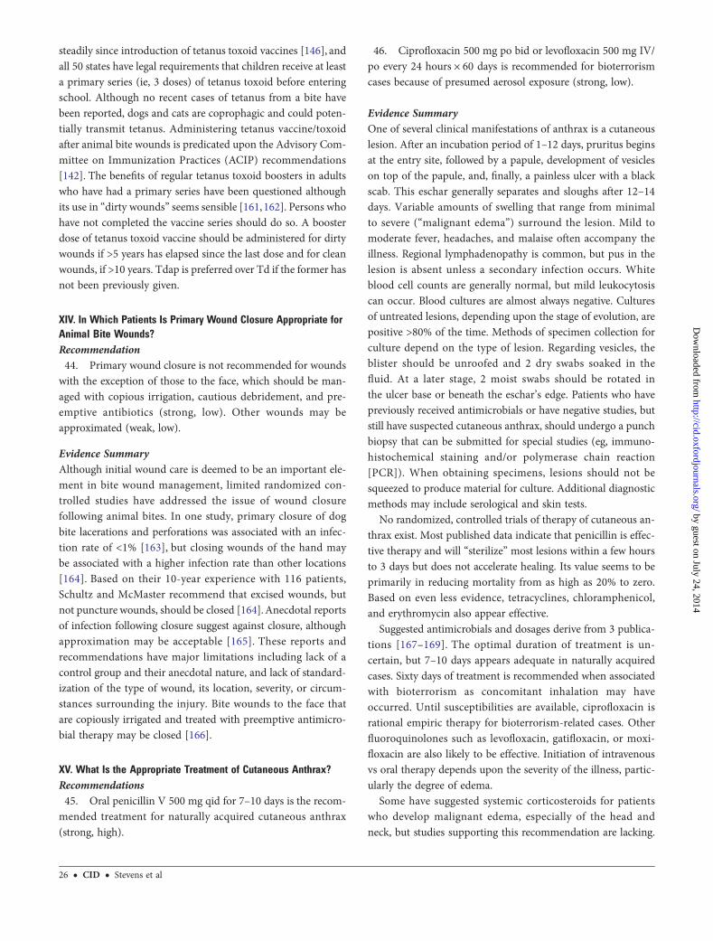
steadily since introduction of tetanus toxoid vaccines [146], andall 50 states have legal requirements that children receive at leasta primary series (ie, 3 doses) of tetanus toxoid before enteringschool. Although no recent cases of tetanus from a bite havebeen reported, dogs and cats are coprophagic and could poten-tially transmit tetanus. Administering tetanus vaccine/toxoidafter animal bite wounds is predicated upon the Advisory Com-mittee on Immunization Practices (ACIP) recommendations[142]. The benefits of regular tetanus toxoid boosters in adultswho have had a primary series have been questioned althoughits use in “dirty wounds” seems sensible [161, 162]. Persons whohave not completed the vaccine series should do so. A boosterdose of tetanus toxoid vaccine should be administered for dirtywounds if >5 years has elapsed since the last dose and for cleanwounds, if >10 years. Tdap is preferred over Td if the former hasnot been previously given.
XIV. In Which Patients Is Primary Wound Closure Appropriate forAnimal Bite Wounds?Recommendation44. Primary wound closure is not recommended for wounds
with the exception of those to the face, which should be man-aged with copious irrigation, cautious debridement, and pre-emptive antibiotics (strong, low). Other wounds may beapproximated (weak, low).
Evidence SummaryAlthough initial wound care is deemed to be an important ele-ment in bite wound management, limited randomized con-trolled studies have addressed the issue of wound closurefollowing animal bites. In one study, primary closure of dogbite lacerations and perforations was associated with an infec-tion rate of <1% [163], but closing wounds of the hand maybe associated with a higher infection rate than other locations[164]. Based on their 10-year experience with 116 patients,Schultz and McMaster recommend that excised wounds, butnot puncture wounds, should be closed [164].Anecdotal reportsof infection following closure suggest against closure, althoughapproximation may be acceptable [165]. These reports andrecommendations have major limitations including lack of acontrol group and their anecdotal nature, and lack of standard-ization of the type of wound, its location, severity, or circum-stances surrounding the injury. Bite wounds to the face thatare copiously irrigated and treated with preemptive antimicro-bial therapy may be closed [166].
XV. What Is the Appropriate Treatment of Cutaneous Anthrax?Recommendations45. Oral penicillin V 500 mg qid for 7–10 days is the recom-
mended treatment for naturally acquired cutaneous anthrax(strong, high).
46. Ciprofloxacin 500 mg po bid or levofloxacin 500 mg IV/po every 24 hours × 60 days is recommended for bioterrorismcases because of presumed aerosol exposure (strong, low).
Evidence SummaryOne of several clinical manifestations of anthrax is a cutaneouslesion. After an incubation period of 1–12 days, pruritus beginsat the entry site, followed by a papule, development of vesicleson top of the papule, and, finally, a painless ulcer with a blackscab. This eschar generally separates and sloughs after 12–14days. Variable amounts of swelling that range from minimalto severe (“malignant edema”) surround the lesion. Mild tomoderate fever, headaches, and malaise often accompany theillness. Regional lymphadenopathy is common, but pus in thelesion is absent unless a secondary infection occurs. Whiteblood cell counts are generally normal, but mild leukocytosiscan occur. Blood cultures are almost always negative. Culturesof untreated lesions, depending upon the stage of evolution, arepositive >80% of the time. Methods of specimen collection forculture depend on the type of lesion. Regarding vesicles, theblister should be unroofed and 2 dry swabs soaked in thefluid. At a later stage, 2 moist swabs should be rotated inthe ulcer base or beneath the eschar’s edge. Patients who havepreviously received antimicrobials or have negative studies, butstill have suspected cutaneous anthrax, should undergo a punchbiopsy that can be submitted for special studies (eg, immuno-histochemical staining and/or polymerase chain reaction[PCR]). When obtaining specimens, lesions should not besqueezed to produce material for culture. Additional diagnosticmethods may include serological and skin tests.
No randomized, controlled trials of therapy of cutaneous an-thrax exist. Most published data indicate that penicillin is effec-tive therapy and will “sterilize” most lesions within a few hoursto 3 days but does not accelerate healing. Its value seems to beprimarily in reducing mortality from as high as 20% to zero.Based on even less evidence, tetracyclines, chloramphenicol,and erythromycin also appear effective.
Suggested antimicrobials and dosages derive from 3 publica-tions [167–169]. The optimal duration of treatment is un-certain, but 7–10 days appears adequate in naturally acquiredcases. Sixty days of treatment is recommended when associatedwith bioterrorism as concomitant inhalation may haveoccurred. Until susceptibilities are available, ciprofloxacin isrational empiric therapy for bioterrorism-related cases. Otherfluoroquinolones such as levofloxacin, gatifloxacin, or moxi-floxacin are also likely to be effective. Initiation of intravenousvs oral therapy depends upon the severity of the illness, partic-ularly the degree of edema.
Some have suggested systemic corticosteroids for patientswho develop malignant edema, especially of the head andneck, but studies supporting this recommendation are lacking.
26 • CID • Stevens et al
by guest on July 24, 2014http://cid.oxfordjournals.org/
Dow
nloaded from

Airway compromise requiring intubation or tracheostomy mayoccur with malignant edema.
RECOMMENDATIONS FOR EVALUATION ANDTREATMENT OF BACILLARY ANGIOMATOSISAND CAT SCRATCH DISEASE
XVI. What Is the Appropriate Approach for the Evaluation andTreatment of Bacillary Angiomatosis and Cat Scratch Disease?Recommendations47. Azithromycin is recommended for cat scratch disease
(strong, moderate) according to the following dosing protocol:
(a) Patients >45 kg: 500 mg on day 1 followed by 250 mgfor 4 additional days (strong, moderate).(b) Patients <45 kg: 10 mg/kg on day 1 and 5 mg/kg for 4more days (strong, moderate).
48. Erythromycin 500 mg qid or doxycycline 100 mg bid for2 weeks to 2 months is recommended for treatment of bacillaryangiomatosis (strong, moderate).
Evidence SummaryIn classic cat scratch disease, a papule or pustule develops from3–30 days following a scratch or a bite. Lymph nodes that drainthe infected area enlarge about 3 weeks after inoculation. Thedisease course varies, but lymphadenopathy generally resolveswithin 1–6 months. In about 10% of cases, the nodes suppurate.Extranodal disease (eg, central nervous system, liver, spleen,bone, and lung) develops in ≤2% of cases. Bartonella henselaecauses most cases of cat scratch disease in immunocompetenthosts. Bacillary angiomatosis, seen in immunocompromised pa-tients, especially with AIDS, can occur from either B. henselaeor Bartonella quintana.
Bacillary angiomatosis typically occurs in individuals withAIDS and has 2 clinical appearances: (1) red papules thatvary in size from a millimeter to several centimeters, numberingfrom 1 to >1000; (2) subcutaneous, painful nodules with theoverlying skin having a normal or dusky hue.
Diagnosis of Bartonella infections may be difficult becausethe organism is fastidious and difficult to grow in culture. Sero-logical testing supports the diagnosis, although there is cross-reactivity between B. henselae and B. quintana as well as witha few other organisms. PCR is a diagnostic option. A positiveWarthin-Starry silver stain of infected lymph node tissue is use-ful to confirm the diagnosis, although it cannot differentiatespecies of Bartonella.
Treatment of cat scratch disease with antimicrobial agentshas had variable, but rarely dramatic, results. A single, dou-ble-blind placebo-controlled study involved 29 patients, 14 ofwhom received azithromycin [170]. The lymph node size re-gressed by 80% at 30 days more frequently in the azithromy-cin-treated patients (P = .02). The recommended dose of
azithromycin for patients weighing ≥45.5 kg (100 lb) is 500 mgon day 1, then 250 mg once daily for 4 additional days; forthose weighing <45.5 kg, the dose is 10 mg/kg orally on day 1,then 5 mg/kg on days 2–5 [124]. Cutaneous bacillary angiomato-sis therapy has not been systematically examined. Based on casereports and small series, either erythromycin (500 mg qid) ordoxycycline (100 mg bid) appears effective [171]. The durationof initial therapy, while not standardized, should be for 2 weeksto 2 months. With relapses, retreatment with prolonged therapy(months) should be entertained until immunocompetence re-turns. HIV-infected patients may require lifelong treatment [171].
XVII. What Is the Preferred Treatment for Erysipeloid?Recommendation49. Penicillin (500 mg qid) or amoxicillin (500 mg 3 times
daily [tid]) for 7–10 days is recommended for treatment of er-ysipeloid (strong, high).
Evidence SummaryErysipeloid is a cutaneous infection caused by Erysipelothrixrhusiopathiae a thin, pleomorphic, non-spore-forming gram-positive rod. It is a zoonosis acquired by handling fish, marineanimals, swine, or poultry. One day to 7 days after exposure, ared maculopapular lesion develops, usually on fingers or hands.Erythema spreads centrifugally, with central clearing. A bluering with a peripheral red halo may appear, giving the lesiona target appearance. Regional lymphangitis/lymphadenopathyoccurs in about one-third of cases. A severe generalized cutane-ous variety also occurs. Systemic symptoms and leukocytosis areunusual. Culture of an aspirate and/or biopsy of the lesion es-tablish the diagnosis; blood cultures are rarely positive. Untreat-ed erysipeloid resolves over about 3–4 weeks, but treatmentprobably hastens healing and may reduce systemic complica-tions. Based on in vitro susceptibilities and anecdotal experienc-es, penicillin is appropriate. Cephalosporins, clindamycin, orfluoroquinolones should be effective for those intolerant of pen-icillin. Erysipelothrix rhusiopathiae is resistant to vancomycin,teicoplanin, and daptomycin [133, 134, 172, 173].
XVIII. What Is the Appropriate Treatment of Glanders?Recommendation50. Ceftazidime, gentamicin, imipenem, doxycycline, or cip-
rofloxacin is recommended based on in vitro susceptibility(strong, low).
Evidence SummaryGlanders, characterized by ulcerating nodular lesions of the skinand mucous membrane, is caused by the aerobic gram-negativerod Burkholderia mallei. Glanders is mainly a disease mainlyof solipeds (eg, horses and mules). Humans become acci-dental hosts either by inhalation or skin contact. Althoughother organs may be involved, pustular skin lesions and
IDSA Practice Guidelines for SSTIs • CID • 27
by guest on July 24, 2014http://cid.oxfordjournals.org/
Dow
nloaded from

lymphadenopathy with suppurative nodes can be a prominentfeature. Optimal therapy of glanders is poorly defined. The or-ganism is susceptible to ceftazidime, gentamicin, imipenem,doxycycline, and ciprofloxacin [174]. A recent laboratory-ac-quired case was successfully treated with imipenem and doxy-cycline for 2 weeks, followed by azithromycin and doxycyclinefor an additional 6 months [175].
XIX. What Is the Appropriate Diagnosis and Treatment of BubonicPlague?Recommendation51. Bubonic plague should be diagnosed by Gram stain and
culture of aspirated material from a suppurative lymph node(strong, moderate). Streptomycin (15 mg/kg intramuscularly[IM] every 12 hours) or doxycycline (100 mg bid po) is recom-mended for treatment of bubonic plague (strong, low). Genta-micin could be substituted for streptomycin (strong, low).
Evidence SummaryPlague results from infection with Yersinia pestis, a facultativeanaerobic gram-negative coccobacillus. It primarily affects ro-dents, being maintained in nature by several species of fleasthat feed on them. Three plague syndromes occur in humans:septicemic, pneumonic, and bubonic. Bubonic plague, themost common and classic form, develops when humans are bit-ten by infected fleas or have a breach in the skin when handlinginfected animals. Domestic cat scratches or bites may also trans-mit bubonic plague. Patients usually develop fever, headache,chills, and tender regional lymphadenopathy 2–6 days aftercontact with the organism. A skin lesion at the portal of entryis sometimes present. Patients with bubonic plague may developsepticemia and secondary plague pneumonia, which is trans-missible person-to-person. Diagnosis can be made by bloodcultures and by aspirating lymph nodes for staining and culture.PCR is available at reference laboratories. Serologic tests mayprovide retrospective confirmation.
No controlled comparative trials of therapy for plague exist.Streptomycin 15 mg/kg every 12 hours (adjusted for renal func-tion) is the treatment of choice, although tetracycline and chlor-amphenicol are also considered appropriate therapy [175, 176].Gentamicin is a reasonable substitute for streptomycin if the lat-ter is not available, although there is but limited experience withgentamicin in the treatment of plague. Based on in vitro suscep-tibilities and murine models, fluoroquinolones are another op-tion. Ciprofloxacin has been suggested as a drug for bothtreatment and prevention of plague due to biowarfare agents de-spite a lack of documented efficacy in humans. The optimal du-ration for treating bubonic plague is unknown, but 10–14 daysis probably adequate. Patients with bubonic plague may developsecondary pneumonic plague and should be placed in respira-tory isolation until after 48 hours of effective drug therapy.
XX. What Is Appropriate for Diagnosis and Treatment forTularemia?Recommendations52. Serologic tests are the preferred method of diagnosing tu-
laremia (weak, low).53. Streptomycin (15 mg/kg every 12 hours IM) or gentami-
cin (1.5 mg/kg every 8 hours IV) is recommended for treatmentof severe cases of tularemia (strong, low).54. Tetracycline (500 mg qid) or doxycycline (100 mg bid
given by mouth) is recommended for treatment of mild casesof tularemia (strong, low).55. Notify the microbiology laboratory if tularemia is sus-
pected (strong, high).
Evidence SummaryFrancisella tularensis, while hardy and persistent in nature, is afastidious, aerobic, gram-negative coccobacillus. Illness canoften be categorized into several fairly distinct syndromes:ulceroglandular, glandular, typhoidal, pneumonic, and oculo-glandular or oropharyngeal. The glandular varieties are gener-ally acquired by handling infected animals, by tick bites, andsometimes by animal bites, especially cats. Biting flies occasion-ally transmit the illness in the United States, while mosquitoesare common vectors in Europe. After an incubation period of3–10 days, the patient typically develops a skin lesion (ulcereschar) at the entry site of the organism along with tenderadenopathy in regional lymph nodes, hence the name ulcero-glandular. In some patients, the skin lesion is inconspicuousor healed by the time they seek medical care, resulting in “glan-dular” tularemia. The illness is often associated with substantialfever, chills, headache, and malaise.
Confirmation of the diagnosis is usually serological. Routinecultures are often negative unless cysteine-supplemented mediaare utilized. The laboratory should be notified when tularemia issuspected because of the health risks posed to laboratory per-sonnel. Unsuspected growth of F. tularemia can cause laboratory-acquired disease. PCR may also be useful for diagnosis.
No prospective controlled or randomized trials of therapy fortularemia have been performed, nor has the optimal duration oftreatment been established. Streptomycin has been consideredthe drug of choice for tularemia for several decades [130].Since then, a few patients have been received fluoroquinolones.Francisella is resistant to most β-lactam antibiotics, which shouldbe avoided. When static drugs such as tetracyclines or chloram-phenicol are used, relapses may be more common, but often thepatients have received brief therapy (ie, <7–10 days).
Acutely ill adults or children should receive an aminoglyco-side, preferably streptomycin or possibly gentamicin. For adults,the regimen for streptomycin is 30 mg/kg/day in 2 divideddoses (no more than 2 g daily) or gentamicin 1.5 mg/kg every8 hours, with appropriate dose adjustment based on renal
28 • CID • Stevens et al
by guest on July 24, 2014http://cid.oxfordjournals.org/
Dow
nloaded from

function. For children, streptomycin should be administered at30 mg/kg/day in 2 divided doses or gentamicin at 6 mg/kg/dayin 3 divided doses [130]. Although no data exist, treatment witha parenteral agent until the acute illness is controlled, followedby an oral agent, seems rational for the duration of 7–10 days.Treatment of severe cases should be extended to 14 days.
For mild to moderate disease, oral tetracycline (500 mg qid)or doxycycline (100 mg bid) is appropriate. A few cases havebeen treated with fluoroquinolones with mixed results [177].Oral levofloxacin (500 mg daily) or ciprofloxacin (750 mgbid) in adults may be reasonable in mild to moderate illness.For oral regimens, patients should receive at least 14 days oftherapy. Despite clinical responses and appropriate treatmentin one study from France, 38.6% of patients relapsed [177].
XXI. What Is the Appropriate Approach to Assess SSTIs inImmunocompromised Patients?Recommendations56. In addition to infection, differential diagnosis of skin le-
sions should include drug eruption, cutaneous infiltration withthe underlying malignancy, chemotherapy- or radiation-in-duced reactions, Sweet syndrome, erythema multiforme, leuko-cytoclastic vasculitis, and graft-vs-host disease amongallogeneic transplant recipients (strong, high).57. Differential diagnosis for infection of skin lesions should
include bacterial, fungal, viral, and parasitic agents (strong,high).58. Biopsy or aspiration of the lesion to obtain material for
histological and microbiological evaluation should always beimplemented as an early diagnostic step (strong, high).
Executive SummarySkin and soft tissues are common sites of infection for HIV-negative patients with a compromised immune system, posinga major diagnostic challenge [178, 179], as the differential diag-nosis is broad and includes drug eruption, skin or soft tissue in-filtration with the underlying malignancy, chemotherapy- orradiation-induced skin reactions, graft-vs-host disease amongallogeneic transplant recipients, Sweet syndrome, erythemamultiforme, and leukocytoclastic vasculitis [180, 181]. Becausethe intensity and type of immune defect diminishes or altersdermatological findings, cutaneous lesions that appear localizedor innocuous may actually be a manifestation of a systemic orpotentially life-threatening infection. The differential diagnosisfor SSTIs in immunocompromised patients is usually widerthan that for immunocompetent patients and often includesbacterial, viral, fungal, and parasitic agents. Organisms thatcause these infections will vary based on the underlying im-mune defects (eg neutropenia, cellular immune defects, iatro-genic related to the use of intravascular catheters), and manyof the infecting organisms are not typically considered
pathogenic in normal hosts (opportunistic organisms such asAspergillus fumigatus). However, infectious agents commonlyfound in immunocompetent patients (eg, S. pyogenes,S. aureus) still need to be entertained in the differential diagno-sis of skin and soft tissue lesions in immunocompromised pa-tients even if the dermatological findings are atypical for thesecommon organisms. A careful epidemiologic history (eg, expo-sure to raw seafood, pets, and travel) should also be obtained inthese patients to consider organisms potentially associated withthese exposures when appropriate (eg, V. vulnificus, B. henselae,cutaneous leishmaniasis). Use of antimicrobial prophylaxis inthese patients has shown to ultimately impact the pathogensthat will be isolated when infection develops, and this informa-tion should be available to the clinician when assessing im-munocompromised patients with skin and soft tissue lesions[182, 183].
After considering the important specific factors concerningthe patient’s immunocompromised status (eg, neutropenia orneutrophil defects, cellular immune defect, presence of intravas-cular catheters) [180, 181], the gross morphologic characteristicsof the skin lesion(s) should be characterized, the extent of theinfection determined (eg, localized vs disseminated), and ap-propriate diagnostic tests undertaken to identify the infectingpathogen. Although blood cultures, tests for detection of anti-gens in blood or vesicular fluid, or nucleic acid amplificationtechniques in body fluids or tissues may be helpful, the mostspecific method for an expedited diagnosis is biopsy or aspira-tion of the lesion to obtain material for histological and micro-biological evaluation. The use of newer molecular methods (eg,gene amplification and sequencing) will likely impact the man-agement algorithms of immunocompromised patients withskin and soft tissue lesions and result in the earlier use of path-ogen-directed antimicrobial therapy [184, 185]. Peripheralblood biomarkers such as galactomannan and 1,3-β-D-glucanhas been well studied over the past 20 years and has been report-ed to be useful in the diagnosis of disseminated fungal infec-tions by several European investigators. However, sensitivityof these tests can be significantly affected by the use of antifun-gal drugs, and in the United States their sensitivity has been re-ported to be lower than in Europe in various populations ofimmunocompromised patients [186].
Empiric antimicrobial therapy should be initiated immedi-ately in these patients on the basis of their underlying disease,primary immune defect, morphology of skin lesions, use ofprior antimicrobial prophylaxis, allergy history, and inherentand local profiles of antimicrobial resistance. Despite aggressiveempiric therapy, treatment failure may occur, and the reasonsfor this lack of response include the following: (1) the initial di-agnosis and/or treatment chosen is incorrect; (2) the etiologicpathogen is already resistant to the antimicrobial agent; (3) re-sistance develops during treatment; (4) if indicated, surgical
IDSA Practice Guidelines for SSTIs • CID • 29
by guest on July 24, 2014http://cid.oxfordjournals.org/
Dow
nloaded from

debridement has not taken place; and/or (5) the patient’s im-mune deficiency is profound and cannot be reversed. Theearly identification of an etiologic agent in immunocompro-mised hosts with SSTIs is essential when deciding whether sur-gical debridement is warranted because microbial resistancemakes dogmatic empiric treatment regimens difficult, if notdangerous. For this reason, skin biopsy material should be ob-tained by an experienced dermatologist and evaluated in con-junction with a pathologist who is familiar with this patientpopulation.
RECOMMENDATIONS FOR SKIN AND SOFTTISSUE INFECTIONS IN CANCER PATIENTSWITH NEUTROPENIA
XXII. What Is the Appropriate Approach to Assess SSTIs inPatients With Fever and Neutropenia?Recommendations59. Determine whether the current presentation of fever and
neutropenia is the patient’s initial episode of fever and neutro-penia, or persistent unexplained fever of their initial episode(after 4–7 days) or a subsequent episode of fever and neutrope-nia (recurrent) (strong, low).60. Aggressively determine the etiology of the SSTI by aspi-
ration and/or biopsy of skin and soft tissue lesions and submitthese for thorough cytological/histological assessments, micro-bial staining, and cultures (strong, low).61. Risk-stratify patients with fever and neutropenia accord-
ing to susceptibility to infection: high-risk patients are thosewith anticipated prolonged (>7 days) and profound neutropenia(absolute neutrophil count [ANC] <100 cells/µL) or with aMultinational Association for Supportive Care (MASCC)score of <21; low-risk patients are those with anticipated brief(<7 days) periods of neutropenia and few comorbidities (strong,low) or with a MASCC score of ≥21 (strong, moderate).62. Determine the extent of infection through a thorough
physical examination, blood cultures, chest radiograph, and ad-ditional imaging (including chest CT) as indicated by clinicalsigns and symptoms (strong, low).
Evidence SummarySSTIs in patients with fever and neutropenia have rarely beencarefully studied as a “separate entity.” Rather, recommenda-tions for these infections are extrapolated from broad groupguidelines that include references to SSTIs and have been devel-oped by professional organizations including IDSA, the Nation-al Comprehensive Cancer Network (NCCN), American Societyof Blood and Marrow Transplantation, the American Society ofClinical Oncology, and the Centers for Disease Control andPrevention [187–193].These guidelines are focused on the diag-nosis and management of specific patient groups (eg, fever and
neutropenia, infection in recipients of hematopoietic stem celltransplant), specific infections (eg, candidiasis, aspergillosis),and iatrogenic infections (eg, intravascular catheter–related in-fection). They are based on published clinical trials, descriptivestudies, or reports of expert committees, and the clinical expe-rience and opinions of respected authorities. Therefore, thissection of the SSTI guideline will focus on existing recommen-dations that demand reinforcement, or that are truly specific toSSTIs.
Neutropenia is defined as an ANC <500 cells/µL, or a neutro-phil count that is expected to decrease to <500 cells/µL within48 hours [187–189]. The development of fever during treat-ment-associated neutropenia is common, but many patientsdo not have an infectious etiology determined [184, 194].More than 20% of patients with chemotherapy-induced neutro-penia develop a clinically documented infection involving theskin and soft tissues, but many are due to hematogenous dis-semination [179].
Cancer patients with fever and neutropenia can be dividedinto low- and high-risk groups [187]. The determination of dif-ferences in patient risk of infection and infectious complicationslevels (high risk and low risk) during the period of neutropeniahas been recognized and further validated since this clinicalguideline was last updated [195, 196]. The MASCC developedand validated a scoring method that formally differentiates be-tween high-risk and low-risk patients [195, 196]. High-risk pa-tients have a MASCC score <21. Low-risk patients have aMASCC score ≥21. Disseminated or complex SSTIs are morelikely to occur among high-risk patients.
Clinical, Laboratory, and Radiological Evaluation of Patients WithFebrile Neutropenia Who Present With Skin and Soft TissueLesionsSigns and symptoms of inflammation and infection are oftendiminished or absent in patients with neutropenia. Skin lesions,no matter how small or innocuous in appearance, should becarefully evaluated. Early involvement of an infectious diseasesspecialist, a surgeon, and a dermatologist familiar with these pa-tients may result in improved outcome. Initial clinical impres-sions should be supplemented with a systemic approach toenhance the diagnosis and management of infection. Blood cul-tures are critical, and at least 2 sets should be obtained. Radio-graphic imaging should be performed as clinically indicated,but can be helpful to define the extent of SSTIs when patientsare neutropenic. Chest/sinus radiologic imaging may identifythe silent or subtle pulmonary site of infection that has resultedin dissemination to skin or soft tissues.
The most specific method for evaluating SSTIs is biopsy oraspiration of the lesion(s) to obtain material for histological, cy-tological, and microbiological evaluation. Prospective studiesevaluating the yield of skin biopsy or aspiration have not been
30 • CID • Stevens et al
by guest on July 24, 2014http://cid.oxfordjournals.org/
Dow
nloaded from

performed in adult immunocompromised patients, but mostclinicians who manage these patients combine blood cultures,serial antigen detection, nucleic acid amplification techniques,radiographic imaging, and a biopsy or aspiration of the abnor-mal skin or soft tissue lesion in the hope of increasing the recov-ery of the offending pathogen and directing pathogen-specificantimicrobial therapy.
This can occur during “initial” episode fever and neutropenia(first episode of neutropenic fever that requires systemic antimi-crobial therapy) or during a “persisting episode” (persistentneutropenic fever unresponsive to broad-spectrum antimicrobi-al therapy beyond days 4–7) or during recurrent episodes offever and neutropenia. This determination helps the cliniciandefine the most likely pathogens and to construct the initial em-piric treatment.
During the initial episode gram-negative bacteria should beprimarily targeted by the initial antibiotic regimen becausethey are associated with high mortality rates. Although gram-positive bacteria are more common, the addition of antibioticswith gram-positive activity including MRSA is not recommend-ed unless physical findings suggestive of inflammation in theskin and soft tissues are present, the patient is hemodynamicallyunstable, and risk factors for MRSA are present. For patientswith a persistent episode of fever and neutropenia or recurrentepisodes, antibiotic-resistant bacterial or fungal pathogens in-cluding Candida and molds become more common [197–199].
Dermatologic manifestations in patients with fever and neu-tropenia include erythematous maculopapular lesions, focal orprogressive cellulitis, cutaneous nodules, “ecthyma gangreno-sum,” and, occasionally, necrotizing fasciitis or myonecrosis[179, 200]. Ecthyma gangrenosum is a cutaneous vasculitiscaused by invasion of the media and adventitia of the vesselwall by bacteria, which may be visible on histologic stains of bi-opsy specimens. Ecthyma gangrenosum frequently begins aspainless erythematous papule(s) that often progress and be-come painful and necrotic within 24 hours. These skin lesionsmay be discrete or multiple, are found preferentially betweenthe umbilicus and the knees, and can increase in size from 1cm to >10 cm in <24 hours. Ecthyma gangrenosum has classi-cally been reported to occur with Pseudomonas aeruginosainfections, but similar lesions may be caused by other Pseudo-monas species, Aeromonas species, Serratia species, S. aureus,Stenotrophomonas maltophilia, S. pyogenes, fungi includingCandida species, Aspergillus, Mucor, and Fusarium, and evenherpes simplex virus (HSV) [201].
In contrast to immunocompetent patients, necrotizing fasci-itis and/or myonecrosis are more frequently associated withgram-negative or polymicrobial pathogens rather than a singlegram-positive bacterium. Necrotizing fasciitis can present aloneor concurrently with myonecrosis in the patient with fever andneutropenia. Rapidly progressive necrotizing SSTIs may
initially be clinically subtle in compromised patients, but MRIscans of the involved area may be helpful in defining the depthof infections. In such infections, immediate surgical explorationby a team experienced in the management of these patients andbroad-spectrum antibiotic therapy targeted at gram-negative,gram-positive, and anaerobic bacteria are essential.
Gram-positive pathogens are now the most common bacteri-al organisms isolated from diagnostic cultures obtained from fe-brile neutropenic patients [197, 198]. These pathogens in orderof decreasing prevalence include coagulase-negative staphylo-cocci, viridans streptococci, enterococci, S. aureus, Corynebacte-rium, Clostridium species, and Bacillus species. SSTIs associatedwith these organisms usually begin as a focal area of tender cu-taneous erythema, a macular or maculopapular eruption, or aclassic cellulitis. Although rare, they can also cause ecthymagangrenosum–like lesions that are often confused with “spiderbites,” superficial and deep abscesses that become apparent fol-lowing marrow recovery, necrotizing fasciitis, myositis, and my-onecrosis. Common infection sites are the groin, axilla, areas ofcutaneous disruption (eg, vascular catheter or bone marrow as-piration sites), or other skin sites that are moist and frequentlyabraded. Hematogenous dissemination of gram-positive bacte-rial organisms to the skin and soft tissue is uncommon exceptfor S. aureus and some Clostridium species. A toxic shock–likesyndrome with associated diffuse erythroderma has been de-scribed with bacteremic toxin–producing streptococci. Painfulmyositis may also occur with S. aureus infections as a compo-nent of hematogenous dissemination.
HSV, varicella zoster virus (VZV), and enteroviruses are rarecauses of cutaneous manifestations in patients with neutropenia[202]. Their presence usually reflects either a disseminated in-fection, or, in the case of HSV, the autoinoculation of virus frommucosal sites to adjacent or distant cutaneous sites. HSV andVZV in compromised patients may appear as vesicles similarto those in normal hosts, or as isolated or multiple benign-look-ing papules with a central eschar (ecthyma gangrenosum–likelesion). VZV in compromised hosts may present with the tradi-tional unilateral dermatome distribution, but may also appear asdiscrete or multiple skin lesions in random distribution. Skinbiopsy is the only reliable method to diagnose cutaneous or dis-seminated HSV or VZV infection; peripheral blood PCR forHSV or VZV can be helpful in these patients.
XXIII. What Is the Appropriate Antibiotic Therapy for Patients WithSSTIs During the Initial Episode of Fever and Neutropenia?Recommendations63. Hospitalization and empiric antibacterial therapy with
vancomycin plus antipseudomonal antibiotics such as cefe-pime, a carbapenem (imipenem-cilastatin or meropenem ordoripenem), or piperacillin-tazobactam are recommended(strong, high).
IDSA Practice Guidelines for SSTIs • CID • 31
by guest on July 24, 2014http://cid.oxfordjournals.org/
Dow
nloaded from

64. Documented clinical and microbiologic SSTIs should betreated based on antimicrobial susceptibilities of isolated organ-isms (strong, high).65. It is recommended that the duration of treatment for most
bacterial SSTIs should be for 7–14 days (strong, moderate).66. Surgical intervention is recommended for drainage of
soft tissue abscess after marrow recovery or for a progressivepolymicrobial necrotizing fasciitis or myonecrosis (strong, low).67. Adjunct colony-stimulating factor therapy (granulocyte
colony-stimulating factor [G-CSF], granulocyte macrophagecolony-stimulating factor [GM-CSF]) or granulocyte transfu-sions are not routinely recommended (weak, moderate).68. Acyclovir should be administered to patients suspected
or confirmed to have cutaneous or disseminated HSV or VZVinfection (strong, moderate).
Evidence SummaryThe appropriate antibiotics for patients with suspected or con-firmed SSTI (initial infection) should be broad-spectrum agentsadministered at the first clinical signs or symptoms of infection[203].No single empiric regimen is superior, but all recommend-ed regimens should meet the following criteria: broad-spectrumantimicrobial activity including P. aeruginosa, bactericidal in theabsence of circulating neutrophils, and low antibiotic-associatedtoxicity (Table 7). Infections caused by gram-negative bacilliincluding P. aeruginosa have been associated with the highestinfection-associated mortality [198, 203]. Despite an increasedprevalence of gram-positive bacteria, antibiotics specificallyaimed against this group of organisms are not required [187,189] unless patients exhibit physical findings of SSTI or cathe-ter-associated infection or are hemodynamically unstable [204].
Antibiotic selection should follow the clinical care guidelinesdeveloped by IDSA and the NCCN [187, 189]. Excellent resultshave been reported for gram-negative infections using broad-spectrum monotherapy with carbapenems, cephalosporins thatpossess antipseudomonal activity, or piperacillin/tazobactam.
For patients in whom vancomycin may not be an option,daptomycin, ceftaroline, or linezolid should be added to the
initial empiric regimen. Linezolid, daptomycin, or ceftarolinehave activity against MRSA [204] and have received FDA ap-proval for the treatment of SSTIs, but have not been compre-hensively studied in patients with neutropenia. The use oflinezolid in this patient population has been associated with de-layed ANC recovery [205, 206].
The combination of ciprofloxacin and amoxicillin-clavula-nate is the preferred oral antibiotic regimen for low-risk patients[207, 208]. Levofloxacin has better gram-positive activity thanciprofloxacin, but is less potent than ciprofloxacin against P.aeruginosa, causing some to suggest that a higher dose of levo-floxacin therapy (750 mg daily) may be required.
Fluoroquinolone prophylaxis should preclude the use of flu-oroquinolones for empiric therapy, and instead broad-spectrumβ-lactam antibiotics should be considered. Intravenous acyclo-vir should be added to the empiric antimicrobial regimen of therare patient who has not been receiving antiviral prophylaxis ef-fective against HSV or VZV, but has developed skin lesions sus-pected or confirmed to be caused by these viruses.
XXIV. What Is the Appropriate Antimicrobial Therapy for PatientsWith SSTIs During Persistent or Recurrent Episodes of Fever andNeutropenia?Recommendations69. Yeasts and molds remain the primary cause of infection-
associated with persistent or recurrent fever and neutropenia;therefore, empiric antifungal therapy (Table 6) should be addedto the antibacterial regimen (strong, high).
(a) Empiric administration of vancomycin or other agentswith gram-positive activity (linezolid, daptomycin, or cef-taroline) should be added if not already being administered(Table 7) (strong, high).(b) Candida species SSTIs should be treated with an echi-nocandin or, if Candida parapsilosis has been isolated, lipidformulation amphotericin B (strong, high) with fluconazoleas an acceptable alternative (strong, moderate). Treatmentshould be for 2 weeks after clearance of bloodstream infectionor resolution of skin lesions (strong, moderate).
Table 6. Standard Doses of Antifungal Agents
Antifungal Agent Oral Dose IV Dose Comments
Fluconazole 100–400 mg every 24 h 800 mg loading dose, then 400 mgdaily
Candida krusei and Candida glabrata areresistant
Voriconazolea 400 mg bid × 2 doses,then 200 mg every 12 h
6 mg/kg IV every 12 h for 2 doses,followed by 4 mg/kg IV every 12 h
Accumulation of cyclodextrin vehicle withIV formulation with renal insufficiency
Posaconazole 400 mg bid with meals N/A Covers MucoralesLipid complexamphotericin B
N/A 5 mg/kg/d Not active against fusaria
Liposomalamphotericin B
N/A 3–5 mg/kg/d Not active against fusaria
Abbreviations: bid, twice daily; IV, intravenous; N/A, not applicable.a The use of patient-specific pharmacokinetics is recommended to improve clinical outcome [247].
32 • CID • Stevens et al
by guest on July 24, 2014http://cid.oxfordjournals.org/
Dow
nloaded from

(c) Aspergillus SSTIs should be treated with voriconazole(strong, high), or, alternatively, lipid formulations of ampho-tericin B, posaconazole, or echinocandin for 6–12 weeks(strong, low). Mucor/Rhizopus infections should be treatedwith lipid formulation amphotericin B (strong, moderate) orposaconazole (strong, low) (Table 6). The addition of an echi-nocandin could be considered based on synergy in murinemodels of mucormycosis and observational clinical data(weak, low).(d) Fusarium species infections should be treated withhigh-dose IV voriconazole or posaconazole (strong, low).(e) Begin treatment for antibiotic-resistant bacterial organ-isms, in patients currently on antibiotics (strong, moderate).(f ) Intravenous acyclovir should be added to the patient’santimicrobial regimen for suspected or confirmed cutaneousor disseminated HSV or VZV infections (strong, moderate).
70. Blood cultures should be obtained, and skin lesions in thispopulation of patients should be aggressively evaluated by cultureaspiration, biopsy, or surgical excision as they may be caused byresistant microbes, yeast, or molds (strong, moderate).71. The sensitivity of a single serum fungal antigen test (1,3-
β-D-glucan or galactomannan tests) is low particularly inpatients receiving antifungal agents, and benefits from laborato-ry tests for fungal antigen or DNA detection remain inconsis-tent (strong, moderate).72. PCR in peripheral blood for HSV and VZV might be
helpful in establishing a diagnosis of disseminated infection inpatients with unexplained skin lesions (weak, moderate).
Evidence SummaryIn patients with persistent unexplained fever of their first epi-sode (after 4–7 days) or recurrent fever, yeast and molds arethe major cause of infection-related morbidity and mortality(Table 7) [187, 189, 203]. These later infections are most com-mon among high-risk patients with prolonged and profoundneutropenia and they should be considered in any patientwith neutropenia and skin and soft tissue lesions suggestive ofinfection. In addition, MRSA should also be considered ifpatients are not receiving antimicrobial agents with activity
against MRSA (eg, vancomycin, linezolid, daptomycin, or cef-taroline) [209]. Multiple antibiotic-resistant gram-negativebacilli are more commonly being recovered from cultures ofblood and soft tissues, and antibiotic modification is necessarywhen their presence is suspected or documented (Table 7)[204]. Treatment of yeast and mold infections should followIDSA and NCCN guideline recommendations [187, 189].
Although skin and soft tissues are less frequent sites of infec-tion in patients with persisting or recurrent fever and neutropenia(<10%), they often represent a site of infection dissemina-tion. Among the responsible pathogens, 10%–15% are causedby antibiotic-resistant gram-negative bacilli; 30%–40% by anti-biotic-resistant gram-positive organisms (coagulase-negativestaphylococci, MRSA, and vancomycin-resistant enterococci),but most (>50%) are caused by yeast or molds [198, 210,211]. In 2012, infections caused by yeast and molds were themajor cause of associated morbidity and mortality in patientswith prolonged and profound neutropenia [198, 210].Diagnosisof fungal infections remains difficult, and benefits from fungalantigen or DNA detection remain inconsistent [212,213]. How-ever, recovery of fungi from aspiration or biopsy of skin or deepsoft tissues warrants aggressive systemic antifungal therapy.Surgical treatment should be also considered in patients withskin and soft tissue changes caused by angioinvasive molds(eg, Mucor, Rhizopus, and Aspergillus).
The incidence of invasive candidiasis prior to the routine useof azole antifungal prophylaxis was 12% in patients with pro-found and prolonged neutropenia [214]. Candida albicansis the most frequently isolated species; however, fluconazole-resistant yeast (ie, Candida krusei and Candida glabrata) areincreasingly common due to the widespread use of azole prophy-laxis [214]. Superficial cutaneous candidiasis presents as intertri-go, vaginitis, balanitis, perleche, and paronychia [215] and rarelycauses dissemination. However, up to 13% of patients with inva-sive disseminated candidiasis develop single or multiple nodularskin lesions [216, 217]. These lesions can appear as discrete pinkto red papules (0.5–1.0 cm) and are usually found on the trunkand extremities [215, 217].Candida skin lesions are usually non-tender, but may develop central pallor, or become hemorrhagic if
Table 7. Standard Doses of Antimicrobial Agents Active Against Multidrug-Resistant Organisms
Antimicrobial IV Dose Comments
Vancomycin 30–60 mg/kg/d in 2–4 divided doses Target serum trough concentrations of 15–20 µg/mL in severe infections
Daptomycin 4–6 mg/kg/d Covers VRE, strains nonsusceptible to vancomycin may be cross-resistant to daptomycinLinezolid 600 mg every 12 h 100% oral bioavailability; so oral dose same as IV dose. Covers VRE and MRSA
Colistin 5 mg/kg load, then 2.5 mg/kgevery 12 h
Nephrotoxic; does not cover gram-positives or anaerobes, Proteus, Serratia, Burkholderia
Abbreviations: IV, intravenous; MRSA, methicillin-resistant Staphylococcus aureus; VRE, vancomycin-resistant enterococci.
IDSA Practice Guidelines for SSTIs • CID • 33
by guest on July 24, 2014http://cid.oxfordjournals.org/
Dow
nloaded from

the patient is thrombocytopenic. Painful myositis can develop asa consequence of hematogenous infection and is most commonwith Candida tropicalis [218, 219]. Muscle and soft tissue abscessformation is uncommon, but when identified it has usually oc-curred following marrow recovery.
Trichosporon beigelii is an uncommon but frequently fatal dis-seminated fungal infection that often involves the skin [220].Dermatologic manifestations vary from multiple erythematousmacules to maculopapular lesions. Biopsy often reveals a mixtureof true hyphae, pseudohyphae, budding yeast, and arthroconidiathat may be easily mistaken for Candida species
Cutaneous mold infections are unusual, but there could belocal infections at sites of IV catheter insertion or at nail bedand cuticle junctions on fingers and toes, or secondary to hema-togenous dissemination [221]. Aspergillus, Rhizopus, and Mucorspecies cause painful erythematous skin nodules that become ne-crotic and can resemble ecthyma gangrenosum because of theirtendency for angioinvasion [222]. Aspergillus species infectionsoccur in 10%–14% of patients with profound and prolonged neu-tropenia, and mortality remains high [223].Aspergillus fumigatusis the most frequently isolated species (50%), followed by Asper-gillus flavus, Aspergillus niger, and Aspergillus terreus. Isolation ofAspergillus from blood cultures is rare, but dissemination is com-monly detected at autopsy [224].LocalMucor infections have oc-curred as a consequence of contaminated bandages or other skintrauma, but patients with pulmonary Mucor infection may alsodevelop secondary cutaneous involvement from presumed hema-togenous dissemination [225, 226].
Fusarium species are frequently identified as the infectingpathogen among patients with prolonged and profound neutro-penia [227]. Patients commonly present with myalgias and per-sistent fever despite antimicrobial therapy. Skin lesions are verycommon (60%–80% of infections), and often begin as multipleerythematous macules with central pallor that quickly evolve topapules and necrotic nodules. The lesions frequently may have aring of erythema surrounding an area of central necrosis. Le-sions localize preferentially to the extremities, especially thefeet, but may also be found on the face and trunk. Blood cul-tures are frequently positive (40%–50%) when cutaneous le-sions appear. Mortality from this infection remains high,although new azole antifungal agents appear promising [227].
RECOMMENDATIONS FOR PATIENTS WITHCELLULAR IMMUNODEFICIENCY
XXV. What Is the Appropriate Approach to Assess SSTIs inPatients With Cellular Immunodeficiency?Recommendations73. Consider immediate consultation with a dermatologist
familiar with cutaneous manifestations of infection in patientswith cellular immune defects (eg, those with lymphoma,
lymphocytic leukemia, recipients of organ transplants, or re-ceiving immunosuppressive drugs such as anti–tumor necrosisfactor (TNF) or certain monoclonal antibodies) (weak, low).74. Consider biopsy and surgical debridement early in the
management of these patients (weak, low).75. Empiric antibiotics, antifungals, and/or antivirals should
be considered in life-threatening situations (weak, moderate).The use of specific agents should be decided with the input ofthe primary team, dermatology, infectious disease, and otherconsulting teams (strong, moderate).
Evidence SummaryPatients with lymphoma or acute or chronic lymphocytic leuke-mia, recipients of hematopoietic stem cell transplant (HSCT) orsolid organ transplant (SOT), patients receiving corticosteroidsand other immunosuppressive drugs (eg, monoclonal antibodies,anti-TNF drugs), and patients with primary cellular immunode-ficiencies are predisposed to infection. These patients are at in-creased risk for infection caused by a select group of bacteria,fungi, viruses, protozoa, and helminths, and some of these path-ogens have the capacity to cause SSTIs. Infection should always behigh in the differential of a skin lesion or skin lesions in patientswith cellular immunodeficiency. These patients may not have sys-temic manifestations of infection, and the initial dermatologicalpresentation may be atypical or misleading. Thus cliniciansshould have a very low threshold to obtain a skin biopsy (Table 6).
Nontuberculous MycobacteriaAlthough most infections occur after primary inoculation atsites of skin disruption or trauma, hematogenous disseminationdoes occur. The most common manifestations of nontuberculousmycobacteria (NTM) infection in SOT recipients include cutane-ous and pleuropulmonary disease, and, in HSCT recipients, cath-eter-related infection and bacteremia [228].Disseminated infectionwith Mycobacterium avium complex occurs preferentially amongpatients with HIV disease, whereas bloodstream and cutaneous in-fections with Mycobacterium fortuitum, Mycobacterium chelonae,Mycobacterium abscessus, Mycobacterium ulcerans, Mycobacte-rium kansasii, Mycobacterium haemophilum, Mycobacteriummarinum, or Mycobacterium mucogenicum are more frequentamong non-HIV-immunocompromised hosts [228]. Dermato-logic manifestations include a poorly resolving cellulitis, painless1- to 2-cm nodules, necrotic ulcers, and subcutaneous abscesses.
Treatment of NTM infections of the skin and soft tissues re-quires prolonged combination therapy (duration, 6–12 weeks)that should consist of a macrolide antibiotic (eg, clarithromy-cin) and a second agent to which the isolate is susceptible. Sur-gical debridement is crucial for cultures and sensitivities and inaddition is necessary to remove devitalized tissue and to pro-mote skin and soft tissue healing. Definitive guidelines for treat-ment of these entities have been published [229].
34 • CID • Stevens et al
by guest on July 24, 2014http://cid.oxfordjournals.org/
Dow
nloaded from

NocardiaCutaneous Nocardia infections usually represent metastatic fociof infection that have originated from a primary pulmonarysource [230]. Nocardia farcinica, Nocardia brasiliensis, andotherNocardia species have been associated with cutaneous dis-ease. The dermatologic manifestations are usually limited tosubcutaneous nodules or abscesses and panniculitis. Soft tissueabscesses are frequently painless and are described as being coldto the touch. The incidence of local and disseminated Nocardiainfections has decreased with the routine use of SMX-TMPprophylaxis for patients who experience prolonged periods ofcellular immune deficiency. SMX-TMP remains the treatmentof choice, but other sulfa antibiotics (eg, sulfadiazine and sulfa-soxazole), amikacin, imipenem, meropenem, third-generationcephalosporins (ceftriaxone and cefotaxime), minocycline, ex-tended-spectrum fluoroquinolones (eg, moxifloxacin), linezol-id, and dapsone are effective in vitro and in animal models(Table 6). Combination therapy with other agents should beconsidered in patients with severe infections or profound andlasting immunodeficiency. Prolonged therapy is important,and the duration of treatment (6–24 months) should takeinto account the presence of disseminated disease and the ex-tent of the patient’s underlying immunosuppression. Surgicaldebridement is recommended for necrotic nodules or large sub-cutaneous abscesses.
FungiCutaneous mold infections have been increasingly reported inimmunocompromised patients with primarily cellular immu-nodeficiency. Skin lesions can occur as a manifestation of a dis-seminated disease, a primary cutaneous inoculation, or in theskin site of a previous IV line [221, 230]. The most commonmolds causing cutaneous manifestations in these patients in-clude Aspergillus, Mucormycosis, Scedosporium, and Fusariumspecies [231–234]. Skin lesions can present as papules, nodules,or ulcers, or with the dermatological appearance of ecthymagangrenosum. Skin biopsy should be performed for diagnosticpurposes and resection of the entire lesion or “debulking” pro-cedures should be considered in cases where there is either asingle lesion or localized disease is present. In instances of As-pergillus species, Scedosporium apiospermum, and Fusariumspecies infections, voriconazole is the best therapeutic option.Amphotericin B is an excellent alternative. Posaconazole isalso a reasonable alternative in combination with amphotericinB or as a transition to oral therapy (Table 7).
Cryptococcal infections originate in the lungs, often withearly hematogenous dissemination to the meninges and skinor soft tissues, but primary cutaneous cryptococcosis also oc-curs [235]. Single or multiple painless skin lesions involvingthe face and scalp develop in 5%–10% of clinically infected pa-tients, and in some patients, these lesions may precede
documented cryptococcal meningitis by several weeks. Cutane-ous cryptococcal infections may appear as papules (often simi-lar to molluscum contagiosum lesions), nodules, pustules,chronic draining necrotic ulcers, or, more subtly, as cellulitis[235]. Cryptococcal cellulitis has occurred in recipients ofblood, bone marrow, or SOT, although the incidence has dra-matically decreased with the prophylactic use of the newerazole agents, particularly fluconazole. Fluconazole is oftenused as initial treatment, for patients with mild infections, orto complete treatment after the patient has shown clinical andmicrobiologic improvement with amphotericin B and 5-flucy-tosine induction therapy [236]. Surgical debridement and/ordrainage are not helpful in the management of skin or soft tis-sue cryptococcal infections.
Cutaneous manifestations of acute progressive disseminatedhistoplasmosis are rare and usually occur in patients with severecellular immune deficiency [237, 238]. Skin lesions appear asnonspecific maculopapular eruptions that become hemorrhag-ic, but oral or cutaneous ulcers are sometimes present, particu-larly in the subacute, disseminated form of the disease.Histopathologic analysis of these skin lesions reveals necrosissurrounding the superficial dermal vessels, and with specialstains, both intracellular and extracellular yeast may be seen.Prompt administration of amphotericin B therapy is the recom-mended treatment for patients with cellular immune deficiencyand acute, life-threatening, progressive disseminated histoplas-mosis. Patients often show a rapid clinical improvement within1–2 weeks, and itraconazole can then replace amphotericin B tocomplete at least 6–12 months of treatment [237]. Patients withillnesses that result in profound and prolonged immune sup-pression should receive long-term suppressive therapy with itra-conazole after the initial treatment course is complete.
VirusesVZV is one of the 2 most frequent herpesviruses to cause cuta-neous infection in immunosuppressed patients [239]. Patientswithout a preceding history of VZV exposure are at significantrisk of developing severe chickenpox if exposed, but herpes zos-ter (also known as shingles) with or without dissemination is amore frequent clinical concern. Between 65% and 70% of adultpatients are seropositive for VZV, and this identifies those pa-tients at risk for future reactivation infection. Herpes zoster oc-curs most frequently during the first year followingchemotherapy treatment, or following receipt of an HSCT ora SOT. Depending on the intensity of treatment or type of trans-plant, 25%–45% of such patients develop dermatomal zoster,with a 10%–20% risk of developing dissemination withoutprompt and effective antiviral therapy. A few patients presentinitially with disseminated cutaneous infection that maymimic atypical varicella, but some patients may present withnonspecific lesions that do not initially have the vesicular
IDSA Practice Guidelines for SSTIs • CID • 35
by guest on July 24, 2014http://cid.oxfordjournals.org/
Dow
nloaded from

appearance of varicella. Herpes zoster typically causes a unilat-eral, vesicular eruption with dermatomal pain that often pre-cedes the skin findings by 24–72 hours (and sometimeslonger). Early lesions are erythematous macules that rapidlyevolve to papules and then to vesicles. The vesicles frequentlycoalesce, form bullae, and scab before healing. Lesions in other-wise healthy hosts continue to erupt for at least 4–6 days, withthe entire disease duration being approximately 2 weeks. How-ever, among immunocompromised hosts, skin lesions may con-tinue to develop over a longer period (7–14 days) and generallyheal more slowly unless effective antiviral therapy is adminis-tered. Without adequate treatment, some immunocompro-mised patients develop chronic ulcerations with persistentviral replication that is complicated by secondary bacterialand fungal superinfections.
High-dose IV acyclovir remains the treatment of choice forVZV infections in compromised hosts. Oral acyclovir, famciclo-vir, and valacyclovir are beneficial for VZV infections in other-wise healthy hosts, but oral therapy should be reserved for mildcases of VZV disease in patients with transient immune sup-pression or as treatment to complete therapy once the patienthas shown a clinical response to IV acyclovir. Recipients of al-logeneic blood and bone marrow transplants routinely take acy-clovir (800 mg bid) or valacyclovir (500 mg bid) during the firstyear following transplant for the prevention of VZV and HSVreactivation [240]. Should skin lesions suspicious of VZV orHSV develop in patients already taking such antivirals, antiviralresistance should be investigated and taken into account in theselection of the empiric regimen.
HSV infections in compromised hosts are almost exclusivelydue to viral reactivation. Orofacial and genital sites are the mostcommon cutaneous locations, but autoinoculation can occur inalmost any area. Infections of the fingernail bed and cuticle(herpetic whitlow) occur because of inoculation of HSV at epi-dermal sites.
Skin lesions are often preceded by localized pain or a tinglingsensation. Early skin lesions are usually focal, erythematous,and maculopapular lesions that evolve to form thin-walled ves-icles and then pustulate before becoming small ulcers. Lesionsfrequently coalesce, and chronic, poorly healing ulcers are char-acteristic of HSV infections among immunocompromisedhosts. Ulcerative lesions rarely include a vesicular componentand thus make the clinical diagnosis of a chronic HSV infectiondifficult. Blood-borne HSV dissemination, manifested by mul-tiple vesicles over a widespread area of the trunk or extremities,is uncommon, but when seen among compromised hosts, it isusually secondary to an HSV-2 infection. Acyclovir is the treat-ment of choice for HSV infections, although famciclovir and va-lacyclovir are also highly effective. The development ofacyclovir-resistant HSV isolates is well described and occursmore frequently among immunocompromised patients [241].
Suppression of HSV reactivation or continued treatment untilthe ulcerated skin or mucosal lesions have totally healed maydecrease the incidence of infections caused by acyclovir-resis-tant HSV strains.
The treatment of acyclovir-resistant HSV isolates requires aprolonged course of intravenous foscarnet, but continuous infu-sion of high-doses of acyclovir has been reported to be success-ful in HSCT patients [242]. Surgery should be avoided inpatients with HSV infections, unless a documented bacterialor fungal abscess is identified.
ParasitesThe skin and soft tissue structures of immunosuppressed pa-tients can also rarely be affected by parasites, including butnot limited to Strongyloides stercoralis [242], free-living ameba(Acanthamoeba species and Balamuthia species) [243], Trypa-nosoma cruzi (Chagas disease) [244], and Sarcoptes scabiei(Norwegian scabies) [245]. A high index of suspicion, a carefulmedical history, and early skin biopsy are important for success-ful diagnosis and successful treatment.
FUTURE DIRECTIONS
Highly specific treatment directed against an identified patho-gen is the ultimate goal of clinical practitioners. In terms of di-agnosis, we currently face major problems in the rapididentification of the pathogen and thus we must still rely onclinical skills and experience. The first decision pathway in-volves determining if the SSTI is caused by an endogenous orexogenous pathogen. Endogenous pathogens can be largely re-stricted to S. aureus or streptococcal species such as groups A, B,C, or G, and together these account for the vast majority ofSSTIs. Previously, the empiric treatment of SSTIs of endoge-nous origin was relatively easy because semisynthetic penicillin,cephalosporins, erythromycin, and clindamycin were effectivetreatment of both S. aureus and streptococcal species. Recently,resistance of S. aureus to methicillin, erythromycin, clindamy-cin, tetracycline, and SMX-TMP has dramatically increased andresistance of streptococci to erythromycin and clindamycin hasbeen reported as well. Therefore, in the future it will be moreimportant than ever to base treatment on cultures and sensitiv-ities. In nonpurulent cellulitis, the clinical isolation rate of apathogen is <20%. This leaves the modern clinician with an un-confirmed diagnosis 80% of the time.
Therefore, this panel supports continued research into therapid diagnosis of causes of cellulitis specifically, but SSTIs ingeneral. This is of particular importance as the FDA has re-quired inclusion of patients with cellulitis into clinical trials.This poses a dilemma for the pharmaceutical industry and in-vestigators as identification of a specific pathogen, as part of the
36 • CID • Stevens et al
by guest on July 24, 2014http://cid.oxfordjournals.org/
Dow
nloaded from

inclusion criteria, is necessary for enrollment in the clinicaltrial.
The priorities for further research are as follows:
1. Rapid and specific diagnostic assays are needed for iden-tification of microbes that cause cellulitis.2. Inexpensive agents are needed that are effective against
groups A, B, C, and G streptococci as well as staphylococci in-cluding MRSA.3. Investigations are needed to determine the pathogenesis of
soft tissue infections caused by streptococci. This should in-clude the respective roles of toxins and host response moleculesin the genesis of redness, swelling, pain, and edema.4. Clinical trials should be performed that include patients
with severe soft tissue infections (eg, necrotizing fasciitis andgas gangrene) and immunocompromised patients. The lack ofevidence-based approaches results in clinical decisions beingmade based on physicians’ best opinion, or extrapolationfrom other patient populations.5. Investigations should determine host and pathogen fac-
tors that result in recurrent cellulitis.6. Larger clinical trials should determine if anti-inflammato-
ry agents are useful or detrimental in the treatment of cellulitisand erysipelas.7. Definitive treatment of SSTIs caused by staphylococci and
streptococci in terms of preferred agents, doses, and duration oftherapy is needed to improve outcomes and potentially reduceantibiotic exposure.
Notes
Acknowledgments. The expert panel expresses its gratitude to the exter-nal reviewers, Drs. Thomas File, ThomasM. Hooton, and George A. Pankey.The panel thanks the IDSA for supporting the development of this guidelineand specifically, Vita Washington for her continued support throughout theguideline development process. We thank Irene Collie and Dr AmyE. Bryant for technical assistance in developing the algorithm in Figure 1.Financial support. Support for these guidelines was provided by the In-
fectious Diseases Society of America.Potential conflicts of interest. The following list is a reflection of what
has been reported to IDSA. To provide thorough transparency, IDSA re-quires full disclosure of all relationships, regardless of relevancy to the guide-line topic. Evaluation of such relationships as potential conflicts of interest isdetermined by a review process that includes assessment by the SPGC chair,the SPGC liaison to the development panel and the board of directors liai-son to the SPGC, and, if necessary, the Conflict of Interest (COI) Task Forceof the Board. This assessment of disclosed relationships for possible COIwill be based on the relative weight of the financial relationship (ie, mone-tary amount) and the relevance of the relationship (ie, the degree to whichan association might reasonably be interpreted by an independent observeras related to the topic or recommendation of consideration). The reader ofthese guidelines should be mindful of this when the list of disclosures is re-viewed. D. L. S. has no current conflicts of interest and currently receivesresearch support from the Department of Veterans Affairs and the NationalInstitutes of Health. A. L. B. has received honoraria from UpToDate. H. F. C.has served as a consultant to Pfizer, AstraZeneca, Theravance, and Trius,and has received stocks/bonds from Merck and Trius. E. P. D. has servedas a consultant; has received grants for clinical research and/or lectured
for honoraria from Bayer, Merck, Wyeth-Ayerst, AstraZeneca, Pfizer,Ortho-McNeil, Cubist, Vicuron, InterMune, Peninsula, Johnson & Johnson,Cepheid, Replidyne, Kimberley-Clark, Targanta, Schering-Plough, Enturia,Optimer Pharmaceuticals, Cadence, Implicit, Cardinal, Durata, 3M, AppliedMedical, and BD-GeneOhm; and has received a clinical trial grant fromTetraphase. E. J. C. G. has served as a consultant to Schering-Plough, Vir-aPharm, Replidyne, Occulus Innovative Sciences, Theravance, Cerexa,Merck, and Optimer Pharmaceuticals; has received honoraria fromMerck, Johnson & Johnson; and has received research grants from Repli-dyne, Occulus Innovative Sciences, Cubist, Theravance, Pfizer, Cerexa,Johnson & Johnson, Merck, and Optimer Pharmaceuticals. S. L. G. has re-ceived stocks/bonds from Optimer Pharmaceuticals, Cubist Pharmaceuti-cals, and Cempra Pharmaceuticals has received honoraria from IDSA(Editor, Clinical Infectious Diseases); has served as a consultant to CempraPharmaceuticals; and has received grants from the National Institutes ofHealth. S. L. K. has served as a consultant to Novartis, Pfizer, and Wyeth;has been a site PI for Cubist, Cerexa, and Optimer; and has received hono-raria from UpToDate and Merck. All other authors report no potentialconflicts.All authors have submitted the ICMJE Form for Disclosure of Potential
Conflicts of Interest. Conflicts that the editors consider relevant to the con-tent of the manuscript have been disclosed.
References
1. Guyatt GH, Oxman AD, Kunz R, et al. Going from evidence to recom-mendations. BMJ 2008; 336:1049–51.
2. Guyatt GH, Oxman AD, Vist GE, et al. GRADE: an emerging consen-sus on rating quality of evidence and strength of recommendations.BMJ 2008; 336:924–6.
3. Guyatt GH, Oxman AD, Kunz R, Vist GE, Falck-Ytter Y, SchunemannHJ. What is “quality of evidence” and why is it important to clinicians?BMJ 2008; 336:995–8.
4. Jaeschke R, Guyatt GH, Dellinger P, et al. Use of GRADE grid to reachdecisions on clinical practice guidelines when consensus is elusive.BMJ 2008; 337:a744.
5. Edelsberg J, Taneja C, Zervos M, et al. Trends in US hospital admis-sions for skin and soft tissue infections. Emerg Infect Dis 2009;15:1516–8.
6. Pallin DJ, Egan DJ, Pelletier AJ, Espinola JA, Hooper DC, CamargoCA Jr. Increased US emergency department visits for skin and soft tis-sue infections, and changes in antibiotic choices, during the emer-gence of community-associated methicillin-resistant Staphylococcusaureus. Ann Emerg Med 2008; 51:291–8.
7. Pallin DJ, Espinola JA, Leung DY, Hooper DC, Camargo CA Jr. Epi-demiology of dermatitis and skin infections in United States physi-cians’ offices, 1993–2005. Clin Infect Dis 2009; 49:901–7.
8. Field MJ, Lohr KN; Institute of Medicine. Committee to Advise thePublic Health Service on Clinical Practice Guidelines, United States.Department of Health and Human Services. Clinical practice guide-lines: directions for a new program. Washington, DC: National Acad-emy Press, 1990.
9. Guyatt GH, Oxman AD, Kunz R, et al. Incorporating considerations ofresources use into grading recommendations. BMJ 2008; 336:1170–3.
10. Schunemann HJ, Oxman AD, Brozek J, et al. Grading quality of evi-dence and strength of recommendations for diagnostic tests and strat-egies. BMJ 2008; 336:1106–10.
11. Chow AW, Benninger MS, Brook I, et al. IDSA clinical practice guide-line for acute bacterial rhinosinusitis in children and adults. Clin InfectDis 2012; 54:e72–e112.
12. Hirschmann JV. Impetigo: etiology and therapy. Curr Clin Top InfectDis 2002; 22:42–51.
13. Durupt F, Mayor L, Bes M, et al. Prevalence of Staphylococcus aureustoxins and nasal carriage in furuncles and impetigo. Br J Dermatol2007; 157:1161–7.
IDSA Practice Guidelines for SSTIs • CID • 37
by guest on July 24, 2014http://cid.oxfordjournals.org/
Dow
nloaded from

14. Koning S, van der Wouden JC, Chosidow O, et al. Efficacy and safetyof retapamulin ointment as treatment of impetigo: randomized dou-ble-blind multicentre placebo-controlled trial. Br J Dermatol 2008;158:1077–82.
15. Wasserzug O, Valinsky L, Klement E, et al. A cluster of ecthyma out-breaks caused by a single clone of invasive and highly infective Strep-tococcus pyogenes. Clin Infect Dis 2009; 48:1213–9.
16. Moran GJ, Krishnadasan A, Gorwitz RJ, et al. Methicillin-resistant S.aureus infections among patients in the emergency department. NEngl J Med 2006; 355:666–74.
17. Duong M, Markwell S, Peter J, Barenkamp S. Randomized, controlledtrial of antibiotics in the management of community-acquired skin ab-scesses in the pediatric patient. Ann Emerg Med 2010; 55:401–7.
18. Rajendran PM, Young D, Maurer T, et al. Randomized, double-blind,placebo-controlled trial of cephalexin for treatment of uncomplicatedskin abscesses in a population at risk for community-acquired methi-cillin-resistant Staphylococcus aureus infection. Antimicrob AgentsChemother 2007; 51:4044–8.
19. Diven DG, Dozier SE, Meyer DJ, Smith EB. Bacteriology of inflamedand uninflamed epidermal inclusion cysts. Arch Dermatol 1998;134:49–51.
20. Gaspari RJ, Resop D, Mendoza M, Kang T, Blehar D. A randomizedcontrolled trial of incision and drainage versus ultrasonographicallyguided needle aspiration for skin abscesses and the effect of methicil-lin-resistant Staphylococcus aureus. Ann Emerg Med 2011; 57:483–91.e1.
21. Macfie J, Harvey J. The treatment of acute superficial abscesses: a pro-spective clinical trial. Br J Surg 1977; 64:264–6.
22. Llera JL, Levy RC. Treatment of cutaneous abscess: a double-blindclinical study. Ann Emerg Med 1985; 14:15–9.
23. O’Malley GF, Dominici P, Giraldo P, et al. Routine packing of simplecutaneous abscesses is painful and probably unnecessary. Acad EmergMed 2009; 16:470–3.
24. Rutherford WH, Hart D, Calderwood JW, Merrett JD. Antibiotics insurgical treatment of septic lesions. Lancet 1970; 1:1077–80.
25. Schmitz GR, Bruner D, Pitotti R, et al. Randomized controlled trial oftrimethoprim-sulfamethoxazole for uncomplicated skin abscesses inpatients at risk for community-associated methicillin-resistant Staph-ylococcus aureus infection. Ann Emerg Med 2010; 56:283–7.
26. Alikhan A, Lynch PJ, Eisen DB. Hidradenitis suppurativa: a compre-hensive review. J Am Acad Dermatol 2009; 60:539–61; quiz 62–3.
27. Humphries AE, Duncan JE. Evaluation and management of pilonidaldisease. Surg Clin North Am 2010; 90:113–24, table of contents.
28. Klempner MS, Styrt B. Prevention of recurrent staphylococcal skininfections with low-dose oral clindamycin therapy. JAMA 1988;260:2682–5.
29. Raz R, Miron D, Colodner R, Staler Z, Samara Z, Keness Y. A 1-yeartrial of nasal mupirocin in the prevention of recurrent staphylococcalnasal colonization and skin infection. Arch Intern Med 1996;156:1109–12.
30. Rahimian J, Khan R, LaScalea KA. Does nasal colonization or mupir-ocin treatment affect recurrence of methicillin-resistant Staphylococcusaureus skin and skin structure infections? Infect Control Hosp Epide-miol 2007; 28:1415–6.
31. Ellis MW, Griffith ME, Dooley DP, et al. Targeted intranasal mupiro-cin to prevent colonization and infection by community-associatedmethicillin-resistant Staphylococcus aureus strains in soldiers: a clusterrandomized controlled trial. Antimicrob Agents Chemother 2007;51:3591–8.
32. Whitman TJ, Herlihy RK, Schlett CD, et al. Chlorhexidine-impregnat-ed cloths to prevent skin and soft-tissue infection in Marine recruits: acluster-randomized, double-blind, controlled effectiveness trial. InfectControl Hosp Epidemiol 2010; 31:1207–15.
33. Wiese-Posselt M, Heuck D, Draeger A, et al. Successful terminationof a furunculosis outbreak due to lukS-lukF-positive, methicillin-
susceptible Staphylococcus aureus in a German village by stringent de-colonization, 2002–2005. Clin Infect Dis 2007; 44:e88–95.
34. Fritz SA, Hogan PG, Hayek G, et al. Household versus individualapproaches to eradication of community-associated Staphylococcusaureus in children: a randomized trial. Clin Infect Dis 2012;54:743–51.
35. Hirschmann JV, Raugi GJ. Lower limb cellulitis and its mimics: partI. Lower limb cellulitis. J Am Acad Dermatol 2012; 67:163 e1–12;quiz 75–6.
36. Dupuy A, Benchikhi H, Roujeau JC, et al. Risk factors for erysipelas ofthe leg (cellulitis): case-control study. BMJ 1999; 318:1591–4.
37. Bjornsdottir S, Gottfredsson M, Thorisdottir AS, et al. Risk factors foracute cellulitis of the lower limb: a prospective case-control study. ClinInfect Dis 2005; 41:1416–22.
38. Perl B, Gottehrer NP, Raveh D, Schlesinger Y, Rudensky B, YinnonAM. Cost-effectiveness of blood cultures for adult patients with cellu-litis. Clin Infect Dis 1999; 29:1483–8.
39. Hook EW 3rd, Hooton TM, Horton CA, Coyle MB, Ramsey PG, TurckM. Microbiologic evaluation of cutaneous cellulitis in adults. ArchIntern Med 1986; 146:295–7.
40. Sachs MK. The optimum use of needle aspiration in the bacteriologicdiagnosis of cellulitis in adults. Arch Intern Med 1990; 150:1907–12.
41. Leppard BJ, Seal DV, Colman G, Hallas G. The value of bacteriologyand serology in the diagnosis of cellulitis and erysipelas. Br J Dermatol1985; 112:559–67.
42. Kielhofner MA, Brown B, Dall L. Influence of underlying disease pro-cess on the utility of cellulitis needle aspirates. Arch Intern Med 1988;148:2451–2.
43. Sigurdsson AF, Gudmundsson S. The etiology of bacterial cellulitis asdetermined by fine-needle aspiration. Scand J Infect Dis 1989;21:537–42.
44. Newell PM, Norden CW. Value of needle aspiration in bacterio-logic diagnosis of cellulitis in adults. J Clin Microbiol 1988;26:401–4.
45. Lebre C, Girard-Pipau F, Roujeau JC, Revuz J, Saiag P, Chosidow O.Value of fine-needle aspiration in infectious cellulitis. Arch Dermatol1996; 132:842–3.
46. Lutomski DM, Trott AT, Runyon JM, Miyagawa CI, Staneck JL, RiveraJO. Microbiology of adult cellulitis. J Fam Pract 1988; 26:45–8.
47. Duvanel T, Auckenthaler R, Rohner P, Harms M, Saurat JH. Quanti-tative cultures of biopsy specimens from cutaneous cellulitis. Arch In-tern Med 1989; 149:293–6.
48. Chartier C, Grosshans E. Erysipelas. Int J Dermatol 1990; 29:459–67.49. Eriksson B, Jorup-Ronstrom C, Karkkonen K, Sjoblom AC, Holm SE.
Erysipelas: clinical and bacteriologic spectrum and serological aspects.Clin Infect Dis 1996; 23:1091–8.
50. Jeng A, Beheshti M, Li J, Nathan R. The role of beta-hemolytic strep-tococci in causing diffuse, nonculturable cellulitis: a prospective inves-tigation. Medicine (Baltimore) 2010; 89:217–26.
51. Bernard P, Toty L, Mounier M, Denis F, Bonnetblanc JM. Early detec-tion of streptococcal group antigens in skin samples by latex particleagglutination. Arch Dermatol 1987; 123:468–70.
52. Bernard P, Bedane C, Mounier M, Denis F, Catanzano G, BonnetblancJM. Streptococcal cause of erysipelas and cellulitis in adults. A micro-biologic study using a direct immunofluorescence technique. ArchDermatol 1989; 125:779–82.
53. Semel JD, Goldin H. Association of athlete’s foot with cellulitis of thelower extremities: diagnostic value of bacterial cultures of ipsilateral in-terdigital space samples. Clin Infect Dis 1996; 23:1162–4.
54. Baddour LM, Bisno AL. Recurrent cellulitis after coronary bypass sur-gery. Association with superficial fungal infection in saphenous venec-tomy limbs. JAMA 1984; 251:1049–52.
55. Eriksson BK. Anal colonization of group G beta-hemolytic strepto-cocci in relapsing erysipelas of the lower extremity. Clin Infect Dis1999; 29:1319–20.
38 • CID • Stevens et al
by guest on July 24, 2014http://cid.oxfordjournals.org/
Dow
nloaded from

56. Jorup-Ronstrom C, Britton S, Gavlevik A, Gunnarsson K, Redman AC.The course, costs and complications of oral versus intravenous penicil-lin therapy of erysipelas. Infection 1984; 12:390–4.
57. HepburnMJ, Dooley DP, Skidmore PJ, Ellis MW, StarnesWF, HasewinkleWC. Comparison of short-course (5 days) and standard (10 days) treat-ment for uncomplicated cellulitis. Arch Intern Med 2004; 164:1669–74.
58. Jenkins TC, Sabel AL, Sarcone EE, Price CS, Mehler PS, Burman WJ.Skin and soft-tissue infections requiring hospitalization at an academicmedical center: opportunities for antimicrobial stewardship. Clin In-fect Dis 2010; 51:895–903.
59. Pallin DJ, Binder WD, Allen MB, et al. Clinical trial: comparative ef-fectiveness of cephalexin plus trimethoprim-sulfamethoxazole versuscephalexin alone for treatment of uncomplicated cellulitis: a random-ized controlled trial. Clin Infect Dis 2013; 56:1754–62.
60. Dall L, Peterson S, Simmons T, Dall A. Rapid resolution of cellulitis inpatients managed with combination antibiotic and anti-inflammatorytherapy. Cutis 2005; 75:177–80.
61. Bergkvist PI, Sjobeck K. Antibiotic and prednisolone therapy of erysip-elas: a randomized, double blind, placebo-controlled study. Scand J In-fect Dis 1997; 29:377–82.
62. Bergkvist PI, Sjobeck K. Relapse of erysipelas following treatment withprednisolone or placebo in addition to antibiotics: a 1-year follow-up.Scand J Infect Dis 1998; 30:206–7.
63. McGee S, Hirschmann J. Use of corticosteroids in treating infectiousdiseases. Arch Intern Med 2008; 168:1034–46.
64. Goettsch WG, Bouwes Bavinck JN, Herings RM. Burden of illness ofbacterial cellulitis and erysipelas of the leg in the Netherlands. J EurAcad Dermatol Venereol 2006; 20:834–9.
65. Jorup-Ronstrom C, Britton S. Recurrent erysipelas: predisposing fac-tors and costs of prophylaxis. Infection 1987; 15:105–6.
66. McNamara DR, Tleyjeh IM, Berbari EF, et al. A predictive model ofrecurrent lower extremity cellulitis in a population-based cohort.Arch Intern Med 2007; 167:709–15.
67. Lewis SD, Peter GS, Gomez-Marin O, Bisno AL. Risk factors for recur-rent lower extremity cellulitis in a U.S. Veterans Medical Center pop-ulation. Am J Med Sci 2006; 332:304–7.
68. Karppelin M, Siljander T, Vuopio-Varkila J, et al. Factors predisposingto acute and recurrent bacterial non-necrotizing cellulitis in hospital-ized patients: a prospective case-control study. Clin Microbiol Infect2010; 16:729–34.
69. Cox NH. Oedema as a risk factor for multiple episodes of cellulitis/erysipelas of the lower leg: a series with community follow-up. Br JDermatol 2006; 155:947–50.
70. Pavlotsky F, Amrani S, Trau H. Recurrent erysipelas: risk factors.J Dtsch Dermatol Ges 2004; 2:89–95.
71. Leclerc S, Teixeira A, Mahe E, Descamps V, Crickx B, Chosidow O.Recurrent erysipelas: 47 cases. Dermatology 2007; 214:52–7.
72. Sjoblom AC, Eriksson B, Jorup-Ronstrom C, Karkkonen K, LindqvistM. Antibiotic prophylaxis in recurrent erysipelas. Infection 1993;21:390–3.
73. Kremer M, Zuckerman R, Avraham Z, Raz R. Long-term antimicrobialtherapy in the prevention of recurrent soft-tissue infections. J Infect1991; 22:37–40.
74. Wang JH, Liu YC, Cheng DL, et al. Role of benzathine penicillin G inprophylaxis for recurrent streptococcal cellulitis of the lower legs. ClinInfect Dis 1997; 25:685–9.
75. Vignes S, Dupuy A. Recurrence of lymphoedema-associated cellulitis(erysipelas) under prophylactic antibiotherapy: a retrospective cohortstudy. J Eur Acad Dermatol Venereol 2006; 20:818–22.
76. Thomas KS, Crook AM, Nunn AJ, et al. Penicillin to prevent recurrentleg cellulitis. N Engl J Med 2013; 368:1695–703.
77. Brennan TA, Leape LL, Laird NM, et al. Incidence of adverse eventsand negligence in hospitalized patients. Results of the Harvard Medi-cal Practice Study I. N Engl J Med 1991; 324:370–6.
78. Mangram AJ, Horan TC, Pearson ML, Silver LC, Jarvis WR. Guidelinefor prevention of surgical site infection, 1999. Hospital Infection
Control Practices Advisory Committee. Infect Control Hosp Epide-miol 1999; 20:250–78; quiz 79-80.
79. Gaynes RP, Culver DH, Horan TC, Edwards JR, Richards C, Tolson JS.Surgical site infection (SSI) rates in the United States, 1992-1998: theNational Nosocomial Infections Surveillance System basic SSI riskindex. Clin Infect Dis 2001; 33(suppl 2):S69–77.
80. Dellinger EP. Approach to the patient with postoperative fever. 3rd ed.Philadelphia: Lippincott Williams & Wilkins, 2004.
81. Burke JF. The effective period of preventive antibiotic action in exper-imental incisions and dermal lesions. Surgery 1961; 50:161–8.
82. Classen DC, Evans RS, Pestotnik SL, Horn SD, Menlove RL, Burke JP.The timing of prophylactic administration of antibiotics and the risk ofsurgical-wound infection. N Engl J Med 1992; 326:281–6.
83. Stone HH, Haney BB, Kolb LD, Geheber CE, Hooper CA. Prophylacticand preventive antibiotic therapy: timing, duration and economics.Ann Surg 1979; 189:691–9.
84. Dellinger EP, Gross PA, Barrett TL, et al. Quality standard for antimi-crobial prophylaxis in surgical procedures. Infectious Diseases Societyof America. Clin Infect Dis 1994; 18:422–7.
85. Bratzler DW, Houck PM. Antimicrobial prophylaxis for surgery: anadvisory statement from the National Surgical Infection PreventionProject. Clin Infect Dis 2004; 38:1706–15.
86. McDonald M, Grabsch E, Marshall C, Forbes A. Single- versus multi-ple-dose antimicrobial prophylaxis for major surgery: a systematic re-view. Aust N Z J Surg 1998; 68:388–96.
87. Steinberg JP, Braun BI, HellingerWC, et al. Timing of antimicrobial pro-phylaxis and the risk of surgical site infections: results from the Trial toReduce Antimicrobial Prophylaxis Errors. Ann Surg 2009; 250:10–6.
88. van Kasteren ME, Mannien J, Ott A, Kullberg BJ, de Boer AS, GyssensIC. Antibiotic prophylaxis and the risk of surgical site infections fol-lowing total hip arthroplasty: timely administration is the most impor-tant factor. Clin Infect Dis 2007; 44:921–7.
89. Bartlett P, Reingold AL, Graham DR, et al. Toxic shock syndromeassociated with surgical wound infections. JAMA 1982; 247:1448–50.
90. Raab MG, O’Brien M, Hayes JM, Graham DR. Postoperative toxicshock syndrome. Am J Orthop (Belle Mead NJ) 1995; 24:130–6.
91. Mandell GL, Bennett JE, Dolin R, eds. Principles and practice of infec-tious diseases. 4th ed. New York: Churchill Livingstone, 1995.
92. Cruse PJE. Wound infections: epidemiology and clinical characteris-tics. CT: Appleton & Lange, 1988.
93. Lee J. Surgical wound infections: surveillance for quality improvement.Boston: Little, Brown and Company, 1995.
94. DeLucia A, Magee JC, Shanley CJ, Simeone DM, Sussman JJ, WahlWL. Review for surgery: scientific principles and practice. Philadel-phia: J.B. Lippincott Company, 1993.
95. Townsend CMJ, Beuchamp RD, Evers BM,Mattox KL, Sabiston S. Thebiologic basis of modern surgical practice. Philadelphia: W.B. Saundersand Company, 2001.
96. Dellinger EP, Evans HL, Van Eaton EG. Hospital Infections Chapter817. In: Ashley SW, Cance WG, Chen H, Jurkovich GJ, NapolitanoLM, Pemberton JH, Riall TS, Swanson JS, Valentine JS, eds. Decker,Ontario, CA: ACS Surgery Online, 2012; 8–26.
97. Dellinger EP. Surgical infections and choice of antibiotics. In: TowsendCM, Beauchamp RD, Evers BM, Mattox K, eds. The Biologic basis formodern surgical practice. Sabiston Textbook of surgery, 16th edition,Philadelphia: WB Saunders Co, 2001; 171–88.
98. Howard R, Simmons RL. Wound infections: epidemiology and clinicalcharacteristics. Surgical infectious diseases. 2nd ed. CT: Appleton &Lange, 1988.
99. Huizinga WK, Kritzinger NA, Bhamjee A. The value of adjuvant sys-temic antibiotic therapy in localised wound infections among hospitalpatients: a comparative study. J Infect 1986; 13:11–6.
100. Meislin HW, Lerner SA, Graves MH, et al. Cutaneous abscesses. An-aerobic and aerobic bacteriology and outpatient management. Ann In-tern Med 1977; 87:145–9.
IDSA Practice Guidelines for SSTIs • CID • 39
by guest on July 24, 2014http://cid.oxfordjournals.org/
Dow
nloaded from

101. Paydar KZ, Hansen SL, Charlebois ED, Harris HW, Young DM. Inap-propriate antibiotic use in soft tissue infections. Arch Surg 2006;141:850–4; discussion 55–6.
102. Bobrow BJ, Pollack CV Jr, Gamble S, Seligson RA. Incision and drain-age of cutaneous abscesses is not associated with bacteremia in afebrileadults. Ann Emerg Med 1997; 29:404–8.
103. Brook I, Frazier EH. Aerobic and anaerobic bacteriology of woundsand cutaneous abscesses. Arch Surg 1990; 125:1445–51.
104. Meislin HW. Pathogen identification of abscesses and cellulitis. AnnEmerg Med 1986; 15:329–32.
105. Dellinger EP. Postoperative wound infection. 2nd ed. Philadelphia:Mosby Year Book, 2001.
106. WyrickWJ Jr, ReaWJ, McClelland RN. Rare complications with intra-venous hyperosmotic alimentation. JAMA 1970; 211:1697–8.
107. Giuliano A, Lewis F Jr, Hadley K, Blaisdell FW. Bacteriology of necro-tizing fasciitis. Am J Surg 1977; 134:52–7.
108. Miller LG, Perdreau-Remington F, Rieg G, et al. Necrotizing fasciitiscaused by community-associated methicillin-resistant Staphylococcusaureus in Los Angeles. N Engl J Med 2005; 352:1445–53.
109. Stevens DL, Tanner MH,Winship J, et al. Severe group A streptococcalinfections associated with a toxic shock-like syndrome and scarlet fevertoxin A. N Engl J Med 1989; 321:1–7.
110. Chelsom J, Halstensen A, Haga T, Hoiby EA. Necrotising fasciitis dueto group A streptococci in western Norway: incidence and clinical fea-tures. Lancet 1994; 344:1111–5.
111. Anaya DA, Dellinger EP. Necrotizing soft-tissue infection: diagnosisand management. Clin Infect Dis 2007; 44:705–10.
112. Zimbelman J, Palmer A, Todd J. Improved outcome of clindamycincompared with beta-lactam antibiotic treatment for invasive Strepto-coccus pyogenes infection. Pediatr Infect Dis J 1999; 18:1096–100.
113. Mulla ZD, Leaverton PE, Wiersma ST. Invasive group A streptococcalinfections in Florida. South Med J 2003; 96:968–73.
114. Tanz RR, Shulman ST, Shortridge VD, et al. Community-based sur-veillance in the united states of macrolide-resistant pediatric pharyn-geal group A streptococci during 3 respiratory disease seasons. ClinInfect Dis 2004; 39:1794–801.
115. Ardanuy C, Domenech A, Rolo D, et al. Molecular characterization ofmacrolide- and multidrug-resistant Streptococcus pyogenes isolatedfrom adult patients in Barcelona, Spain (1993-2008). J AntimicrobChemother 2010; 65:634–43.
116. Noels H, Weber C. Catching up with important players in atheroscle-rosis: type I interferons and neutrophils. Curr Opin Lipidol 2011;22:144–5.
117. Jaggi P, Beall B, Rippe J, Tanz RR, Shulman ST. Macrolide resistanceand emm type distribution of invasive pediatric group A streptococcalisolates: three-year prospective surveillance from a children’s hospital.Pediatr Infect Dis J 2007; 26:253–5.
118. Stevens DL. Dilemmas in the treatment of invasive Streptococcus pyo-genes infections. Clin Infect Dis 2003; 37:341–3.
119. Kaul R, McGeer A, Norrby-Teglund A, et al. Intravenous immuno-globulin therapy for streptococcal toxic shock syndrome—a compara-tive observational study. The Canadian Streptococcal Study Group.Clin Infect Dis 1999; 28:800–7.
120. Darenberg J, Ihendyane N, Sjolin J, et al. Intravenous immunoglobulinG therapy in streptococcal toxic shock syndrome: a European random-ized, double-blind, placebo-controlled trial. Clin Infect Dis 2003;37:333–40.
121. Laucks SS 2nd. Fournier’s gangrene. Surg Clin North Am 1994;74:1339–52.
122. Eke N, Echem RC, Elenwo SN. Fournier’s gangrene in Nigeria: a re-view of 21 consecutive patients. Int Surg 2000; 85:77–81.
123. Sissolak D, Weir WR. Tropical pyomyositis. J Infect 1994; 29:121–7.124. Pannaraj PS, Hulten KG, Gonzalez BE, Mason EO Jr, Kaplan SL. In-
fective pyomyositis and myositis in children in the era of community-acquired, methicillin-resistant Staphylococcus aureus infection. ClinInfect Dis 2006; 43:953–60.
125. Gafur OA, Copley LA, Hollmig ST, Browne RH, Thornton LA, Craw-ford SE. The impact of the current epidemiology of pediatric muscu-loskeletal infection on evaluation and treatment guidelines. J PediatrOrthop 2008; 28:777–85.
126. Crum NF. Bacterial pyomyositis in the United States. Am J Med 2004;117:420–8.
127. Garcia-Lechuz JM, Cuevas O, Castellares C, Perez-Fernandez C, Cer-cenado E, Bouza E. Streptococcus pneumoniae skin and soft tissue in-fections: characterization of causative strains and clinical illness. Eur JClin Microbiol Infect Dis 2007; 26:247–53.
128. Theodorou SJ, Theodorou DJ, Resnick D. MR imaging findings of pyo-genic bacterial myositis (pyomyositis) in patients with local muscletrauma: illustrative cases. Emerg Radiol 2007; 14:89–96.
129. Turecki MB, Taljanovic MS, Stubbs AY, et al. Imaging of musculoskel-etal soft tissue infections. Skeletal Radiol 2010; 39:957–71.
130. Browne LP, Mason EO, Kaplan SL, Cassady CI, Krishnamurthy R,Guillerman RP. Optimal imaging strategy for community-acquiredStaphylococcus aureus musculoskeletal infections in children. PediatrRadiol 2008; 38:841–7.
131. Lin MY, Rezai K, Schwartz DN. Septic pulmonary emboli and bacter-emia associated with deep tissue infections caused by community-ac-quired methicillin-resistant Staphylococcus aureus. J Clin Microbiol2008; 46:1553–5.
132. Campbell KM, Vaughn AF, Russell KL, et al. Risk factors for commu-nity-associated methicillin-resistant Staphylococcus aureus infectionsin an outbreak of disease among military trainees in San Diego, Cali-fornia, in 2002. J Clin Microbiol 2004; 42:4050–3.
133. Weigelt J, Itani K, Stevens D, Lau W, Dryden M, Knirsch C. Linezolidversus vancomycin in treatment of complicated skin and soft tissue in-fections. Antimicrob Agents Chemother 2005; 49:2260–6.
134. Arbeit RD, Maki D, Tally FP, Campanaro E, Eisenstein BI.The safety and efficacy of daptomycin for the treatment of complicatedskin and skin-structure infections. Clin Infect Dis 2004; 38:1673–81.
135. File TM, Wilcox MH, Stein GE. Summary of ceftaroline fosamil clin-ical trials and clinical safety. Clin Infect Dis 2012; (suppl 3):S173–80.
136. Stevens DL, Aldape MJ, Bryant AE. Life-threatening clostridial infec-tions. Anaerobe 2012; 18:254–9.
137. Stevens DL, Laine BM, Mitten JE. Comparison of single and combina-tion antimicrobial agents for prevention of experimental gas gangrenecaused by Clostridium perfringens. Antimicrob Agents Chemother1987; 31:312–6.
138. Stevens DL, Maier KA, Laine BM, Mitten JE. Comparison of clinda-mycin, rifampin, tetracycline, metronidazole, and penicillin for effica-cy in prevention of experimental gas gangrene due to Clostridiumperfringens. J Infect Dis 1987; 155:220–8.
139. Heimbach D. Use of hyperbaric oxygen. Clin Infect Dis 1993;17:239–40.
140. Goldstein EJ, Citron DM, Finegold SM. Dog bite wounds and infec-tion: a prospective clinical study. Ann Emerg Med 1980; 9:508–12.
141. Goldstein EJ, Citron DM. Comparative activities of cefuroxime, amox-icillin-clavulanic acid, ciprofloxacin, enoxacin, and ofloxacin againstaerobic and anaerobic bacteria isolated from bite wounds. AntimicrobAgents Chemother 1988; 32:1143–8.
142. Kroger AT, Atkinson WL, Marcuse EK, Pickering LK. General recom-mendations on immunization: recommendations of the AdvisoryCommittee on Immunization Practices (ACIP). MMWR RecommRep 2006; 55:1–48.
143. Goldstein EJ, Citron DM, Merriam CV, Warren YA, Tyrrell K,Fernandez H. Comparative in vitro activity of ertapenem and 11other antimicrobial agents against aerobic and anaerobic pathogensisolated from skin and soft tissue animal and human bite wound in-fections. J Antimicrob Chemother 2001; 48:641–51.
144. Brakenbury PH, Muwanga C. A comparative double blind study ofamoxycillin/clavulanate vs placebo in the prevention of infectionafter animal bites. Arch Emerg Med 1989; 6:251–6.
40 • CID • Stevens et al
by guest on July 24, 2014http://cid.oxfordjournals.org/
Dow
nloaded from
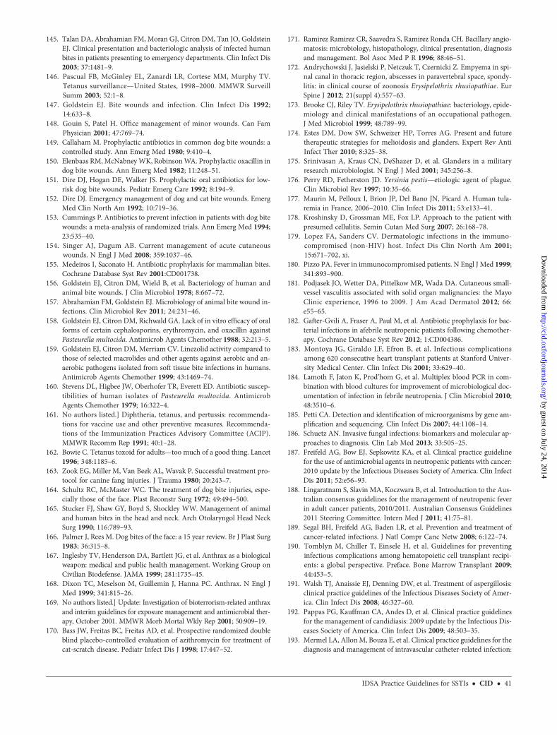
145. Talan DA, Abrahamian FM, Moran GJ, Citron DM, Tan JO, GoldsteinEJ. Clinical presentation and bacteriologic analysis of infected humanbites in patients presenting to emergency departments. Clin Infect Dis2003; 37:1481–9.
146. Pascual FB, McGinley EL, Zanardi LR, Cortese MM, Murphy TV.Tetanus surveillance—United States, 1998–2000. MMWR SurveillSumm 2003; 52:1–8.
147. Goldstein EJ. Bite wounds and infection. Clin Infect Dis 1992;14:633–8.
148. Gouin S, Patel H. Office management of minor wounds. Can FamPhysician 2001; 47:769–74.
149. Callaham M. Prophylactic antibiotics in common dog bite wounds: acontrolled study. Ann Emerg Med 1980; 9:410–4.
150. Elenbaas RM,McNabneyWK, RobinsonWA. Prophylactic oxacillin indog bite wounds. Ann Emerg Med 1982; 11:248–51.
151. Dire DJ, Hogan DE, Walker JS. Prophylactic oral antibiotics for low-risk dog bite wounds. Pediatr Emerg Care 1992; 8:194–9.
152. Dire DJ. Emergency management of dog and cat bite wounds. EmergMed Clin North Am 1992; 10:719–36.
153. Cummings P. Antibiotics to prevent infection in patients with dog bitewounds: a meta-analysis of randomized trials. Ann Emerg Med 1994;23:535–40.
154. Singer AJ, Dagum AB. Current management of acute cutaneouswounds. N Engl J Med 2008; 359:1037–46.
155. Medeiros I, Saconato H. Antibiotic prophylaxis for mammalian bites.Cochrane Database Syst Rev 2001:CD001738.
156. Goldstein EJ, Citron DM, Wield B, et al. Bacteriology of human andanimal bite wounds. J Clin Microbiol 1978; 8:667–72.
157. Abrahamian FM, Goldstein EJ. Microbiology of animal bite wound in-fections. Clin Microbiol Rev 2011; 24:231–46.
158. Goldstein EJ, Citron DM, Richwald GA. Lack of in vitro efficacy of oralforms of certain cephalosporins, erythromycin, and oxacillin againstPasteurella multocida. Antimicrob Agents Chemother 1988; 32:213–5.
159. Goldstein EJ, Citron DM, Merriam CV. Linezolid activity compared tothose of selected macrolides and other agents against aerobic and an-aerobic pathogens isolated from soft tissue bite infections in humans.Antimicrob Agents Chemother 1999; 43:1469–74.
160. Stevens DL, Higbee JW, Oberhofer TR, Everett ED. Antibiotic suscep-tibilities of human isolates of Pasteurella multocida. AntimicrobAgents Chemother 1979; 16:322–4.
161. No authors listed.] Diphtheria, tetanus, and pertussis: recommenda-tions for vaccine use and other preventive measures. Recommenda-tions of the Immunization Practices Advisory Committee (ACIP).MMWR Recomm Rep 1991; 40:1–28.
162. Bowie C. Tetanus toxoid for adults—too much of a good thing. Lancet1996; 348:1185–6.
163. Zook EG, Miller M, Van Beek AL, Wavak P. Successful treatment pro-tocol for canine fang injuries. J Trauma 1980; 20:243–7.
164. Schultz RC, McMaster WC. The treatment of dog bite injuries, espe-cially those of the face. Plast Reconstr Surg 1972; 49:494–500.
165. Stucker FJ, Shaw GY, Boyd S, Shockley WW. Management of animaland human bites in the head and neck. Arch Otolaryngol Head NeckSurg 1990; 116:789–93.
166. Palmer J, Rees M. Dog bites of the face: a 15 year review. Br J Plast Surg1983; 36:315–8.
167. Inglesby TV, Henderson DA, Bartlett JG, et al. Anthrax as a biologicalweapon: medical and public health management. Working Group onCivilian Biodefense. JAMA 1999; 281:1735–45.
168. Dixon TC, Meselson M, Guillemin J, Hanna PC. Anthrax. N Engl JMed 1999; 341:815–26.
169. No authors listed.] Update: Investigation of bioterrorism-related anthraxand interim guidelines for exposure management and antimicrobial ther-apy, October 2001. MMWR Morb Mortal Wkly Rep 2001; 50:909–19.
170. Bass JW, Freitas BC, Freitas AD, et al. Prospective randomized doubleblind placebo-controlled evaluation of azithromycin for treatment ofcat-scratch disease. Pediatr Infect Dis J 1998; 17:447–52.
171. Ramirez Ramirez CR, Saavedra S, Ramirez Ronda CH. Bacillary angio-matosis: microbiology, histopathology, clinical presentation, diagnosisand management. Bol Asoc Med P R 1996; 88:46–51.
172. Andrychowski J, Jasielski P, Netczuk T, Czernicki Z. Empyema in spi-nal canal in thoracic region, abscesses in paravertebral space, spondy-litis: in clinical course of zoonosis Erysipelothrix rhusiopathiae. EurSpine J 2012; 21(suppl 4):557–63.
173. Brooke CJ, Riley TV. Erysipelothrix rhusiopathiae: bacteriology, epide-miology and clinical manifestations of an occupational pathogen.J Med Microbiol 1999; 48:789–99.
174. Estes DM, Dow SW, Schweizer HP, Torres AG. Present and futuretherapeutic strategies for melioidosis and glanders. Expert Rev AntiInfect Ther 2010; 8:325–38.
175. Srinivasan A, Kraus CN, DeShazer D, et al. Glanders in a militaryresearch microbiologist. N Engl J Med 2001; 345:256–8.
176. Perry RD, Fetherston JD. Yersinia pestis—etiologic agent of plague.Clin Microbiol Rev 1997; 10:35–66.
177. Maurin M, Pelloux I, Brion JP, Del Bano JN, Picard A. Human tula-remia in France, 2006–2010. Clin Infect Dis 2011; 53:e133–41.
178. Kroshinsky D, Grossman ME, Fox LP. Approach to the patient withpresumed cellulitis. Semin Cutan Med Surg 2007; 26:168–78.
179. Lopez FA, Sanders CV. Dermatologic infections in the immuno-compromised (non-HIV) host. Infect Dis Clin North Am 2001;15:671–702, xi.
180. Pizzo PA. Fever in immunocompromised patients. N Engl J Med 1999;341:893–900.
181. Podjasek JO, Wetter DA, Pittelkow MR, Wada DA. Cutaneous small-vessel vasculitis associated with solid organ malignancies: the MayoClinic experience, 1996 to 2009. J Am Acad Dermatol 2012; 66:e55–65.
182. Gafter-Gvili A, Fraser A, Paul M, et al. Antibiotic prophylaxis for bac-terial infections in afebrile neutropenic patients following chemother-apy. Cochrane Database Syst Rev 2012; 1:CD004386.
183. Montoya JG, Giraldo LF, Efron B, et al. Infectious complicationsamong 620 consecutive heart transplant patients at Stanford Univer-sity Medical Center. Clin Infect Dis 2001; 33:629–40.
184. Lamoth F, Jaton K, Prod’hom G, et al. Multiplex blood PCR in com-bination with blood cultures for improvement of microbiological doc-umentation of infection in febrile neutropenia. J Clin Microbiol 2010;48:3510–6.
185. Petti CA. Detection and identification of microorganisms by gene am-plification and sequencing. Clin Infect Dis 2007; 44:1108–14.
186. Schuetz AN. Invasive fungal infections: biomarkers and molecular ap-proaches to diagnosis. Clin Lab Med 2013; 33:505–25.
187. Freifeld AG, Bow EJ, Sepkowitz KA, et al. Clinical practice guidelinefor the use of antimicrobial agents in neutropenic patients with cancer:2010 update by the Infectious Diseases Society of America. Clin InfectDis 2011; 52:e56–93.
188. Lingaratnam S, Slavin MA, Koczwara B, et al. Introduction to the Aus-tralian consensus guidelines for the management of neutropenic feverin adult cancer patients, 2010/2011. Australian Consensus Guidelines2011 Steering Committee. Intern Med J 2011; 41:75–81.
189. Segal BH, Freifeld AG, Baden LR, et al. Prevention and treatment ofcancer-related infections. J Natl Compr Canc Netw 2008; 6:122–74.
190. Tomblyn M, Chiller T, Einsele H, et al. Guidelines for preventinginfectious complications among hematopoietic cell transplant recipi-ents: a global perspective. Preface. Bone Marrow Transplant 2009;44:453–5.
191. Walsh TJ, Anaissie EJ, Denning DW, et al. Treatment of aspergillosis:clinical practice guidelines of the Infectious Diseases Society of Amer-ica. Clin Infect Dis 2008; 46:327–60.
192. Pappas PG, Kauffman CA, Andes D, et al. Clinical practice guidelinesfor the management of candidiasis: 2009 update by the Infectious Dis-eases Society of America. Clin Infect Dis 2009; 48:503–35.
193. Mermel LA, AllonM, Bouza E, et al. Clinical practice guidelines for thediagnosis and management of intravascular catheter-related infection:
IDSA Practice Guidelines for SSTIs • CID • 41
by guest on July 24, 2014http://cid.oxfordjournals.org/
Dow
nloaded from

2009 update by the Infectious Diseases Society of America. Clin InfectDis 2009; 49:1–45.
194. Legrand M, Max A, Peigne V, et al. Survival in neutropenic patientswith severe sepsis or septic shock. Crit Care Med 2012; 40:43–9.
195. Paesmans M, Klastersky J, Maertens J, et al. Predicting febrile neutro-penic patients at low risk using the MASCC score: does bacteremiamatter? Support Care Cancer 2011; 19:1001–8.
196. Klastersky J, Paesmans M, Georgala A, et al. Outpatient oral antibioticsfor febrile neutropenic cancer patients using a score predictive forcomplications. J Clin Oncol 2006; 24:4129–34.
197. Lanoix JP, Schmit JL, Douadi Y. Bacterial lung sepsis in patients withfebrile neutropenia. Curr Opin Pulm Med 2012; 18:175–80.
198. Mebis J, Jansens H, Minalu G, et al. Long-term epidemiology of bac-terial susceptibility profiles in adults suffering from febrile neutropeniawith hematologic malignancy after antibiotic change. Infect Drug Re-sist 2010; 3:53–61.
199. Nordmann P, Cuzon G, Naas T. The real threat of Klebsiella pneumo-niae carbapenemase-producing bacteria. Lancet Infect Dis 2009;9:228–36.
200. Johnston DL, Waldhausen JH, Park JR. Deep soft tissue infections inthe neutropenic pediatric oncology patient. J Pediatr Hematol Oncol2001; 23:443–7.
201. Reich HL, Williams Fadeyi D, Naik NS, Honig PJ, Yan AC. Nonpseu-domonal ecthyma gangrenosum. J Am Acad Dermatol 2004; 50:S114–7.
202. Wade JC. Viral infections in patients with hematological malignancies.Hematology Am Soc Hematol Educ Program 2006; 2006:368–74.
203. Pizzo PA. Management of fever in patients with cancer and treatment-induced neutropenia. N Engl J Med 1993; 328:1323–32.
204. Kosmidis CI, Chandrasekar PH. Management of gram-positivebacterial infections in patients with cancer. Leuk Lymphoma 2012;53:8–18.
205. Tattevin P, Camus C. What can we learn from studies comparing line-zolid with vancomycin in neutropenic patients when vancomycin dosag-es are not optimized? Clin Infect Dis 2006; 42:1813–4; author reply 14–5.
206. Jaksic B, Martinelli G, Perez-Oteyza J, Hartman CS, Leonard LB, TackKJ. Efficacy and safety of linezolid compared with vancomycin in arandomized, double-blind study of febrile neutropenic patients withcancer. Clin Infect Dis 2006; 42:597–607.
207. Freifeld A, Marchigiani D, Walsh T, et al. A double-blind comparisonof empirical oral and intravenous antibiotic therapy for low-risk febrilepatients with neutropenia during cancer chemotherapy. N Engl J Med1999; 341:305–11.
208. Freifeld AG, Sepkowitz KA. No place like home? Outpatient manage-ment of patients with febrile neutropenia and low risk. J Clin Oncol2011; 29:3952–4.
209. Spellberg B, Ibrahim A, Roilides E, et al. Combination therapy for mu-cormycosis: why, what, and how? Clin Infect Dis 2012; 54(suppl 1):S73–8.
210. Wingard JR, Santos GW, Saral R. Differences between first andsubsequent fevers during prolonged neutropenia. Cancer 1987;59:844–9.
211. Wade JC. Management of infection in patients with acute leukemia.Hematol Oncol Clin North Am 1993; 7:293–315.
212. Leeflang MM, Debets-Ossenkopp YJ, Visser CE, et al. Galactomannandetection for invasive aspergillosis in immunocompromized patients.Cochrane Database Syst Rev 2008; 4:CD007394.
213. Maertens J, Theunissen K, Lodewyck T, Lagrou K, Van Eldere J.Advances in the serological diagnosis of invasive Aspergillusinfections in patients with haematological disorders. Mycoses 2007;50(suppl 1):2–17.
214. Segal BH, Almyroudis NG, Battiwalla M, et al. Prevention and earlytreatment of invasive fungal infection in patients with cancer and neu-tropenia and in stem cell transplant recipients in the era of newerbroad-spectrum antifungal agents and diagnostic adjuncts. Clin InfectDis 2007; 44:402–9.
215. Mays SR, Bogle MA, Bodey GP. Cutaneous fungal infections in the on-cology patient: recognition andmanagement. Am J Clin Dermatol 2006;7:31–43.
216. Goodrich JM, Reed EC, Mori M, et al. Clinical features and analysis ofrisk factors for invasive candidal infection after marrow transplanta-tion. J Infect Dis 1991; 164:731–40.
217. Bodey GP, Luna M. Skin lesions associated with disseminated candi-diasis. JAMA 1974; 229:1466–8.
218. Wingard JR, Merz WG, Saral R. Candida tropicalis: a major patho-gen in immunocompromised patients. Ann Intern Med 1979;91:539–43.
219. Jarowski CI, Fialk MA, Murray HW, et al. Fever, rash, and muscle ten-derness. A distinctive clinical presentation of disseminated candidiasis.Arch Intern Med 1978; 138:544–6.
220. Walsh TJ, Newman KR, Moody M, Wharton RC, Wade JC. Tricho-sporonosis in patients with neoplastic disease. Medicine 1986;65:268–79.
221. Allo MD, Miller J, Townsend T, Tan C. Primary cutaneous aspergillo-sis associated with Hickman intravenous catheters. N Engl J Med 1987;317:1105–8.
222. Petrikkos G, Skiada A, Lortholary O, Roilides E, Walsh TJ, Kontoyian-nis DP. Epidemiology and clinical manifestations of mucormycosis.Clin Infect Dis 2012; 54(suppl 1):S23–34.
223. Kontoyiannis DP, Marr KA, Park BJ, et al. Prospective surveillance forinvasive fungal infections in hematopoietic stem cell transplant recip-ients, 2001–2006: overview of the Transplant-Associated InfectionSurveillance Network (TRANSNET) Database. Clin Infect Dis 2010;50:1091–100.
224. Kontoyiannis DP, Sumoza D, Tarrand J, Bodey GP, Storey R, Raad II.Significance of aspergillemia in patients with cancer: a 10-year study.Clin Infect Dis 2000; 31:188–9.
225. Gartenberg G, Bottone EJ, Keusch GT, Weitzman I. Hospital-acquiredmucormycosis (Rhizopus rhizopodiformis) of skin and subcutaneoustissue: epidemiology, mycology and treatment. N Engl J Med 1978;299:1115–8.
226. Anaissie E. Opportunistic mycoses in the immunocompromised host:experience at a cancer center and review. Clin Infect Dis 1992; 14(suppl 1):S43–53.
227. Nucci M, Anaissie E. Fusarium infections in immunocompromisedpatients. Clin Microbiol Rev 2007; 20:695–704.
228. Doucette K, Fishman JA. Nontuberculous mycobacterial infection inhematopoietic stem cell and solid organ transplant recipients. Clin In-fect Dis 2004; 38:1428–39.
229. Griffith DE, Aksamit T, Brown-Elliott BA, et al. An official ATS/IDSAstatement: diagnosis, treatment, and prevention of nontuberculousmycobacterial diseases. Am J Respir Crit Care Med 2007; 175:367–416.
230. Ambrosioni J, Lew D, Garbino J. Nocardiosis: updated clinical reviewand experience at a tertiary center. Infection 2010; 38:89–97.
231. Thomas LM, Rand HK, Miller JL, Boyd AS. Primary cutaneous asper-gillosis in a patient with a solid organ transplant: case report and re-view of the literature. Cutis 2008; 81:127–30.
232. Shinohara MM, George E. Scedosporium apiospermum: an emergingopportunistic pathogen thatmust bedistinguished fromAspergillus andother hyalohyphomycetes. J Cutan Pathol 2009; 36(suppl 1):39–41.
233. Miyamoto H, Hayashi H, Nakajima H. Cutaneous mucormycosisin a patient with acute lymphocytic leukemia. J Dermatol 2005;32:273–7.
234. Halpern M, Balbi E, Carius L, et al. Cellulitis and nodular skin lesionsdue to Fusarium spp in liver transplant: case report. Transplant Proc2010; 42:599–600.
235. Abuav R, McGirt LY, Kazin RA. Cryptococcal panniculitis in an im-munocompromised patient: a case report and review of the literature.Cutis 2010; 85:303–6.
236. Perfect JR, Dismukes WE, Dromer F, et al. Clinical practice guidelinesfor the management of cryptococcal disease: 2010 update by the
42 • CID • Stevens et al
by guest on July 24, 2014http://cid.oxfordjournals.org/
Dow
nloaded from

Infectious Diseases Society of America. Clin Infect Dis 2010; 50:291–322.
237. Wheat LJ, Freifeld AG, Kleiman MB, et al. Clinical practice guidelinesfor the management of patients with histoplasmosis: 2007 update bythe Infectious Diseases Society of America. Clin Infect Dis 2007;45:807–25.
238. Freifeld AG, Wheat LJ, Kaul DR. Histoplasmosis in solid organ trans-plant recipients: early diagnosis and treatment. Curr Opin OrganTransplant 2009; 14:601–5.
239. Shiley K, Blumberg E. Herpes viruses in transplant recipients: HSV,VZV, human herpes viruses, and EBV. Infect Dis Clin North Am2010; 24:373–93.
240. Erard V, Guthrie KA, Varley C, et al. One-year acyclovir prophylaxisfor preventing varicella-zoster virus disease after hematopoietic celltransplantation: no evidence of rebound varicella-zoster virus diseaseafter drug discontinuation. Blood 2007; 110:3071–7.
241. Erard V, Wald A, Corey L, Leisenring WM, Boeckh M. Use of long-term suppressive acyclovir after hematopoietic stem-cell transplanta-tion: impact on herpes simplex virus (HSV) disease and drug-resistantHSV disease. J Infect Dis 2007; 196:266–70.
242. Basile A, Simzar S, Bentow J, et al. Disseminated Strongyloides stercor-alis: hyperinfection during medical immunosuppression. J Am AcadDermatol 2010; 63:896–902.
243. Walia R, Montoya JG, Visvesvera GS, Booton GC, Doyle RL. A case ofsuccessful treatment of cutaneous Acanthamoeba infection in a lungtransplant recipient. Transpl Infect Dis 2007; 9:51–4.
244. Godoy HL, Guerra CM, Viegas RF, et al. Infections in heart transplantrecipients in Brazil: the challenge of Chagas’ disease. J Heart LungTransplant 2010; 29:286–90.
245. Sandhu K, Gupta S, Kumar B, Dhandha R, Udigiri NK, Minz M. Thepattern of mucocutaneous infections and infestations in renal trans-plant recipients. J Dermatol 2003; 30:590–5.
246. Pickering LK. Committee on Infectious Diseases, American Academyof Pediatrics. Antimicrobial agents and related therapy. 26th ed. ElkGrove Village, IL: American Academy of Pediatrics, 2003.
247. Pascual A, Csajka C, Buclin T, et al. Challenging recommendedoral and intravenous voriconazole doses for improved efficacyand safety: population pharmacokinetics-based analysis of adultpatients with invasive fungal infections. Clin Infect Dis 2012;55:381–90.
IDSA Practice Guidelines for SSTIs • CID • 43
by guest on July 24, 2014http://cid.oxfordjournals.org/
Dow
nloaded from