clinical aides for increasing retention for removable
TRANSCRIPT

Republic of IraqMinistry of Higher Education
And scientific ResearchUniversity of Baghdad
College of Dentistry
Clinical Aides for increasing Retention forRemovable Implant supported prosthesis
A project
Submitted to Collage of Dentistry, University of Baghdad. Department of
Prosthodontics in fulfillment for the requirement to award the degree
B.D.S
Done byNarjis Hashim Zayed
5th Grade
SupervisorLec. Dr. Mustafa Mahdi Jassim
B.D.S, M.Sc.
Baghdad Iraq
2018 A.D 1439 A.H

Supervisor DeclarationThis is to certify that the organization and preparation of this
project have been made by the under graduated student " Narjis Hashim
Zayed" under my supervision at the College of Dentistry, University of
Baghdad in a partial fulfillment of requirements of the degree of B.D.S in
Prosthodontic Dentistry.
Signature
SupervisorLec. Dr. Mustafa Mahdi Jassim
B.D.S, M.Sc.

Dedicationo This work is dedicated to my family, My father and
Mother and My friends for their great support and for
always believing in me.
o To my supervisor for his guidance and support thank you
from all my heart.
Narjis

I
AcknowledgementThanks and praise to "ALLAH" the almighty for inspiring and giving me
the strength, willingness and patience to complete this work.
My deepest gratitude and sincere appreciation to Prof. Dr. Hussain F. Al-
Huwaizi, dean of the college of Dentistry, University of Baghdad, for this
kind care and continuous support for the post graduated students.
I would like to thank Prof. Dr. Nidhal H. Ghaib, Assistant Dean for
Scientific Affairs, for her advice and support.
My sincere appreciation and thank fullness to Prof. Raghdad Kareem
Jassim, chairman of the prosthodontics dentistry Department, University
of Baghdad, for her Keen interest, help, scientific support and
encouragement.
A million words would be too short to express my genuine appreciation to
my Supervisor Dr. Mustafa Mahdi Jassim who supported me
throughout this project and introduced me to the wonders of science.
Thank you for your invaluable guidance and for your tremendous effect
to help me finish this project. Thank you for your time, advice, care and
motivation during the entire research period. I am very greateful for your
supervision and I owe you the greatest degree of respect and appreciation.
Being your student was a prilego – I have learnt so much from you.
Thank you for being your student was a privilege –I have learnt so much
from you. Thank you for being the perfect mentor. I can never really
thank you enough.

II
AbstractMany patients have problems adapting to their complete dentures,
especially to the mandibular prosthesis. The totally edentulous patient has
several options for implant treatment today, including both fixed and
removable solutions. While fixed prosthesis may appear more attractive
by restoring the patient closer to a truly ‘dentate’ status many patients are
also favourable to receive a removable appliance. This removable choice
has become increasingly popular during the recent global financial
downturn. Many patients, especially those who are uncomfortable with
dentures, have a provision for increased retention and support with
implant supported overdentures. The relatively few reports on patient-
based assessment of the outcome and on the functional effect of such
therapy have shown better quality of life, greater patient satisfaction,
better chewing and speaking performance, higher jaw-closing force, and
less bone loss after implant-supported prosthetic reconstructions than
with a conventional complete denture. This article presents a case report
in which teeth were replaced with an implant supported overdenture.

III
List of ContentsSubject Page No.Introduction 1Review of Literature 3
42.Greater Prosthesis Stability 6
74.Improved Maintenance 8
88
7.Implant Supported vs. Implant Retained 88.Types of Implant-Supported Overdentures 99.Narrow-Body Implant Overdentures 13
Supported and retainedOver Denture
13
1512. O-rings attachment 16
1617
15.Locator (self-aligning) attachment 15162123
Patients and MethodsCase 1Case 2 30Discussion 34Conclusion 35References 36

IV
List of TableTable Subject Page No.
1 Guidance for Overdentures 10
List of FiguresFigure Subject Page No.1 Narrow-diameter implant overdenture 132 Attachments evaluated from left to right: ERA, Saturno
O-Ring, Locator, Ball.15
3 attachment – resilient 174 Ball abutments on two implants in the mandible. These
single components leave the implants unsplinted. Whenplaced, the overdenture will then stabilize the implants(secondary splinting). They form the patrix, i.e. the partof the attachment system connected to the implant.
18
5 Intaglio view of the mandibular overdenture. The tworubber O-rings for the ball abutments form the matrix,being the receptacle component. The matrix designs areincorporated within the undersurface of the overdentureand they are adjustable and/or replaceable
19
6 Locator attachment 207 Magnet attachments 218 Mandibular implant bar. A bar construction splints the
implants directly together, also without the overdentureplaced, providing higher stability to implants andprosthesis
22
9 Definitive overdenture with Hader bar clips 2210 telescopic attachment 24

Introduction
1
Introduction
Despite the decline in the prevalence of edentulism (complete
loss of teeth), there is a high number of patients who are completely
edentulous1 and wearing conventional complete dentures. (Warreth
A., 2015), Before the era of dental implants, complete edentulous jaws
were restored with conventional complete dentures, as this was the
only option available. (Carlsson, 2010), Use of conventional complete
dentures is associated with several problems, such as lack of denture
stability, support and retention. (Sohrabi,2012), These problems lead to
discomfort, reduction in chewing ability and, at times, may be socially
embarrassing. Consequently, the patients’ psychological and social
well-being are negatively affected. In certain clinical situations,
denture fixatives may offer a solution, but this approach is not always
practical or cost-effective. (Hillerup.,1982) Therefore, several surgical
techniques have been used to improve the conventional complete
denture outcome.(Hochstedler,1998., Schwartz-Arad.et al., 2003)
However، surgical approaches are not without risks and may lead to
several complications and improper treatment results. Thus, dental
implants may provide a solution to these problems. The routine use of
an implant with a complete denture is associated with improvements
in retention, stability, function, perception and comfort.
(Doundoulakis,2003) Hence, an edentulous mandible may be restored
with a fixed or removable implant-supported complete denture. The
fixed prosthesis has the advantage of being secured to the implants

Introduction
2
and is not removed by the patient. However, it is expensive, as more
implants are required to support it, and it is technically more
demanding than the removable prosthesis. On the other hand،
removable implant-supported/retained overdentures (RISOs) result in
an aesthetically more pleasing outcome, better access for oral
hygiene, lower cost and use of fewer implants compared to fixed
implant-supported/retained overdentures (FISOs). Phonetics may also
be improved and lip support can be enhanced with the use of RISOs
as the lost tissue can be compensated for by the denture. RISOs
provide an alternative option when financial constraints preclude the
use of an FISO. It is also a solution in certain complex restorative
situations where the missing teeth are associated with soft tissue and
bone loss, and both require replacement. Therefore, RISOs may
provide a solution to the problems linked to the use of conventional
complete dentures or FISOs. The use of the mandibular conventional
complete denture is more problematic than that of the maxillary
conventional denture due to several factors such as thin mucosal
coverage of the edentulous ridge, a reduced support area and the
mobility of the floor of the mouth, the movement of the mandible and
the tongue. These factors make the use of dental implants and
attachments a common practice to overcome their adverse
consequences.
Chapter One Review of Literature
3
Review of LiteratureImplant supported prosthesis
Implant-supported overdentures offer many practical advantages over
conventional complete dentures and removable partial dentures. These include
decreased bone resorption; reduced or eliminated prosthesis movement; better
esthetics; improved tooth position; better occlusion, including improved
occlusal load direction, increased occlusal function and maintenance of the
occlusal vertical dimension. In addition, implant supported overdentures
improve phonetics, the patient’s psychological outlook and quality of
life.(Warreth A., 2015).
Conventional dentures rely upon the residual alveolar ridge and mucosa
for support and retention. Many patients have problems adapting to their
complete dentures, especially to the mandibular prosthesis. The widespread use
of denture adhesives is one indication that these prostheses generally provide
inadequate comfort and function.(Carlsson, 2010)
Studies show implant-supported overdentures have superior retention to
conventional dentures. Regardless of the type of attachment system used bar,
ball or magnet, patients are significantly more satisfied with implant-supported
overdentures than with conventional dentures. Patients find implant-supported
overdentures significantly more stable and rate their ability to chew a wider
variety of foods as significantly easier, thus improving their nutritional state.
Furthermore, they find implant-supported overdentures more comfortable and
speech easier.(Sohrabi,2012)
The implant-supported overdenture may reduce the amount of soft tissue
coverage and extension of the prosthesis which is especially important for new
denture wearers restoration of the edentulous mandible with a conventional
denture is no longer the most appropriate choice of prosthetic treatment. The
implant-supported overdenture has become the standard of care.(Hillerup,1982)

Chapter One Review of Literature
4
Numerous studies show cumulative success rates for all implant
supported overdentures, with implant-supported overdentures placed in the
mandible enjoying a slightly higher success rate than implant-supported
overdentures placed in the maxilla. The major indications for a mandibular
implant-supported over denture are lack of retention or stability, poor function
and speech, tissue sensitivity and soft tissue abrasions.(Hochstedler,1998)
1.Less Bone Resorption
One advantage of implant-supported full bridges and dentures is that they
function like tooth roots, which preserves jaw bone. Dental implants integrate
with the jawbone and dramatically reduce the rate of bone loss attributed to
conventional dentures. Edentulism is characterized by atrophy of the jaw bone.
Studies show rapid resorption an average of 4mm occurs during the first year
after tooth loss and thereafter decreases to 0.5mm per year. Over a five year
period, 5.2mm of vertical bone height will be lost under complete dentures.
Bone loss under complete dentures continues with the mandible experiencing a
fourfold greater vertical bone loss than the maxilla. In contrast, (Schwartz-
Arad.et al., 2003) found that 70 percent of their patients with implant-
supported overdentures lost less than .2mm bone in the first year. (Misch.,2008)
found that only .6mm of bone will typically be lost over a five year period and
long-term resorption may remain as low as .1mm per year in patients with
overdentures supported by multiple implants. (Doundoulakis,2003)
Crestal bone loss (CBL) around implants supporting overdentures appears
to be affected by factors such as location (maxilla or mandible), attachment
system, and number of implants supporting the overdenture. Location in the
maxilla or the mandible appears to most influence CBL. Studies show implants
in the mandible exhibit less CBL than implants in the maxilla, which could be
attributed to the difference in bone quality in the maxilla and the mandible and
to different loading circumstances. The maxilla normally has less density and
quantity of bone than the mandible. The resorbed mandible usually has dense

Chapter One Review of Literature
5
compact bone with an oak-like quality or a combination of thick porous
compact bone on the outside and course trabecular bone on the inside. Because
the percentage of bone at the implant interface is 70 to 80 percent, mandibular
implants are the most successful.(Sarandha, 2007)
Regardless of the type of attachment system used bar, ball or magnet
patients are significantly more satisfied with implant-supported overdentures
than with conventional dentures.( Bambara GE.,2015).
Advantages of implant-supported overdentures:
1. Patients find implant-supported overdentures significantly more stable
2. rate their ability to chew a wider variety of foods as significantly easier, thus
improving their nutritional state.
3. They find implant-supported overdentures more comfortable and speech
easier.
4. The implant-supported overdenture may reduce the amount of soft tissue
coverage and extension of the prosthesis which is especially important for
new denture wearers or those who have low gagging thresholds. Timing of
implant loading appears to be a factor in the success of implant-supported
overdentures. Immediate loading techniques, a newer approach to implant-
supported restorations, Recently, more two-implant mandibular
overdentures are being placed as an affordable alternative to prostheses
requiring several implants. The choice of implant site for these overdentures
should be governed by the quantity, quality and volume of available bone,
along with the size and curvature of the anterior arch.

Chapter One Review of Literature
6
2.Greater Prosthesis Stability
The greater stability of an implant-supported overdenture derives from
the mechanical attachment of the implant support system retaining the
restoration.(Ahmed YA.,2016)
A mandibular denture may move Slightly during function. Under these
conditions, predetermined occlusal contacts and the control of masticatory
forces are nearly impossible. Implants stabilize the prosthesis and the patient is
able to consistently reproduce a determined centric occlusion. Lateral forces
may cause a horizontal movement of a conventional prosthesis and cause soft
tissue abrasions and accelerated bone loss.( Alsiyabi AS.,2005)
An implant-supported overdenture limits lateral movements and
consequently minimizes soft tissue trauma. In addition, complete dentures often
move vertically during mandibular movement and speech. The contraction of
the mentalis, buccinator, or mylohyoid muscles may lift the denture off the soft
tissue. As a consequence, the teeth may touch during speech and cause clicking
noises.(Gulizio MP,2005)
The implant-supported overdenture remains in place during mandibular
movement which allows the tongue and perioral musculature to resume a more
normal function since they are not required to control mandibular denture
movement. While prosthesis retention has been found to be good for magnets,
balls and clips, bars were the most retentive. Studies have shown balls
experienced more complications.(Trakas T,2006)
Overdenture bars may be cemented or screw-retained. Cemented bars
present the advantages of more passive fit, reduced cost and an easier bar
impression technique. (Jemt et al.,1990) showed a decrease in occlusal force
when the bar connecting implants was removed and attributed it to the loss of
support, stability and retention.

Chapter One Review of Literature
7
The prosthesis support and range of motion should be part of the initial
diagnosis. Proven, simple, predictable and cost-effective devices limited to a
minimum of hardware may present the best options. The more sophisticated the
attachment, the more complex the fabrication and maintenance procedures.(
Alsiyabi AS,2005).
3. Better Esthetics, Tooth Position and OcclusionIn severe resorption cases, implant-supported overdentures may be more
esthetic than a fixed restoration. Bone loss dictates the appearance of the
inferior third of the face. An implant-supported overdenture provides improved
support for the lips and soft tissues of the face allowing the teeth to be the same
length as natural teeth. When there is marked loss of alveolar height, the teeth
on a conventional fixed restoration will be very long. The presence of a large
labial flange in a conventional denture may result in exaggerated facial contours
for the patient with recent extractions. (Gulizio MP,2005)
Implant-supported prostheses do not require as great a labial extension or
as much extended soft tissue coverage as is necessary for a conventional
denture. An implant-supported overdenture can provide the soft tissue support
to the facial features often required for a patient with advanced bone loss.
(Misch.,2008) found the maximum occlusal force of a patient with
dentures may improve 300 percent with an implant-supported prosthesis. This
improves the chewing efficiency of patients with an implant-supported
overdenture by 20 percent over the bite strength of patients with a conventional
denture. (Misch.,2008) also cites a study of chewing efficiency comparing
complete denture wearers with implant-supported overdentures. Patients with
conventional dentures needed 1.5 to 3.6 times the number of chewing strokes as
patients with implant-supported overdentures. .( Hochstedler,1998)

Chapter One Review of Literature
8
4.Improved MaintenanceHygiene conditions and home maintenance procedures are improved with
an overdenture compared with a fixed prosthesis. The overdenture may be
extended over the abutments to prevent food entrapment during function.
Professional maintenance is also improved as peri-implant probing is diagnostic
and easier around a bar. (Yamada RH., 2011)
5.Indications of Implant Denture
1. Edentulous patient with history of difficulty in wearing removable dentures.
2. When there is severe change in complete denture bearing tissues.
3. Poor oral muscular coordination.
4. Para-functional habits that compromise prosthesis stability.
5. Unrealistic patient expectations for complete dentures.
6. Hyperactive gag reflex.
7. Low tissue tolerance of supporting mucosa.
6.Contraindications of Implant Denture1. High dose irradiated patients.
2. Patient with psychiatric problems such as psychosis, dysthorphobia.
3. Hematological systemic disorders.
4. Pathology of hard and soft tissues.
5. Patient with drug, alcohol or tobacco chewing abuse.
7.Implant Supported vs. Implant RetainedOne of the first treatment planning decisions is whether the prosthesis
will be implant supported or implant retained. The main difference is that a
sufficient number of implants must be placed for the prosthesis to be totally
implant supported. (Jemt et al.,1990)
Usually, six implants positioned in the lower arch and eight implants
placed in the upper arch will suffice. In most cases, the maxillary arch needs at
least two more implants placed than the mandibular arch because the quality

Chapter One Review of Literature
9
and quantity of maxillary bone is not as good as mandibular bone. In contrast,
an implant-retained case will rely on support from the implants as well as the
soft tissue. Two to five implants placed in the mandible will necessitate an
implant-retained, soft tissue-supported prosthesis. Both implant and soft tissue
will be supporting areas and will share the occlusal load. The prosthesis will be
stable, retentive, rigid, or resilient, depending on the attachments
used.(Carlsson, G.E.,2010., Hillerup, S.,1982)
8.Types of Implant-Supported OverdenturesMany variations in treatment planning implant overdentures are available.
One approach is to use freestanding implant attachment abutments, and it is not
unusual for dentists to utilize anywhere from two to six in any given arch.
Their ability to allow the prosthesis to be rigid or resilient enables an
improved treatment planning flexibility with greater decision-making regarding
how much load the implants would endure.
Also, loadbearing areas of the palate and tuberosities can be utilized to
minimize the amount of denture base in other areas.(Hussain Z.,2007) Bars can
also be made resilient in the vertical direction by using a Dolder bar (Preat
Corporation) or an Ackermann clip on a Hader Bar® (Sterngold) with 0.6-mm
spacers, which can mitigate occlusal loads on implants. Intra- and extra-bar
attachments can also be utilized, which can be rigid or resilient.
The following guidelines can be used to treatment plan implant
overdentures. As the number of implants decrease, the length of a possible
cantilever also decreases and the amount of tissue support increases (Table 1).

Chapter One Review of Literature
10
Table 1: Guidance for Overdentures(Heckmann SM,2001).
Implant number and location Attachment optionsMandibular arch configuration
5-6 implants between the mentalforamen
Clip bar / implant – supported;cantilever optional
5 implants between the mentalforamen
Clip bar and tissue supported withdistal bar attachments
4 implants between the mentalforamen
Clip bar and tissue supported withORA and / or distal ERA, SFI-Bar'(Sterngold); cantilevers optional; 4separate attachments can be used
without a bar3 implants between the mental
foramenClip bar and tissue – supported
attachments can be used on the distalend of the bar; 3 separate attachments
can be used without a bar2 implants in anterior Clip bar and tissue supported; 2
separate attachments can be usedwithout a bar; SFI-Bar can also be
usedMaxillary arch configurations
8 implants between the molar areas Clip bar and implant supportedwithout a palate; cantilevers optional
6 implants between the premolar areas Clip bar and implant supportedwithout a palate. Cantilevers optional
4 implants between the first premolarareas
Clip bar and tissue supported. SFI-Bar.ORA and ERAs or ball attachments;
palate optional; no cantilevers

Chapter One Review of Literature
11
Type 1
This overdenture uses a combination of implant and soft tissue support
and is usually more effective in the mandible than in the maxilla. Two implants
are generally used in the cuspid positions. The prosthesis is implant retained.
The freestanding attachment abutments can be rigid and allow for both implant
and soft tissue support concurrently or allow for soft tissue support up to 0.6
mm, which protects the implants from being overloaded. The attachment
abutments can be ball attachments (eg, Preci Clix, Preat Corporation, ),
extracoronal resilient attachments, (eg, ERA®, Sterngold), o-ring attachments
(eg, ORA, Sterngold), or LOCATOR® attachments (Zest Anchors, ), depending
on how much soft tissue support is necessary.(Steffen RP,2004., Felton DA,
2005).
Type 2
This overdenture uses a straight round bar connecting two or more
implants or a curved bar supporting two or more implants with two distal
rotational attachments. The prosthesis is implant retained and supported by soft
tissue and implants or, depending on the selection of implant attachment
abutments, soft tissue supported. The distinguishing feature is that the
overdenture is supported by a round or oval bar that allows for rotation. Four
implants may be used in this scenario.(Kakar., 2001).

Chapter One Review of Literature
12
Type 3
This overdenture is similar to a Type 2 treatment planning option.
However, if four implants are placed in and around the cuspid area, the anterior
rotational clip and distal ball or other rotational attachments will work together
to generate less torquing forces around the implants. With four implants, the
midline clip functions as an indirect retainer, preventing the posterior base
portion of the overdenture from rotating away from the posterior edentulous
ridge areas during function. Another advantage of the midline clip is that it
provides for a tripod effect, or third reference point to determine a plane. By
balancing the mucosal-supported area with the bar/implant-supported area,
rotation of the overdenture can be minimized. The midline clip should provide
only a positive seat when the overdenture is engaged. It functions in a Type 3
case as an indirect retainer, which is basically a vertical stop for a removable
partial denture framework, so the free-end saddles do not move from the
tissue.(Walton JN, 2002).

Chapter One Review of Literature
13
Type 4
Type 1, Type 2, and Type 3 overdentures receive a portion of their
support from the soft tissue areas. The distinguishing feature of Type 4
overdenture is that the overdenture is completely supported by bars and
implants. The overdenture base may actually contact the mucosal tissue, but any
support is inadvertent. Because this overdenture is completely implant
supported, it requires the same number of implants for support as a fixed
prosthesis. Type 1, Type 2, and Type 3 options are usually treatment planned
for the lower arch, while Type 4 is frequently planned for the upper arch. A
minimum of five implants is planned for the mandibular arch, whereas six are
planned for the maxilla. This all depends on many factors, and many
practitioners will opt for six implants in the mandible and eight in the maxilla,
just to ensure that the implants are not overloaded.(Yamada RH,2000).
9. Narrow-Body Implant OverdenturesMany narrow-body implant systems on the market are used for implant
overdentures. Four implants are usually placed on the mandibular arch between
the mental foramen, and an overdenture is fabricated over them (Figure 2). Most
allow the overdenture to rotate only (eg, MDI Mini Dental Implants, 3M
ESPE).( Rodrigues RC,2009).
Chapter One Review of Literature
14
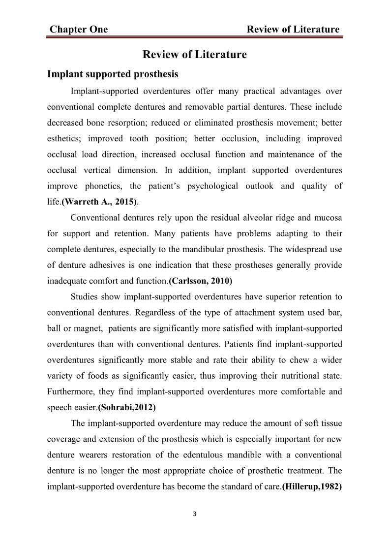
Some allow the prosthesis to rotate as well as allow for vertical
movement and soft tissue compression (eg, Shatkin FIRST, www.
shatkinfirst.com). The Atlas® narrow body implant system
(Dentatus, www.dentatus.com) provides for a total soft tissue-supported
appliance by using a tough silicone material called Tuf-Link™, which functions
as both a reline and a retentive element. (Bambara GE. 2015).
Figure 1. Narrow-diameter implant overdenture. Bambara GE. 2015.
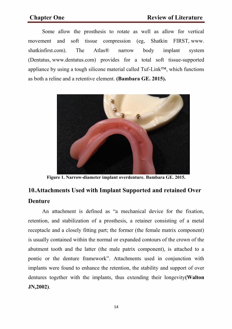
10.Attachments Used with Implant Supported and retained Over
DentureAn attachment is defined as “a mechanical device for the fixation,
retention, and stabilization of a prosthesis, a retainer consisting of a metal
receptacle and a closely fitting part; the former (the female matrix component)
is usually contained within the normal or expanded contours of the crown of the
abutment tooth and the latter (the male patrix component), is attached to a
pontic or the denture framework”. Attachments used in conjunction with
implants were found to enhance the retention, the stability and support of over
dentures together with the implants, thus extending their longevity(Walton
JN,2002).

Chapter One Review of Literature
15
The selection of the attaching mechanism for an implantretained over
denture depend on : cost effectiveness, amount of retention needed, expected
level of oral hygiene, amount of available bone, patient’s social status, patient’s
expectation, maxilla mandibular relationship, inter-implant distance, and status
of the antagonistic jaw (Winkler S,2002).
According to Retentive Means the Attachments Can Be Classified into
1. Frictional, mechanical, frictional and mechanical and magnetic
attachments(Majer HJ, 1992).
2. The retentive force of the locator, ball and magnetic attachments is gained
through mechanical interlocking, frictional contact or magnetic forces of
attraction between the patrices and matrices (Gulizio MP,2005).
3. Attachments used to connect the denture and implants are fabricated
either by machine milling an alloy or custom casted from plastic patterns.
4. Machine-milled attachments are commonly used on the individual
implant, while custom-cast attachments in the bar design are popular.
Both designs have shown satisfactory results in terms of implant success
and patient satisfaction (Doundoulakis, J.H.,2003., Jiménez-Lopez V
1999).
5. The attachments used to retain implant over denture include stud, bar,
magnets and telescopic attachments.
11.Stud attachmentStud attachments consisted of a matrix part which is frictionally retained
over the patrix stud and incorporated into the denture resin either by the means
of a transfer coping system and the creation of a master cast incorporating a
replica of the attachment or directly in the mouth using self cured or light
polymerized resin (Gotfredsen K,1993).
The stud attachments are classified according to function into resilient
and non-resilient attachments. Resilient attachments permit some tissue ward

Chapter One Review of Literature
16
vertical and rotational movements, thus protecting the underlying abutments or
implants against overload. However, resilient attachments usually require a
large space and might cause posterior mandibular resorption with the vertical
movement of the denture. On the other hand, the non-resilient type do not
permit any movement of the overdenture during function and were commonly
employed when the interocclusal space was limited (9). One of the main
advantages of stud attachments is the ability of its use in cases with V-shaped
arches where straight connection between the implants can affect the tongue
space. (Steffen RP,2004., Felton DA, 2005).
Fig 2. Attachments evaluated from left to right: ERA, Saturno O-Ring, Locator, Ball.
Stud attachments include
12. O-rings attachment
It consists of a titanium patrix unit and an easily replaceable rubber-ring
matrix unit that is retained in a metal retainer ring. It transfers the amount of
stress to the abutments and provides an excellent shock resorbing effect during
function (Gotfredsen K,1993).
(Rodrigues et al. 2009) evaluated the retention force of an O-ring
attachment system in different inclinations to the ideal path of insertion and
Chapter One Review of Literature
17
concluded that when the O-rings attachments were properly placed parallel each
other, the retention were adequate for longer time and the retentive capacity of
O-ring was affected by implant inclinations (Carlsson, G.E.,2010).
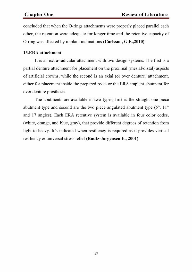
13.ERA attachment
It is an extra-radicular attachment with two design systems. The first is a
partial denture attachment for placement on the proximal (mesial/distal) aspects
of artificial crowns, while the second is an axial (or over denture) attachment,
either for placement inside the prepared roots or the ERA implant abutment for
over denture prosthesis.
The abutments are available in two types, first is the straight one-piece
abutment type and second are the two piece angulated abutment type (5°. 11°
and 17 angles). Each ERA retentive system is available in four color codes,
(white, orange, and blue, gray), that provide different degrees of retention from
light to heavy. It’s indicated when resiliency is required as it provides vertical
resiliency & universal stress relief (Budtz-Jorgensen E., 2001).

Chapter One Review of Literature
18
Figure 3. attachment – resilient
14.Ball attachmentsThe ball and socket attachments consist of a metal ball (patrix portion) which is
screwed into the fixture, where the matrix part is incorporated in the fitting
surface of the denture.
The matrix part may be one of the following types:
(A)- The O-ring in which the retentive element is rubber ring. It’s better to have
parallel implants otherwise the rubber ring will wear within a few weeks.
(B)- A metal part as in dalbo system. This permits less resilience however the
retentive forces are almost twice those obtained with the O-ring system.
(C)- A spherical metal anchor in which the female part contains a spring. These
attachments have advantage of being resilient and easily activated (Taylor TD
2005).

Chapter One Review of Literature
19
Ball attachments are among the simplest of all stud attachments widely used
because of their low cost, ease of handling, minimal chair side time
requirements and their possible applications with both root and implant-
supported prostheses (Meyer M,2001).
In comparison, done between over dentures retained by ball and socket
attachment and another design retained by two clips on a bar connecting the two
implants, regarding stresses on the peri implant bone. The result revealed that
stress on peri implant bone was greater with the clip/bar than that of ball
attachment (Hillerup S.,1982).
Fig 4: Ball abutments on two implants in the mandible. These single components leavethe implants unsplinted. When placed, the overdenture will then stabilize the implants(secondary splinting). They form the patrix, i.e. the part of the attachment systemconnected to the implant.

Chapter One Review of Literature
20
Fig 5: Intaglio view of the mandibular overdenture. The two rubber O-rings for the ball
abutments form the matrix, being the receptacle component. The matrix designs are
incorporated within the undersurface of the overdenture and they are adjustable and/or
replaceable.
15.Locator (self-aligning) attachment1. The locator attachment system is an attachment system with self-aligning
feature and has dual retention (inner and outer).
2. Locator attachments come in different colors (white, pink and blue) and
each has different retentive value.
3. Additional features are the extended range attachments, which can be used
to correct implant angulation up to 20o they are offered in green, which has
standard retention, and red, which has extra-light retention (Jiménez-Lopez
V.,1999).
4. The reduced height of this attachment is an advantagous for cases with
limited interocclusal space or when retrofitting an existing old denture
(Kakar., 2001).
5. Locator attachment will also accommodate divergent implants up to 20
degrees. A variety of abutment heights, angulations correction and different

Chapter One Review of Literature
21
levels of retention are available that help to create the optimum overdenture
restoration for each case (Kakar., 2001).
Fig.6. Locator attachment
16.Magnet attachmentsMagnetic retention is a popular method of attaching removable prosthesis
to either retained roots or osseo integrated implants. The magnet is usually
cylindrical or dome shaped attached to the fitting surface of the acrylic resin
base of the over denture. The magnetic keeper casted to a metal coping
cemented to root surface or screwed over the implant fixture (Sadowsky
SJ,2000).
The magnet system used for over denture retention incorporates the
magnet into the overdenture which is a neodymium-iron-boron alloy or a
cobalt-samarium alloy. The second part of the magnetic system is the
ferromagnetic keeper which is screwed into the implants (The Academy of
Prosthodontics.,2005).

Chapter One Review of Literature
22
The retention force of magnet attachments in implant-retained mandibular
overdenture treatment is markedly less than the retention force of ball and bar-
clip attachments (Warreth A.,2015).
The immediate loading of magnet attachment-retained mandibular
implant overdentures is considered as a viable treatment option in cases of
complete edentulous patient that increase retention and stability of conventional
dentures (Rothman A,2002).
Fig.7. Magnet attachments
17.Bar attachments
The bar attachment consists of a metallic bar that splints two or more
implants or natural teeth spanning the edentulous ridge between them and a
sleeve (suprastructure) incorporated in the over denture which clips over the
original bar to retain the denture. The bar attachments are available in wide
variety of forms; they could be prefabricated or custom made (Faria AC,2009).
There are two basic types based upon the shape and the action performed.
Bar joint that permit some degree of rotation or resilient movement between the
two components.
Spacers should be provided to ensure a small gap between the sleeve and
the bar during processing. Bar joints are subdivided into two types: single

Chapter One Review of Literature
23
sleeve and multiple sleeves; the single sleeve has to run straight without
allowing the anteroposterior curvature of the arch, so it is used in square arches.
On the other hand, the multiple sleeves can follow the curvature of the
arch. It also enables the use of more than one clip. Bar units that provide rigid
fixation of the over denture allowing no movement between the sleeve and the
bar (Yamada RH,2000).
The bar and clip attachments are probably the most widely used
attachments for implant tissue supported over dentures as they offer greater
mechanical stability and more wear resistance than solitary attachments. In
addition, short distal extensions from rigid bars can be achieved which
contribute to the stabilization and prevent shifting of the denture (Yamada
RH,2000., Faria AC,2009).
Fig 8: Mandibular implant bar. A bar construction splints the implants directly
together, also without the overdenture placed, providing higher stability to implants
and prosthesis.
Fig 9: Definitive overdenture with Hader bar clips.

Chapter One Review of Literature
24
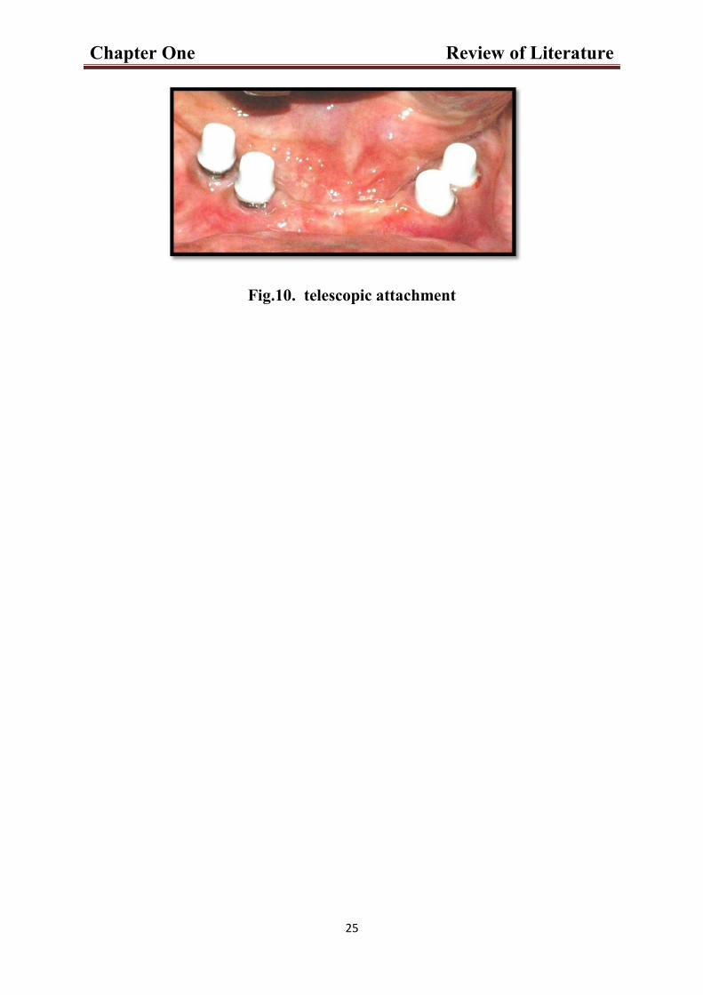
18. Telescopic attachment
Telescopic crowns are also known as a double crown, crown and sleeve
coping (CSC). These crowns consist of an inner or primary telescopic coping,
permanently cemented to an abutment, and a congruent detachable outer or
secondary telescopic crown, rigidly connected to a detachable prosthesis
(Warreth A.,2015).
The use of telescopic retainers has been expanded to include implant
retained prostheses to make use of their enormous advantages. These retainers
provide excellent retention resulting from frictional fit between the crown and
the sleeve. They also provide better force distribution due to the circumferential
relation of the outer crown to the abutment which make axial transfer of
occlusal load that produce less rotational torque on the abutment by improving
the crown root ratio so preserving the tooth and alveolar bone (Piermatti
J,2002).
According to wall design telescopic retainers can be classified into
parallel sided crowns. tapered (conical shaped) crowns and crowns with
additional attachments (Kang K, 2006).
Telescopic retained restoration has the advantage of the ease of
removability. This encourages the patient for repeated cleaning and
maintenance purposes. Moreover, the over dentures self-finding mechanism in
telescopic constructions facilitated prosthesis insertion considerably. This
construction seemed to be an effective treatment modality for geriatric patients
with serious systemic diseases as in Parkinson’s diseases (Rothman A,2002).
Chapter One Review of Literature
25
Fig.10. telescopic attachment

Chapter Two Patient and Method
25
Case 1.
Female patient with 65 years old with good medical history with lower anterior
teeth with severe periodontitis, so the treatment plan was extracted the
remaining natural teeth in the lower arch.
Figure (1): OPG x-ray for patient showing the lower remaining natural teeth
Figure (2)The fixture (Dentarim, Germany) immediately insertion after extractionwith bone augmentaion and membrane .

Chapter Two Patient and Method
26
Figure (3). Final impression of lower and upper arch
Figure (4): Make spaces in the denture for attachment

Chapter Two Patient and Method
27
Figure (5): Putting gingival former after 12 weeks for 10 day
A B
Figure (6).Torque Ratchet used to precisely apply a specific torque to screw the abutment to
the fixture.

Chapter Two Patient and Method
28
Figure (7). Attachment of patrix part to the implant part (stage 2).
Figure (8). Placement of matrix in the denture after finishing
Figure (9). Lower denture is performed

Chapter Two Patient and Method
29
Figure (10). Denture inside the patient mouth
Figure (11). Before and after insertion of dentures (finish the case)

Chapter Two Patient and Method
30
Figure (12). Lateral view after treatment

Chapter Two Patient and Method
31
Case 2Male patient with 55 years old with good medical history, the completely
edentolus with difficulty and wearing of the complete denture.
The work start with construction of complete denture with steps after the
implant operation about 4 month and good healing of the soft tissue covering.
Figure (1). Consideration regarding providing good space for the matrix part in
the denture so slight reduction from the length of the upper and lower anterior
teeth was done, the implant procedure started with reduction from the inner side
of the denture in the site of implant.

Chapter Two Patient and Method
32
Figure (2). Position of the implant in the lower and upper arches (length 9mm,diameter3.5mm)using Dentem Implant.
Figure (3). Attachment of the patrix part for verification

Chapter Two Patient and Method
33
Figure (4). Put the pieces(thin sheet) of wax around the matrix part in the patient
mouth.
The denture or matrix part for the implanted placed on the ball head of the
fixture and small piecesof wax placed under the matrix so that during fixation
no acrylic will be entrapted inside the matrix -patrix complex.
Figure (5). Fast set cold cure acrylic was matrix and put inside the holes thatwas done in the denture for the upper and lower denture and the denture fullyseated with occlusion ,after about 5 min time for curing the acrylic ,the dentureputout axcess acrylic removed, finished and polished.

Discussion
34
DiscussionThe results of the previous study showing that special consultation
regarding the treatment plan of implant supported or retained denture must be
done accurately as the position and the implant with optimal benefit regarding
the retention depend on many factors the occlusal relation of U/L arch ,degree
of bone resorption ,available interarch space ,biting forces ,type of implant, oral
hygiene, technical skills ,all those factors must be credent and inter connected to
obtain the best result for each case (Doundoulakis, J.H,2003).
so that special cooperation between the prosthodontic and implantologist
and the technician all must be worked together for obtaining the final result that
meet with patient desire and expectation.(Carlsson, G.E., 2010)

Conclusion
35
ConclusionImplant supported overdenture is more stable, retentive and
improves the mastication and speech dramatically. Edentulous patients
often do not get accustomed to wear conventional dentures. Their support
is compromised by progressive bone resorption that will increase
patient’s instability, insecurity and discomfort. Implant supported
overdentures also have a positive influence on adjacent peri-implant bone
levels. It creates a sensorimotor feedback that seems to facilitate the
overall functional experience with implant abutments under overdentures.
Overdenture use is a cheaper treatment than fixed prosthesis. Overdenture
use will prevent future aesthetic or phonetic problems in cases with lip
support loss or with an interocclusal space larger than 15 mm.

References
36
References
A Ahmed YA. Attachments Used with Implant Supported Over Denture.
Adv Dent & Oral Health. 2016; 1(2): 555560. DOI:
10.19080/ADOH.2016.01.555560.
Alsiyabi AS, Felton DA, Cooper LF (2005) The role of
abutmentattachment selection in resolving inadequate interarch distance:
a clinical report. J Prosthodont 14(3): 184-190.
B Bambara GE. Prosthetic Options for Implant-Supported Overdentures
Myriad choices make it possible to restore health and function. Inside
Dentistry 2015; 11(10).
Brewer AA, Fenton AH (1973) The overdenture. Dent Clin North Am
17(4): 723-746.
Budtz-Jorgensen E (2001) Prosthodontics for the elderly: diagnosis and
treatment. Chicago: quintessence publishing Co, USA.
C Carlsson, G.E., Omar, R. The future of complete dentures in oral
rehabilitation. A critical review. J Oral Rehabil 2010; 37 (2): 143-156.

References
37
D Doundoulakis, J.H., Eckert, S.E., Lindquist, C.C., Jeffcoat, M.K. The
implant supported overdenture as an alternative to the complete
mandibular denture. J Am Dent Assoc 2003; 134 (11): 1455-1458.
G Gotfredsen K, Holm B, Sewerin I, Harder F, Hjörting-Hansen E, et al.
(1993) Marginal tissue response adjacent to Astra Dental Implants
supporting overdentures in the mandible. Clin Oral Implants Res 4(2):
83-89.
Gulizio MP, Agar JR, Kelly JR, Taylor TD (2005) Effect of implant
angulation upon retention of overdenture attachments. J Prosthodont
14(1): 3-11.
H Heckmann SM, Winter W, Meyer M, Weber HP, Wichmann MG (2001)
Overdenture attachment selection and the loading of implant and denture-
bearing area. Part 2: A methodical study using five types of attachment.
Clin Oral Implants Res 12(6): 640-647.
Hillerup, S. Preprosthetic mandibular vestibuloplasty with buccal
mucosal graft. A 2- year follow-up study. Int J Oral Surg 1982; 11 (2):
81-88.
Hochstedler, J.L., Finger, I.M. Preprosthetic surgery. Gen Dent 1998; 46
(6): 626- 630.

References
38
J Jiménez-Lopez V (1999) Oral rehabilitation with implant-supported
prostheses. In Implant supported mandibular overdenture Chicago,
Berlin, London, Paris: Quintessence publishing Co, USA.
K Kakar (2001) Oral implantology 1st edn. New delhi. Jaypee Brothers
Medical Publishers Pvt Ltd.
Krennmair G, Ulm C (2001) The symphyseal single-tooth implant for
anchorage of a mandibular complete denture in geriatric patients: a
clinical report. Int J Oral Maxillofac Implants 16(1): 98-104.
Krennmair G, Weinlander M, Krainhofner M, Piehslinger E (2006)
Implant-supported mandibular overdentures retained with ball or
telescopic crown attachments: a 3-year prospective study. Int J
Prosthodont 19(2): 164-170.
M Majer HJ, (1992) The Stern ERA attachment. Exacting retention made
easy. J Can Dent Assoc 58(8): 615.
S Sadowsky SJ, Caputo AA (2000) Effect of anchorage systems and
extension base contact on load transfer with mandibular implantretained
overdentures. J Prosthet Dent 84(3): 327-334.
References
39
Sarandha DL, Hussain Z, Uthkarsh. Textbook of Complete Denture
PROSTHODONTICS. Jaypee Brothers Medical Publishers First Edition
2007; Ch. 22, P: 166.
Sohrabi, K., Mushantat, A., Esfandiari, S., Feine, J. How successful are
smalldiameter implants? A literature review. Clin Oral Implants Res
2012; 23 (5): 515-525.
Steffen RP, White V, Markowitz NR (2004) The use of ball clip
attachments with an implant-supported primary-secondary bar
overdenture. J Oral Implantol 30(4): 234-239.
T The Academy of Prosthodontics (2005) The glossary of prosthodontic
terms. J Prosthet Dent 94(1): 10-92.
Trakas T, Michalakis K, Kang K, Hirayama H (2006) Attachment
systems for implant retained overdentures: a literature review. Implant
Dent 15(1): 24-34.
W Walton JN, MacEntee MI, Glick N (2002) One-year prosthetic outcomes
with implant overdentures: a randomized clinical trial. Int J Oral
Maxillofac Implants 17(3): 391-398.
Warreth A., Byrne C., Alkadhimi AF., Woods E and Sultan A.
Mandibular implant-supported overdentures: attachment systems, and
number and locations of implants – Part I. Journal of the Irish Dental
Association 2015; 61 (2): 93-97.
Winkler S, Piermatti J, Rothman A, Siamos G (2002) An overview of the
O-ring implant overdenture attachment: clinical reports. J Oral Implantol
28(2): 82-86. 16.

References
40
Rodrigues RC, Faria AC, Macedo AP, Sartori IA, de Mattos Mda G, et al.
(2009) An in vitro study of non-axial forces upon the retention of an O-
ring attachment. Clin Oral Implants Res 20(12): 1314-1319.
Y Yamada RH, Gorin DV, Marinello RF, Rosen MA and Russo SP.
Implant-Supported Overdentures: The Standard of Care for Edentulous
Patients. Perio Donta Letter. Summer 2000.