clinical atlas of ophthalmic disease · 2015-07-31 · clinical atlas of canine and feline...
TRANSCRIPT


CLINICAL ATLAS OF
CANINE AND FELINE
OPHTHALMIC DISEASE

CLINICAL ATLAS OFCANINE AND FELINEOPHTHALMIC DISEASE
DOUGLAS W. ESSON, BVSC, MRCVS, DVM, DACVO,Veterinary ophthalmologist and clinical director of Eye Care for Animals,Tustin, California, USA
This edition first published 2015 ©2015 by John Wiley & Sons Inc
Editorial Offices: 1606 Golden Aspen Drive, Suites 103 and 104, Ames, Iowa 50010, USAThe Atrium, Southern Gate, Chichester, West Sussex, PO19 8SQ, UK9600 Garsington Road, Oxford, OX4 2DQ, UK
For details of our global editorial offices, for customer services and for information about how to apply for permission to reuse the copyright material inthis book please see our website at www.wiley.com/wiley-blackwell.
Authorization to photocopy items for internal or personal use, or the internal or personal use of specific clients, is granted by Blackwell Publishing,provided that the base fee is paid directly to the Copyright Clearance Center, 222 Rosewood Drive, Danvers, MA 01923. For those organizations thathave been granted a photocopy license by CCC, a separate system of payments has been arranged. The fee codes for users of the Transactional ReportingService are ISBN-13: 978-1-1188-4077-1/2015
Designations used by companies to distinguish their products are often claimed as trademarks. All brand names and product names used in this book aretrade names, service marks, trademarks or registered trademarks of their respective owners. The publisher is not associated with any product or vendormentioned in this book.
The contents of this work are intended to further general scientific research, understanding, and discussion only and are not intended and should not berelied upon as recommending or promoting a specific method, diagnosis, or treatment by health science practitioners for any particular patient. Thepublisher and the author make no representations or warranties with respect to the accuracy or completeness of the contents of this work and specificallydisclaim all warranties, including without limitation any implied warranties of fitness for a particular purpose. In view of ongoing research, equipmentmodifications, changes in governmental regulations, and the constant flow of information relating to the use of medicines, equipment, and devices, thereader is urged to review and evaluate the information provided in the package insert or instructions for each medicine, equipment, or device for, amongother things, any changes in the instructions or indication of usage and for added warnings and precautions. Readers should consult with a specialistwhere appropriate. The fact that an organization or Website is referred to in this work as a citation and/or a potential source of further information doesnot mean that the author or the publisher endorses the information the organization or Website may provide or recommendations it may make. Further,readers should be aware that Internet Websites listed in this work may have changed or disappeared between when this work was written and when it isread. No warranty may be created or extended by any promotional statements for this work. Neither the publisher nor the author shall be liable for anydamages arising herefrom.
Library of Congress Cataloging-in-Publication Data
Esson, Douglas W., author.Clinical atlas of canine and feline ophthalmic disease / Douglas W. Esson.
p. ; cm.Includes index.ISBN 978-1-118-84077-1 (cloth)1. Dogs–Diseases–Atlases. 2. Cats–Diseases–Atlases. 3. Eye–Diseases–Atlases. 4. Veterinary ophthalmology–Atlases. I. Title.[DNLM: 1. Dog Diseases–Atlases. 2. Eye Diseases–veterinary–Atlases. 3. Cat Diseases–Atlases. 4. Ophthalmologic Surgical Procedures–Atlases.
SF 992.E92]
SF992.E92 E87 2015636.089′77–dc23
2015006624
A catalogue record for this book is available from the British Library.
Wiley also publishes its books in a variety of electronic formats. Some content that appears in print may not be available in electronic books.
Set in 10/12pt Minion by Laserwords Private Limited, Chennai, India
1 2015
CONTENTS
Preface ixAcknowledgments xi
Section 1. Anatomy and Diagnostics 1Chapter 1. Normal Ocular Anatomy 2Chapter 2. Normal Pigmentary Variations 4Chapter 3. The Normal Canine Fundus 6Chapter 4. The Normal Feline Fundus 8Chapter 5. The Normal Subalbinotic Fundus 10Chapter 6. Normal Myelination Variations 12Chapter 7. The Ocular Examination 14
Section 2. Diseases of the Eyelids 17Chapter 8. Eyelid Agenesis 18Chapter 9. Eyelid Laceration 20Chapter 10. Distichiasis 22Chapter 11. Ectopic Cilia 24Chapter 12. Trichiasis 26Chapter 13. Tear Film Wicking Syndrome 28Chapter 14. Entropion 30Chapter 15. Ectropion 32Chapter 16. Combined Entropion-Ectropion 34Chapter 17. Macropalpebral Fissure 36Chapter 18. Chalazion 38Chapter 19. Juvenile Pyoderma 40Chapter 20. Immune-Mediated
Blepharoconjunctivitis 42Chapter 21. Autoimmune Blepharitis 44Chapter 22. Eosinophilic
Folliculitis/Furunculosis 46Chapter 23. Adverse Drug Reactions (ADRs) 48Chapter 24. Dermatomyositis 50Chapter 25. Demodex-Associated Blepharitis 52Chapter 26. Dermatophytosis 54Chapter 27. Radiation Induced
Blepharoconjunctivitis 56Chapter 28. Apocrine Hidrocystoma 58Chapter 29. Sebaceous Adenoma/Epithelioma 60Chapter 30. Histiocytoma 62
Chapter 31. Eyelid Melanocytoma 64Chapter 32. Eyelid Melanoma 66Chapter 33. Cutaneous Epitheliotropic Lymphoma
(CEL) 68Chapter 34. Eyelid Squamous Cell Carcinoma
(SCC) 70Chapter 35. Mast Cell Tumor (MCT) 72Chapter 36. Fibrosarcoma 74
Section 3. Diseases of the Conjunctiva,Nasolacrimal System, and ThirdEyelid 77Chapter 37. Allergic Conjunctivitis 78Chapter 38. Dacryocystitis 80Chapter 39. Symblepharon 82Chapter 40. Herpesviral-Associated
Conjunctivitis 84Chapter 41. Third Eyelid Gland Prolapse (Cherry
Eye) 86Chapter 42. Keratoconjunctivitis Sicca (Dry Eye) 88Chapter 43. Scrolled Third Eyelid Cartilage 90Chapter 44. Medial Canthal Pocket Syndrome 92Chapter 45. Onchocerciasis 94Chapter 46. Thelazia 96Chapter 47. Papilloma 98Chapter 48. Conjunctival Melanoma 100Chapter 49. Conjunctival
Hemangioma/Hemangiosarcoma 102Chapter 50. Adenoma/Adenocarcinoma 104Chapter 51. Conjunctival Lymphoma 106Chapter 52. Conjunctival squamous Cell
Carcinoma 108
Section 4. Corneoscleral Disease 111Chapter 53. Dermoid 112Chapter 54. Corneal Dystrophy 114Chapter 55. Corneal Degeneration 116Chapter 56. Corneal Endothelial
Decompensation 118Chapter 57. Scleritis 120Chapter 58. Nodular Granulomatous Episcleritis 122
v
vi Contents
Chapter 59. Chronic Superficial Keratitis (CSK) 124Chapter 60. Eosinophilic Keratoconjunctivitis 126Chapter 61. Herpesviral-Associated Keratitis 128Chapter 62. Canine Multifocal Immune-Mediated
Punctate Keratitis (MIPK) 132Chapter 63. Endotheliitis 134Chapter 64. Bullous Keratopathy 136Chapter 65. Feline Corneal Sequestrum 138Chapter 66. Pigmentary Keratitis 140Chapter 67. Corneal Abscessation 142Chapter 68. Corneal Hemorrhage 144Chapter 69. Corneoscleral Laceration 146Chapter 70. Spontaneous Chronic Corneal Epithelial
Defects (SCCEDs) 148Chapter 71. Stromal Ulcerative Keratitis 150Chapter 72. Descemetocele 152Chapter 73. Keratomalacia (Melting Ulcers) 154Chapter 74. Corneal Perforation 156Chapter 75. Epithelial Inclusion Cysts 158Chapter 76. Corneal Foreign Body 160Chapter 77. Limbal Melanocytoma 162Chapter 78. Fungal Keratitis 164Chapter 79. Corneoscleral
Hemangioma/Hemangiosarcoma 166Chapter 80. Corneoscleral Lymphoma 168Chapter 81. Corneal Squamous Cell Carcinoma 170
Section 5. Diseases of the Uvea 173Chapter 82. Persistent Pupillary Membranes
(PPMs) 174Chapter 83. Iris Colobomas 176Chapter 84. Senile Iris Atrophy 178Chapter 85. Uveal Cysts 180Chapter 86. Merle Ocular Dysgenesis (MOD) 182Chapter 87. Feline Anterior Uveitis 184Chapter 88. Canine Anterior Uveitis 186Chapter 89. Golden Retriever–Associated Uveitis and
Glaucoma 188Chapter 90. Vaccine-Associated Uveitis 190Chapter 91. Uveodermatologic Syndrome
(UDS)-Associated Uveitis 192Chapter 92. Hyphema 194Chapter 93. Aqueous Lipidosis 196Chapter 94. Iris Bombe’ 198Chapter 95. Feline Iris Melanosis (FIM) 200Chapter 96. Uveal Melanoma 202Chapter 97. Uveal Adenoma/Adenocarcinoma 204Chapter 98. Uveal Lymphoma 206
Section 6. Diseases of the Lens 209Chapter 99. Microphakia/Spherophakia 210Chapter 100. Persistent Hyaloid Vasculature
(PHV) 212Chapter 101. Nuclear Sclerosis 214Chapter 102. Immature Cataract 216Chapter 103. Mature Cataract 218Chapter 104. Hypermature Cataract 220Chapter 105. Phacolytic Uveitis 222Chapter 106. Phacoclastic Uveitis 224Chapter 107. Anterior Lens Luxation 226Chapter 108. Posterior Lens Luxation 228Chapter 109. Feline Post-Traumatic Ocular
Sarcoma 230
Section 7. Vitreoretinal Disease 233Chapter 110. The Retinal Dysplasias (RDs) 234Chapter 111. Oculoskeletal Dysplasia (OSD) 236Chapter 112. Collie Eye Anomaly (CEA) 238Chapter 113. The Retinal Atrophies (RAs) 240Chapter 114. Vitreal Degeneration/Herniation 242Chapter 115. Retinal Toxicity 244Chapter 116. SARDs/IMR 246Chapter 117. Hypertensive Retinopathy 248Chapter 118. Feline Chorioretinitis 250Chapter 119. Canine Chorioretinitis 252Chapter 120. Retinal Pigment Epithelial Dystrophy
(RPED) 254Chapter 121. Uveodermatologic Syndrome
(UDS)-Associated Chorioretinitis 256Chapter 122. Primary (Bullous) Retinal
Detachment 258Chapter 123. Rhegmatogenous Retinal Detachment
(RRD) 260Chapter 124. Chorioretinal Lymphoma 262Chapter 125. Myeloma 264
Section 8. Diseases of the Globe andOrbit 267Chapter 126. Microphthalmia 268Chapter 127. Phthisis Bulbus 270Chapter 128. Orbital Cellulitis 272Chapter 129. Extraocular Myositis (EOM) 274Chapter 130. Zygomatic Sialoadenitis 276Chapter 131. Orbital Fat Pad Prolapse 278Chapter 132. Orbital Foreign Bodies 280Chapter 133. Endophthalmitis/Panophthalmitis 282Chapter 134. Proptosis of the Globe 284Chapter 135. Peripheral Nerve Sheath Tumor
(PNST) 286Chapter 136. Retrobulbar Neoplasia 288
Contents vii
Section 9. The Glaucomas 291Chapter 137. Congenital Glaucoma 292Chapter 138. Primary Glaucoma 294Chapter 139. Secondary (Postinflammatory)
Glaucoma 296Chapter 140. Feline Aqueous Humor Misdirection
Syndrome (AHMS) 298Chapter 141. Pigmentary Glaucoma 300Chapter 142. Golden Retriever-Associated Uveitis and
Glaucoma 302Chapter 143. Buphthalmos 304
Section 10. Neuro-OphthalmicDisease 307Chapter 144. Optic Nerve Hypoplasia 308Chapter 145. Lysosomal Storage Disease (LSD) 310Chapter 146. Sympathetic Denervation (Horner’s
Syndrome) 312Chapter 147. Ophthalmoplegia 314Chapter 148. Neuroparalytic Keratitis/Hemifacial
Paralysis 316Chapter 149. Neurogenic KCS and Xeromycteria 318Chapter 150. Optic Neuritis/Meningitis 320
Index 323


PREFACE
It is some years since I made the conscious decision to further my postgraduate knowledge of the complex and fascinatingworld of veterinary ophthalmology. With the passing of the intervening years, I have had the immense good fortune tointeract with a number of clinician-scientists, all of whom have generously shared their time, experience, and knowledgewith me. In particular, I am indebted in this regard to Drs. Peter Bedford, Randy Scagliotti, Kirk Gelatt, Paul Miller, DickDubielzig, Bill Dawson, and Mark Sherwood. I am frequently asked to give lectures about various ophthalmic topics toaudiences ranging from students to experienced ophthalmologists, and it is most commonly through photographic imagesthat I am able to share my own thoughts and experience. The field of veterinary ophthalmology, detailed ophthalmic texts,and the peer-reviewed ophthalmic literature represent a sometimes challenging and potentially confusing arena and, as aconsequence, I have sought here to provide the busy general practitioner with a clear, systematic, repeatable clinical pictureof the most frequently encountered ophthalmic conditions in small animal practice. Images are intentionally presented inthe same way as cases would be encountered in practice. In order to expand this project beyond that of simply an imageatlas, I have also tried to provide clear, concise, updated, and clinically relevant information as it pertains to each of theseconditions. This information has been supported with a small number of relevant references for those who to wish to readfurther.
Any potential drug side effects are identified by this shaded colored box.
ix

ACKNOWLEDGMENTS
Many individuals have contributed to the development of this book. The project, from its inception, has been shaped bymy editors and publishers at Wiley, with special thanks to Erica Judisch, Nancy Turner, and Catriona Cooper. The imageswithin this volume have mostly been sourced from my own collection; however, I have relied on friends and colleagues todiscuss clinical presentations, help source cases (and in some instances provide images) in order to achieve completeness.In this regard, I acknowledge Drs. Dustin Dees, Anne-Michelle Armour, Nicole McClaren, Randy Scagliotti, Al MacMillan,Christin Chapman, David Wilkie, Matthew Fife, Nancy Park, Anastasia Komenou, Jennifer Urbanz, Peter Bedford, DavidWilliams, David Donaldson, Julius Brinkis, Dilip Bhalerao, Emily Moeller, Francesca Venturi, Neal Wasserman, JoannaNorman, Keith Collins, Steve Sissler, Melanie Church. Ashley Stich, Laura Wilson, Rudayna Gubash, Allison Kirby, NickMillichamp, Mark Haskins, Gwen Lynch, and Kristina Narfstrom. I am additionally grateful to a number of the affiliated spe-cialists with whom I work, for their willingness to source and discuss cases commonly presented to the services of internalmedicine, dermatology, and oncology. In this regard, I particularly acknowledge Drs. Wayne Rosenkrantz, Colleen Mendel-sohn, Melissa Hall, Julie Bulman-Fleming, and David Bommarito. The time taken to collate and describe these conditionswas allocated by Karen Webster and sponsored by Eye Care for Animals as part of its ongoing commitment to advancingthe field of veterinary ophthalmology. Finally but most importantly I am endlessly grateful to my wife Sara, herself a giftedand passionate veterinary ophthalmologist. Without her endless assistance, clinical expertise, and patient advice, this bookwould simply not have been possible.
For Sara and Justin With all my love Doug Esson Tustin, California, 2014
xi


Section 1
Anatomy and Diagnostics
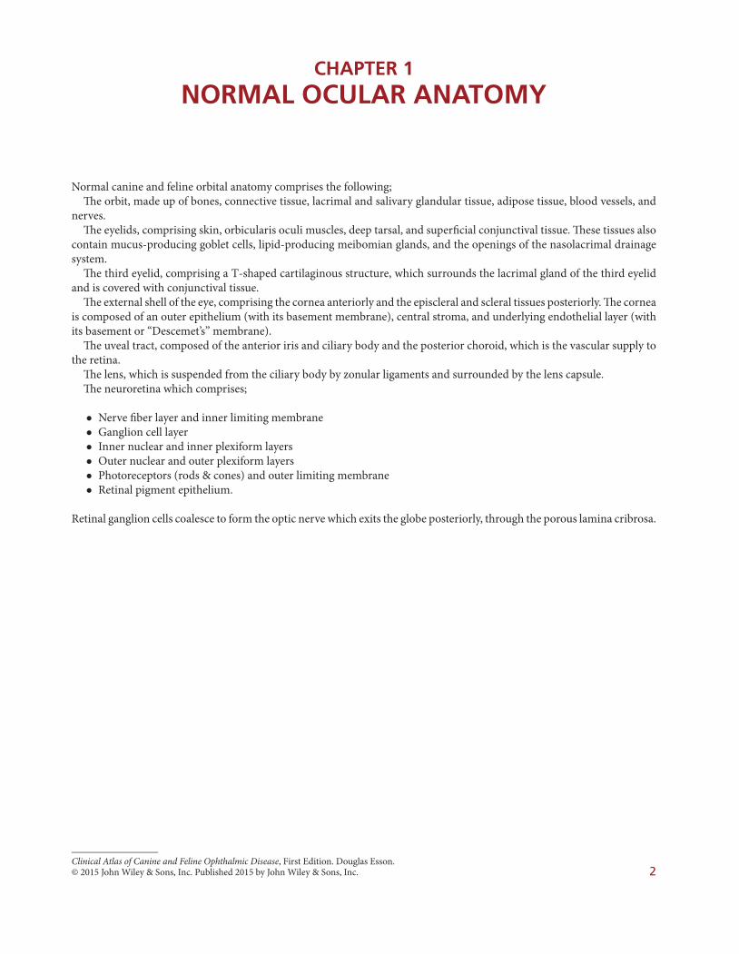
CHAPTER 1NORMAL OCULAR ANATOMY
Normal canine and feline orbital anatomy comprises the following;The orbit, made up of bones, connective tissue, lacrimal and salivary glandular tissue, adipose tissue, blood vessels, and
nerves.The eyelids, comprising skin, orbicularis oculi muscles, deep tarsal, and superficial conjunctival tissue. These tissues also
contain mucus-producing goblet cells, lipid-producing meibomian glands, and the openings of the nasolacrimal drainagesystem.
The third eyelid, comprising a T-shaped cartilaginous structure, which surrounds the lacrimal gland of the third eyelidand is covered with conjunctival tissue.
The external shell of the eye, comprising the cornea anteriorly and the episcleral and scleral tissues posteriorly. The corneais composed of an outer epithelium (with its basement membrane), central stroma, and underlying endothelial layer (withits basement or “Descemet’s” membrane).
The uveal tract, composed of the anterior iris and ciliary body and the posterior choroid, which is the vascular supply tothe retina.
The lens, which is suspended from the ciliary body by zonular ligaments and surrounded by the lens capsule.The neuroretina which comprises;
• Nerve fiber layer and inner limiting membrane• Ganglion cell layer• Inner nuclear and inner plexiform layers• Outer nuclear and outer plexiform layers• Photoreceptors (rods & cones) and outer limiting membrane• Retinal pigment epithelium.
Retinal ganglion cells coalesce to form the optic nerve which exits the globe posteriorly, through the porous lamina cribrosa.
Clinical Atlas of Canine and Feline Ophthalmic Disease, First Edition. Douglas Esson.© 2015 John Wiley & Sons, Inc. Published 2015 by John Wiley & Sons, Inc. 2

Choroid Sclera
Vitreous body
Retina
Optic nerve
Posterior chamber
Zonular ligaments
Anterior chamber
Lens
Cornea
Iris
Ciliary body
Figure 1.1 Normal ocular anatomy.
3
CHAPTER 2NORMAL PIGMENTARY VARIATIONS
Both canine and feline irides may demonstrate a range of pigmentary variations. True ocular albinism (the complete lack ofpigment) is rare. The term subalbinism describes pigment dilution resulting in variably grey to bluish colored iridal tissue,a finding common in animals with light hair coat colors. The term heterochromia iridis describes variable combinations ofpigment within either one or both irides.
Clinical Atlas of Canine and Feline Ophthalmic Disease, First Edition. Douglas Esson.© 2015 John Wiley & Sons, Inc. Published 2015 by John Wiley & Sons, Inc. 4

Figure 2.1 Normal variation in pigmentation between leftand right eyes.
Figure 2.2 Normal heterochromic variation inpigmentation within a light colored iris.
Figure 2.3 Normal heterochromic variation inpigmentation within a dark colored iris.
Figure 2.4 Normal blue iris (the “red reflex” of thesubalbinotic fundus being visible through the pupil).
5
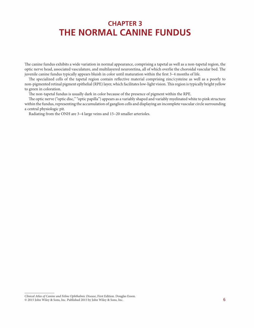
CHAPTER 3THE NORMAL CANINE FUNDUS
The canine fundus exhibits a wide variation in normal appearance, comprising a tapetal as well as a non-tapetal region, theoptic nerve head, associated vasculature, and multilayered neuroretina, all of which overlie the choroidal vascular bed. Thejuvenile canine fundus typically appears bluish in color until maturation within the first 3–4 months of life.
The specialized cells of the tapetal region contain reflective material comprising zinc/cysteine as well as a poorly tonon-pigmented retinal pigment epithelial (RPE) layer, which facilitates low-light vision. This region is typically bright yellowto green in coloration.
The non-tapetal fundus is usually dark in color because of the presence of pigment within the RPE.The optic nerve (“optic disc,” “optic papilla”) appears as a variably shaped and variably myelinated white to pink structure
within the fundus, representing the accumulation of ganglion cells and displaying an incomplete vascular circle surroundinga central physiologic pit.
Radiating from the ONH are 3–4 large veins and 15–20 smaller arterioles.
Clinical Atlas of Canine and Feline Ophthalmic Disease, First Edition. Douglas Esson.© 2015 John Wiley & Sons, Inc. Published 2015 by John Wiley & Sons, Inc. 6
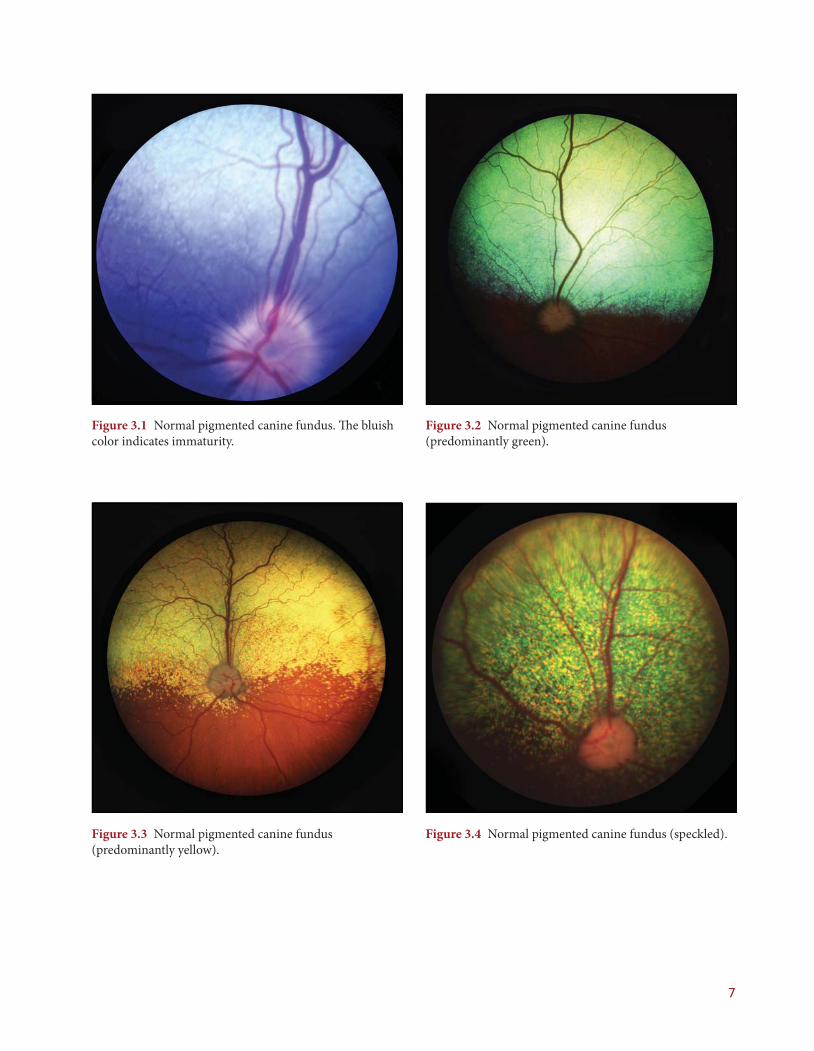
Figure 3.1 Normal pigmented canine fundus. The bluishcolor indicates immaturity.
Figure 3.2 Normal pigmented canine fundus(predominantly green).
Figure 3.3 Normal pigmented canine fundus(predominantly yellow).
Figure 3.4 Normal pigmented canine fundus (speckled).
7

CHAPTER 4THE NORMAL FELINE FUNDUS
The feline fundus exhibits a wide variation in normal appearance, comprising a relatively large tapetal as well as non-tapetalregion, the optic nerve head, associated vasculature, and multilayered neuroretina, all of which overly the choroidal vascularbed.
The specialized cells of the tapetal region contain reflective material comprising zinc/riboflavin as well as a poorly tonon-pigmented retinal pigment epithelial (RPE) layer, which facilitates low-light vision. This region is typically bright yellowto green in coloration.
The non-tapetal fundus is usually dark in color due to the presence of pigment within the RPE.The optic nerve (“optic disc,” “optic papilla”) appears as a small, circular, unmyelinated white to grey structure within the
fundus, representing the accumulation of ganglion cells.Three major pairs of arterioles as well as larger venules radiate from the ONH.
Clinical Atlas of Canine and Feline Ophthalmic Disease, First Edition. Douglas Esson.© 2015 John Wiley & Sons, Inc. Published 2015 by John Wiley & Sons, Inc. 8

Figure 4.1 Normal pigmented feline fundus(predominantly green), note the poorly myelinated opticnerve head.
Figure 4.2 Normal pigmented feline fundus(predominantly green), note the poorly myelinated opticnerve head.
Figure 4.3 Normal pigmented feline fundus(predominantly yellow), note the poorly myelinated opticnerve head.
Figure 4.4 Normal pigmented feline fundus(predominantly yellow), note the poorly myelinated opticnerve head.
9
CHAPTER 5THE NORMAL SUBALBINOTIC FUNDUS
Dogs or cats displaying blue irides, heterochromic irides, and/or merled coat coloration, typically display “subalbinotic”fundi. In these animals, the tapetal region may be variably reduced to absent in association with a variable to complete lackof pigment within the non-tapetal fundus. As a result, underlying choroidal vasculature is visible against the white scleralbackground. The subalbinotic fundus represents a normal variation in coloration.
Clinical Atlas of Canine and Feline Ophthalmic Disease, First Edition. Douglas Esson.© 2015 John Wiley & Sons, Inc. Published 2015 by John Wiley & Sons, Inc. 10

Figure 5.1 Normal (canine) subalbinotic fundus.Choroidal vessels are clearly visible against the whitescleral background.
Figure 5.2 Normal (canine) subalbinotic fundus.Choroidal vessels are clearly visible against the whitescleral background.
Figure 5.3 Normal (canine) subalbinotic fundus.Choroidal vessels are clearly visible against the whitescleral background.
Figure 5.4 Normal (feline) subalbinotic fundus.Choroidal vessels are clearly visible against the whitescleral background.
11

CHAPTER 6NORMAL MYELINATION VARIATIONS
The optic nerve head (ONH) comprises coalescing ganglion cells as they converge before exiting the globe caudally throughthe porous lamina cribrosa. This region is variably myelinated; poorly in the cat and variably in the dog. Variations in theamount of myelin present may result in range of normal appearances when this region is visualized.
Clinical Atlas of Canine and Feline Ophthalmic Disease, First Edition. Douglas Esson.© 2015 John Wiley & Sons, Inc. Published 2015 by John Wiley & Sons, Inc. 12
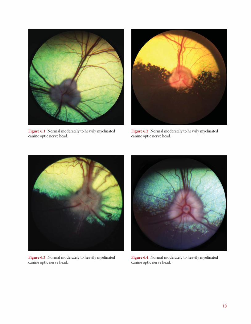
Figure 6.1 Normal moderately to heavily myelinatedcanine optic nerve head.
Figure 6.2 Normal moderately to heavily myelinatedcanine optic nerve head.
Figure 6.3 Normal moderately to heavily myelinatedcanine optic nerve head.
Figure 6.4 Normal moderately to heavily myelinatedcanine optic nerve head.
13
CHAPTER 7THE OCULAR EXAMINATION
The ophthalmic examination should comprise the following components.
HISTORYSignalment, pre-existing medical/surgical history (including travel history), and/or current medications.
PRESENTING COMPLAINTIdentification of the presenting ophthalmic complaint.
DISTANT “HANDS-OFF” EXAMINATIONPatient is allowed to move around freely—demonstrating mentation, neurological status, and visual ability.
BRIEF GENERAL PHYSICAL EXAMINATIONIncludes assessment of mucous membranes, oral cavity, external ear canals, thoracic auscultation, palpation of lymph nodesand abdomen and body temperature.
CLOSE UP “HANDS-ON” EXAMINATIONIncludes careful palpation of the skull and orbits, noting any deformity, asymmetry, crepitus, or discomfort.
NEURO-OPHTHALMIC EXAMINATION• Palpebral reflex (closure of eyelids upon tactile stimulus)• Menace response (eyelid closure and/or head withdrawal in response to menacing hand gesture)• Dazzle reflex (closure of eyelids in response to bright focal light source being shined into eye)• Pupillary light reflex (PLR) (direct and consensual reflex pupillary miosis in response to a focal light source).
SEGMENTAL ANTERIOR EXAMINATIONA focal light source is used to examine the eyelids, conjunctival surfaces, third eyelid, sclera, cornea, anterior chamber (AC),iris, lens, and anterior vitreous face.
FUNDIC EXAMINATIONThe posterior segment is examined using either a focal light source and handheld lens or an ophthalmoscope.
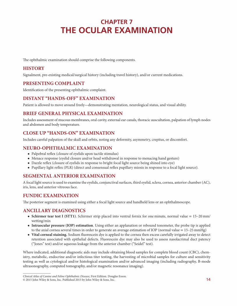
ANCILLARY DIAGNOSTICS• Schirmer tear test I (STT1). Schirmer strip placed into ventral fornix for one minute, normal value = 15–20 mm/
wetting/min• Intraocular pressure (IOP) estimation. Using either an applanation or rebound tonometer, the probe tip is applied
to the axial cornea several times in order to generate an average estimation of IOP (normal value = 15–25 mmHg)• Vital corneal staining. Sodium fluorescein dye is applied to the cornea then excess carefully irrigated away to detect
retention associated with epithelial defects. Fluorescein dye may also be used to assess nasolacrimal duct patency(“Jones” test) and/or aqueous leakage from the anterior chamber (“Seidel” test).
Where indicated, additional diagnostic aids may include obtaining blood samples for complete blood count (CBC), chem-istry, metabolic, endocrine and/or infectious titer testing, the harvesting of microbial samples for culture and sensitivitytesting as well as cytological and/or histological examination and/or advanced imaging (including radiography, B-modeultrasonography, computed tomography, and/or magnetic resonance imaging).
Clinical Atlas of Canine and Feline Ophthalmic Disease, First Edition. Douglas Esson.© 2015 John Wiley & Sons, Inc. Published 2015 by John Wiley & Sons, Inc. 14

Figure 7.1 Estimation of intraocular pressure (IOP) usinga rebound tonometer (“Tonovet”). The probe-tip is gentlyallowed to contact the axial corneal surface (without theuse of local anesthetic) by pressing the measurementbutton. Several readings are taken, so that aberrantreadings may be disregarded.
Figure 7.2 Measurement of lacrimal function usinggradated (“Schirmer”) tear strips. The tip of each strip isfolded and placed into the lower medial fornix for oneminute and the resultant STT1 value recorded. This testshould be performed before the installation of any topicalagents.
Figure 7.3 Vital staining of the corneal surface usingfluorescein-impregnated strips. Strips are moistened usinga physiologic solution and gently touched to the sclerallimbus. Excess stain is then carefully irrigated away usingeyewash to prevent “pooling” of residual stain.
Figure 7.4 Visualization of the fundus using a simplehandheld indirect lens, positioned just in front of the eyeand parallel to the posterior segment. Where an indirectophthalmoscope is unavailable, a small flashlight ortransilluminator, held adjacent to the examiner’s head, willsuffice as a distant focal light source.
15
