clinical case: axiom px axiom 2 - anthogyr.be · the absence of maxillary lateral incisors creates...
TRANSCRIPT

AGENESIS OF MAXILLARY LATERAL INCISORS
Two cases managed with Anthogyr Axiom® 2.8 and Axiom® PX implants.
CLINICAL CASE: AXIOM® PX / AXIOM® 2.8
Dr Francis BAILLY
w Doctor in Dental Surgeryw M.D., University School of Medicine, Lyon, Francew Degree in Oral and Maxillofacial Implantologyw Attended the Advanced Training Course on soft tissue management
and bone grafting by Prof. Khoury, Schellenstein, Germanyw Formerly attached to Lyon hospitals

2
The absence of maxillary lateral incisors creates major aesthetic and functional problems due to their location in the aesthetic zone.In adolescent patients, the possible options are space maintenance and replacement of the missing teeth, or orthodontic space closure with canine substitution9,13,14,15. Proper recontouring or reshaping of the canines after removal of the orthodontic appliances greatly improves the aesthetic outcome19.There are also adult patients who have gone untreated, and at some time in their lives are confronted with this problem. We see quite a lot of such cases12. We are presenting here two distinct clinical cases.
Clinical case n°1
An 18-year-old female patient with a bilateral agenesis of the maxillary lateral incisors was referred to us by a colleague, Dr G. Prost. He had started orthodontic space opening treatment when the patient was 14 years old.Agenesis is often associated with bone hypoplasia, which results in insufficient bone volume due to the presence of thin concave ridges18.Thanks to the Axiom® 2.8 implant (Anthogyr), a distance of 1.5 mm can be maintained between the implants and the adjacent teeth16. But above all, it avoids the need for a two-stage surgery with a first stage for bone healing, and a second stage several months later for placement of the implants.
Fig.1: Panoramic X-ray taken at the end of the orthodontic treatment with the retaining wires in situ.
Fig.2: Clinical view prior to surgical treatment.
Fig.3: A removable prosthesis was used until jaw growth was completed.
Figs.4a & 4b: Lateral views showing the spaces achieved with the orthodontic treatment.
Figs.5a & 5b: Accurate measurements of interdental spaces are made on CBCT (cone beam computed tomography) reconstructions.
Figs.6a & 6b: As it was anticipated during clinical examination, it would not be possible to use standard implants without prior bone reconstruction.
5a
1
1
2
3
4a
4b
5a 5b
6a 6b

CLINICAL CASE - AXIOM® PX / AXIOM® 2.8
3
Figs.7a & 7b: Control X-rays taken after placement of two 14 mm long Axiom® 2.8 implants.
Figs.8a & 8b: Following elevation of two mini-flaps preserving the gingival papillae, only 2 instruments were necessary to place the implants: an initial drill (2 mm diameter) and a stepwise drill (2.6 mm diameter).
Anthogyr recommends a subcrestal positioning of the implant, so as to enhance aesthetics of soft tissues4.
Owing to the small diameter of the implant, placement is easy. However, bone apposition is mandatory to preserve the peri-implant tissue and ensure a long-term aesthetic outcome.
Fig.9: Xenograft using bovine origin material (Cerabone®).
Fig.10: Graft is covered with a collagen membrane (Jason®).
Fig.11: Graft fills completely the bone defect.
7a
7b
8b
8a
10
9
11
4
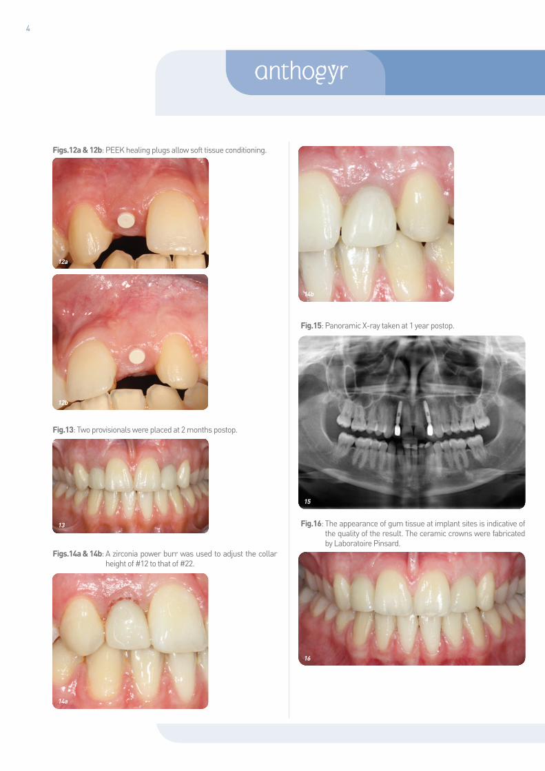
Figs.12a & 12b: PEEK healing plugs allow soft tissue conditioning.
Fig.13: Two provisionals were placed at 2 months postop.
Figs.14a & 14b: A zirconia power burr was used to adjust the collar height of #12 to that of #22.
Fig.15: Panoramic X-ray taken at 1 year postop.
Fig.16: The appearance of gum tissue at implant sites is indicative of the quality of the result. The ceramic crowns were fabricated by Laboratoire Pinsard.
12a
12b
13
14a
14b
15
16

CLINICAL CASE - AXIOM® PX / AXIOM® 2.8
5
Fig.17: The wonderful smile of this young patient speaks for itself!
In such a case, multidisciplinary team coordination is essential to define the most appropriate treatment plan and most effective workflow.Implant selection is crucial.The Axiom® 2.8 implant (Fig. 18) is specially designed for use in the incisor region, in cases of restricted mesiodistal space or low bone volume.Both the implant and the prosthetic components have a 2.8 mm outside diameter. The unique “two-part” design of this implant provides optimal intraoperative flexibility. In particular, it makes it possible to cement the crown ex situ.Platform-switching offers the advantage of preserving hard tissue1,2,6,8. The Morse taper connection with its 1.5° and 4 mm length angle ensures a tight bacterial seal. It prevents bacterial accumulation at the implant-abutment interface which is responsible for local inflammation4. This unique connection facilitates accurate positioning of the abutment and offers superior mechanical strength7,10.The SafeLock® calibrated instrument (Fig. 19) guarantees fully controlled and reproducible impaction of abutments. Abutments are available in four gingival heights (1, 2.5, 4 and 5.5 mm) and four angulations (0°, 7°, 15°, and 23°).Temporary abutments and healing plugs are avalaible in four gingival heights (1, 2.5, 4 and 5.5 mm). A cover plug has been designed for use in two-stage surgeries.
Clinical case n°2
A 60-year-old female patient with agenesis of the maxillary lateral incisors and retention of primary canines complained of severe mobility of her canines.Fig. 20: The maxillary canines are substituting for the lateral incisors. Besides, primary teeth are not only mobile but also highly deteriorated.
Fig. 21: Radiological examination of the maxillary arch shows significant resorption of primary teeth roots.
Figs. 22a & 22b: Mobility and severe damage to the primary canines are obvious.
17
18
20
19
21
22a
22b

6
At least, the primary canines had preserved the bone volume so that immediate post-extraction loading of the implants could be considered11,17.
We selected two 4x14 mm Axiom® PX implants (Anthogyr) (Fig. 23). Axiom® PX implants are specially designed for these situations and provide optimal anchorage.
In case of flapless surgery, as in this case report, a radiograph is essential to ensure the fit of the pillar and bone remodeling is sometimes necessary as it was the case for the 22 (Fig. 24b). The fit of the abutment of the implant placed in 12 was good (Fig. 24a).
Figs. 25a & 25b: Placement and immediate loading of provisionals helped preserve the integrity of gum tissue5. They were placed out of occlusion and the patient was happy with a new bright smile right after the procedure.
Figs. 26a & 26b: Ceramic crowns were placed at 4 months postop.At this stage, it is still possible and rather easy to recontour the canines to a more ideal lateral incisor shape and size by using composite resin buildups or porcelain veneers. In the present case, the patient did not feel the need to. Ceramic crowns were fabricated by Laboratoire Bienfait.
Figs. 27a & 27b: Control X-rays taken at 1 year postop show good bone stability.
Fig. 28: Panoramic view at 1 year postop.
Fig. 29: Good functional and aesthetic outcome was achieved with minimal surgical invasiveness.
As for CASE 1, implant selection was an important determining factor for successful outcome.The Axiom® PX implant (Fig. 23) is specially designed for immediate post-extraction placement and low-density bone.Its symmetrical double threads (self-drilling and self-tapping) and conical shape are key to easy insertion and optimal anchorage in bone.Its reverse conical neck contributes to preservation of cortical bone and remodeling of alveolar bone.It uses the same surgical kit as the Axiom® REG implant (Fig. 30).Thanks to their unique connection system, Axiom® REG and Axiom® PX implants are compatible with the full range of dental restorations.Constant emergence profile is maintained from the healing screw
22b
23
24a 24b
25a 25b
26a 26b
27a 27b
28
29

CLINICAL CASE - AXIOM® PX / AXIOM® 2.8
7
or temporary abutment to the permanent abutment, which guarantees perfect fit to soft tissue.In this patient, primary anchorage strength (over 30 N.cm) was sufficient to allow immediate loading17.
Conclusion
In patients with agenesis of maxillary lateral incisors, when it comes to choose the most appropriate treatment, only two options are available. One option is orthodontic closure of the spaces and substitution of the missing teeth with canines, and the second is to open or maintain the spaces for placement of implants. The need for prior bone reconstruction seems to be the main factor that may work against the implant solution.With its small diameter and unique connection system, the Axiom® 2.8 implant expands the indications and above all, it greatly facilitates the procedure.The Axiom® PX implant is the ideal solution for patients who meet all the criteria that make them eligible for immediate post-extraction implant placement.By providing a strong, reliable anchorage, the Axiom® PX implant permits immediate loading in a large number of cases.
Fig. 30: Surgical kit for Axiom® REG/PX implants.
References1. Al-Nsour M, Chan HL, Wang HL. Effect of the Platform-Switching Technique
on Preservation of Peri-implant Marginal Bone: A Systematic Review. Int J Oral Maxillofac Implants. 2012 Janv; 27 (1): 138-145.
2. Becker J, Ferrari D, Kirsh A, Shaer A: Influence of platform switching on crestal bone changes at non submerged titanium implant. J. Clin. Periodontol 2007, Dec 34: 1089-1096.
3. Cordaro, L., et al. (2010). Submerged vs. non-submerged healing of implants for single-tooth replacement in the esthetic zone. Results from a multicenter RCT. European Association for Osseointegration 19 Annual Scientific Meeting. Glasgow: abstract 053.
4. Dibart et al. In vitro evaluation of the implant-abutment bacterial seal: the locking taper system. The international journal of oral et maxillofacial implants (2005) vol. 20 (5) pp.732-7.
5. Evans, C. D. and Chen,S. T. (2008). Esthetic outcomes of immediate implant placements. Clin Oral Implants Res 19(1): 73)80.
6. Gardner, D. M. (2005). Platform switching as a mean to achieving implant esthetics. N Y State Dent J 71(3): 34-7.
7. Hansson, S. (2003). A conical implant-abutment interface at the level of the marginal bone improves the distribution of stresses in the supporting bone. An axisymmetric finite element analysis. Clin Oral Implants Res 14(3): 286-93.
8. Lazzara, R. J. and Porter, S. S. (2006). Platform switching: a new concept in implant dentistry for controlling postrestorative crestal bone levels. Int J Periodontics Restorative Dent 26(1): 9-17.
9. Millard BJ, Taylor NG. Lateral thinking: the management of missing upper lateral incisors. Br Dent J 1995; 179 (3): 99-106.
10. Norton, MR. An in vitro evaluation of the strength of an internal conical interface compared to a butt joint interface in implant design. Cin Oral Implants Res 1997; 8:290-298.
11. Penarrocha M, Uribe R, Balaguer J. Immediate implants after extraction. A review of the current situation. Med Oral 2004; 9: 234-42.
12. Polder BJ, Van T, Hof MA, Van der Linden, Frans. PGM, Kuijpers. Jag AM. A meta analysis of the prevalence of dental agenesis of permanent tooth. Community Dentistry and Oral Epidemiology. Juin 2001 vol 32 ; issue 3 : 217-226.
13. Richardson G, Russell KA. Congenitally missing maxillary lateral incisor and orthodontic treatment considerations for the single-tooth implant. J Can Dent Assoc 2001; 67(1) : 25-28.
14. Samama Y. Agenesies. Orthod Fr 1986; 57 Pt 2: 559-577.15. Samama Y, Menceur S, Bouniol H. L’agénésie des incisives latérales
maxillaires: données actuelles sur les solutions thérapeutiques en cas d’ouverture des espaces. International Orthodontics 2005; 3: 115-127.
16. Tarnow DP, Cho S, Wallace ST: The effect of inter implant distance on the height of the inter implant bone crest. J. Of. Periodontol. 2000; 71: 546-549.
17. Wang HL, Ormiaer Z, Palti A, Perel ML, Trisi P, Sammartino G. Consensus conference on immediate loading: the single tooth and partiel edentulous areas. Implant dent. 2006 Dec; 15(4): 324-333.
18. Yap AK, Klineberg I. Dental implants in patients with ectodermal dysplasia and tooth agenesis: a critical review of the literature. Int J Prosthodont. 2009; 22 (3): 268-276.
19. Zachrisson BU. Improving the esthetic outcome of canine substitution for missing maxillary lateral incisors. World J Orthod. 2007; 8(1): 72-79.
Author: Dr Francis Bailly - 2, place Pierre Sémard - 38200 VIENNE - FRANCE
Photos credit : Dr F. Bailly - Anthogyr
30

C_C
LIN
ICA
L_00
4_G
B-2
014-
04
2 237, Avenue André Lasquin 74700 Sallanches - FRANCEPhone: +33 (0)4 50 58 02 37 Fax: +33 (0)4 50 93 78 60
www.anthogyr.com
simeda®
customized &
solutions
A
IMPLANTEO®a