clinical case reports using cytoplast™ gtr barrier …€¦ · next, a subperiosteal pocket was...
TRANSCRIPT

Clinical Case ReportsUsing Cytoplast™ GTRBarrier Membranes

Presented by Osteogenics Biomedical, Inc.4620 71st Street | Bldg 78Lubbock, TX 79424888.796.1923osteogenics.com
Educational Disclaimer
The authors, editors and publisher have exerted considerable care to ensure that drug, device and material selection and applications set forth in this publication are in accordance with current recommendations and practice at the time of publication. However, in view of ongoing research, changes in government regulations, and the constant flow of informa-tion related to implant procedures, grafting and implant materials and techniques, the reader is urged to check the package insert, prior to use, for any material discussed for any changes in indications and dosage and for additional warnings and precautions. This is particularly important when the recommended agent is new or infrequently employed.
Readers need to be aware of the potential risks of using limited knowledge when inte-grating techniques and procedures that are new to them into their practices, particularly if their training has not included supervised clini-cal experience to ensure that participants have attained competence. Treatment decisions are personal choices made by individual dentists exercising their own professional judgment in each situation. Readers need to consult their own professional colleagues and advisers for professional advice. Osteogenics Biomedical, Inc. does not warrant the accuracy, com-pleteness or timeliness of the information presented and is not responsible for any claim, injury, damage or loss arising from the use of or reliance upon the material presented or techniques demonstrated, whether those claims are asserted by members of the health-care professions or any other person.

Table of Contents
Extraction, Immediate Implant Placement and Guided Bone Regeneration Using a Flapless Approach
A Dual-Layer Membrane Technique for Immediate Implant Placement in the Esthetic Zone
Minimally Invasive Socket Reconstruction Using a High-Density Titanium-Reinforced PTFE Membrane
Immediate Implant Placement and Socket Reconstruction Using a High-Density Titanium-Reinforced PTFE Membrane
Ridge Augmentation with Immediate Implant Placement Using a High-Density Titanium-Reinforced PTFE Membrane
Guided Bone Regeneration Using a High-Density Titanium-Reinforced PTFE Membrane and Cortico-cancellous Block Graft
The Use of Tenting Screws with High-Density Titanium-Reinforced PTFE Membrane
Implant Site Development Using a Bovine Collagen Membrane and Allogeneic Bone
Guided Bone Regeneration Using a Bovine Collagen Membrane, Platelet-rich Plasma and Allogeneic Bone Putty
Ridge Preservation usingPorcine Xenograft and dPTFE Membrane
Multi-site Ridge Preservation/Reconstruction using Porcine Xenograft and dPTFE Membrane
The Cytoplast™ Technique: Extraction Site Grafting Without Primary Closure
Selection of Applicable References
4
6
8
10
12
14
16
18
20
22
24
26
28

4
Extraction, Immediate Implant Placement and Guided Bone Regeneration Using a Flapless Approach Barry K. Bartee, DDS, MD
This is a 60 year-old female who presented with a crown-root fracture of a non-vital maxillary right central incisor. The crown was temporarily stabilized with composite resin bonded to the adjacent teeth (Fig 1).
Extraction of the tooth and immediate im-plant placement was planned. To minimize soft and hard tissue recession, a flapless, minimally invasive extraction technique was employed (Fig 2).
The tooth root was extracted using only an intrasulcular incision. A #15 blade was used to sever the periodontal ligament and create space for root luxation and elevation (Fig 3).
Next, a subperiosteal pocket was created on the buccal and palatal aspect of the socket using a micro periosteal elevator (Fig 4).
Following luxation and initial elevation of the root with the micro elevator, the tooth was removed with forceps (Fig 5).
The interdental papillae were carefully under-mined and elevated. This can be done with a small periosteal elevator or curette (Fig 6).
All remaining soft tissue was removed from the interior and margins of the socket with a sharp curette (Fig 7).
The implant osteotomy was done in the stan-dard fashion, with the implant being placed against the palatal wall of the socket (Fig 8).
Fig 1 Fig 2
Fig 3 Fig 4
Fig 5 Fig 6
Fig 7 Fig 8
osteogenics.com 1.888.796.1923 5
Fig 9 Fig 10
Fig 11 Fig 12
Fig 13 Fig 14
Fig 15 Fig 16
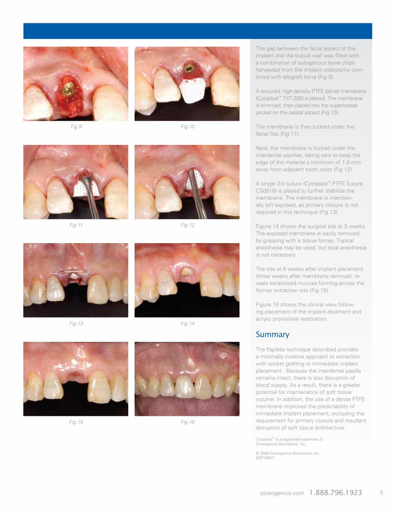
The gap between the facial aspect of the implant and the buccal wall was filled with a combination of autogenous bone chips harvested from the implant osteotomy com-bined with allograft bone (Fig 9).
A textured, high-density PTFE barrier membrane (Cytoplast™ TXT-200) is placed. The membrane is trimmed, then placed into the superiosteal pocket on the palatal aspect (Fig 10).
The membrane is then tucked under the facial flap (Fig 11).
Next, the membrane is tucked under the interdental papillae, taking care to keep the edge of the material a minimum of 1.0 mm away from adjacent tooth roots (Fig 12).
A single 3-0 suture (Cytoplast™ PTFE Suture; CS0518) is placed to further stabilize the membrane. The membrane is intention-ally left exposed, as primary closure is not required in this technique (Fig 13).
Figure 14 shows the surgical site at 3 weeks. The exposed membrane is easily removed by grasping with a tissue forcep. Topical anesthesia may be used, but local anesthesia is not necessary.
The site at 6 weeks after implant placement (three weeks after membrane removal), re-veals keratinized mucosa forming across the former extraction site (Fig 15).
Figure 16 shows the clinical view follow-ing placement of the implant abutment and acrylic provisional restoration.
Summary
The flapless technique described provides a minimally invasive approach to extraction with socket grafting or immediate implant placement. Because the interdental papilla remains intact, there is less disruption of blood supply. As a result, there is a greater potential for maintenance of soft tissue volume. In addition, the use of a dense PTFE membrane improves the predictability of immediate implant placement, excluding the requirement for primary closure and resultant disruption of soft tissue architecture.
Cytoplast™ is a registered trademark of Osteogenics Biomedical, Inc.
© 2008 Osteogenics Biomedical, Inc.BBFY0607

6
A Dual-Layer Membrane Technique for Immediate Implant Placement in the Esthetic Zone Barry K. Bartee, DDS, MD
This is a 60 year-old female who presented with a crown-root fracture of the maxillary right central incisor. The crown was retained with denture adhesive (Fig 1a and b). A thin gingival biotype and multiple, adjacent por-celain fused to metal restorations increased the esthetic risk in this case. To minimize soft and hard tissue recession, a minimally invasive extraction technique and immediate implant placement combined with guided tissue regeneration was planned.
The tooth root was extracted using only an intrasulcular incision and elevation with a micro periosteal elevator. Following curet-tage of the socket, an implant was placed towards the palatal wall of the socket. A thin buccal plate was noted. The gap between the implant and the buccal wall of the socket (2.5 mm) was grafted with demineralized allograft bone and beta tricalcium phosphate (Cerasorb®, Riemser Arzneimittel AG) (Fig 2).
To thicken the soft tissue while maintaining the natural position of the mucogingival junc-tion, a dual layer GTR technique was used, employing a cross-linked type 1 bovine col-lagen membrane covered with a high-density PTFE (dPTFE) barrier membrane (Fig 3).
To stabilize the barrier membranes, a subperiosteal pocket was developed on the facial and palatal aspect of the socket. Next, the bovine collagen membrane (Cytoplast™ RTM Collagen) was placed to extend approxi-mately 5 mm beyond the socket margins (Fig 4). To protect the collagen membrane and further stabilize the site, a textured dPTFE membrane (Cytoplast™ TXT-200) was placed over the collagen (Fig 5).
Closure was achieved with a criss-cross 3-0 PTFE suture (Cytoplast™ PTFE Suture) (Fig 6). Note that primary closure was not required due to the presence of the dPTFE membrane and its ability to remain exposed without epithelial or bacterial penetration. The suture was removed at 2 weeks, and the soft tissue overlying the exposed mem-brane demonstrated healing without signs of inflammation.
Fig 1a Fig 1b
Fig 2 Fig 3
Fig 4 Fig 5
Fig 6 Fig 7

osteogenics.com 1.888.796.1923 7
After 4 weeks, the dPTFE membrane was removed non-surgically with topical anesthe-sia. (Fig 8a and 8b). Immediately following removal of the dPTFE barrier, the collagen membrane is observed intact and with a developing blood supply (Fig 9).
After four months of healing, the soft tissue is stable with full interproximal papillae (Fig 10) and preservation of the natural mucogin-gival architecture. To aid in development of soft tissue contours, a removable tempo-rary partial denture was used with an ovate pontic. Radiograpically, there is good bone density adjacent to the implant and mainte-nance of the interdental crest.
The restorative phase included placement of a custom Procera zirconia abutment (Fig 11) and a processed acrylic restoration. After 12 weeks of provisional loading, the soft tissues were stable, with preservation of anatomical contours.
Summary
This case demonstrates the use of a du-al-layer technique for immediate placement of implants into extraction sockets. While bone formation and successful integration will occur with a gap as wide as 2.0 mm, as much as 56% of the buccal-palatal width is lost during the early healing phase.1 This loss of tissue thickness can result in apical migration of the gingival margin, loss of the interdental papilla and discoloration of the soft tissues due to show-through of the underlying dental implant. This technique, using the principles of guided tissue regen-eration combined with augmentation of the gap, results in preservation of the natural contours, even in high-risk sites.
1. Botticelli D, Berglundh T, Lindhe J. Hard-tissue alterations following immediate implant placement in extraction sites.J Clin Periodontol 2004 Oct;31(10):820-8.
Cytoplast™ is a registered trademark of Osteogenics Biomedical, Inc.
Cerasorb® is a registered trademark of Riemser Arzneimittel AG.
© 2008 Osteogenics Biomedical, Inc.BBJJ0607
Fig 8a Fig 8b
Fig 9 Fig 10
Fig 11 Fig 12

8
Minimally Invasive Socket Reconstruction Using a High-Density Titanium-Reinforced PTFE Membrane Barry K. Bartee, DDS, MD
A flapless and minimally invasive approach to socket reconstruction, facilitated by the unique characteristics of high-density titanium-reinforced PTFE membrane is illus-trated in this case. The patient, a 50 year-old female, presented with a severe buccal wall defect secondary to a vertical root fracture (Fig 1). A chronic fistula was present, but was not actively draining at the time of surgery. The tooth was removed using an intrasulcular incision without reflecting the interdental papillae (Fig 2).
Upon curettage and exploration of the socket, the entire buccal wall was found to be missing. Granulation tissue, which was adherent to the facial flap, was removed with sharp dissection (Fig 3) and the socket was irrigated with sterile saline. Next, a subperi-osteal pocket was developed on the facial and palatal aspect of the socket, extending 3 mm beyond the defect margins (Fig 4).
A combination of mineralized and demineral-ized allograft bone was mixed with approxi-mately 25 mg of clindamycin and placed into the socket (Fig 5). A high-density titanium-re-inforced PTFE membrane (Cytoplast™ Ti-250 Anterior Narrow) was shaped to completely cover the facial defect and to cover the coro-nal aspect of the socket, overlapping the de-fect margins by 3 mm. The membrane was introduced into the facial pocket first (Fig 6) then under the palatal flap (Fig 7) and finally tucked under the interdental papillae, taking care to keep the margins of the membrane at least 1 mm from the roots of the adjacent teeth.The single titanium strut facilitates precise placement and stabilization of the de-vice. Adaptation of the flap to the membrane surface was achieved with a single 3-0 PTFE suture (Cytoplast™ PTFE Suture; CS0518) (Fig 8). Note that primary closure was not attempted in an effort to preserve the soft tissue architecture of the site.
Fig 1 Fig 2
Fig 3 Fig 4
Fig 5 Fig 6
Fig 7 Fig 8

osteogenics.com 1.888.796.1923 9
After 3 weeks of healing, the soft tissue around the exposed membrane exhibited no inflammation (Fig 9). After four weeks of healing, the membrane was removed non-surgically by simply removing it through the socket opening. At 6 months of healing, there was adequate ridge width for place-ment of a dental implant as well as mainte-nance of the soft tissue architecture (Figs 10 and 11a & b).
A biopsy taken at the time of implant place-ment revealed the presence of 80% vital bone (Fig12). (Histology by Michael Rohrer, DDS, MS.) Complete regeneration of the socket and facial bone contour was evident at the time of implant placement, six months following the grafting procedure (Fig 13).
The implant was exposed at 4 months and restored with a zirconium abutment and all-ceramic restoration (Fig 14). The post-treat-ment radiograph demonstrates total regen-eration of the socket defect and maintenance of the interproximal height of bone (Fig 15).
Summary
There are several advantages of a high-density titanium-reinforced PTFE membrane. In defects where an entire wall is missing, there is a tendency for loss of volume as the underlying graft material undergoes consolidation and replacement by vital bone. The addition of the titanium strut provides support to the overlying soft tissue prevent-ing its collapse into the defect, resulting in increased bone volume. Additionally, in a minimally invasive technique such as the one illustrated, the presence of the strut allows the surgeon to precisely position the mem-brane under flaps with minimal dissection and flap reflection.
Cytoplast™ is a registered trademark of Osteogenics Biomedical, Inc.
© 2008 Osteogenics Biomedical, Inc.BBJJ0607
Fig 9 Fig 10
Fig 11a Fig 11b
Fig 12 Fig 13
Fig 14 Fig 15

10
Immediate Implant Placement and Socket Reconstruction Using a High-Density Titanium-Reinforced PTFE Membrane Barry K. Bartee, DDS, MD
A 55 year-old female presented for implant placement in a recent extraction site. Surgical exposure revealed fibrous healing at the buc-cal and coronal aspect of the site, requiring augmentation simultaneous with implant placement (Fig 1 and Fig 2) to regenerate the buccal bone contour.
A high-density titanium-reinforced PTFE membrane in a single-tooth configuration (Cytoplast™ Ti-250 Anterior Narrow) was trimmed to fit over the defect and then curved over an instrument handle to provide three-dimensional support and stability (Fig 3a and Fig 3b).
Mineralized bone allograft was placed into the defect (Fig 4) and covered with the mem-brane. The membrane is trimmed to remain 1.0 mm away from the roots of the adjacent teeth, and to extend 3 to 5 mm beyond the defect margins (Fig 5).
Primary closure was achieved using a 3-0 PTFE suture (Cytoplast™ PTFE Suture; CS0518) (Fig 6). After four months of un-eventful healing, the soft tissue covering the membrane appears healthy prior to implant exposure and abutment placement (Fig 7).
Fig 1 Fig 2
Fig 3a Fig 3b
Fig 4 Fig 5
Fig 6 Fig 7

osteogenics.com 1.888.796.1923 11
Four months after implant placement, re-generation of hard tissue is evident radio-graphically (Fig 8). Exposure of the barrier is accomplished using a u-shaped incision with apical advancement of the keratinized gingiva ( Fig 9). The high-density PTFE membrane is easily removed through a conservative inci-sion due to limited soft tissue ingrowth into the barrier (Fig 10).
Clinically, restoration of the full width of keratinized gingiva was observed at the time of abutment placement (Fig 11). After soft tissue healing, the restorative components were placed and the implant was restored with a porcelain fused to metal restoration (Fig 12 and Fig 13).
Summary
This case report demonstrates the success-ful augmentation of a localized defect involv-ing the entire buccal plate of a recent extrac-tion site. The use of a titanium-reinforced, high-density PTFE membrane provides predictable space-making and regenerative function without the risks associated with highly porous, expanded PTFE devices such as Gore-Tex®.
Cytoplast™ is a registered trademark of Osteogenics Biomedical, Inc.
© 2008 Osteogenics Biomedical, Inc.BBDI0607
Fig 8 Fig 9
Fig 10 Fig 11
Fig 12 Fig 13

12
Ridge Augmentation with Immediate Implant Placement Using a High-Density Titanium-Reinforced PTFE Membrane Marco Ronda, DDS
This is a 49 year old female who presented for implant placement in the left posterior mandible. Preoperative radiographs reveal inadequate bone height for ideal implant placement and restoration (Fig 1).
Three tapered implants were placed at sec-ond bicuspid, first molar and second molar areas, and the vertical defect was measured from crestal height to the neck of the implant (Fig 2 and 3). The defect measurements at the implant positions were 9 mm, 8 mm and 4 mm respectively. The implant measure-ments were 3.7 mm x 10 mm, 4.7 mm x 11.5 mm and 4.7 mm x 8 mm, respectively.
The alveolar ridge was decorticated and a high-density titanium-reinforced PTFE mem-brane (Cytoplast™ Ti-250 XL) was secured lingually with two pins (Fig 4). This membrane configuration is ideal to cover three implants. The membrane was then bent to a desired three-dimensional shape to provide stability while utilizing the implants as tenting support.
A combination (50:50 ratio) of mineralized cortical and cancellous allograft was hydrated with PRGF and placed around the implants and to the desired crestal height (Fig 5). The membrane was then draped over the graft and trimmed 1 mm from the adjacent tooth and secured with three pins buccally and two pins crestally (Fig 6).
Advancement of the buccal flap is accom-plished by the use of a periosteal releasing incision along the full length of the flap. Care is taken to avoid damaging the neurovas-cular bundle (Fig 7). On the lingual side a new technique developed by the author for the extension of the flap was used (Fig 8). (Ronda M, Stacchi C. A Novel Approach for the Coronal Advancement of the Buccal Flap.Int J Periodontics Restorative Dent. 2015 Nov-Dec;35(6):795-801.)
Fig 1 Fig 2
Fig 3 Fig 4
Fig 5 Fig 6
Fig 7 Fig 8
osteogenics.com 1.888.796.1923 13
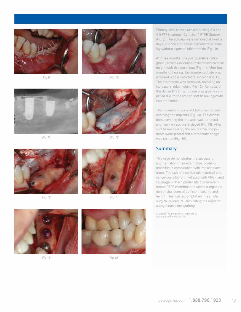
Primary closure was achieved using 3-0 and 4-0 PTFE sutures (Cytoplast™ PTFE Suture) (Fig 9). The sutures were removed at twelve days, and the soft tissue demonstrated heal-ing without signs of inflammation (Fig 10).
At three months, the postoperative radio-graph provides evidence of increased alveolar height with this technique (Fig 11). After four months of healing, the augmented site was exposed with a mid-crestal incision (Fig 12).The membrane was removed, revealing an increase in ridge height (Fig 13). Removal of the dense PTFE membrane was greatly sim-plified due to the limited soft tissue ingrowth into the barrier.
The presence of compact bone can be seen overlying the implants (Fig 14). The excess bone covering the implants was removed and healing caps were placed (Fig 15). After soft tissue healing, the restorative compo-nents were placed and a temporary bridge was seated (Fig. 16).
Summary
This case demonstrates the successful augmentation of an edentulous posterior mandible in combination with implant place-ment. The use of a combination cortical and cancellous allograft, hydrated with PRGF, and coverage with a high-density titanium-rein-forced PTFE membrane resulted in regenera-tion of vital bone of sufficient volume and height. This was accomplished in a single surgical procedure, eliminating the need for autogenous block grafting.
Cytoplast™ is a registered trademark of Osteogenics Biomedical, Inc.
Fig 9 Fig 10
Fig 11 Fig 12
Fig 13 Fig 14
Fig 15 Fig 16
14
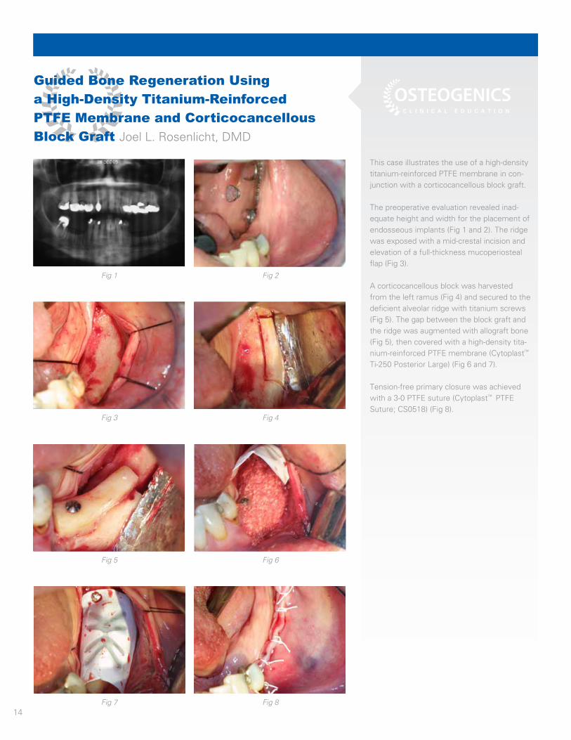
Guided Bone Regeneration Using a High-Density Titanium-Reinforced PTFE Membrane and Corticocancellous Block Graft Joel L. Rosenlicht, DMD
This case illustrates the use of a high-density titanium-reinforced PTFE membrane in con-junction with a corticocancellous block graft.
The preoperative evaluation revealed inad-equate height and width for the placement of endosseous implants (Fig 1 and 2). The ridge was exposed with a mid-crestal incision and elevation of a full-thickness mucoperiosteal flap (Fig 3).
A corticocancellous block was harvested from the left ramus (Fig 4) and secured to the deficient alveolar ridge with titanium screws (Fig 5). The gap between the block graft and the ridge was augmented with allograft bone (Fig 5), then covered with a high-density tita-nium-reinforced PTFE membrane (Cytoplast™
Ti-250 Posterior Large) (Fig 6 and 7).
Tension-free primary closure was achieved with a 3-0 PTFE suture (Cytoplast™ PTFE Suture; CS0518) (Fig 8).
Fig 1 Fig 2
Fig 3 Fig 4
Fig 5 Fig 6
Fig 7 Fig 8

osteogenics.com 1.888.796.1923 15
The postoperative panoramic radiograph demonstrates the increased alveolar height achievable with this technique (Fig 9).
8 months later, the membrane was exposed with a mid-crestal incision. (Fig 10 and 11). Compared to expanded PTFE membranes, removal of the dense PTFE membrane is greatly simplified due to the limited soft tissue ingrowth and attachment to the barrier.
An increase in ridge height and width was achieved allowing placement of implants into ideal position (Fig 12 and 13).
Cytoplast™ is a registered trademark of Osteogenics Biomedical, Inc.
© 2009 Osteogenics Biomedical, Inc.BBJJ0607
Fig 9 Fig 10
Fig 11 Fig 12
Fig 13

16
The Use of Tenting Screws with High-Density Titanium-Reinforced PTFE Membrane Joel L. Rosenlicht, DMD
A 45 year-old male presented with a substan-tial loss of buccal bone contour and in need of an endosseous implant to replace the maxillary left lateral incisor (Fig 1a-1c).
The alveolar ridge was surgically exposed and decorticated in preparation for bone grafting (Fig 2).
A titanium tenting screw 5.0 mm in length and specifically designed for guided tissue regeneration (JLR Tenting Screw Kit, KLS Martin L.P., Jacksonville, FL) was placed to augment the ridge to a predetermined contour (Fig 3).
A composite particulate graft, consisting of demineralized bone putty combined with beta-tricalcium phosphate granules, was then placed and covered with a high-density titanium-reinforced PTFE membrane (Cyto-plast™ Ti-250 Posterior Large) and primary closure was achieved using a 3-0 PTFE suture (Cytoplast™ PTFE Suture; CS0518) (Fig 4a and 4b).
After 6 months of healing, the augmented site was exposed (Fig 5a) and the membrane was removed (Fig 5b), revealing dense corti-cal bone under the membrane.
Fig 1a
Fig 2 Fig 3
Fig 4a Fig 4b
Fig 5a Fig 5b
Fig 1b Fig 1c

osteogenics.com 1.888.796.1923 17
Upon removal of the tenting screw (Fig 5c), it is apparent total reconstruction of the ridge contour, up to the height predetermined by the tenting screw and membrane, was achieved.
A CT scan taken prior to the removal of the tenting screw and membrane reveals a sub-stantial increase in width, from 2.9 mm to 8.5 mm, greatly facilitating implant placement in the proper three-dimensional position. (Fig 6a - 6c).
Cytoplast™ is a registered trademark of Osteogenics Biomedical, Inc.
© 2009 Osteogenics Biomedical, Inc.BBJJ0607
Fig 5c Fig 5d
Fig 6a Fig 6b Fig 6c

18
Implant Site Development Using a Bovine Collagen Membrane and Allogeneic Bone Barry K. Bartee, DDS, MD
A 48 year-old female presented for implant replacement of the maxillary right first molar, which had been extracted 6 months previ-ously. There was a substantial hard tissue de-fect requiring augmentation prior to implant placement (Fig 1a and Fig 1b). The original plan was to augment the site in two stages. First, a particulate graft would be used to expand the soft tissue envelope, and then an autogenous block graft would be placed.
The initial surgical exposure of the healing socket revealed soft tissue extending up to and including the antral floor (Fig 2a). After removal of the soft tissue, the antral membrane was found to be intact, as well as the palatal wall and the mesial and distal bony walls. The buccal plate and floor of the socket were missing (Fig 2b).
Allogeneic bone putty (Regenaform® Moldable Allograft Paste, Exactech Dental Biologics) was mixed according to the manufacturer’s directions, placed into the defect, and shaped to restore the contour of the ridge (Fig 3).
A bovine collagen guided tissue regeneration membrane (Cytoplast™ RTM Collagen) was trimmed to fit over the graft (Fig 4 and Fig 5). Primary closure was achieved over the membrane and graft using 3-0 PTFE sutures (Cytoplast™ PTFE Suture; CS0518) (Fig 6).
Fig 1a Fig 1b
Fig 2a Fig 2b
Fig 3 Fig 4
Fig 5 Fig 6
osteogenics.com 1.888.796.1923 19
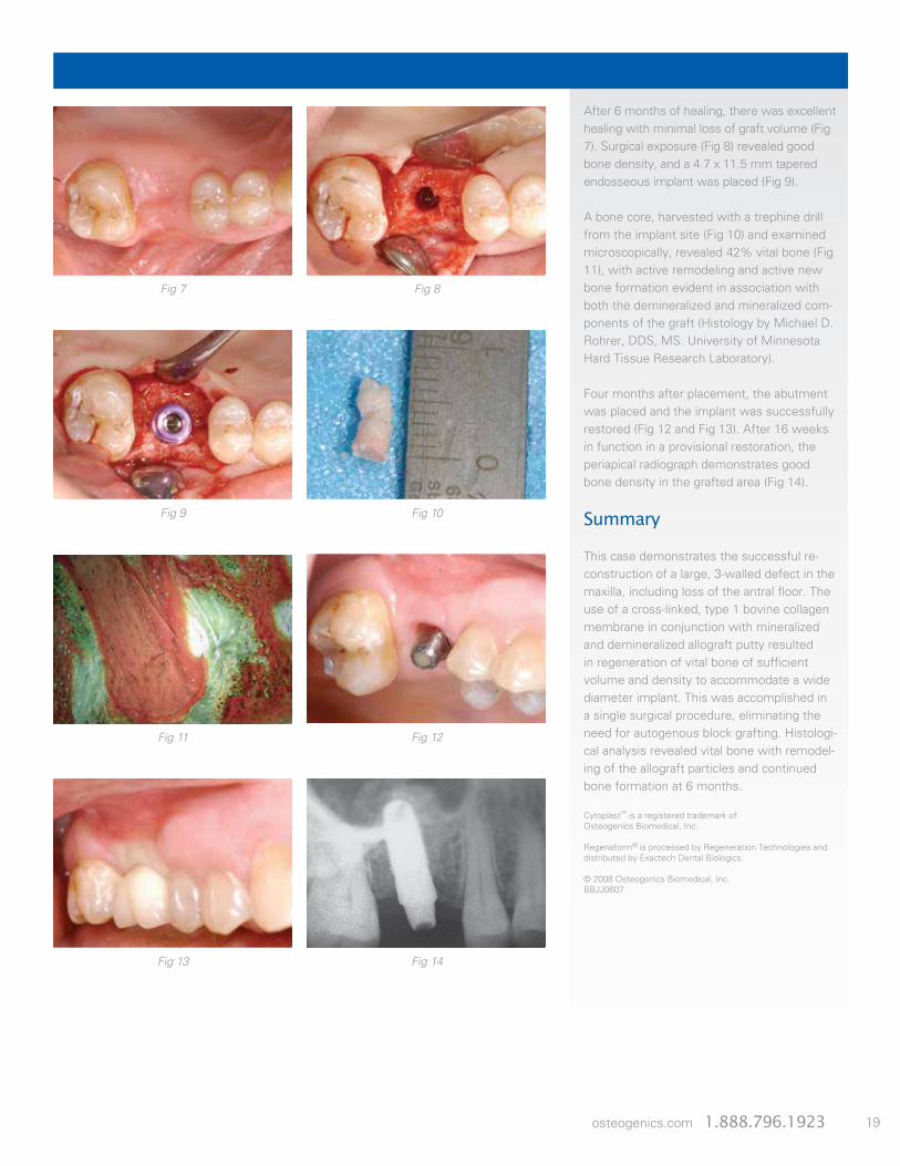
After 6 months of healing, there was excellent healing with minimal loss of graft volume (Fig 7). Surgical exposure (Fig 8) revealed good bone density, and a 4.7 x 11.5 mm tapered endosseous implant was placed (Fig 9).
A bone core, harvested with a trephine drill from the implant site (Fig 10) and examined microscopically, revealed 42% vital bone (Fig 11), with active remodeling and active new bone formation evident in association with both the demineralized and mineralized com-ponents of the graft (Histology by Michael D. Rohrer, DDS, MS. University of Minnesota Hard Tissue Research Laboratory).
Four months after placement, the abutment was placed and the implant was successfully restored (Fig 12 and Fig 13). After 16 weeks in function in a provisional restoration, the periapical radiograph demonstrates good bone density in the grafted area (Fig 14).
Summary
This case demonstrates the successful re-construction of a large, 3-walled defect in the maxilla, including loss of the antral floor. The use of a cross-linked, type 1 bovine collagen membrane in conjunction with mineralized and demineralized allograft putty resulted in regeneration of vital bone of sufficient volume and density to accommodate a wide diameter implant. This was accomplished in a single surgical procedure, eliminating the need for autogenous block grafting. Histologi-cal analysis revealed vital bone with remodel-ing of the allograft particles and continued bone formation at 6 months.
Cytoplast™ is a registered trademark of Osteogenics Biomedical, Inc.
Regenaform® is processed by Regeneration Technologies and distributed by Exactech Dental Biologics.
© 2008 Osteogenics Biomedical, Inc.BBJJ0607
Fig 7 Fig 8
Fig 9 Fig 10
Fig 11 Fig 12
Fig 13 Fig 14

20
A 43 year-old female presented for replace-ment of the mandibular right first molar and second premolar. The teeth had been extracted 20 years previously. There was a combined hard and soft tissue defect requiring augmentation prior to implant placement (Fig 1).
A mid-crestal incision was used to expose the atrophic edentulous ridge. A surgical burr was used to decorticate the bone in prepara-tion for grafting (Fig 2).
Allogeneic bone putty (Regenaform® Mold-able Allograft Paste, Exactech Dental Biolog-ics) was hydrated with PRP and then mixed with autogenous cortical bone harvested with a bone scraper (Fig 3a and Fig 3b).
A cross-linked type 1 bovine collagen mem-brane (Cytoplast™ RTM Collagen) was placed over the graft (Fig 4a and Fig 4b). Primary closure was achieved with 3-0 PTFE sutures (Cytoplast™ PTFE Suture; CS0518) (Fig 5a and Fig 5b).
Fig 1 Fig 2
Fig 3a Fig 3b
Fig 4a Fig 4b
Fig 5a Fig 5b
Guided Bone Regeneration Using a Bovine Collagen Membrane, Platelet-rich Plasma and Allogeneic Bone Putty Barry K. Bartee, DDS, MD
osteogenics.com 1.888.796.1923 21
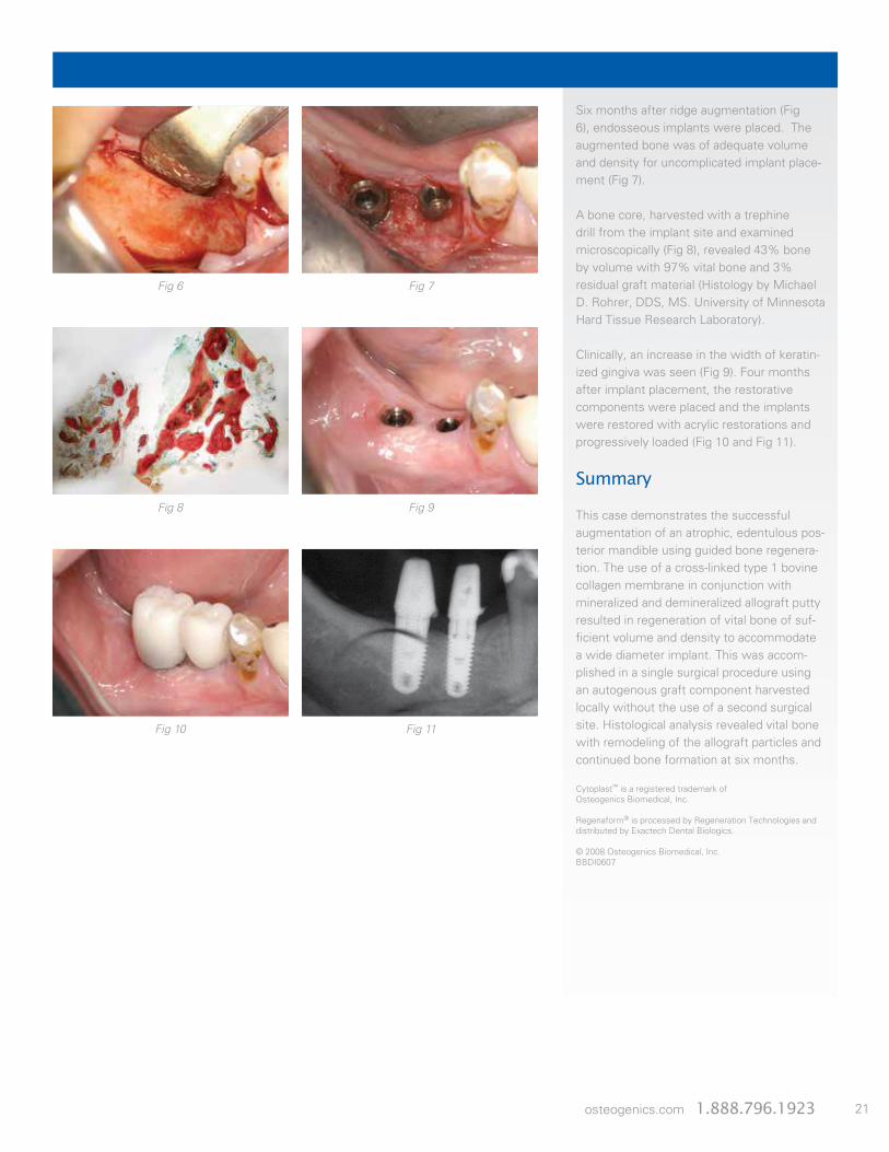
Six months after ridge augmentation (Fig 6), endosseous implants were placed. The augmented bone was of adequate volume and density for uncomplicated implant place-ment (Fig 7).
A bone core, harvested with a trephine drill from the implant site and examined microscopically (Fig 8), revealed 43% bone by volume with 97% vital bone and 3% residual graft material (Histology by Michael D. Rohrer, DDS, MS. University of Minnesota Hard Tissue Research Laboratory).
Clinically, an increase in the width of keratin-ized gingiva was seen (Fig 9). Four months after implant placement, the restorative components were placed and the implants were restored with acrylic restorations and progressively loaded (Fig 10 and Fig 11).
Summary
This case demonstrates the successful augmentation of an atrophic, edentulous pos-terior mandible using guided bone regenera-tion. The use of a cross-linked type 1 bovine collagen membrane in conjunction with mineralized and demineralized allograft putty resulted in regeneration of vital bone of suf-ficient volume and density to accommodate a wide diameter implant. This was accom-plished in a single surgical procedure using an autogenous graft component harvested locally without the use of a second surgical site. Histological analysis revealed vital bone with remodeling of the allograft particles and continued bone formation at six months.
Cytoplast™ is a registered trademark of Osteogenics Biomedical, Inc.
Regenaform® is processed by Regeneration Technologies and distributed by Exactech Dental Biologics.
© 2008 Osteogenics Biomedical, Inc.BBDI0607
Fig 6 Fig 7
Fig 8 Fig 9
Fig 10 Fig 11

22
Ridge Preservation usingPorcine Xenograft and dPTFE MembraneGustavo Avila-Ortiz, DDS, MS, PhDAssociate Professor and Graduate Program Director, Department of Periodontics, University of Iowa College of Dentistry
Chris Barwacz, DDS, FAGDAssistant Professor, Department of Family Dentistry, University of Iowa College of Dentistry
A 47-year old female with no contributory medical history presented with a cervical tooth fracture on the right maxillary lat-eral incisor (Fig 1a & 1b). A thorough clinical examination was conducted. All periodontal parameters were normal, except for BOP on the mid-facial. Plaque control was adequate. A CBCT scan and a periapical radiograph indicated normal interproximal bone levels, and the thickness of the buccal bone ranged from 0.6 to 0.9 mm (Fig 2a & 2b). Different treatment options were considered, including tooth conservation and tooth-supported FPD, but patient opted for tooth replacement with an implant-supported prosthesis.
The remaining tooth structure was extracted in a minimally invasive fashion to avoid dam-age to the supporting hard and soft tissue structures (Fig 3). Following meticulous debridement and irrigation of the socket, sub-periosteal pockets were created on the buccal and lingual aspects of the alveolar ridge. A dPTFE (dense polytetrafluoroethylene) mem-brane (Cytoplast™ TXT Singles) was gently tucked into the buccal pocket and porcine-de-rived cancellous xenograft particles (Zcore™) were placed in the socket, up to the level of the crestal bone (Fig 4). The dPTFE membrane was then tucked into the lingual pocket and a horizontal cross mattress suturing technique using PTFE suture (Cytoplast™ PTFE Suture 4-0) was placed to secure the membrane and stabilize the soft tissue margins (Fig 5). The application of a dPTFE membrane allows for a conservative regenerative approach that does not require primary closure via buccal flap advancement. Immediately following this ridge preservation procedure, a periapi-cal radiograph was taken to verify that the xenograft particles were level with the crestal bone (Fig 6). Before dismissal, patient was instructed to clean the exposed membrane area by carefully swabbing twice daily with a cotton pellet soaked in Chlorhexidine 0.12% aqueous solution.
Suture removal occurred at the one week post-op appointment and soft tissue healing was progressing as predicted. The dPTFE membrane was visible through the soft tissue
Fig 1a Fig 1b
Fig 2bFig 2a
Fig 3 Fig 4
Fig 5 Fig 6

osteogenics.com 1.888.796.1923 23
and the membrane had remained in the origi-nal site of placement.
At 3 weeks, non-disturbed healing continued. Keratinized tissue was preserved and no signs of inflammation or swelling were present (Fig 7).
At 6 weeks, the membrane was removed using tissue forceps (Fig 8a). Upon clinical examination, the soft tissue architecture had remained intact. Slight bleeding could be observed around the perimeter of the wound and in the area immediately underlying the membrane, which is normal (Fig 8b).
At 12 weeks, the soft tissue appears mature and a supracrestal increase in the band of keratinized tissue was visible. Esthetics of the anterior ridge have been preserved at an optimal level (Fig 9).
At 20 weeks, another CBCT scan was obtained. Radiographic evaluation of the site revealed that ridge volume and bone density were adequate to proceed with the plan to surgically place an implant following a com-puter guided surgery protocol (Fig 10). The implant was placed using a steriolithic surgical guide at 5 months after tooth extraction. Using a 2.75 mm trephine and the surgical guide, a core of the grafted site was taken for histologic and histomorphometric analysis (Fig 11). The amount of newly formed vital bone was approximately 35%. Excellent integra-tion of the remaining xenograft particles was observed throughout the sample in absence of any inflammatory infiltrate, which illustrates the biocompatibility and the osteoconductive properties of this grafting material (Fig 12a & 12b).
At 1 week following implant placement, an abundance of keratinized tissue is seen, with a portion encroaching the healing abutment (Fig 13).
The final single-tooth implant crown was delivered at 10 weeks after implant placement using a custom zirconia abutment that was hand-layered with porcelain. A natural emer-gence profile and a very satisfactory esthetic and functional outcome was achieved (Fig 14). A periapical radiograph was taken to verify the maintenance of normal marginal bone levels and absence of pathosis (Fig 15).
Cytoplast™ and Zcore™ are registered trademarks of Osteogenics Biomedical, Inc.
Fig 8a
Fig 8b Fig 9
Fig 10 Fig 11
Fig 12a Fig 12b
Fig 13 Fig 14 Fig 15
Coronal Coronal
Apical Apical
Fig 7

24
Multi-site Ridge Preservation/Reconstruction Using Porcine Xenograft and dPTFE MembraneDan Cullum, DDS
A 68 year-old female patient presented with refractory periodontal disease in the maxillary right first and second molar teeth. The patient requested removal of the failing teeth and implant reconstruction as she was concerned with loss of function. The clinical exam revealed recession, bleeding on probing and 10 mm pocket depths (Fig 1-2). Radiographic evaluation revealed vertical bony defects with furcation involvement (Fig 3-4). Her oral hygiene at the time of the exam appeared adequate. The medical history was non-con-tributory. A delayed approach was selected due to concern for achieving control of the periodontal disease, with preservation and reconstruction of the horizontal, vertical and intra-bony defects. Re-evaluation was planned after healing to confirm adequate periodontal disease control prior to reconstruction with implant supported restorations.
The teeth were extracted using minimally in-vasive protocols to preserve the residual bony housing and soft tissue architecture. Signifi-cant interproximal and inter-furcal vertical bony defects were observed (Fig 5) and the sockets were carefully debrided. Minimal peripheral periosteal elevation was done for placement of dPTFE (dense polytetrafluoroethylene) membranes (Cytoplast™ TXT 200) after graft-ing. The extraction defects were then grafted with porcine-derived cancellous xenograft particles (Zcore™) saturated with autologous venous blood (Fig 6).
The dPTFE membranes were trimmed and placed in the sub-periosteal space between the periosteum and the existing bony housing. Next, the soft tissues and membrane are stabilized using 4-0 PTFE (Cytoplast™) with a combination of mattress and interrupted sutures (Fig 7). The impervious nature of the dPTFE membranes allows direct exposure to the oral cavity without risk of bacterial penetration into the surgical site. Advan-tages of the open grafting technique include preservation of the muco-gingival junction and maintenance of vestibular depth, as the exposed dPTFE membrane minimalizes the need for flap elevation and avoids the need for flap advancement for primary closure.
Fig 1 Fig 2
Fig 4Fig 3
Fig 5 Fig 6
Fig 7 Fig 8

osteogenics.com 1.888.796.1923 25
At two weeks post-op, undisturbed healing of the sites was noted. Slight discoloration and plaque accumulation on the dPTFE mem-branes was noted, with no signs of inflamma-tion or swelling of the surgical site (Fig 8). At that time the sutures were removed.
At five weeks the patient returned and the membrane was removed. The surgical sites demonstrated excellent healing, uncontami-nated by bacteria (Fig 9).
After six months the soft tissues have com-pletely healed and a wide band of attached gingival tissue can be observed (Fig 10). The soft tissue architecture and vestibular depth has been maintained as a direct result of avoid-ing flap elevation and advancement for primary closure at the initial surgery. Radiographic im-ages demonstrate excellent bony healing and ridge dimensions (Fig 11 - 12a & b).
The implant surgical site was exposed with a palatally incised, buccal- based flap for planned apical flap positioning on closure. The grafted sites were fully incorporated and excellent bone volume preservation for implant place-ment was seen. X-Nav® dynamic navigational surgery was used to harvest a bone core with a 3mm trephine at the grafted first molar site for histological evaluation (Fig 13a & 16). Implant site preparation was completed using dynamic navigation and a combined oseotome sinus floor elevation at the second molar site (Fig 13b).
5.5 mm wide platform conical connection implants were then placed at the molar sites (Fig 14). Expanded emergence PEEK healing abutments were used to develop proper emergence profile. Chromic gut (4-0) suture was used with an interrupted suturing technique to approximate the wound margins and stabilize the apically-positioned flap (Fig 15). Immediate post-op radiograph confirms implant placement.
At four months post-implant placement the patient was released for restoration. Defini-tive restorations were placed with an optimal functional and esthetic result (Fig 17) and the post treatment radiograph can be seen in Figure 18.
Cytoplast™ and Zcore™ are registered trademarks of Osteogenics Biomedical, Inc.
X-Nav® is a registered trademark of X-Nav Technologies, LLC.
Fig 10
Fig 11 Fig 12a & 12b
Fig 13a & 13b Fig 14
Fig 17
Fig 15
Fig 18
Fig 16
Fig 9

26
1. Preoperative view. To maximize the result of ridge preservation procedures, techniques designed to minimize trauma to the alveolar bone, such as the use of periotomes and surgical sectioning of ankylosed roots should be considered.
2. All soft tissue remnants should be removed with sharp curettage. Special care should be taken to remove all soft tissue at the apical extent of the socket of endodontically treated teeth. Bleeding points should be noted on the cortical plate. If necessary, decortication of the socket wall should be done with a #2 round burr to improve blood supply.
3. A subperiosteal pocket is created with a micro periosteal elevator or small curette, extend-ing 3-5 mm beyond the socket margins on the palatal and the facial aspect of the socket. In the esthetic zone, rather than incising and elevating the interdental papilla, it is left intact and undermined in a similar fashion. The Cytoplast™ high-density PTFE membrane will be tucked into this subperiosteal pocket.
4. Particulate graft material can be placed into the socket with a syringe or with a curette. Ensure that the material is evenly distributed throughout the socket. However, the particles should not be densely packed to preserve ample space for blood vessel ingrowth.
5. The Cytoplast™ high-density PTFE membrane is trimmed to extend 3-5 mm beyond the socket walls and then tucked subperiosteally under the palatal flap, the facial flap and under-neath the interdental papilla with a curette. The membrane should rest on bone 360° around the socket margins, if possible. Note that minimal flap reflection is necessary to stabilize the membrane.
6. Ensure that there are no folds or wrinkles in the membrane and that it lies passively over the socket. To prevent bacterial leakage under the membrane, take care to avoid puncturing the membrane, and do not overlap two adjacent pieces of membrane material.
7. The membrane is further stabilized with a criss-cross Cytoplast™ PTFE suture. Alternatively, interrupted sutures may be placed. The PTFE sutures, which cause minimal inflammatory response, are left in place for 10 to 14 days.
8. The membrane is removed, non-surgically, in 21 to 28 days. Sockets with missing walls may benefit from the longer time frame. Topical anesthetic is applied, then the membrane is grasped with a tissue forcep and removed with a gentle tug.
9. Studies have shown that by 21-28 days there is a dense, vascular connective tissue matrix in the socket and early osteogenesis is observed in the apical 2/3 of the socket.
10. Immediately following membrane removal, a dense, highly vascular, osteoid matrix is observed. The natural position of the gingival margin has been left intact because primary closure was not necessary. The dense PTFE membrane has contained the graft material and prevented epithelial migration into the socket.
11. The socket at 6 weeks. Keratinized gingiva is beginning to form over the grafted socket. The natural soft tissue architecture is preserved, including the interdental papillae. New bone is beginning to form in the socket. Over the next 6 to 10 weeks, increasing thickness of tra-beculae and mineralization will result in load bearing bone suitable for implant placement.
2.
3.
4.
5.
6.
7.
8.
9.
10.
11.
1.™ Technique
Extraction Site Grafting Without Primary Closure

osteogenics.com 1.888.796.1923 27
Selection of Applicable References
Membrane References
• Ronda M, Stacchi C. A Novel Approach for the Coronal Advancement of the Buccal Flap. Int J Periodontics Restorative Dent. 2015 Nov-Dec;35(6):795-801.
• Urban IA, Monje A, Wang HL. Vertical RidgeAugmentation and Soft Tissue Reconstructionof the Anterior Atrophic Maxillae: A Case Series.Int J Periodontics Restorative Dent. 2015 Sep-Oct;35(5):613-23.
• Al-Hezaimi K, Iezzi G, Rudek I, Al-Daafas A, Al-Hamdan K, Al-Rasheed A, Javed F, Piattelli A,Wang HL. Histomorphometric Analysis of Bone Re-generation Using a Dual Layer of Membranes (dPTFE Placed Over Collagen) in Fresh Extraction Sites: A Canine Model. J Oral Implantol. 2015 Apr;41(2):188-95.
• Ronda M, Rebaudi A, Torelli L, Stacchi C. Expandedvs. dense polytetrafluoroethylene membranes in vertical ridge augmentation around dental implants: a prospective randomized controlled clinical trial. Clin Oral Implants Res. 2014 Jul;25(7):859-66.
• Barboza EP, Stutz B, Mandarino D, Rodrigues DM,Ferreira VF. Evaluation of a dense polytetrafluoro-ethylene membrane to increase keratinized tissue: a randomized controlled clinical trial. Implant Dent.2014 Jun;23(3):289-94.
• Urban IA, Lozada JL, Jovanovic SA, Nagursky H, Nagy K. Vertical Ridge Augmentation with Titanium-Reinforced, Dense-PTFE Membranes and a Combina-tion of Particulated Autogenous Bone and Anorganic Bovine Bone-Derived Mineral: A Prospective Case Series in 19 Patients. Int J Oral Maxillofac Implants. 2014 Jan-Feb;29(1):185-93.
• Carbonell JM, Martin IS, Santos A, Pujol A, SanzMoliner JD, Nart J. High-density polytetrafluo-roethylene membranes in guided bone and tissue regeneration procedures: a literature review. Int J Oral Maxillofac Surg. 2014 Jan;43(1):75-84.
• Vittorini Orgeas G, Clementini M, De Risi V, de Sanctis M. Surgical techniques for alveolar socket preservation: a systematic review. Int J Oral Maxillofac Implants. 2013 Jul-Aug;28(4):1049-61.
• Bagoff R, Mamidwar S, Chesnoiu-Matei I, Ricci J, Alexander H, Tovar N. Socket preservation and sinus augmentation using a medical grade calcium sulfate hemihydrate and mineralized irradiated cancellous
bone allograft composite. J Oral Implantol. 2013 Jun;39(3):363-71.
• Waasdorp, J., Feldman, S. Bone regeneration around immediate implants utilizing a dense polytetra-fluoroethylene membrane without primary closure: A report of 3 cases. J Oral Implantol. 2013;39:355-361.
• Zafiropoulos GG, Deli G, Vittorini G, Hoffmann O. Implant placement and immediate loading with fixed restorations in augmented sockets. Five year results. A case report. J Oral Implantol. 2013; 39:372-379.
• Al-Hezaimi K, Rudek I, Al-Hamdan KS, Javed F, Nooh N, Wang HL. Efficacy of using a dual layer of membrane (dPTFE placed over collagen) for ridge pres-ervation in fresh extraction sites: a micro-computed tomographic study in dogs. Clin Oral Implants Res. 2012; (Epub ahead of print).
• Annibali S, Bignozzi I, Sammartino G, La Monaca G, Cristalli MP. Horizontal and Vertical Ridge Augmentation in Localized Alveolar Deficient Sites: A Retrospective Case Series. Implant Dent. 2012 Jun;21(3):175-185.
• Levin B. Immediate temporization of immediate implants in the esthetic zone: Evaluating survival and bone maintenance. Compendium 2011;32:52-62.
• Lee JY, Kim YK, Yun PY, Oh JS, Kim SG.Guided bone regeneration using two types of non-resorbable barrier membranes. J Korean Assoc Oral Maxillofac Surg 2010;36:275-9.
• Park S-Y, Kye S-B, Yang S-M, Shin S-Y.The effect of non-resorbable membrane on buccal bone healing at an immediate implant site: an experi-mental study in dogs. Clin. Oral Impl. Res. xx, 2010; 000–000. doi: 10.1111/j.1600-0501.2010.01995.x
• Barboza EP, Stutz B, Ferreira VF; Carvalho W. Guided bone regeneration using nonexpanded polytet-rafluoroethylene membranes in preparation for dental implant placements – A report of 420 cases. Implant Dent. 2010;19:2-7.
• Zafiropoulos GG, Deli G, Bartee BK, Hoffman O. Single-tooth implant placement and loading in fresh and regenerated extraction sockets. Five-year results: A case series using two different implant designs. J Periodontol. 2010;81:604-615.
• Fotek PD, Neiva RF, Wang HL. Comparison of dermal matrix and polytetrafluoroethylene membrane for socket bone augmentation: A clinical and histologic study. J Periodontol. 2009;80:776-785.
• Hoffman O, Bartee BK, Beaumont C, Kasaj A, Deli G, Zafiropoulos GG. Alveolar bone preservation in extraction sockets using non-resorbable dPTFE membranes: A retrospective non-randomized study. J Periodontol. 2008;79:1355-1369.
• Barber HD, Lignelli J, Smith BM, Bartee BK. Using a dense PTFE membrane without primary closure to achieve bone and tissue regeneration. J Oral Maxillofac Surg. 2007;65:748-752.
• Walters SP, Greenwell H, Hill M, Drisko C, Pickman K, Scheetz JP. Comparison of porous and non-porous teflon membranes plus a xenograft in the treatment of vertical osseous defects: A clinical reentry study. J Periodontol. 2003;74:1161-1168.
• Bartee BK. Extraction site reconstruction for alveo-lar ridge preservation. Part 1: Rationale and material selection. J Oral Implantol. 2001;27:187-193.
• Bartee BK. Extraction site reconstruction for alveolar ridge preservation. Part 2: Membrane-assisted surgical technique. J Oral Implantol. 2001;27:194-197.
• Lamb JW III, Greenwell H, Drisko C, Henderson RD, Scheetz JP, Rebitski G. A comparison of porous and non-porous teflon membranes plus demineralized freeze-dried bone allograft in the treatment of class II buccal/lingual furcation defects: A clinical reentry study. J Periodontol. 2001;72:1580-1587.
• Bartee BK. Evaluation of a new polytetrafluoro-ethylene guided tissue regeneration membrane in healing extraction sites. Compend Contin Educ Dent 1998;19:1256-1264.
• Bartee BK, Carr JA. Evaluation of a high-density polytetrafluoroethylene (n-PTFE) membrane as a bar-rier material to facilitate guided bone regeneration in the rat mandible. J Oral Implantol. 1995;21:88-95.
• Bartee BK. The use of high-density polytetrafluoro-ethylene membrane to treat osseous defects: Clinical reports. Implant Dent. 1995;4:21-26.
Suture References
• Silverstein LH, Kurtzman GM, Shatz PC. Suturing for optimal soft-tissue management. J Oral Implantol. 2009;35:82-90.
• Silverstein LH. Suturing principles: Preserving needle edges during dental suturing. PPAD. 2005;17:562-564.

4620 71st StreetBuilding 78-79Lubbock, TX 79424www.osteogenics.com 1.888.796.1923
Revised 8.17