clinical chest radiography interpretation part 1...clinical chest radiography interpretation part 1...
TRANSCRIPT
Clinical Chest Radiography Interpretation
Part 1
Theresa M Campo DNP, FNP-C, ENP-BC, FAANP
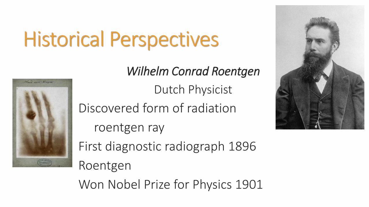
Historical Perspectives
Wilhelm Conrad Roentgen
Dutch Physicist
Discovered form of radiation
roentgen ray
First diagnostic radiograph 1896
Roentgen
Won Nobel Prize for Physics 1901
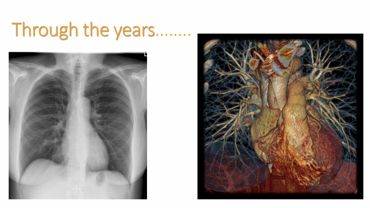
Through the years……..

Image Production
•Strahlung Ray (X-Ray) from cathode tube
•Attenuation of the ray
•Radiant energy – short wave – greater ability to penetrate objects
•Cassette
•Image
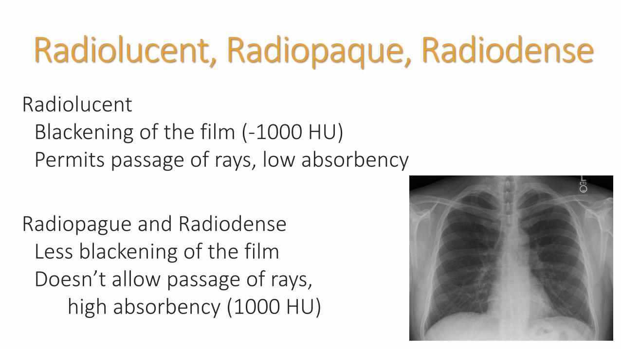
Radiolucent, Radiopaque, Radiodense
RadiolucentBlackening of the film (-1000 HU)Permits passage of rays, low absorbency
Radiopague and RadiodenseLess blackening of the filmDoesn’t allow passage of rays,
high absorbency (1000 HU)

Radiographic Densities
Gas (air)Black
FatGray-Black
Soft tissue (water)Gray
Bone (metal)White

Radiographic Contrast
Image Quality
MotionScatter
Magnification
Thickness
Distortion
The BasicsFoundational Concepts
Anatomy and Physiology
Pathophysiology
Shades of Gray
2 dimensional image of 3 dimensional body

Plain RadiographsAir – Fluid Levels
Visualize stones
Identify gross abnormalities that may lead to further testing
Foreign body identification
Diagnosis or Finding
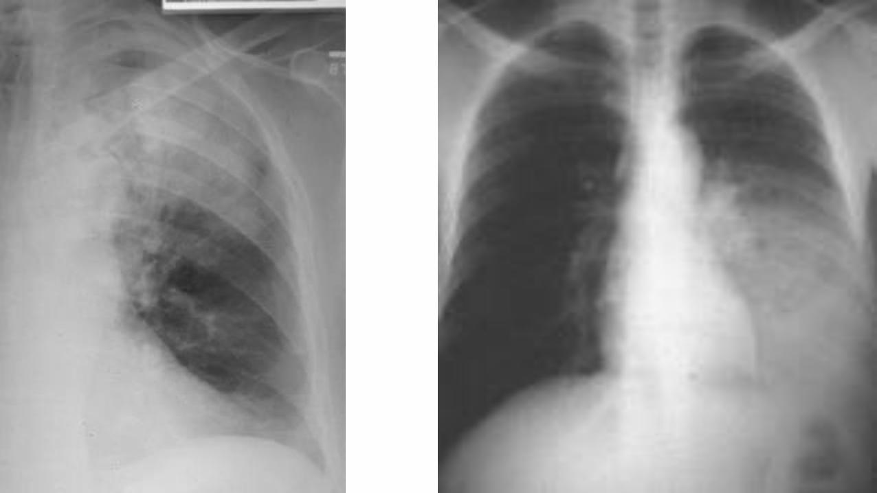
Right Middle Lobe Infiltrate
Pneumonia
Radiopague Foreign Body
Gangrene
THE RULES
Obtain a thorough history and physical examination
Order when necessary
Evaluate the entire radiograph
Re-examine the patient and the radiograph
Rule of 2s
Failsafe measures

High energy ionizing radiationAtom loses electron – ionizedPhoton with >15 electron volts is capable of ionization
Radiation exposure has been researched since the atomic bomb exposure. However, it has been observed since the early 1900s
Increased use of plain radiographs, nuclear medicine and CT scans has increased population exposure rates
Interrupts cell DNA causing mutations
Organs and tissues – varying sensitivities
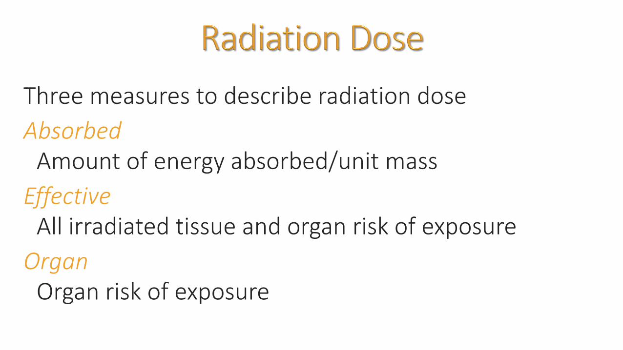
Three measures to describe radiation dose
AbsorbedAmount of energy absorbed/unit mass
EffectiveAll irradiated tissue and organ risk of exposure
OrganOrgan risk of exposure
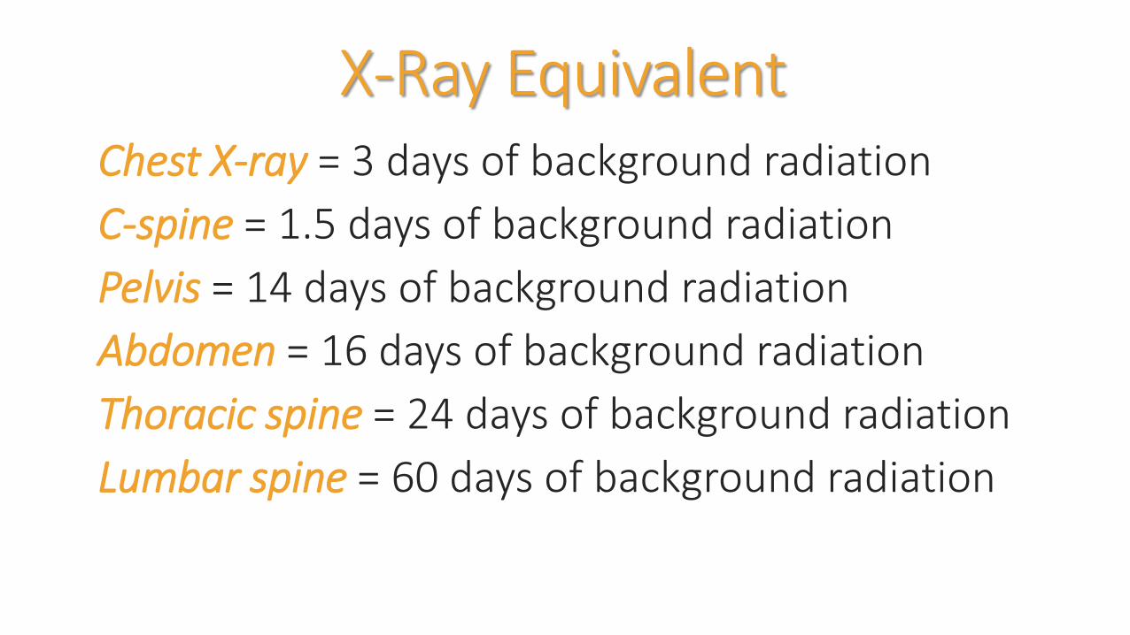
X-Ray EquivalentChest X-ray = 3 days of background radiation
C-spine = 1.5 days of background radiation
Pelvis = 14 days of background radiation
Abdomen = 16 days of background radiation
Thoracic spine = 24 days of background radiation
Lumbar spine = 60 days of background radiation

Plain Radiographs0.02 mSv Chest X-Ray
CT scan2.0 mSv Head20-60 mSv Chest, Abdomen and Pelvis
Nuclear Medicine10-25 mSv (sestamibi scan – dual isotope scanning)

Ionizing Radiation Medical Imaging
Classified as carcinogenic
Patients get multiple tests
Statistically significant increases in cancer with doses over 50mSv

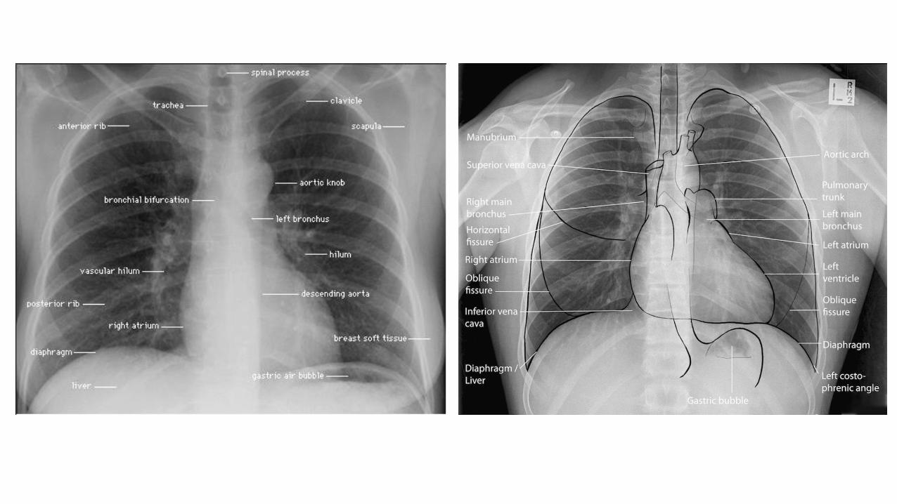
Chest Anatomy
http://www.medcyclopaedia.com/upload/book%20of%20radiology/chapter18/nic_k18_915.jpg

PositioningPosterior Anterior (PA)
Facing the cartridge
Supine Anterior Posterior (AP)Only in the critical patient
Lateral Position
Lateral Decubitus

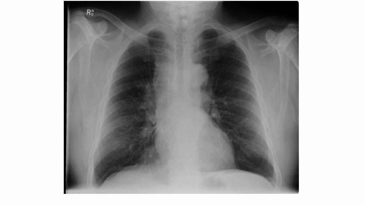
Normal PA and Lateral

PA vs AP• Lung markings more distinct• Heart is smaller• Clavicles are superimposed over
upper lungs• Cervical and thoracic vertebrae
more clearly visible
• Heart appears larger than normal• Lung volumes are shallow• Clavicles usually higher
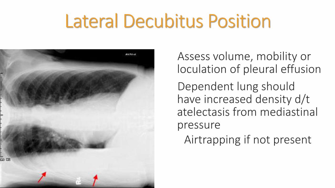
Lateral Decubitus Position
Assess volume, mobility or loculation of pleural effusion
Dependent lung should have increased density d/t atelectasis from mediastinalpressure
Airtrapping if not present

ABC’s of Interpretation
Adequacy, Airway
Breathing
Circulation
Diaphragm
Edges
Skeleton, Soft Tissue
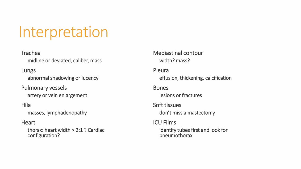
InterpretationTrachea
midline or deviated, caliber, mass
Lungs abnormal shadowing or lucency
Pulmonary vessels artery or vein enlargement
Hila masses, lymphadenopathy
Heart thorax: heart width > 2:1 ? Cardiac configuration?
Mediastinal contour width? mass?
Pleura effusion, thickening, calcification
Bones lesions or fractures
Soft tissues don’t miss a mastectomy
ICU Films identify tubes first and look for pneumothorax

Adequacy
Normal Inspiration
Penetration
Rotation
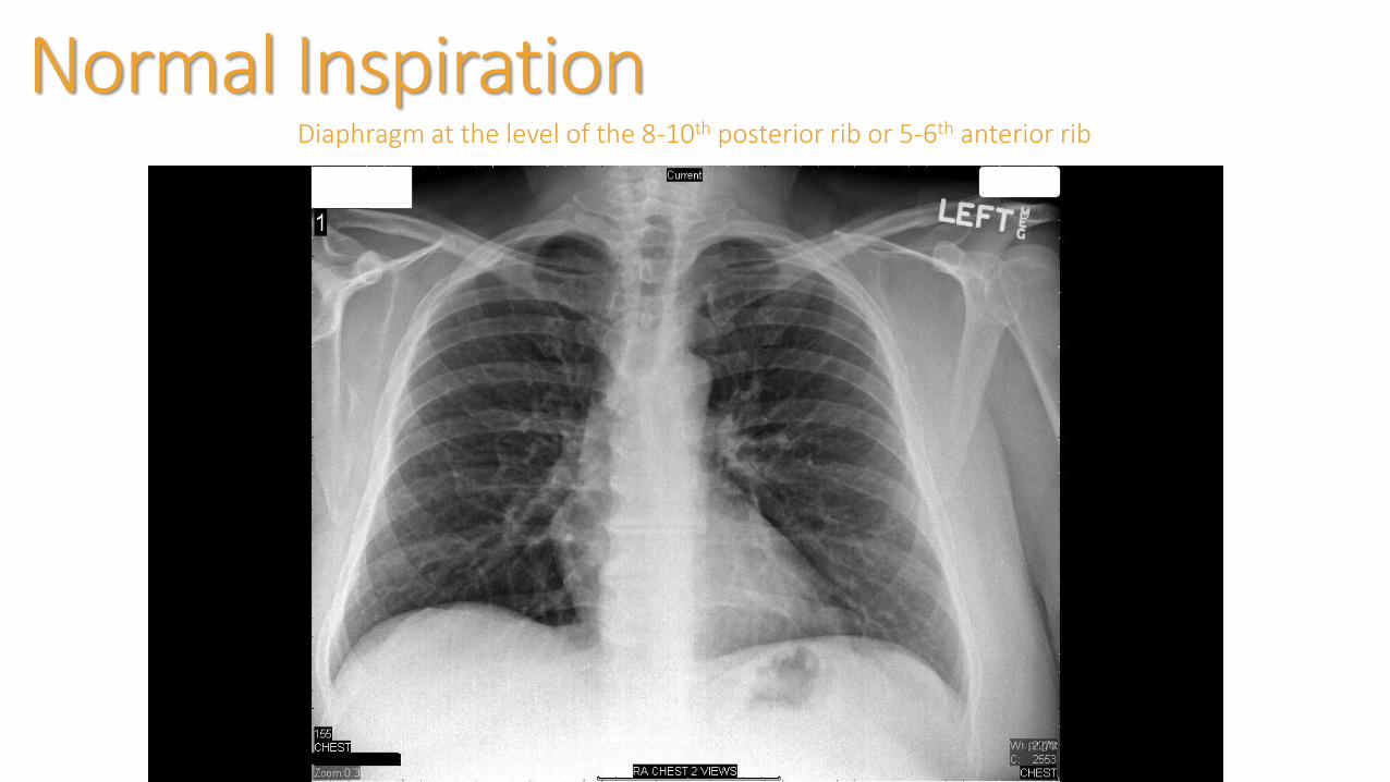
Normal InspirationDiaphragm at the level of the 8-10th posterior rib or 5-6th anterior rib

Poor Inspiration
Expiration
Desirable to evaluate a patient with:Suspected pneumothoraxSuspected foreign body in bronchus
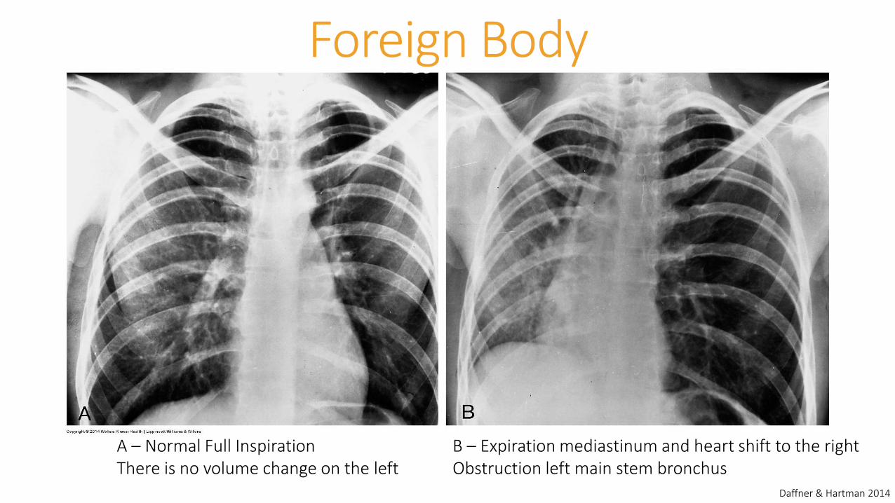
Foreign Body
A – Normal Full Inspiration B – Expiration mediastinum and heart shift to the rightThere is no volume change on the left Obstruction left main stem bronchus
Daffner & Hartman 2014
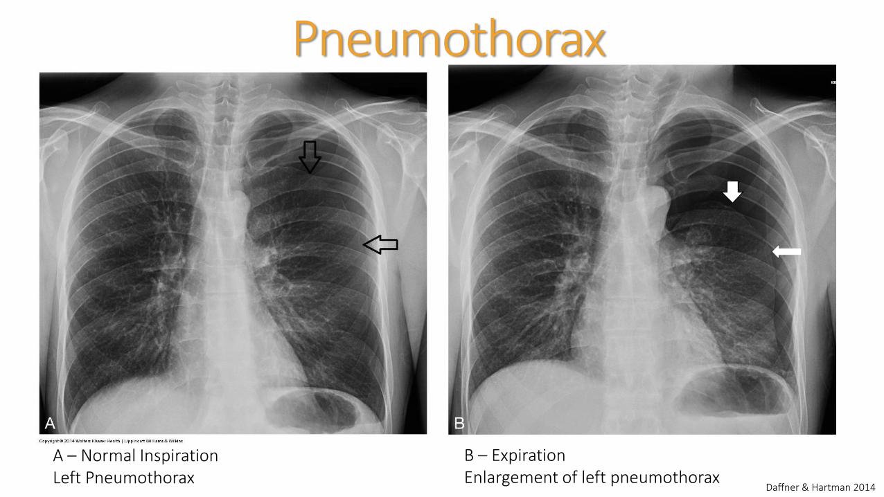
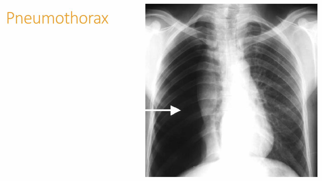
Pneumothorax
A – Normal InspirationLeft Pneumothorax
B – ExpirationEnlargement of left pneumothorax
Daffner & Hartman 2014

PenetrationPA
Thoracic disc spaces should be barely visible through the heart with vertebral bodies not visible
Over-penetration = Dark
Under-penetration = Light
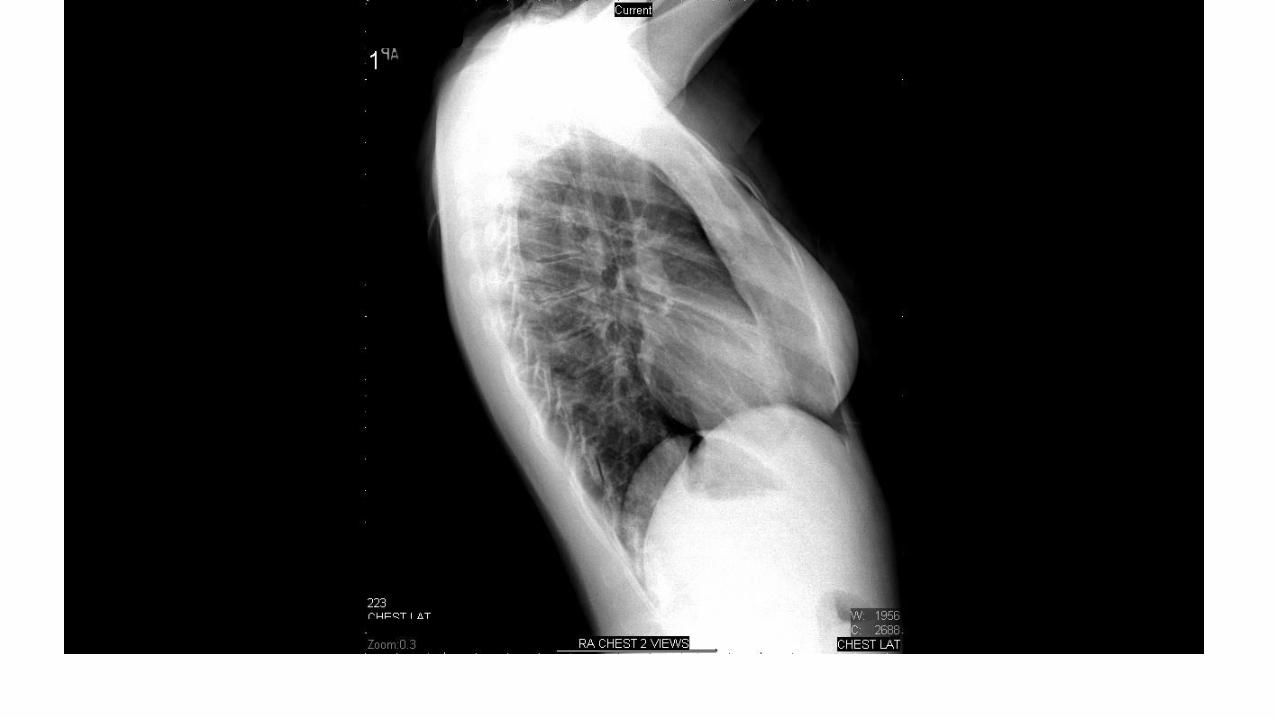
LateralShould see 2 sets of ribs
Sternal edge may be visible
Vertebrae appear darker as you move caudally
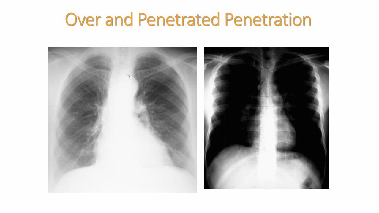
Over and Penetrated Penetration

Rotation• May result in distortion of
normal anatomic structures
• Clavicle heads and spinal processes should be symmetrical

Airway
Trachea midline and seen to the carina (bifurcation T4-T5)
Slowly angles downward to the thoracic inlet (retrotracheal
line 3mm)Bronchogram
may be normal or abnormalAir filled tube surrounded by soft tissue

The Rest of the A, B, Cs
Breathing (Bird cages)
Cardiac/circulation
Diaphragm
Edges
Skeleton and Soft Tissue

What do you see????
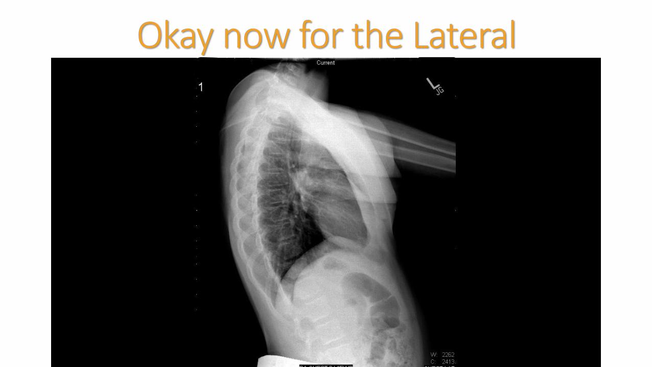
Okay now for the Lateral


MediastinumCentral chest between lungs and heart
Divided into three regionsAnterior – area between sternum and front of heart and great vesselsMiddle – area between anterior and posterior pericardium
Includes: pericardium, heart, aortic arch, proximal brachialcephalic vessels, pulmonary veins/arteries, trachea, main bronchus, and lymph nodes
Posterior – area behind the heart and trachea including vertebral bodies

Mediastinum

Pediatric Considerations
Can be challengingLook different in children
Different diseasesChange with age
Limited patient cooperationThymus can cause confusion
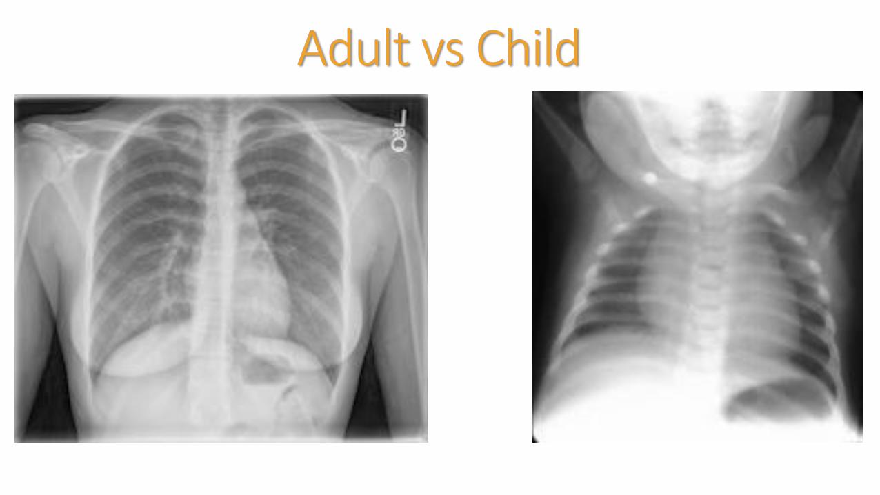
Adult vs Child
DifferencesHeart
Newborn hearts can be more than ½ the width of the chest
Good inspiration needed to judge heart size
Poor inspiration can significantly change the look and position of the heart
Thymus
Increases in size fro birth through puberty BUT child grows so it appears smaller with age
Can be variable in size and appearance (i.e. shrink rapidly due to illness or grow due to chemotherapy)
Look for a BronchogramOutline of airway that is made visible by surrounding alveoli with fluid or exudate
When visualized diagnostic for air space disease
6 causes
normal expiration
lung consolidation
pulmonary edema
Non-obstructive pulmonary atelectasis
severe interstitial disease
Neoplasm
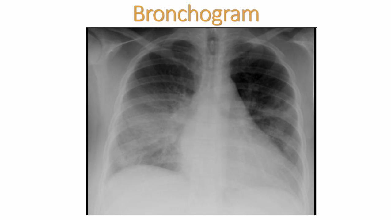
Bronchogram
AtelectasisCondition of volume loss in some portion of lung
May involve sub-segment, segment, lobe or entire lung
Increased density usually linear
Collapse or incomplete expansion of the lung or part of the lung
Segmental and sub-segmental collapse may show linear, curvilinear, wedge shaped opacities
AtelectasisCauses
ObstructiveMost commonBronchus obstructed by mucous plug, neoplasm, or foreign body
CompressiveNormal lung compressed by tumor, emphysematous bulla or heart enlargement
CicatrizationOrganizing scar tissueMost often after healing granulomatous disease (i.e. TB), pulmonary infarct or trauma
AdhesiveInactivation of surfactant (example: hyaline membrane disease)
PassiveNormal compliance of the lung with pneumothorax or pleural effusionAirway remains patent
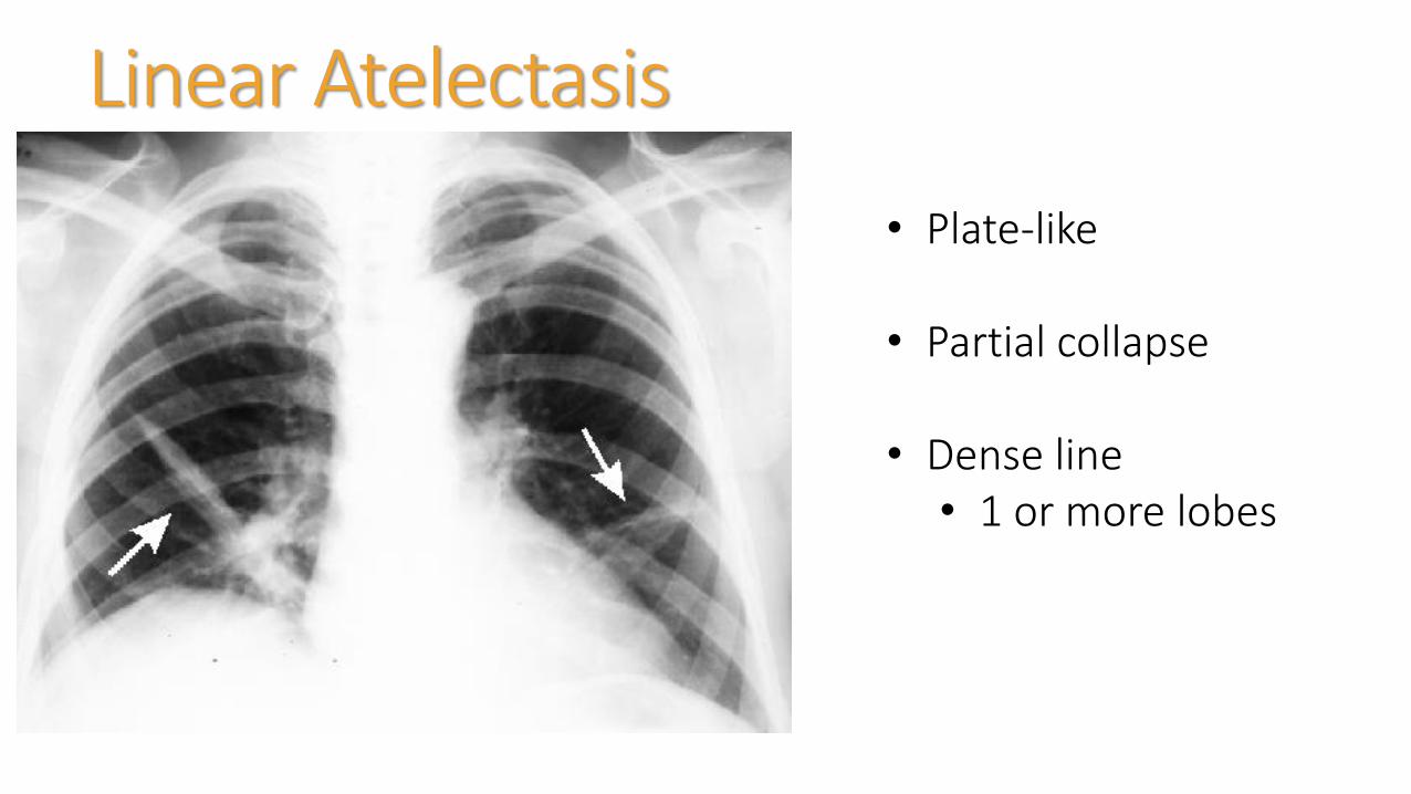
Linear Atelectasis
• Plate-like
• Partial collapse
• Dense line • 1 or more lobes
Pulmonary EdemaTwo basic types
Cardiogenic
increased hydrostatic pulmonary capillary pressure
Non-cardiogenic
altered capillary membrane permeability or decreased plasma oncotic pressure
NOT CARDIAC (Pneumonic)
Near-drowning, Oxygen therapy, Transfusion or Trauma, CNS disorder, ARDS, Aspiration, or Altitude sickness, Renal disorder or Resuscitation, Drugs, Inhaled toxins, Allergic Alveolitis, Contrast or Contusion
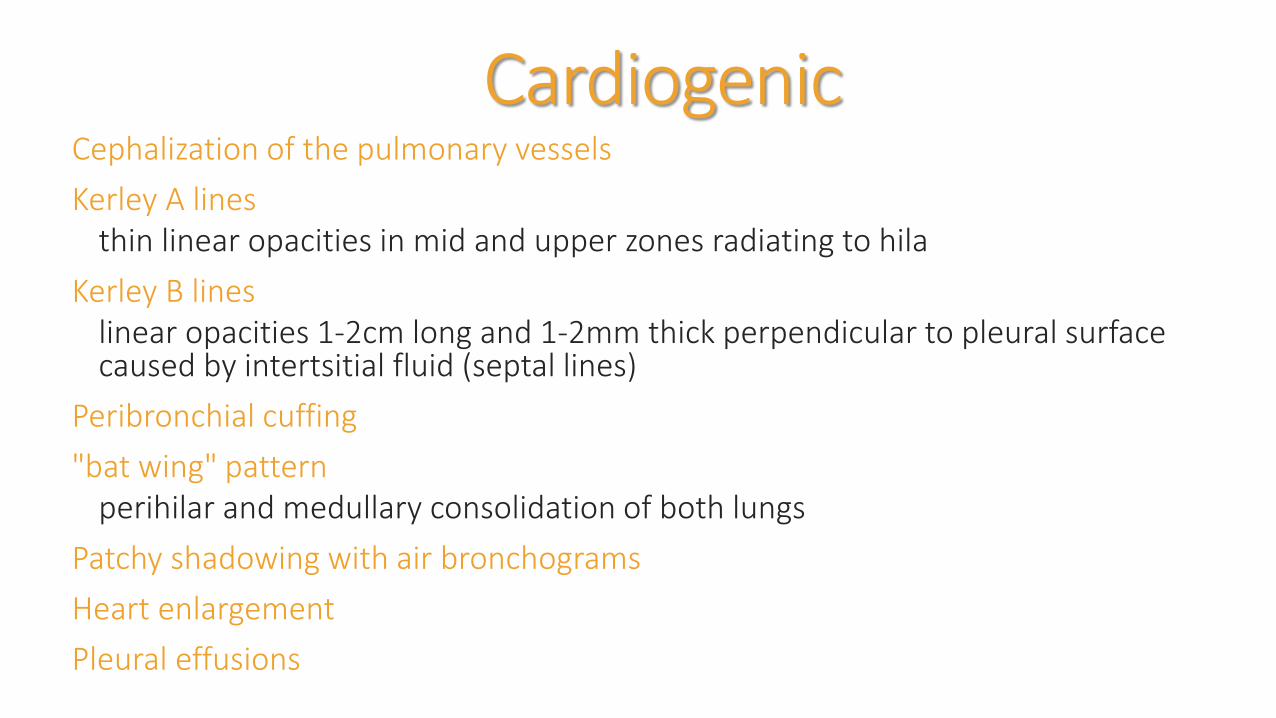
CardiogenicCephalization of the pulmonary vessels
Kerley A linesthin linear opacities in mid and upper zones radiating to hila
Kerley B lines linear opacities 1-2cm long and 1-2mm thick perpendicular to pleural surface caused by intertsitial fluid (septal lines)
Peribronchial cuffing
"bat wing" patternperihilar and medullary consolidation of both lungs
Patchy shadowing with air bronchograms
Heart enlargement
Pleural effusions
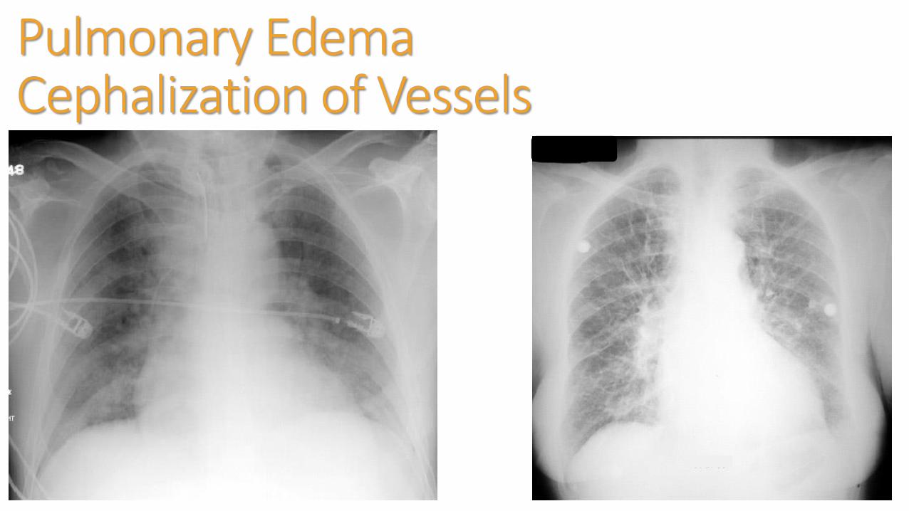
Pulmonary EdemaCephalization of Vessels

Bat Wing Pattern

Pulmonary Edema

Diffuse Pulmonary Edema
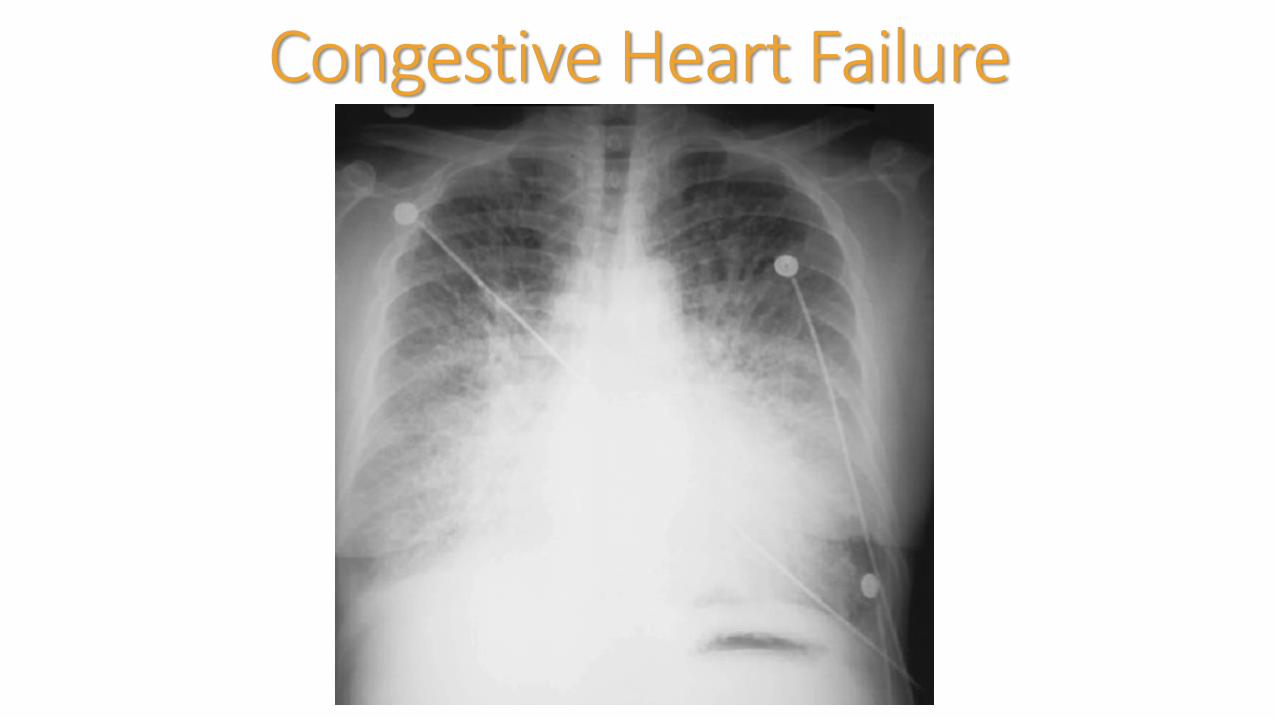
Congestive Heart Failure
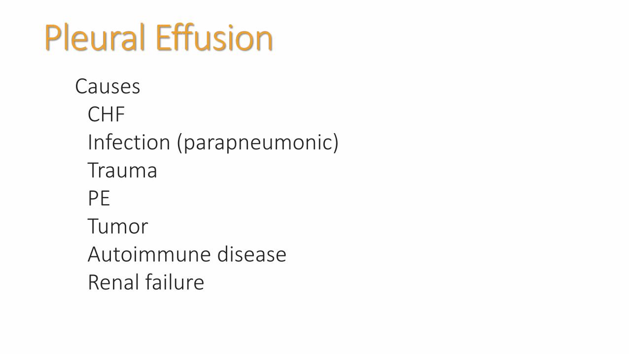
Pleural EffusionCauses
CHFInfection (parapneumonic)TraumaPETumorAutoimmune diseaseRenal failure
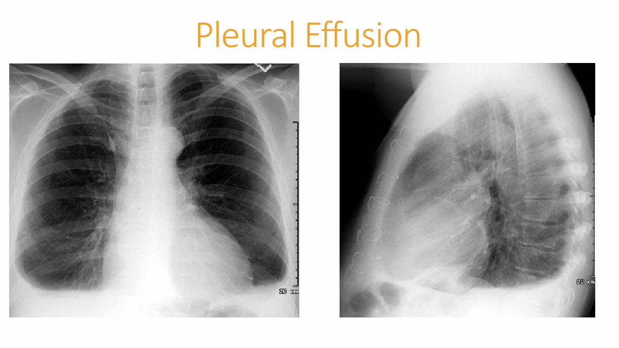
Pleural Effusion
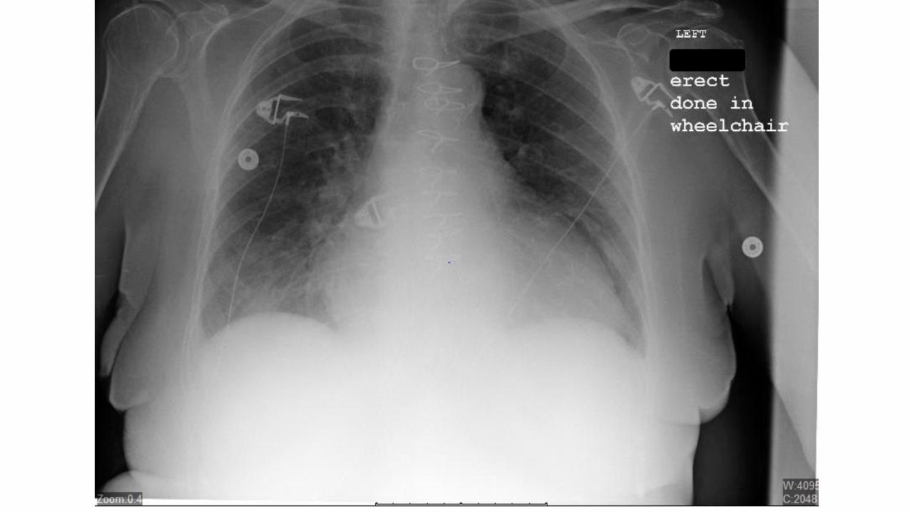
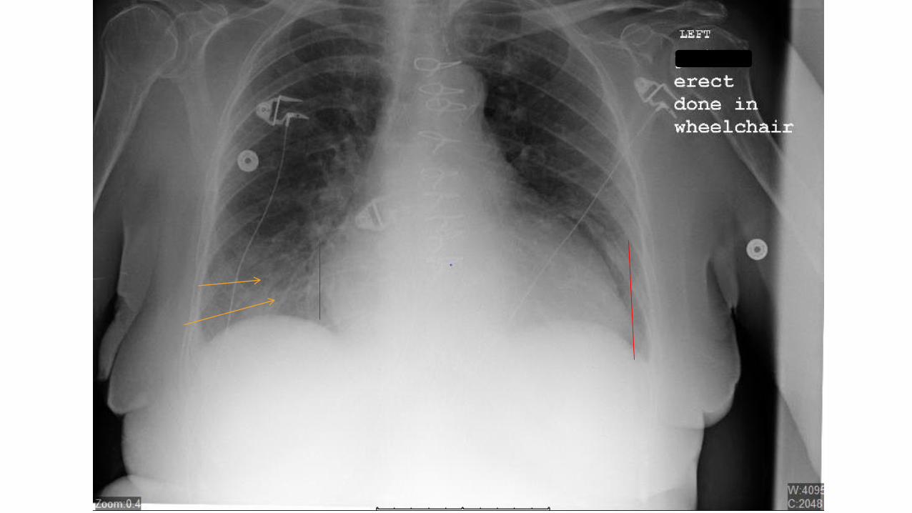
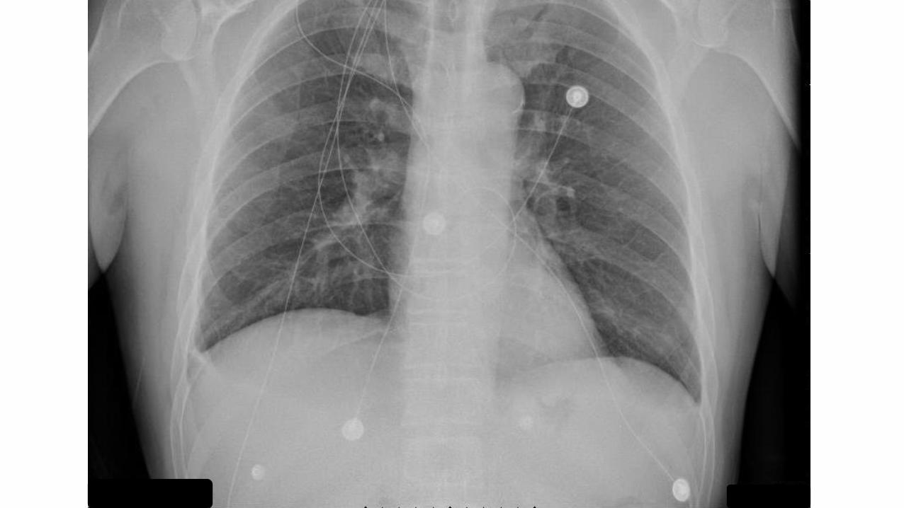
88 year old Female
Presents with complaints of shortness of breath
PMH – arthritis, hypercholesterolemia, HTN, CAD, pulmonary HTN,
PSH – CABG, Cataracts, Aortic Valve Repair
Allergy – codeine, PCN
PE – lungs decreased with bibasilar crackles
Vital signs – BP 146/85, HR 85, RR 16, T 97F, pulse Ox 95% RA

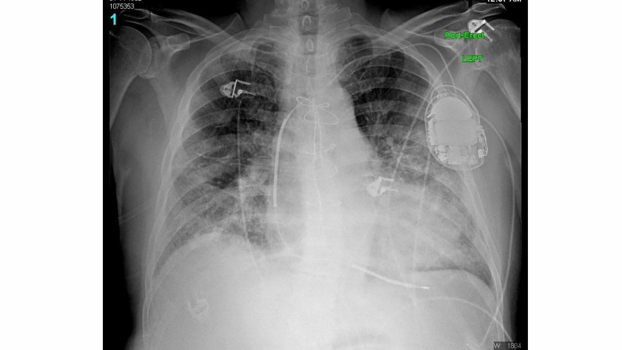
78 year old Male
Presents with shortness of breath for 1 day progressively getting worse.
PMH – CAD, HTN, Hypercholesterolemia
PSH – CABG, pacemaker or AICD patient and family not sure
Allergies – none
PE – pale, diaphoretic, in mild respiratory distress. Mild JVD. Lungs with course diffuse rhonchi. S1 S2 no M/G/C
Vital signs – BP148/90, HR 102, RR 28, T 97.9F pulse Ox 94% RA
52 Year Old MaleComplaints of not feeling well, chest tightness, and racing heart. Denied SOB or fever
PMH – alcohol abuse, depression
PSH – none
Allergies – none
Social – alcohol use daily 1 bottle of scotch; tobacco 1-2 PPD
PE – unremarkable
Vital signs – BP 154/103, HR 138, RR 27, T 100.8, pulse Ox 97% RA


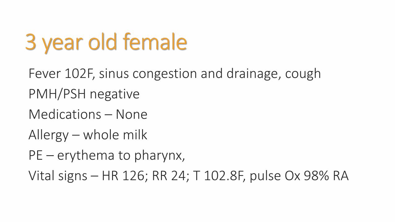
3 year old femaleFever 102F, sinus congestion and drainage, cough
PMH/PSH negative
Medications – None
Allergy – whole milk
PE – erythema to pharynx,
Vital signs – HR 126; RR 24; T 102.8F, pulse Ox 98% RA

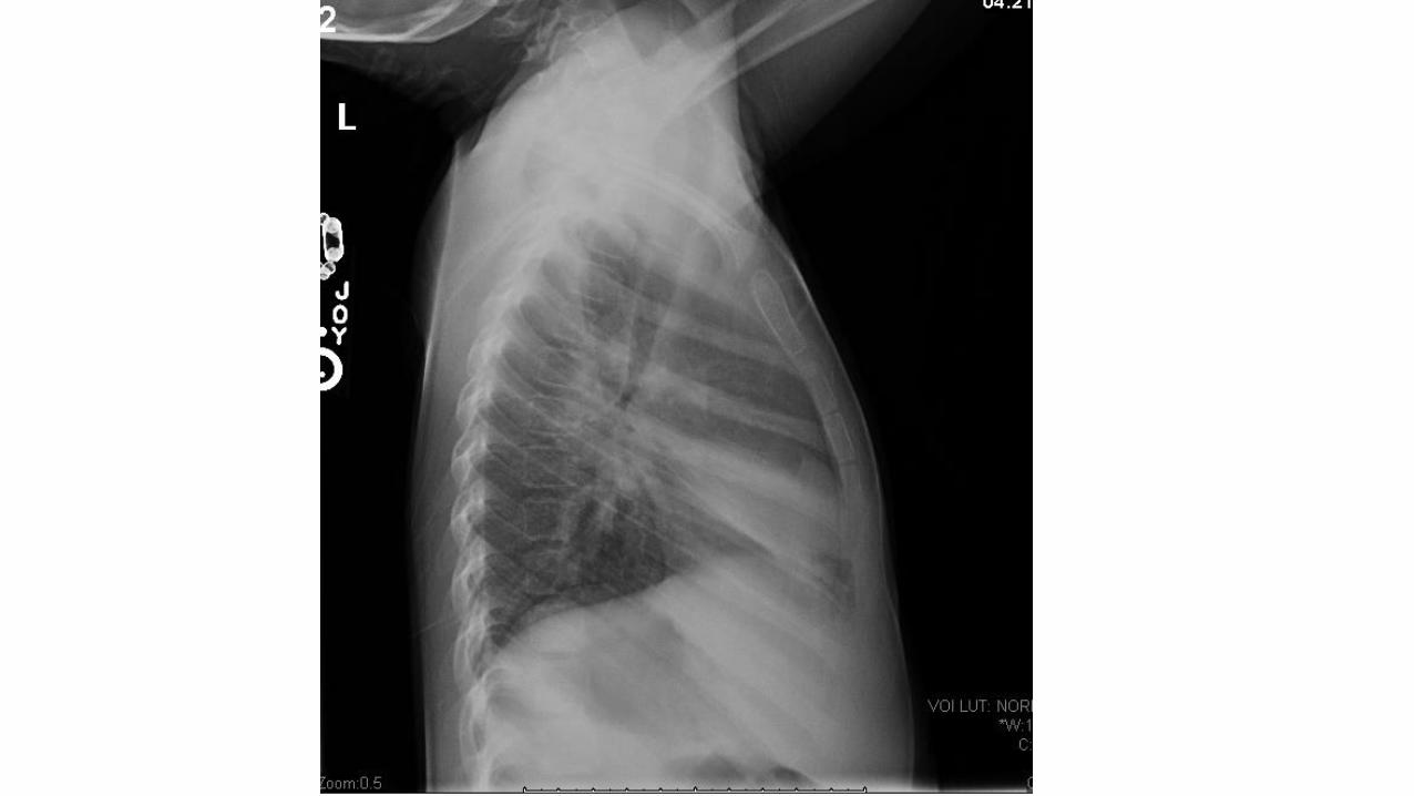
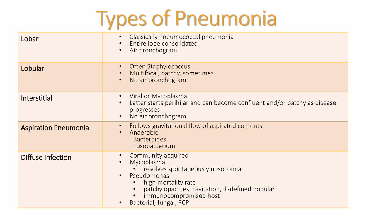
Types of PneumoniaLobar • Classically Pneumococcal pneumonia
• Entire lobe consolidated • Air bronchogram
Lobular • Often Staphylococcus• Multifocal, patchy, sometimes • No air bronchogram
Interstitial • Viral or Mycoplasma• Latter starts perihilar and can become confluent and/or patchy as disease
progresses• No air bronchogram
Aspiration Pneumonia • Follows gravitational flow of aspirated contents• Anaerobic
BacteroidesFusobacterium
Diffuse Infection • Community acquired• Mycoplasma
• resolves spontaneously nosocomial • Pseudomonas
• high mortality rate• patchy opacities, cavitation, ill-defined nodular• immunocompromised host
• Bacterial, fungal, PCP


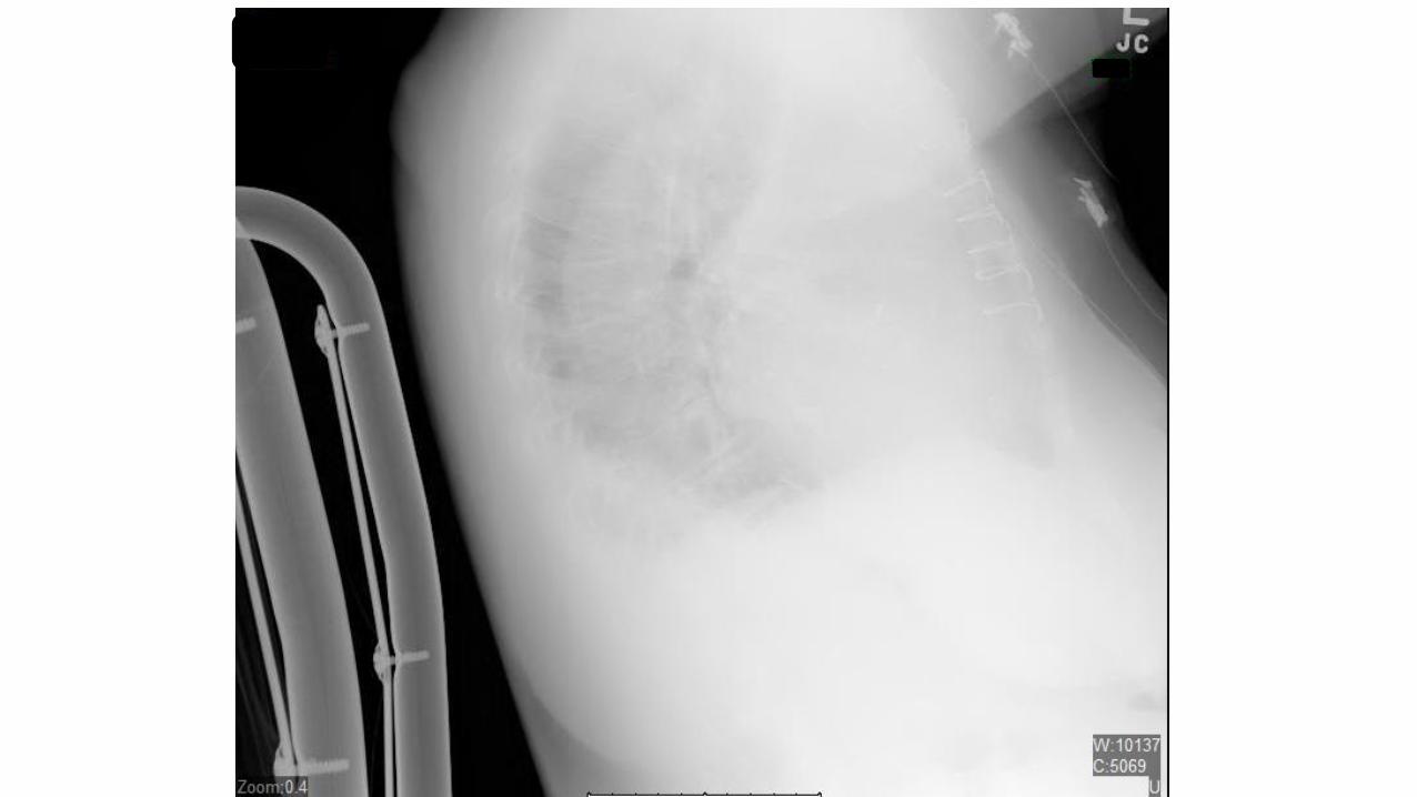
32 Year Old Male
Aortic Valve Replacement Post-op 5 days
Chest tube removed
C/O mild SOB
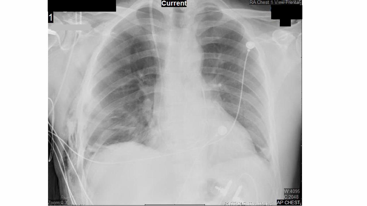

PneumomediastinumStreaky lucencies over the mediastinum that may extend into the neck, and elevation of the parietal pleura along the mediastinal
borders
Causes• Asthma• Surgery • Traumatic tracheobronchial rupture• Abrupt changes in intrathoracic pressure (vomiting, coughing,
exercise, parturition)• Ruptured esophagus• Barotrauma• Smoking crack cocaine

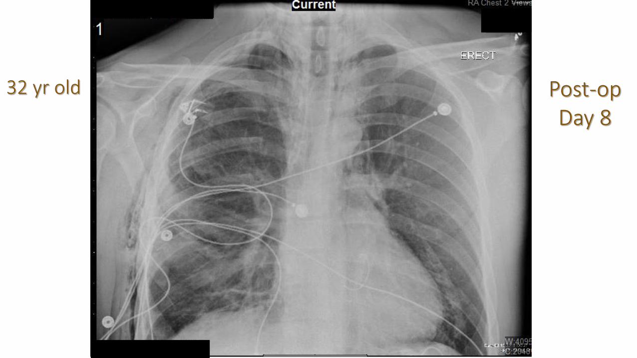
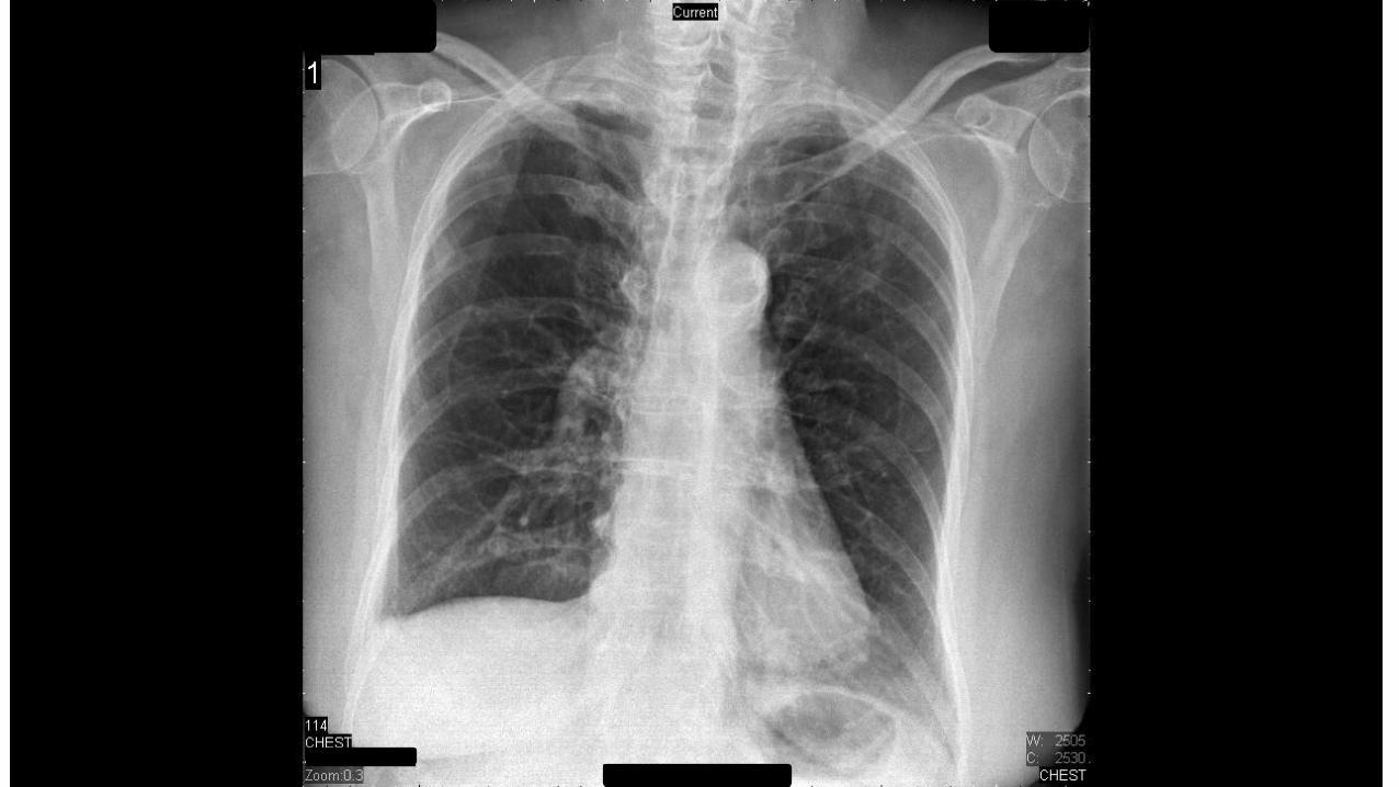
32 yr old Post-opDay 8


Pneumothorax
CausesIdiopathicAsthmaCOPDPulmonary infectionNeoplasmMarfan syndromeSmoking cocaineTraumaProvider
Pneumothorax

Traumatic Injuries


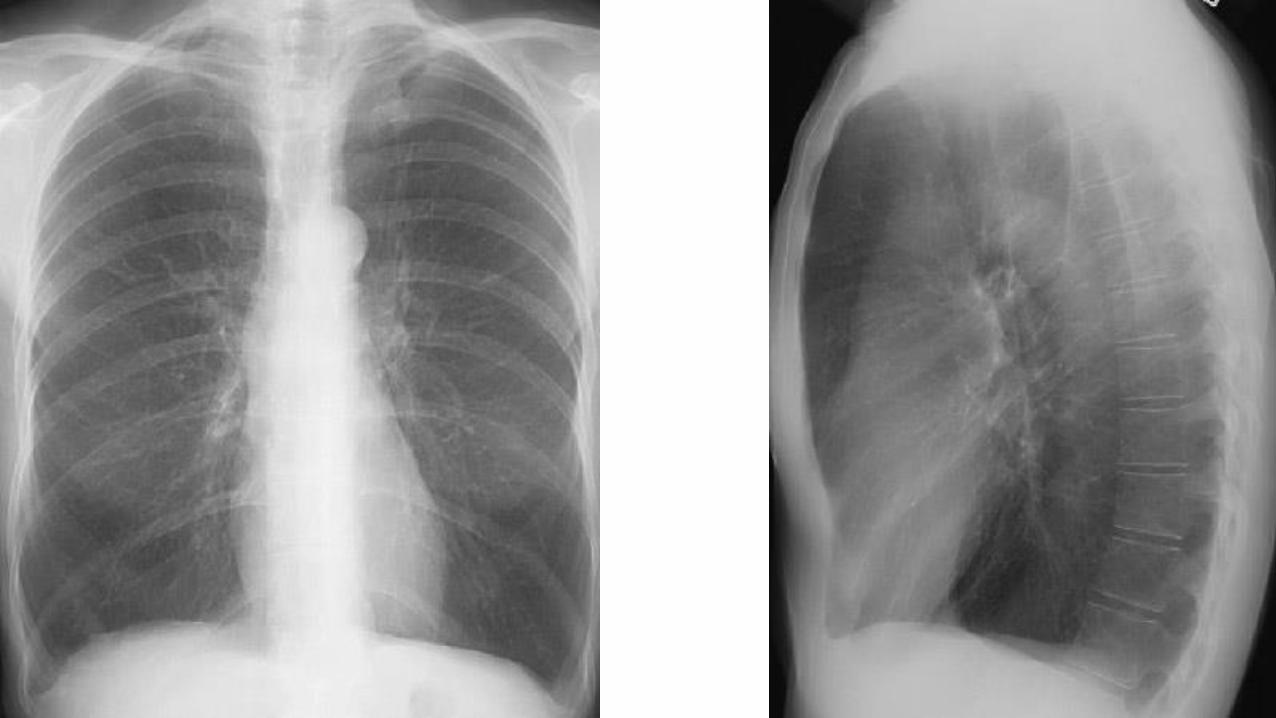
What Do You Think????
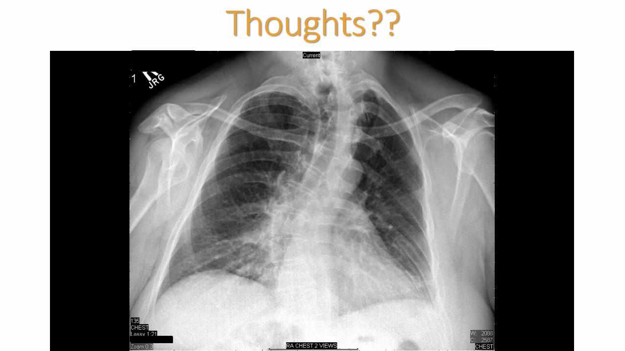
Thoughts??

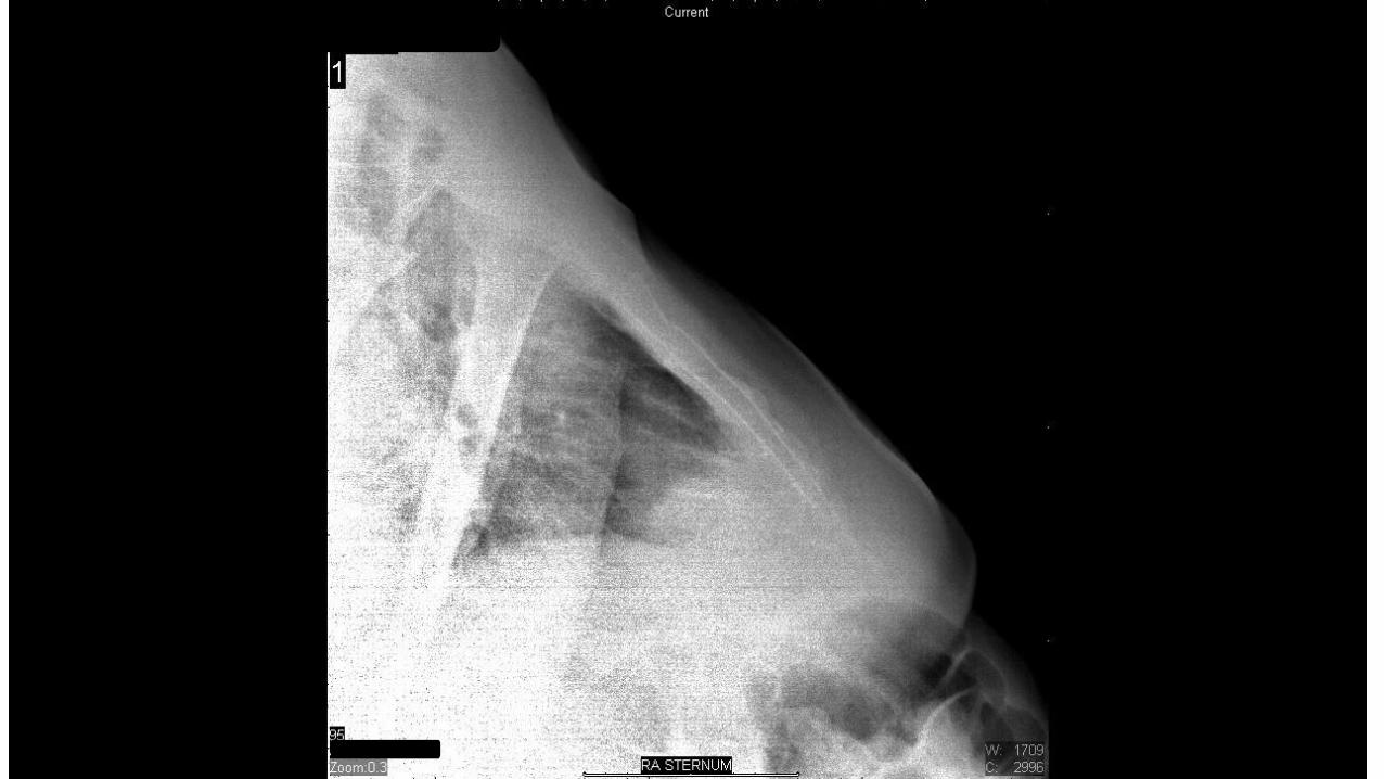

Sternum Fracture
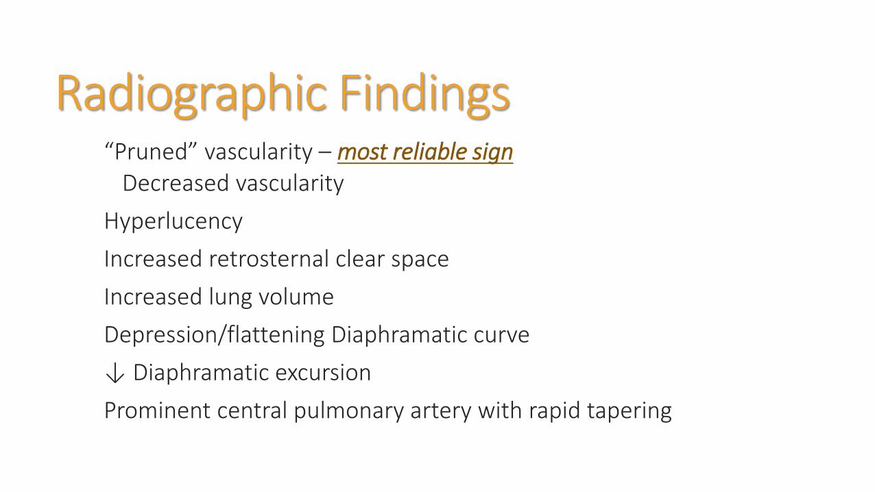
Radiographic Findings“Pruned” vascularity – most reliable sign
Decreased vascularity
Hyperlucency
Increased retrosternal clear space
Increased lung volume
Depression/flattening Diaphramatic curve
↓ Diaphramatic excursion
Prominent central pulmonary artery with rapid tapering
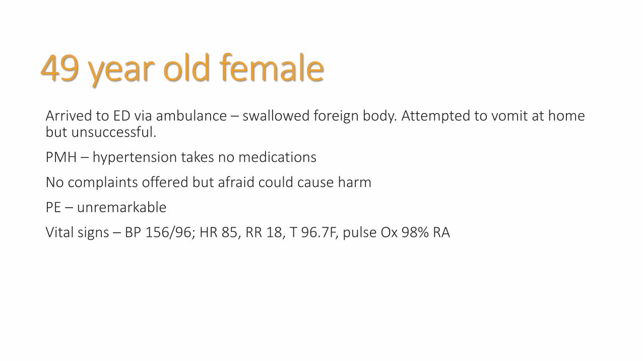
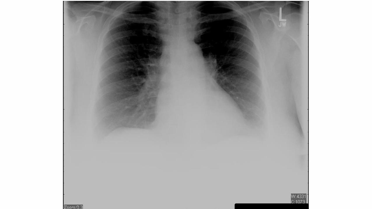
49 year old femaleArrived to ED via ambulance – swallowed foreign body. Attempted to vomit at home but unsuccessful.
PMH – hypertension takes no medications
No complaints offered but afraid could cause harm
PE – unremarkable
Vital signs – BP 156/96; HR 85, RR 18, T 96.7F, pulse Ox 98% RA
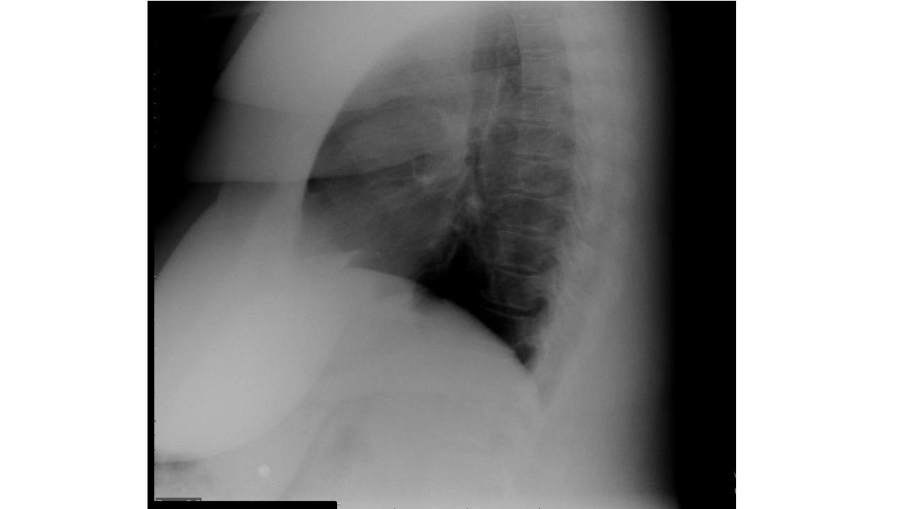
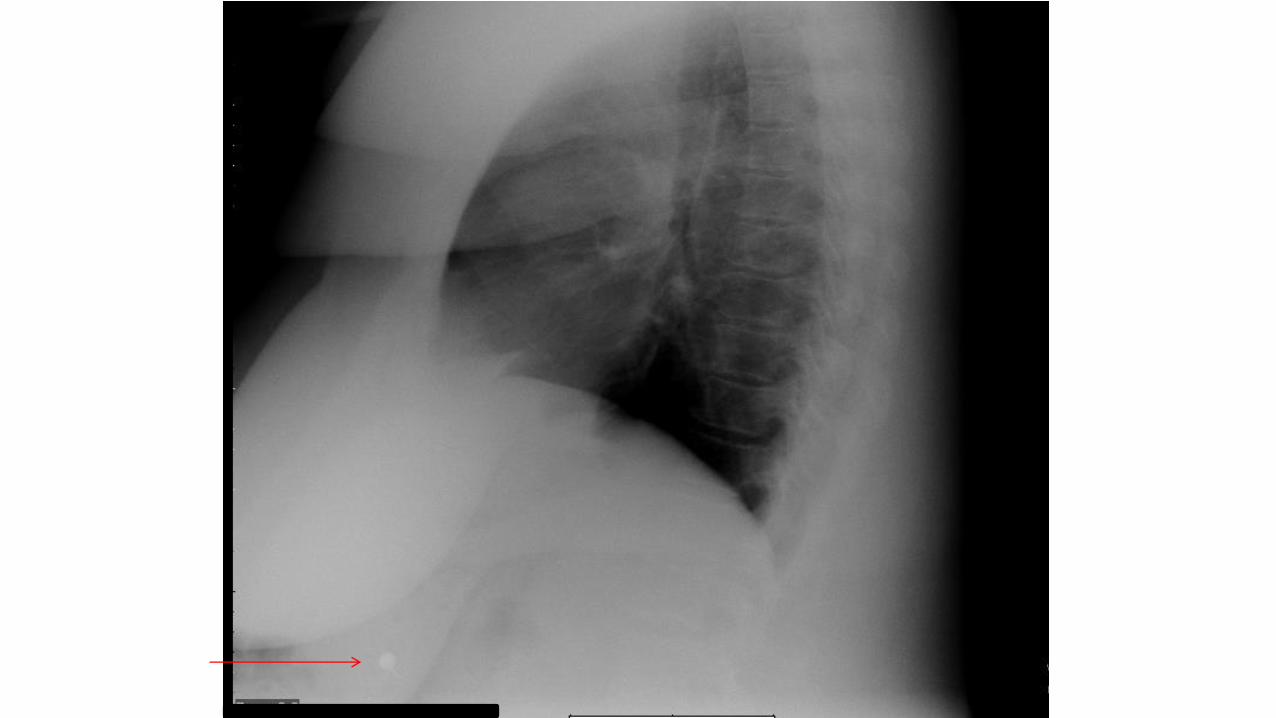
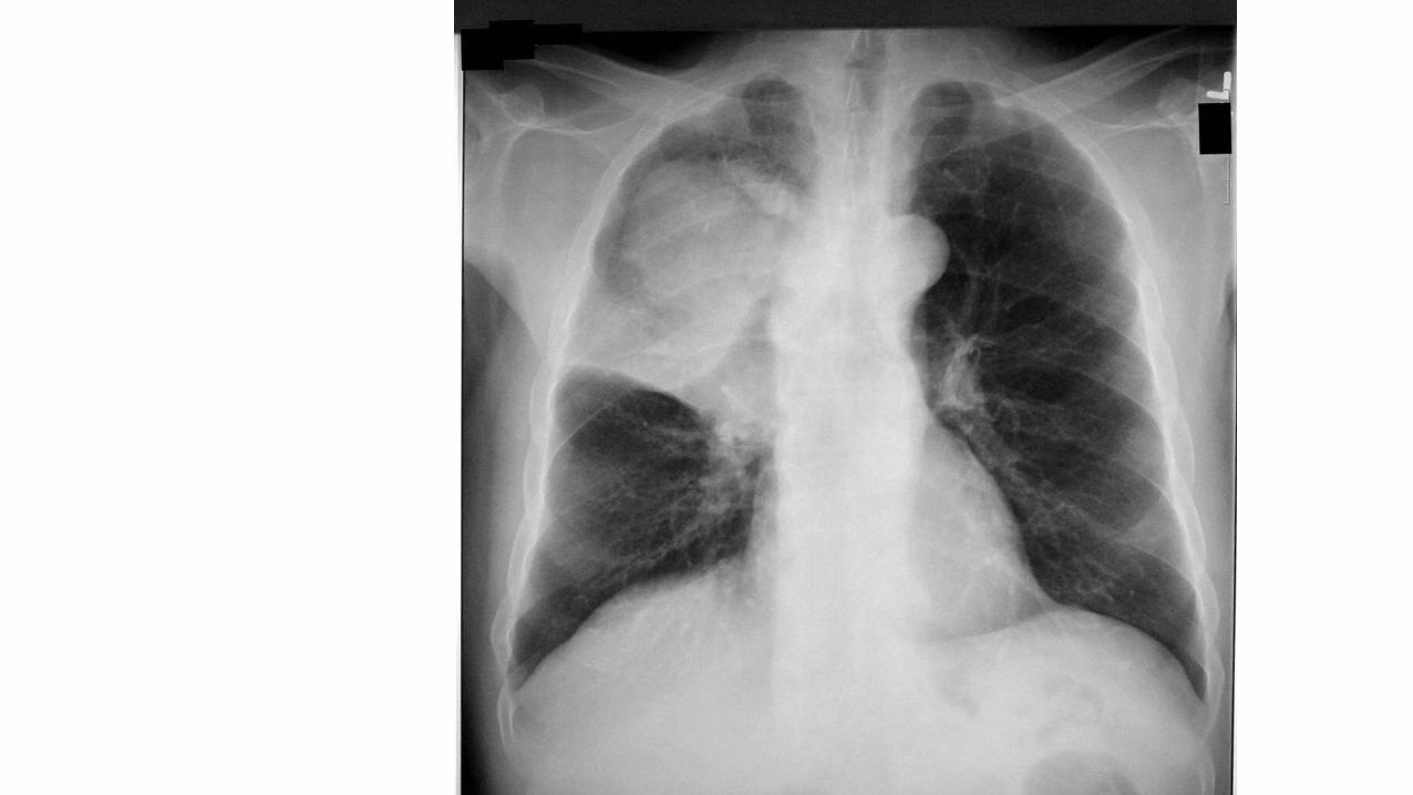
Masses and TumorsLung and Mediastinal masses are common
Solitary pulmonary nodulesTumorsGranulomas
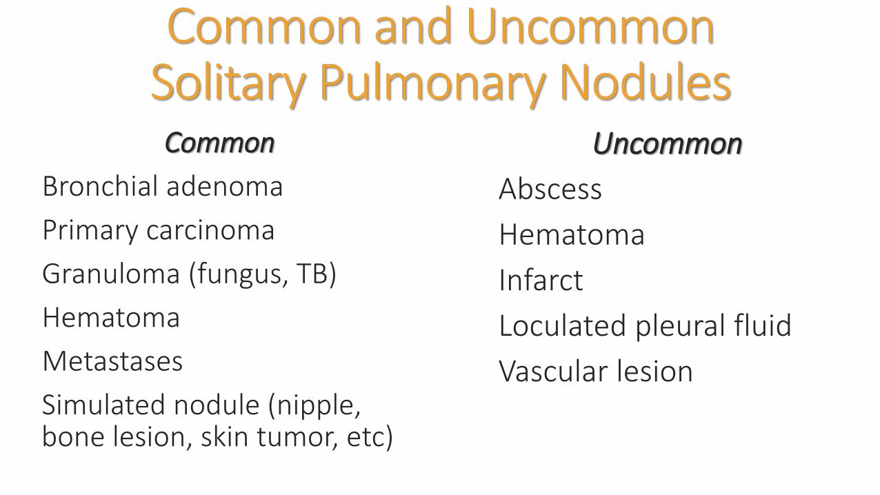
Common and Uncommon Solitary Pulmonary Nodules
Common
Bronchial adenoma
Primary carcinoma
Granuloma (fungus, TB)
Hematoma
Metastases
Simulated nodule (nipple, bone lesion, skin tumor, etc)
Uncommon
Abscess
Hematoma
Infarct
Loculated pleural fluid
Vascular lesion
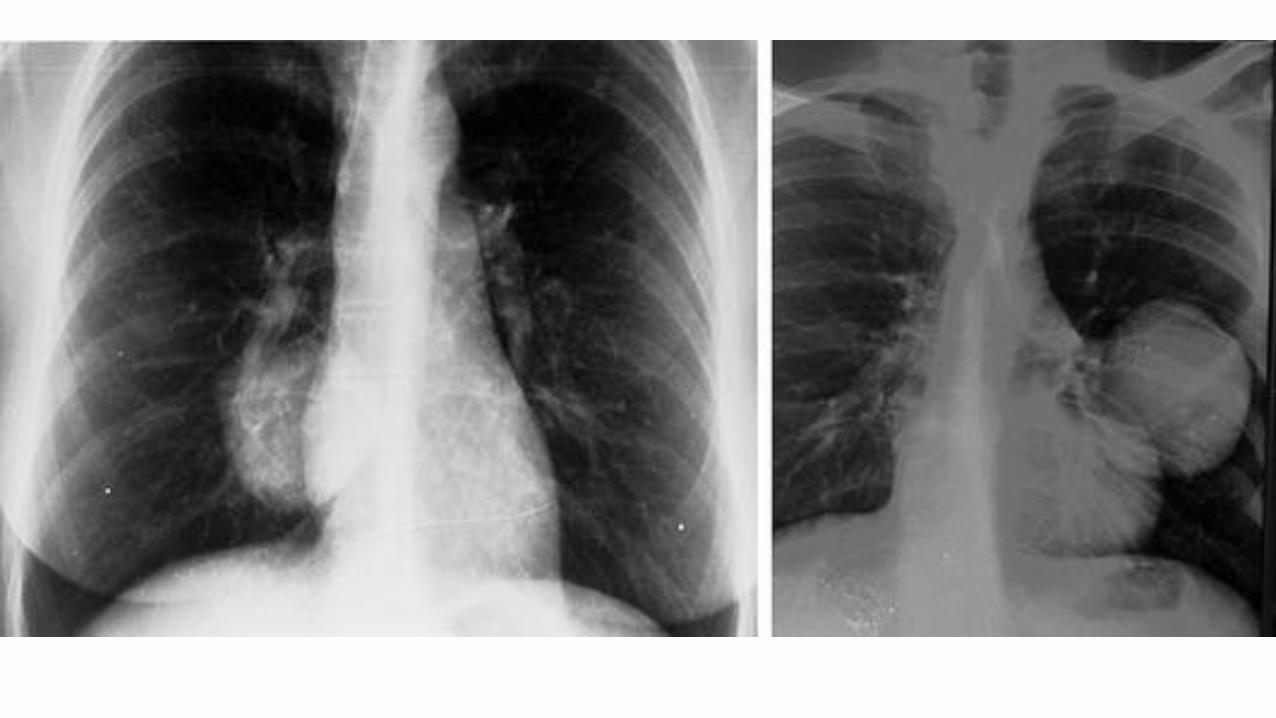

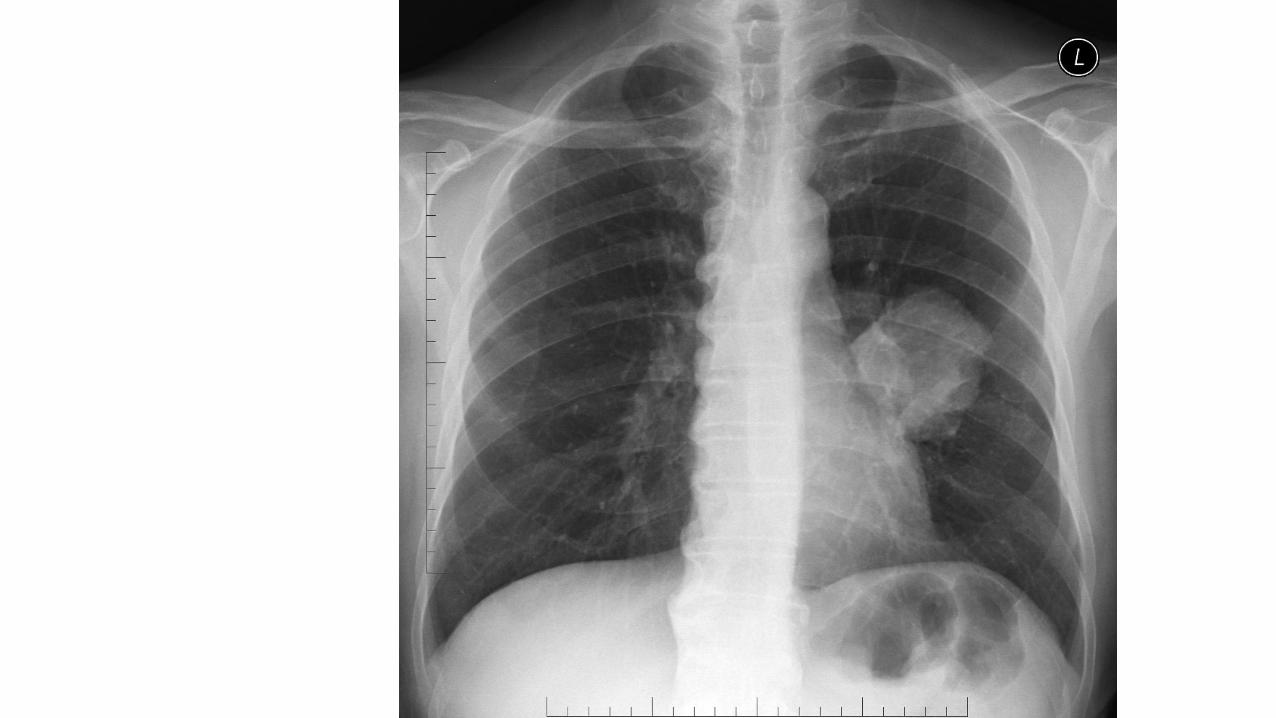
Cavitary MassesPulmonary nodules which may cavitate
Most Common
Carcinoma
Necrotizing infections (abscesses)
Metastatic lesions (usually squamous cell)
Others Causes
Fungal or TB infection
Hematoma
Pneumatoceles

Cavitary MassesWall Thickness
>15 mm more likely to be malignant
<4 mm more likely to be benign
Not specific enough further testing needs to be done (i.e. biopsy)


Evaluating Pulmonary NodulesNeed to find the epicenter of the pulmonary mass (nodule)
Allows for identification of where the mass began (i.e. lung, mediastinum, pleura, chest wall)
Spiculated margins
Lesions doubling in diameter actually increase 8-fold in volume
Utilize old studiesChest radiograph CTMRI PET imaging

Spiculated Margins
Mediastinal MassesCan be difficult to differentiate from pulmonary parenchymal masses
Majority of primary mediastinal massesOccur anterior compartment1/3 middle compartmentRemainder posterior compartment
Majority show extrapulmonary signsObtuse margins with pleuraCentered outside the lung
Mediastinal MassesAnterior compartment
Lymphoma, thymomas, teratomas – most commonHernias and cysts – other abnormalities
Middle mediastinumLymph nodes, metastatic disease, sarcoidosis, infection/inflammation
Behind the heartHiatel/paraesophageal hernia
Posterior compartmentNeurogenic tumor (paraspinous mass)
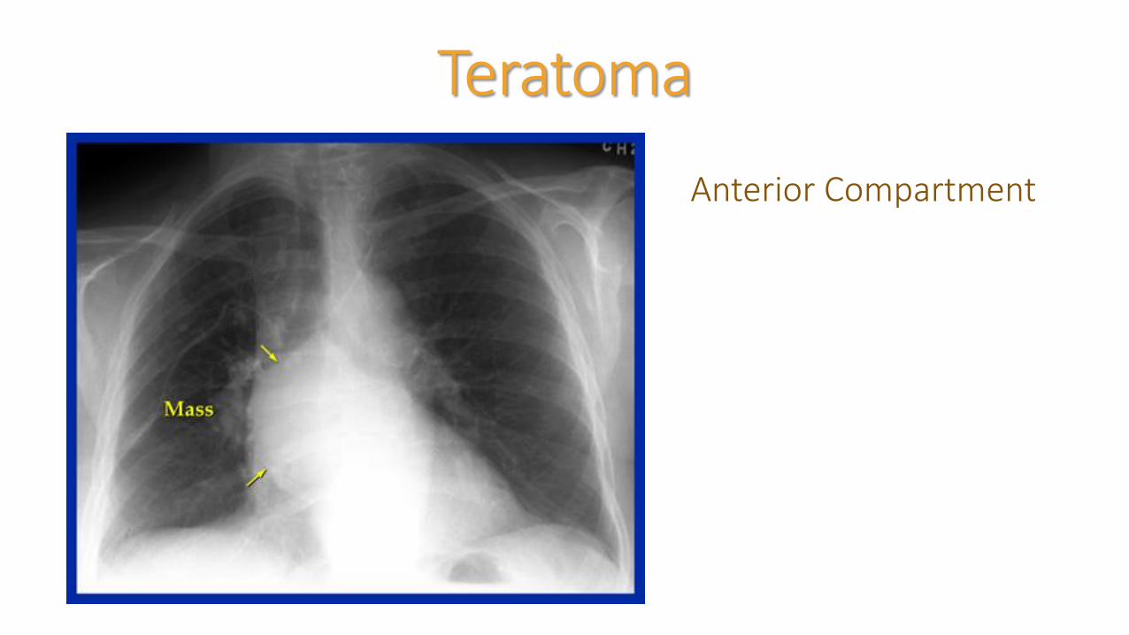
Teratoma
Anterior Compartment
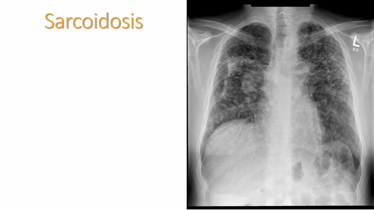
Sarcoidosis

Lymphoma

Hiatel Hernia
• Air/fluid levels
• Posterior to heart
TuberculosisPrimary
Healthy individuals – may have no chest x-ray findings even with a positive PPD
Inflammatory response
“Primary inflammatory complex” or “Ranke complex” – calcified nodules and thoracic lymph nodes
Not reliable sign (i.e. fungal infections, histoplasmosis)
Immunocompromised/chronically ill
Nonspecific consolidation
Cavity nodule/mass with air/fluid levels – ominous sign for transmissible disease
Small miliary nodules
Necrotizing adenopathy
Pleural effusions
TuberculosisSecondary
Reactivation of dormant infection
Infection thrives on oxygen
Particularly upper lobes
Consolidation with or without cavitation and adenopathy
End Stage
Fibrosis
Scarring with volume loss
Shift of fissures and/or vessels
Calcification
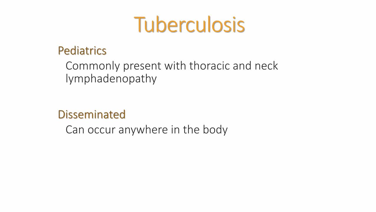
TuberculosisPediatrics
Commonly present with thoracic and neck lymphadenopathy
DisseminatedCan occur anywhere in the body

Primary Inflammatory ComplexRanke Complex
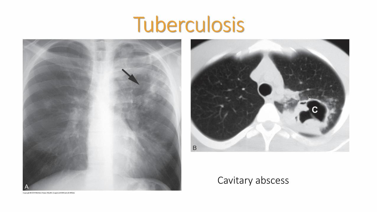
Tuberculosis
Cavitary abscess
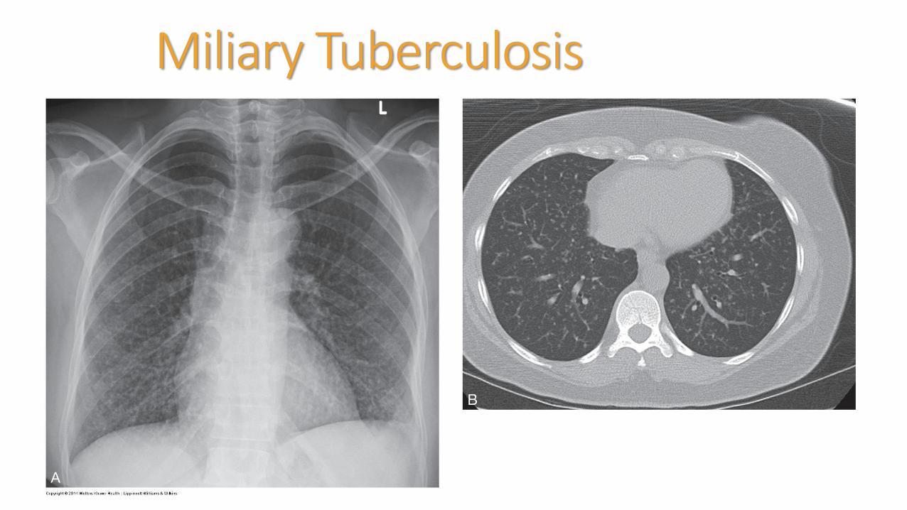
Miliary Tuberculosis
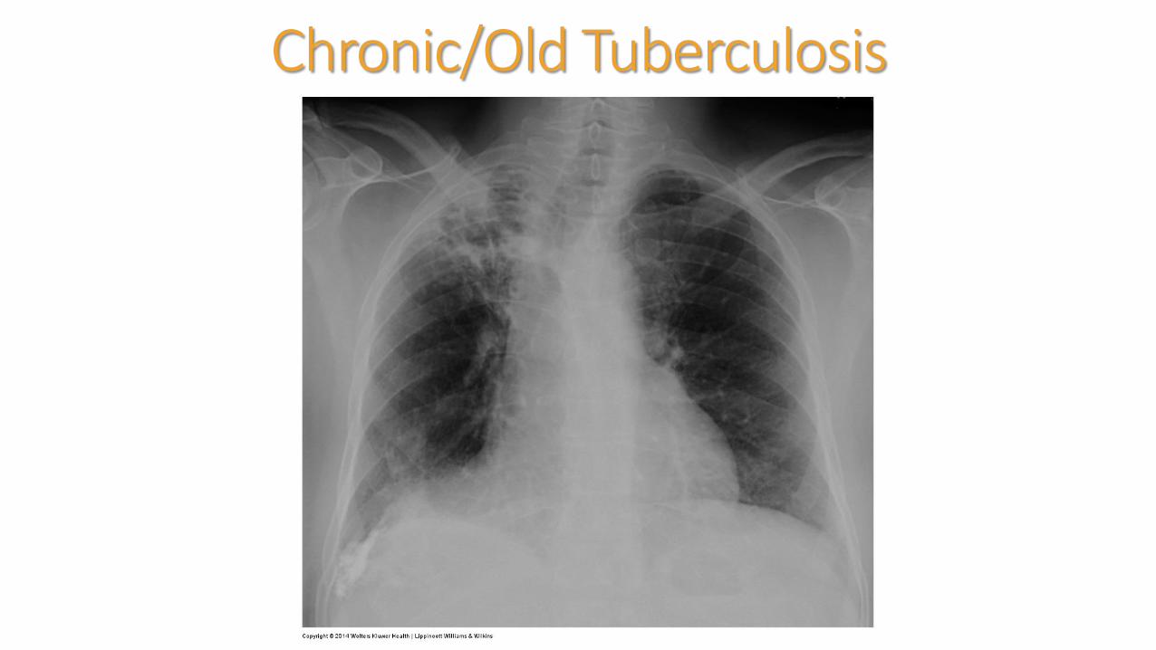
Chronic/Old Tuberculosis

Thank you!!Theresa M Campo DNP, APN, NP-C, ENP-BC, FAANP
Drexel University
Co-Director FNP Track
Associate Clinical Professor

ReferencesCollins & Stern. (2008). Chest Radiology: The Essentials. Wolters Kluwer/LWW: Phila, PA.
Daffner & Hartman (2014). Clinical Radiology: The Essentials 4th ed. WoltersKluwer/LWW: Phila, PA.
Hermann et al. (2012). Best practices in digital radiography: White paper. American Society of Radiologic Technologies accessed from http://www.asrt.org/docs/whitepapers/asrt12_bstpracdigradwhp_final.pdf