clinical dentistry endodontic cone beam...
TRANSCRIPT

CLINICAL28 www.dentistry.co.uk Dentistry
Endodontic cone beam CT Richard Kahan discusses a case study using cone beam computed tomography
Richard Kahan is an endodontist working in Harley Street and the ex-director of endodontic courses at UCL Eastman CPD. He has lectured on endodontics and technology and has set up the Academy of Advanced
Endodontics to teach the fundamentals of endodontics to GDPs through extended mentoring within his practice. years experience of endodontic CBCT his clinic has become a referral centre for complex cases used by both endodontists and GDPs. For more information visit www.endodontics.co.uk
Figure 1: Pre-operative periapical radiograph. Widening of the pdl observed around the mesial root of the LR7. Pdl intact around the mesial and distal roots of the LR6
Figure 5: CBCT Axial slice – this slice confirms the position of the lesion at the LL6 distal root between the cortical plates and also shows the presence of a single canal in the mesial root of the LR7
Figure 2: CBCT Saggital slice – widening of the pdl space around the mesial root of the LR7 (arrowed) can be observed. Also arrowed at the LR7 is a distal vertical periodontal defect. The width of this defect (seen axially) suggests it is likely to be of periodontal aetiology and not a vertical root fracture
Figure 6: Post-operative periapical radiograph
Figure 3: CBCT Saggital slice – a 2.5mm diameter periapical lesion is associated with the distal root of the LR6. There is also widening of the pdl space around the distal root of the LR7
Figure 4: CBCT Coronal slice – the lesion associated with the distal root of the LR6 is positioned between the buccal and lingual cortical plates and therefore invisible on a traditional 2D periapical radiograph
Radiography has an important role in the assessment of morphology and the diagnosis of pathology of the pulp and root canal system, but conventional planar radiography only provides a two-dimensional representation of this complex anatomy. The limited 2D representation might not yield sufficient information for the clinician to fully diagnose pathological states and therefore effectively treatment plan.
Limited volume cone beam computed tomography (CBCT) provides high-resolution three-dimensional undistorted images of the maxillo-facial skeleton, including the teeth and their surrounding tissues. Although the effective radiation dose used in CBCT is higher than that of conventional radiographic techniques, it is substantially lower than conventional CT1. The advantages of this relatively new technology in all fields of dentistry have not been overlooked and guidelines for its safe use have been prescribed by SEDENTEXCT (http://www.sedentexct.eu/content/guidelines-cbct-dental-and-maxillofacial-radiology).
CBCT has a demonstrated efficacy in a large number of endodontic applications, including but not limited to the investigation of complex dental anatomy and hidden pathology2.
I have been using CBCT for diagnosis, treatment decision-making and planning, along with surgical guidance for the past five years in my specialist practice. I believe that the advantages of the extra dimension and superior resolution substantially enhances the level of advice and treatment on offer to patients, reducing failure with effective diagnosis and increasing success and efficiency through the accurate identification of canal anatomy and the surrounding tissues.
This series of case discussions highlight the use of CBCT in clinical endodontics and how it is used to enhance diagnosis, decision-making, treatment planning and the treatment itself.
Case discussion 1 – a hidden lesion Clinical details A 56-year-old female patient with no relevant medical history was referred to the practice by a restorative consultant for root canal treatment of her lower right second molar (LR7). The patient had presented a week earlier to the referring dentist complaining of an intermittent pain in her lower right jaw. The dentist had identified the LR7 as the source of the pain and carried out a pulpectomy. He was only able to identify and negotiate a distal canal and a single mesial canal. A week following the emergency treatment the pain was still present and the referral requested further treatment through the location of a missed mesial canal.
At the time of consultation, the patient described the pain as intermittent, and moderate to severe. The timing was mostly random, lasting for hours, occasionally disturbing sleep, and it would invariably follow eating. It was poorly localised towards the posterior part of the right mandible with the pain radiating up towards the right ear. It was controlled with regular analgesics. Following the pulpectomy, the pain had worsened briefly but was now similar to that prior to the emergency procedure.
Clinical examination revealed a temporary filling in the LR7 which was slightly tender to percussion (TTP). The tooth did not respond to vitality tests (endo-ice and electric pulp testing (EPT)). The lower right first molar
(LR6) had a well fitting bonded precious metal full veneer crown and the lower right second premolar (LR5) was a bonded precious metal full veneer crown on an implant. Neither LR5 or LR6 was TTP and without visible dentine around the LR6 it was not possible to carry out an electric pulp test. Periodontal probing depths around all lower right posterior teeth were within normal limits and there were no discernible occlusal issues.
A periapical radiograph (Figure 1) showed a deep restoration in the LR7 with widening of the periodontal ligament (pdl) space associated with the mesial root apex. Apart from some sclerosis in the distal canal of the LR6 the pdl is clearly intact around this root and no other pathology was noted.
A limited volume (4cm x 4cm) cone beam computed tomograph was taken of the lower right mandible using a Morita Epochs 3D (80kV, 5mA, 9.4 sec). The scan confirmed widening of the pdl at the mesial root of LR7
(Figure 2), but also revealed a 2.5mm diameter lesion associated with the distal root of the LR6 (Figures 3 and 4). The scan also showed the presence of a single canal in the mesial root of the LR7 (Figure 5).
A diagnosis of chronic periapical periodontitis (CPP) associated with pulpal inflammation and necrosis was made for the LR7 and also CPP for the LR6 associated with likely necrosis or partial necrosis. As the LR7 had already been opened and dressed without pain resolution, a pulpectomy of the LR6 was carried out through the crown. The pulpal tissues in both mesial and distal canals were found to be necrotic and the canals were dressed with calcium hydroxide.
Within two to three days, the patient reported a resolution of her pain symptoms. Endodontic treatment of both the LR6 and LR7 was subsequently completed without complication (Figure 6).
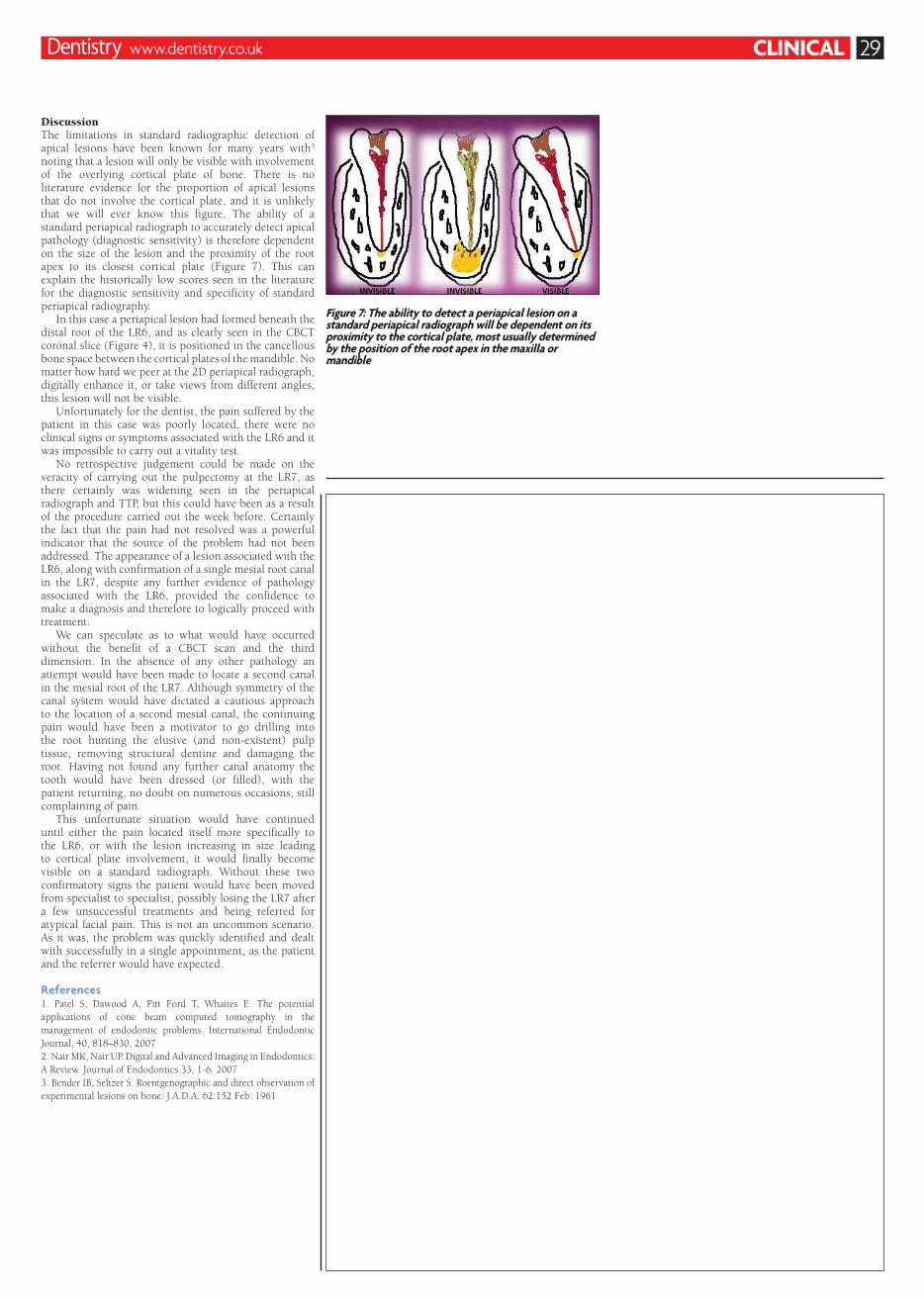
Figure 7: The ability to detect a periapical lesion on a standard periapical radiograph will be dependent on its proximity to the cortical plate, most usually determined by the position of the root apex in the maxilla or mandible
CLINICAL 29Dentistry www.dentistry.co.uk
Discussion The limitations in standard radiographic detection of apical lesions have been known for many years with3 noting that a lesion will only be visible with involvement of the overlying cortical plate of bone. There is no literature evidence for the proportion of apical lesions that do not involve the cortical plate, and it is unlikely that we will ever know this figure. The ability of a standard periapical radiograph to accurately detect apical pathology (diagnostic sensitivity) is therefore dependent on the size of the lesion and the proximity of the root apex to its closest cortical plate (Figure 7). This can explain the historically low scores seen in the literature for the diagnostic sensitivity and specificity of standard periapical radiography.
In this case a periapical lesion had formed beneath the distal root of the LR6, and as clearly seen in the CBCT coronal slice (Figure 4), it is positioned in the cancellous bone space between the cortical plates of the mandible. No matter how hard we peer at the 2D periapical radiograph, digitally enhance it, or take views from different angles, this lesion will not be visible.
Unfortunately for the dentist, the pain suffered by the patient in this case was poorly located, there were no clinical signs or symptoms associated with the LR6 and it was impossible to carry out a vitality test.
No retrospective judgement could be made on the veracity of carrying out the pulpectomy at the LR7, as there certainly was widening seen in the periapical radiograph and TTP, but this could have been as a result of the procedure carried out the week before. Certainly the fact that the pain had not resolved was a powerful indicator that the source of the problem had not been addressed. The appearance of a lesion associated with the LR6, along with confirmation of a single mesial root canal in the LR7, despite any further evidence of pathology associated with the LR6, provided the confidence to make a diagnosis and therefore to logically proceed with treatment.
We can speculate as to what would have occurred without the benefit of a CBCT scan and the third dimension. In the absence of any other pathology an attempt would have been made to locate a second canal in the mesial root of the LR7. Although symmetry of the canal system would have dictated a cautious approach to the location of a second mesial canal, the continuing pain would have been a motivator to go drilling into the root hunting the elusive (and non-existent) pulp tissue, removing structural dentine and damaging the root. Having not found any further canal anatomy the tooth would have been dressed (or filled), with the patient returning, no doubt on numerous occasions, still complaining of pain.
This unfortunate situation would have continued until either the pain located itself more specifically to the LR6, or with the lesion increasing in size leading to cortical plate involvement, it would finally become visible on a standard radiograph. Without these two confirmatory signs the patient would have been moved from specialist to specialist, possibly losing the LR7 after a few unsuccessful treatments and being referred for atypical facial pain. This is not an uncommon scenario. As it was, the problem was quickly identified and dealt with successfully in a single appointment, as the patient and the referrer would have expected.
References 1. Patel S, Dawood A, Pitt Ford T, Whaites E. The potential applications of cone beam computed tomography in the management of endodontic problems. International Endodontic Journal, 40, 818–830, 20072. Nair MK, Nair UP. Digital and Advanced Imaging in Endodontics: A Review. Journal of Endodontics 33, 1-6. 2007 3. Bender IB, Seltzer S. Roentgenographic and direct observation of experimental lesions on bone: J.A.D.A. 62:152 Feb. 1961