clinical deterioration in community acquired infections...
TRANSCRIPT

Title Clinical deterioration in community acquired infectionsassociated with lymphocyte upsurge in immunocompetent hosts
Author(s) Cheng, VCC; Wu, AKL; Hung, IFN; Tang, BSF; Lee, RA; Lau,SKP; Woo, PCY; Yuen, KY
Citation Scandinavian Journal Of Infectious Diseases, 2004, v. 36 n. 10,p. 743-751
Issued Date 2004
URL http://hdl.handle.net/10722/48638
Rights
Scandinavian Journal of Infectious Diseases. Copyright ©Informa Healthcare.; This work is licensed under a CreativeCommons Attribution-NonCommercial-NoDerivatives 4.0International License.

This is a pre-published versionThis is a pre-published version
Clinical deterioration in community acquired infections associated
with lymphocyte upsurge in immunocompetent hosts
VINCENT C.C. CHENG1, ALAN K.L. WU2, IVAN F.N. HUNG1, BONE
S.F. TANG1, RODNEY A. LEE1, SUSANNA K.P. LAU1, PATRICK C.Y.
WOO1, KWOK-YUNG YUEN1
From the 1Division of Infectious Diseases, Centre of Infection, Queen Mary Hospital,
The University of Hong Kong; Hong Kong Special Administrative Region, China and the
2Division of Infectious Diseases, Department of Medicine and Therapeutics, Prince of
Wales Hospital, Hong Kong Special Administrative Region, China.
K.Y.Yuen, Division of Infectious Diseases, Centre of Infection, Queen Mary Hospital,
The University of Hong Kong, Hong Kong Special Administrative Region, China (Tel:
+852-28554892, Fax: +852-28551241, E-mail: [email protected])
1

ABSTRACT Clinical deterioration during the course of community-acquired infections can occur as a
result of an exaggerated immune response of the host towards the inciting pathogens,
leading to immune-mediated tissue damage. Whether a surge in the peripheral
lymphocyte count can be used as a surrogate marker indicating the onset of
immunopathological tissue damage is not known. In this study, we reported the clinical
presentations and outcomes of a cohort of immunocompetent patients with non-
tuberculous community acquired infections who experienced clinical deteriorations
during hospital stay (N=85). 12 (14.1%) patients had a surge in lymphocyte count
preceding their clinical deteriorations, and their diagnoses included viral pneumonitis (4),
viral encephalitis (3), scrub typhus (2), leptospirosis (1), brucellosis (1), and dengue
hemorrhagic fever (1). The clinical manifestations during deterioration ranged from
interstitial pneumonitis (6), airway obstruction (1), CNS disturbances (4), and systemic
capillary leak syndrome (1), all of which were thought to represent immunopathological
tissue damages. When compared with patients without lymphocyte surge, these patients
were more likely to be infected with fastidious / viral pathogens (0 vs 12; p <0.05), in
addition to having lower mean baseline lymphocyte counts (403 ± 181 vs 1143 ± 686
cells/µL; p <0.05). We postulate that the peripheral lymphocyte count may be a useful
surrogate marker indicating the presence of immunopathological damage during clinical
deterioration in certain infectious diseases.
2

INTRODUCTION
The outcome of clinical infectious diseases depends on both the virulence of the microbes
and the appropriateness of host immune responses. An intact innate immunity with or
without subsequent adaptive immunity towards the infecting organisms remains the most
important factor in eradicating or controlling the infections. However, clinical
deterioration as a result of immunopathological damage may occur as a result of an
overwhelming rebound of the immune system, either temporally related to the withdrawal
of immunosuppressive therapy such as steroid or cytotoxic agents, or secondary to
treatment of microbes with potent immunosuppressive effects on the host such as HIV. It
is manifested as immunorestitution disease, which has been described in previous studies
[1,2,3]. In addition, immune-mediated tissue damage can also develop in either
immunosuppressed or immunocompetent patients infected with Mycobacterium
tuberculosis, with disease manifestation or exacerbation during anti-tuberculosis therapy,
a phenomenon that has been given the term ‘paradoxical response’ [4-9]. Recent studies
showed that an upsurge of the absolute lymphocyte counts and conversion of tuberculin
skin test from negative to positive occurred during paradoxical deterioration [10,11],
even for patients co-infected with HIV [7]. A low baseline lymphocyte count with a
subsequent greater upsurge is one of the risk factors for development of a paradoxical
response [5].
Apart from tuberculosis, clinical deterioration during the course of illness can also occur
in immunocompetent patients affected by certain infectious diseases, giving rise to so-
called biphasic illness patterns. For instance, leptospirosis is characterized by an initial
3

bacteremic phase lasting 4 to 7 days, may be followed by an immune-mediated
meningitis, uveitis, and pneumonitis, causing multiple organ failure [12,13]. Dengue
virus infection may also present with ‘saddleback' biphasic fever pattern, followed by
increased vascular permeability which can result in hypovolemic shock in some cases
[14]. However, the association of an upsurge of absolute lymphocyte count with
clinical deterioration in these patients has not been reported.
In this article, we report a cohort of immunocompetent patients with non-tuberculous
community acquired infections in whom clinical deterioration occurred during an upsurge
of the absolute lymphocyte count. The clinical spectrum of this phenomenon and its
possible mechanisms are discussed.
4

MATERIALS AND METHODS
Patients
All clinical data were collected prospectively over a 24-month period (January 2000 –
December 2001) on patients referred for infectious disease (ID) consultation in a tertiary
hospital (Queen Mary Hospital, Hong Kong, a 1,350-bed teaching hospital).
Definition of clinical deterioration involving upsurge of lymphocyte count
For the purpose of this study, clinical deterioration involving upsurge of the lymphocyte
count was defined as an acute symptomatic deterioration of a (presumably) pre-existing
infection, which is temporally related to an increase in the absolute lymphocyte count.
The acute symptomatic deterioration included worsening of neurological symptoms such
as mental confusion, disorientation, and convulsions; worsening of respiratory symptoms
such as respiratory distress and development of pulmonary infiltrates and pleural
effusion; and hypotension with multiple organ failure.
Surge in absolute lymphocyte count was defined as rise of lymphocyte count of ≥ 500
cells / μL if the baseline lymphocyte count was less than 500 cells / μL, or double the
baseline value when the lymphocyte count was more than 500 cells / μL, within a 2-day
period.
Exclusion criteria
Patients receiving immunosuppressive therapy (systemic steroids, cytotoxic agents, a
combination of steroid and cytotoxic agents, or irradiation) or known to have
immunodeficiency or infected with HIV were excluded from this study as these
5

conditions may suppress the absolute lymphocyte count. Patients with chronic diseases
such as diabetes mellitus, chronic renal failure, and chronic liver disease were also
excluded from this study as they may have abnormal lymphocyte function. Patients with
nosocomial infections were excluded because in-patient interventions may result in
changes in lymphocyte count. Patients with diagnosis of M. tuberculosis infections were
also excluded as they were analyzed separately in the context of paradoxical response
[4,5,10]
Time to development of clinical deterioration involving upsurge of lymphocyte count
The time to development of clinical deterioration involving upsurge of lymphocyte
counts was defined as the interval between the initial clinical symptoms and the onset of
symptomatic deterioration as defined above.
The consultation procedure has been described previously [15,16]. Briefly, detailed
history taking, physical examination and review of case notes were performed for each
patient. Serial lymphocyte counts with subsequent surges, and the occurrence of
symptomatic clinical deteriorations, were recorded and monitored for each patient.
Statistical evaluation
The characteristics of patients with or without lymphocyte surge during clinical
deterioration were compared. The chi-square test was used for categorical variables.
Continuous variables were tested by the Student’s t test. A P value of < 0.05 was
considered significant. A statistical package (SPSS 10.0; SPSS Hong Kong, Hong Kong)
was used for all analyses.
6
RESULTS
During the 24-month period, there were 3518 inpatient ID consultations, of which 357
(10.1%) patients with median age of 59, range 2-98, were immunocompetent patients
with clinical and microbiological evidence of non-tuberculous community acquired
infections. Eighty-five patients had clinical deterioration during follow up (table 1). Of
these 85 patients, 12 (14.1%) had an upsurge of lymphocyte count during clinical
deterioration (table 2). Eight were male and four were female, with median age of 35,
range 17-75. Diagnoses in these patients included viral pneumonitis (4), viral
encephalitis (3), scrub typhus (2), leptospirosis (1), brucelllosis (1), and dengue
hemorrhagic fever (1) (table 2). The median duration between the onset of symptoms to
diagnosis was 6 days (8 days for bacterial infections and 2.5 days for viral infections).
The median time of clinical deterioration was 10 days from symptom onset. But the
median ‘time to clinical deterioration’ was 6 days and 11.5 days for viral and bacterial
infection respectively. The clinical manifestations during deterioration included acute
respiratory distress syndrome as a result of interstitial pneumonitis (6), severe upper
airway obstruction (1), mental confusion with or without convulsions (4), and capillary
leak syndrome (1). When compared with patients without lymphocyte surge, these
patients were more likely to be suffering from infection by fastidious/intracellular
bacteria or virus (p < 0.05), in addition to having a lower baseline lymphocyte count (403
± 181 cells / μL; p < 0.05), a higher lymphocyte count during clinical deterioration (1813
± 941 cells / μL; p <0.05), and a greater change in lymphocyte count between baseline
and clinical deterioration (1410 ± 954 cells / μL; p <0.05). Although empirical
antibiotics or antiviral agents were initiated in 8 (66.6%) out of 12 patients, appropriate
7

antibiotics or antiviral agents were given in 5 patients after the onset of clinical
deterioration. One out of 12 patients died of adult respiratory distress syndrome, whereas
3 (25%) of them developed permanent functional deficits including left-sided
hemiparesis, impairment of cognitive function, and chronic renal failure. The median
length of stay in hospital was 24 days, range 8-269.
Case reports
Case 1
A 29 year-old man was admitted to hospital for persistent high swinging fever and
headache 9 days after occupational exposure to a scrub area in Hong Kong. On
admission, he had a temperature of 38.3oC. Physical examination revealed conjunctival
haemorrhage, diffuse maculopapular rash, hepatomegaly, and a 2 cm eschar on his right
shin. Preliminary investigations showed a normal white cell count (7110 cells / μL,
normal range 400-1100 cells / μL) but his lymphocyte count was depressed (370 cells /
μL, normal range 150-400 cells / μL). The platelet count was low (47 x 109 /L, normal
range 150-400 x 109 /L). Liver function tests showed elevated alanine and aspartate
aminotransferase (ALT) 311 U/L (normal range 6-53 U/L) and aspartate
aminotransferase levels and renal function was well. The chest radiograph showed
pulmonary congestion. A clinical diagnosis of Rickettsial infection was made and he was
given doxycycline 100 mg bd. However, he deteriorated rapidly with progressive liver
impairment, renal impairment and respiratory distress, and required mechanical
ventilation in intensive care unit 3 days after admission. Repeated chest radiographs
demonstrated acute pulmonary edema. The hemoglobin dropped from 15.4 g/dL to 12.7
8
g/dL over 3 days and the platelet count further decreased to 36 x 109 /L. A sudden
upsurge of lymphocyte count (2670 cells / μL) was observed at the time of deterioration,
which further increased to 3250 cells / μL. The clinical features were compatible with
endothelitis, capillary leak syndrome and multiple organ failure as a result of
immunopathological damage due to severe rickettsial infection. After receiving
antibiotics and methylprednisolone, he gradually recovered. His lymphocyte count
decreased to 1200 cells / μL on the day of extubation. He had positive Orientia
tustusgamushi IgM serology by immunofluoresence and the Weil-Felix OXK component
demonstrated a four-fold rise in titre from 1:80 to 1:320 over a 2-week interval.
Case 2
A 27-year-old man presented with fever, chills, and abdominal discomfort 7 days after
occupational exposure to a scrub area in Hong Kong. On admission, he was alert but had
a temperature of 38oC. Physical examination showed maculopapular rash, hepatomegaly,
and a 1 cm eschar in his right groin. Preliminary investigations revealed a total white cell
count of 5600 cells / μL with lymphopenia (300 cells / μL). The platelet count was low
(27 x 109 /L). Liver function tests were deranged whereas renal function tests were
normal. A clinical diagnosis of Rickettsial infection was made and he was given
doxycycline 100 mg bd. However he deteriorated rapidly with respiratory distress, and
required mechanical ventilation in the intensive care unit for 3 days after admission. A
chest radiograph demonstrated acute pulmonary edema. On the day of deterioration,
there was a sudden upsurge of lymphocyte (3700 cells / μL), which further increased to
3900 cells / μL one day later. The clinical features were compatible with endothelitis
and capillary leak as a result of immunopathological damage secondary to severe
9

rickettsial infection. He was given intravenous minocycline 100 mg q12h and
levofloxacin 500 mg q24h. He subsequently improved and was extubated after 5 days of
mechanical ventilation. The lymphocyte count decreased to 1500 cells / μL on the day of
extubation. He had positive Orientia tustusgamushi IgM serology and the Weil-Felix
OXK component demonstrated a three fold serial elevation from 1:20 to 1:160 over a 2-
week interval. He had a full recovery after a 4-week course of doxycycline 100 mg bd.
Case 3
A 39-year-old man presented with fever, generalized malaise, bilateral calf pain, and
abdominal discomfort 6 days after a heavy rainstorm with flooding in the city. On
admission, he had conjuctival suffusion and hepatomegaly. Investigations showed a
normal white cell count (8700 cells / μL), but the lymphocyte count was decreased (400
cells / μL). His platelet count was 35 x 109 /L. The liver function was mildly elevated.
The renal function tests were normal. A clinical diagnosis of leptospirosis was made and
he was given intravenous penicillin 1 MU q6h. Fever subsided after 3 days of therapy.
On day 5, however, he rapidly deteriorated with recurrent fever, hypotension and
deranged liver and renal function. A chest radiograph revealed bilateral pulmonary
infiltrates. A sudden surge in the lymphocyte count from 400 to 1100 cells / μL was
observed on the day of deterioration. He was continued with intravenous penicillin for 1
week and recovered after a 2-month convalescent period. IgM antibodies to Leptospria
interrogans were demonstrated by enzyme-linked immunosorbent assay.
Case 4
A 72-year-old lady with a history of dementia presented with low-grade fever, poor
appetite, malaise, and generalized bone pain for 2 weeks. On admission, she was found
10

to have pancytopenia with hemoglobin of 7.5 g/dL, a total white cell count of 3100 cells /
μL (lymphocyte of 300 cells / μL), and a platelet count of 48 x 109 /L. Hematological
malignancy was suspected but bone marrow examination was unremarkable. On day 8,
while her lymphocyte count surged to 1200 cells / μL, she became mentally confused,
and disorientated in time, place, and person. Lumbar puncture was not performed
because of thrombocytopenia. She was given intravenous cefoperzone-sulbactam 1 gm
q8h as empirical therapy but fever persisted. Serological evidence of brucellosis was
obtained by serum agglutination test, in which there was a 4-fold rise in BM from 1:20 to
1:320 over a 10-day interval. Magnetic resonance imaging of brain revealed diffuse
ischemia in brainstem and thalamus. She was treated as neurobrucellosis with
doxycycline 100 mg bd and rifampicin 600 mg qd for 6 months. Both her mental state
and pancytopenia recovered with therapy. She probably acquired the infection by
ingestion of raw meat because of inadequate cooking.
Case 5
A 33-year-old man presented with fever, chills, headache, sore throat, cough and myalgia
7 days after returning from Bangladesh. Physical examination revealed a fine diffuse
macular rash over the body. On admission, he had a normal white cell count (8400 cells /
μL) but had lymphopenia (400 cells / μL). His platelet count was 168 x 109 /L and the
liver and renal function tests were normal. A clinical diagnosis of dengue fever was
made. However, he had a progressive drop in platelet count (25 x 109 /L) and blood
pressure after 3 days of admission. A chest radiograph revealed bilateral pleural
effusions. A surge of the lymphocyte count (2100 cells / μL) was observed on the day of
deterioration. The liver function tests were also deranged when assayed 3 days after the
11
upsurge of lymphocytes. He gradually improved and pleural effusion resolved after 1
month. Paired serum demonstrated a rising titer from < 10 to 1:2560 towards dengue
type 2 over a 4-day interval. The dengue PCR on his acute serum was also positive.
Case 6
A 75-year-old man presented with painful reactivation of varicella zoster in the
dermatomal distribution of L1. On admission, he was conscious and afebrile. The white
cell count was normal (4100 cells / μL), but the lymphocyte count was low (700 cells /
μL). He was given oral famciclovir 500 mg tds and the skin lesions gradually subsided.
However, after 10 days of therapy, he developed generalized malaise, mental confusion,
and convulsions, suggestive of encephalitis. The lymphocyte count increased to (1600
cells / μL) on the day of deterioration. Cerebrospinal fluid (CSF) revealed lymphocytic
pleocytosis with a total cell count of 293 cells / μL and a lymphocyte count of 201 cells /
μL. His CSF was polymerase chain reaction (PCR) positive for varicella zoster virus but
a viral culture of CSF was negative. He gradually improved with intravenous acyclovir
500 mg q8h for 14 days.
Case 7
A 27-year-old man presented with neck pain, neck stiffness, and progressive headache for
1 week. On admission, he had a temperature of 37.6 oC. The white cell count and
lymphocyte count were 5610 and 760 cells / μL respectively. Lumbar puncture revealed
a total cell count of 819 and a lymphocyte count of 606 cells / μL. He was treated with
intravenous acyclovir 500 mg ivi q8h. On day 3, he developed mental confusion with a
surge in peripheral lymphocyte count to 2000 cells / μL. Anti-tuberculous therapy was
started in view of the clinical deterioration. He gradually improved neurologically after 1
12

weeks of acyclovir and 4 days of anti-tuberculous treatment. CSF was PCR positive for
HSV and for mycobacteria. Anti-tuberculous drugs were withheld and he fully recovered
after 21 days of acyclovir.
Case 8
A 35-year-old woman presented with a 3-day history of fever, headache, nausea, and
vomiting. On admission, she was mentally alert. Physical examination was
unremarkable except for a low-grade fever of 37.8 oC. Her white cell count was normal
(6200 cells / μL) and the lymphocyte count was low (500 cells / μL). On day 3, she
developed mental confusion with auditory hallucination and convulsion. Computerized
tomography of brain revealed a 3 cm hypodense lesion in the right temporal area.
Magnetic resonance scanning showed an abnormal right temporal lobe with mild
hypointense signal on T-1 weighted image and a heterogeneous hyperintense signal on T-
2 weighted image, consistent with encephalitis. CSF showed a total cell count of 130
cells / μL and lymphocyte of 125 cells / μL. Her blood lymphocyte count increased from
500 to 1400 cells / μL on the day of deterioration. She was treated with intravenous
acyclovir 500 mg q8h for 21 days and recovered with a residual deficit in abstract
thinking. The CSF was HSV-1 PCR positive.
Case 9
A 71-year-old woman was admitted for a 2-day history of sore throat and nonproductive
cough. On admission, the white cell and lymphocyte counts were 2900 and 300 cells /
μL respectively. She was treated conservatively as community acquired viral infection.
However, two days after admission, she deteriorated with respiratory distress and
inspiratory stridor requiring mechanical ventilation. Her lymphocyte count surged from
13

300 to 800 cells / μL at the time of deterioration. Bronchoscopic examination showed
swollen vocal cords and subglottic edema. A nasopharyngeal aspirate for direct antigen
detection was positive for parainfluenza virus type 3. She required prolonged ventilation
for 28 days and finally recovered on day 35.
Case 10
A 17-year-old female with mental retardation presented with flu-like symptoms for 2
days. On admission, her white cell count was normal (5600 cells / μL), with a depressed
lymphocyte count (300 cells / μL). A chest radiograph was unremarkable. On day 3, she
developed progressive respiratory distress and required mechanical ventilation. The
chest radiograph showed bilateral interstitial opacities suggestive of pneumonitis. At the
time of deterioration, her lymphocyte count increased from 1300 (day 3) to 3180 cells /
μL (day 5). A nasopharyngeal aspirates showed influenza A virus by direct antigen
detection. She succumbed on day 10 from adult respiratory distress syndrome despite
therapy with amantidine and intravenous cefotaxime.
Case 11
A 67-year-old man with a history of chronic obstructive airway disease (COAD) was
admitted complaining of shortness of breath for 2 days. On admission, his lymphocyte
count was 400 cells / μL and the chest radiograph showed hyperinflated lung fields. He
was diagnosed as having an exacerbation of COAD, and treatment with bronchodilators.
Three days after admission he developed worsening respiratory distress, and a chest
radiograph revealed interstitial pneumonitis. This was associated with a surge in the
lymphocyte count of 1100 cells / μL. He was intubated and mechanically ventilated in
the intensive care unit. A nasopharyneal aspirate was positive for influenza A virus by
14

direct antigen detection. He was extubated on day 7 and transferred to a convalescent
hospital for further management.
Case 12
A 64-year-old man with an underlying history of chronic obstructive airway disease was
admitted to hospital for increasing shortness of breath for 1 week. On admission, he was
noted to have leucocytosis (white cell count 13700 cells / μL) and lymphopenia (total
lymphocyte count 100 cells / μL). He was diagnosed as having a COAD exacerbation
and treated with bronchodilators. Three days after admission he deteriorated with
increasing respiratory distress, and chest radiographs revealed bilateral interstitial
pneumonitis. He developed progressive respiratory failure and was subsequently
intubated and mechanically ventilated. The lymphocyte count surged to 900 μ/L. A
nasopharyngeal aspirate was positive for respiratory syncytial virus. He gradually
recovered after 8 days of ventilation.
15

DISCUSSION
Clinical deterioration associated with lymphocyte surge is not a rare phenomenon,
especially in the context of certain infectious diseases [1-4,10]. In our experience, it
constitutes 14% of immunocompetent hosts who experienced clinical deterioration during
the course of community-acquired infections. In addition to the classical examples of
leptospirosis and dengue virus infection in which a “biphasic” clinical pattern may occur,
infections caused by fastidious/intracellular bacteria such as Orientia tsutsugamushi,
Brucella species, and other commonly encountered viruses such as influenza A virus,
parainfluenza virus, respiratory syncytial virus, herpes simplex virus and varicella zoster
virus were also observed in our patients with clinical deterioration during the course of
illness. Endothelium-rich organs such as lungs and brain were the most commonly
involved systems during clinical deterioration.
It is not surprising to observe an upsurge of the lymphocyte count in patients suffering
from fastidious/intracellular bacteria and viruses because an adaptive immune response is
required for their clearance [17]. However, the clinical outcome of infection is dependent
on the appropriateness and exactness of the development of adaptive immunity. An
overwhelming and exaggerated immune response may lead to excessive
immunopathological damage at the tissue level [1]. This phenomenon is exemplified by
scrub typhus, caused by O. tsutsugamushi, which is a gram negative obligate intracellular
bacterium transmitted by the bites of infected mite. The pathologic damage of scrub
typhus included disseminated vasculitis and perivasculitis of small blood vessels as a
result of direct invasion and proliferation of O. tsutsugamushi in the vascular endothelial
16

cells, which may lead to multiple organ failure [24]. In experimental studies, transient
immunosuppression occurred during acute infection [18]. Subsequent development of
specific T lymphocytic response, especially T-helper 1 cells, and development of delay-
type hypersensitivity were important for protective immunity [20-23]. However, an
overwhelming immune response may contribute to the development of acute respiratory
distress syndrome, which is caused by interstitial infiltration of lymphocytes without any
evidence of vasculitic damage [25]. However, the serial lymphocyte counts were not
mentioned during clinical deterioration in these case reports.
As for viral infection, dengue hemorrhagic fever occurred as a result of massive T
lymphocyte activation leading to high level of inflammatory cytokine production such as
interferon γ, interleukin 2, tumor necrosis factor α. Overproduction of cytokines that
affect monocytes, endothelial cells, and hepatocytes may result in capillary leakage and
deranged liver function [26,27]. Development of T cell mediated immunity in respiratory
virus infection has been associated with immunopathological damage in lungs in various
experimental models [28,29]. Influenza infection in CD8 T-cell depleted mice presented
with less significant histological evidence of inflammatory injury which suggested that
host T cells played a role in the immunopathological process [30]. Further study
demonstrated that antigen-specific CD8 T cells were capable of recognizing an alveolar
epithelial autoantigen, which triggered an inflammatory cascade and resulted in lung
damage [31]. In contrast to CD8 T cell mediated damage in influenza virus infection,
CD4 T cells have been shown to be detrimental to the host by mediating
immunopathological damage in respiratory syncytial virus infection [32].
17

It is possible that the upsurge in lymphocyte count may have been caused by specific
microbial components or products. One interesting example illustrating some of the
potential mechanisms may be found in the model of pertussis infection. Infection by
Bordetella pertussis has been reported to result in peripheral blood lymphocytosis in
infants [33,34]. The expression of L-selectin (CD62L), an important cell surface adhesion
molecule mediating extravasation of blood lymphocytes into tissues and homing of
lymphocytes to lymph nodes, has been found to be marked reduced in infants with
pertussis infection [33,34]. In addition, it has been shown in animal models that
expression of other lymphocyte adhesion molecules such as CD11a and CD18 (LFA-1)
are also reduced in macaques treated with pertussis toxin [35]. The significant down-
regulation of these adhesion molecules may prevent lymphocyte migration to areas of
infection and homing of T and B cells to peripheral lymphoid tissues, resulting in
peripheral blood lymphocytosis.
In additional to the microbial factors, specific host factors might have contributed to the
upsurge in lymphocytes. Various cytokine levels can affect the lymphocyte count; for
instance, increase in lymphocyte count has been shown to correlate with response to
interleukin 2 therapy for malignant conditions [36,37]. Even acute stress may lead to
changes in lymphocyte count as a result of changes in integrin expression and
lymphocyte trafficking in the body [38]. Thus, it is possible that the cytokine response or
stress induced by some of the microbes may have contributed to the lymphocyte upsurge.
However, the scope of our study does not allow us to measure the various cytokine levels
or to determine the pattern of expression of adhesion molecules on the lymphocytes.
18

Besides having lymphocyte upsurge during hospitalizations, many of our patients also
had low lymphocyte counts on admission. Lymphopenia is not uncommonly observed in
the acute phase of viral infections, such as during infection by respiratory syncytial virus
[39], avian influenza [40] and even SARS [41]; in can also occur in the setting of severe
sepsis [42]. One limitation of our study is that we could not differentiate whether the
lymphocyte upsurge observed in our patients actually reflect initial migration of
lymphocytes to infected tissues followed by subsequent rebound, or the effect of
microbial and/or cytokine mediated lymphocyte apoptosis followed by reconstitution
later in disease course. Further studies are warranted to delineate the underlying
immunopathological mechanisms involved in this interesting phenomenon.
The exact mechanism mediating the immunopathology in various types of clinical
infectious diseases requires further investigation. However, peripheral lymphocyte
counts may act as a surrogate marker indicating the onset of possible
immunopathological damage, which may be useful to infectious disease specialists who
are involved in the care of these patients. Studies on lymphocyte subsets and cytokine
levels would be useful in understanding this clinical entity.
19

Reference
1. Cheng VC, Yuen KY, Chan WM, Wong SS, Ma ES, Chan RM.
Immunorestitution disease involving the innate and adaptive response. Clin Infect
Dis. 2000; 30: 882-92.
2. Cheng VC, Yuen KY, Wong SS, Woo PC, Ho PL, Lee R, Chan RM.
Immunorestitution diseases in patients not infected with HIV. Eur J Clin
Microbiol Infect Dis. 2001; 20: 402-6.
3. Cheng VC, Hung IF, Wu AK, Tang BS, Chu CM, Yuen KY. Lymphocyte surge
as a marker for immunorestitution disease due to Pneumocystis jiroveci
pneumonia in HIV-negative immunosuppressed hosts. Eur J Clin Microbiol
Infect Dis 2004; 23: 512-4.
4. Cheng VC, Ho PL, Lee RA, Chan KS, Chan KK, Woo PC, Lau SK, Yuen KY.
Clinical spectrum of paradoxical deterioration during antituberculosis therapy in
non-HIV-infected patients. Eur J Clin Microbiol Infect Dis. 2002; 21: 803-9.
5. Cheng VC, Yam WC, Woo PC, Lau SK, Hung IF, Wong SP, Cheung WC, Yuen
KY. Risk factors for development of paradoxical response during anti-
tuberculosis therapy in HIV-negative patients. Eur J Clin Microbiol Infect Dis
2003; 22: 597-602.
6. Narita M, Ashkin D, Hollender ES, Pitchenik AE. Paradoxical worsening of
tuberculosis following antiretroviral therapy in patients with AIDS. Am J Respir
Crit Care Med 1998; 158:157-61.
20
7. Wendel KA, Alwood KS, Gachuhi R, Chaisson RE, Bishai WR, Sterling TR.
Paradoxical worsening of tuberculosis in HIV-infected persons. Chest 2001; 120:
193-7.
8. Orlovic D, Smego RA Jr. Paradoxical tuberculous reactions in HIV-infected
patients. Int J Tuberc Lung Dis 2001; 5: 370-5.
9. Navas E, Martin-Davila P, Moreno L, Pintado V, Casado JL, Fortun J, Perez-Elias
MJ, Gomez-Mampaso E, Moreno S. Paradoxical reactions of tuberculosis in
patients with the acquired immunodeficiency syndrome who are treated with
highly active antiretroviral therapy. Arch Intern Med 2002; 162: 97-9.
10. Cheng VC, Woo PC, Lau SK, Cheung CH, Yung RW, Yam LY, Yuen KY.
Peripartum tuberculosis as a form of immunorestitution disease. Eur J Clin
Microbiol Infect Dis. 2003; 22: 313-7.
11. Valdez LM, Schwab P, Okhuysen PC, Rakita RM. Paradoxical subcutaneous
tuberculous abscess. Clin Infect Dis 1997; 24: 734.
12. Sperber SJ, Schleupner CJ. Leptospirosis: a forgotten cause of aseptic meningitis
and multisystem febrile illness. South Med J. 1989; 82: 1285-8.
13. Hill MK, Sanders CV. Leptospiral pneumonia. Semin Respir Infect. 1997; 12: 44-
9.
14. Henchal EA, Putnak JR. The dengue viruses. Clin Microbiol Rev. 1990; 3: 376-
96.
15. Yuen KY, Seto WH, Chau PY. An evaluation of inpatient consultations
conducted by clinical microbiologists in a teaching hospital. J Infection 1992; 25:
29-37.
21

16. Luk WK, Wong SS, Yuen KY, Ho PL, Woo PC, Lee R, Peiris JS, Chau PY:
Inpatient emergencies encountered by an infectious disease consultative service.
Clin Infect Dis 1998; 26: 695-701.
17. Blumberg RS, Schooley RT. Lymphocyte markers and infectious diseases. Semin
Hematol 1985; 22: 81-114.
18. Jerrells TR. Immunosuppression associated with the development of chronic
infections with Rickettsia tsutsugamushi: adherent suppressor cell activity and
macrophage activation. Infect Immun 1985; 50: 175-82.
19. Kasuya S, Nagano I, Ikeda T, Goto C, Shimokawa K, Takahashi Y. Apoptosis of
lymphocytes in mice induced by infection with Rickettsia tsutsugamushi. Infect
Immun 1996; 64: 3937-41.
20. Jerrells TR, Osterman JV. Host defenses in experimental scrub typhus: delayed-
type hypersensitivity responses of inbred mice. Infect Immun 1982; 35: 117-23.
21. Jerrells TR, Osterman JV. Development of specific and cross-reactive lymphocyte
proliferative responses during chronic immunizing infections with Rickettsia
tsutsugamushi. Infect Immun 1983; 40: 147-56.
22. Palmer BA, Hetrick FM, Jerrells TJ. Production of gamma interferon in mice
immune to Rickettsia tsutsugamushi. Infect Immun 1984; 43: 59-65.
23. Hickman CJ, Stover CK, Joseph SW, Oaks EV. Murine T-cell response to native
and recombinant protein antigens of Rickettsia tsutsugamushi. Infect Immun
1993; 61: 1674-81.
24. Strickman D, Smith CD, Corcoran KD, Ngampochjana M, Watcharapichat P,
Phulsuksombati D, Tanskul P, Dasch GA, Kelly DJ. Pathology of Rickettsia
22

tsutsugamushi infection in Bandicota savilei, a natural host in Thailand. Am J
Trop Med Hyg 1994; 51: 416-23.
25. Park JS, Jee YK, Lee KY, Kim KY, Myong NH, Seo PW. Acute respiratory
distress syndrome associated with scrub typhus: diffuse alveolar damage without
pulmonary vasculitis. J Korean Med Sci 2000; 15: 343-5.
26. Rothman AL, Ennis FA. Immunopathogenesis of Dengue hemorrhagic fever.
Virology 1999; 257: 1-6.
27. Lei HY, Yeh TM, Liu HS, Lin YS, Chen SH, Liu CC. Immunopathogenesis of
dengue virus infection. J Biomed Sci 2001; 8: 377-88.
28. Julkunen I, Melen K, Nyqvist M, Pirhonen J, Sareneva T, Matikainen S.
Inflammatory responses in influenza A virus infection. Vaccine 2000; 19 (Suppl
1): S32-7.
29. Varga SM, Braciale TJ. RSV-induced immunopathology: dynamic interplay
between the virus and host immune response. Virology 2002; 295: 203-7.
30. Scherle PA, Palladino G, Gerhard W. Mice can recover from pulmonary influenza
virus infection in the absence of class I-restricted cytotoxic T cells. J Immunol
1992; 148: 212-7.
31. Enelow RI, Mohammed AZ, Stoler MH, Liu AN, Young JS, Lou YH, Braciale
TJ. Structural and functional consequences of alveolar cell recognition by CD8(+)
T lymphocytes in experimental lung disease. J Clin Invest 1998; 102: 1653-61.
32. Graham BS, Bunton LA, Wright PF, Karzon DT. Role of T lymphocyte subsets in
the pathogenesis of primary infection and rechallenge with respiratory syncytial
virus in mice. J Clin Invest 1991; 88: 1026-33.
23

33. Hodge G, Hodge S, Markus C, Lawrence A, Han P. A marked decrease in L-
selectin expression by leucocytes in infants with Bordetella pertussis infection:
leucocytosis explained? Respirology. 2003; 8: 157-62.
34. Hudnall SD, Molina CP. Marked increase in L-selectin-negative T cells in
neonatal pertussis. The lymphocytosis explained? Am J Clin Pathol. 2000; 114:
35-40.
35. Schenkel AR, Pauza CD. Pertussis toxin treatment in vivo reduces surface
expression of the adhesion integrin leukocyte function antigen-1 (LFA-1). Cell
Adhes Commun. 1999; 7: 183-93.
36. Fumagalli LA, Vinke J, Hoff W, Ypma E, Brivio F, Nespoli A. Lymphocyte
counts independently predict overall survival in advanced cancer patients: a
biomarker for IL-2 immunotherapy. J Immunother. 2003; 26: 394-402.
37. Fumagalli L, Lissoni P, Di Felice G, Meregalli S, Valsuani G, Mengo S, Rovelli
F. Pretreatment serum markers and lymphocyte response to interleukin-2 therapy.
Br J Cancer. 1999; 80: 407-11.
38. Bosch JA, Berntson GG, Cacioppo JT, Dhabhar FS, Marucha PT. Acute stress
evokes selective mobilization of T cells that differ in chemokine receptor
expression: a potential pathway linking immunologic reactivity to cardiovascular
disease. Brain Behav Immun. 2003; 17: 251-9.
39. Roe MF, Bloxham DM, White DK, Ross-Russell RI, Tasker RT, O'Donnell DR.
Lymphocyte apoptosis in acute respiratory syncytial virus bronchiolitis. Clin Exp
Immunol. 2004; 137: 139-45.
24

25
40. Yuen KY, Chan PK, Peiris M, Tsang DN, Que TL, Shortridge KF, Cheung PT,
To WK, Ho ET, Sung R, Cheng AF. Clinical features and rapid viral diagnosis of
human disease associated with avian influenza A H5N1 virus. Lancet. 1998; 351:
467-71.
41. Wong RS, Wu A, To KF, Lee N, Lam CW, Wong CK, Chan PK, Ng MH, Yu
LM, Hui DS, Tam JS, Cheng G, Sung JJ. Haematological manifestations in
patients with severe acute respiratory syndrome: retrospective analysis. BMJ.
2003; 326: 1358-62.
42. Hotchkiss RS, Tinsley KW, Swanson PE, Schmieg RE Jr, Hui JJ, Chang KC,
Osborne DF, Freeman BD, Cobb JP, Buchman TG, Karl IE. Sepsis-induced
apoptosis causes progressive profound depletion of B and CD4+ T lymphocytes
in humans. J Immunol. 2001; 166: 6952-63.

Table 1. Characteristics of immunocompetent patients with clinical deterioration following culture-documented community acquired infection Patients without
upsurge of lymphocyte
counts (n = 73)
Patients with upsurge of lymphocyte
counts (n=12)
P value
Age (mean ± S.D.) 59.7 ± 20.0 46.3 ± 21.5 NS Sex (male: female) 1.5: 1 2: 1 NS Predominant infecting microbes Non-fastidious bacteria 73 0 < 0.05 Fastidious bacteria 0 4 * < 0.05 Viruses 0 8 # < 0.05 Reasons for clinical deterioration Primary infective complications 21 0 0.03 Nosocomial infective complications 32 0 < 0.05 Antibiotic side effects 13 0 NS Others non infective complications 7 12 < 0.05
Acute respiratory distress syndrome 0 6 < 0.05 Upper airway obstruction 0 1 < 0.05 Mental confusion +/- convulsion 0 4 < 0.05 Capillary leak syndrome 0 1 < 0.05 Gastrointestinal bleeding 3 0 NS Deep vein thrombosis 2 0 NS Atrial fibrillation 1 0 NS Accidental fracture 1 0 NS
Baseline lymphocyte count (cells/μL), mean ± S.D.
1143 ± 686 403 ± 181 < 0.05
Lymphocyte count at clinical deterioration (cells/μL), mean ± S.D.
1081 ± 539# 1813 ± 941 < 0.05
Change in lymphocyte count between baseline and clinical deterioration (cells/μL), mean ± S.D.
335 ± 304 1410 ± 954 < 0.05
* Brucella species (1), Leptospira species (1), Orientia tsutsugamushi (2); # dengue virus (1), herpes simplex virus (2), influenza A virus (2), parainfluenza virus (1), respiratory syncytial virus (1), varicella zoster virus (1).
26

Table 2. Characteristics of patients with clinical deterioration associated with upsurge of lymphocyte
Case no.
Sex / age
Infectious diagnosis (organisms)
Lym count - baseline (cells/ μL)
Lym count - during deterioration (cells/ μL)
Duration between onset of symptom to diagnosis
Duration between onset of symptom to deterioration
Antimicrobial therapy before deterioration
Clinical features during deterioration
Therapy after deterioration
Outcome & (length of stay in hospital)
1 M/29 Scrub typhus (Orientia tsutsugamushi)
370 2670 9 days 12 days Doxycycline given 2 day before deterioration
Respiratory distress, interstitial pneumonitis requiring mechanical ventilation, acute liver and renal failure
Chloram-phenicol, minocycline, levofloxacin, methy-prednisolone
Recovered (72 days)
2 M/27 Scrub typhus (Orientia tsutsugamushi)
300 3700 7 days 10 days Doxycycline given 2 day before deterioration
Respiratory distress, interstitial pneumonitis requiring mechanical ventilation
Doxycycline Recovered (24 days)
3 M/39 Leptospirosis (Leptospira interrogans)
400 1100 6 days 11 days Penicillin given 5 days before deterioration
Respiratory distress, interstitial pneumonitis, acute liver and renal failure
Penicillin Recovered with residual renal dysfunction (17 days)
4 F/72 Brucellosis 300 1200 38 days 22 days Cefoperazone-sulbactam 2 days before deterioration
Mental confusion, disorientation in time, place, and person
Doxycycline, rifampicin for 6 months
Recovered (269 days)
5 M/33 Dengue hemorrhage fever (Dengue virus)
400 2100 3 days 6 days Nil Pleural effusion, thrombocytopenia, liver dysfunction
Supportive Recovered (8 days)
27
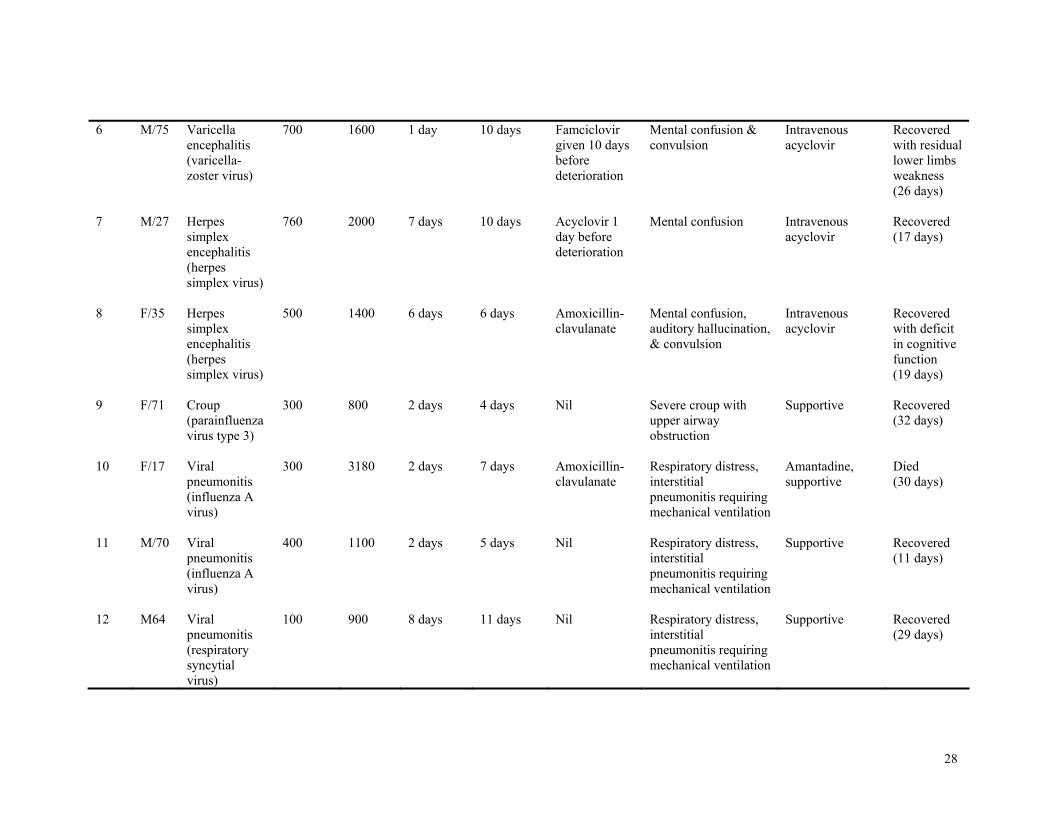
6 M/75 Varicella encephalitis (varicella-zoster virus)
700 1600 1 day 10 days Famciclovir given 10 days before deterioration
Mental confusion & convulsion
Intravenous acyclovir
Recovered with residual lower limbs weakness (26 days)
7 M/27 Herpes simplex encephalitis (herpes simplex virus)
760 2000 7 days 10 days Acyclovir 1 day before deterioration
Mental confusion Intravenous acyclovir
Recovered (17 days)
8 F/35 Herpes simplex encephalitis (herpes simplex virus)
500 1400 6 days 6 days Amoxicillin-clavulanate
Mental confusion, auditory hallucination, & convulsion
Intravenous acyclovir
Recovered with deficit in cognitive function (19 days)
9 F/71 Croup (parainfluenza virus type 3)
300 800 2 days 4 days Nil Severe croup with upper airway obstruction
Supportive Recovered (32 days)
10 F/17 Viral pneumonitis (influenza A virus)
300 3180 2 days 7 days Amoxicillin-clavulanate
Respiratory distress, interstitial pneumonitis requiring mechanical ventilation
Amantadine, supportive
Died (30 days)
11 M/70 Viral pneumonitis (influenza A virus)
400 1100 2 days 5 days Nil Respiratory distress, interstitial pneumonitis requiring mechanical ventilation
Supportive Recovered (11 days)
12 M64 Viral pneumonitis (respiratory syncytial virus)
100 900 8 days 11 days Nil Respiratory distress, interstitial pneumonitis requiring mechanical ventilation
Supportive Recovered (29 days)
28

29