clinical diagnosis of achalasia: how reliable is the...
TRANSCRIPT
Can J Gastroenterol Vol 20 No 5 May 2006 335
Clinical diagnosis of achalasia: How reliable is thebarium x-ray?
I El-Takli MD FRCPC1, P O’Brien MB ChB FRCPC2, WG Paterson MD FRCPC1
Gastrointestinal Diseases Research Unit and Departments of 1Medicine and 2Radiology, Queens University, Kingston, OntarioCorrespondence: Dr William G Paterson, Hotel Dieu Hospital, 166 Brock Street, Kingston, Ontario, K7L 5G2.
Telephone 613-544-3400 ext 2292, fax 613-544-3114, e-mail [email protected] for publication November 14, 2005. Accepted January 24, 2006
I El-Takli, P O’Brien, WG Paterson. Clinical diagnosis of
achalasia: How reliable is the barium x-ray? Can J
Gastroenterol 2006;20(5):335-337.
Manometry is considered to be the gold standard for the diagnosis of
achalasia. However, many physicians believe that contrast radiography,
classically showing esophageal dilation with bird-beak narrowing of the
gastroesophageal junction, is also accurate in either diagnosing or
excluding the disorder. The aim of the current study was to determine
the accuracy of barium x-ray in the diagnosis of achalasia. The radio-
logical diagnosis of all patients manometrically diagnosed with acha-
lasia (using conventional criteria) between January 1994 and June
1998 were reviewed. A total of 51 cases of achalasia were identified.
Thirteen patients were excluded because they either did not have con-
trast radiography before a manometric diagnosis or had their x-rays per-
formed more than six months previously. Of the remaining
38 patients, achalasia was stated as a diagnostic possibility in the
radiologists report in only 22 (58%) of those patients. Achalasia was
not considered in the remaining 16 patients: two were reported as nor-
mal, four as having stenosis/narrowing in distal esophagus, two as hav-
ing presbyesophagus, one as having mild gastroesophageal reflux and
seven as having nonspecific dysmotility. To determine the reason for the
diagnostic failure of the barium x-ray, an expert gastrointestinal radiolo-
gist reviewed 12 of the nondiagnostic x-rays in a blinded fashion,
interspersed with 10 randomly selected esophageal-contrast radi-
ographs from control subjects to avoid bias. Of these initially nondi-
agnostic x-rays in achalasia patients, typical radiological features of
achalasia were deemed to be present in 50%. The present study indi-
cates that contrast radiography lacks sensitivity in the diagnosis of
achalasia. This is not only due to radiologist oversight but also because
of the absence of the characteristic radiological features in many
cases. This reinforces the important role of esophageal manometry in
patients with persistent nonstructural dysphagia.
Key Words: Contrast radiography; Dysphagia; Esophageal motor
disorder; Esophagus; Manometry
Le diagnostic clinique d'achalasie: Quelle est lafiabilité du repas baryté?
La manométrie est considérée comme le diagnostic de référence de l’acha-
lasie. Cependant, de nombreux médecins sont d’avis que la radiographie
de contraste, qui révèle une dilatation classique de l’œsophage avec rétré-
cissement en bec d’oiseau de la jonction gastro-œsophagienne, est égale-
ment précise pour diagnostiquer ou exclure ce trouble. La présente étude
visait à déterminer la précision du repas baryté dans le diagnostic d’acha-
lasie. Le diagnostic radiologique de tous les patients atteints d’une acha-
lasie diagnostiquée par manométrie (au moyen des critères classiques)
entre janvier 1994 et juin 1998 a été évalué. Un total de 51 cas d’acha-
lasie a été repéré. Treize patients ont été exclus parce qu’ils n’avaient pas
subi de radiographie de contraste avant le diagnostic par manométrie ou
que leur radiographie de contraste avait été exécutée plus de six mois
auparavant. Le rapport du radiologiste citait l’achalasie parmi les possibil-
ités diagnostiques chez seulement 22 (58 %) des 38 patients restants.
L’achalasie n’a pas été envisagée chez les 16 autres patients : on a consi-
déré que deux étaient normaux et que quatre souffraient de sténose ou de
rétrécissement de l’œsophage distal, deux, de presbyœsophage, un, de
reflux gastro-œsophagien léger et sept, de dysmotilité non spécifique. Pour
déterminer la raison de l’échec diagnostique du repas baryté, un radiologiste
spécialisé en gastroentérologie a analysé 12 des rayons X non diagnostiques
en aveugle, mêlés à 10 radiographies œsophagiennes de contraste
provenant de sujets témoins choisis aléatoirement, afin d’éviter les biais.
Les caractéristiques radiologiques classiques d’achalasie ont été constatées
dans 50 % des rayons X non diagnostiques de patients atteints d’achalasie.
D’après la présente étude, la radiographie de contraste n’est pas assez
sensible pour diagnostiquer l’achalasie. Cette non-sensibilité s’explique
non seulement par une omission de la part du radiologiste, mais
également par l’absence de caractéristiques radiologiques de nombreux
cas. Ce constat renforce le rôle important de la manométrie
œsophagienne chez les patients atteints d’une dysphagie fonctionnelle
persistante.
Achalasia is a primary esophageal motor disorder caused bydegeneration of inhibitory nitrergic neurons within the
myenteric plexus, which leads to impaired relaxation of thelower esophageal sphincter (LES) and loss of peristalsis in thesmooth muscle segment of the esophageal body (1). Thefailure of LES relaxation appears to be the crucial factorleading to dysphagia, because therapy which effectivelyablates the LES pressure barrier usually leads to markedsymptom improvement (2-5).
At the time of presentation, patients usually experienceslowly progressive dysphagia for months or even years.
Typically, barium contrast studies reveal a dilated, aperistalticesophagus with bird-beak narrowing at the gastroesophagealjunction because of functional obstruction caused bynonrelaxation of the LES (6).
Although manometry is considered to be the gold standardfor the diagnosis of achalasia, there is a common belief that bar-ium studies are as accurate as primary screening tests whenachalasia is suspected on clinical grounds. Small studies in thepast showed variable results. Ott et al (6) reported radiographicsensitivity of 95% for achalasia; however, more recent studies(7,8) have reported that the diagnosis of achalasia was suggested
ORIGINAL ARTICLE
©2006 Pulsus Group Inc. All rights reserved
eltakli_9316.qxd 4/21/2006 1:31 PM Page 335
by videofluoroscopy in less than two-thirds of patients who hadmanometrically proven achalasia. The reasons for this apparentlack of sensitivity were not clear.
The aim of the present study was to evaluate the diagnosticaccuracy of barium x-rays in the diagnosis of achalasia and todetermine the reasons for its diagnostic failure.
PATIENTS AND METHODSAll patients manometrically diagnosed with achalasia at Hotel
Dieu Hospital (Kingston, Ontario) between January 1994 and
December 1998 were identified. Standard manometric criteria
were used to make the diagnosis (9). Patients all had the typical
manometric features of achalasia, including complete aperistalsis
and either absent or markedly impaired (less than 50%) swallow-
induced LES relaxation. Detailed chart review was performed to
exclude cases of secondary achalasia and to identify reports of
esophageal barium contrast studies. If patients were referred in
from other institutions, the respective radiology departments were
contacted to determine if barium contrast studies had been per-
formed before manometry. Patients were excluded if they had no
barium x-ray before manometry or if their barium x-ray was per-
formed more than six months before the manometric study. In
addition, patients who had their initial diagnosis before January
1994 and were returning for follow-up manometry were excluded.
Original radiological reports were reviewed and classified as to
whether the diagnosis of achalasia was considered by the reporting
radiologist.
To determine the reason for the diagnostic failure, an expert
gastrointestinal radiologist reviewed the nondiagnostic x-rays in a
blinded fashion interspersed with 10 randomly selected
esophageal-contrast radiographs from control subjects who under-
went barium swallow for reasons other than dysphagia. The
control radiographs had been interpreted as either being normal or
showing minor reflux only. Three criteria were used by our radiologist
to diagnose achalasia:
• dilation of the esophagus;
• bird-beak narrowing of the gastroesophageal junction; and
• evidence of stasis.
If all three criteria were present, the x-ray was interpreted as
diagnostic of achalasia, if two of the three criteria were present, the
x-ray was interpreted as possible achalasia and if one or none of the
criteria were present, the x-ray was interpreted as nondiagnostic of
achalasia.
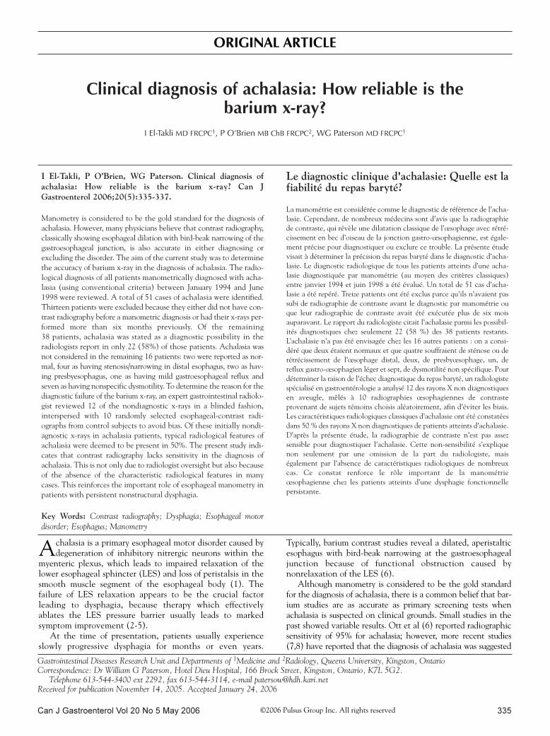
RESULTSOf the 51 patients who had manometric diagnosis of achalasiaduring the five-year period, five were excluded because theirx-rays were performed more than six months before themanometry, two because they were initially diagnosed before1994 and six because no x-rays were performed before manometry.Of the remaining 38 patients, barium swallow or cine esophagramwas suggestive of achalasia (as per the radiologist report) in22 patients (58%) and nondiagnostic in 16 patients (42%)(Figure 1). Seven of the nondiagnostic x-rays were interpretedas nonspecific motility disorder, four as stenosis/narrowing indistal esophagus, two as presbyesophagus, two as normal andone as gastroesophageal reflux disease.
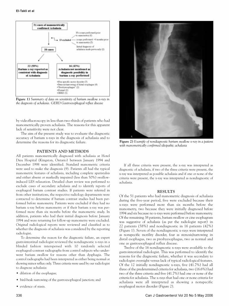
Twelve of the 16 nondiagnostic x-rays were available to thegastrointestinal radiologist. This was performed to identify thereasons for the diagnostic failure, whether it was secondary toradiologist oversight versus lack of typical radiological features.Of the 12 initially nondiagnostic x-rays, five (41.7%) had allthree of the predetermined criteria for achalasia, two (16.6%) hadtwo of the three criteria and five (41.7%) had one or none of thecriteria for achalasia. The x-rays that had one or none criteria forachalasia were all interpreted as showing a nonspecificesophageal motor disorder (Figure 2).
El-Takli et al
Can J Gastroenterol Vol 20 No 5 May 2006336
Figure 1) Summary of data on sensitivity of barium swallow x-ray inthe diagnosis of achalasia. GERD Gastroesophageal reflux disease
Figure 2) Example of nondiagnostic barium swallow x-ray in a patientwith manometrically confirmed idiopathic achalasia
eltakli_9316.qxd 4/21/2006 1:31 PM Page 336

DISCUSSIONThe present study demonstrates that radiography missed thediagnosis of achalasia in 42% of the patients. The reason formissing the diagnosis varied. In five of 12 of the missed casesin which barium x-rays were available for review, it wasclearly secondary to radiologist oversight in that all three ofthe classic criteria were present. However, in most of theremaining cases, the classic radiographic features ofachalasia were absent.
The early diagnosis of achalasia has major clinicalsignificance because delays in the diagnosis results in delaysin the institution of effective therapy, with resultingmorbidity and impaired quality of life. Videofluoroscopyclearly carries a much better chance of detecting motilitydisorders, including achalasia, than static films. Ourradiologist was only able to retrospectively review spot films,but the radiologist who originally performed the studypresumably would have viewed the fluoroscopic images. It couldalso be argued that we underestimated the sensitivity ofcontrast-radiography, because any radiographic evidence ofesophageal dysmotility in a patient with dysphagia would triggera request for manometry to firmly establish the diagnosis.However, we do not believe this approach is widely practicedin Canada, particularly given the limited access toesophageal manometry. In addition, nonspecific esophagealmotor abnormalities are common in asymptomatic individuals(10) despite the very low incidence of achalasia.
In a series of 33 patients with manometrically confirmedachalasia reported by Howard et al (7), barium swallowx-ray was considered consistent with or suggestive of
achalasia in 45% and 21% of patients, respectively; in theremainder, achalasia was not considered. A more recentstudy by Schima et al (8) showed similar results in that thediagnosis of achalasia was suggested using barium swallowvideofluoroscopy in only 58% of the cases that were diag-nosed manometrically. Of the remainder, 34% were reportedto show nonspecific motor disorders while 8% were reportedas normal. These results are comparable with the results ofthe present study. The results of the study by Ott et al (6)showing a sensitivity of 95% for detecting achalasia byradiographic studies has never been reproduced insubsequent studies.
The current study did not address the issue of specificityof the barium swallow x-ray in the diagnosis of achalasiawhen the classic x-ray features were present. To date, thereare no published studies addressing this issue; however, wehave seen rare cases where the barium swallow was thoughtto have the characteristic features of achalasia, yetmanometric criteria for the diagnosis were absent.
CONCLUSIONContrast radiography lacks sensitivity in the diagnosis ofachalasia. This can be partially rectified by increasingawareness among radiologists regarding the diagnostic fea-tures of the disease. However, it is clear that classic x-rayfeatures may not be present in some patients. Thus, to avoidmisdiagnosing patients with a readily treatable disease,manometry should be performed in all patients withpersisting esophageal-type dysphagia but negativeendoscopy and radiological examinations.
Barium x-ray in achalasia
Can J Gastroenterol Vol 20 No 5 May 2006 337
REFERENCES1. Paterson WG. Etiology and pathogenesis of achalasia.
Gastrointest Endosc Clin N Am 2001;11:249-66.2. Spiess AE, Kahrilas PJ. Treating achalasia: From whalebone to
laparoscope. JAMA 1998;280:638-42.3. Csendes A, Braghetto I, Henriquez A, Cortes C. Late results of
prospective randomized study comparing forceful dilatation andoesophagomyotomy in patients with achalasia. Gut1989;30:299-304.
4. Gelfond M, Rozen P, Gilat T. Isosorbide dinitrate and nifedipine treatment of achalasia: A clinical, manometric and radionuclide evaluation. Gastroenterology 1982;83:963-9.
5. Pasricha PJ, Rai R, Ravich WJ, Hendrix TR, Kalloo AN. Botulinumtoxin for achalasia: Long term outcome and predictors of outcome.Gastroenterology 1996;110:1410-5.
6. Ott DJ, Richter JE, Chen YM, Wu WC, Gelfand DW, Castell DO. Esophageal radiography and manometry: Correlation in 172 patientswith dysphagia. Am J Roentgenol 1987;149:307-11.
7. Howard PJ, Maher L, Pryde A, Cameron EW, Heading RC. Five yearprospective study of the incidence, clinical features, and diagnosis ofachalasia in Edinburgh. Gut 1992;33:1011-5.
8. Schima W, Ryan JM, Harisinghani M, et al. Radiographic detectionof achalasia: Diagnostic accuracy of videofluoroscopy. Clin Radiol1998;53:372-5.
9. Paterson WG, Marciano-D’Amore DA, Beck IT, DaCosta LR.Esophageal manometry with provocative testing in patients withnoncardiac angina-like chest pain. Can J Gastroenterol 1991;5:51-7.
10. Ekberg O, Feinberg MJ. Altered swallowing function in elderlypatients without dysphagia: Radiologic findings in 56 cases. Am J Roentgenol 1991;156:1181-4.
eltakli_9316.qxd 4/21/2006 1:31 PM Page 337

Submit your manuscripts athttp://www.hindawi.com
Stem CellsInternational
Hindawi Publishing Corporationhttp://www.hindawi.com Volume 2014
Hindawi Publishing Corporationhttp://www.hindawi.com Volume 2014
MEDIATORSINFLAMMATION
of
Hindawi Publishing Corporationhttp://www.hindawi.com Volume 2014
Behavioural Neurology
EndocrinologyInternational Journal of
Hindawi Publishing Corporationhttp://www.hindawi.com Volume 2014
Hindawi Publishing Corporationhttp://www.hindawi.com Volume 2014
Disease Markers
Hindawi Publishing Corporationhttp://www.hindawi.com Volume 2014
BioMed Research International
OncologyJournal of
Hindawi Publishing Corporationhttp://www.hindawi.com Volume 2014
Hindawi Publishing Corporationhttp://www.hindawi.com Volume 2014
Oxidative Medicine and Cellular Longevity
Hindawi Publishing Corporationhttp://www.hindawi.com Volume 2014
PPAR Research
The Scientific World JournalHindawi Publishing Corporation http://www.hindawi.com Volume 2014
Immunology ResearchHindawi Publishing Corporationhttp://www.hindawi.com Volume 2014
Journal of
ObesityJournal of
Hindawi Publishing Corporationhttp://www.hindawi.com Volume 2014
Hindawi Publishing Corporationhttp://www.hindawi.com Volume 2014
Computational and Mathematical Methods in Medicine
OphthalmologyJournal of
Hindawi Publishing Corporationhttp://www.hindawi.com Volume 2014
Diabetes ResearchJournal of
Hindawi Publishing Corporationhttp://www.hindawi.com Volume 2014
Hindawi Publishing Corporationhttp://www.hindawi.com Volume 2014
Research and TreatmentAIDS
Hindawi Publishing Corporationhttp://www.hindawi.com Volume 2014
Gastroenterology Research and Practice
Hindawi Publishing Corporationhttp://www.hindawi.com Volume 2014
Parkinson’s Disease
Evidence-Based Complementary and Alternative Medicine
Volume 2014Hindawi Publishing Corporationhttp://www.hindawi.com