clinical examination of the temporomandibular joint · clinical examination of the...
TRANSCRIPT
© Copyright 2013 Elsevier, Ltd. All rights reserved.
Clinical examination of the temporomandibular joint
CHAPTER CONTENTS
Pain . . . . . . . . . . . . . . . . . . . . . . . . . . . . e202
Pain referred from the temporomandibular joint . . . . . . . . . . . . . . . . . . . . . . . . . e202Pain referred to the temporomandibular joint area . . . . . . . . . . . . . . . . . . . . . . . . . e202
History . . . . . . . . . . . . . . . . . . . . . . . . . . . e203
Inspection . . . . . . . . . . . . . . . . . . . . . . . . . e204
Functional .examination . . . . . . . . . . . . . . . . . . e204
Active movements . . . . . . . . . . . . . . . . . . e204Resisted movements . . . . . . . . . . . . . . . . e204
Palpation . . . . . . . . . . . . . . . . . . . . . . . . . . e206
Technical .investigations . . . . . . . . . . . . . . . . . e206
The most characteristic symptoms of disorders of the tempo-romandibular joint (TMJ) are orofacial pain, noises in the joint, limitation of movement – mouth opening – or a combination of these. Limitation may present suddenly as locking or may be slowly progressive.
Pain
Pain in the TMJ area usually has a local cause and is seldom referred to any distance.
The patient should also be asked about the influence of chewing, yawning, swallowing or talking. If pain is present on one of these, a disorder of the TMJ is most likely. Some disorders of the cervical spine (see Section 2) and the parotid gland may exceptionally also provoke pain on swallowing.
A clear description of the type of pain should always be sought. A sharp severe pain tends to suggest an arthrogenic problem, a diffuse ache of less intensity points more to a mus-cular disorder.
A painful click may be the consequence of subluxation of the intra-articular meniscus. Pain coming on spontaneously and progressively increasing over some weeks is often the result of arthritis. Continuous dull pain felt in the area of the mastica-tory muscles and usually worse at the end of the day may indicate myalgia.
Pain referred from the temporomandibular joint
Pain of the TMJ structures may arise from the masticatory muscles or from the joint itself. The main inert structures that can give rise to pain are the intracapsular tissues located pos-terior to the condyle: the posterior part of the meniscus, the meniscus attachments to the capsule, the capsule and the retromeniscal fat pad.1 Pain is often accompanied by headache, earache or pain in the postauricular area. Pain arising from the TMJ sometimes refers to the maxilla.
Pain referred to the temporomandibular joint area
Occasionally pain is referred from the neck. When there is doubt, a preliminary examination of the neck must be performed.
Other structures may give rise to painful conditions in the TMJ area and can be divided into neurological and non-neurological disorders.
Neurological .disorders
Atypical facial neuralgiaThis can be uni- or bilateral and is of unknown origin. It usually causes a burning sensation, pins and needles, and continuous pain, in cycles of severity: it may occur after dental procedures.
Clinical examination of the temporomandibular joint
e203© Copyright 2013 Elsevier, Ltd. All rights reserved.
ipsilateral facial redness. Attacks of severe headache in or around the eyes, usually unilaterally, come on within 5–10 minutes and last from about 45 minutes to a few hours. Attacks occur in clusters.6
Temporal arteritisThis is one of the manifestations of a giant-cell arteritis, an autoimmune process.7 It is usually seen unilaterally in males over 50 years of age and is frequently associated with polymy-algia rheumatica. It is characterized by a knocking pain around the temporal vessels. The skin overlying the artery is red, swollen and warm. The erythrocyte sedimentation rate is raised.
Leaking cerebral aneurysmIt has an explosive onset of headache, nausea and vomiting, together with photophobia and stiffness of the neck. Aneurysm at the level of the posterior communicating artery may be followed by pain in the first division of the trigeminal nerve. It is the commonest cause of so-called ophthalmoplegic migraine.8
History
Questions are asked about the onset of pain, its nature, locali-zation, intensity and duration. The examiner should discover which factors increase or relieve pain.
As well as taking a history of pain, a number of other aspects should be discussed with the patient:
• Does the joint click? In an anterior subluxating meniscus, the normal relation between meniscus and condyle is disturbed, giving rise to a click on opening the mouth.
• Is movement limited, either in range or by locking? If there is a diminished range of opening of the mouth, did the limitation come on suddenly or was it more progressive? If ‘sudden’ locking is mentioned, can the patient still open or close the mouth? Inability to open suggests meniscus displacement, which is usually unilateral, and in which at least 1 cm of mouth opening is always retained. If closing is impossible, a luxation of the mandibular condyle is most likely. Excessive limitation coming on rapidly may be the result of hysteria or of tetanus; mouth opening is impossible in these circumstances. A limitation of slow development is usually the outcome of arthrosis of the TMJ.
• Is there crepitus? Crepitus is the result of movement across an irregular surface because of advanced changes in the joint. It may be present in osteoarthrosis.
• Does the patient suffer from clenching or grinding? This occurs mainly at night in stressed people. The patient may not be aware of it, relatives may have to be asked.
• Is there tinnitus, vertigo or a hearing problem? Vertigo may result from differences in vestibular impulses as a result of TMJ problems. Other symptoms, such as mild deafness, a sensation of fullness in the ear and tinnitus, may also be present.
Trigeminal nerve neuritisThis may be encountered in patients of 45–60 years of age. It affects females more often than males and the right side more frequently than the left. The patients complain of unilateral shooting pain, from the ear towards the temporal area and the maxilla, sometimes even in the forehead and towards the pharynx. The cause of the pain may be so obscure that unnec-essary dental extraction takes place. Pain is seldom accompa-nied by diminished sensitivity but characteristic trigger points are often found. Stimulation of these, even sometimes by light touch, results in pain felt elsewhere, which is followed by a refractory period of up to 30 seconds during which stimulation does not lead to new pain. The pain attacks seldom last longer than a few seconds. They may recur at irregular intervals, sometimes on a daily, weekly or even a monthly basis. They are isolated or come on in clusters.2
Herpes zoster oticus infectionThis can give rise to dysaesthesia preceding the characteristic vesicles. No true trigger points are present. About 15% of all peripheral facial palsies is caused by this virus.3
Idiopathic peripheral facial palsy (Bell’s palsy)This is a disorder of the facial nerve, probably the result of a cranial neuritis.4,5
It mainly affects patients between 20 and 50 years of age. It is seldom painful although at the onset some pain around the ear may be felt. It gives rise to a palsy of the facial muscles, characterized by lowering of the ipsilateral side of the mouth. It may also cause diminished pain sensibility, changes in taste, diminished lachrymation and increased salivation.
Peripheral neuropathyThis is usually the result of diabetes, long-standing temporal arteritis or Raynaud’s syndrome. It usually leads to a burning sensation and loss of sensibility on lips, cornea or conjunctivae.
Non-neurological .disorders
Otitis media, otitis externa and parotitisThese all give rise to pain in the TMJ area. Pain usually remains local and increases on pressure on the tragus (otitis) or parotid gland (parotitis epidemica). These conditions mainly affect children and are usually accompanied by fever.
Paranasal sinusitisThis results in a constant, knocking pain usually felt around the orbits, sometimes radiating towards the cheek and into the teeth.
Infection of the teeth and dental abscessTooth infections are followed by pain in the cheek on percus-sion which is provoked or increased by eating sugary food. An abscess gives rise to local swelling of the gingiva.
Cluster headache (Horton’s neuralgia)This predominantly affects males, is unilaterally localized, and is associated with increased lachrymation, rhinitis and
The Temporomandibular Joint
e204© Copyright 2013 Elsevier, Ltd. All rights reserved.
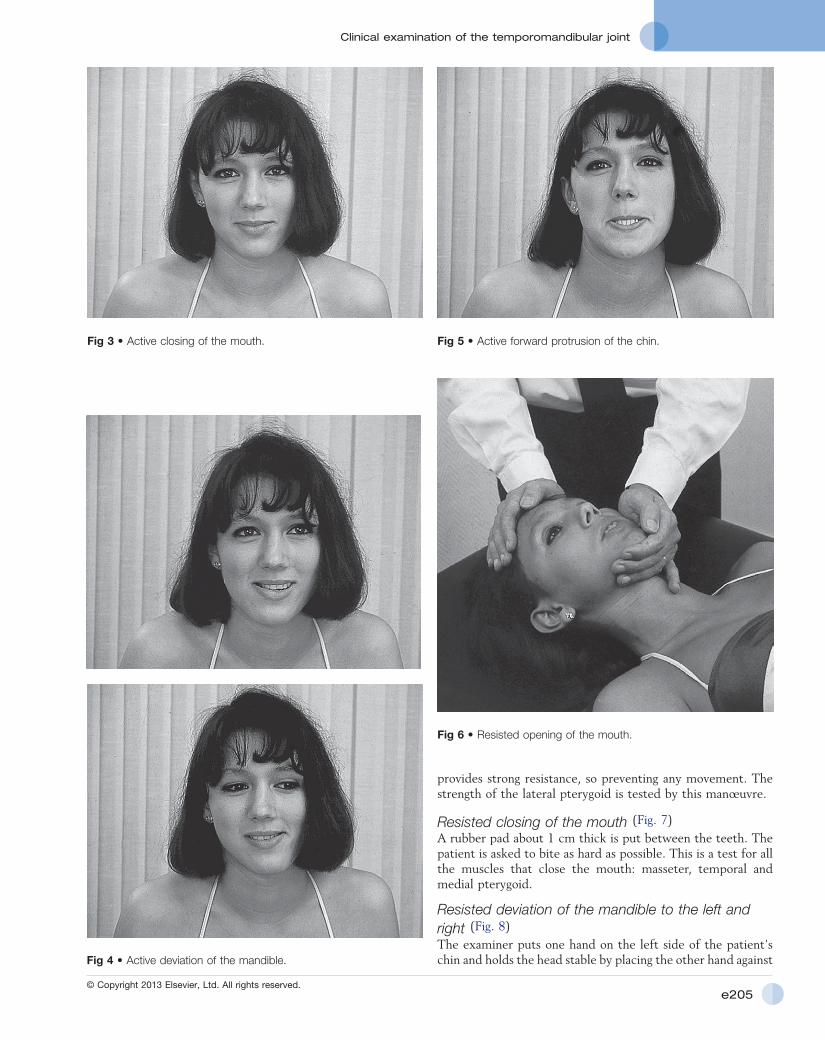
Active forward protrusion of the chin (Fig. 5)This is performed by the lateral and medial pterygoid, mas-seter, geniohyoid and digastric muscle. When it is disturbed, this is usually the consequence of an inert problem.
Resisted movements
Resisted opening of the mouth (Fig. 6)The examiner places one hand underneath the patient’s chin, the other on the vertex. With the mouth open about 1 cm, the patient is now asked to open further while the examiner
• Have there been changes in sensibility? These can indicate peripheral neuropathy. It frequently affects the lips, cornea and conjunctivae. In atypical facial neuralgia, severe diminished facial sensibility is often found. Trigeminal neuritis is seldom accompanied by disturbed sensibility.
Inspection
On inspection, attention must be paid to local swelling, defor-mation, deviation of the chin and teeth wear.
Swelling may be the result of a bacterial or an inflammatory arthritis (frequently rheumatoid, seldom due to psoriasis or gout), or in children10 may be caused by an inflammation of the parotid gland.
In Bell’s palsy, there is lowering of the ipsilateral side of the mouth and a smoothing out of wrinkles.
Severe inflammatory disorders of the TMJ area during child-hood may result in asymmetrical development of the lower face because of disturbance of the growth centre in the man-dible. Advanced arthrosis may lead to asymmetry of face and head and to narrowing of the external auditory canal. Synovitis usually causes an ipsilateral deviation when the mouth is opened and a contralateral deviation when closed.9
Abnormal wear and tear of the teeth may be a sign of bruxism or grinding. Malocclusion and missing teeth may result in a TMJ problem. A bilateral relationship between the teeth and TMJs exists. Changes in the dental relationship, as in malocclusion and missing teeth, may lead to adaptation in the TMJ. Problems with the joint can cause changes in dental occlusion.
Functional examination
Active movements
The influence of all five active movements on pain, range of movement, deviation, abnormal sounds and crepitus are noted.
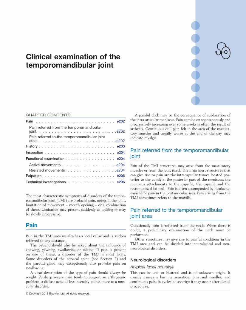
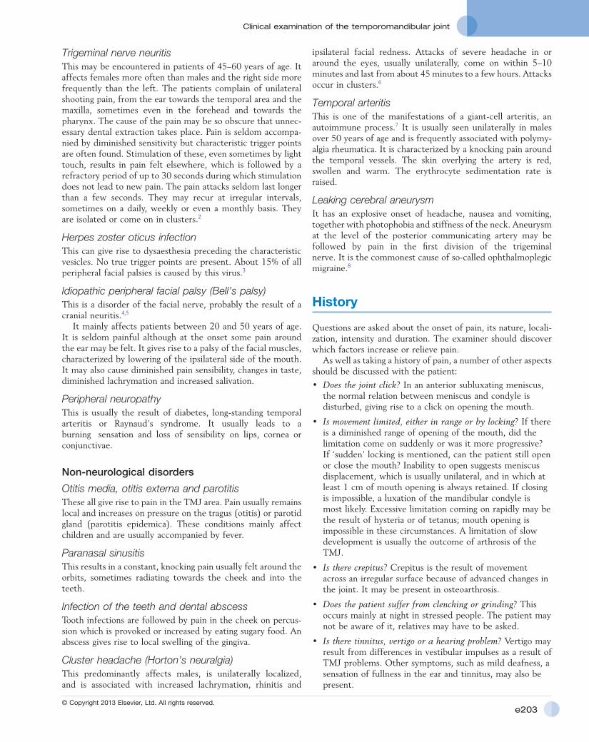
Active opening of the mouth (Fig. 1)Because it is difficult to measure the range of motion of the TMJ in degrees, the interincisal distance at maximum opening is used. It is about 36–38 mm in adults but may vary between 30 and 67 mm, depending on sex and age.11,12 A practical and quick way of checking range of motion is to ask the patient to insert the knuckles in between the front teeth (Fig. 2).
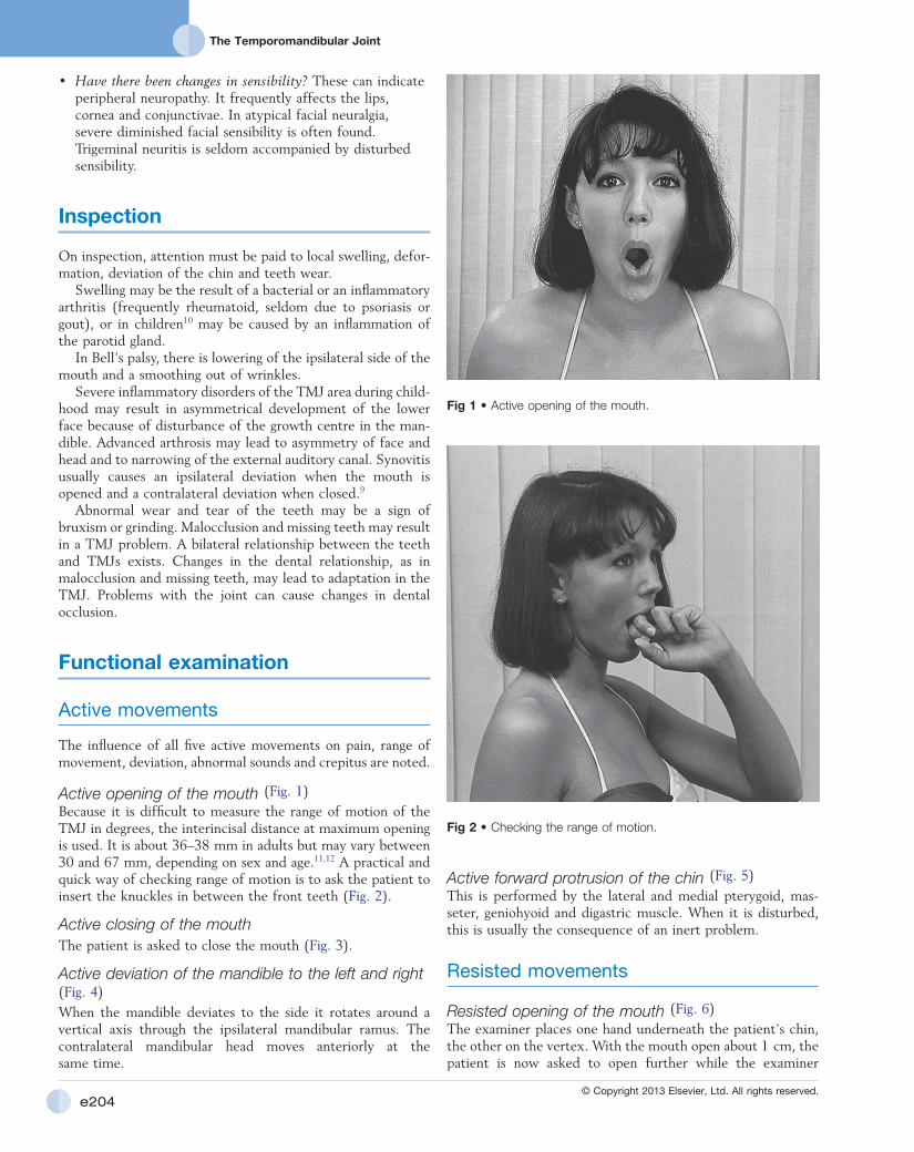
Active closing of the mouthThe patient is asked to close the mouth (Fig. 3).
Active deviation of the mandible to the left and right (Fig. 4)When the mandible deviates to the side it rotates around a vertical axis through the ipsilateral mandibular ramus. The contralateral mandibular head moves anteriorly at the same time.
Fig 1 • Active opening of the mouth.
Fig 2 • Checking the range of motion.
Clinical examination of the temporomandibular joint
e205© Copyright 2013 Elsevier, Ltd. All rights reserved.
provides strong resistance, so preventing any movement. The strength of the lateral pterygoid is tested by this manœuvre.
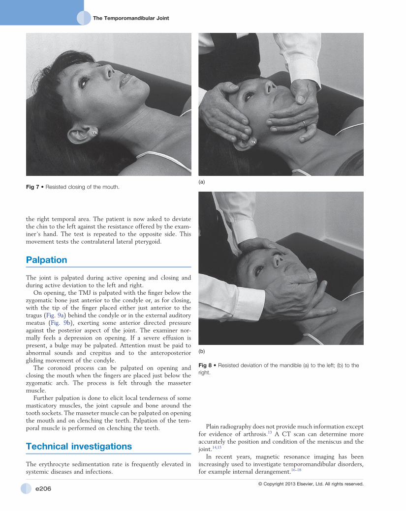
Resisted closing of the mouth (Fig. 7)A rubber pad about 1 cm thick is put between the teeth. The patient is asked to bite as hard as possible. This is a test for all the muscles that close the mouth: masseter, temporal and medial pterygoid.
Resisted deviation of the mandible to the left and right (Fig. 8)The examiner puts one hand on the left side of the patient’s chin and holds the head stable by placing the other hand against
Fig 3 • Active closing of the mouth.
Fig 4 • Active deviation of the mandible.
Fig 5 • Active forward protrusion of the chin.
Fig 6 • Resisted opening of the mouth.
The Temporomandibular Joint
e206© Copyright 2013 Elsevier, Ltd. All rights reserved.
Plain radiography does not provide much information except for evidence of arthrosis.13 A CT scan can determine more accurately the position and condition of the meniscus and the joint.14,15
In recent years, magnetic resonance imaging has been increasingly used to investigate temporomandibular disorders, for example internal derangement.16–18
the right temporal area. The patient is now asked to deviate the chin to the left against the resistance offered by the exam-iner’s hand. The test is repeated to the opposite side. This movement tests the contralateral lateral pterygoid.
Palpation
The joint is palpated during active opening and closing and during active deviation to the left and right.
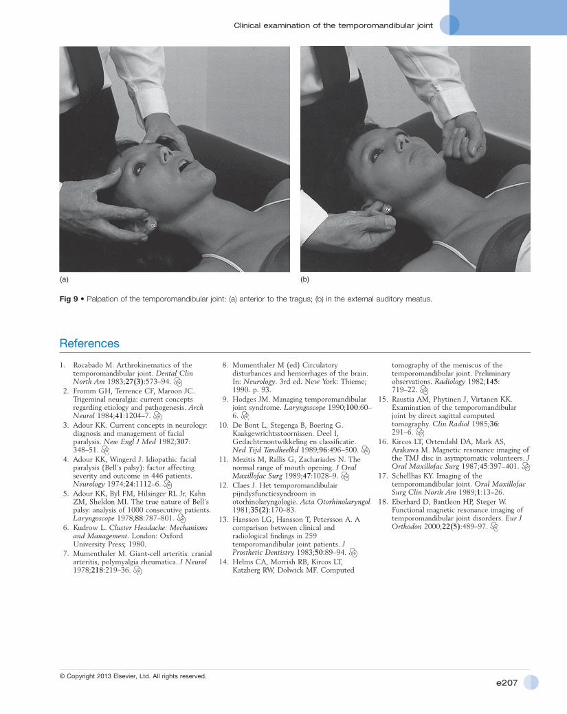
On opening, the TMJ is palpated with the finger below the zygomatic bone just anterior to the condyle or, as for closing, with the tip of the finger placed either just anterior to the tragus (Fig. 9a) behind the condyle or in the external auditory meatus (Fig. 9b), exerting some anterior directed pressure against the posterior aspect of the joint. The examiner nor-mally feels a depression on opening. If a severe effusion is present, a bulge may be palpated. Attention must be paid to abnormal sounds and crepitus and to the anteroposterior gliding movement of the condyle.
The coronoid process can be palpated on opening and closing the mouth when the fingers are placed just below the zygomatic arch. The process is felt through the masseter muscle.
Further palpation is done to elicit local tenderness of some masticatory muscles, the joint capsule and bone around the tooth sockets. The masseter muscle can be palpated on opening the mouth and on clenching the teeth. Palpation of the tem-poral muscle is performed on clenching the teeth.
Technical investigations
The erythrocyte sedimentation rate is frequently elevated in systemic diseases and infections.
Fig 7 • Resisted closing of the mouth.
Fig 8 • Resisted deviation of the mandible (a) to the left; (b) to the right.
(a)
(b)
Clinical examination of the temporomandibular joint
e207© Copyright 2013 Elsevier, Ltd. All rights reserved.
Fig 9 • Palpation of the temporomandibular joint: (a) anterior to the tragus; (b) in the external auditory meatus.
(a) (b)
References
1. Rocabado M. Arthrokinematics of the temporomandibular joint. Dental Clin North Am 1983;27(3):573–94.
2. Fromm GH, Terrence CF, Maroon JC. Trigeminal neuralgia: current concepts regarding etiology and pathogenesis. Arch Neurol 1984;41:1204–7.
3. Adour KK. Current concepts in neurology: diagnosis and management of facial paralysis. New Engl J Med 1982;307:348–51.
4. Adour KK, Wingerd J. Idiopathic facial paralysis (Bell’s palsy): factor affecting severity and outcome in 446 patients. Neurology 1974;24:1112–6.
5. Adour KK, Byl FM, Hilsinger RL Jr, Kahn ZM, Sheldon MI. The true nature of Bell’s palsy: analysis of 1000 consecutive patients. Laryngoscope 1978;88:787–801.
6. Kudrow L. Cluster Headache: Mechanisms and Management. London: Oxford University Press; 1980.
7. Mumenthaler M. Giant-cell arteritis: cranial arteritis, polymyalgia rheumatica. J Neurol 1978;218:219–36.
8. Mumenthaler M (ed) Circulatory disturbances and hemorrhages of the brain. In: Neurology. 3rd ed. New York: Thieme; 1990. p. 93.
9. Hodges JM. Managing temporomandibular joint syndrome. Laryngoscope 1990;100:60–6.
10. De Bont L, Stegenga B, Boering G. Kaakgewrichtsstoornissen. Deel I, Gedachtenontwikkeling en classificatie. Ned Tijd Tandheelkd 1989;96:496–500.
11. Mezitis M, Rallis G, Zachariades N. The normal range of mouth opening. J Oral Maxillofac Surg 1989;47:1028–9.
12. Claes J. Het temporomandibulair pijndysfunctiesyndroom in otorhinolaryngologie. Acta Otorhinolaryngol 1981;35(2):170–83.
13. Hansson LG, Hansson T, Petersson A. A comparison between clinical and radiological findings in 259 temporomandibular joint patients. J Prosthetic Dentistry 1983;50:89–94.
14. Helms CA, Morrish RB, Kircos LT, Katzberg RW, Dolwick MF. Computed
tomography of the meniscus of the temporomandibular joint. Preliminary observations. Radiology 1982;145:719–22.
15. Raustia AM, Phytinen J, Virtanen KK. Examination of the temporomandibular joint by direct sagittal computed tomography. Clin Radiol 1985;36:291–6.
16. Kircos LT, Ortendahl DA, Mark AS, Arakawa M. Magnetic resonance imaging of the TMJ disc in asymptomatic volunteers. J Oral Maxillofac Surg 1987;45:397–401.
17. Schellhas KY. Imaging of the temporomandibular joint. Oral Maxillofac Surg Clin North Am 1989;1:13–26.
18. Eberhard D, Bantleon HP, Steger W. Functional magnetic resonance imaging of temporomandibular joint disorders. Eur J Orthodon 2000;22(5):489–97.