clinical guidelines and brief operating instructions …c1-preview.prosites.com/106982/wy/docs/kavo...
TRANSCRIPT

Clinical Guidelines andBrief Operating InstructionsKaVo DIAGNOdent®
NEW!!
EZ-Cal
Faster Calibration
PostScript BildKavo Logo 4c_RZ.eps
Listen! It's caries.


Introduction 1
Indications for use 1
Instructions for use 2
Correlation of DIAGNOdent values 4
Special notes 5
Research 6
Brief operating instructions 7
Assembly 7
Function 9
Calibration 10
Examining tooth surfaces 11
Scanning teeth 12
Dental treatment sequence 14
Additional user information 15
Frequently asked questions 17
References 19
Safety disclosure 20
Clinical Guidelines
Table of contents
KaVo DIAGNOdent 2095
INTRODUCTION
The DIAGNOdent is intendedfor use as an aid in detectingcaries. It provides informationto supplement the clinician’svisual observations, considera-tion of patient histories, andinformation from other diagnos-tic modalities, resulting in anoverall risk assessment and atreatment determination. It is nota stand alone diagnostic tool.DIAGNOdent may also be usedas an aid in monitoring progres-sion or arrestment of caries bycomparing a patient’s readingsfrom visit to visit. All DIAGNOdentreadings must be evaluated inthe context of other diagnosticinformation.
Only caries on occlusal or flatsurfaces of teeth, which areaccessible to the probes, canbe examined for caries. TheDIAGNOdent does not detectinterproximal caries, subgingivalcaries, or secondary cariesunder crowns, inlays, or com-posite/amalgam restorations.
INDICATIONS FOR USE
The DIAGNOdent is intended foruse as an aid in detecting carieson thoroughly cleaned teeth and inmonitoring progression or arrest-ment of caries by comparing apatient’s readings from visit to visit.
PRIOR TO USE OF THEDIAGNOdent, THE DENTISTSHOULD:
Take a medical history and obtaininformation regarding diet, oralhygiene and past caries history.Examine radiographs, if available.
Perform an initial dental exami-nation, which includes a visualexamination, using, if possible, mag-nification and an intraoral camera.
Form a risk assessment of thepatient from this information.
Clean the teeth using a prophybrush or powder jet cleaner, e.g.KaVo PROPHYflex II, or otheracceptable means. Dry the teeth.
Identify suspicious tooth sur-faces requiring further examina-tion with DIAGNOdent.
Clin
ical
Gui
delin
es
1
Clinical guidelines
KaVo DIAGNOdent 2095

INSTRUCTIONS FOR USE
The DIAGNOdent should be usedon suspicious sites identifiedafter careful visual examinationand thorough cleaning and dry-ing of the teeth.
Prior to use; the DIAGNOdentmust be calibrated with theselected probe tip and a patient-specific zero base line must beestablished. Please refer to thesetopics in the Brief OperatingInstructions section of this booklet.
During examination of suspicioussites, the tip of the handpieceshould be in light contact withthe surface of the tooth andshould be slowly rotated or rockedin a pendulum-like manner whencontacting fissures or areas ofconcern, e.g., discoloration,enamel defects or areas thatproduce a sharp change in theaudible signal. The higher thereading the greater the fluores-cence of the site. The highestreading obtained can be refer-enced in the context of treat-ment considerations.
It is important to clean and drythe teeth prior to using theDIAGNOdent. Powder jet cleanersare rapid and effective in remov-ing stain and debris from com-plex occlusal anatomy. Prophypaste residue can be difficult toremove from deep grooves andfissures.1
Clinical Guidelines
2
Instructions for use
KaVo DIAGNOdent 2095

Seemingly intact molar, as perceived visually and radiographically.Corresponding histolgical cross-sections reveal actual extent of caries.(taken from Lussi 1993).
Clin
ical
Gui
delin
es
3
Detecting the invisible
KaVo DIAGNOdent 2095

* Taken from Lussi; See “Research Supporting DIAGNOdent ScaleReadings,” (page 6).** In unusual cases of virulent disease, preparation may be a courseof action when a value between 20 and 30 is recorded. *** Regardless of course of action taken to treat a specific lesion,preventive therapy may be indicated based upon caries risk.
Table 1-1
Please note:
Alloy restorations (amalgam/gold) exhibit little or no fluorescence values.Composites/ceramics/cements emit their own fluorescence values.Therefore, it should be understood that any restorative material can mis-represent the health of the underlying tooth structure.
Plaque, tartar and discolorations generally exhibit very high fluorescencevalues. All deposits must be removed to permit better evaluation.
Possible Course of Action*DIAGNOdent No Preventive Record & Sealant Preparation
Values Action Therapy Monitor
0-5 •
5-10 • •
10-15 • • • •
15-20 • • •
20-25 •*** • • •**
25-30 •*** • • •**
30+ •*** •
Clinical Guidelines
4
Correlation of DIAGNOdent values to possiblecourse of action
KaVo DIAGNOdent 2095

1. The DIAGNOdent should beused only after the teeth havebeen thoroughly cleaned and dried.
2. The device detects fluores-cence of a chromophore, not nec-essarily bacteria, which is a surro-gate marker that correlates withthe grade of caries development.
3. False positives may arisebecause of fluorescence due toplaque or calculus in fissures or todiscoloration, tartar, food and sim-ilar materials which may be lodgedin or on the surface of the teeth. TheDIAGNOdent may also respond tosome restorative materials whichinclude but are not limited to com-posite resins, sealants and ceram-ic restorations due to fluorescentcomponents of these restorations.A distinction among these possiblecauses of a signal based solely onthe displayed value is not possi-ble; the dentist must assess eachsuspicious site to determinewhether caries is present.
4. Very high readings, e.g.,greater than 80, may indicate thatthe teeth are not thoroughlycleaned or free of debris. In suchsituations it is suggested that thetooth or area(s) be cleaned, driedand re-examined.
5. Arrested caries MAY be indicat-ed by readings which are thesame, nearly the same, or lower,over a series of examinations.Final assessment is dependentupon an examination of such sitesby the dentist by other diagnosticmodalities.
6. If the overall clinical assess-ment, taking into account all infor-mation other than that provided bythe DIAGNOdent, leads to a con-clusion that restoration is indicated,conservative methods should beused to determine the depth of thesuspected caries. THE DIAGNOdentIN NO CASE SHOULD BEVIEWED AS AN INDICATOR OFTHE DEPTH TO WHICH THECLINICIAN SHOULD EXCAVATE!
7. The changing sound during anexamination may cause anxiety insome patients. In such cases, theaudible signal can be turned aslow as possible or turned off com-pletely at the discretion of the den-tist. Refer to page 9 of this bookletfor instructions on changing thetone control.
Clin
ical
Gui
delin
es
5
Special notes regarding use of the DIAGNOdent
KaVo DIAGNOdent 2095

Extensive pre-clinical researchestablishes that laser-induced fluo-rescence is correlated with materi-als in carious lesions,2 and that thescale readings obtained by theDIAGNOdent correlate with gradesof carious lesions.3,4,5 Resultsobtained by DIAGNOdent comparefavorably to results achieved byother means of detection.6
Clinical research supports thevalue of the DIAGNOdent as an aidto diagnosis of caries.7,8,9,10,11,12
While the scale readings given bythe device are valuable informa-tion, they should be used in thecontext of the other informationobtained by the clinician by meansof visual examination, patient histo-ries, radiographs and other diag-nostic modalities. They should notbe relied on as a sole diagnostictool. The chart “Correlation ofDIAGNOdent Values to PossibleCourses of Action” (page 4) isbased on the research and sug-gestions of Lussi.13 Specifically,making the following correlationsbased upon in vivo studies:
0 - 14 no caries, or histological enamelcaries limited to the outer half of theenamel thickness.
15 - 20 histological caries extendingbeyond the outer half, but confined tothe enamel.
21 - 99 histological dentinal caries
and provide the following treatmentrecommendations:
0 - 14 no active care is advised.
15 - 20 preventive care is advised.
21 - 30 preventive or operative care isadvised depending on the patient’scaries risk, the recall interval etc.
>30 operative and preventive care isadvised.
THE SCALE READINGS FROMDIAGNOdent SHOULD NOT BEVIEWED AS CONCLUSIVE OFTHE PRESENCE, ABSENCE,PROGRESSION, ARRESTMENT,OR GRADE OF CARIES, OR BEDETERMINATIVE OF THE CHOICEOF TREATMENT, BUT SHOULDBE INTERPRETED IN THE CON-TEXT OF OTHER DIAGNOSTICINFORMATION!
Clinical Guidelines
6
Research supporting DIAGNOdent’s scale readings
KaVo DIAGNOdent 2095
NOTES
Please note the followingpoints
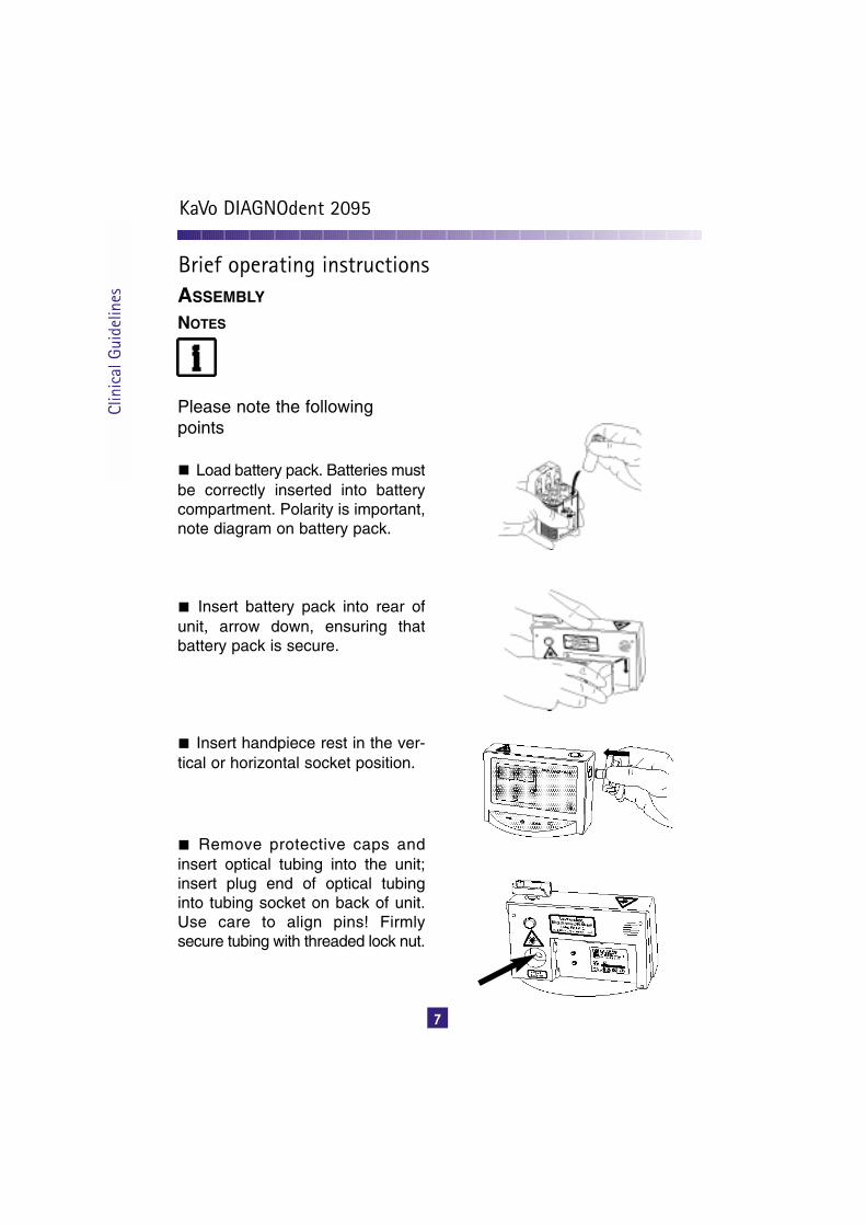
Load battery pack. Batteries mustbe correctly inserted into batterycompartment. Polarity is important,note diagram on battery pack.
Insert battery pack into rear ofunit, arrow down, ensuring thatbattery pack is secure.
Insert handpiece rest in the ver-tical or horizontal socket position.
Remove protective caps andinsert optical tubing into the unit;insert plug end of optical tubinginto tubing socket on back of unit.Use care to align pins! Firmlysecure tubing with threaded lock nut.
ASSEMBLY
7
Brief operating instructions
KaVo DIAGNOdent 2095
Clin
ical
Gui
delin
es

Remove protective cap fromhandpiece end of tubing. Slidethe gripping sleeve over the endof handpiece with light axialpressure, clicking into place,aligning KaVo logo withembossed arrow symbol. ( )
Gently seat light probe tip ontothe gripping sleeve and snap intoplace with a slight axial pressure.When seated properly the A or Bsymbol on the tip will align withKaVo logo on gripping sleeve.
Ensure proper connection ofhandpiece.
To remove the gripping sleeveand the laser probe tip, gently rotateradially. Do not pull!
Place handpiece correctly in thehandpiece rest! (magnetic holder)
Operating Instructions
8
Assembly
KaVo DIAGNOdent 2095
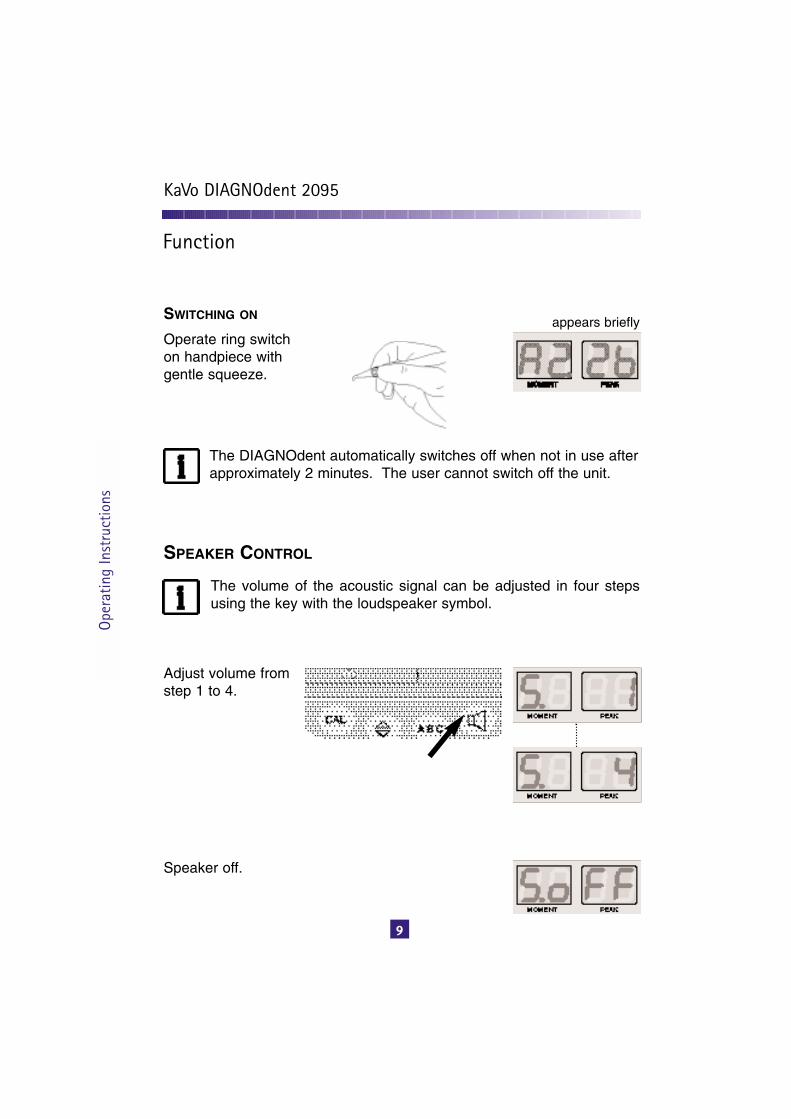
SWITCHING ON
Operate ring switchon handpiece withgentle squeeze.
appears briefly
The DIAGNOdent automatically switches off when not in use afterapproximately 2 minutes. The user cannot switch off the unit.
SPEAKER CONTROL
The volume of the acoustic signal can be adjusted in four stepsusing the key with the loudspeaker symbol.
Adjust volume fromstep 1 to 4.
Speaker off.
Ope
ratin
g In
stru
ctio
ns
9
Function
KaVo DIAGNOdent 2095

Device must be calibrat-ed before use, with desiredprobe tip in place.
Press the CAL key andrelease. Azero will appearin the Moment displayfollowed by a secondzero in the Peak display.
The display changes toa two-digit number and aletter e.g.“ 67b or 58c”, etc.Simultaneously, the unitbegins to emit an audi-ble tone. Gently placethe probe tip on the cen-ter of the circular cali-bration disc, which isaffixed inside the sterili-zation cassette.* Whileholding the tip in place,the two digit number willalso appear under theMoment display and theaudible tone will cease.This indicates that theprobe tip calibrationprocess is complete andthe device is ready foruse.
Switch unit on.
Ensure that the LED oncontrol panel is in the cor-rect position which cor-responds with tip selected.
Select tip.Tip A: conical shape forfissure areas. Tip B: broad, flat designfor flat surfaces.
A
B
Operating Instructions
10
Calibration
KaVo DIAGNOdent 2095
*Note: The two-digit numberindicates the device cali-bration standard, whichshould match the numberprinted on the circular discfound in the sterilizationcassette (see page 15).

ESTABLISH ZERO BASE LINE
EXAMINING TOOTH SURFACES
Check the calibration frequentlyusing the ceramic standard discprovided in the sterilization cas-sette. In the event of a deviationgreater than +/- 3 from the refer-ence value of the ceramic stan-dard, a new calibration must beperformed.
NOTE: Due to influences whichaffect the optical properties ofthe probe tips e.g. wear andsterilization, it is recommendedthat calibration should be per-formed rountinely with probe tipchanges. The additional timerequired to recalibrate will resultin optimal performance of theDIAGNOdent.
Due to slight, natural variances inthe fluorescense of healthy toothstructure, it is recommended toestablish a zero base line, specificto each patient. Prior to scanningtooth surfaces, select one anatom-ical reference point on a tooth sur-face which is apparently healthy.The middle third, facial surface of anon-restored tooth is ideal.
Hold the probe tip against the toothat right angles to the surface, whilegently maintaining squeeze pres-sure on gray ring switch of thehandpiece. “Set 0” will appear onthe display, also confirmed by asecond audible beep, indicatingthat the zero base line is estab-lished.
The anatomical location where thezero base line was establishedshould be documented in thepatient’s dental record for futurereference e.g. DIAGNOdent zerobase line: mid facial #8.
After scanning tooth surfaces it isimportant to eliminate the influenceof the patient-specific zero baseline from the device. Simply holdthe tip in the air and hold the grayring switch until “Set 0” appears onthe display also confirmed by thesecond audible beep.
Clean and dry the teeth.
Ope
ratin
g In
stru
ctio
ns
11
KaVo DIAGNOdent 2095

Place probe tip on tooth surfacewith light contact. If the audible sig-nal is enabled, the tone will soundwhen the DIAGNOdent reaches aminimum value threshold and con-tinues to increase proportionatelywith increased values.
In fissure areas, the tip must beslowly rocked in a pendulousmotion as to carefully scan theadjacent periphery of the site atvarious angles.
The operator will observe the val-ues on the display: the Momentdisplay indicates the real-timevalue that the probe tip is measur-ing, continuously changing as theprobe tip moves. The Peak displaycaptures the maximum valuemeasured in the sequence. ThePeak value is reset by the operatorby actuating the ring switch with abrief squeeze.
In practice, the operator will scantooth surfaces while monitoring theaudible signal. An increase in tonepitch will alert the operator toobserve the display values. Thiswill allow the operator to pin-pointthe exact site which corresponds tothe elevated values.
Operating Instructions
12
Scanning teeth
KaVo DIAGNOdent 2095

Currentvalue
Highest valuedetermined
Calibration positions of theindividual probes
Battery chargestatus
Calibration
Standardvalue setting
Level selec-tion for probetip calibration
Loudspeaker
These brief Operating Instructions cover only the important operating functions. Beforestarting the unit for the first time and when the handling of this medical product, it is essen-tial to read the instructions for use included in the delivery.
Ope
ratin
g In
stru
ctio
ns
13
KaVo DIAGNOdent 2095
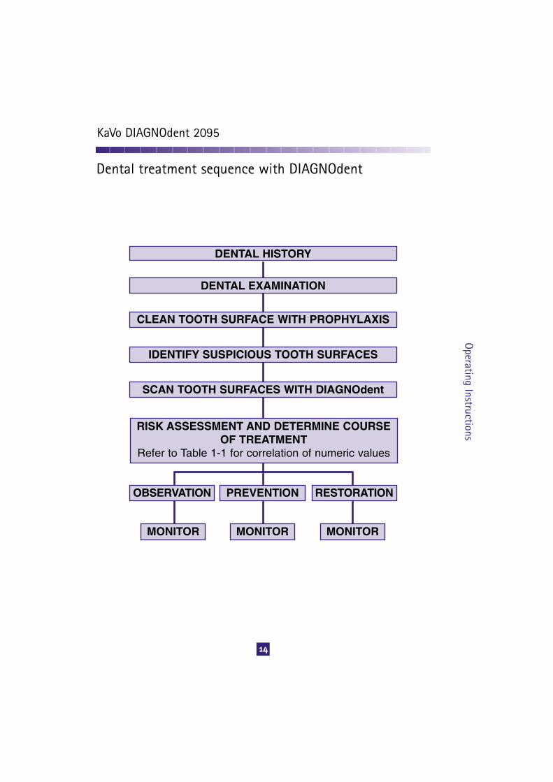
DENTAL HISTORY
DENTAL EXAMINATION
CLEAN TOOTH SURFACE WITH PROPHYLAXIS
IDENTIFY SUSPICIOUS TOOTH SURFACES
SCAN TOOTH SURFACES WITH DIAGNOdent
RISK ASSESSMENT AND DETERMINE COURSE OF TREATMENT
Refer to Table 1-1 for correlation of numeric values
OBSERVATION RESTORATIONPREVENTION
MONITOR MONITORMONITOR
Operating Instructions
14
Dental treatment sequence with DIAGNOdent
KaVo DIAGNOdent 2095

CHECKING THE STANDARD VALUE
Press key.
The display showse.g. c... and two dig-its. The letter and thetwo digits must beidentical to the onesprinted on the ceram-ic standard disc [1]provided in the sterili-zation cassette usedfor calibration.
[1]
Note: The standard value displayed in the DIAGNOdent mustmatch the standard (cassette) used to calibrate the probe tips. Inthe event that multiple or replacement cassettes are used with thedevice, the standard value must be reprogrammed in theDIAGNOdent to match the respective value standard (cassette)used for probe tip calibration.
CHANGING THE STANDARD VALUE
Press the “up/downarrow” key to viewstandard value.Actuate gray ring onhandpiece to changestandard value.Press the “up/downarrow” key again toenter new standardvalue.
Switch unit on.
Switch unit on.
Ope
ratin
g In
stru
ctio
ns
15
Additional user information
KaVo DIAGNOdent 2095

BATTERY INDICATOR
Indicator flashes at apower of approx. 20%
When the power fallsbelow 20% the dis-play shows ACC thenLO.
At the same time, anacoustic signal isheard and the unitswitches off.
TROUBLESHOOTINGRefer to OperatingInstructions deliveredwith product.
User Inform
ation
KaVo DIAGNOdent 2095
16

1. What is the DIAGNOdent actuallymeasuring?A. The DIAGNOdent measures laserfluorescence within tooth structure. Asthe incident laser light is propagatedinto the site, two-way handpiece opticsallows the unit to simultaneously quan-tify the reflected laser light energy. Atthe specific wavelength that theDIAGNOdent laser operates, cleanhealthy tooth structure exhibits little orno fluorescence, resulting in very lowscale readings on the display.However, carious tooth structure willexhibit fluorescence, proportionale tothe degree of caries, resulting in elevat-ed scale readings on the display.
2. What is the difference betweenthe Moment and Peak?A. The Moment is the number that isoccurring at that exact spot on the toothand changes as you move across thetooth. The Peak is the highest numberrecorded before the unit is reset. Asyou are doing your examination it is thePeak number which you should look at.
3. What do you mean by a cleantooth?A. To get consistent readings it isessential to have a clean tooth. This isespecially important when monitoringvalues at future examinations. We sug-gest that the teeth are cleaned thor-oughly using any acceptable means.The use of a powder jet cleaner, e.g.KaVo PROPHYflex 2, is a rapid andeffective method to clean stain anddebris from complex occlusal anatomy.
4. Can the unit diagnose inter-proxi-mal caries?A. No. Limited accessibility to theembrasure prevents accurate readingof approximal surfaces.
5. Can the unit be used around exist-ing composite resin restorations?A. No. Because composite resins canfluoresce, prompting elevated readings,the DIAGNOdent should not be used onthese materials.
6. Can the unit read caries under anexisting amalgam?A. If there is caries at the margin it willgive an accurate reading; however if thecaries is under the floor of the amalgamthe reading will not be accurate.
7. If I see stain under a sealant will theunit tell me if this is decay or not?A. No. The sealant must first be removedand then an accurate reading can be taken.
8. Can the DIAGNOdent be used to deter-mine if caries excavation is complete?A. Not always; some conservative pre-paration designs, particularly those withsmall access openings, limit proper tipangulations within a preparation.Furthermore, independent research8
indicates that when used in deeppreparations, in close proximity to thepulp, elevated values may be obtained,possibly resulting from fluorescence ofunderlying pulp and not necessarily asa result of caries. Therefore, the use ofother methods to determine extent ofaffected tooth structure should beemployed in these situations.
Use
r In
form
atio
n
17
Frequently asked questions
KaVo DIAGNOdent 2095

9. What do you mean by risk assessment?A. It is necessary to consider a patient’sdental history, dietary sugar intake, oralhygiene, history of maintaining regularrecalls, caries status (the number of cari-ous teeth in their mouth), as well as anyother information such as that from ra-diographs or other diagnostic modalities.
10. What does a very high number mean?A. This may occur when there is an openlesion at the surface or if there is a lot ofdebris where the reading is being taken.The tooth should be cleaned thoroughlyand a second reading taken.
11. Can the unit be used on both pri-mary and permanent teeth?A. Studies have shown the unit is equallyaccurate in both primary and permanentteeth.
12. How much change in the readingbetween visits is considered significant?A. The margin of error is (+) or (-) 3 so achange greater than this would be nec-essary before the tooth should be con-sidered to exhibit a changing condition.
13. Can the tone be used to identifycaries?A. No. The audible tone function is anoperator convenience, which allows thedentist to focus on the tooth while scan-ning, only sounding variably as differentvalue thresholds are reached. The signalis intended to direct the operator to thevisual display. Tone volume can be changedor altogether switched off as desired.
14. I cannot calibrate the unit, I con-tinuously get an error message on thedisplay.A. Nearly all calibration errors are attrib-uted to user error. It is necessary to followcalibration instructions carefully. If errorpersists, contact KaVo Customer Service.
15. As the device is a laser, is protectiveeyewear required?A. No. The device is harmless whenused as directed.
16. How deep does the laser penetratethe tooth?A. Approximately 2mm.
17. Which tip should I use, A or B?A. Tip A is conical in shape and designedfor fissure areas. Tip B is broader anddesigned for flat surfaces.
18. What is the recommended asep-sis protocol for the DIAGNOdent?A. Clean the control unit and the tubingthese surfaces with a soft cloth dampenedwith a mild soap solution. The remaininghandpiece sheath and tips can be steamsterilized. Chemical vapor sterilization isalso acceptable, however, componentswill eventually discolor and degrade atan accelerated rate. Many clinicianssterilize the tips but prefer to barrier pro-tect the sleeve portion. Other cliniciansfit barriers over the entire sleeve and tipassembly to eliminate the need for tipsterilization. When tips are barrier pro-tected, unit must be calibrated with barrierin place. Tips can be cleaned with a softcotton swab moistened with water. Donot use surface disinfectants on the tripor sleeve components! See OperatingManual for additional cleaning information.
19. Does ambient light affectDIAGNOdent readings?A. The photo-optic measuring system inthe DIAGNOdent has filters to eliminatethe influences of most ambient light.However, when calibrating the device,avoid exposing the probe tip to directhalogen light.
User Inform
ation
18
Frequently asked questions
KaVo DIAGNOdent 2095

1 Garcia-Godoy, F., Medlock, JW.: “AnSEM study of the effects of air-polishing offissues surfaces.” QuintessenceInternational Vol. 19 1988, Special Reprint
2 Konig, K., Fleming, G., and Hibst, R.:“Laser-induced autofluorescence spec-troscopy of dental caries.” Cell Mol Biol(Noisy-le-grand) 1998 Dec;44(8):1293-300
3 Lussi, A., et al.: “Performance of a LaserFluorescence System for Detection ofOcclusal Caries.” 45th ORCA Congress,1998, Abst. #87, p. 297
4 Lussi, A., et al.: “Performance andReproducibility of a Laser FluorescenceSystem for Detection of Occlusal Caries invitro.” Caries Research, 1999; 33:261-266
5 Lussi, A., et al.: “Reproducibility of aLaser Fluorescence System for Detectionof Occlusal Caries.” 45th ORCA Congress,1998, Abst. #88, p. 297
6 Klimm, W., et al.: “Comparison of ThreeNon-Invasive Methods for Early OcclusalCaries Assessment in vitro.” 46th ORCA,Congress, 1999, Abst. #48, p. 297
7 Reich, E., et al.: “Clinical Validation of aLaser Caries Diagnosis System.” 45thORCA Congress, 1998, Abst. #89, p. 297
8 Reich, E., et al.: “Clinical CariesDiagnosis Compared to DIAGNOdentEvaluation.” 46th ORCA Congress, 1999,Abst. #54, p. 299
9 Lussi, et al.: “Clinical Performance of theLaser Fluorescence System DIAGNOdentfor Detection of Occlusal Caries.” 46thORCA Congress, 1999, Abst. #55, p. 299
10 Longbottom, et al.: “HistologicalValidation of in vivo Measurements Usingthe DIAGNOdent Device: A Three-CentreStudy.” 46th ORCA Congress, 1999, Abst.#58, p. 300
11 Braun, A., et al.: “Effects of tooth clean-ing on laser fluorescence measurementsof the tooth surface.” Conference Paper,Dtsch. Zahnarztl Z 54 (1999) 3
12 Lussi, A., and Hibst, R.: “Methods forOcclusal Caries Detection Used in DailyPractice,” Indiana Conference 1999
13 Lussi, A.: “Clinical Performance of theLaser Fluorescence System DIAGNOdentfor Detection of Occlusal Caries” (inGerman), Acta Med Dent Helv 5: 15-19(2/2000)
Use
r In
form
atio
n
19
References
KaVo DIAGNOdent 2095
20
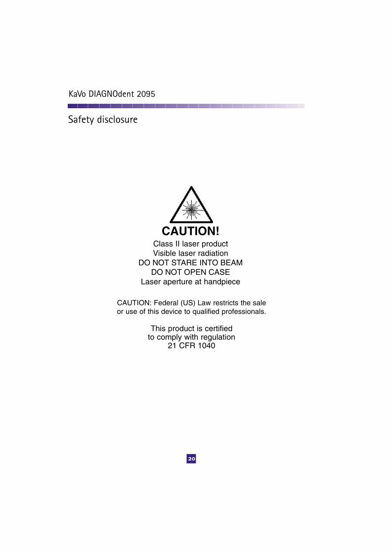
CAUTION!Class II laser productVisible laser radiation
DO NOT STARE INTO BEAMDO NOT OPEN CASE
Laser aperture at handpiece
CAUTION: Federal (US) Law restricts the saleor use of this device to qualified professionals.
This product is certified to comply with regulation
21 CFR 1040
Safety disclosure
KaVo DIAGNOdent 2095

KaVo America.340 East Main Street, Lake Zurich, Illinois 60047
Tel: 1-888-KaVo USA (1-888-528-6872)Fax: 847-550-6825 www.kavousa.com1.
001.
5328
/ 0
8.02
DM
S /
sk