clinical highs and lows of dk/t - johnson & johnson · clinical highs and lows of dk/t a...
TRANSCRIPT
Contact Lens Monthly
In the first of a two-part series, Professor Noel Brennan and Dr Philip Morgan look at the latest terminology and theories about oxygenation of the cornea through silicone hydrogel lenses
Clinical highs and lows of Dk/t
Al though s i l icone hydrogel (SiH) contact lenses have fallen short of their original goal, solving the continuous wear-infectious keratitis
problem, they undeniably provide a superior environment than hydrogels by promoting normal corneal metabolic activity. While there is consensus on this benefit, it is less clear how high we have to go in terms of oxygen transmissibility (Dk/t) within the SiH category to achieve an optimal physiological result. (For simplicity, units of 10-9(cm/sec)(mlO2/ml.mmHg) are omittedwhere Dk/t values are given in the text.) Theoretical modelling backed up by equivalent oxygen percentage (EOP)-based empirical data suggests that there will be little difference between any of the SiH lenses.1-6 However, some authors argue that one should expect continuing benefits by increasing Dk/t within the SiH range.7-9
While the modelling and laboratory findings are important to contemplate, the ultimate test of these differences will be found in clinical performance. In this, the first of a two-part series, we address the clinical perspective to arrive at the conclusion that there is little to be gained in strict physiological terms by pushing Dk/t values beyond those provided at the low end of the SiH range. In part two, we will highlight important differences in performance within the SiH category that are not oxygen based and should be the real consideration for selecting between different lens brands.
The law of diminishing returns The law of diminishing returns must apply when considering the relation between oxygen reaching the cornea and Dk/t. Let us suppose a contact lens with Dk/t equal to 100 leads to a partial pressure of oxygen at the front surface of the cornea of 100mmHg (from available data, this is most probably a conservative estimate). Under normal open eye wearing conditions, doubling the Dk/t to 200 will not lead to a partial pressure of 200mmHg. It cannot, because the partial pressure of
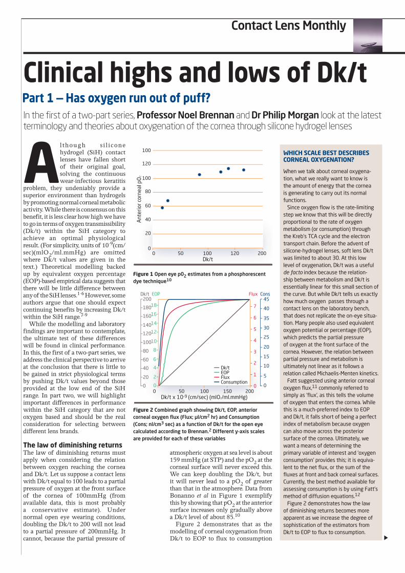
atmospheric oxygen at sea level is about 159 mmHg (at STP) and the pO2 at thecorneal surface will never exceed this. We can keep doubling the Dk/t, but it will never lead to a pO2 of greaterthan that in the atmosphere. Data from Bonanno et al in Figure 1 exemplify this by showing that pO2 at the anteriorsurface increases only gradually above a Dk/t level of about 85.10
Figure 2 demonstrates that as the modelling of corneal oxygenation from Dk/t to EOP to flux to consumption
Part 1 — Has oxygen run out of puff?
When we talk about corneal oxygena-tion, what we really want to know is the amount of energy that the cornea is generating to carry out its normal functions.
Since oxygen flow is the rate-limiting step we know that this will be directly proportional to the rate of oxygen metabolism (or consumption) through the Kreb’s TCA cycle and the electron transport chain. Before the advent of silicone-hydrogel lenses, soft lens Dk/t was limited to about 30. At this low level of oxygenation, Dk/t was a useful de facto index because the relation-ship between metabolism and Dk/t is essentially linear for this small section of the curve. But while Dk/t tells us exactly how much oxygen passes through a contact lens on the laboratory bench, that does not replicate the on-eye situa-tion. Many people also used equivalent oxygen potential or percentage (EOP), which predicts the partial pressure of oxygen at the front surface of the cornea. However, the relation between partial pressure and metabolism is ultimately not linear as it follows a relation called Michaelis-Menten kinetics.
Fatt suggested using anterior corneal oxygen flux,11 commonly referred to simply as ‘flux’, as this tells the volume of oxygen that enters the cornea. While this is a much-preferred index to EOP and Dk/t, it falls short of being a perfect index of metabolism because oxygen can also move across the posterior surface of the cornea. Ultimately, we want a means of determining the primary variable of interest and ‘oxygen consumption’ provides this; it is equiva-lent to the net flux, or the sum of the fluxes at front and back corneal surfaces. Currently, the best method available for assessing consumption is by using Fatt’s method of diffusion equations.12
Figure 2 demonstrates how the law of diminishing returns becomes more apparent as we increase the degree of sophistication of the estimators from Dk/t to EOP to flux to consumption.
WHiCH sCAle besT DesCribes CorneAl oxygenATion?
Figure 1 Open eye p02 estimates from a phosphorescent dye technique10
Figure 2 Combined graph showing Dk/t, EOP, anterior corneal oxygen flux (Flux; μl/cm2 hr) and Consumption (Cons; nl/cm3 sec) as a function of Dk/t for the open eye calculated according to Brennan.2 Different y-axis scales are provided for each of these variables
0 50 120 200100Dk/t
Ant
erio
r cor
neal
pO
2
100
80
60
40
100
120
20
0
0 50 150 200100
Dk/t
Dk/t x 10-9 (cm/sec) (mlO2/ml.mmHg)
Dk/t EOP Flux Cons200
180
160
140
120
100
80
60
40
20
0
718
16
14
12
10
8
6
4
2
0
45
40
35
30
25
20
15
10
5
0
6
5
4
3
2
1
0
EOPFluxConsumption
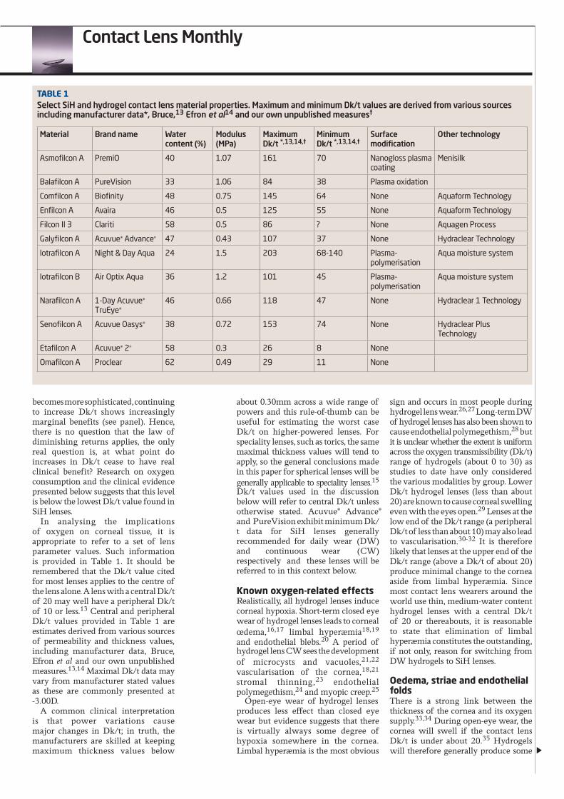
becomes more sophisticated, continuing to increase Dk/t shows increasingly marginal benefits (see panel). Hence, there is no question that the law of diminishing returns applies, the only real question is, at what point do increases in Dk/t cease to have real clinical benefit? Research on oxygen consumption and the clinical evidence presented below suggests that this level is below the lowest Dk/t value found in SiH lenses.
In analysing the implications of oxygen on corneal tissue, it is appropriate to refer to a set of lens parameter values. Such information is provided in Table 1. It should be remembered that the Dk/t value cited for most lenses applies to the centre of the lens alone. A lens with a central Dk/t of 20 may well have a peripheral Dk/t of 10 or less.13 Central and peripheral Dk/t values provided in Table 1 are estimates derived from various sources of permeability and thickness values, including manufacturer data, Bruce, Efron et al and our own unpublished measures.13,14 Maximal Dk/t data may vary from manufacturer stated values as these are commonly presented at -3.00D.
A common clinical interpretationis that power variations causemajor changes in Dk/t; in truth, themanufacturers are skilled at keepingmaximum thickness values below
about 0.30mm across a wide range of powers and this rule-of-thumb can be useful for estimating the worst case Dk/t on higher-powered lenses. For speciality lenses, such as torics, the same maximal thickness values will tend to apply, so the general conclusions made in this paper for spherical lenses will be generally applicable to speciality lenses.15 Dk/t values used in the discussion below will refer to central Dk/t unless otherwise stated. Acuvue® Advance® and PureVision exhibit minimum Dk/t data for SiH lenses generally recommended for daily wear (DW) and continuous wear (CW) respectively and these lenses will be referred to in this context below.
Known oxygen-related effects Realistically, all hydrogel lenses induce corneal hypoxia. Short-term closed eye wear of hydrogel lenses leads to corneal œdema,16,17 limbal hyperæmia18,19 and endothelial blebs.20 A period of hydrogel lens CW sees the development of microcysts and vacuoles,21,22 vascularisation of the cornea,18,21 stromal thinning,23 endothelial polymegethism,24 and myopic creep.25
Open-eye wear of hydrogel lenses produces less effect than closed eye wear but evidence suggests that there is virtually always some degree of hypoxia somewhere in the cornea. Limbal hyperæmia is the most obvious
sign and occurs in most people during hydrogel lens wear.26,27 Long-term DW of hydrogel lenses has also been shown to cause endothelial polymegethism,28 but it is unclear whether the extent is uniform across the oxygen transmissibility (Dk/t) range of hydrogels (about 0 to 30) as studies to date have only considered the various modalities by group. Lower Dk/t hydrogel lenses (less than about 20) are known to cause corneal swellingeven with the eyes open.29 Lenses at thelow end of the Dk/t range (a peripheralDk/t of less than about 10) may also leadto vascularisation.30-32 It is thereforelikely that lenses at the upper end of theDk/t range (above a Dk/t of about 20)produce minimal change to the corneaaside from limbal hyperæmia. Sincemost contact lens wearers around theworld use thin, medium-water contenthydrogel lenses with a central Dk/tof 20 or thereabouts, it is reasonableto state that elimination of limbalhyperæmia constitutes the outstanding,if not only, reason for switching fromDW hydrogels to SiH lenses.
oedema, striae and endothelial folds There is a strong link between the thickness of the cornea and its oxygen supply.33,34 During open-eye wear, the cornea will swell if the contact lens Dk/t is under about 20.35 Hydrogels will therefore generally produce some
TAble 1Select SiH and hydrogel contact lens material properties. Maximum and minimum Dk/t values are derived from various sources including manufacturer data*, Bruce,13 Efron et al14 and our own unpublished measures†
Material Brand name Water content (%)
Modulus (MPa)
Maximum Dk/t *,13,14,†
Minimum Dk/t *,13,14,†
Surface modification
Other technology
Asmofilcon A PremiO 40 1.07 161 70 Nanogloss plasma coating
Menisilk
Balafilcon A PureVision 33 1.06 84 38 Plasma oxidation
Comfilcon A Biofinity 48 0.75 145 64 None Aquaform Technology
Enfilcon A Avaira 46 0.5 125 55 None Aquaform Technology
Filcon II 3 Clariti 58 0.5 86 ? None Aquagen Process
Galyfilcon A Acuvue® Advance® 47 0.43 107 37 None Hydraclear Technology
lotrafilcon A Night & Day Aqua 24 1.5 203 68-140 Plasma-polymerisation
Aqua moisture system
lotrafilcon B Air Optix Aqua 36 1.2 101 45 Plasma-polymerisation
Aqua moisture system
Narafilcon A 1-Day Acuvue® TruEye®
46 0.66 118 47 None Hydraclear 1 Technology
Senofilcon A Acuvue Oasys® 38 0.72 153 74 None Hydraclear Plus Technology
Etafilcon A Acuvue® 2® 58 0.3 26 8 None
Omafilcon A Proclear 62 0.49 29 11 None
Contact Lens Monthly
Contact Lens Monthly
corneal swelling even when the eyes are open. If this amount is more than about 5 per cent, the clinician will also be able to see posterior stromal striae and when it swells by more than about 10 per cent, endothelial folds appear.36 None of the SiH lenses would be expected to produce any corneal swelling while the eyes are open and there is evidence to support this contention.37
During overnight eye closure, there will be some swelling of the cornea even without contact lens wear. The amount has been estimated to be between 0.7 and 5.5 per cent.38 The amount varies with lens wearing experience; Cox et al found 3.8 per cent swelling in non-contact lens wearers, 2.0 per cent in subjects adapted to DW hydrogel lenses and 0.7 per cent in those adapted to CW hydrogel lenses.39 All contact lenses will cause further swelling in the closed eye state above these baselines. Thin, medium-water content hydrogels will generally cause about 8 per cent further swelling and hydrogels with lower Dk/t than these will cause greater amounts.40 SiH lenses cause much less œdema. The SiH lens with the lowest Dk/t that is approved for CW, PureVision, only causes about 2 per cent swelling additional to the baseline amount.40 The SiH with the highest Dk/t, Focus Night & Day, causes about 1 per cent more swelling.41 It is unclear whether these small amounts of 1 to 2 per cent swelling seen with SiH lenses in addition to that which occurs without a contact lens in place are indicative of harmful hypoxic stress and whether the small differences between these lenses are important. In the absence of a known link between swelling and corneal pathology and given the large proportional difference between SiH and hydrogel lenses, we propose that the degree of swelling occurring with SiH lenses is of no consequence.
Microcysts Holden et al stated that, of various compromises that CW of hydrogels may produce, ‘the most easily observable condition indicative of epithelial compromise is microcysts’.42 Sweeney et al state that ‘in clinical trials microcysts are used as the classic marker of hypoxia’. Hickson and Papas measured the incidence of microcysts in people who do not wear contact lenses to be 49 per cent, although none showed more than 5 microcysts per cornea.43 DW of hydrogel lenses does not seem to influence this incidence;42,44 consequently DW of SiH lenses would not be expected to increase this prevalence, a premise based on
the default that no-one has deemed it worthy of measuring.
In contrast, the mean number of microcysts in subjects wearing hydrogel contact lenses after five years of CW has been reported as 17 ± 21, with virtually all eyes showing at least one microcyst.23 Microcyst numbers inversely correlate with Dk/t during closed eye wear, with the Dk/t level at which they fall to baseline numbers estimated at around 50.45 Given that the central Dk/t of the PureVision is about 90, it is no surprise that microcysts are not generally reported as a problem with SiH lenses. Brennan et al reported on wear of 3 different type of SiH lenses and found an incidence of 30 to 59 per cent, which is consistent with the Hickson-Papas baseline but a higher incidence of 9 to 17 per cent with greater than 10 microcysts.46 They studied the lenses with highest and lowest Dk/t values in the EW category for SiH but found no evidence of a relation between this most important marker of hypoxia and Dk/t within this group.
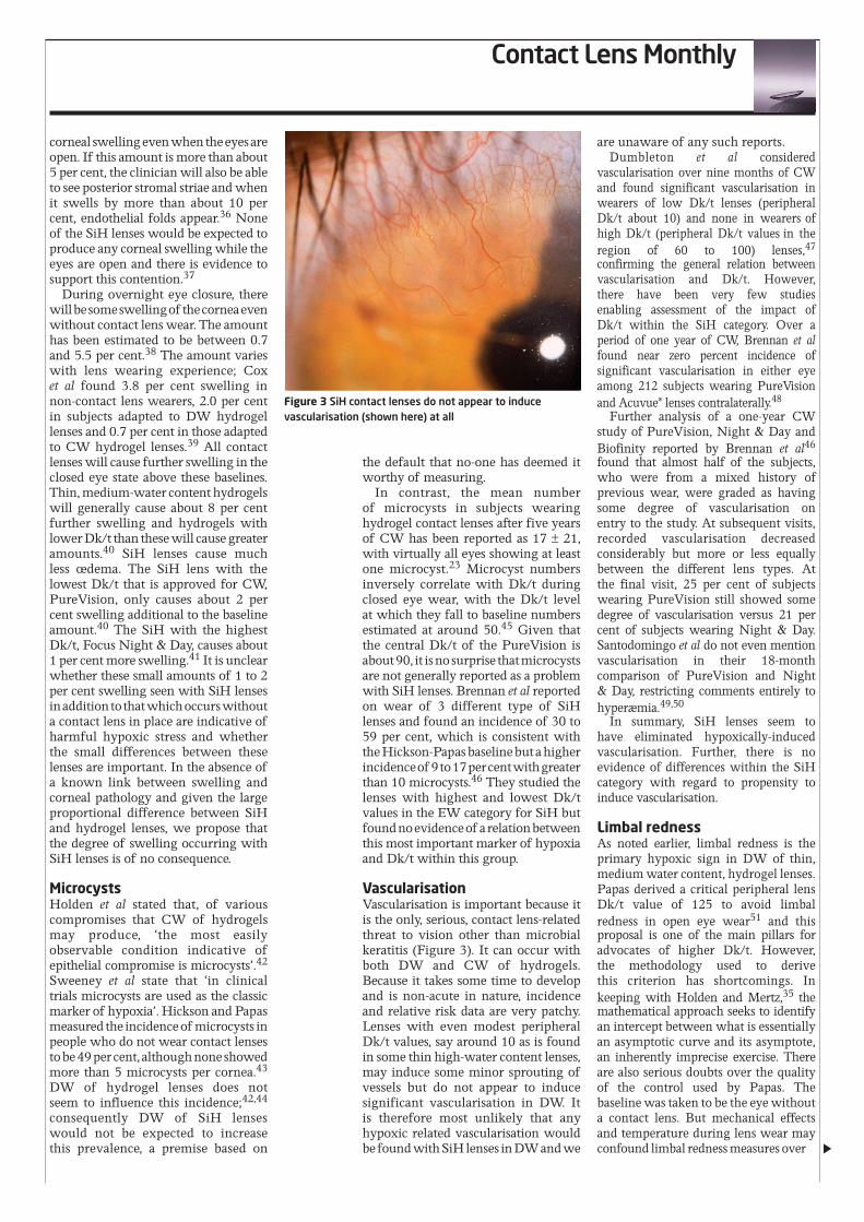
Vascularisation Vascularisation is important because it is the only, serious, contact lens-related threat to vision other than microbial keratitis (Figure 3). It can occur with both DW and CW of hydrogels. Because it takes some time to develop and is non-acute in nature, incidence and relative risk data are very patchy. Lenses with even modest peripheral Dk/t values, say around 10 as is found in some thin high-water content lenses, may induce some minor sprouting of vessels but do not appear to induce significant vascularisation in DW. It is therefore most unlikely that any hypoxic related vascularisation would be found with SiH lenses in DW and we
are unaware of any such reports. Dumbleton et al considered
vascularisation over nine months of CW and found significant vascularisation in wearers of low Dk/t lenses (peripheral Dk/t about 10) and none in wearers of high Dk/t (peripheral Dk/t values in the region of 60 to 100) lenses,47 confirming the general relation between vascularisation and Dk/t. However, there have been very few studies enabling assessment of the impact of Dk/t within the SiH category. Over a period of one year of CW, Brennan et al found near zero percent incidence of significant vascularisation in either eye among 212 subjects wearing PureVision and Acuvue® lenses contralaterally.48
Further analysis of a one-year CW study of PureVision, Night & Day and Biofinity reported by Brennan et al46
found that almost half of the subjects, who were from a mixed history of previous wear, were graded as having some degree of vascularisation on entry to the study. At subsequent visits, recorded vascularisation decreased considerably but more or less equally between the different lens types. At the final visit, 25 per cent of subjects wearing PureVision still showed some degree of vascularisation versus 21 per cent of subjects wearing Night & Day. Santodomingo et al do not even mention vascularisation in their 18-month comparison of PureVision and Night & Day, restricting comments entirely to hyperæmia.49,50
In summary, SiH lenses seem to have eliminated hypoxically-induced vascularisation. Further, there is no evidence of differences within the SiH category with regard to propensity to induce vascularisation.
limbal redness As noted earlier, limbal redness is the primary hypoxic sign in DW of thin, medium water content, hydrogel lenses. Papas derived a critical peripheral lens Dk/t value of 125 to avoid limbal redness in open eye wear51 and this proposal is one of the main pillars for advocates of higher Dk/t. However, the methodology used to derive this criterion has shortcomings. In keeping with Holden and Mertz,35 the mathematical approach seeks to identify an intercept between what is essentially an asymptotic curve and its asymptote, an inherently imprecise exercise. There are also serious doubts over the quality of the control used by Papas. The baseline was taken to be the eye without a contact lens. But mechanical effects and temperature during lens wear may confound limbal redness measures over
Figure 3 SiH contact lenses do not appear to induce vascularisation (shown here) at all
and above the influence of Dk/t. To this end, we surveyed the literature
for support to Papas’ criterion of 125. Our search identified seven studies that compared differences between two different SiH lenses in the degree of induced limbal hyperæmia in both DW and CW.46,49,52-56 Since the peripheral Dk/t values of all the SiH lenses fall below 125 and no two values are the same, we would expect to find significant differences in the degree of limbal redness in all of these studies if Papas finding is clinically important. In none of these studies was there a difference reported. This suggests that, not only is the criterion of 125 of little relevance in the clinical world, but that a figure of 37, the minimum peripheral Dk/t found in DW silicone-hydrogel lenses, is adequate to avoid clinically important open-eye limbal redness.
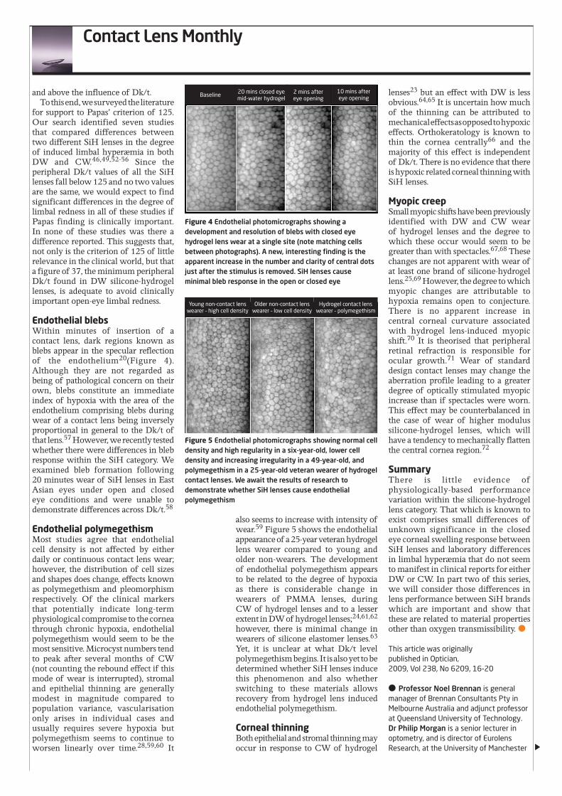
endothelial blebs Within minutes of insertion of a contact lens, dark regions known as blebs appear in the specular reflection of the endothelium20(Figure 4). Although they are not regarded as being of pathological concern on their own, blebs constitute an immediate index of hypoxia with the area of the endothelium comprising blebs during wear of a contact lens being inversely proportional in general to the Dk/t of that lens.57 However, we recently tested whether there were differences in bleb response within the SiH category. We examined bleb formation following 20 minutes wear of SiH lenses in East Asian eyes under open and closed eye conditions and were unable to demonstrate differences across Dk/t.58
endothelial polymegethism Most studies agree that endothelial cell density is not affected by either daily or continuous contact lens wear; however, the distribution of cell sizes and shapes does change, effects known as polymegethism and pleomorphism respectively. Of the clinical markers that potentially indicate long-term physiological compromise to the cornea through chronic hypoxia, endothelial polymegethism would seem to be the most sensitive. Microcyst numbers tend to peak after several months of CW (not counting the rebound effect if this mode of wear is interrupted), stromal and epithelial thinning are generally modest in magnitude compared to population variance, vascularisation only arises in individual cases and usually requires severe hypoxia but polymegethism seems to continue to worsen linearly over time.28,59,60 It
also seems to increase with intensity of wear.59 Figure 5 shows the endothelial appearance of a 25-year veteran hydrogel lens wearer compared to young and older non-wearers. The development of endothelial polymegethism appears to be related to the degree of hypoxia as there is considerable change in wearers of PMMA lenses, during CW of hydrogel lenses and to a lesser extent in DW of hydrogel lenses;24,61,62 however, there is minimal change in wearers of silicone elastomer lenses.63 Yet, it is unclear at what Dk/t level polymegethism begins. It is also yet to be determined whether SiH lenses induce this phenomenon and also whether switching to these materials allows recovery from hydrogel lens induced endothelial polymegethism.
Corneal thinning Both epithelial and stromal thinning may occur in response to CW of hydrogel
lenses23 but an effect with DW is less obvious.64,65 It is uncertain how much of the thinning can be attributed to mechanical effects as opposed to hypoxic effects. Orthokeratology is known to thin the cornea centrally66 and the majority of this effect is independent of Dk/t. There is no evidence that there is hypoxic related corneal thinning with SiH lenses.
Myopic creep Small myopic shifts have been previously identified with DW and CW wear of hydrogel lenses and the degree to which these occur would seem to be greater than with spectacles.67,68 These changes are not apparent with wear of at least one brand of silicone-hydrogel lens.25,69 However, the degree to which myopic changes are attributable to hypoxia remains open to conjecture. There is no apparent increase in central corneal curvature associated with hydrogel lens-induced myopic shift.70 It is theorised that peripheral retinal refraction is responsible for ocular growth.71 Wear of standard design contact lenses may change the aberration profile leading to a greater degree of optically stimulated myopic increase than if spectacles were worn. This effect may be counterbalanced in the case of wear of higher modulus silicone-hydrogel lenses, which will have a tendency to mechanically flatten the central cornea region.72
summary There is little evidence of physiologically-based performance variation within the silicone-hydrogel lens category. That which is known to exist comprises small differences of unknown significance in the closed eye corneal swelling response between SiH lenses and laboratory differences in limbal hyperæmia that do not seem to manifest in clinical reports for either DW or CW. In part two of this series, we will consider those differences in lens performance between SiH brands which are important and show that these are related to material properties other than oxygen transmissibility. ●
This article was originally published in Optician, 2009, Vol 238, No 6209, 16-20
● Professor Noel Brennan is general manager of Brennan Consultants Pty in Melbourne Australia and adjunct professor at Queensland University of Technology. Dr Philip Morgan is a senior lecturer in optometry, and is director of Eurolens Research, at the University of Manchester
Figure 4 Endothelial photomicrographs showing a development and resolution of blebs with closed eye hydrogel lens wear at a single site (note matching cells between photographs). A new, interesting finding is the apparent increase in the number and clarity of central dots just after the stimulus is removed. SiH lenses cause minimal bleb response in the open or closed eye
Figure 5 Endothelial photomicrographs showing normal cell density and high regularity in a six-year-old, lower cell density and increasing irregularity in a 49-year-old, and polymegethism in a 25-year-old veteran wearer of hydrogel contact lenses. We await the results of research to demonstrate whether SiH lenses cause endothelial polymegethism
Baseline 20 mins closed eye mid-water hydrogel
2 mins after eye opening
10 mins after eye opening
Young non-contact lens wearer – high cell density
Older non-contact lens wearer – low cell density
Hydrogel contact lens wearer – polymegethism
Contact Lens Monthly
Contact Lens Monthly
Clinical highs and lows of Dk/t
The first part of this two-part series considered differences between silicone-hydrogel (SiH) contact lenses in terms of their effect on ocular
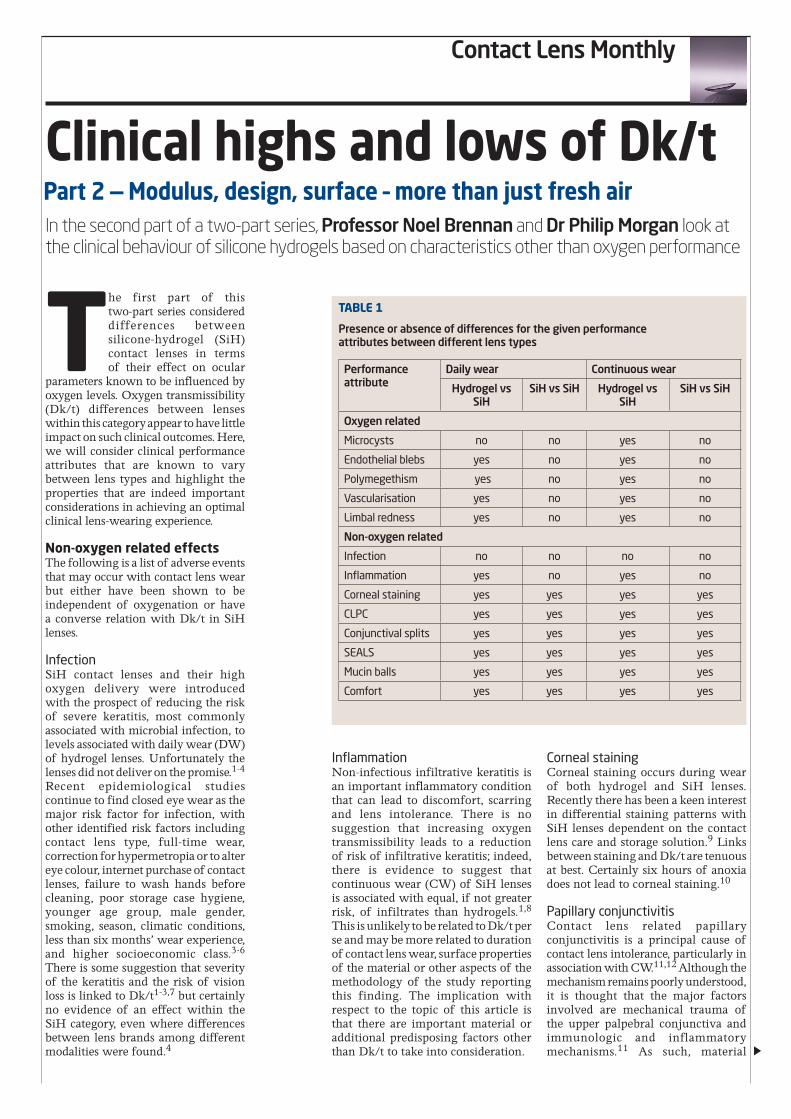
parameters known to be influenced by oxygen levels. Oxygen transmissibility (Dk/t) differences between lenses within this category appear to have little impact on such clinical outcomes. Here, we will consider clinical performance attributes that are known to vary between lens types and highlight the properties that are indeed important considerations in achieving an optimal clinical lens-wearing experience.
Non-oxygen related effects The following is a list of adverse events that may occur with contact lens wear but either have been shown to be independent of oxygenation or have a converse relation with Dk/t in SiH lenses.
Infection SiH contact lenses and their high oxygen delivery were introduced with the prospect of reducing the risk of severe keratitis, most commonly associated with microbial infection, to levels associated with daily wear (DW) of hydrogel lenses. Unfortunately the lenses did not deliver on the promise.1-4 Recent epidemiological studies continue to find closed eye wear as the major risk factor for infection, with other identified risk factors including contact lens type, full-time wear, correction for hypermetropia or to alter eye colour, internet purchase of contact lenses, failure to wash hands before cleaning, poor storage case hygiene, younger age group, male gender, smoking, season, climatic conditions, less than six months’ wear experience, and higher socioeconomic class.3-6 There is some suggestion that severity of the keratitis and the risk of vision loss is linked to Dk/t1-3,7 but certainly no evidence of an effect within the SiH category, even where differences between lens brands among different modalities were found.4
Inflammation Non-infectious infiltrative keratitis is an important inflammatory condition that can lead to discomfort, scarring and lens intolerance. There is no suggestion that increasing oxygen transmissibility leads to a reduction of risk of infiltrative keratitis; indeed, there is evidence to suggest that continuous wear (CW) of SiH lenses is associated with equal, if not greater risk, of infiltrates than hydrogels.1,8 This is unlikely to be related to Dk/t per se and may be more related to duration of contact lens wear, surface properties of the material or other aspects of the methodology of the study reporting this finding. The implication with respect to the topic of this article is that there are important material or additional predisposing factors other than Dk/t to take into consideration.
Corneal staining Corneal staining occurs during wear of both hydrogel and SiH lenses. Recently there has been a keen interest in differential staining patterns with SiH lenses dependent on the contact lens care and storage solution.9 Links between staining and Dk/t are tenuous at best. Certainly six hours of anoxia does not lead to corneal staining.10
Papillary conjunctivitis Contact lens related papillary conjunctivitis is a principal cause of contact lens intolerance, particularly in association with CW.11,12 Although the mechanism remains poorly understood, it is thought that the major factors involved are mechanical trauma of the upper palpebral conjunctiva and immunologic and inflammatory mechanisms.11 As such, material
Part 2 — Modulus, design, surface – more than just fresh airIn the second part of a two-part series, Professor Noel Brennan and Dr Philip Morgan look at the clinical behaviour of silicone hydrogels based on characteristics other than oxygen performance
Table 1
Presence or absence of differences for the given performance attributes between different lens types
Performance attribute
Daily wear Continuous wear
Hydrogel vs SiH
SiH vs SiH Hydrogel vs SiH
SiH vs SiH
Oxygen related
Microcysts no no yes no
Endothelial blebs yes no yes no
Polymegethism yes no yes no
Vascularisation yes no yes no
Limbal redness yes no yes no
Non-oxygen related
Infection no no no no
Inflammation yes no yes no
Corneal staining yes yes yes yes
CLPC yes yes yes yes
Conjunctival splits yes yes yes yes
SEALS yes yes yes yes
Mucin balls yes yes yes yes
Comfort yes yes yes yes
Contact Lens Monthly
modulus, lens surface characteristics and lens design are implicated as the contact lens related causative factors.
Conjunctival splits Lofstrom and Kruse recently identified a new finding arising from the use of SiH contact lenses.13 In certain patients, conjunctival splits and fringes have been observed near to where the edge of the contact lens sits. For the most part, the subjects appear to be symptomless and there do not appear to be serious ramifications. Mechanical effects are most likely the cause as CW causes a greater effect than DW.14 Material modulus and lens design are implicated as the lens related causative factors.
SEALs Superior epithelial arcuate lesions (SEALs) are an infrequent occurrence of lens wear that give rise to concern as they present a consistent breach in the corneal epithelial surface. They may occur more commonly with SiH lenses. It is currently thought that SEALs are produced by mechanical chaffing as a result of inward pressure of the upper lid, in an area where the peripheral corneal topography and lens design, rigidity, and surface characteristics combine to create excessive ‘frictional’ pressure and abrasive shear force on the epithelial surface.15
Mucin balls Mucin balls are small spheroidal structures that are visible beneath the surface of a contact lens and seem to occur with greater frequency with SiH lenses used for CW. While they are generally considered of limited clinical consequence, they can become inclusion bodies within the corneal epithelium.16 Aside from patient factors involved in their development, lens modulus,
design and surface properties appear to be key aetiological factors.17
Refractive error Unwanted orthokeratology effects have been noticed during wear of SiH lenses, particularly those of high power.18 Lenses with higher modulus and a flatter back optic zone radius than the cornea are likely to lead to this effect by compressing and thus flattening the central cornea. This effect may act in the opposite manner to the myopic creep effect reported with hydrogel lenses.
Discomfort Of the factors that govern success in contact lens wear, comfort is the most important.19,20 A number of articles have recently appeared suggesting the use of SiH lenses leads to greater comfort than is achieved with hydrogels.21-24 Despite the apparent consistency between the studies, the position remains debatable. The typical design of these studies has been to switch hydrogel lens wearers to SiH lenses. However, the absence of a masked control group means that the reports of increased comfort may arise from a number of possible biases. Importantly, none of these papers makes a conclusive link to oxygen levels beneath a contact lens and such a link is most unlikely. In truth, there may be a false sense of comfort when oxygen levels are low, since hypoxia has been shown to induce corneal hypoesthesia.25 Our research suggests that some hydrogel lenses are more comfortable than some SiH lenses.26 Material modulus, lens design and surface properties such as lubricity are the principal determinants of comfort and there will be differences in comfort levels between SiH lenses as there are when comparing hydrogel lenses. Any possible relation between comfort and
oxygen levels is far outweighed by these other lens properties.
Negative effects of higher Dk/t Table 1 compares differences between hydrogel and SiH lenses, and within the SiH group, across a range of oxygen and non-oxygen related performance attributes from clinical studies and current beliefs. In addition to the absence of effects within the SiH group in terms of oxygen related properties, there is a range of possible negative effects associated with higher Dk/t values. It is important to emphasise that these are not a direct effect of higher oxygenation but as a consequence of the material properties necessary to achieve high Dk.
Material Dk is generally a function of the proportions of silicone, water and oxygen-impermeable components. In turn the proportion of silicone will tend to be proportional to modulus of the material. While it was initially thought that a higher modulus might produce beneficial effects from greater tear exchange, it has become apparent that it is associated with numerous negative consequences. From the list above, it seems that through its association with higher lens modulus, higher Dk/t may be associated with increased frequency of CLPC, conjunctival splits, SEALs, mucin balls, refractive error changes and discomfort.
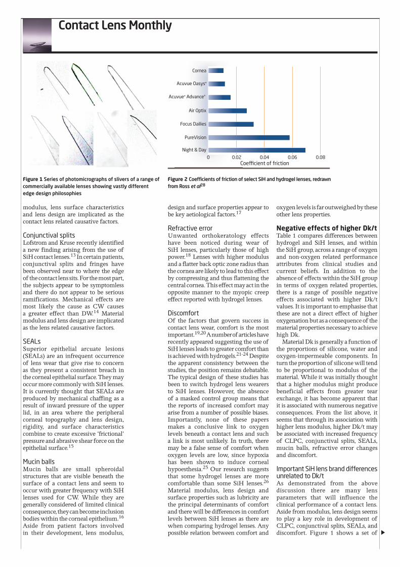
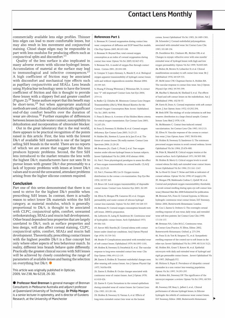
Important SiH lens brand differences unrelated to Dk/t As demonstrated from the above discussion there are many lens parameters that will influence the clinical performance of a contact lens. Aside from modulus, lens design seems to play a key role in development of CLPC, conjunctival splits, SEALs, and discomfort. Figure 1 shows a set of
Figure 1 Series of photomicrographs of slivers of a range of commercially available lenses showing vastly different edge design philosophies
Figure 2 Coefficients of friction of select SiH and hydrogel lenses, redrawn from Ross et al28
Coefficients of friction of selectSiH and hydrogel lenses
0 0.02 0.04Coefficient of friction
0.06 0.08
Cornea
Acuvue Oasys®
Acuvue® Advance®
Air Optix
Focus Dailies
PureVision
Night & Day
Contact Lens Monthly
commercially available lens edge profiles. Thinner lens edges can lead to more comfortable lenses, but may also result in less movement and conjunctival staining. Chisel shape edges may be responsible in part with lens modulus for producing effects such as conjunctival splits and discomfort.13
Quality of the lens surface is also implicated in many adverse events with silicone-hydrogel lenses. Accumulation of material at the surface may lead to immunological and infective consequences.27 A high coefficient of friction may be associated with discomfort and mechanical type effects such as papillary conjunctivitis and SEALs. Lens brands using Hydraclear technology seem to have the lowest coefficient of friction and this is thought to provide these lenses with a slippery feel and greater comfort (Figure 2).28 Some authors report that this benefit may be short-term,29 but when appropriate analytical methods are used, clinically and statistically significant end-of-day comfort benefits over the duration of wear are obvious.30 Further examples of differences between lenses include water content, susceptibility to dehydration and incorporation of ultraviolet blocker.
Out in the great laboratory that is the real world, there appears to be practical recognition of the points raised in this article. First, the lens with the lowest Dk/t of available SiH materials is one of the largest selling SiH brands in the world. There are no reports of which we are aware that suggest that this lens produces hypoxic problems. Second, the first SiH lens introduced to the market remains the lens with the highest Dk/t; manufacturers have not seen fit to pursue lenses with greater Dk/t due presumably to a lack of hypoxic problems with lenses at lower Dk/t values and to avoid the unwanted, attendant problems arising from the higher silicone contents required.
Conclusion Part one of this series demonstrated that there is no need to strive for the highest Dk/t possible when prescribing SiH lenses. In contrast, there is actually reason to select lower Dk materials within the SiH category, as material modulus, which is generally proportional to Dk/t, is thought to be associated with CLPC, conjunctival splits, comfort, unwanted orthokeratology, SEALs and mucin ball development. Other brand dependent lens properties that are largely unrelated to Dk/t, such as surface properties and lens design, will also affect corneal staining, CLPC, conjunctival splits, comfort, SEALs and mucin ball development. Theoretically, prescribing contact lenses with the highest possible Dk/t is a fine concept but only where other aspects of lens behaviour match. In reality, different lens brands behave quite differently. Practically the greatest clinical success with SiH lenses will be achieved by closely considering the range of parameters of available lenses and basing the selection on everything but Dk/t. ●
● Professor Noel Brennan is general manager of Brennan Consultants in Melbourne Australia and adjunct professor at Queensland University of Technology. Dr Philip Morgan is a senior lecturer in optometry, and is director of Eurolens Research, at the University of Manchester
References Part 11. Brennan N. Corneal oxygenation during contact lens
wear: comparison of diffusion and EOP-based flux models.
Clin Exp Optom 2005; 88:103-108.
2. Brennan N. Beyond flux: total corneal oxygen
consumption as an index of corneal oxygenation during
contact lens wear. Optom Vis Sci 2005; 82:467-472.
3. Brennan NA. A model of oxygen flux through contact
lenses. Cornea 2001; 20:104-108.
4. Compan V, Lopez-Alemany A, Riande E, et al. Biological
oxygen apparent transmissibility of hydrogel contact lenses
with and without organosilicon moieties. Biomat 2004;
25:359-365.
5. Huang P, Zwang-Weissman J, Weissman BA. Is contact
lens “T” still important? Contact Lens Ant Eye 2004;
27:9-14.
6. Radke CJ, Chhabra M. Minimum Contact Lens Oxygen
Transmissibility (Dk/L) With Monod Kinetics for the
Corneal Oxygen Consumption Rate. Invest. Ophthalmol.
Vis. Sci. 2005; 46:904-.
7. Fonn D, Bruce A. A review of the Holden-Mertz criteria
for critical oxygen transmission. Eye Contact Lens 2005;
31:247-251.
8. Fonn D, Sweeney D, Holden B, et al. Corneal oxygen
deficiency. Eye Contact Lens 2005; 31:23-27.
9. Holden BA, Stretton S, Lazon de la Jara P, et al. The
future of contact lenses: Dk really matters. Contact Lens
Spectrum 2006; 21:20-28.
10. Bonanno JA, Clark C, Pruitt J, et al. Tear oxygen
tension under silicone-hydrogel lenses in human subjects.
Invest Ophthalmol Vis Sci 2008; 49:E-abstract 4846.
11. Fatt I. New physiological paradigms to assess the effect
of lens oxygen transmissibility on corneal health. CLAOJ
1996; 22:25-29.
12. Fatt I, Freeman RD, Lin D. Oxygen tension
distributions in the cornea: a re-examination. Exp Eye Res
1974; 18:357-365.
13. Bruce AS. Local oxygen transmissibility of disposable
contact lenses. Contact Lens Anterior Eye 2003; 26:189-
196.
14. Efron N, Morgan PB, Cameron ID, et al. Oxygen
permeability and water content of silicone hydrogel
contact lens materials. Optom Vis Sci 2007; 84:328-337.
15. Brennan N. Corneal oxygen consumption beneath toric
soft lenses. 2008:In preparation.
16. Leibowitz H, Laing R, Sandstrom M. Continuous wear
of hydrophilic contact lenses. Arch Ophthalmol 1973;
89:306-310.
17. Sarver MD, Staroba JE. Corneal edema with contact
lenses under closed-eye conditions. AmJ Optom Physiol
Opt 1978; 55:739-743.
18. Binder P. Complications associated with extended wear
of soft contact lenses. Ophthalmol 1979; 86:1093-1101.
19. Holden B, Sweeney D, Swarbrick H, et al. The vascular
response to long-term extended contact lens wear. Clin
Exp Optom 1986; 69:112-119.
20. Zantos S, Holden B. Transient endothelial changes soon
after wearing soft contact lenses. Am J Optom Physiol Opt
1977; 54:856-858.
21. Zantos S, Holden B. Ocular changes associated with
continuous wear of contact lenses. Aust J Optom 1978;
61:418-426.
22. Zantos S. Cystic formations in the corneal epithelium
during extended wear of contact lenses. Int Contact Lens
Clin 1983; 10:128-146.
23. Holden B, Sweeney D, Vannas A, et al. Effects of
long-term extended contact lens wear on the human
cornea. Invest Ophthalmol Vis Sci 1985; 26:1489-1501.
24. Schoessler J. Corneal endothelial polymegathism
associated with extended wear. Int Contact Lens Clin
1983; 10:148-156.
25. Dumbleton KA, Chalmers RL, Richter DB, et al.
Changes in myopic refractive error with nine months’
extended wear of hydrogel lenses with high and low
oxygen permeability. Optom Vis Sci 1999; 76:845-849.
26. Ruben M, Brown N, Lobascher D, et al. Clinical
manifestations secondary to soft contact lens wear. Br J
Ophthalmol 1976; 60:529-531.
27. McM nnies CW, Chapman-Davies A, Holden BA.
The vascular response to contact lens wear. Am J Optom
Physiol Opt 1982; 59:795-799.
28. MacRae S, Matsuda M, Shelland S, et al. The effects
of hard and soft contact lenses on the endothelium. Am J
Ophthalmol 1986; 102:50-57.
29. Farris R, Donn A. Corneal respiration with soft contact
lenses. J Am Optom Assoc 1972; 43:292-294.
30. Roth HW. The etiology of ocular irritation in soft lens
wearers: distribution in a large clinical sample. Contact
Intraoc Lens Med J 1978; 4:38.
31. McMonnies C. Contact lens-induced corneal
vascularization. Int Contact Lens Clin 1983; 10:12-21.
32. Efron N. Vascular response of the cornea to contact
lens wear. J Am Optom Assoc 1987; 58:836-846.
33. Holden B, Sweeney D, Sanderson G. The minimum
precorneal oxygen tension to avoid corneal oedema. Invest
Ophthalmol Vis Sci 1984; 25:476-480.
34. Polse K, Mandell R. Critical oxygen tension at the
corneal surface. Arch Ophthalmol 1971; 84:505-508.
35. Holden B, Mertz G. Critical oxygen levels to avoid
corneal edema for daily and extended wear contact lenses.
Invest Ophthalmol Vis Sci 1984; 25:1161-1167.
36. La Hood D, Grant T. Striae and folds as indicators of
corneal œdema. Optom Vis Sci 1990; 67 (suppl.):196.
37. Morgan PB, Maldonado-Codina C, Quhill W, et al.
Central and peripheral oxygen transmissibility thresholds
to avoid corneal swelling during open eye soft contact lens
wear.J Biomed Mat Res 2008:Submitted for publication.
38. Brennan NA, Fonn D, Corneal hypoxia, in Silicone-
hydrogels: continuous wear contact lenses, D.F. Sweeney,
Editor. 2004, Butterworth-Heinemann: London.
39. Cox I, Zantos S, Orsborn G. The overnight corneal
swelling response of non-wear, daily wear, and extended
wear soft lens patients. Int Contact Lens Clin 1990;
17:134-137.
40. Brennan NA, Coles M-LC, Continuous wear,
in Contact Lens Practice, N. Efron, Editor. 2002,
Butterworrth-Heinemann: Oxford. p. 275-294.
41. Fonn D, du Toit R, Simpson TL, et al. Sympathetic
swelling response of the control eye to soft lenses in the
other eye. Invest Ophthalmol Vis Sci 1999; 40:3116-3121.
42. Holden BA, Grant T, Kotow M, et al. Epithelial
microcysts with daily and extended wear of hydrogel and
rigid gas permeable contact lenses. . Invest Ophthalmol Vis
Sci 1987; 28(Suppl):372.
43. Hickson S, Papas E. Prevalence of idiopathic corneal
anomalies in a non contact lens-wearing population.
Optom Vis Sci 1997; 74:293-297.
44. Holden BA, Sweeney DF. The significance of the
microcyst response: a review. Optom Vis Sci 1991; 68:703-
707.
45. Sweeney DF, Keay L, Jalbert I, et al., Clinical
performance of silicone hydrogel lenses, in Silicone
hydrogels: the rebirth of continuous wear contact lenses,
D.F. Sweeney, Editor. 2000, Butterworth-Heinemann:
This article was originally published in Optician, 2009, Vol 238, No 6218, 26-30.
Contact Lens Monthly
Oxford. p. 90-149.
46. Brennan NA, Coles ML, Connor HR, et al. A 12-month
prospective clinical trial of comfilcon A silicone-hydrogel
contact lenses worn on a 30-day continuous wear basis.
Cont Lens Anterior Eye 2007; 30:108-118.
47. Dumbleton KA, Chalmers RL, Richter DB, et al.
Vascular response to extended wear of hydrogel lenses
with high and low oxygen permeability. Optom Vis Sci
2001; 78:147-151.
48. Brennan NA, Coles M-LC, Levy B, et al. One-Year
Prospective Clinical Trial of balafilcon A (PureVision)
Silicone-hydrogel Contact Lenses Used on a 30-Day
Continuous Wear Schedule. Ophthalmol 2002; 109:1172-
1177.
49. Santodomingo-Rubido J, Wolffsohn JS, Gilmartin B.
Changes in ocular physiology, tear film characteristics, and
symptomatology with 18 months silicone hydrogel contact
lens wear. Optom Vis Sci 2006; 83:73-81.
50. Santodomingo-Rubido J, Wolffsohn JS, Gilmartin B.
Adverse events and discontinuations during 18 months
of silicone hydrogel contact lens wear. Eye Contact Lens
2007; 33:288-292.
51. Papas E. On the relationship between soft contact lens
oxygen transmissibility and induced limbal hyperaemia.
Exp Eye Res 1998; 67:125-131.
52. Brennan NA, Coles ML, Ang JH. An evaluation of
silicone-hydrogel lenses worn on a daily wear basis. Clin
Exp Optom 2006; 89:18-25.
53. Guillon M, Maissa C. The effect of optimising silicone
hydrogels key physical properties on extended wear clinical
performance. Invest Ophthalmol Vis Sci 2006; 49:ARVO
E-abstract 2382.
54. Maldonado-Codina C, Morgan PB, Schnider CM, et
al. Short-term physiological response in neophyte subjects
fitted with hydrogel and silicone-hydrogel contact lenses.
Optom Vis Sci 2004; 81:911-921.
55. Morgan PB, Efron N. Comparative clinical
performance of two silicone hydrogel contact lenses for
continuous wear. Clin Exp Optom 2002; 85:183-192.
56. Papas E, Willcox MDP. Reducing the consequences
of hypoxia: the ocular redness response. Contact Lens
Spectrum 2006; 21 (suppl.):32-37.
57. Inagaki Y, Akahori A, Sugimoto K, et al. Comparison
of corneal endothelial bleb formation and disappearance
processes between rigid gas-permeable and soft contact
lenses in three classes of dk/l. Eye Contact Lens 2003;
29:234-237.
58. Brennan NA, Coles M-LC, Connor RM, et al. Short-
term corneal endothelial response to wear of silicone-
hydrogel contact lenses in east asian eyes. Eye Contact Lens
2008; 34:317-321.
59. Chang SW, Hu FR, Lin LL. Effects of contact lenses
on corneal endothelium -a morphological and functional
study. Ophthalmologica 2001; 215:197-203.
60. Yamauchi K, Hirst LW, Enger C, et al. Specular
microscopy of hard contact lens wearers II.
Ophthalmology 1989; 96:1176-1179.
61. Carlson KH, Bourne WM. Endothelial morphologic
features and function after long-term extended wear of
contact lenses. Arch Ophthalmol 1988; 106:1677-1679.
62. Stocker EG, Schoessler JP. Corneal endothelial
polymegathism induced by PMMA contact lens wear.
Invest Ophthalmol Vis Sci 1985; 26:857-863.
63. Schoessler JP, Barr JT, Freson DR. Corneal endothelial
observations of silicone elastomer contact lens wearers. Int
Contact Lens Clin 1984; 11:337.
64. Liu Z, Pflugfelder SC. The effects of long-term contact
lens wear on corneal thickness, curvature, and surface
regularity. Ophthalmology 2000; 107:105-111.
65. Myrowitz EH, Melia M, O’Brien TP. The relationship
between long-term contact lens wear and corneal
thickness. Clao J 2002; 28:217-220.
66. Alharbi A, Swarbrick HA. The effects of overnight
orthokeratology lens wear on corneal thickness. Invest
Ophthalmol Vis Sci 2003; 44:2518-2523.
67. Binder P. Myopic extended wear with the Hydrocurve
II soft contact lens. Ophthalmol 1983; 90:623-626.
68. Harris MG, Sarver MD, Polse KA. Corneal curvature
and refractive error changes associated with wearing
hydrogel contact lenses. Am J Optom Physiol Opt 1975;
52:313-319.
69. Fonn D, MacDonald KE, Richter D, et al. The ocular
response to extended wear of a high Dk silicone hydrogel
contact lens. Clin Exp Optom 2002; 85:176-182.
70. Patel S. Changes in myopic refractive error with nine
months’ extended wear of hydrogel lenses with high and
low oxygen permeability. Optom Vis Sci 2000; 77:285.
71. Smith EL, Greeman P, Ho A, et al. Methods and
apparatuses for altering relative curvature of field and
positions of peripheral off-axis focal positions. US patent
7025460 2006.
72. Szczotka-Flynn LB. Unintended Ortho-k Effects From
Silicone Hydrogel Lenses. Contact Lens Spectrum 2004;
19(8):19-20.
References Part 2 1. Morgan PB, Efron N, Hill EA, et al. Incidence of
keratitis of varying severity among contact lens wearers.
Br J Ophthalmol 2005; 89:430-436.
2. Schein OD, McNally JJ, Katz J, et al. The incidence of
microbial keratitis among wearers of a 30-day silicone
hydrogel extended-wear contact lens. Ophthalmology
2005; 112:2172-217
3. Stapleton F, Keay L, Edwards K, et al. The Incidence
of contact lens-related microbial keratitis in Australia.
Ophthalmology 2008; 115:1655-1662.
4. Dart JK, Radford CF, Minassian D, et al. Risk factors
for microbial keratitis with contemporary contact lenses: a
case-control study. Ophthalmology 2008; 115:1647-1654.
5. Morgan PB, Efron N, Brennan NA, et al. Risk factors for
the development of corneal infiltrative events associated
with contact lens wear. Invest Ophthalmol Vis Sci 2005;
46:31363143.
6. Stapleton F, Keay LJ, Sanfilippo PG, et al. Relationship
between climate, disease severity, and causative organism
for contact lens-associated microbial keratitis in Australia.
AmJ Ophthalmol 2007; 144:690-698.
7. Keay L, Edwards K, Naduvilath T, et al. Factors affecting
the morbidity of contact lens-related microbial keratitis:
a population study. Invest Ophthalmol Vis Sci 2006;
47:4302-4308.
8. Szczotka-Flynn L, Diaz M. Risk of corneal inflammatory
events with silicone hydrogel and low dk hydrogel
extended contact lens wear: a meta-analysis. Optom Vis Sci
2007; 84:247-256.
9. Andrasko G, Ryen K. Corneal staining and comfort
observed with traditional and silicone hydrogel lenses and
multipurpose solution combinations. Optometry 2008;
79:444-454.
10. O’Leary DJ, Wilson G, Henson DB. The effect of
anoxia on the human corneal epithelium. Am J Optom
Physiol Opt 1981; 58:472-476.
11. Skotnitsky CC, Naduvilath TJ, Sweeney DF, et al.
Two presentations of contact lens-induced papillary
conjunctivitis (CLPC) in hydrogel lens wear: local and
general. Optom Vis Sci 2006; 83:27-36.
12. Spring TF. Reaction to hydrophilic lenses. Med J Aust
1974; 1:449-450.
13. Lofstrom T, Kruse A. A Conjunctival Response to
Silicone Hydrogel Lens Wear: A new finding reveals
how silicone hydrogel lenses may affect the conjunctival
epithelium. Contact Lens Spectrum 2005; 20(9):42-45.
14. Santodomingo-Rubido J, Wolffsohn J, Gilmartin B.
Conjunctival epithelial flaps with 18 months of silicone
hydrogel contact lens wear. Eye Contact Lens 2008;
34:35-38.
15. Holden BA, Stephenson A, Stretton S, et al. Superior
epithelial arcuate lesions with soft contact lens wear.
Optom Vis Sci 2001; 78:9-12.
16. Pritchard N, Jones L, Dumbleton K, et al. Epithelial
inclusions in association with mucin ball development in
high-oxygen permeability hydrogel lenses. Optom Vis Sci
2000; 77:68-72.
17. Tan J, Keay L, Jalbert I, et al. Mucin balls with wear of
conventional and silicone hydrogel contact lenses. Optom
Vis Sci 2003; 80:291-297.
18. Szczotka-Flynn LB. Unintended Ortho-k Effects From
Silicone Hydrogel Lenses. Contact Lens Spectrum 2004;
19(8):19-20.
19. Pritchard N, Fonn D, Brazeau D. Discontinuation of
contact lens wear: a survey. Int Contact Lens Clin 1999;
26:157-162.
20. Young G, Veys J, Pritchard N, et al. A multi-centre
study of lapsed contact lens wearers. Ophthalmic Physiol
Opt 2002; 22:516-527.
21. Dillehay SM, Miller MB. Performance of Lotrafilcon
B silicone hydrogel contact lenses in experienced low-Dk/t
daily lens wearers. Eye Contact Lens 2007; 33:272-277.
22. Dumbleton K, Keir N, Moezzi A, et al. Objective and
subjective responses in patients refitted to daily-wear
silicone hydrogel contact lenses. Optom Vis Sci 2006;
83:758-768.
23. Long B, McNally J. The clinical performance of
a silicone hydrogel lens for daily wear in an Asian
population. Eye Contact Lens 2006; 32:65-71.
24. Riley C, Young G, Chalmers R. Prevalence of ocular
surface symptoms, signs, and uncomfortable hours of wear
in contact lens wearers: the effect of refitting with daily-
wear silicone hydrogel lenses (senofilcon a). Eye Contact
Lens 2006; 32:281-286.
25. Millodot M, O’Leary DJ. Effect of oxygen deprivation
on corneal sensitivity. Acta Ophthalmol (Copenh) 1980;
58:434-439.
26. Brennan NA, Coles M-LC, Connor HR, et al. Silicone
hydrogel contact lens comfort. Optom Vis Sci 2007;
84:e-abstract 070037.
27. Brennan NA, Coles M-LC. Deposits and
symptomatology with soft contact lens wear. Int Contact
Lens Clin 2000; 27:75-100.
28. Ross G, Nasso M, Franklin V, et al. Silicone hydrogels:
Trends in products and properties. Ophthalmic Res 2005;
2005:27&162.
29. Dumbleton KA, Woods CA, Jones LW, et al. Comfort
and adaptation to silicone hydrogel lenses for daily wear.
Eye Contact Lens 2008; 34:215-223.
30. Brennan NA, Coles M-LC, Connor HRM, et al.
Silicone hydrgel contact lens comfort. Optom Vis Sci 2007;
84:E-abstract 070037.
Reprinted from | Optician | 2009