clinical practice guidelines for physical therapy in ... · pdf file1 kngf guidelines for...
TRANSCRIPT

1
KNGF guidelines for physical therapy in patients with chronic ankle pain
V-10/2003/US
IntroductionThese guidelines, which have been produced by the
Royal Dutch Society for Physical Therapy (KNGF),
describe the diagnostic and therapeutic processes
involved in providing physical therapy
(physiotherapy) for patients with chronic ankle
sprains. The choices made in deriving the
recommended diagnostic and therapeutic procedures
are explained in the discussion section of the
document, entitled “Review of the evidence”. The
abbreviations and key concepts used are explained in
an attached list of abbreviations and glossary.
For whom are these guidelines intended?
These guidelines are primarily directed at physical
therapists (physiotherapists) who may have to treat
patients with long-lasting ankle complaints that
result from inversion injury to the ankle. The
physical therapist involved is expected to have
knowledge about the various stages of the process of
recovery from a chronic ankle sprain and to have the
skills needed to distinguish between a correct and an
incorrect walking pattern and to apply the principles
of appropriate progressive training programs. To aid
the healing process, the possession of specific skills,
in for example the use of bandages, tapes and braces,
is mandatory. Proper use of these guidelines requires
the therapist’s treatment area, or practice, to be large
enough to allow proper analyses of gait to be made
and to enable indoor sports and training activities to
be performed.
Chronic ankle sprain versus acute ankle sprain
These guidelines on chronic ankle sprain can be seen
as a logical sequel to KNGF guidelines on acute ankle
sprain, which describe the diagnostic and therapeutic
processes involved in providing physical therapy for
patients with acute lateral ankle sprains. Recovery
from an acute ankle sprain to being able to perform
normal daily activities usually takes about six weeks.
The present guidelines describe the treatment of long
lasting ankle complaints. In particular, they
concentrate on functional instability in chronic ankle
complaints. Functional instability is defined as the
persistence of residual complaints, such as a feeling
that the ankle is “giving way” or a “(a feeling of)
recurrent spraining”, or both, after an ankle sprain.
Usually, pain and swelling are absent, but, when they
do occur, they are the consequences of overuse or of a
new inversion trauma. Functional instability can lead
to behavioral adaptations, such as an aberrant
Clinical practice guidelines for physical therapy
in patients with chronic ankle sprain
RA de Bie PT PhDI, MAMB Heemskerk PTII, AF Lenssen PT MScIII, SR van Moorsel PTIV, G Rondhuis PTV,
DJ Stomp PT MScVI, RAHM Swinkels PT MScVII, HJM Hendriks PT PhDVIII
I RA de Bie PT PhD, associate professor of clinical epidemiology and chair, Department of Epidemiology, Maastricht University, Maastricht,
the Netherlands.
II MAMB Heemskerk PT, physical therapist and project staff member, Department of Research and Development, Dutch Institute of Allied
Health Professions, Amersfoort, the Netherlands.
III AF Lenssen PT MSc, hospital-based physical therapist, research coordinator and human movement scientist, Department of Physiotherapy,
University Hospital Maastricht, Maastricht, and lecturer, Faculty of Physiotherapy, Hogeschool Zuid, Maastricht, the Netherlands.
IV SR van Moorsel PT, hospital-based physical therapist, Department of Physiotherapy, University Medical Center, Catholic University of
Nijmegen (KUN), Nijmegen, the Netherlands.
V G Rondhuis PT, private-practice physical therapist, Hilversum, the Netherlands.
VI DJ Stomp PT MSc, physical therapist, human movement scientist and project staff member, Department of Research and Development, Dutch
Institute of Allied Health Professions, Amersfoort, the Netherlands.
VII RAHM Swinkels PT MSc, private-practice physical therapist and human movement scientist, physiotherapy and manual therapy practice ‘de
Coevering’, Geldrop, the Netherlands.
VIII HJM Hendriks PT PhD, physical therapist, clinical epidemiologist and guidelines coordinator, Department of Research and Development,
Dutch Institute of Allied Health Professions, Amersfoort, and Department of Epidemiology, Maastricht University, Maastricht the Netherlands.

walking pattern or the avoidance of normal daily
activities, or to problems with physical activities at
work or with participating in sport at the desired
level.
Factors involved in the occurrence or persistence of
functional instability are thought to include:
mechanical instability (i.e., loose capsular ligaments
in the ankle), disturbed proprioception, reduced
muscle strength, slow muscle reaction times, reduced
mobility, and inappropriate complaint-related
behavior. There are also indications that the
inadequate or incomplete treatment of an acute ankle
sprain may increase the chance of residual
complaints.
Other conditions that may lead to chronic ankle
complaints but are not included in the definition of
functional instability are: impingement due to
osteochondral lesions, osteophytes or ‘loose bodies’,
osteoarthrosis and systemic diseases, rupture of the
distal tibiofibular syndesmosis complex, sinus tarsi
syndromes, and subtalar instability. These guidelines
offer a brief description of these conditions for the
purpose of differential diagnosis (see the section on
differential diagnosis in the review of the evidence).
The treatment strategies usually adopted in these
conditions are also mentioned.
Epidemiology
In the Netherlands, about 600,000 people sprain their
ankles each year. About half visit a general
practitioner or hospital emergency department. Some
75% of sprained ankles are inversion injuries.
Irrespective of the severity of the initial trauma and
of the type of treatment given, a considerable number
of persons with acute lateral ankle sprains experience
residual complaints. Literature estimates indicate that
the prevalence varies between 10% and 60%, whereas
that for functional instability lies between 10% and
40%.
Diagnosis
During diagnosis, the physical therapist assesses
impairments in the patient’s body structures and in
physiological and psychological functioning, and the
extent to which the patient’s activities and
participation in normal life are limited. A proper
analysis of the health problem should enable
conclusions to be drawn about its extent and severity
and about possible ways of modifying it. Thereafter, a
treatment plan is devised in co-operation with the
patient. The starting point is the patient’s description
of the health problem.
Referral
Referral by a general practitioner or medical specialist
is required before these guidelines can be
implemented. This is mandatory in the Netherlands
because, in the country, patients do not have open
access to a physical therapist. They must be referred
by a general practitioner or another physician. The
referral documentation should include data that
indicate the existence of residual complaints after an
acute ankle sprain. Additional relevant medical data
should also be incorporated into the referral
documentation.
History-taking
History-taking should include details of:
• demographic factors;
• the health problem as described by the patient,
treatment goals, and expectations of treatment.
History of the disorder
Causal factors:
• Did the ankle suffer an inversion trauma?
- When did the first inversion trauma occur?
- How did the trauma occur?
• Is the current condition the result of a relapse?
- When did the most recent inversion trauma
take place?
Course of the complaint over time:
• Which medical and therapeutic interventions
were employed and what were the results?
• How did the pain and swelling develop over time?
• When was it possible to return to normal daily
activities, work and sporting activities?
Assessment of current condition
• Investigate impairments in the patient’s body
structures and functions, activity limitations, and
participation restrictions:
- body structures (e.g., possible damage to
ligaments);
- Is the patient in pain at present, during or
2
KNGF guidelines for physical therapy in patients with chronic ankle pain
V-10/2003/US

after exercise, or when resting?
- body functions (e.g., coordination,
proprioception and kinesthesia);
- Does the patient report a feeling that the
ankle is giving way or that he or she is
actually spraining the ankle?
- If so, how many times per day or week does
this feeling occur, and when does it occur:
during normal daily living activities, during
exercise, or when tired?
- If so, does symptom severity increase?
- If so, which symptoms occur and how long
do they last?
- Activity limitations (e.g., in performing
specific tasks or activities related to work,
housekeeping, sport or leisure pursuits);
- Participation restrictions (e.g., in activities in
and around the house, at work, in
housekeeping, or in sports at a level that was
normal before the initial trauma);
- If there are problems, what are they?
• Assess use of external support;
- Does the patient still use tape or a brace to
provide support?
- When, why and how often?
• Assess personal factors;
- How does the patient behave in response to
the condition?
- Is there a balance between the patient’s actual
load-bearing capacity and that required for
normal daily living activities and work? Is the
required capacity level attainable?
Physical examination
Observation:
• pain and its localization;
• the amount of swelling and the reason for the
swelling;
• static posture and any postural abnormalities.
Palpation (only required for the purpose of
differential diagnosis):
• look for tender spots and signs of inflammation.
Assessment of functioning:
• ask the patient to perform load-bearing
movements and pay special attention to dorsal
flexion of the ankle;
• Is full load-bearing by the foot possible? Does any
pain, giving way, or fear of movement occur
during full load-bearing?
• Is the gait pattern normal (use the recommended
gait analysis technique)?
• Is the patient able to stand on the affected leg
with eyes open and closed?
• Is the patient able to jump on the affected leg,
and subsequently stand still on the affected leg?
• Is the patient able to walk on toes and heels?
• Is the patient able to perform twin tasks during
complex load-bearing activities?
Recommended measuring instruments
These guidelines recommend the use of two
measuring instruments for assessing and evaluating
the patient’s functional status:
• An instrument for patient-specific complaints is
used to assess the patient’s functional status. In
practice, the patient is asked to select three daily
activities that he or she considers important and
that are unavoidable, such as walking, climbing
stairs or running. The difficulties the patient has
in executing these activities is subsequently scored
on three separate visual analogue scales (VAS).
• The Nijmegen gait analysis scale (GALN) is used to
assess and describe the patient’s gait. It comprises
13 items, each of which refers to an aspect of the
patient’s manner of walking and involves the
evaluation of different anatomical structures (e.g.,
the trunk, pelvis, knee and ankle).
Analysis
The central goal of the diagnostic process is to
determine whether physical therapy is justified and
necessary. Physical therapy should be able to
influence the factors that contribute to the chronic
ankle condition. The relevant factors should be
classified according to whether they have a positive
or negative influence on the chronic nature of the
condition. Because the condition is chronic, the
physical therapist must describe the relevant
impairments in body function or structure, the
resulting disabilities and participation problems, and
their interrelationship. When there is no relationship
between the relevant impairments, disabilities and
participation problems, additional attention should
be paid to:
• personal factors;
• external factors, such as a return to work; and
3
KNGF guidelines for physical therapy in patients with chronic ankle pain
V-10/2003/US

• patient compliance.
Conclusions
• The physical therapist must determine that
physical therapy is justified.
• The physical therapist must determine whether
functional instability is present.
- Patients with long-duration functional
instability who have recently suffered tissue
damage should initially be treated according to
the guidelines on acute ankle sprain. When
the patient is able to bear full weight on the
foot and able to flex the foot normally, and the
recent swelling has diminished, the procedures
in these guidelines on chronic ankle sprain can
be followed.
- When there is functional instability without
new tissue damage, the guidelines on chronic
ankle sprain should be applied.
• If there is a concomitant complaint related to a
condition specified in the table on differential
diagnosis (see the section on differential diagnosis
in the review of the evidence), the guidelines on
chronic ankle sprain should be applied.
After considering the above-mentioned points
together with the patient, treatment goals and a
treatment plan can be formulated.
Therapy
Throughout therapy, the patient’s description of the
health problem is of central importance. The target
performance level to be achieved at the end of
treatment must be consistent with the patient’s
individual requirements.
Physical therapy treatment goals:
• to achieve optimal functional recovery in terms of
the patient’s functions and skills, with a return to
the highest achievable or desired level of activities
and participation; and
• to prevent relapses, exacerbations and further
dysfunction.
Treatment subgoals:
• to optimize load-bearing and load-carrying
capacity;
• to achieve a normal dynamic gait; and
• to achieve active stability,
by improving relevant functions (e.g., coordination
and balance, strength and endurance).
Structure of physical therapy
Primarily, therapy focuses on the return to a normal
mode of walking and to unperturbed functioning of
the ankle during normal daily activities. When this
has been achieved, therapy could be aimed at
attaining a higher level of daily activity, which may
include heavy work, physically demanding hobbies
or participation in sport.
Throughout therapy, a stepwise approach that is
tuned to the increasing load-bearing capacity of the
patient is recommended. Therapy can be intensified
by increasing the level of difficulty of general
exercises and load-bearing exercises, and by
increasing the speed, duration and dynamic quality
of practiced movements. At a later stage, training of
specific skills, such as heavy lifting, climbing stairs,
running or jumping, can be carried out. The exercises
and training given should be appropriate for the
specific demands being made on the ankle. For
example, if the patient wants to participate in a
particular sporting activity, training of all aspects of
the activity in question should be carried out. This
involves analyzing both the demands made by the
sport and the relevant characteristics of the person
participating in it (i.e., the patient). The exercises
should gradually be increased to the level the patient
wants to achieve.
Content of physical therapy
The physical therapist:
1. gives information and advice;
2. administers the use of tape, bandages or braces, if
necessary; and
3. provides exercise for specific functions and skills.
Giving information and advice
• inform the patient about the expected rate of
recovery. If incomplete recovery is expected, this
should be discussed with the patient. Goals that
are achievable, in terms of the patient’s functions
and activities, should be set together with the
patient. The achievement of subgoals should, as
far as possible, follow a time-contingent strategy.
• instruct the patient how to adjust the load
4
KNGF guidelines for physical therapy in patients with chronic ankle pain
V-10/2003/US

imposed by normal daily activities to the load-
bearing capacity of the ankle and how to increase
load-bearing capacity gradually over time. It
should be explained that symptoms or signs such
as pain, swelling, stiffness and loss of function can
indicate, perhaps temporary, overloading.
• suggest, if necessary, the temporary use of tape,
bandages or a brace to alleviate any symptoms
occurring during causative movements or when
returning to, perhaps heavy, work.
• point out the significance of exercising at home
and stress the importance of adopting a correct
manner of walking and a good stance during the
performance of normal daily life activities.
Use of tape, braces or bandages
• if the patient indicates that the ankle is being, or
feels as though it is being, repeatedly sprained,
tape, a brace or bandages can be used during
therapy.
• the use of tape, bandages or a brace is advisable if
the patient returns to, perhaps heavy, work or to
sporting activities. When good muscular stability
has been achieved and functional exercises can be
performed satisfactorily, it is advisable to reduce
the use of tape, bandages and braces.
Exercising functions and skills
• a symmetrical and dynamic gait should be
strongly encouraged to prevent the patient from
causing the condition to persist;
• all relevant daily life activities should be exercised,
such as standing up, sitting, and using stairs.
Training coordination and balance
• static balance exercises should have an increasing
level of difficulty (e.g., the eyes could be open or
closed, the size of the supporting platform could
be varied, static and moving surfaces could be
used, a wobble board could be used, and external
factors could be applied to disturb balance);
• dynamic balance should be exercised (e.g., single
tasks or functional exercises with twin tasks on
different types of surface). Finally, mental tasks
could be incorporated during balance tasks (e.g.,
calculations);
• simple taping could be used to heighten
proprioceptive responses in the ankle while the
patient is standing still or moving.
Training strength and endurance
• the strength and endurance exercises given should
also be incorporated into the patient’s normal
daily activities.
Increasing the range of motion
• any increase in the active or passive range of
motion of the ankle should immediately be
followed by proprioception training and stability
exercises to reinforce the new increase.
Evaluation
In assessing whether the patient is able to perform
activities that require an increased load-bearing
capacity, the physical therapist can use the
recommended measurement instruments (i.e., the
measuring instrument for patient-specific complaints
and the Nijmegen gait analysis scale) and can directly
assess body functions and impairments in body
functions. The presence of pain, swelling or a
decrease in movement quality after either performing
exercises or increasing the level of normal daily
activities indicate that the load was too great.
Periodic evaluation of treatment results should take
place after three and six weeks, and possibly also after
nine and 12 weeks, depending on the duration of
therapy. During each evaluation, the patient’s
progress should be compared with a baseline
measurement or with the results of earlier
evaluations. Progress can be either subjective (e.g.,
assessed in terms of changes in the severity of
symptoms reported by the patient, such as pain, the
feeling that the ankle is giving way, and the ease of
performing normal daily activities) or objective (e.g.,
assessed in terms of gait, muscle strength,
coordination, endurance and load-bearing capacity).
After six weeks of therapy, benefits should be
demonstrable. If no improvement is registered, the
physiotherapist (physical therapist) should contact
the referring physician.
Relapses prevention
To reduce the chance of a recurrent ankle sprain, or
relapse, the following advice should be followed:
5
KNGF guidelines for physical therapy in patients with chronic ankle pain
V-10/2003/US

6
KNGF guidelines for physical therapy in patients with chronic ankle pain
V-10/2003/US
• do not use taping or a brace as a standard
precaution during training or regular sporting
activities. Reserve the use of these techniques for
competition sports and for high-risk sports such as
contact or indoor sports. Not only does this
reduce the chance of a new injury occurring, but
it also reduces the extent of the damage should a
relapse occur. A sports brace is preferable to
taping;
• advise the patient to buy new sports shoes if the
old ones are worn out. No specific advice can be
given concerning the use of high-top or low-top
footwear;
• advise and instruct the patient, after finishing
therapy, to pay attention to sport-specific as well
as proprioception training;
• give instruction on a program of home exercises.
Concluding treatment and reporting
If high demands are to be placed on the ankle, for
example during professional sporting activities,
treatment can continue until the desired load-bearing
level has been reached. These demands may be very
specific and may, therefore, mean that the therapist
has to have special skills.
At the end of treatment, the referring physician
should receive a written report detailing the
diagnosis, treatment goals, treatment results, and the
advice and instruction given to the patient. For
details, see the KNGF guidelines on communicating
with and reporting back to general practitioners. To
ensure good communication between general
practitioner and physical therapist, guiding principles
are specified on five elements of communication:
indication setting, consultation, referral letters,
contact during treatment, and reporting.

IntroductionDefinition of KNGF guidelines
Guidelines produced by the Royal Dutch Society for
Physical Therapy (KNGF) are defined as “guidelines
whose production is directed by a central body, that
are developed systematically, that are written by
experts, and that deal with the systematic process of
physical therapy in certain health problems and with
various (organizational) aspects of the profession”.1,2
Goals of KNGF guidelines
The general goals of KNGF guidelines can be divided
into two areas.1–3 Firstly, there are goals that are
relevant to individual physical therapists and,
secondly, there are goals that are relevant to the
physical therapy profession as a whole. It is
important to note that these guidelines are intended
for exclusive use within the physical therapy
profession. The KNGF uses guidelines as instruments
for ensuring quality control within the profession
and for improving the quality of care offered by the
profession.
For individual physical therapists, the relevant
guideline goals are:
• to support decision-making;
• to provide a point of reference for education and
orientation;
• to provide criteria for self-evaluation and peer-
group assessment; and
• to guide future developments in the desired
direction.
For the physical therapy profession as a whole, the
relevant guideline goals are:
• to ensure that evidence-based care is available,
and to distinguish between conclusions derived
from scientific research and conclusions based on
expert consensus; and
• to increase the uniformity of care and, thus,
improve the quality of care.
Research shows that there are large variations in the
treatment goals, the interventions employed in, and
the overall use of physical therapy.4 Therefore, these
guidelines also set out:
• to alter the care provided in a way that takes
scientific research into account (i.e., evidence-
based care); and
• to delineate the tasks and responsibilities of
professional bodies, to provide some insight into
the areas of concern of professional bodies, and to
stimulate cooperation between practitioners of
different healthcare disciplines.
The KNGF guidelines on chronic ankle sprain
encapsulate a methodical approach to the diagnostic
and therapeutic processes involved in providing
physical therapy for patients with chronic ankle
complaints. They concentrate on the concept of
functional instability, which is defined as the
persistence of residual complaints, such as a feeling
that the ankle is “giving way” or a “(a feeling of)
relapsed spraining”, or both, after an ankle sprain.
Other chronic ankle complaints are briefly described
for the purpose of differential diagnosis.
The goals of physical therapy, which must take into
account the individual patient’s needs and desires, are
to guarantee optimal functional recovery and to
prevent relapses and exacerbations. To achieve this
goal, the patient has to learn to balance actual load-
bearing with load-bearing capacity. The recovery of a
normal dynamic gait and the return to active stability
are also prime targets of therapy. The physical
therapy interventions used to achieve these goals are
the provision of advice, the provision of exercises for
specific functions and skills, and the provision of
support for the healing process through the
administration of tape, braces and bandages.
The guidelines describe physical therapy
interventions in patients with long-lasting or chronic
ankle complaints. Usually, patients who suffer a first
ankle sprain or a relapse should be treated in
accordance with the guidelines on acute ankle sprain.
The chronic ankle sprain guidelines should never be
used to extend treatment beyond that recommended
by the acute ankle sprain guidelines. The chronic
ankle sprain guidelines are intended for a different
7
KNGF guidelines for physical therapy in patients with chronic ankle pain
V-10/2003/US
Review of the evidence

category of patients.
The treatment recommended by the chronic ankle
sprain guidelines differs in a number of crucial ways
from that recommended by the acute ankle sprain
guidelines:
1. When the healing that took place after the initial
acute ankle sprain did not lead to complete
functional recovery, the chronic ankle sprain
guidelines should be used. Analysis of the
patient’s health problem becomes more complex.
It becomes necessary to investigate why the
patient did not fully recover, and to determine
which factors are hindering complete recovery
and whether those factors can be influenced by
physical therapy.
2. When a complaint is long-lasting, specific factors
associated with chronic disease processes start to
emerge. There may be a loss of strength,
coordination and general endurance, which may
have negative effects on the patient’s levels of
activity and social participation. The recovery
process and the need for physical therapy will last
longer.
The present guidelines take into account the
intervention strategies used for treating ankle
complaints occurring as residual complaints that were
described in the KNGF guidelines on acute ankle
sprain5 and in the (Dutch) Collaborating Center for
Quality Assurance in Healthcare (CBO) consensus
document entitled “Diagnosis and treatment of acute
ankle sprain”.6 They also link up with the Dutch
College of General Practitioners (NHG) publication on
standards entitled “Ankle distortion”,7 in which a
referral for physical therapy is mentioned as a first
line of treatment for residual complaints such as a
feeling that the ankle is “giving way”, a “(a feeling of)
relapsed spraining” or a loss of muscle strength, or
some combination of these complaints. Referral to a
surgeon is considered a secondary option.
Clinical considerations
The chronic ankle sprain guidelines working group
was interested in answering the following clinical
questions:
Extent of the problem:
• How many patients suffer from residual
complaints after an acute ankle sprain?
• How many patients with these residual
complaints also suffer from functional instability?
Diagnosis:
• Which elements of the physical therapy
diagnostic approach are reliable, valid and useful
in daily practice?
• Which data derived from the physical therapy
diagnostic process are essential for defining
treatment goals, for devising a treatment plan,
and for making a prognosis?
Therapy:
• Which interventions and elements of advice are
most useful, as indicated by evidence-based
research or the views of the working group?
Working group composition and methodology
In August 2001, a multidisciplinary working group of
experts was formed to answer the above clinical
questions. The core members of the working group
comprised physical therapists and researchers with
the desired type of expertise who had been part of the
core group working on the acute ankle sprain
guidelines and who were willing to participate in
developing these guidelines. In determining the
composition of the chronic ankle sprain guidelines
working group, an attempt was made to strike a
balance between members with practical experience,
subject experts, and individuals with a scientific
background. All members of working group declared
in advance that there were no conflicts of interest
that would influence their involvement in the
development of these guidelines. Development took
place between August 2001 and February 2002.
These guidelines were developed in accordance with a
published method for the development and
implementation of physical therapy guidelines.3 This
publication gives practical information on finding
appropriate literature, which details search terms,
sources of information, and the time frame within
which retrieved literature should have been
published. In addition, inclusion and exclusion
criteria applicable to retrieved material are specified,
as is the level of scientific evidence necessary for
making recommendations. In the absence of
scientific evidence, recommendations can be
formulated on the basis of consensus statements from
8
KNGF guidelines for physical therapy in patients with chronic ankle pain
V-10/2003/US

9
KNGF guidelines for physical therapy in patients with chronic ankle pain
V-10/2003/US
core members of the working group and external
experts. The consensus reached should preferably be
unanimous.
Administrative staff carried out scientific literature
selection and the entire working group discussed
critical appraisal and the results. The scientific
evidence and its strength were summarized for each
intervention. Evidence was summarized and graded
systematically by an evidence-based guidelines
discussion group (Evidence Based Richtlijnen Overleg
list), which was formed under the auspices of the
(Dutch) Collaborating Center for Quality Assurance
in Healthcare (CBO). In addition to good scientific
evidence, other factors were also considered
important in establishing recommendations for the
physical therapy of chronic ankle sprains: reaching a
general consensus, cost-effectiveness, the availability
of resources, the availability of the necessary expertise
and educational facilities, organizational factors, and
consistency with other monodisciplinary or
multidisciplinary guidelines.
After a draft of the guidelines was drawn up, it was
sent to a panel of experts and to professional
organizations. The purpose was to reach a consensus,
to achieve consistency with the views of other
professional bodies and organizations, and to achieve
consistency with other monodisciplinary or
multidisciplinary guidelines, such as the Dutch
College of General Practitioners (NHG) publication on
standards entitled “Ankle distortion”7 and the CBO
consensus document entitled “Diagnosis and
treatment of acute ankle sprain”.6
Guideline appraisal by practicing physical
therapists
Before the guidelines were published and
implemented, they were systematically appraised by
their intended users in a validation process.
Consequently, the chronic ankle sprain guidelines
have been evaluated by physical therapists working
in different healthcare settings. Information on the
guidelines was collected by questionnaire, which was
structured using defined quality criteria for guidelines
being developed by central bodies. Any comments or
criticisms made by participating physical therapists
were recorded and discussed by the guidelines
working group. Where necessary, the guidelines were
adapted. The final recommendations on physical
therapy practice were based on a combination of the
scientific evidence available, the other important
factors listed above, and the results of appraisal by
intended users.
Composition and implementation of the
guidelines
The guidelines comprise three parts: the practice
guidelines themselves, a schematic summary of the
most important points in the guidelines, and a review
of the evidence. This division was chosen for practical
and educational reasons. All three parts can be read
separately. In addition to the guidelines, an expert
guide has also been developed and published to
promote implementation in clinical practice.
Guideline implementation should be carried out in
accordance with the recommendations of the
published implementation strategy.3,8
A novel aspect of guideline use is access through the
internet. There is a chronic ankle sprain guidelines
internet site that enables physical therapists to
consult the guidelines online, to update his or her
knowledge about theoretical aspects of the guidelines,
and to search for related topics on the internet. An
important aspect of the internet site is that it
provides the opportunity to ‘chat’ interactively with
colleagues. In this way, physical therapists can discuss
the guidelines and help each other implement and
integrate them into their daily practice. This internet
forum is moderated by the working group.
This review of the evidence explains how the
recommendations made in the chronic ankle sprain
guidelines were derived. Wherever possible, the
guidelines are evidence-based. Where there is
insufficient or no scientific evidence, the
recommendations made are based on the experience
and views of working group experts. Information on
the epidemiology, etiology and diagnosis of chronic
ankle sprain was obtained by a systematic search of
the scientific literature. The following databases were
searched: MEDLINE (from 1980–2001), CINAHL (from
1980–2001), the Cochrane Library rehabilitation and
related therapies field database (from 1990–2001,
accessed at Maastricht University in the Netherlands),

and the DocOnline database of the Documentation
Center at the Dutch Paramedical Institute in
Amersfoort, the Netherlands (from 1990–2001). The
keywords used were: physiotherapy, physical therapy,
ankle, inversion, chronic, instability, stability,
diagnosis, prognosis, treatment, prevention,
measurement instruments, guidelines, prevalence,
incidence, randomized clinical trial, randomized
controlled trial, meta-analysis, review, and systematic
review. Reference tracking was used to obtain
additional material. The articles used were written in
English, German or Dutch.
Epidemiology
In the Netherlands, about 600,000 people sprain their
ankles each year, half during sporting activities.
About 300,000 visit a general practitioner or a
hospital emergency department. Some 75% of
sprained ankles are inversion injuries.9 Irrespective of
the severity of the initial trauma and of the treatment
given, whether it involves an operation, use of a
plaster cast or functional treatment, a considerable
number of persons with acute lateral ankle sprains
experience residual complaints.10 The reported
percentage varies between 10% and 60%.11–15 The
differences in prevalence rate can be attributed to
variations in the nature of the residual complaints
observed. In addition, differences in follow-up time
also play a role in the reported prevalence rate
variation.10,11 The prevalence of functional instability
is also variable: one year after the initial sprain,
10–40% of patients report a feeling that the ankle is
“giving way”, instability, or “inversion of the
ankle”.11,12,15,16 Two to five years after the initial
trauma, an increasing number (27–45%) mention “a
feeling of giving way” and “inversion of the ankle”.
Delineating the health problem
The flow chart in Figure 1 shows the decision
diagram used in deciding whether an ankle
complaint should be treated according to these
guidelines.
Etiology
Factors responsible for the occurrence and
maintenance of functional instability include
mechanical instability, loss of muscle strength,
delayed muscle reaction time, decreased mobility,
peripheral nerve lesions, and inappropriate behavior
in response to the complaint. These factors are
strongly correlated. Probably, a combination of
mechanical instability and reduced neuromuscular
control due to abnormalities in proprioception are
largely responsible for the failure of dynamic
stability.17–19
Mechanical instability
Movements that exceed the normal physiological
range of motion of the joint most likely cause the
mechanical instability that occurs after an inversion
trauma involving straining of the lateral joint
capsule. Ligament fiber endings separate and do not
heal as a functional unit. The result is scar tissue
formation and capsular thickening.20,21 The resulting
laxity limits the passive dampening of motion that
normally occurs in minor sprains16,22 and could be
responsible for delayed sensory responses in the
lateral ankle joint capsule.13 Mechanical instability
can be demonstrated by the presence of an anterior
drawer sign (in the sagittal plane) or a positive talar
tilt test (in the frontal plane), or both.13,23 Most
studies assessing factors related to functional
instability demonstrate objective mechanical
instability in only 2-4% of patients.11,24–29
Impaired proprioception
Proprioception is defined as “the ability to detect the
position and motion of the body or body part relative
to the surroundings by means of mechanosensory
afferent information from joints, muscles, tendons
and the skin”.30 Intact proprioception plays a key role
in afferent-efferent neuromuscular reflex chains,
thereby ensuring dynamic joint stability.
Impaired proprioception, as evaluated by asking the
patient to stand on one leg or on a wobble board, has
been demonstrated in patients with functional
instability.16,26,31–34 However, Tropp and other
researchers33,35,36 were unable to show this balance
defect but did find that athletes who exhibited poorer
balance when standing on one leg before the sport
season had a greater chance of suffering ankle
sprains. Positional sense and passive movement sense
also seem to be disturbed in patients with chronically
unstable ankles.27,34,37–41
10
KNGF guidelines for physical therapy in patients with chronic ankle pain
V-10/2003/US

11
KNGF guidelines for physical therapy in patients with chronic ankle pain
V-10/2003/US
Figure 1. Decision diagram used for deciding how to treat an ankle complaint.
Treatment
according to
chronic
ankle sprain
guidelines
Acute
?paired
ankle
function
Trauma
Functional
instability
With newtissue damage
Treatment
according to
acute ankle
sprain
guidelines
Chronisch
No trauma
Infectious
Osteoarthritic
Systemic
Post-traumatic
● Osteochondral lesions
● Osteophytes
Sinus tarsi sydrome
Subtalar
instability
Distal tibiofibular
syndesmosis rupture
Without newtissue damage
With soft tissueimpingement
Without soft tissueimpingement
● Loose bodies ● Osteochondritis
dissecans
▼▼
▼
▼
Discuss with
referring
physician

Disturbed proprioception is thought to influence
functional instability when damage and subsequent
scar tissue formation occurs in the joint capsule and
tendons during inversion injury. The lesion,16 the
resulting laxity13,42 and the functional morphological
adaptations that result from decreased use after the
injury43 may lead to a local loss of mechanosensory
efferent (type-II) impulses and to decreased passive
movement sense.27 These articular mechanosensory
impulses are thought to influence gamma motor
neurons, thereby directly exciting the alpha motor
neurons involved in musculoskeletal functioning.13
However, there is no consensus about the extent to
which decreased stability can be attributed to the loss
of function of receptors in the joint. Recent
experimental26,44,45 and positional sense research39
demonstrates that receptors in muscle-tendon bridges
play a major role. Efferent muscle spindles, which
react to muscle lengthening, and Golgi tendon
receptors are thought to be responsible for triggering
stabilizing reactions in muscles.
Central motor control
Lephart et al.18 distinguish three subsystems through
which the central nervous system receives
information: the somatosensory system (see the
definition of proprioception), the vestibular system,
and the visual system. The integrated information
flow from these systems is involved in motor control
at three different levels: in the spine, via reflexes and
central modulation (e.g., for unconscious joint
stabilization); in the brainstem, for posture and
balance; and in higher brain centers, which mediate
conscious interactions between the individual, the
action and the surroundings.
Recent literature acknowledges that different levels of
the central nervous system play very active roles in
local motor control, including active joint
stabilization. The higher brain centers, which have
central programs for motor control, are thought to be
responsible for the bilateral effects that can be seen in
unilateral disorders. For example, decreased
conduction speed occurs in the nervi peronei after
unilateral inversion trauma,46 decreased balance in
both legs is seen on the wobble board when there is
only unilateral functional instability,31,35 and
training programs for proprioception35 and muscle
strength47 in one leg result in a general training effect
in both legs. However, not all authors agree with this
hypothesis. Laskowski and colleagues note: “since
cortical routes are considered too lengthy to prevent
injury, the shorter loops (i.e., spinal reflexes) are
considered more important in preventive
mechanisms”.48
Delayed muscle reaction time
It has been demonstrated that peroneal muscles have
a protective function because unstable ankles are less
able to dampen sudden inversion movements than
stable ankles.49 Patients with chronic instability have
been found to have delayed reactions in peroneal
muscles.13,32,50,51 Moreover, decreased conduction
speed in nervi peronei following an ankle trauma
could contribute to this phenomenon.46,52
However, not all research demonstrates this
mechanism.49,53,54
Decreased muscle strength
Studies of the relationship between muscle strength
and functional instability have produced conflicting
results. Nevertheless, adequate peroneal muscle
activity and strength are considered essential for good
balance.55,56 A direct relationship has been found
between loss of strength in the peroneal41,57,58 and
invertor muscles59 and chronic ankle instability. In
particular, peroneal muscle strength is considered
important for quick movements.41 However, other
authors do not regard muscle strength as being
primarily associated with functional
instability.17,24,26,27,60
Altered mobility
Limited dorsal flexion in healthy individuals is
thought to increase the risk of an inversion injury
five fold.61 In this hypothesis, decreased mobility
leads to functional morphological adaptations in the
number of mechanosensors. However, McKnight et
al.60 were unable to demonstrate any differences in
joint mobility between individuals with functional
instability, individuals without a history of ankle
sprain, and individuals who took part in a
proprioception rehabilitation program.
Inadequate way of coping with complaints
Behavioral factors, such as overloading the joint,
12
KNGF guidelines for physical therapy in patients with chronic ankle pain
V-10/2003/US

limiting weight-bearing because of fearfulness, and
adopting an incorrect gait pattern, are strongly linked
to the patient’s character and to his or her social and
cultural background.
Inadequate or insufficient treatment of acute ankle
sprains
Renström and others20,23 state that “inadequate
rehabilitation is the primary cause of residual
disability after ankle sprains”. The treatment of acute
ankle sprains described in the acute ankle sprain
guidelines is intended to produce a complete cure,
without any residual symptoms, and to prevent the
development of chronic complaints and instability.
In these guideline recommendations, building up
adequate load-bearing capacity is a central feature of
training. Adequate and sufficient rehabilitation of
acute ankle sprains has been shown to be effective in
limiting the risk of residual complaints, including
functional instability.6,23,25,62,63 Functional instability
in itself is considered a risk factor for the
development of a new ankle sprain.
Diagnosis
The diagnostic process should reveal which
underlying disorder is responsible for the patient’s
condition. For example, it should be possible to
determine why the patient has a residual complaint
after the inversion trauma and why functional
recovery has not occurred. It is important to
differentiate between functional instability and other
chronic complaints because of the implications for
therapy. In order to distinguish between these
different factors, the physical therapist has to be able
to assess coordination, mobility, strength, load-
bearing capacity, and the extent and cause of the
patient’s pain. Moreover, during history-taking, the
patient’s own goals and expectations should be
ascertained. The following questions must be
answered:
• What level of functioning does the patient want
to achieve?
• Does the patient participate in any sports and
what impact do sporting activities have on the
patient’s life?
• Which other activities does the patient consider
to be important?
• What does the patient expect from therapy?
Answers to these questions are important because the
patient’s own preferences play a central role in
devising treatment goals and the overall treatment
plan. At the end of the diagnostic process, the
physical therapist should be able to state clearly
which treatment goals are attainable.
A previous inversion injury is a major risk factor for a
relapse.28,64,65 Therefore, the physical therapist
should find out as much as possible about the initial
trauma, recovery from the trauma, and any relapses.
During history-taking, the patient should be asked
whether he or she is afraid of the ankle ‘giving way’.
This fear could be assessed using a visual analogue
scale, as described below in the discussion of
measurement instruments.
Pain and swelling may occur because of new tissue
damage. These phenomena are mainly useful for
differential diagnosis. In particular, it is important to
know if pain or swelling occurs while performing
activities or resting, the location of the pain, and
whether there is pressure pain. It should be
remembered that the presence of pain and swelling
can indicate overloading.
No relationship has been found between the anatomy
of the foot and the occurrence of instability.28
The way in which a patient deals with his or her
complaints depends on the patient’s characteristics,
his or her background, and interactions between the
patient and his or her personal environment. The
patient may be afraid of movement and, possibly, of
“breaking something”. In addition, he or she may
also lack knowledge about the correct relationship
between load and load-bearing capacity, thereby
overloading the joint.
Investigations of active movement should take place
with the joint under load. Dorsal flexion is the
movement that is most likely to lead to pain and
functional problems.56 About 20 to 30 degrees of
dorsal flexion55 is needed for running.67 Pain-free
dorsal flexion is a vital determinant of walking speed
and contralateral step length. If dorsal flexion is
found to be limited or painful during history-taking,
active movement investigation or observation of the
13
KNGF guidelines for physical therapy in patients with chronic ankle pain
V-10/2003/US

patient’s walking pattern, ankle movement should be
investigated passively.
Proprioception can be assessed by asking the patient
to stand on one leg with eyes open and then with
eyes closed. Differences in stance and pain behavior
between left and right sides can be observed from the
front.31 It is important to be certain that this test is
appropriate for the particular patient. By using this
test, postural swaying and control over static balance
can be evaluated to determine how neuromuscular
control is influenced by the combined afferent input
from peripheral, vestibular and visual systems.26,30,31
Neuromuscular control itself also influences the test
results as it is involved in ensuring that afferent
stimuli are processed in such a way as to evoke an
appropriate reaction.68 Closing the eyes eliminates
visual input and sensory feedback becomes more
important. Subsequently, the patient can be asked to
hop on one leg, including the injured leg. The
therapist should check the patient’s ability to balance
on the affected leg after the exercise.
Patients can be asked to walk alternately on their toes
or heels to assess proprioception and pain. Only a few
steps on the heels are needed to demonstrate gait
asymmetry. When assessing strength, the patient can
be asked to walk for a longer period and the point at
which fatigue starts can be recorded. In addition, the
number of times the toe or heel is lifted (i.e., to a
relative distance between toe and heel of more than 1
cm from the floor) while the patient stands on one
leg for a minute can be noted. This can demonstrate
differences between left and right sides.69
These tests can be repeated after a number of therapy
sessions to evaluate the effects of therapy. When
training for strength, it is necessary to carry out a
strength assessment. When training for
proprioception, proprioception assessment is called
for.
If the patient has performed satisfactorily on the
above-mentioned tests, the patient’s quality of
movement during heavy loading and during twin
tasks can then be assessed. For instance, tasks
involving activities the patient regards as being
important or problematic can be practiced or balance
can be tested on an unstable surface such as an
exercise mat, wobble board or trampoline. Ultimately,
these activities could be made more complex by
requiring the patient to throw or catch a ball at the
same time.
Anterior drawer test
Use of the anterior drawer test has not been included
in these guidelines because its prognostic value is
limited and therapeutic management of the patient is
not influenced by the test results. However, the test
does give an impression of mechanical instability and
could, therefore, be performed. The talofibular
anterior ligament is the structure most likely to
sustain damage during an inversion trauma. If this
ligament is ruptured, exorotation (i.e., anterolateral
rotation) instability is observable in the ankle joint.
Under anesthesia, this can be demonstrated as a
positive anterior drawer sign. Without local
anesthesia, the test is less reliable10 because of pain,
reactive muscle spasms and swelling. Van Dijk et al.10
assessed the validity of the anterior drawer test when
carried out four to five days after the initial trauma
and found a specificity of 74% and a sensitivity of
86%. Carrying out this clinical test without
anesthesia is probably better tolerated in the subacute
or chronic phase. The reliability of the anterior
drawer test is probably better in these phases.70
The prerequisites for carrying out the anterior drawer
test are that the patient is able to relax, that the test is
not painful, and that the physical therapist has
explained what will happen. The following sequence
of events then occurs:
• the patient lies on his or her back or is seated
while the upper leg is supported and the lower leg
hangs free;
• the physical therapist takes hold of the heel,
supports the sole of the foot using the forearm,
and maintains plantar flexion in the foot (i.e.,
10–15 degrees);
• the physical therapist holds the front of the lower
leg with the other hand at a position 10 cm above
the ankle joint; and
• the physical therapist moves the relaxed foot
ventrally while the lower leg is held in position.
The test is regarded as giving a positive result when
the foot slides ventrally relative to the lower leg by at
14
KNGF guidelines for physical therapy in patients with chronic ankle pain
V-10/2003/US

least 1 cm more than occurs in the healthy leg.
Recommended measurement instruments
The guidelines advise use of the following two
measurement instruments to assist the physical
examination: the visual analogue scale for patient-
specific complaints and the Nijmegen gait analysis
scale. These scales can be used to monitor the
patient’s progress during therapy. It is advisable to
assess the patient’s functional status and the severity
of any problems performing normal daily activities at
baseline, halfway through therapy, and at the end of
therapy.
Patient-specific complaints instrument
The instrument for patient-specific complaints71,72 is
used to assess the patient’s functional status. Firstly,
the patient chooses the three activity-related
complaints that he or she regards as being most
important. The activities involved should be
important to the patient, should hinder the
performance of daily tasks, and should be performed
regularly, at least once a week. The patient should
estimate the amount of effort spent on carrying out
these activities and score it on three visual analogue
scales (VAS). This instrument is responsive in assessing
patient-specific complaints in daily practice, and
takes little time to apply.71,73 Moreover, testing does
not require specific knowledge.
The guideline working group regards the instrument
for patient-specific complaints, as applied to the
patient’s three main complaints, as useful for
assessing the severity of complaints in patients with
functional instability of the ankle and for evaluating
the effect of therapy.
Nijmegen gait analysis scale
The Nijmegen gait analysis scale can be used to assess
and describe the patient’s gait pattern. The working
group regards a normal gait pattern as being essential
for recovery. An abnormal walking pattern is thought
to cause the complaint to persist. The Nijmegen gait
analysis scale is a measurement instrument used by
the St Radboud University Medical Center in
Nijmegen, the Netherlands, to analyze gait patterns
in patients with disorders affecting the lower
extremities. It consists of 13 items, each of which
refers to an aspect of the patient’s gait and involves
the evaluation of different anatomical structures (e.g.,
the trunk, pelvis, knee and ankle). It indicates
whether or not gait training for a particular item
should be a primary goal of treatment.
Unpublished research76 on the Nijmegen gait analysis
scale shows that intra-rater reliability is fair to good
for both experienced and inexperienced raters, with
intra-rater correlation coefficients (ICCs) of 0.52 and
0.70, respectively, and good for expert raters, with an
intra-rater correlation coefficient of 0.71. Inter-rater
reliability is reasonable to fair for both experienced
and inexperienced raters, with inter-rater correlation
coefficients of 0.40 and 0.43, respectively, and
reasonable for expert raters, with an inter-rater
correlation coefficient of 0.54.
Use of the Nijmegen gait analysis scale can help guide
physical therapy. Standard scores are available,
enabling the comparison of individual patient data
with reference values. However, as training in the use
of this scale increases its reliability, additional
training is necessary. The working group
acknowledges that recommending use of the
Nijmegen gait analysis scale is open to question as
there is a lack of published data on its reliability and
validity. Given that there are no better alternatives,
however, the scale can be regarded as a useful tool for
helping therapists to standardize their observations of
gait patterns.
The guideline working group regards gait training as
an important aspect of therapy in patients with
chronic ankle sprains. Therefore, use of the Nijmegen
gait analysis scale for diagnosis and therapy
evaluation is recommended.
Differential diagnosis
The presence of residual complaints such as pain, a
feeling that the ankle is ‘giving way’, or activity
limitations can also be due to disorders other than
chronic ankle sprain. It is important, therefore, that
the disorders listed in Table 1 are identified if they are
present as they may require a different approach to
that advised in these guidelines.
15
KNGF guidelines for physical therapy in patients with chronic ankle pain
V-10/2003/US

16
KNGF guidelines for physical therapy in patients with chronic ankle pain
V-10/2003/US
1. (Osteo)chondral lesionsand osteophytes, with orwithout impingement
1a. Soft tissue impingement,scar tissue78
2. Loose bodies,osteochondritis dissecans
3. Subtalar (mechanical)instability
4. Sinus tarsi syndrome
5. Distal tibiofibularsyndesmosis rupture
6. Arthrosis
Table 1. Other disorders that can cause chronic ankle complaints. Notes: Disorders 1 to 4 are all residual
complaints associated with inversion trauma. Disorders 5 and 6 can also occur in the absence of preceding
inversion trauma. All these disorders may be accompanied by functional instability.
Compression fractures,cartilage damage anduncontrolled formation oftalar or tibial bone, or both,with, as possibleconsequences, anteromedial oranterolateral impingement
The thickened capsule impinges,probably because ofinflammation
As for disorder 1 but with loosefragments
The estimated prevalence is 10%in patients with functionalinstability. However, there is noproven relationship betweensubtalar motion and theoccurrence of symptoms28
Lesion of the talar calcanealligament that results in swellingin the sinus tarsi
The distal tibiofibularsyndesmosis is stabilized by fourligaments. Of these, the anteriordistal tibiofibular ligament ismost affected. Increasedmobility of the fibula isinvoked. The incidence isestimated to about 1% in allankle sprains. The result is alonger recovery period.80,81
Possibly the result of aninversion or exorotation dorsalflexion trauma23
The incidence is low comparedto that in hip or knee arthrosis.Risk factors include the presenceof incongruent joint planes thatresult in intra-articulartrauma6,10,79 and the presenceof osteophytes
Continued swelling (synovitis),pressure pain and activity-related pain that hinder thebuild up of load-bearing,stiffness, functional limitations,and limited and painful dorsalflexion resulting from ventralimpingement79
Mostly anterior pain, swellingand limited dorsal flexion, pluslimited synovitis
Intermittent pain, swelling andclicking, and limited synovitis
Similar to those in functionalinstability, but with localpressure pain in the subtalarjoint.23 No reliable diagnosis ispossible42
Feeling of ‘giving way’ andpressure pain 2 cm anterior anddistal to the tip of the lateralmalleolus, on the sinus tarsi23
Initially, the disorder presents asa normal inversion injury withlimited swelling. Persistentpressure pain in ventralsyndesmoses, and an abnormalgait pattern because of pressurepain and dorsal flexion pain.The exorotation stress test andsqueeze test give positive results
Starting pain and startingstiffness, activity-related pain,occasional instability, and dorsalflexion more (painfully) limitedthan plantar flexion
Additional investigation by CT,magnetic resonance imaging orarthroscopy is indicated, andreferral to a doctor required
Additional investigation byarthroscopy is indicated, andreferral to a doctor required
Additional investigation byarthroscopy is indicated, andreferral to a doctor required
The same as for functionalinstability, as described in theseguidelines
Rest and administration of non-steroidal anti-inflammatorydrugs, in consultation with thereferring physician
Early recognition is important.Building up of load-bearingcapacity should be slower thanfor a normal inversion injury.Support with tape or a bracemay be necessary for a longerperiod.80 Exercise therapyshould be given in accordancewith the guidelines on acuteor chronic ankle sprain
Improve range of dorsal flexion.Provide functional exercisetherapy to improve gait pattern,proprioception and musclestrength. Provide frequent low-intensity training, involving, forexample, cycling or theperformance of normal dailyactivities. Give advice onregulating activity andalternating it with resting. Shoeadaptation.81 If complaintspersist, refer to a surgeon or to ageneral practitioner for non-steroidal anti-inflammatorydrugs
Disorder Description Symptoms Recommended actions
Therapy
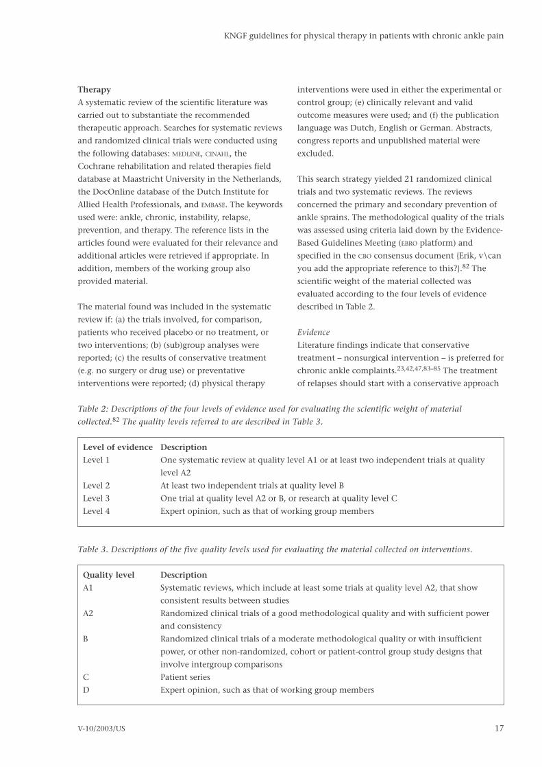
A systematic review of the scientific literature was
carried out to substantiate the recommended
therapeutic approach. Searches for systematic reviews
and randomized clinical trials were conducted using
the following databases: MEDLINE, CINAHL, the
Cochrane rehabilitation and related therapies field
database at Maastricht University in the Netherlands,
the DocOnline database of the Dutch Institute for
Allied Health Professionals, and EMBASE. The keywords
used were: ankle, chronic, instability, relapse,
prevention, and therapy. The reference lists in the
articles found were evaluated for their relevance and
additional articles were retrieved if appropriate. In
addition, members of the working group also
provided material.
The material found was included in the systematic
review if: (a) the trials involved, for comparison,
patients who received placebo or no treatment, or
two interventions; (b) (sub)group analyses were
reported; (c) the results of conservative treatment
(e.g. no surgery or drug use) or preventative
interventions were reported; (d) physical therapy
interventions were used in either the experimental or
control group; (e) clinically relevant and valid
outcome measures were used; and (f) the publication
language was Dutch, English or German. Abstracts,
congress reports and unpublished material were
excluded.
This search strategy yielded 21 randomized clinical
trials and two systematic reviews. The reviews
concerned the primary and secondary prevention of
ankle sprains. The methodological quality of the trials
was assessed using criteria laid down by the Evidence-
Based Guidelines Meeting (EBRO platform) and
specified in the CBO consensus document {Erik, v\can
you add the appropriate reference to this?}.82 The
scientific weight of the material collected was
evaluated according to the four levels of evidence
described in Table 2.
Evidence
Literature findings indicate that conservative
treatment – nonsurgical intervention – is preferred for
chronic ankle complaints.23,42,47,83–85 The treatment
of relapses should start with a conservative approach
17
KNGF guidelines for physical therapy in patients with chronic ankle pain
V-10/2003/US
Level of evidence Description
Level 1 One systematic review at quality level A1 or at least two independent trials at quality
level A2
Level 2 At least two independent trials at quality level B
Level 3 One trial at quality level A2 or B, or research at quality level C
Level 4 Expert opinion, such as that of working group members
Table 2: Descriptions of the four levels of evidence used for evaluating the scientific weight of material
collected.82 The quality levels referred to are described in Table 3.
Table 3. Descriptions of the five quality levels used for evaluating the material collected on interventions.
Quality level Description
A1 Systematic reviews, which include at least some trials at quality level A2, that show
consistent results between studies
A2 Randomized clinical trials of a good methodological quality and with sufficient power
and consistency
B Randomized clinical trials of a moderate methodological quality or with insufficient
power, or other non-randomized, cohort or patient-control group study designs that
involve intergroup comparisons
C Patient series
D Expert opinion, such as that of working group members

lasting 8–12 weeks.86 If complaints persist, surgery
can be considered. The most important form of
conservative therapy is reported to be exercise
therapy augmented by taping or bracing.23,42,65
However, the relative effectiveness of different
conservative treatment options remains unclear. Most
published articles are of limited methodological
quality and contain inconsistent definitions of the
interventions and outcome measures
employed.65,87,88
Exercise therapy
To ensure that patients with chronic ankle
complaints regain optimal functioning of the ankle,
it appears advisable that treatment should be as
varied and intense as possible.42,86,89,92 Treatment
should focus on training proprioception, increasing
muscle strength, increasing mobility, normalizing the
gait pattern, and training for sporting activities, if
necessary.23,42 Ideally, the patient’s performance in
these areas is assessed at the start of treatment and,
depending on the findings, exercise therapy should
focus on the weakest area or areas. The effects of
therapy should be evaluated at regular intervals. The
project group acknowledges that, at the moment, no
valid instruments for the standardized assessment of
proprioception, strength, mobility, gait pattern or
functional instability in chronic ankle complaints are
available for physical therapy practices. There is a
need for appropriate clinical measurement techniques
in physical therapy.
Conclusions
On the basis of the material collected, the project
group concludes that the evidence on exercise
therapy is at evidence level 2 and that the quality of
the studies retrieved89–92 is at quality level B.
It is likely, then, that the treatment of ankle
complaints should consist of an exercise program
that is as varied and intense as possible if optimal
ankle functioning is to be achieved.
Training proprioception
The effect of training proprioception using a wobble
board has been investigated in healthy
individuals93–95 and in patients with ankle
sprains.25,62,96,97 All studies show that coordination
and balance exercises lead to increased functional
stability of the ankle. In their review of prevention,
Verhagen et al.87 concluded that athletes who have
had a previous ankle sprain and who receive
proprioception training have the same risk of a new
ankle sprain as individuals who have not had a
previous ankle sprain. Vaes et al.49 showed that, to
train supination dampening in the standing position,
the height of the wobble board should be sufficient
to enable a tilt of more than 30 degrees.
Delayed reaction time in the peroneus longus muscle
can be improved by rehabilitation therapy.98
However, the impulse given to disturb balance should
be sufficiently strong.49 Nevertheless, there is an
absence of data proving that proprioception training
shortens the reaction time to such an extent that the
ankle is better protected against trauma. In research
into dynamic stability in the knees of healthy
individuals, Wojtys et al.99 concluded that muscle
fatigue leads to a delayed muscular reaction to
sudden joint translations. Matsusaka et al.97 studied
the additional effect of applying non-elastic tape
around the lateral malleolus during wobble board
training in 22 persons with functional instability of
the ankle. The researchers hypothesized that taping
may improve the afferent input from skin receptors,
thereby improving the efficacy of proprioception
training. On the basis of their primary outcome
measure of ‘maintaining position’, they found that
the experimental group reached their reference level
two weeks earlier than the control group.
Conclusions
On the basis of the material collected, the working
group concludes that the evidence on proprioception
training is at evidence level 2 and that the quality of
the studies retrieved25,35,96,97,100 is at quality level B.
It is likely, then, that coordination exercises and
balance training help patients with ankle complaints
to regain functional stability of the ankle.
However, as other evidence on proprioception
training is at evidence level 4, the working group
postulates that training using a wobble board as
stand-alone therapy does not fully provide
proprioception training. It is advised, therefore, that
18
KNGF guidelines for physical therapy in patients with chronic ankle pain
V-10/2003/US

optimal use is made of the patient’s normal daily
activities and of exercises for specific sports.
Proprioception should be trained using the full range
of ankle motion in order to activate
mechanoreceptors at all possible joint angles. In
particular, this principle should be applied using the
range of movement regained after mobilization.
Increasing muscle strength
There is little evidence available on the effect of
strength training in patients with chronic ankle
complaints. However, strength training is often an
integral part of exercise programs.23,33,42,85,86,89
It seems likely that a particular strength level is
associated with, and is a prerequisite for, a particular
level of muscular stability. Moreover, active muscular
contraction, by means of mechanisms in muscle
spindles, influences proprioception.101 Muscle
training is thought to decrease the risk of relapse and
to have a positive influence on proprioception.25
Other research93 suggests that muscle training is as
effective as proprioception training in improving
joint stability and balance. Uh et al.102 and Kannus et
al.47 found that, in healthy individuals, strength
training had a positive effect in not only the leg that
had undergone training, but also in the contralateral
leg that had not. This is the so-called cross-over
effect. In addition, Wojtys et al.99 found that muscle
fatigue leads to decreased coordination and, thereby,
to the deterioration of dynamic stability in the knees
of healthy individuals.
Conclusions
On the basis of the material collected, the project
group concludes that the evidence on strength training
is at evidence level 3 and that the quality of the studies
retrieved47,93,99,102 is at quality level C.
It is possible, then, that strength training has a
positive effect on the recovery of functional
instability in the ankle.
Other evidence on strength training is at evidence
level 4, and the working group believes that, in
patients with chronic ankle complaints, the exercise
program should involve enough repetitions and be of
sufficient intensity to train muscle endurance.
Increasing mobility
There is no evidence available on the effectiveness of
manipulative techniques in patients with
functionally unstable ankles. In patients with acute
ankle sprains, the use of anterior-posterior talocrural
mobilization techniques in addition to RICE (i.e., rest,
ice, compression, elevation) protocols has a positive
effect in achieving earlier pain-free dorsal flexion and
in improving walking speed.67
Conclusions
On the basis of the material collected, the working
group concluded that the evidence on mobilization
techniques is at evidence level 4.
The project group believes that, in patients with
chronic ankle complaints, the range of motion of the
ankle should be maximized by active exercises. Only
when this is not sufficient, should passive techniques
be employed.
Electrotherapy, laser therapy and ultrasound therapy
There is no evidence in the literature justifying the
use of electrotherapy, laser therapy or ultrasound
therapy in patients with chronic ankle complaints.103
Conclusions
On the basis of the material collected, the working
group concluded that the evidence on electrotherapy,
laser therapy and ultrasound therapy is at quality
level A1.
The project group states that, in patients with chronic
ankle complaints, use of electrotherapy, laser therapy
or ultrasound therapy does not, in general, increase
the effectiveness of exercise programs.
19
KNGF guidelines for physical therapy in patients with chronic ankle pain
V-10/2003/US

Relapse prevention
Tape and braces
Recent extensive reviews87,88 show strong evidence, at
evidence level 1, that the provision of external
support prevents ankle sprain relapses. Other
literature reviews confirm this finding.104,105 The use
of tape or a brace reduces the frequency of ankle
sprains, especially in individuals who have had
previous sprains. A reduction of 40–50% has been
reported.25,88,106,107 Taping and bracing also reduce
ankle sprain severity.87 The fit of the tape or brace
must be optimal if maximum patient compliance is
to be obtained.108–111 Vaes et al.104 distinguish
between four types of support: wraps (i.e., non-
adhesive bandages), straps (i.e., adhesive elastic
bandages), tape (i.e., adhesive non-elastic bandages)
and braces (i.e., a manufactured support that may be
tailor-made to fit).
Braces
Extensive research has been carried out into the
functioning of braces and into comparisons of the
different types of brace. To date, no single type of
brace has proved superior to the others. However,
each has its own advantages and disadvantages with
regard to fit and comfort. The more rigid the brace,
the less comfortable it is to wear and the greater the
restriction on joint movement. The task of deciding
which type of brace is most appropriate is influenced
by data from history-taking, the results of the
physical examination, and the individual patient’s
wishes and needs. Literature reviews show that most
studies have been performed on the usefulness of
ankle braces for preventing ankle sprains during
sporting activities.28,88,109,112–116 Braces have been
shown to be useful in preventing relapses in high-risk
sports.
Tape
Taping has a minimal effect on sporting
performance.65,104,117 Moreover, it is claimed to be
“useful for prevention, both in healthy and injured
(chronically sprained) athletes”.87,88
Taping has two functions.118,119 Firstly, it exerts a
mechanical or stabilizing effect by limiting plantar
flexion and inversion movements. This is very
important if there is weakness or secondary atrophy,
or both, of the evertor muscles.25 Lohrer et al.120
noted that, in addition to movement being limited
during inversion, movement speed is also reduced.
However, Manfroy et al.121 argued that these effects
exist for only a short period after tape has been
applied. After about 40 minutes of participation in a
sport, no difference can be observed between the
taped ankle and the normal ankle in terms of
resistance to inversion.121 Secondly, an influence on
proprioceptive reflexes in ankle muscles has been
reported. However, the effect of taping on
proprioception is controversial. Refshauge et al.122
and Konradsen and Bohsen-Ravn32 concluded that
there is no evidence that taping enhances
proprioception. Nevertheless, both positive and
negative effects on the reaction time and on the
moment of contraction of the peroneal muscles have
been reported. Karlsson et al.13 showed that taping a
mechanically stable ankle decreases the muscle
reaction time. Matsusaka et al.97 claim that
KNGF-richtlijn Stress urine-incontinentie
20 51.1001.01.02
Table 4. The interventions for treating patients with chronic ankle strain reviewed in these guidelines ranked
according to the level of evidence of their effectiveness.
Evidence level Strength of evidence Intervention
1 Strong Bracing
Taping
2 Intermediate Proprioception training
3 Limited Strength training
4 None or unclear Electrotherapy, laser therapy and ultrasound therapy
Manipulative therapies
Special shoes
Gait training

individuals with chronic ankle instability recover two
weeks faster because taping has a positive influence
on afferent input.
Different taping techniques have not been compared
extensively in terms of the degree of mechanical
restriction they exert on the ankle.119 The basket-
weave method, with ‘stirrups’ and a ‘heel lock’, has
been described as the best way to apply tape.124,125 In
addition, Coumans’ taping has also been reported to
be effective.15 On a theoretical basis, Riezebos et al.126
postulate that applying tape limits exorotation of the
lower leg and prevents exorotation and adduction of
the talus relative to the calcaneus. It is assumed that
inversion traumas occur primarily during rotation in
the (mainly) horizontal plane.
Comparison of tape and braces
Braces are even more effective in preventing ankle
sprain relapses than tape.87 However, taping results in
a 25% greater reduction in the range of joint motion
before sporting activities than the use of a brace.127
During sporting activities, however, there is a
considerable loosening effect with tape, which leads
to a reduction in efficacy of 40–50% within hours
depending on the type of activity.124,128 In contrast,
braces show a loosening affect of only 4.5–12%.127
Moreover, with braces, the initial rigidity can be
easily regained by pulling on the straps or tightening
the laces. Both taping and the use of braces have
comparable effects on mechanical stability, as
assessed after activities.127,129
Advantages and disadvantages of tape and braces
In addition to the considerable loosening effect,
taping also has other disadvantages. The cost of the
materials is high, there may be skin irritation due to
the adhesive, and tape is hard to remove. One
advantage of taping is that the fit is highly
individual. In contrast, braces are less likely to irritate
the skin and are easy to put on. The disadvantages are
that it is difficult to find the right size (i.e., the
optimal fit) and that patients cannot wear shoes with
their braces, with the result that skin irritation may
occur.130 A good brace should balance comfort and
support112 and is, therefore, an individual choice.
Some authors think that the use of tape and braces is
beneficial until coordination training has
ended25,107,119 and in situations where there is a
greater than average risk of injury25, for example,
when the patient is fatigued during a sporting
activity, is involved in a high-risk sport, or has
experienced a relapse.
The working group holds the opinion that the
continuous use of taping or a brace is not supported
by the literature. The effect of the continuous use of
taping or bracing on proprioception is unknown. The
intention of the treatment recommended by these
guidelines is to optimize the recovery of
neuromuscular function. The working group believes
that the continuous use of taping or a brace could
possibly have a negative influence on functional
stability over the long term.
Conclusions
On the basis of the material collected, the working
group concludes that the evidence on the use of
taping and braces is at evidence level 1 and that the
quality of the studies retrieved87,88 is at quality level
A1.
External support reduces the risk of ankle sprain in
high-risk sports and, in general, prevents ankle sprain
relapses and reduces sprain severity. Physical
therapists should inform patients about these facts
and advise accordingly.
Other evidence on the use of taping and braces is at
evidence level 4, with the quality of the studies
retrieved87,88 being at quality level A1.
The project group believes that the routine use of
external support during participation in sports or
during heavy work could have a negative influence
on functional stability over the long term. The
project group holds the opinion that the recovery of
functional stability should be the endpoint of
treatment and advises the gradually diminishing use
of external support.
Shoes
Evidence on the optimal use of footwear is
contradictory. Some authors recommend high-top
shoes107,114 whereas others recommend low-top
21
KNGF guidelines for physical therapy in patients with chronic ankle pain
V-10/2003/US

shoes.131 Barret et al.132 found no strong relationship
between shoe type and type of ankle injury. Verhagen
et al.87 concluded that the use of new shoes probably
has a greater preventive effect than shoe height.
The project group holds the opinion that it is
important that the patient’s shoe type is appropriate
for normal daily life activities, work and sporting
activities, and for the type of surface encountered.
The timely renewal of footwear is advisable.
Treatment frequency and duration
The frequency of treatment depends on the
intervention being used and the stage of treatment.
The project group recommends that more guidance
should be given on complex processes, such as
relearning a walking pattern or learning specific
exercises, than on strength or proprioception
training. The role of the physical therapist is both
educational and correctional, thereby requiring
frequent contact with the patient (i.e., two to three
times a week). The patient should be able to perform
strength and proprioception training at home if
properly instructed. Here, the role of the physical
therapist is similar to coaching, and contact with the
patient need only occur once a week or fortnight.
There is no scientific evidence on how timing
influences the effectiveness of interventions for
chronic ankle sprain. The project group holds the
opinion that an improvement should be noted in the
patient, as assessed using the instrument for patient-
specific complaints, by at least six weeks after the
start of therapy. If this is not the case, the patient
should be referred back to his or her doctor.
Updating these guidelinesThese guidelines are based on currently available
evidence. At the moment, there is a moderate
amount of scientific evidence on the use of physical
therapy in chronic ankle sprains. However, a
significant part of the guideline recommendations is
based on expert consensus. Therefore, the
recommendations given in these guidelines might
have to change as a result of new knowledge and
scientific findings. The method of guideline
development and implementation that was followed
in the production of these guidelines states that all
guidelines should be revised within three to five years
of the first publication.3,133 The Royal Dutch Society
for Physical Therapy (KNGF), together with members
of the current working group, plans to update these
guidelines in 2006. Nevertheless, if new scientific
developments necessitate an earlier update, this
would also be carried out.
The legal status of the guidelinesThese guidelines are not legal documents. They detail
knowledge and contain recommendations, which are
based on the results of scientific research, that
therapists should follow in order to provide
appropriate healthcare. Since the recommendations
made in the guidelines apply to the ‘average’ patient,
therapists must use their professional judgement to
decide when to deviate from the guidelines if that is
required in a particular patient’s situation. Any
deviation from guideline recommendations must be
justified and documented.2,3
AcknowledgementsSpecial thanks for their help in developing these
guidelines go to members of the panel of experts.
These are, in alphabetical order: Dr R Bening and Dr
N van Dijk of the Dutch Orthopedic Association, Dr
A Goudswaard of the Dutch College of General
Practitioners, Dr W Hullegie of the Dutch Association
of Sports Physiotherapy and Dr C Ottevanger of the
(Dutch) Association for Sports Medicine. We would
also like to thank all the physical therapists who
commented on the draft version of these guidelines
and who evaluated its applicability for clinical
practice.
The Dutch Ministry of Healthcare, Sport and Welfare
(VWS) funded the work on the guidelines within the
context of a development program entitled
“Bevordering Kwaliteitsbeleid Paramedische Zorg
(BKPZ)”. The development of these guidelines was
carried out independently of the funding
organization and the funding organization has had
no influence whatsoever on their contents.
22
KNGF guidelines for physical therapy in patients with chronic ankle pain
V-10/2003/US

1 Hendriks HJM, Reitsma E, van Ettekoven H. Centrale
richtlijnen in de fysiotherapie. Ned Tijdschr Fysiother
1996;106:2-11.
2 Hendriks HJM, van Ettekoven H, Reitsma E, Verhoeven ALJ,
van der Wees P. Methode voor centrale richtlijnontwikkeling
en implementatie in de fysiotherapie. Amersfoort, the
Netherlands: KNGF/NPi/CBO; 1998.
3. Hendriks HJM, Bekkering GE, van Ettekoven H, Brandsma JW,
van der Wees P, de Bie RA. Development and implementation
of national practice guidelines: a prospect for continuous
quality improvement in physiotherapy. Introduction to the
method of guideline development. Physiother 2000;86:547.
4 Roebroeck ME, Dekker J, Oostendorp RAB, Bosveld W.
Physiotherapy for patients with lateral ankle sprains: a
prospective survey of practice patterns in Dutch primary
health care. Physiother 1998;84:421-32.
5 De Bie RA, Hendriks HJM, Lenssen AF, van Moorsel SR, Opraus
KWF, Remkes WFA, et al. KNGF-richtlijn Acuut Enkelletsel.
Amersfoort, the Netherlands: KNGF/NPi; 1998.
6 CBO consensus diagnostiek en behandeling van het acute
enkelletsel. Utrecht, the Netherlands: CBO; 1998.
7 Goudswaard AN, Thomas S, van den Bosch WJHM, Geijer
RMM. NHG-Standaard Enkeldistorsie (eerste herziening).
Huisarts Wet 2000;43:32-7.
8 Hendriks HJM, van Ettekoven H, Bekkering GE, Verhoeven
ALJ. Implementatie van KNGF-richtlijnen. Fysiopraxis 2000;9:9-
13.
9 Mulder S, Bloemhoff A, Harris S. Ongevallen in Nederland,
opnieuw gemeten. Amsterdam, the Netherlands: Stichting
Consument en Veiligheid; 1995.
10 Van Dijk CN. Review of the literature. In: Van Dijk CN. On
diagnostic strategies in patients with severe ankle sprain
(thesis). Amsterdam, the Netherlands: Universiteit van
Amsterdam; 1994;5-51.
11 Zeegers AVCM. Het supinatieletsel van de enkel. Resultaten
(thesis). Utrecht, the Netherlands: Universiteit Utrecht;
1995;83-131.
12 Møller-Larsen F, Wethelund JO, Jurik AG, de Carvalho A, Lucht
U. Comparison of three different treatments for ruptured
lateral ankle ligaments. Acta Orthop Scand 1988;59:564-6.
13 Karlsson J, Andreasson GO. The effect of external ankle
support in chronic lateral ankle joint instability: an
electromyographic study. Am J Sports Med 1992;20:257-61.
14 Povacz P, Unger F, Miller K, Tockner R, Resch H. A randomized,
prospective study of operative and non-operative treatment of
injuries of the fibular collateral ligaments of the ankle. J Bone
Joint Surg 1998;80-A:345-51.
15 Van Moppes FI, van den Hoogenband CR. Diagnostic and
therapeutic aspects of inversion trauma of the ankle joint
(thesis). Maastricht, the Netherlands; 1982.
16 Freeman MAR, Dean MRE, Hanham IWF. The etiology and
prevention of functional instability of the foot. J Bone Joint
Surg 1965;47B:678-84.
17 Lentell GL, Katzman LL, Walters MR. The relationship between
muscle function and ankle stability. J Orthop Sports Phys Ther
1990;11:605-11.
18 Lephart SM, Pincivero DM, Giraldo JL, Fu FH. The role of
proprioception in the management and rehabilitation of
athletic injuries. Am J Sports Med 1997;25:130-7.
19 Konradsen L, Olesen S, Hansen HM. Ankle sensorimotor
control and eversion strength after acute ankle inversion
injuries. Am J Sports Med 1998;26:72-7.
20 DeMaio M, Paine R, Drez D. Chronic lateral ankle instability
inversion sprains: Part I. Orthopedics 1992;15:87-96.
21 DeMaio M, Paine R, Drez D. Chronic lateral ankle instability
inversion sprains: Part II. Orthopedics 1992;15:241-8.
22 Staples OS. Result study of ruptures of lateral ligament of the
ankle. Clin Orthop 1972;85:50-8.
23 Renström PAF, Konradsen L. Ankle ligament injuries. Br J
Sports Med 1997;31:11-20.
24 Vaes PH, Duquet W, van den Broeck R. Enkeldistorsietrauma
en enkelinstabiliteit. In: Jaarboek Fysiotherapie. 1997:1-45.
25 Tropp H, Askling C, Gillquist J. Prevention of ankle sprains.
Am J Sports Med 1985;13:259-62.
26 Ryan L. Mechanical stability, muscle strength and
proprioception in the functionally unstable ankle. Aust J
Physiother 1994;40:41-7.
27 Lentell G, Baas B, Lopez D, McGuire L, Sarrels M, Snyder P. The
contributions of proprioceptive deficits, muscle function, and
anatomic laxity to functional instability of the ankle. J Orthop
Sports Phys Ther 1995;21:206-15.
28 Louwerens JWK. The position of the rear foot at the moment
of heel contact and chronic lateral instability: a video analysis
(thesis). Rotterdam, the Netherlands: Erasmus Universiteit
Rotterdam; 1996;61-8.
29 Birmingham TB, Chesworth BM, Hartsell HD, Stevenson AL,
Lapenskie GL, Vandervoort AA. Peak passive resistive torque at
maximum inversion range of motion in subjects with
recurrent ankle inversion sprains. J Orthop Sports Phys Ther
1997;25:342-8.
30 Lephart SM, Pincivero DM, Rozzi SL. Proprioception of the
ankle and knee. Sports Med 1998;25:149-55.
31 Fridén T, Zätterström R, Lindstrand A, Mortiz U. A
stabilometric technique for evaluation of lower limb
instabilities. Am J Sports Med 1989;17:118-22.
32 Konradsen L, Bohsen-Ravn J. Prolonged peroneal reaction time
in ankle instability. Int J Sports Med 1991;12:290-2.
33 Leanderson J, Eriksson E, Nilsson C, Wykman A.
Proprioception in classical ballet dancers: a prospective study
of the influence of an ankle sprain on proprioception in the
ankle joint. Am J Sports Med 1996;24:370-4.
34 Forkin DM, Koczur C, Battle R, Newton RA. Evaluation of
kinesthetic deficits indicative of balance control in gymnasts
with unilateral chronic ankle sprains. J Orthop Sports Phys
Ther 1996;23:245-50.
35 Rozzi SL, Lephart SM, Sterner R, Kuligowski L. Balance training
for persons with functionally unstable ankles. J Orthop Sports
Phys Ther 1999;29:478-86.
36 Tropp H, Ekstrand J, Gillquist J. Factors affecting stabilometry
recordings of single limb stance. Am J Sports Med
1984;12:185-8.
37 Garn SN, Newton A. Kinesthetic awareness in subjects with
multiple ankle sprains. Phys Ther 1988;68:1667-71.
38 Glencross D, Thornton E. Position sense following joint injury.
J Sports Med Phys Fitness 1981;21:23-7.
39 Konradsen L, Ravn JB, Sorenson AI. Proprioception at the
ankle: the effect of anaesthetic blockade of ligament receptors.
J Bone Joint Surg Br 1993;75:433-6.
40 Boyle J, Negus V. Joint position sense in the recurrently
sprained ankle. Aust J Physiother 1998;44:159-63.
23
KNGF guidelines for physical therapy in patients with chronic ankle pain
V-10/2003/US
References

41 Hartsell HD. The effects of external bracing on joint position
sense awareness for the chronically unstable ankle. J Sport
Rehab 2000;9:279-89.
42 Karlsson J, Faxén E, Eriksson BI. Ankle joint ligament injuries:
prevention, evaluation and treatment. Phys Rehab Med
1996;8:183-200.
43 Oostendorp RAB. Functionele instabiliteit na het
inversietrauma van enkel en voet: een effectonderzoek
pleisterbandage versus pleisterbandage gecombineerd met
fysiotherapie. Geneesk Sport 1987;20:45-55.
44 Burke D, Gandevia SC, Macefield G. Responses to passive
movement of receptors in joint, skin and muscle of the
human hand. J Physiol 1988;Aug:347-61.
45 Clarke FJ, Grigg P, Chapin JW. The contribution of articular
receptors to proprioception with the fingers in humans. J
Neurophysiology 1989;61:186-93.
46 Kleinrensink GJ, Stoeckart R, Meulstee J, Kaulesar Sukul DMKS,
Vleeming A, Snijders CJ, et al. Lowered motor conduction
velocity of the peroneal nerve after inversion trauma. Med Sci
Sports Exerc 1994;26:877-83.
47 Kannus P, Alosa D, Cook L, Johnson RJ, Renström P, Pope M,
et al. Effect of one-legged exercise on the strength, power and
endurance of the contralateral leg. Eur J Appl Physiol
1992;64:117-26.
48 Laskowski ER, Newcomer-Aney K, Smith J, van Deursen RW.
[author - article title?] In: Laskowski. Proprioception. Sci
Principals Sports Rehab 2000;11:323-40.
49 Vaes PH, van Gheluwe B, Duquet W. Control of acceleration
during sudden ankle supination in people with unstable
ankles. J Orthop Sports Phys Ther 2001;31:741-52.
50 Konradsen L, Bohsen-Ravn J. Ankle instability caused by
prolonged peroneal reaction time. Acta Orthop Scand
1990;61:388-90.
51 Löfvenberg R, Karrholm J, Sundelin G, Ahlgren O. Prolonged
reaction time in patients with chronic lateral instability of the
ankle. Am J Sports Med 1995;23:414-7.
52 Brunt D, Andersen JC, Hunstman B, Reinhert LB, Thorell AC,
Sterling JC. Postural responses to lateral perturbation in
healthy subjects and ankle sprain patients. Med Sci Sports
Exerc 1992;24:171-6.
53 Fernandes N, Allison GT, Hopper D. Peroneal latency in
normal and injured ankles at varying angles of perturbation.
Clin Orthop Rel Res 2000;375:193-201.
54 Beynnon BD, Renström PA, Alosa DM, Baumhauer JF, Vacek
PM. Ankle ligament injury risk factors: a prospective study of
college athletes. J Orthop Res 2001;19:213-20.
55 Louwerens JWK, van Linge B, de Klerk LWL, Mulder PGH,
Snijders CJ. Peroneus longus and tibialis anterior muscle
activity in the stance phase. Acta Orthop Scand 1995;66:517-
23.
56 Balduini FC, Vegso JJ, Torg JS, Torg E. Management and
rehabilitation of ligamentous injuries to the ankle. Ankle
inversion injury and hypermobility: effect on hip and ankle
muscle electromyography onset latency. Sports Med
1987;4:364-80.
57 Bosien WR, Staples OS, Russell S. Residual disability following
acute ankle sprains. J Bone Joint Surg 1955;37A:1237-43.
58 Tropp H. Pronator muscle weakness in functional instability of
the ankle joint. Int J Sports Med 1986;7:291-4.
59 Wilkerson GB, Pinerola JJ, Caturano RW. Invertor vs. evertor
peak torque and power deficiencies associated with lateral
ankle ligament injury. J Orthop Sports Phys Ther 1997;26:78-
86.
60 McKnight CM, Armstrong CW. The role of ankle strength in
functional ankle instability. J Sport Rehab 1997;6:21-9.
61 Pope R, Herbert R, Kirwan J. Effects of ankle dorsiflexion range
and pre-exercise calf muscle stretching on injury risk in army
recruits. Aus Physiother 1998;44:165-72.
62 Wester JU, Jespersen SM, Nielsen KD. Wobble board training
after partial sprains of the lateral ligaments of the ankle: a
prospective randomized study. J Orthop Sports Phys Ther
1996;23:332-6.
63 Bahr R. Recent advances: sport medicine. BMJ 2001;323:328-
31.
64 McKay GD, Goldie PA, Payne WR, Oakes BW. Ankle injuries in
basketball: injury rate and risk factors. Br J Sports Med
2001;35:103-8.
65 Thacker SB, Stroup DF, Branche CM. The prevention of ankle
sprains in sports: a systematic review of the literature. Am J
Sports Med 1999;27:753-60.
66 Lankhorst GJ, Timmer M. Invaliditeits- en arbeids
(on)geschiktheidsschattingen. Ned Tijdschr Geneesk
1990;3:215-7.
67 Green T, Refshauge K, Crosbie J, Adams R. A randomized
controlled trial of a passive accessory joint mobilization on
acute ankle inversion sprains. Phys Ther 2001;81:984-94.
68 Laskowski ER, Newcomer-Aney K, Smith J. Proprioception. Sci
Principals Sports Rehab 2000;11:323-40.
69 Kaikkonen A, Kannus P, Järvinen M. A performance test
protocol and scoring scale for the evaluation of ankle injuries.
Am J Sports Med 1994;22:462-9.
70 Van Dijk CN. The relative merits of physical examination and
arthrography after supination trauma of the ankle (thesis).
Amsterdam, the Netherlands: Universiteit van Amsterdam;
1994;71-126.
71 Beurskens AJHM, de Vet HC, Koke AJ. Responsiveness of
functional status in low back pain: a comparison of different
instruments. Pain 1996;65:71-6.
72 Beurskens AJ, de Vet HC, Koke AJ. A patient-specific approach
for measuring functional status in low back pain. J Manipul
Physiol Ther 1999;22:144-8.
73 Huskisson EC. Measurement of pain. Lancet 1974;9:1127-31.
74 Miller MD, Daron G, Ferris MD. Measurement of subjective
phenomena in primary care research: the visual analogue
scale. Fam Pract Res J 1993;13:15-24.
75 Wevers ME, Lowe NK. A critical review of visual analogue
scales in the measurement of clinical phenomena. Res Nurs
Health 1990;13:227-36.
76 Brunnekreef JJ (namens de werkgroep). Betrouwbaarheid van
de ganganalyse lijst Nijmegen. Jaarcongres Fysiotherapie. Den
Haag, the Netherlands. 2-3 November 2001.
77 Van Dijk CN. On the cause of ‘Medial ankle pain’ after severe
sprain (thesis). Amsterdam, the Netherlands: Universiteit van
Amsterdam; 1994;139-62.
78 Van Dijk CN, Tol JL, Verheyen CCP. A prospective study of
prognostic factors concerning the outcome of arthroscopic
surgery for anterior ankle impingement. Am J Sports Med
1997;25:737-45.
79 Shea MP, Manoli A. Foot fellow’s review: osteochondral lesions
of the talar dome. Foot Ankle 1993;14:48-55.
80 Brosky T, Nyland J, Nitz AJ, Caborn DNM. The ankle
ligaments: consideration of syndesmotic injury and
implications for rehabilitation. J Orthop Sports Phys Ther
1995;21:197-205.
24
KNGF guidelines for physical therapy in patients with chronic ankle pain
V-10/2003/US

81 Hopkinson WJ, St Pierre P, Ryan JB, Wheeler JH. Syndesmosis
sprains of the ankle. Foot Ankle 1990;10:325-30.
82 Richtlijnontwikkeling binnen het Kwaliteitsinstituut voor de
Gezondheidszorg CBO: handleiding voor werkgroepleden.
Utrecht, the Netherlands: Kwaliteitsinstituut voor de
Gezondheidszorg CBO; 2000.
83 Munk B, Holm-Christensen K, Lind T. Long-term outcome
after ruptured lateral ankle ligaments: a prospective study of
three different treatments in 79 patients with 11-year follow-
up. Acta Orthop Scand 1995;66:452-4.
84 Ogilvie-Harris DJ, Gilbart M. Treatment modalities for soft
tissue injuries of the ankle: a critical review. Clin J Sport Med
1995;5:175-86.
85 Shrier I. Treatment of lateral collateral ligament sprains of the
ankle: a critical appraisal of the literature. Clin J Sport Med
1995;5:187-95.
86 Safran MR, Benedetti RS, Bartolozzi III AR, Mandelbaum BR.
Lateral ankle sprains: a comprehensive review. Part 1: etiology,
pathoanatomy, histopathogenesis and diagnosis. Med Sci
Sports Exerc 1999;31:S429-37
87 Verhagen EA, van Mechelen W, de Vente W. The effect of
preventive measures on the incidence of ankle sprains. Clin J
Sport Med 2000;10:291-6.
88 Handoll HHG, Rowe BH, Quinn KM, de Bie RA. Interventions
for preventing ankle ligament injuries (Cochrane Review). The
Cochrane Library. Oxford, UK: Update Software; 2001.
89 Heidt RS, Sweeterman LM, Carlonas RL, Traub JA, Tekulve FX.
Avoidance of soccer injuries with preseason conditioning. Am
J Orthop Soc Sports Med 2000;28:659-62.
90 Holme E, Magnusson SP, Becher K, Bieler T, Aagaard P, Kjær M.
The effect of supervised rehabilitation on strength, postural
sway, position sense and re-injury risk after acute ankle
ligament sprain. Scand J Med Sci Sports 1999;9:104-9.
91 Söderman K, Werner S, Pietilä T, Engström B, Alfredson H.
Balance board training: prevention of traumatic injuries of the
lower extremities in female soccer players? Knee Surg Sports
Traumatol Arthrosc 2000;8:356-63.
92 Bahr R, Lian Ø, Bahr IA. A two-fold reduction in the incidence
of acute ankle sprains in volleyball after the introduction of an
injury prevention program: a prospective cohort study. Scand J
Med Sci Sports 1997;7:172-7.
93 Blackburn T, Guskiewicz KM, Petschauer MA, Prentice WE.
Balance and joint stability: the relative contributions of
proprioception and muscular strength. J Sport Rehab
2000;9:315-28.
94 Waddington G, Adams R, Jones A. Wobble board (ankle disc)
training effects on the discrimination of inversion
movements. Aus Physiother 1999;45:95-101.
95 Hoffman M, Payne VG. The effects of proprioceptive ankle
disk training on healthy subjects. J Orthop Sports Phys Ther
1995;21:90-3.
96 Bernier JN, Perrin DH. Effect of coordination training on
proprioception of the functionally unstable ankle. J Orthop
Sports Phys Ther 1998;27:264-75.
97 Matsusaka N, Yokoyama S, Tsurusak T. Effect of ankle disk
training combined with tactile stimulation to the leg and foot
on functional instability of the ankle. Am J Sports Med
2001;29:25-30.
98 Sheth P, Yu B, Laskowski ER, An KN. Ankle disk training
influences reaction times of selected muscles in a simulated
ankle sprain. Am J Sports Med 1997;25(4):538-43.
99 Wojtys EM, Wylie BB, Huston LJ. The effects of muscle fatigue
on neuromuscular function and anterior tibial translation in
healthy knees. Am J Sports Med 1996;24:615-21.
100 Gauffin H, Tropp H, Odenrick P. Effect of ankle disk training
on postural control in patients with functional instability of
the ankle joint. Int J Sports Med 1988;9:141-4.
101 Gandevia SC, McCloskey DI, Burke D. Kinesthetic signals and
muscle contraction. Trends Neuroscience 1992;15:62-5.
102 Uh BS, Beynnon BD, Helie BV, Alosa DM, Renström PA. The
benefit of a single-leg strength training program for the
muscles around the untrained ankle. Am J Sports Med
2000;28:568-73.
103 Gezondheidsraad. De effectiviteit van fysische therapie;
electrotherapie, lasertherapie, ultrageluidbehandeling. Den
Haag, the Netherlands: Gezondheidsraad; 1999.
104 Vaes PH, Duquet W, van Gheluwe B. Werkzaamheid van
enkelbandages en enkelortheses. In: Jaarboek Fysiotherapie.
2000:49-66.
105 Sitler M, Ryan J, Wheeler B, McBride J, Arciero R, Anderson J,
et al. The efficacy of a semi-rigid ankle stabilizer in reducing
acute ankle injuries in basketball. A randomized clinical study
at West Point. Am J Sports Med 1994;22:454-61.
106 Thorndike AJ. Athletic injuries: prevention, diagnosis and
treatment. Philadelphia: Lea and Febiger; 1956.
107 Garrick JC, Requa RK. Role of external support in the
prevention of ankle sprains. Med Sci Sports 1973;5:200-3.
108 Sitler MR, Ryan J, Wheeler B, McBride J, Arciero R, Anderson J,
et al. The efficacy of a semi-rigid ankle stabilizer in reducing
acute ankle injuries in basketball. Am Orthop Soc Sports Med
1994;22:454-61.
109 Gross MT, Everts JR, Roberson SE, Roskin DS, Young KD. Effect
of Donjoy ankle ligament protector and Aircast Sport-Stirrup
ortheses on functional performance. J Orthop Sports Phys
Ther 1994;19:150-6.
110 Gross MT, Ballard CL, Mears HG, Watkins EJ. Comparison of
Donjoy ankle ligaments protector and Aircast Sport-Stirrup
orthoses in restricting foot and ankle motion before and after
exercise. J Orthop Sports Phys Ther 1992;16:60-7.
111 Gross MT, Lapp AK, Davis M. Comparison of Swede-O-
Universal ankle support and Aircast Sport-Stirrup orthoses and
ankle tape in restricting eversion-inversion before and after
exercise. J Orthop Sports Phys Ther 1991;13:11-9.
112 Alves JW, Alday RV, Ketcham DL, Lentell GL. A comparison of
the passive support provided by various ankle braces. J Orthop
Sports Phys Ther 1992;15:10-8.
113 Beynnon BD, Renström PA. The effect of bracing and taping in
sports. Ann Chir Gynaecol 1991;80:230-8.
114 Shapiro MS, Kabo JM, Mitchell PW, Loren G, Tsenten M. Ankle
sprain prophylaxis: an analysis of the stabilizing effects of
braces and tape. Am J Sports Med 1994;22:78-82.
115 Johnson RE, Veale JR, McCarthy GJ. Comparative study of
ankle support devices. J Am Podiatr Med Assoc 1994;84:107-
14.
116 Stiggelbout M, van Mechelen W. De preventie waarde van de
enkelbrace in vergelijking tot enkeltaping ter voorkoming van
enkeldistorsies bij sporters. Geneesk Sport 1993;26: [author -
page numbers please]
117 Hamer PW, Munt AM, Harris CD, James NC. The influence of
ankle strapping on wobbleboard performance, before and after
exercise. Aust Physiother 1992;38:85-92.
118 Firer P. Effectiveness of taping for the prevention of ankle
ligaments sprains. Br J Sports Med 1990;24:47-50.
119 Karlsson J, Swärd L, Andréasson GO. The effect of taping on
25
KNGF guidelines for physical therapy in patients with chronic ankle pain
V-10/2003/US

ankle stability. Sports Med 1993;16:210-5.
120 Lohrer H, Alt W, Gollhofer A. Neuromuscular properties and
functional aspects of taped ankles. Am J Sports Med
1999;27:69-75.
121 Manfroy PP, Ashton-Miller JA, Wojtys EM. The effect of
exercise, prewrap, and athletic tape on the maximal active and
passive ankle resistance to ankle inversion. Am J Sports Med
1997;25:156-63.
122 Refshauge KM, Kilbreath SL, Raymond J. The effect of
recurrent ankle inversion sprain and taping on proprioception
at the ankle. Med Sci Sports Exerc 2000;32:10-5.
123 Glick JM, Gorden RB, Nishimoto D. The prevention and
treatment of ankle injuries. Am J Sports Med 1976;4:136-41.
124 Rarick GL, Bigley G, Karst R, Malina RM. The measurable
support of the ankle joint by conventional methods of taping.
J Bone Joint Surg 1962;44:1183-90.
125 Van Wingerden BAR. Tape en bandagetechnieken. Lochem,
the Netherlands: Uitgeverij De Tijdstroom; 1982.
126 Riezebos C, Lagerberg A. Inversietrauma van de enkel,
ontstaansmechanismen, risicofactoren en preventie. Tijdsch
Fysiother 1998;16:16-47.
127 Brunch RP, Bednarski K, Holland D. Ankle joint support: a
comparison of reusable lace-on braces with taping and
wrapping. Phys Sport Med 1985;13:59-62.
128 Fumich RM, Ellison AE, Geurin GJ, Grace PD. The measured
effect of taping on combined foot and ankle motion, before
and after exercise. Am J Sports Med 1981;9:165-70.
129 Hughes LH, Stetts DM. A comparison of ankle taping and
semi-rigid support. Phys Sports Med 1983;11:99-103.
130 Leemrijse C. Gaat bij chronische instabiliteit van de enkel de
voorkeur uit naar bandageren/tapen of pushbraces? Respons
1990;1:1-2.
131 Rovere GD, Clarke TL, Yates CS. Retrospective comparison of
taping and ankle stabilizers in preventing ankle injuries. Am J
Sports Med 1988;16:228-33.
132 Barrett JR, Tanji JL, Drake C, Fuller D, Kawasaki RI, Fenton RM.
High- versus low-top shoes for the prevention of ankle sprains
in basketball players. Ankle inversion injury and
hypermobility: effect on hip and ankle muscle
electromyography onset latency. Am J Sports Med
1993;21:582-5.
133 Hendriks HJM, van Ettekoven H, van der Wees P. Eindverslag
van het project Centrale richtlijnen in de fysiotherapie (Deel
I): Achtergronden en evaluatie van het project. Amersfoort,
the Netherlands: KNGF/NPi/CBO; 1998.
134 Van Mechelen W, Kemper HCG, Voorn WJ, de Jongh HR.
Prevention of running injuries by warming-up, cool-down,
and stretching exercises. Am J Sports Med 1993;21:711-9.
135 Pienkowski D, McMorrow M, Shapiro R, Caborn DNM, Stayton
J. The effect of ankle stabilizers on athletic performance. Am J
Sports Med 1995;23:757-62.
26
KNGF guidelines for physical therapy in patients with chronic ankle pain
V-10/2003/US

27
KNGF guidelines for physical therapy in patients with chronic ankle pain
V-10/2003/US

28
KNGF guidelines for physical therapy in patients with chronic ankle pain
V-10/2003/US