clinical practice recommendations for needling of...
TRANSCRIPT
Clinical Practice Recommendations for
Needling of Arteriovenous Fistulae and Grafts
for Haemodialysis

i
Foreword
For people undergoing haemodialysis, vascular access is their lifeline. Protecting
and maintaining that access, be it an arteriovenous fistula or graft, is paramount to
achieve the best dialysis possible. Nurses working in dialysis units require specific
training and development of keys skills and competencies to proficiently needle
dialysis access. To date the lack of national standards has impacted on patient care
as highlighted by the 2017 CKD-PREM survey reporting vast variation in patient
experience with needling across centres in England and Wales
(https://www.renalreg.org/projects/prem).
The publication of these recommendations could not be timelier and will enable all
healthcare professionals working in renal units to improve their knowledge, skills,
quality of care and the experience of patients undergoing dialysis with arteriovenous
fistulae and grafts. The users of this document will be able to both benchmark and
challenge their own clinical practice. Nurses working in dialysis access need to
measure evaluate and report in local and national clinical audits, participate in
research and quality improvement projects.
The authors are to be congratulated on creating an in depth guide which includes
theory, practical skill sets and tools that can be used in the workplace. The
development of these recommendations demonstrates the value of collaborative
working across multi-professional groups to share their expert knowledge and vast
experience of managing vascular access.
I look forward to seeing these recommendations being implemented in renal units
across the UK, influencing practice and making a difference to patients.
Karen Jenkins
British Renal Society Vice President Clinical Practice

ii
Contents
Introduction ................................................................................................................ 1
Methodology ............................................................................................................... 3
Summary of Clinical Practice Recommendations ....................................................... 4
Clinical Practice Recommendations ......................................................................... 18
Recommendation A: Principles of a Good Needling Technique ............................... 18
Recommendation B: Technical Principles to aid Decision Making Prior to Needle
Insertion ................................................................................................................... 21
Recommendation C: Procedural Principles for Good Needle Insertion .................... 25
Recommendation D: Assessment of the AV Access prior to Needling ..................... 30
Recommendation E: Definitions of Needling Techniques ......................................... 33
Recommendation F: Choosing the Needling Technique and Planning Needling ..... 38
Recommendation G: Rope Ladder Needling Technique .......................................... 44
Recommendation H: Buttonhole Needling Technique .............................................. 45
Recommendation I: Area Puncture Needling Technique.......................................... 53
Recommendation J: Needling of New AV Access .................................................... 56
Recommendation K: Use of Nurse-Led Ultrasound to Assist with Needling ............. 59
Recommendation L: Managing Anxiety during Needling .......................................... 62
Recommendation M: Involving Patients in Care of their Vascular Access ............... 66
Recommendation N: Teaching Patients how to Self-Needle .................................... 69
Recommendation O: Staff Training to Perform Needling of AV Access ................... 74
Conclusion ............................................................................................................... 76
References ............................................................................................................... 78
Appendix 1 - Systematic Literature Search Strategy ................................................ 85
Appendix 2 – Arteriovenous Fistula / Graft (AVF/AVG) Pre-Needle Insertion
Assessment Tool ...................................................................................................... 86
Appendix 3 – ACCESS Assessment ........................................................................ 88
Appendix 4 – Needling Decision Making Model ....................................................... 89
Appendix 5 – Area Puncture Action Chart ................................................................ 90
Appendix 6 - Acknowledgments ............................................................................... 91
1
Introduction
Epidemiological data has shown the safety and longevity advantage of arteriovenous
(AV) fistula as access modality of first choice in comparison to AV graft and central
venous catheter (CVC) (1, 2). The Renal Association guidelines (UK) reflect this,
currently recommending use of AV fistula first, AV graft as the second option and
CVC as last option (3). Timely placement of AV fistula is essential to reduce the
need for the use of CVC which carry highest risk of access related complications,
hospitalisation, patient morbidity and mortality (4). Multiple factors may influence the
survival of AVF: age, frailty, sex, race, diabetes, smoking, hypotension, body mass
index, thrombosis, infection, aneurysm formation, timing of referral to the surgeons,
surgical techniques and skills, vessel size and use of adjuvant therapies such as
antiplatelet agents and infrared, timing and technique of cannulation (5).
Most national and international guidelines on haemodialysis access have mainly
focussed on areas of good practice in the MDT approach in the management of the
access pathway in establishing timely AV fistulae and maintenance of patency,
prevention and treatment of access related complications (3, 6). Published robust
and evidence based recommendations of cannulation of AV fistulae and grafts are
lacking and clinical practice varies widely.
Needling of AV fistula or graft prior to haemodialysis is an important part of the
haemodialysis process. Successful needling is required to perform the
haemodialysis treatment using the AV access. This requires good needle insertion at
each haemodialysis session. Incorrect techniques can lead to complications
including stenosis and aneurysm development, infections, haematoma,
pseudoanuerysm, bleeding and pain (7-11). Ensuring a good technique will reduce
such complications and prolong the life-span of the AV access.
It is less well recognised is that needling is of significant concern to haemodialysis
patients. In the first Patient Reported Experience Measure (PREM) in the UK, needling
was identified as one of 3 issues of concern for renal patients
(https://www.renalreg.org/projects/prem/). This is further reiterated in qualitative research
(8). In a study exploring dialysis characteristics important to patients and caregivers,
needling was 4th. most important factor, after survival and factors related to lack of
interference with day to day life (12). Taylor’s (13) qualitative study highlights the anxiety
and pain haemodialysis patients experience during needling, relating this to nurses with
poor needling skills. Casey’s (14) systematic review of qualitative studies on patient’s
experience of haemodialysis vascular access, found that needling was indeed a major
issue. There is valid concern that patients will choose CVC to avoid poor needling
experiences (14, 15). A patients’ experience survey in Canada did indicate patient
satisfaction with AV fistula in comparison to other types of access (16), however a later
survey identified needling as a major issue of concern for patients (17). To promote AV

2
access use and patient choice of AV access, providing an optimal patient experience of
needle insertions is essential.
The renal community has a strong desire to promote AV access use. However work
needs to be done to minimise complications and ensure patients have a better
experience of needling. This will ensure that the benefits of AV access are optimised,
AV access life-span is prolonged and patients are more likely to choose AV access
for haemodialysis. These recommendations aim to outline best practice in needling
to achieve these aims, balancing minimisation of complications with an improved
patient experience of needling. Whilst we encourage patients to self-needle, these
recommendations are targeted at registered nurses and unregistered staff who
needle multiple AV access. However, some elements will be relevant to patient’s
who self-needle.
NB. 1) AV access has been used to denote both AV fistulae and AV grafts, in points relevant to both. When a point is relevant to only AV fistula of AV grafts, the individual term is used. 2) When referring to buttonhole, rope ladder and area puncture techniques, the document uses these terms in relation to the definitions described Recommendation E.
3
Methodology
Initially these recommendations intended to be evidence based. However, initial
examination of the research indicated wide variation in results, disagreement as to
best practice recommendations from this research and little relation to practical
experiences of experts involved in developing the recommendations. Therefore,
these recommendations are based on expert consensus opinion.
Consensus opinion has not been identified through any formal methodology except
discussion and agreement among co-authors. This was achieved through monthly
telephone conference calls from August 2016 to January 2018. Telephone
conference calls were support by the Kidney Quality Improvement Partnership
(KQuIP) and included 16 nurses from 14 units across the UK, including paediatric
nurses and nurses from Scotland and Wales, as well as England. Contributors were
encouraged throughout to discuss their own units practice and endorse
recommendations that they would implement in their own unit, ensuring practical
application of the recommendations. Co-authors were provided the opportunity to
review and edit drafts of each section, to ensure agreement with the content. When
practice was not agreed upon, options have been provided as to what could be best
practice to include all opinions.
To ensure no research findings were discounted without consideration, a literature
search was performed in systematic manner by a clinical librarian (outlined in
Appendix 1). This identified a number of unknown articles which were reviewed by
the co-authors. No changes were made to the recommendations following this
process, but further sources of evidence were incorporated. These
recommendations have only referred to research or quality improvement evidence
and eliminated expert opinion as a source. Both qualitative and quantitative findings
were included in rationales and context provided as to their interpretation.
Following initial drafting of the recommendations, these were reviewed by:
British Renal Society (BRS) Council
Vascular Access Society of Britain and Ireland (VASBI) council
RA – BRS Patient Safety
Welsh Vascular Access Nurses group
Scottish Vascular Access Nurses group
Comments through this consultation were considered alongside discussions to
formulate the recommendations.

4
Summary of Clinical Practice Recommendations
Recommendation A: Principles of a Good Needling Technique
1) A good needling technique will:
a. Minimise damage to the AV access during needling
b. Minimise complications from needling
c. Minimise pain and anxiety related to needling
2) A good needling technique will result in either a successful needling or failed attempt at needling of
the AV access; with the minimum amount of damage to the vessel and surrounding tissue, whilst
minimising pain and inspiring confidence in the patient.
3) A good needling technique involves using either rope ladder or buttonhole needling technique to
plan needling.
4) Prior to needle insertion, documentation of previous needling should be reviewed along with a
documented needling plan.
5) Prior to needle insertion, a good assessment of the vessel should provide a clear idea of the depth
and direction of the needle insertion which will result in the correct position of the needle.
6) If needle insertion is not performed by the patient, prior to needle insertion, the ‘needler’ should
discuss previous needling attempts with the patient.
7) The decision on where and how to insert the needle should occur in discussion with the patient
and include consideration of:
a. How to best perform either buttonhole or rope ladder technique
b. Directions from the needling plan
c. Conclusions from their assessment of the vessel
d. The patient’s opinions on how the needle insertion should be performed.
8) Once entering the skin, the needle insertion route should take the most direct route to the vein and
not follow a tortuous route to the vein.
9) The needle insertion movement should be accurate, considered, gentle and continuous,
minimising pain and discomfort for the patient.
10) Needle insertion is a balance between prompt insertion of the needle and a gentle technique, so
whilst insertion should not be rapid, it also should not be unnecessarily prolonged.
11) The needle insertion should aim to finish with the tip of the needle in the centre of the AV access
vessel.
12) If insertion is unsuccessful at first attempt, then further attempts may be required through retraction
and reinsertion of the needle.
13) Once an individual has attempted 2 unsuccessful needle insertions on one occasion at the same
site, if available, they should gain help from another staff member who can needle, rather than
persevering with the needle insertion themselves.
14) Registered nurses and non-registered staff who cannulate AV access should constantly aspire to
develop a good needling technique. This is on-going development of a skill that is never complete.
Skill development can progress through regular needling practice, observation of and guidance
from experienced, skilled needlers.
15) A good needling technique requires self-awareness, with the person inserting the needle
acknowledging their limitations.
16) Needling should always be an empathetic procedure. The correct balance between sympathy and
pragmatism should acknowledge the pain and anxiety caused by the procedure alongside
recognising the necessity of the procedure.

5
Recommendation B: Technical Principles to aid Decision Making Prior to
Needle Insertion
Prior to each needle insertion, there are a number of decisions the person inserting the needle needs
to make. This section provides advice on what needles to use and how to decide where to put the
needle.
1) A pre-needling assessment including a “Look, listen, feel” assessment should be performed
prior to every needling, which will aid decision-making on needling sites.
2) The needling decision making model (Appendix 4) should be used to identify appropriate
needling technique.
3) The needling strategy should follow the needling technique as defined in Recommendation E
and be planned as described in Recommendation F.
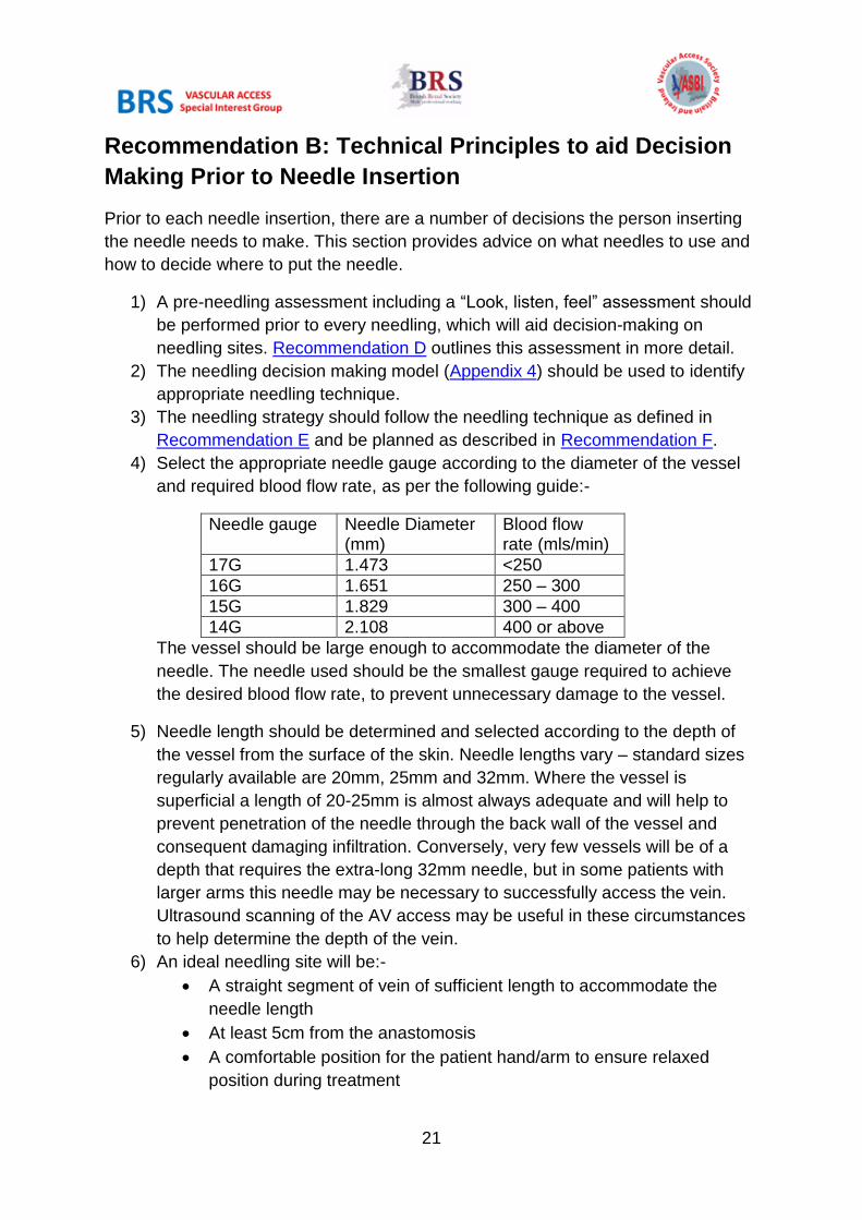
4) Select the appropriate needle gauge according to the diameter of the vessel and required
blood flow rate.
5) Needle length should be determined and selected according to the depth of the vessel from
the surface of the skin.
6) An ideal needling site will be:-
A straight segment of vein of sufficient length to accommodate the needle length
At least 5cm from the anastomosis
A comfortable position for the patient hand/arm to ensure relaxed position during
treatment
7) The identification of good sites for needling will depend very much on the length, depth,
tortuosity and general condition of the vessel in addition to any recent needling problems, as
well as the planned needling technique. This decision requires clinical judgement using the
skill and expertise of the person inserting the needle.
8) The following areas should be avoided, regardless of the needling plan:-
Swollen, hard or bruised areas
Areas with any scabs / damaged skin integrity
Red/inflamed areas, erythema, oozing
If possible, aneurysmal areas should be avoided.
9) Needle sites should be positioned at least 5cm apart, if possible, to avoid recirculation.
10) A tourniquet should be used for all needle insertions into AV fistulae, unless the AV fistula is
aneurysmal. A tourniquet is not needed for AV grafts.
11) The needle should be inserted bevel up.
12) The angle of insertion of the needle should be dictated by the minimum angle needed to
reach the depth of the vessel to allow the needle tip to sit in the centre of the vessel.
13) Ideally both needles should be inserted in an antegrade direction, with the needle pointing
away from the anastomosis / with the flow of blood. If required, the arterial needle can be
inserted in a retrograde direction, with the needle facing towards the anastomosis / against
the flow of blood.
14) Once the needle has been inserted, the needle should not be routinely rotated. This should
be avoided if possible, as this can lead to damage to the vessel wall known as ‘coring’.
6
Recommendation C: Procedural Principles for Good Needle Insertion
When inserting the needle, there are various practice points that minimise complications during
needle insertion. This includes good infection control practices, securing the needle correctly to
prevent dislodgement and use of safety needles. The points are outlined in the recommendations
below.
1) Prior to needle insertion, patients should wash their hands and the AV access site. Octenidine
dihydrochloride wash is ideal to use and is preferable to soap and water.
2) Safety needles should be used in all needle insertions to prevent the risk of needle stick
injuries to healthcare workers.
3) Healthcare staff performing needle insertion should wear either sterile or non-sterile gloves,
dependant on local policy.
4) If non-sterile gloves are worn, the needle site should not be touched once disinfected prior to
needle insertion.
5) Prior to needle insertion, the planned needle insertion site should be cleaned using 2%
chlorhexidine gluconate with 70% isopropyl alcohol, ensuring this has sufficient dry time (at
least 30 seconds) prior to initiating needle insertion. If the patient is allergic to chlorhexidine,
then Povidone Iodine solutions or octenidine dihydrochloride can be used to disinfect prior to
needle insertion.
6) The principles of Aseptic Non-Touch technique (ANTT) should be adhered to during the
needle insertion procedure. This includes:
a. Handwashing prior to the procedure
b. Use of an aseptic field. A sterile aseptic field is recommended for needle insertion i.e.
sterilised dressing pack, not a tray cleaned with disinfectant wipes
c. Key part protection, avoiding touching key parts and minimising exposure is essential
throughout the needle insertion.
7) Needles can either be flushed with 0.9% Saline or inserted dry, dependant on local policies. If
inserted dry, care needs to be taken to ensure the blood in the needle does not clot prior to
connection to haemodialysis.
8) Tape that covers the needle insertion site needs to be clean, which can be achieved through
a number of methods:
a. Sterile tape as part of the dressing pack used for needle insertion
b. Single use rolls of tape for individual patients.
Alternatively, gauze can be used over the needle insertion site or the needle insertion site can
be left exposed to avoid taping directly over this (Figure 1).
Rolls of tape should always be stored in a designated clean area and not in staff members’
pockets.
9) Following insertion, the needle should be taped either using the chevron method (Figure 2) or
H technique (Figure 3), to prevent needle dislodgement.
10) Once inserted, rotation of the needle should be avoided, to prevent ‘coring’ and damage to
the vessel wall.

7
Recommendation D: Assessment of the AV Access prior to Needling
1) Prior to needling, a history should be taken from the patient about the AV access. This should
include any changes since their last haemodialysis session and any problems during
haemodialysis sessions.
2) A look, listen, feel assessment of the AV access should be performed prior to needling, by the
person who will be inserting the needles (aka as the ‘needler’).
3) When inspecting the AV access, the ‘needler’ should visually observe the skin over the
vessel, the previous needling sites and the general condition of the limb.
4) Any signs of problems with increasing aneurysm size, new aneurysms, skin integrity or
infection should be escalated to medical / surgical teams immediately, to minimise the risk of
life threatening bleed.
5) The full length of vessel including the anastomosis should be palpated prior to disinfecting the
skin for needling.
6) Palpation of the vessel prior to needling allows identification of:
a. Direction of the vessel for needle insertion including identifying tortuosity
b. Length of vessel used for needle insertion
c. Diameter of the vessel
d. Depth of the vessel
e. Areas to avoid inserting the needle, including hard areas, areas where the vein dips
away and interference from collateral vessels
7) The thrill should be palpated prior to needling.
8) The bruit following the length of AV access should be auscultated using a stethoscope prior to
needle insertion.
9) The ‘Pre-Needle Insertion Assessment Tool’ document in Appendix 2 should be used to
record the look, listen, feel assessment and identify appropriate escalation pathways for
detection of complications.
10) It is recognised this describes a basic assessment prior to needle insertion. More in-depth
assessment of the AV access should occur 1-3 monthly using the ACCESS tool outlined in
Appendix 3.
11) It is recommended that haemodialysis nurses have an awareness of the signs of declining
access function to incorporate these into their regular assessment of the vessel.
Recommendation E: Definitions of Needling Techniques
1) We recommend use of rope ladder or buttonhole needling techniques and avoidance of area
puncture technique.
2) Selecting the needling technique to be used is an individual decision for each patient.
Recommendation F and Appendix 4 outline which technique is best utlised, in which situation.
We recommend rope ladder needling for both AV fistulae and grafts.
3) Rope ladder technique is defined as needling sites that move progressively up the vessel in a
systematic manner, with each needle site approximately 0.5-1cm above the previous site.
4) To perform rope ladder technique, the following criteria should be adhered to:
a. Rope ladder should utilise as much length of the vessel as possible, using the full
length of the vessel in a uniform manner
b. Once the highest needle site is reached, needling should start at the bottom of the
vessel again. We do not recommend moving up and down the vessel, as this can
easily degrade into area puncture.
c. Needling sites should cover:

8
i. A minimum of 8cm length of vessel if both arterial and venous needle sites
join up
ii. Minimum of 5cm if arterial and venous sites do not join up.
5) Rope ladder can be performed in 2 ways:
a. Zip rope ladder where needling sites are in the centre of vessel and each needling
site is above the previous site. This can be used with all AV fistulae and grafts.
b. Traditional rope ladder where needling sites move side to side as well as up the
vessel. This can be used with AV fistulae or graft with diameter wider than 0.9mm
and may minimise damage from repetitive needling over short lengths. However,
caution needs to be taken to perform this technique in a systematic manner.
c. Figure 4 demonstrates the 2 types of rope ladder.
6) How to best perform needling with rope ladder technique is outlined in Recommendation G.
7) Area puncture technique is used to categorise needling sites that do not meet the criteria for
rope ladder or butttonhole technique:
a. A needling site covers less than 5cm
b. Needling sites do not progress systematically up the vessel, but are ad hoc in a
similar area
8) Whilst we do not recommend area puncture, we recognise there may be situations where it is
impossible or unfeasible to avoid using area puncture technique. Recommendation I outlines
how to avoid area puncture and what to do if performing this technique.
9) We recommend buttonhole technique for use with AV fistulae.
10) Buttonhole needling involves needling each needling site in the same manner during each
needle insertion. This means at each needling site, entering the skin through the same hole,
following the same route to the vein and entering the vein in the same place each time. Figure
5 shows the elements of a buttonhole needling site.
11) Buttonhole needling involves:
a. Removing the scab from the previous needle insertion prior to the present needle
insertion
b. A track development phase where consistent needling with sharp needles is
performed
c. Needling with blunt needles once the track is developed
12) Buttonhole needling can involve 2-4 needling sites on one vessel. More than 2 needling sites
can allow resting of sites especially when patients dialyse more than 3 times a week.
13) How to best perform buttonhole needling is outlined in Recommendation H.
Recommendation F: Choosing the Needling Technique and Planning Needling
Recommendation F.1 – Choosing the Needling Technique
1) Decisions as to which needling technique should be made with the patient. Individual factors
and patient preference will influence the decision made. Whilst recommendations have been
made in this section to guide this decision, they should always be taken in context for
individual circumstances and patient choice.
2) Selecting the correct technique for individual patients is essential to ensure successful use of
the AV access for haemodialysis with minimal complications.
3) We recommend either rope ladder or buttonhole technique is chosen for AV fistulae and rope
ladder for AV grafts.
4) There are some AV fistula characteristics that will indicate either buttonhole or rope ladder
technique is better as follows:

9
a. Rope ladder technique (as outlined in Recommendation E) requires systematic
needle insertions over either:
i. At least 5cm segment for one needle insertion site
ii. At least 8cm segment where needle sites are on the same length of vessel
If this length of vessel is not available, then rope ladder is not possible. Buttonhole is
the recommended technique for short AV fistula segments, but it is recognised there
are situations where this is not possible. In this situation, area puncture may be the
only option and should be managed as per Recommendation I.
b. Buttonhole technique should be utilised in patients with a low infection risk. In patients
with a high infection risk, if the segment available is too short for rope ladder, then
area puncture may be the only option and should be managed as per
Recommendation I.
c. Buttonhole technique may be a better technique for patients with a vessel where
needle insertions are difficult or the patient is very anxious about needle insertions
due to a needle phobia. If these patients have a high infection risk, then rope ladder
may the better option. Again, in patients with a high infection risk, if the segment
available is too short for rope ladder, the area puncture may be the only options and
should be managed as per Recommendation I.
d. These factors are outlined in the Needling Decision Making Model in Appendix 4.
Units who do not utilise buttonhole technique, will inevitably require use of area
puncture technique, although area puncture is not recommended as a technique.
Recommendation F.2 – Infection Screening for Patients with AV Access
1) All patients undergoing needle insertion into AV access should undergo screening for MRSA
and MSSA including their AV access site, a minimum of every 3 months.
2) Decolonisation should occur for patients who are positive for MRSA. There is no maximum
recommended times that decolonisation should be undertaken, however twice is a common
maximum number. Decolonisation protocols should be pragmatic, taking consideration that
haemodialysis patients may undergo these on multiple occasions.
3) Patients should be individually risk assessed by the renal team before undertaking buttonhole
technique. A recommended infection screening tool for this purpose is included in Appendix 4
and based on work completed at Royal Berkshire renal unit (18).
4) Root cause analysis should be undertaken in all bacteraemia episodes in haemodialysis
patients.
Recommendation F.3 – Planning Needle Insertions
1) We recommend all patients who have needles inserted to AV access regularly have a plan for
needle insertions.
2) The needling plan should consist of paper documentation that needs to be completed by all
haemodialysis nursing staff inserting the needles. It should be designed to promote
communication to ensure consistent needle insertions between staff members.
3) The needling plan should be considered a guide rather than prescriptive piece of
documentation. The assessment pre-needle insertion may detect factors that warrant
divergence from the needling plan.
4) The needling plan should develop with each assessment of the AV access and individual staff
members should be encouraged to add their expertise to the plan, maintaining their autonomy
and decision making in this process.
10
5) The needling plan needs to include:
a. The arm position for optimal needle insertion
b. Images to demonstrate the needling section of the AV access, possibly with potential
needle sites.
c. Whether a tourniquet should be used
d. How to best insert the needle, including clearly defined information on both the depth
(angle from skin to hub) and the direction of needle insertion.
6) The needling plan should be designed to include patients in needling decisions, either being
accessible to patients or patient held.
7) Identifying suitable sites on a mature AV access undergoing rope ladder should also be given
due time and consideration to ensure uniform use and development of the full length of the
vessel, thus avoiding area puncture which may have damaging consequences. Experienced
staff should have the courage and confidence to attempt to use new sites along the vein.
Recommendation G: Rope Ladder Needling Technique
1) Rope ladder needle insertions should be planned with the same consideration as buttonhole,
as per Recommendation F.
2) Decide on needle type and size, as per Recommendation B.
3) Prior to needle insertion, assess the AV access using the look, listen, feel assessment, as per
Recommendation D. Decide on needling sites and direction and angle of needle insertion, as
per Recommendation E. To perform rope ladder, these should be 0.5-1cm above previous
sites, unless contra-indicated or commencing needling at the bottom of the vessel, as
previous sites reached the top of the needling segment.
4) Ensure the patient washes the area around and over the AV access prior to commencing
needle insertion.
5) Prepare aseptic field and don personal protective equipment (PPE) as per local policy,
including sterile / non-sterile gloves, a plastic apron and protective eye wear, as per
Recommendation C.
6) Cleanse the needling sites as per Recommendation C.
7) Apply tourniquet to AV fistula but not AV graft, as per Recommendation B.
8) If not performing dry needle insertion, prime the needles with Normal saline 0.9%, maintaining
a non-touch technique, as per Recommendation C.
9) Insert needle as determined during pre-needle insertion assessment, using a gentle, non-
touch technique, as per Recommendations A and Recommendation C. The needle should be
bevel up with both needles ideally facing antegrade, unless retrograde needling is used for
the arterial needle, as per Recommendation B.
10) Secure needles using chevron or ‘H’ taping technique, as per Recommendation C.
11) Avoid rotating the needle once inserted, as per Recommendation B.
12) Flush the needle to check for patency and to confirm adequate flow.
Recommendation H: Buttonhole Needling Technique
Recommendation H1: Track Development and Needle Insertion for Buttonhole
Needling of AV Fistulae
a) Track Development
1) The depth and direction of needle insertion need to be consistent during the track
development phase, to allow a consistent collagen track to develop.
11
2) A tourniquet should be used during track development, as per Recommendation B.
Tourniquet use should be consistent, with either use at all times or not at all.
3) Buttonhole track development should ideally occur with established AV fistulae only, to
ensure the vein does not change over time.
4) Track development on the arterial and venous needle sites can occur at different times,
dependant on the maturity of each site.
5) When the sharp needle glides in place with no resistance, dull/blunt needles can be
attempted. This should occur in 6-12 needle insertions using sharp needles.
6) During track development, the patient should be encouraged to discuss the sensations during
needling, recalling the positioning of the limb and hand that is optimal for them, to promote
information exchange between the patient and needler as to how to best insert the needles.
7) If the patient is planning to insert their own needles, then they should be encouraged to
develop the track themselves.
8) If the track is not established and blunt needles are unable to be inserted after 12 sessions of
sharp needle insertion, further assessment of the buttonhole sites should occur with
consideration as to whether different sites need to be developed.
9) Difficult track development, especially with deep tracks with a lot of subcutaneous tissue, can
be supported by insertion of polycarbonate pegs.
b) Buttonhole Sites
1) Buttonhole sites should adhere to the principles outlined in Recommendation B.
2) Avoid developing buttonhole sites on dips, curves, aneurysms on the fistula vein or any area
with abnormal skin integrity.
3) 1 patient can have 3-4 active buttonhole sites at one time, to allow rotation of sites, as per
Recommendation E.
4) Use of specific buttonhole sites should be reviewed in the following situations and cessation
of use of sites with these issues should be considered:
Hubbing of the site
Sharp needle insertion required regularly to cannulate the buttonhole site
Signs of infection at the site
Enlarging entry site or signs of tissue damage
Prolonged bleeding from buttonhole site
Significant pain/discomfort during insertion
c) Buttonhole Needle Insertion
1) Prior to needle insertion, the needler should encourage the patient to describe how the needle
is best inserted, promoting consistent needle insertion.
2) The arm and hand position should remain consistent through track development and further
blunt needle insertion to ensure alignment of the track and vein.
3) Communication should continue following the track development phase, to ensure all
needlers are aware of the track direction, as per Recommendation F.
4) The blunt needle should glide down the track and not require excessive force to cannulate.
The force applied during needling can be minimised by holding the tubing rather than the
needle wings during needling.
5) The external steel shaft of the needle should never be ‘wetted’ with sterile or non-sterile
solutions prior to insertion, as this practice increases the risk of contamination leading to a
potential infection.

12
6) On insertion of the needle, 1-2mm of steel should be visible to prevent hubbing of the needle
site.
7) The needle insertion should adhere to those principles outlined in Recommendations A, B,
and C.
d) Troubleshooting Buttonhole Needling
1) If the blunt needle is not entering the vein smoothly, check the arm and hand position of the
patient and track direction to ensure the needling technique and track position remains
consistent at all times.
2) If one blunt needle will not enter the vein, needling should be attempted with a second blunt
needle.
3) If a second blunt needle cannot be inserted, the needler who developed the track may insert a
sharp needle.
4) It is not advised that a sharp needle is used in the track by anyone other than those involved
in track development of that individual track. If a sharp needle insertion is performed by a
needler not involved in the track development, then this needle should be inserted at least 2
cm above the buttonhole site. If space is not available above the buttonhole site, then a site at
least 2cm below the buttonhole site can be used.
Recommendation H2: Disinfection Procedure and Scab Removal Prior to Buttonhole
Needling of AV Fistulae
1) All patients should wash their hands and fistula limb, as per Recommendation C.
2) The needling sites should be cleaned using solutions outlined in Recommendation C.
Needling sites should be disinfected immediately before and after scab removal.
3) Softening of scabs prior to removal is not recommended.
4) Sterile tweezers or sterile picks which are supplied with the dull/blunt needles or separately,
should be used to remove the scab.
5) To prevent infectious complications, the complete scab should be removed prior to needling
of the buttonhole site.
Recommendation H3: Mupirocin use with Buttonhole Needling of AV Fistulae
1) Topical 2% mupirocin ointment / cream should be applied to the needling sites of all patients
undergoing buttonhole technique, who are considered to have a high infection risk. The
ointment / cream should be applied following needle removal and cessation of bleeding from
needling site, after each haemodialysis treatment and left in place for approximately 12 hours.
2) All patients receiving 2% mupirocin ointment / cream regularly should undergo screening for
mupirocin resistant Staphylococcus Aureus.
3) Patients who develop mupirocin resistance must not continue to use 2% mupirocin, until
mupirocin sensitivity is restored. Each case should be risk assessed and consideration given
as to whether buttonhole technique should be discontinued or an alternative antibacterial
used.
13
Recommendation I: Area Puncture Needling Technique
1) Area puncture needling technique should be avoided wherever possible and we recommend the
use of rope ladder or buttonhole needling technique on all AV fistulae and grafts, whenever it is
possible.
2) Area puncture may be the only option on an AV fistula if:
a. There is not the length available to perform rope ladder
b. Buttonhole needling technique is not feasible, due to concerns about infection.
3) If area puncture is necessary in an AV fistula, the needling segment should cover as large an area
as possible and attempt to progress up the vein in a similar systematic manner to rope ladder
needling technique.
4) Area puncture should never be used on AV graft. An AV graft will always supply a suitable length
for rope ladder.
5) Area puncture of AV fistula is a better alternative for vascular access provision for haemodialysis,
than using a tunnelled CVC.
6) Needling of new AV fistula should be planned to avoid area puncture. Rope ladder is normally
feasible in new AV fistula, before aneurysms and tortuosity develop from poor area puncture.
7) Needling practice should be audited regularly, using the definitions developed in these
recommendations, to identify how many patients undergo rope ladder, buttonhole and area
puncture.
If area puncture of an AV fistula is identified as in use, the following conditions should apply:
8) Assessment of the vessel to ensure that an alternative needling technique cannot be employed.
9) Increased assessment and surveillance of the AV fistula should be implemented to monitor for the
signs of complications.
10) Refer the patient to the vascular team to assess alternatives to area puncture and assess the risk
of complications developing.
A flow chart outlining what to do if area puncture is identified in use on an AV fistula is in Appendix 5.
Recommendation J: Needling of New AV Access
1) Newly developed fistula veins can be very delicate and fragile. Needling should be carried out
very carefully and as gently as possible and only by experienced nurses.
2) AV fistula should be considered mature to needle when the vessel diameter reaches 0.6cm.
3) AV grafts should be first needled 2-4 weeks after insertion, once swelling and bruising has
settled in the surrounding tissues. ’Early cannulation’ AV grafts can be needled within 24
hours, dependant on manufacturer instructions.
4) The patient should be prepared for the first needle insertion as outlined in Recommendation
M.
5) The person inserting the needle will need to decide whether to attempt one or two needle
insertions
6) If one needle only is inserted into the AV access, then haemodialysis can either be performed
using single needle dialysis or using one CVC lumen as one point of access.
7) The first needle insertion for new AV access should be performed by nursing staff
experienced at needle insertion.
8) If only one needle is inserted successfully, a plan should be developed for progression to
double needle dialysis promptly within 1-2 weeks, ensuring adequate dialysis. As soon as the
vessel is mature enough, 2 needles should be inserted for each haemodialysis session.

14
9) Initial needle insertion into the AV fistula may need to be nearer to the anastomosis, to ensure
successful needle insertion. As the vein is cannulated more, needle sites can be moved
further from the anastomosis.
10) Buttonhole track development is not recommended until the AV fistula is mature and
established for use in haemodialysis, as per Recommendation H.
11) 17g needles should be used for the first 3-6 needle insertions into AV fistulae. If, after two
weeks, needling has been problem free, then progression to 16G will be appropriate, whilst
also increasing blood flow rates as above.
12) Needle gauge for AV grafts should follow manufacturer instructions. Early cannulation AV
graft may require small needles (17g) for early needle insertions, whilst other AV grafts may
be able to commence needle insertion using 15g needles.
13) CVC may remain in-situ whilst commencing use of AV access. This provides an alternative
form of access if needle insertion is problematic or may be used in conjunction with the AV
access as one form of arterial / venous line access. However, haemodialysis nurses need to
consider safe management of the CVC when not utilised for haemodialysis and we
recommend prompt removal of the CVC once use of the AV access is established.
Recommendation K: Use of Nurse-Led Ultrasound (US) to Assist with Needling
1) US images can be used:
a. To assess maturity of the AV fistula vein, to identify if it is ready for needling
b. To assess the AV fistula vein / graft prior to needling if it is difficult to palpate or has
previously been difficult to cannulate
c. To view the AV fistula vein / graft during needling, undertaking US imaging concurrently
with needling, allowing assessment of the vessel’s response to needle insertion.
2) US assessment of an AV access should complement clinical assessment and never replace the
initial look, listen, feel clinical assessment.
3) US images interpreted by a registered nurse to demonstrate abnormal findings should be referred
to an experienced US practitioner for interpretation.
4) Nurses who use and interpret US images need training on how to use the US machine correctly,
how to interpret the US images and how to detect signs of complications or abnormal anatomy.
5) US imaging in this context is only recommended for use by experienced, senior registered nurses.
These are nurses defined as those who have achieved the ‘Gold’ Standard of the ‘VASBI / BRS
VA National Needling Competency’.
Recommendation L: Managing Anxiety during Needling
1) Preparation information: all patients should have access to written and/or web based
information about having an AV access.
2) Needle desensitization: There will be some children, young people and adults who will
have a fear of needles. Desensitisation work may be needed over a longer period of time
with a gradual move to using a fistula needle.
3) Provide a calm environment: The environment can contribute greatly to any anxiety or
fear a child, young person or adult may be feeling when beginning to use their AV access.
Where possible try to create a calm and relaxed environment utilising equipment such as
music and fibre optics.
4) Written AV Access plans: Prior to accessing the AV access it is helpful for some
patients to feel they have some control over what happens when their AV access is used.

15
This can be achieved through a written plan devised with the child, young person or adult
giving them simple choices and responsibilities as part of the procedure.
5) Visual routine: Some children, young people and adults prefer to have a visual routine
using pictures or photographs of the procedure as this helps them to understand what
needs to happen during the routine of accessing their AV access.
6) Distraction: From the preparation sessions the child, young person or adult may decide
that they do not want to be involved directly in the procedure but would rather be
distracted.
Recommendation M: Involving Patients in Care of their Vascular Access
1) It is critical that patients have the opportunity to become involved with the care and
management of their vascular access as early as possible, ideally in the preparation stages
before starting haemodialysis. The ideal time to begin this process is when a patient starts pre
dialysis education.
2) Pre-dialysis clinic discussions should outline vascular access options. The patient’s
involvement in this choice and the subsequent implementation of that vascular access care is
critical.
3) Educational material about vascular access should be available for all patients who are about
to begin dialysis.
4) Pre dialysis educational material should not just include information on their vascular access
but also what to expect from their first needle insertion or vascular access use alongside
connection to haemodialysis.
5) Information provided to patients on vascular access care and needling should be consistent
between staff and with local renal unit needling policies.
6) If the patient is keen to insert their own needles then they should be encouraged and assisted
to do this, using techniques recommended in Recommendation N.
7) Continuing careful assessment of the needling sites is critical for sustainable vascular access.
The engaged patient will be best placed to assess and note any changes or difficulties with
needling, facilitating timely and appropriate intervention. All patients should be shown how to
assess changes over time, especially in detecting complications.
8) In order for patients to increase their involvement in their vascular access care, there needs to
be a multi-disciplinary approach from the whole team.
Recommendation N: Teaching Patients how to Self-Needle
1) Before embarking on training for self-needling, ensure the patient is a willing participant in
inserting their own needles, giving them the chance to make an informed choice.
2) Timing needs to be carefully considered when to commence or even ask the patient about
inserting their own needles.
3) An initial step to self-needling may be to allow the patient to commence their training by
removing their needles, as this would allow them to get used to handling the needles and allow
them to gain confidence in holding the needles.
4) Needle insertion requires dexterity. Patients may require support from nursing staff to perform
some elements of the process.
5) Whilst buttonhole technique is often cited as the best technique for patients to insert their own
needles, we do not recommend buttonhole technique as the default technique for self-needling.
We recommend that the decision is made with the patient and the needling technique used
takes into consideration patient preference and clinical considerations, in line with

16
Recommendation F, as well as identifying which technique is easiest for the patient to
complete.
6) If patients do plan to self-needle using buttonhole technique, we recommend where possible the
patient develops their own ‘tracks’, as outlined in Recommendation H1.
7) Teaching a patient to cannulate is different from teaching nurses the same skill and the
approach to this is different. Patients should not be expected to learn needle insertion from
information designed for nursing staff.
8) Knowledge levels will differ within your patient groups meaning an individual teaching plan for
each patient is required.
9) The training to teach patients how to insert the needles themselves should include:
a. What the fistula / graft is and how it works
b. The differences between rope ladder, buttonhole and area puncture
c. The look, listen, feel assessment including recognising problems and how to escalate
problems
d. Feeling the vessel prior to needle insertion, becoming familiar with the structure of the
vessel and direction that the blood flow is going in.
e. The correct cleaning techniques and the importance of the skin site preparation
process, especially the appropriate drying times for the solution chosen.
f. Applying a tourniquet, if used
g. How to hold the needle for needle insertion
h. Guidance on how to insert the needle
i. How to flush the needle
j. How to tape the needle
10) The patient will need to be taught not just how to wash their hands and access, but also the
reasons why these procedures are carried out.
11) Confidence needs to be built in the patient who is learning this new and alien skill to them.
12) The time needed for training must be set aside and not rushed; the patient must feel they are
being given adequate time to learn these new skills.
13) Information for children and young adults will need to be tailored dependent on their
developmental age and ability, and revised as per local teaching protocol for different ages.
14) On-going support and patient understanding of problems and how to overcome them must also
be added to the teaching programme.
15) Once self-needling, the patient is likely to experience problems during difficult needle insertions.
Patients should be supported through difficult needle insertions so that they continue to needle
themselves. For home patients, this may mean occasional in-centre sessions or home sessions
with support from nursing staff.
16) As the patient becomes more familiar with the needle insertion procedure and gains in
confidence, there needs to be monitoring pathways to ensure proper technique is being
followed at all times. This should include regular re-assessment of technique of self-needling.
Recommendation O: Staff Training to Perform Needling of AV Access
1) All healthcare staff (registered or unregistered) who are learning to insert needles into AV
fistulae and grafts must have a theoretical understanding of:
i. What is an AV fistula and graft, including relevant anatomy and physiology
ii. Different needle insertion techniques, including their risks and complications
2) Following theoretical teaching, all healthcare staff (registered or unregistered) that are
learning to insert needles into AV fistulae and grafts should have a period of supervised
clinical practice, using staff experienced at this procedure to supervise learners.

17
3) An assessment of competency of needle insertion for AV fistulae and grafts should occur
for all healthcare staff (registered or unregistered), prior to performing this skill
independently. No-one should cannulate an AV access independently, without this
assessment.
4) All healthcare staff (registered or unregistered) who perform needle insertion on AV
fistulae and grafts should be:
i. Reassessed every 3 years
ii. Receive an annual theoretical update.
5) Regular monthly audits should occur of needling practice, to ensure everyday practice
adheres to infection control and local needle insertion protocols.

18
Clinical Practice Recommendations
Recommendation A: Principles of a Good Needling
Technique
1) A good needling technique will:
a. Minimise damage to the AV access during needling
b. Minimise complications from needling
c. Minimise pain and anxiety related to needling
2) A good needling technique will result in either a successful needling or failed
attempt at needling of the AV access; with the minimum amount of damage to the
vessel and surrounding tissue, whilst minimising pain and inspiring confidence in
the patient.
3) A good needling technique involves using either rope ladder or buttonhole
needling technique to plan needling.
4) Prior to needle insertion, documentation of previous needling should be reviewed
along with a documented needling plan.
5) Prior to needle insertion, a good assessment of the vessel should provide a clear
idea of the depth and direction of the needle insertion which will result in the
correct position of the needle. Needle insertion should not continue without this
clear idea of depth and direction.
6) If needle insertion is not performed by the patient, prior to needle insertion, the
‘needler’ should discuss previous needling attempts with the patient. The patient
should be encouraged to disclose their views on how to best insert the needle,
how previous needle insertions progressed from their perspective and what may
have caused previous problems.
7) The decision on where and how to insert the needle should occur in discussion
with the patient and include consideration of:
a. How to best perform either buttonhole or rope ladder technique
b. Directions from the needling plan
c. Conclusions from their assessment of the vessel
d. The patient’s opinions on how the needle insertion should be performed.
8) Once entering the skin, the needle insertion route should take the most direct
route to the vein and not follow a tortuous route to the vein.
9) The needle insertion movement should be accurate, considered, gentle and
continuous, minimising pain and discomfort for the patient.
10) Needle insertion is a balance between prompt insertion of the needle and a gentle
technique, so whilst insertion should not be rapid, it also should not be
unnecessarily prolonged.
11) The needle insertion should aim to finish with the tip of the needle in the centre of
the AV access vessel.

19
12) If insertion is unsuccessful at first attempt, then further attempts may be required
through retraction and reinsertion of the needle. If the needle is fully withdrawn
from the skin at any time during the needling attempt, or clotting of the needle
lumen is suspected, then another unused, clean needle must always be used to
continue the attempt at needling, to prevent infection from an already used needle.
Once fully removed away from the skin, a used fistula needle must never be
reinserted into the skin above the vessel during a needle insertion attempt.
13) Once an individual has attempted 2 unsuccessful needle insertions on one
occasion at the same site, if available, they should gain help from another staff
member who can needle, rather than persevering with the needle insertion
themselves.
14) Registered nurses and non-registered staff who cannulate AV access should
constantly aspire to develop a good needling technique. This is on-going
development of a skill that is never complete. Skill development can progress
through regular needling practice, observation of and guidance from experienced,
skilled needlers.
15) A good needling technique requires self-awareness, with the person inserting the
needle acknowledging their limitations. Staff who needle need awareness of the
type of AV fistula / graft they can needle successfully and ask for assistance with
AV access outside their skill level. However experienced guidance can be used to
help inexperienced staff insert needles into AV access outside their current skill
level and promote their skill development.
16) Needling should always be an empathetic procedure. The correct balance
between sympathy and pragmatism should acknowledge the pain and anxiety
caused by the procedure alongside recognising the necessity of the procedure.
Rationale for Recommendation A
It is imperative that needlers of AV access develop a good needling technique to
minimise damage to the AV access from repetitive needling and minimise the anxiety
and pain patients’ experience from repetitive needling.
Very little evidence has been produced as to what makes a good needling technique.
Harwood (15) performed a qualitative research study to explore this with haemodialysis
nurses. They identified multiple aspects that contribute to successful needle insertion,
including (15):
Accurate assessment of the AV access prior to needling
Involving patients in their needling, which they named patient-centred care
Practising the needling skill regularly
Teamwork, using experienced needlers to support skill development in less
experienced needlers

20
Self-awareness in needlers of their own needling skill level
Correct training of staff who will insert needles into AV access.
Points for Future Consideration
Needling is a little explored phenomena that is about more than the physical act of
inserting the needle (15). Research is urgently needed to examine the patient’s
experience of needling, what leads to successful needling and how to best develop a
good needling technique. Any work that examines needling practices for AV access used
for haemodialysis always needs to consider the patient’s perspective of needling as part
of the evaluation, with consideration that this is more complex than just a pain score.
Concepts raised in this section will be further explored throughout the recommendations.
21
Recommendation B: Technical Principles to aid Decision
Making Prior to Needle Insertion
Prior to each needle insertion, there are a number of decisions the person inserting
the needle needs to make. This section provides advice on what needles to use and
how to decide where to put the needle.
1) A pre-needling assessment including a “Look, listen, feel” assessment should
be performed prior to every needling, which will aid decision-making on
needling sites. Recommendation D outlines this assessment in more detail.
2) The needling decision making model (Appendix 4) should be used to identify
appropriate needling technique.
3) The needling strategy should follow the needling technique as defined in
Recommendation E and be planned as described in Recommendation F.
4) Select the appropriate needle gauge according to the diameter of the vessel
and required blood flow rate, as per the following guide:-
Needle gauge Needle Diameter (mm)
Blood flow rate (mls/min)
17G 1.473 <250
16G 1.651 250 – 300
15G 1.829 300 – 400
14G 2.108 400 or above
The vessel should be large enough to accommodate the diameter of the
needle. The needle used should be the smallest gauge required to achieve
the desired blood flow rate, to prevent unnecessary damage to the vessel.
5) Needle length should be determined and selected according to the depth of
the vessel from the surface of the skin. Needle lengths vary – standard sizes
regularly available are 20mm, 25mm and 32mm. Where the vessel is
superficial a length of 20-25mm is almost always adequate and will help to
prevent penetration of the needle through the back wall of the vessel and
consequent damaging infiltration. Conversely, very few vessels will be of a
depth that requires the extra-long 32mm needle, but in some patients with
larger arms this needle may be necessary to successfully access the vein.
Ultrasound scanning of the AV access may be useful in these circumstances
to help determine the depth of the vein.
6) An ideal needling site will be:-
A straight segment of vein of sufficient length to accommodate the
needle length
At least 5cm from the anastomosis
A comfortable position for the patient hand/arm to ensure relaxed
position during treatment
22
7) The identification of good sites for needling will depend very much on the
length, depth, tortuosity and general condition of the vessel in addition to any
recent needling problems, as well as the planned needling technique. This
decision requires clinical judgement using the skill and expertise of the person
inserting the needle.
8) The following areas should be avoided, regardless of the needling plan:-
Swollen, hard or bruised areas
Areas with any scabs / damaged skin integrity
Red/inflamed areas, erythema, oozing
If possible, aneurysmal areas should be avoided.
9) Needle sites should be positioned at least 5cm apart, if possible, to avoid
recirculation.
10) A tourniquet should be used for all needle insertions into AV fistulae, unless
the AV fistula is aneurysmal. A tourniquet is not needed for AV grafts. The
tourniquet should be applied for assessment of vessel depth and direction to
determine needle insertion. However, the tourniquet must never be applied
for longer than 1-2 minutes. To avoid long periods of application, the
tourniquet may need to be released and reapplied during difficult needle
insertions.
11) The needle should be inserted bevel up.
12) The angle of insertion of the needle should be dictated by the minimum angle
needed to reach the depth of the vessel to allow the needle tip to sit in the
centre of the vessel. This decision requires clinical judgement using the skill
and expertise of the person inserting the needle.
13) Ideally both needles should be inserted in an antegrade direction, with the
needle pointing away from the anastomosis / with the flow of blood. If
required, the arterial needle can be inserted in a retrograde direction, with the
needle facing towards the anastomosis / against the flow of blood. This
decision requires clinical judgement using the skill and expertise of the person
inserting the needle.
14) Once the needle has been inserted, the needle should not be routinely
rotated. This should be avoided if possible, as this can lead to damage to the
vessel wall known as ‘coring’.
Rationale for Recommendation B
The decision making process prior to needle insertion is complex, requiring the skill
and knowledge of the person inserting the needle. Each needle insertion will be
individual, according to the anatomy of the individual and the current condition of the
23
vessel. However there are a number of technical principles that can be applied to
assist in this decision making.
Fulker (19) identified that placing the needle tip in the centre of the lumen of the
vessel created less turbulent flow and shear wall stress, which theoretically reduces
the formation of stenosis in the vessel. Using a shallow angle also reduced
turbulence (19). This theory is developed from computer simulations and has not
been tested on real vessels. However, it is in line with sensible needling practice.
Achieving the needle tip in the centre of the vessel will maximise flow rates, minimise
flow problems and reduce the risk of infiltration. A shallow angle reduces the risk of
infiltration when guiding the needle along the vessel length, as does use of shorter
needles. Whilst there is little research evidence for this practice, it was identified that
common practices we have recommended minimise damage to the vessel during
needle insertion, including:
Using the smallest needle gauge required
Inserting the needle bevel up (20)
Antegrade needle insertion (20)
Needling a distance away from the anastomosis
Avoid rotating the needle once insitu (20).
Whilst other recommendations have prescribed needle angles (6, 21), current
evidence dictates that the most important aspect of needle insertion is to insert the
needle into the centre of the vessel successfully. This requires clinical judgement of
angle in relation to the depth of the vessel, not a prescriptive depth that ignores
individual variation in vessel depth. Needles should be inserted ideally 5cm apart to
optimise clearance during the haemodialysis treatment (22).
Areas on the vessel where there are signs of complications should be avoided, to
prevent exacerbation of complications. Ideally, aneurysmal areas should be avoided
to prevent exacerbation of the aneurysm formation. However, this is not always
possible in a tortuous and aneurysmal fistula. Aneurysmal fistula will often need to
undergo area puncture, so should follow the recommendation given in
Recommendation I.
Tourniquet use is thought to dilate the vessel making the needle insertion easier and
less painful. However, as an AV graft is a rigid tube, the dilation effect with a
tourniquet does not occur, meaning there is no benefit to tourniquet use in this
context. Tourniquet use does compress the vessel to achieve dilation, hence why
prolonged compression is not recommended to prevent thrombosis from static blood
in the vessel.

24
Points for Future Consideration
Little research evidence is available for many of the principles referenced to. Often
what is decided as good in day to day clinical practice is correct, due to real time
feedback on the results. However, these principles still need testing. Laboratory
simulation is useful to identify good principles. However this approach is limited as
they are not applied to ‘real-world’ settings. Simulated findings need to be interpreted
with caution with consideration of application into clinical practice. We recommend
further research into these principles in rigorous but simple studies, alongside
common sense interpretation of results by needling experts.
Plastic cannulas to replace metal needles are a new innovation introduced to allow
limb movement during haemodialysis, increase patient comfort and reduce
infiltrations. Some units are implementing plastic cannulas to minimise these
complications. It is strongly recommended that service evaluations of using these
cannulas, including patient experience measures, are performed. Units are cautious
of use, due to expense and unknown complications. If this practice is beneficial, then
the benefits need to be identified and shared to improve future practice.

25
Recommendation C: Procedural Principles for Good
Needle Insertion
When inserting the needle, there are various practice points that minimise
complications during needle insertion. This includes good infection control practices,
securing the needle correctly to prevent dislodgement and use of safety needles.
The points are outlined in the recommendations below.
Clinical Practice Recommendations
1) Prior to needle insertion, patients should wash their hands and the AV access
site. Octenidine dihydrochloride wash is ideal to use and is preferable to soap
and water. However, if octenidine dihydrochloride wash is not available or the
patient is allergic to this, then soap and water is the next suitable alternative.
Whilst chlorhexidine washes will disinfect the area, there is concern that this
will be too abrasive to use on the skin regularly. If the patient is unable to
access a sink, skin cleansing wipes are an alternative.
2) Safety needles should be used in all needle insertions to prevent the risk of
needle stick injuries to healthcare workers. Safety needles include blunt
needles for buttonhole needling or sharp needles with a protection or
retraction device to cover the needle once removed. Sharp safety principles
should always be adhered to during needle insertion and removal.
3) Healthcare staff performing needle insertion should wear either sterile or non-
sterile gloves, dependant on local policy. This may differ for AV fistulae and
graft needle insertion, with some units preferring sterile gloves for AV grafts
needle insertion.
4) If non-sterile gloves are worn, the needle site should not be touched once
disinfected prior to needle insertion. If the needle site requires touching
following disinfection, for example in a difficult needle insertion, then sterile
gloves should be worn.
5) Prior to needle insertion, the planned needle insertion site should be cleaned
using 2% chlorhexidine gluconate with 70% isopropyl alcohol, ensuring this
has sufficient dry time (at least 30 seconds) prior to initiating needle insertion.
If the patient is allergic to chlorhexidine, then Povidone Iodine solutions or
octenidine dihydrochloride can be used to disinfect prior to needle insertion.
6) The principles of Aseptic Non-Touch technique (ANTT) should be adhered to
during the needle insertion procedure. This includes:
a. Handwashing prior to the procedure
b. Use of an aseptic field. A sterile aseptic field is recommended for
needle insertion i.e. sterilised dressing pack, not a tray cleaned with
disinfectant wipes
c. Key part protection, avoiding touching key parts and minimising
exposure is essential throughout the needle insertion. Key parts

26
include the ends of syringes, both ends of the needle, the skin once
disinfected and tape if it covers the needle site. Once disinfected the
skin around the needle insertion site can be touched once wearing
gloves, but the needle insertion site should not be directly touched.
7) Needles can either be flushed with 0.9% Saline or inserted dry, dependant on
local policies. If inserted dry, care needs to be taken to ensure the blood in the
needle does not clot prior to connection to haemodialysis.
8) Tape that covers the needle insertion site needs to be clean, which can be
achieved through a number of methods:
a. Sterile tape as part of the dressing pack used needle insertion
b. Single use rolls of tape for individual patients.
Alternatively, gauze can be used over the needle insertion site or the needle
insertion site can be left exposed to avoid taping directly over this (Figure 1).
Rolls of tape should always be stored in a designated clean area and not in
staff members’ pockets.
9) Following insertion, the needle should be taped either using the chevron
method (Figure 2) or H technique (Figure 3), to prevent needle dislodgement.
The H technique may be easier for patients who cannulate themselves.
10) Once inserted, rotation of the needle should be avoided, to prevent ‘coring’
and damage to the vessel wall.
Figure 1 Taping Method using Gauze over the needle site

27
Figure 2: Poster from Oxford Renal Unit demonstrating safe taping methods
Figure 3: Image from Manchester renal unit demonstrating the H technique
Rationale for Recommendation C
Good infection control practice for needle insertion is essential to prevent infections.
Whilst AV access is considered to have lower infection rates than CVC, it remains an
invasive procedure that can lead to life-threatening infections. There is much
disagreement as to the best infection control practices, with little evidence base for
various practices. Practice varies across units and discussion indicated that glove
selection, flushing of needles with 0.9% Saline and taping practice varies with little

28
rationale for this. However, using a aseptic non-touch approach was agreed to be
most likely to reduce infection.
The solution used to disinfect needling sites is also believed to be important in
preventing infections. The EPIC guidelines (23) recommend 2% chlorhexidine
gluconate / 70% isopropyl alcohol as this has a rapid (30 seconds) and persistent
(up to 48 hours) antimicrobial activity on the skin. Povidone iodine can be used to
disinfect skin prior to needling, but needs to be applied for 2-3 minutes for its full
bacteriostatic action to take effect and must be allowed to dry prior to needling.
Therefore, whilst used routinely by some units (10), this is a less pragmatic
disinfectant but can be used if the patient is allergic to chlorhexidine. However, it is
important to ensure when using products for skin preparation that manufacturers
advice is adhered to which should include technique of application, contact time and
drying times to effectively kill bacteria. Octenidine dihydrochloride has been used by
1 unit when chlorhexidine sensitivity developed, with good results (24). Water or
0.9% saline solution (even if it sterile) has no disinfectant properties and is not
recommended.
Correct taping of the needles is also essential to prevent venous needle
dislodgement (25). Again, variation still occurs across the UK, but the taping
techniques recommended are considered best practice. Majority of units use a
chevron / butterfly techniques, but H technique has been identified as safe and easy
technique for patients who insert their own needles.
Points for Future Consideration
There continues to be variation across the UK and disagreement as to the best
infection control practices to prevent infection during needle insertions. Studies need
to examine the following points:
It is unclear whether 0.5% or 2% chlorhexidine (both with 70% isopropyl
alcohol) is the best cleaning solution to use pre needling of AV access. Whilst
2% is recommended in many general guidelines, it is unclear whether the
repetitive use on skin may cause complications related to either skin necrosis
or sensitivity (25). Work needs to be done on what is the optimal cleaning
solution for regular use on AV access. 0.5% chlorhexidine / 70% isopropyl
alcohol is used in some centres within the UK, with no cited problems with
infections.
Octenidine dihydrochloride has been identified by one unit as an appropriate
cleaning solution for patients with chlorhexidine allergy (24). Further
investigation needs to occur to identify if this could be suitable alternative
disinfectant to chlorhexidine.

29
It is unclear whether use of sterile gloves is necessary for needle insertion or
whether a non-touch technique suffices. Some units use non-sterile gloves for
all needle insertions, whilst others use sterile gloves for AV graft needle
insertions. Units also vary as to whether those who insert their own needles
should wear gloves. Work needs to be done to identify the best technique.
Flushing of needles with 0.9% Saline varies across the UK. Some units do not
use 0.9% Saline flushes prior to needle insertion, predominantly as it can
introduce a further vector for infection. However some units use 0.9% Saline
flush to prevent clotting in the needle. Neither approach is evidenced and
further work is needed to identify best practice.
30
Recommendation D: Assessment of the AV Access prior to
Needling
1) Prior to needling, a history should be taken from the patient about the AV
access. This should include any changes since their last haemodialysis
session and any problems during haemodialysis sessions. Difficulty with
needle insertion, aspiration of clots and prolonged bleeding can all be signs of
developing complications in an AV access.
2) A look, listen, feel assessment of the AV access should be performed prior to
needling, by the person who will be inserting the needles (aka as the
‘needler’).
3) When inspecting the AV access, the ‘needler’ should visually observe the skin
over the vessel, the previous needling sites and the general condition of the
limb. This will identify:
a. Aneurysms
b. Compromised skin integrity over the AV access
c. Swelling
d. Rashes
e. Signs of infection including redness and oozing
f. Collateral veins.
The needler should inspect the colour of extremities, identifying any signs of
ischaemia indicating ‘steal’ syndrome.
4) Any signs of problems with increasing aneurysm size, new aneurysms, skin
integrity or infection should be escalated to medical / surgical teams
immediately, to minimise the risk of life threatening bleed.
5) The full length of vessel including the anastomosis should be palpated prior to
disinfecting the skin for needling. This allows assessment of the vessel for
needle insertion and function of the AV access.
6) Palpation of the vessel prior to needling allows identification of:
a. Direction of the vessel for needle insertion including identifying
tortuosity
b. Length of vessel used for needle insertion
c. Diameter of the vessel
d. Depth of the vessel
e. Areas to avoid inserting the needle, including hard areas, areas where
the vein dips away and interference from collateral vessels
7) The thrill should be palpated prior to needling. This should be a vibrating
sensation, not a pulsation or thumping sensation. The thrill should lessen as
palpation progresses up the vessel.
8) The bruit following the length of AV access should be auscultated using a
stethoscope prior to needle insertion. This should be:

31
a. A whooshing noise that is uninterrupted and regularity is in line with the
patients’ normal pulse / heart rate
b. Gradually diminishes / fades / becomes quieter as you move up the
vessel, with no sudden interruption of the sound.
c. The pitch should be long and low, with no whistling or high pitch
sounds.
d. Asculated at both needling sites, otherwise the needling sites are too
far from the anastomosis
9) The ‘Pre-Needle Insertion Assessment Tool’ document in Appendix 2 should
be used to record the look, listen, feel assessment and identify appropriate
escalation pathways for detection of complications.
10) It is recognised this describes a basic assessment prior to needle insertion.
More in-depth assessment of the AV access should occur 1-3 monthly using
the ACCESS tool outlined in Appendix 3. ACCESS stands for:
a. Assessment
b. Cannulation
c. Circulation
d. Evaluation
e. Signs / symptoms
f. Surveillance
Further recommendations are planned for long-term surveillance, but this is
too complex to include in more detail in these recommendations.
11) It is recommended that haemodialysis nurses have an awareness of the signs
of declining access function to incorporate these into their regular assessment
of the vessel. These include:
a. Altered arterial / venous pressures
b. Reduced or interrupted blood flow rate
c. Reduced Kt/V / URR
d. Needling difficulties
e. Prolonged bleeding.
Intermittent surveillance results should be used to detect signs of altered flow
through the vessel, indicating stenosis development, allowing prompt referral
for salvage procedures before function is significantly reduced.
32
Rationale for Recommendation D
A good pre-needle insertion assessment and ongoing-surveillance of the AV access
is essential to:
a) achieve a good needle insertion (15)
b) detect and minimise complications (26).
The pre-needle insertion assessment allows identification of the direction and angle
of needle insertion, providing a clear planned trajectory of the needle. This includes
obtaining an accurate history from patients about previous needle experiences as
well as a physical look, listen, feel assessment of the vessel. This is more likely to
ensure success first time with needle insertion, with qualitative research identifying
this as a trait of successful needlers (15).
The pre-needle insertion assessment also ensures signs of complications are
detected promptly. The look, listen, feel assessment can identify signs of infection,
issues with skin integrity, signs of steal syndrome and alteration in flow through the
vessel indicating development of a stenosis. Detection allows prompt response to
these signs, providing treatment of the complication before it escalates to a life-
threatening issue or causes failure of the AV access.
More in-depth surveillance can be performed less frequently to also detect the signs
of complications, especially decline of function related to stenosis development.
Jackson et al (26) performed a systematic review of studies that examined physical
assessment and surveillance. They identified that both practices can be useful at
detecting complications, but combining both approaches as a complete assessment
of the vessel, provided the best sensitivity to detect complications. Using both these
tools can help you refer in a timely manner to the appropriate person/department for
further assessment or intervention to maintain the patency of the AV access.
Consensus agreement indicates that the obtaining a patient history of needling and a
basic look, listen, feel assessment provides an essential basis for detecting
complications and interpreting more in-depth surveillance results.

33
Recommendation E: Definitions of Needling Techniques
1) We recommend use of rope ladder or buttonhole needling techniques and
avoidance of area puncture technique.
2) Selecting the needling technique to be used is an individual decision for each
patient. Recommendation F and Appendix 4 outline which technique is best
utilised, in which situation. We recommend rope ladder needling for both AV
fistulae and grafts.
3) Rope ladder technique is defined as needling sites that move progressively up
the vessel in a systematic manner, with each needle site approximately 0.5-
1cm above the previous site.
4) To perform rope ladder technique, the following criteria should be adhered to:
a. Rope ladder should utilise as much length of the vessel as possible,
using the full length of the vessel in a uniform manner
b. Once the highest needle site is reached, needling should start at the
bottom of the vessel again. We do not recommend moving up and
down the vessel, as this can easily degrade into area puncture.
c. Needling sites should cover:
i. A minimum of 8cm length of vessel if both arterial and venous
needle sites join up
ii. Minimum of 5cm if arterial and venous sites do not join up.
5) Rope ladder can be performed in 2 ways:
a. Zip rope ladder where needling sites are in the centre of vessel and
each needling site is above the previous site. This can be used with all
AV fistulae and grafts.
b. Traditional rope ladder where needling sites move side to side as well
as up the vessel. This can be used with AV fistula or graft with
diameter wider than 0.9mm and may minimise damage from repetitive
needling over short lengths. However, caution needs to be taken to
perform this technique in a systematic manner.
c. Figure 4 demonstrates the 2 types of rope ladder.
6) How to best perform needling with rope ladder technique is outlined in
Recommendation G.
7) Area puncture technique is used to categorise needling sites that do not meet
the criteria for rope ladder or buttonhole technique:
a. A needling site covers less than 5cm
b. Needling sites do not progress systematically up the vessel, but are ad
hoc in a similar area
8) Whilst we do not recommend area puncture, we recognise there may be
situations where it is impossible or unfeasible to avoid using area puncture
technique. Recommendation I outlines how to avoid area puncture and what
to do if performing this technique.

34
9) We recommend buttonhole technique for use with AV fistulae.
10) Buttonhole needling involves needling each needling site in the same manner
during each needle insertion. This means at each needling site, entering the
skin through the same hole, following the same route to the vein and entering
the vein in the same place each time. Figure 5 shows the elements of a
buttonhole needling site.
11) Buttonhole needling involves:
a. Removing the scab from the previous needle insertion prior to the
present needle insertion
b. A track development phase where consistent needling with sharp
needles is performed
c. Needling with blunt needles once the track is developed
12) Buttonhole needling can involve 2-4 needling sites on one vessel. More than
2 needling sites can allow resting of sites especially when patients dialyse
more than 3 times a week.
13) How to best perform buttonhole needling is outlined in Recommendation H.
Figure 4: 2 Types of Rope Ladder

35
Figure 5: Buttonhole Needle Site
Rationale for Recommendation E
Kronung (9) first described rope ladder, buttonhole and area puncture technique. In
his article, he hypothesised that rope ladder and buttonhole led to better preservation
of AV access vessel through minimising aneurysm and stenosis development.
Stenosis is well accepted as the biggest cause of AV access failure and needling
techniques that avoid aneurysm and stenosis formation are optimal. Since then
needling culture has developed to recommend rope ladder and buttonhole technique
as superior to area puncture.
Various studies have examined the efficacy of buttonhole and rope ladder technique,
as demonstrated in various systematic reviews, including Ren et al (11), Wong et al
(27), Muir et al (28), Kotwal et al (29) and Grudzinski et al (30). However, strong
evidence cannot be drawn due to the poor quality of studies (27, 30). 2 systematic
reviews have produced conflicting recommendations with Ren et al (11)
recommending buttonhole as the standard technique (in China), whilst Wong et al
(27) recommend avoidance of buttonhole unless necessary (in Canada).
5 randomised control trials have been completed to compare various needling
technique:
MacRae et al in Canada (31, 32) – rope ladder to buttonhole
Vaux et al in Reading, UK (33) – buttonhole to standard practice
Chow et al in Australia (34) – rope ladder to buttonhole
Struthers et al in Scotland (35) – buttonhole to rope ladder
Toma et al (36) – buttonhole to standard practice
Results from these studies disagree, making recommendations based on these
findings difficult.

36
Whilst many studies have examined buttonhole technique, there is less evidence to
show that rope ladder leads to better AV fistula or graft survival in comparison to
area puncture. Parisotto et al’s (37, 38) observational study identified that both
buttonhole and rope ladder have less risk of complications than area puncture and
Rajoo (39) identified that area puncture was associated with increased complication
rate. Current practice accepts that avoidance of area puncture with a preference for
buttonhole or rope ladder technique is best practice. Other current recommendations
all recommend rope ladder and buttonhole techniques with avoidance of area
puncture (3, 6, 21). All contributors are aware of anecdotal cases where area
puncture has led to significant problems with AV access.
Historically, claims have been made that units perform rope ladder. Consensus
opinion agrees that few patients actually receive rope ladder and the majority receive
area puncture. Vaux et al (33) recognise this dilemma and Parisotto’s (20)
international study, including the UK, identified only 28.2% of patients receive rope
ladder, where as 65.8% receive area puncture.
For the first time, we have endeavoured to define rope ladder from area puncture
technique. This has never been done before and with no evidence base, these
definitions are developed through consensus opinion.
Points for Future Consideration
As highlighted, the research base for needling techniques, especially that rope
ladder is better than area puncture, is poor. Studies are needed to compare needling
techniques. Comparison between buttonhole and rope ladder needs to be careful to
ensure both techniques are performed correctly and consistently. Without this
assurance, studies cannot claim to compare the 2 techniques.
Currently we have no concept as to the frequency of use of each technique within
the UK. Majority of units will claim that rope ladder is their pre-dominant technique.
However this cannot be assumed to be correct without application of definitions that
delineate area puncture from rope ladder. Units should apply these definitions to
audit their needling practice; ensuring buttonhole and rope ladder are performed
consistently. An example of how this audit can change practice was demonstrated by
Lister Renal unit through a poster presentation at BRS conference in 2017 (40).
National data, using these definitions, is needed to identify needling practice patterns
and guide quality improvement initiatives in this area.
Buttonhole technique is currently only recommended for AV fistulae and not for AV
grafts. This is based on the theoretical high risk of infection and graft rupture if
buttonhole is used on AV grafts. Reported experience from a single centre indicated
that buttonhole technique in carefully selected patients was successful (41, 42).

37
However, clinical trials and qualitative patient focussed studies are needed to
evaluate the benefits and risks related to buttonhole and rope ladder on AV grafts.

38
Recommendation F: Choosing the Needling Technique and
Planning Needling
Recommendation F.1 – Choosing the Needling Technique
1) Decisions as to which needling technique should be made with the patient.
Individual factors and patient preference will influence the decision made.
Whilst recommendations have been made in this section to guide this
decision, they should always be taken in context for individual circumstances
and patient choice.
2) Selecting the correct technique for individual patients is essential to ensure
successful use of the AV access for haemodialysis with minimal
complications. We do not recommend ‘blanket’ unit-wide use of buttonhole or
rope ladder without individual consideration of each patient’s circumstances.
3) We recommend either rope ladder or buttonhole technique is chosen for AV
fistulae and rope ladder for AV grafts.
4) There are some AV fistula characteristics that will indicate either buttonhole or
rope ladder technique is better as follows:
a. Rope ladder technique (as outlined in Recommendation E) requires
systematic needle insertions over either:
i. At least 5cm segment for one needle insertion site
ii. At least 8cm segment where needle sites are on the same
length of vessel
If this length of vessel is not available, then rope ladder is not possible.
Buttonhole is the recommended technique for short AV fistula
segments, but it is recognised there are situations where this is not
possible. In this situation, area puncture may be the only option and
should be managed as per Recommendation I.
b. Buttonhole technique should be utilised in patients with a low infection
risk. In patients with a high infection risk, if the segment available is too
short for rope ladder, then area puncture may be the only option and
should be managed as per Recommendation I.
c. Buttonhole technique may be a better technique for patients with a
vessel where needle insertions are difficult or the patient is very
anxious about needle insertions due to a needle phobia. If these
patients have a high infection risk, then rope ladder may the better
option. Again, in patients with a high infection risk, if the segment
available is too short for rope ladder, the area puncture may be the
only options and should be managed as per Recommendation I.
d. These factors are outlined in the Needling Decision Making Model in
Appendix 4. Units who do not utilise buttonhole technique, will

39
inevitably require use of area puncture technique, although area
puncture is not recommended as a technique.
Rationale for Recommendation F.1
These recommendations have been developed based on criteria outlined in
Recommendation E.
To add to the evidence discussed in Recommendation E, it has been identified that
excluding patients with a high infection risk from buttonhole technique negates the
risk of serious infections (43). This has been identified within units who have
contributed to these recommendations, including work disseminated by Royal
Berkshire renal unit via conference abstracts (18).
Buttonhole technique has been associated with less painful needle insertion (44-49),
although the evidence for this is not completely conclusive. Rope ladder has been
identified as having a higher rate of acute complications including multiple needle
attempts and haematoma (48, 50) and is more stressful for patients (47). It is
indicated that buttonhole is a better option for patients who are anxious or
experience difficult needle insertions (51).
For children, it is recognised that it may be difficult to perform rope ladder due to the
short length of vessel. Buttonhole needling is used in some units now to avoid area
puncture and findings in one study has shown that use of buttonhole has been
successful in children (52, 53).
Recommendation F.2 – Infection Screening for Patients with AV
Access
1) All patients undergoing needle insertion into AV access should undergo
screening for MRSA and MSSA including their AV access site, a minimum of
every 3 months.
2) Decolonisation should occur for patients who are positive for MRSA. There is
no maximum recommended times that decolonisation should be undertaken,
however twice is a common maximum number. Decolonisation protocols
should be pragmatic, taking consideration that haemodialysis patients may
undergo these on multiple occasions.
3) Patients should be individually risk assessed by the renal team before
undertaking buttonhole technique. The following factors should be considered
as to whether buttonhole technique is safe to use or should be avoided:
i. MSSA and MRSA positive patients (until negative from decolonisation)
ii. Patients with mupirocin-resistant strains of Staphylococcus Aureus

40
iii. Patients with a history of recurring infections, particularly vascular
access infections
iv. Patients with a prosthetic heart valve, pacemaker or history of
endocarditis
v. Patients with poor personal hygiene
vi. Patients with poor adherence to recommended cleaning techniques
(e.g. refuse to wash arm prior to needle insertion; patients who insert
their own needles and do not adhere to correct procedure)
vii. Patients with skin conditions that lead to scratching of the area around
the buttonhole needle sites
viii. Patients considered a high infection risk due to other factors
A recommended infection screening tool for this purpose is included in
Appendix 4 and based on work completed at Royal Berkshire renal unit (18).
4) Root cause analysis should be undertaken in all bacteraemia episodes in
haemodialysis patients.
Rationale for Recommendation F.2
Screening of renal dialysis patients for MRSA was recommended in ‘Saving Lives’
(54). Whilst recommendations have been updated, renal dialysis patients continue to
be considered at high risk of infections and screening for MRSA is still recommended
in this population (55). Introduction of screening for MRSA and subsequent isolation
and decolonisation of identified patients has been associated with a reduction in
bacteraemias (56), especially in high risk areas including Intensive Care Units (57,
58) and dialysis units (59, 60). Therefore, screening of dialysis patients is crucial in
preventing infections. Not only does this allow decolonisation and appropriate
isolation, but also allows high risk patients to be ‘de-selected’ from the use of
buttonhole technique.
Evidence for screening of MSSA is less obvious, as some studies have shown that
screening, decolonisation and other techniques are less effective at preventing
MSSA bacteraemias (61). Therefore, recommendations for MSSA screening are less
clear. However, Tacconelli et al’s (60) reviewed the effect of decolonisation of
dialysis patients for all Staphylococcus Aureus positive screens and found
decolonisation to be effective in all cases. Considering this and the higher risk of
infection of buttonhole technique compared to other techniques, screening for MSSA
leading to a risk assessment and deselection of ‘high-risk’ patients, may prevent
infections associated with buttonhole technique.
Root cause analysis of all bacteraemia episodes in patients who undergo needle
insertions will allow identification of the cause of the infection. This will guide further

41
practice in preventing infections and identifying patients at high risk, in who
buttonhole technique should be discontinued or avoided.
Recommendation F.3 – Planning Needle Insertions
1) We recommend all patients who have needles inserted to AV access regularly
have a plan for needle insertions. This should consider previous needle
insertions and what will happen with future needle insertions. Neither
buttonhole technique nor rope ladder can be adhered to without a needling
plan.
2) The needling plan should consist of paper documentation that needs to be
completed by all haemodialysis nursing staff inserting the needles. It should
be designed to promote communication to ensure consistent needle insertions
between staff members. This record is less necessary and not mandatory
where the same person inserts the needle each time. However this person
can never be a member of haemodialysis nursing staff as staff will always
rotate between shifts and leave including unplanned leave.
3) The needling plan should be considered a guide rather than prescriptive piece
of documentation. The assessment pre-needle insertion may detect factors
that warrant divergence from the needling plan. Whilst consistent needle
insertion is encouraged, it is recognised that vessel characteristics may vary
between sessions.
4) The needling plan should develop with each assessment of the AV access
and individual staff members should be encouraged to add their expertise to
the plan, maintaining their autonomy and decision making in this process.
5) The needling plan needs to include:
a. The arm position for optimal needle insertion
b. Images to demonstrate the needling section of the AV access, possibly
with potential needle sites. Figure 6 shows an example of good use of
acetate to map the AV access and plan needle sites, as used in
Portsmouth renal unit.
c. Whether a tourniquet should be used
d. How to best insert the needle, including clearly defined information on
both the depth (angle from skin to hub) and the direction of needle
insertion. This could be performed by comparing the fistula vessel and
needle position to a clock face. This information is more likely to vary
with rope ladder needle insertions than buttonhole needle insertions.
6) The needling plan should be designed to include patients in needling
decisions, either being accessible to patients or patient held. Ideally this
should be part of a package that engages the patient in their vascular access

42
care, as outlined in Recommendation M. Patients are in an optimal position to
ensure consistent needle insertions in line with the needling plan.
7) Identifying suitable sites on a mature AV access undergoing rope ladder
should also be given due time and consideration to ensure uniform use and
development of the full length of the vessel, thus avoiding area puncture
which may have damaging consequences. Experienced staff should have the
courage and confidence to attempt to use new sites along the vein.
Figure 6 Image of an acetate used to plan Rope Ladder Needling from Portsmouth Renal Unit
Rationale for Recommendation F.3
Nursing staff patterns result in haemodialysis patients undergoing needle insertions
by multiple staff members. Methods that lead to successful needle insertions should
be communicated between multiple nursing staff who will perform the needle
insertion. Communication in haemodialysis units is often a challenge due to the large
numbers of patients undergoing haemodialysis in one unit. Therefore nursing teams
need to consider how they will communicate the needle insertion plan to the whole
team.
Both rope ladder and buttonhole technique require needle insertions to be planned.
Rope ladder requires systematic progression up the vessel with needle insertions, so
those inserting the needles need to understand this planned progression for each
individual patient, to ensure they follow this systematically. Buttonhole technique
requires consistent needle insertion, to ensure each needle is inserted in the same
manner. Due to challenges with communication, this needs to be communicated
between needlers.
Currently, no studies have been identified which outline the optimal way to do this.

43
Points for Future Consideration
Which needling technique is best for which individual patient has not yet been
answered by research findings. The majority of studies on needling techniques make
comparisons with the assumption that one technique is best for all haemodialysis
patients. Future research needs to consider what individual considerations lead to
choosing the correct needling technique.
Engaging patients to facilitate shared decision making about the needling technique
to be used, whilst ideal, requires good patient information and facilitation. This is
discussed further in Recommendation M. Patient information to support this will be
extensive, requiring careful design. This is beyond the scope of these
recommendations, but requires consideration and work from national organisations.

44
Recommendation G: Rope Ladder Needling Technique
Rope ladder needling should adhere to all the principles in other sections of the
recommendations. Whilst rope ladder needling incorporates all of these principles,
the needle insertion procedure is outlined below. This also describes a basic needle
insertion procedure, which can also be used for area puncture and provides context
for the recommendations on buttonhole technique in Recommendation H.
1) Rope ladder needle insertions should be planned with the same consideration
as buttonhole, as per Recommendation F.
2) Decide on needle type and size, as per Recommendation B.
3) Prior to needle insertion, assess the AV access using the look, listen, feel
assessment, as per Recommendation D. Decide on needling sites and
direction and angle of needle insertion, as per Recommendation E. To
perform rope ladder, these should be 0.5-1cm above previous sites, unless
contra-indicated or commencing needling at the bottom of the vessel, as
previous sites reached the top of the needling segment.
4) Ensure the patient washes the area around and over the AV access prior to
commencing needle insertion.
5) Prepare aseptic field and don personal protective equipment (PPE) as per
local policy, including sterile / non-sterile gloves, a plastic apron and
protective eye wear, as per Recommendation C.
6) Cleanse the needling sites as per Recommendation C.
7) Apply tourniquet to AV fistula but not AV graft, as per Recommendation B.
8) If not performing dry needle insertion, prime the needles with Normal saline
0.9%, maintaining a non-touch technique, as per Recommendation C.
9) Insert needle as determined during pre-needle insertion assessment, using a
gentle, non-touch technique, as per Recommendation A and
Recommendation C. The needle should be bevel up with both needles ideally
facing antegrade, unless retrograde needling is used for the arterial needle,
as per Recommendation B.
10) Secure needles using chevron or ‘H’ taping technique, as per
Recommendation C.
11) Avoid rotating the needle once inserted, as per Recommendation B.
12) Flush the needle to check for patency and to confirm adequate flow.

45
Recommendation H: Buttonhole Needling Technique
Recommendation H1: Track Development and Needle Insertion for
Buttonhole Needling of AV Fistulae
a) Track Development
1) The depth and direction of needle insertion need to be consistent during the
track development phase, to allow a consistent collagen track to develop. This
is best performed following these recommendations:
i. Track development should ideally involve only one person (aka as
needler) completing the needle insertion.
ii. If 1 needler is not feasible, then track development can occur between a
maximum of 3 needlers. In this instance, systems need to be in place to
ensure communication between all needlers, to ensure each needler
implements the same technique, using the same angle and depth each
time (see Recommendation F).
iii. If the patient is to self-cannulate, if feasible, the patient should be
supported to be the sole needler during the track development phase.
2) A tourniquet should be used during track development, as per
Recommendation B. Tourniquet use should be consistent, with either use at
all times or not at all.
3) Buttonhole track development should ideally occur with established AV
fistulae only, to ensure the vein does not change over time. The period of time
between first needle insertion and established AV fistulae is not set and
should be decided on an individual basis. Factors that can indicate an
established AV fistula ready for track development include (this is not an
exhaustive list):
a) The vein length, depth and diameter is not expected to mature and change
rapidly.
b) Adequate vein length to insert 2 needles reasonable distance apart – what
this distance is will vary dependant on the fistula type, but further maturing
of the vein to allow needling higher up the vein should not be expected at
this stage. As a minimum, each needle should be 5cm apart and at least
5cm from the anastomosis.
c) Both needle sites have vein diameter of more than 6mm
d) Ability to cannulate both sites without problem, allowing the needle to enter
the vein in one uninterrupted movement.
e) Both sites can be cannulated with the needle gauge required to provide
the desired blood flow rate, as per Recommendation B.
4) Track development on the arterial and venous needle sites can occur at
different times, dependant on the maturity of each site. If the arterial site is

46
mature, but the segment of vein for the venous site still requires further
maturation, the arterial buttonhole site can be established whilst performing
rope ladder technique further up the vein.
5) When the sharp needle glides in place with no resistance, dull/blunt needles
can be attempted. This should occur in 6-12 needle insertions using sharp
needles.
6) During track development, the patient should be encouraged to discuss the
sensations during needling, recalling the positioning of the limb and hand that
is optimal for them, to promote information exchange between the patient and
needler as to how to best insert the needles.
7) If the patient is planning to insert their own needles, then they should be
encouraged to develop the track themselves. For patients to insert the
needles themselves, they will approach the vessel at a different angle to
another person performing this. Patients developing their own tracks will
ensure greater success when inserting blunt / dull needles themselves and
develop the skills required to troubleshoot problems in later needle insertions.
8) If the track is not established and blunt needles are unable to be inserted after
12 sessions of sharp needle insertion, further assessment of the buttonhole
sites should occur with consideration as to whether different sites need to be
developed.
9) Difficult track development, especially with deep tracks with a lot of
subcutaneous tissue, can be supported by insertion of polycarbonate pegs.
Polycarbonate pegs are inserted after needle removal and remain in place
until the next needle insertion (33, 36, 52, 53, 62, 63).
b) Buttonhole Sites
1) Buttonhole sites should adhere to the principles outlined in Recommendation
B.
2) Avoid developing buttonhole sites on dips, curves, aneurysms on the fistula
vein or any area with abnormal skin integrity.
3) 1 patient can have 3-4 active buttonhole sites at one time, to allow rotation of
sites, as per Recommendation E. This is particularly useful for patients who
dialyse more than 3 times a week.
4) Use of specific buttonhole sites should be reviewed in the following situations
and cessation of use of sites with these issues should be considered:
Hubbing of the site
Sharp needle insertion required regularly to cannulate the buttonhole
site
Signs of infection at the site
Enlarging entry site or signs of tissue damage
Prolonged bleeding from buttonhole site
Significant pain/discomfort during insertion

47
c) Buttonhole Needle Insertion
1) Prior to needle insertion, the needler should encourage the patient to describe
how the needle is best inserted, promoting consistent needle insertion.
2) The arm and hand position should remain consistent through track
development and further blunt needle insertion to ensure alignment of the
track and vein.
3) Communication should continue following the track development phase, to
ensure all needlers are aware of the track direction, as per Recommendation
F.
4) The blunt needle should glide down the track and not require excessive force
to cannulate. The force applied during needling can be minimised by holding
the tubing rather than the needle wings during needling.
5) The external steel shaft of the needle should never be ‘wetted’ with sterile or
non-sterile solutions prior to insertion, as this practice increases the risk of
contamination leading to a potential infection.
6) On insertion of the needle, 1-2mm of steel should be visible to prevent
hubbing of the needle site.
7) The needle insertion should adhere to those principles outlined in
Recommendations A, B, and C.
d) Troubleshooting Buttonhole Needling
1) If the blunt needle is not entering the vein smoothly, check the arm and hand
position of the patient and track direction to ensure the needling technique
and track position remains consistent at all times.
2) If one blunt needle will not enter the vein, needling should be attempted with a
second blunt needle.
3) If a second blunt needle cannot be inserted, the needler who developed the
track may insert a sharp needle.
4) It is not advised that a sharp needle is used in the track by anyone other than
those involved in track development of that individual track. If a sharp needle
insertion is performed by a needler not involved in the track development,
then this needle should be inserted at least 2 cm above the buttonhole site. If
space is not available above the buttonhole site, then a site at least 2cm
below the buttonhole site can be used.
Rationale for Recommendation H1
Buttonhole needling relies on consistent development of a collagen track of scar
tissue that the needle follows to the AV fistula vein during needling (64). This

48
ensures the needle enters the vein at the same place during each needle insertion
(64). The track development phase is crucial in ensuring a consistent track is
developed. During this phase sharp needles are used to develop the track, but this
needs to be done in exactly the same manner each time. A single needler is
recommended to avoid potential inconsistencies with track alignment. However it is
acknowledged this is difficult to achieve in busy in-centre units, so a method of
communication amongst staff is vital to minimise false track formation (65). A
maximum of 3 nurses can be used during track formation, which still allows
consistent tracks to be formed. If there are plans for self-care or home haemodialysis
then the patient could be encouraged to form the tracks (66). False tracks can occur
if there have been multiple needlers or the original angle of entry is not followed. It is
thought false tracks could be reservoirs for infection, although this is only a theory
with no research evidence to justify it. Other aspects that may also alter the AV
fistula vein need to be consistent during the track formation phase i.e. arm
placement, hand placement, use of tourniquet.
The issues highlighted as consideration for cessation of the use of buttonhole sites
have been associated with poor outcomes with buttonhole needling. Hubbing is
when the hub of the needle is pushed into the skin causing the needle entrance to
stretch, becoming concave and over granulated. Hubbing (67) and sharp needle
insertion into established buttonhole tracks have both been associated with an
increase in infections. With hubbing, the scab becomes difficult to remove,
increasing the infection risk. Concern has also been raised that repetitive needling in
the same area during buttonhole needling can lead to problems with skin integrity
over the AV fistula, increasing the haemorrhage risk. As with any needling technique,
close monitoring of the skin should always occur to identify any degradation in skin
integrity.
To ensure successful blunt needle insertion, the blunt needle needs to glide down
the track. To allow this to occur the needling needs to copy exactly the technique
used during the track development phase. Therefore, conditions for needling need to
remain consistent. Whilst blunt needles are used to minimise the damage to the
collagen track, excessive force should not be used with a blunt needle as this can
still damage the track.
Unsuccessful needle insertion is a frequent complication of buttonhole needling (66).
Long periods between needling the site can make the track harder to cannulate, with
sites often harder to cannulate after a 2 day break from haemodialysis. Changes in
tissue condition, which can particularly be altered by fluid status in a haemodialysis
patients, can also alter the track making it problematic to cannulate. Difficulties with
placing a blunt needle have the potential for increased sharp usage which is not
recommended. Inserting a sharp needle into an established track can potentially cut
the established track and lead to creation of false tracks, which in turn makes placing
49
a blunt needle even more difficult. Prolonged intermittent usage of sharp needles
and extensive needle probing to overcome the misalignment will damage the existing
track and create false tracks, which is also associated with an increased infection
risk. If other troubleshooting methods fail, placing a sharp needle at least 2cm above
the buttonhole site avoids trauma to the established track.
Points for Future Consideration
Not all advice included in these recommendations has yet been clarified by research.
However, these are good practice points identified by experienced renal nurses
across multiple units, who are experienced at performing buttonhole needling.
Points for further investigation, consideration or basis for further projects could
include:
Is there a technique that assists with blunt needle insertion, reducing missed
needling attempts? Once such recommended practice was twisting the
needle, but there is concern this may stretch the buttonhole site.
Why does the transition onto blunt needles fail in some individual patients?
What are the major reasons for unsuccessful needle insertion?
Does inserting the needle bevel up or bevel down alter the needling and is
this different for buttonhole needling?
Supercaths / Angiocaths have been used in units to assist with track development
(62, 68). These are plastic cannulas which remain in-situ for the whole track
development period. They are used as the access for haemodialysis and are flushed
and capped in between treatments. These have not yet been researched and are not
licensed to be left in place in between treatments, so cannot yet be recommended.
However 2 units are known to have good experiences of using these for buttonhole
track development, as they eliminate the need for multiple needle insertions during
the track development phase. The identified benefits indicate research is required
into this practice to ensure it achieves its potential without increasing complication
rates. It is not recommended they are used close to the elbow due to known
breakages when in place (69).
Recommendation H2: Disinfection Procedure and Scab Removal
Prior to Buttonhole Needling of AV Fistulae
1) All patients should wash their hands and fistula limb, as per Recommendation
C.

50
2) The needling sites should be cleaned using solutions outlined in
Recommendation C. Needling sites should be disinfected immediately before
and after scab removal.
3) Softening of scabs prior to removal is not recommended.
4) Sterile tweezers or sterile picks which are supplied with the dull/blunt needles
or separately, should be used to remove the scab.
5) To prevent infectious complications, the complete scab should be removed
prior to needling of the buttonhole site.
Rationale for Recommendation H2
With buttonhole technique the key points need to be good disinfection of the
needling sites pre and post scab removal and the correct and careful removal of the
scab at the buttonhole site (10, 70) and disinfection of the needling site before and
after scab removal (70-72) is thought to reduce infectious complications. It has been
identified that during buttonhole, the track can become colonised by bacteria (73), so
good disinfection of the site prior to needle insertion is essential.
Complete whole scab removal is recommended (10) to prevent particles, which
contain bacteria, from entering the blood stream. It is important that this step should
not be performed hastily (10). Soaking of scabs to soften prior to removal is thought
to cause the scab to rupture into multiple pieces and so is not recommended. Scab
removal needs to occur with a blunt, sterile object to prevent complications from
utilising invasive tools, which can include scraped, ragged or torn tissue at the mouth
of the tunnel (10) leading to wide tunnel mouths and infections from using non-sterile
tools (70). Wide tunnel mouths may encourage entry of bacteria into the track and
may result in large, often bulbous, scabs post treatment which can lead to infection
(67).
Points for Future Consideration
Whilst the above recommendations have been able to be made on best available
evidence, there are some aspects that still require further investigation or
clarification. Covering the scabs with disinfectant soaked gauze for 1-2 minutes prior
to scab removal may be associated with prevention of infections related to
buttonhole needling. However, this is of yet unproven and requires further
investigation to clarify whether this makes a difference.

51
Recommendation H3: Mupirocin use with Buttonhole Needling of
AV Fistulae
1) Topical 2% mupirocin ointment / cream should be applied to the needling sites
of all patients undergoing buttonhole technique, who are considered to have a
high infection risk. The ointment / cream should be applied following needle
removal and cessation of bleeding from needling site, after each
haemodialysis treatment and left in place for approximately 12 hours.
2) All patients receiving 2% mupirocin ointment / cream regularly should undergo
screening for mupirocin resistant Staphylococcus Aureus.
3) Patients who develop mupirocin resistance must not continue to use 2%
mupirocin, until mupirocin sensitivity is restored. Each case should be risk
assessed and consideration given as to whether buttonhole technique should
be discontinued or an alternative antibacterial used.
Rationale for Recommendation H3
The routine application of mupirocin ointment or cream post dialysis to all patients
with native AV fistulae on buttonhole needling method has proved successful at
preventing bacteraemia episodes (74-76). However, there is concern that application
of mupirocin ointment or cream regularly to all patients, will lead to the development
of mupirocin resistant Staphylococcus Aureus in individuals (77, 78). Therefore,
resistance to mupirocin should be monitored. Mupirocin application should be
discontinued as soon as resistance is identified. When using mupirocin routinely, the
risk of developing mupirocin resistance should be considered as this is considered a
more virulent form of Staphylococcus Aureus.
Points for Future Consideration
The following aspects are not yet clarified and could be points for further
investigation, consideration or basis for further projects:
Nesrallah et al (74, 75) recommend the use of topical 2% mupirocin cream
use for all patients undergoing buttonhole technique. However, it is unclear
whether long term use will lead to problematic mupirocin resistance. This risk
needs to be assessed and until ascertained, use for all patients cannot be
recommended. Units who use mupirocin routinely should contribute to the
evidence base by publishing their resistance rates.
Alternatives to 2% mupirocin ointment / cream need to be evaluated for
patients with mupirocin resistant Staphylococcus Aureus. Inadine gauze

52
(Nesrellah), octenidine dihydrochloride and Naseptin cream are alternatives
that require evaluation.

53
Recommendation I: Area Puncture Needling Technique
1) Area puncture needling technique should be avoided wherever possible and we
recommend the use of rope ladder or buttonhole needling technique on all AV
fistulae and grafts, whenever it is possible.
2) Area puncture may be the only option on an AV fistula if:
a. There is not the length available to perform rope ladder
b. Buttonhole needling technique is not feasible, due to concerns about
infection.
With an AV fistula with only a short needling segment (e.g. tortuous AV fistulae or
small adult / child AV fistula), this is not a simple assessment and requires an
assessment of the risks of area puncture against the risks of infection from
buttonhole.
3) If area puncture is necessary in an AV fistula, the needling segment should cover
as large an area as possible and attempt to progress up the vein in a similar
systematic manner to rope ladder needling technique.
4) Area puncture should never be used on AV graft. An AV graft will always supply a
suitable length for rope ladder.
5) Area puncture of AV fistula is a better alternative for vascular access provision for
haemodialysis, than using a tunnelled CVC.
6) Needling of new AV fistula should be planned to avoid area puncture. Rope ladder
is normally feasible in new AV fistula, before aneurysms and tortuosity develop
from poor area puncture.
7) Needling practice should be audited regularly, using the definitions developed in
these recommendations, to identify how many patients undergo rope ladder,
buttonhole and area puncture.
If area puncture of an AV fistula is identified as in use, the following conditions should
apply:
8) Assessment of the vessel to ensure that an alternative needling technique cannot
be employed.
a. If rope ladder or buttonhole is possible and will lead to avoidance of area
puncture, then the following can be used to promote avoidance of area
puncture for individual patients:
i. Education for the staff cannulating the AV fistula, about avoiding
area puncture and how to perform rope ladder / buttonhole
ii. Individual education for the patient about avoiding area puncture and
how to perform rope ladder / buttonhole
iii. Plan future needling for rope ladder / buttonhole needling, using
written documentation to communicate the plan

54
iv. Consider the use of distraction for adults or play therapy with
children to improve the needling experience, promoting confidence
in the use of rope ladder
b. If rope ladder or buttonhole is possible, but the patient refuses to be
cannulated in this manner, following review by the vascular team, this
should be documented and recorded in the nursing and medical notes.
9) Increased assessment and surveillance of the AV fistula should be implemented
to monitor for the signs of complications. The frequency of this surveillance should
be dictated by clinical assessment of the risk to the individual patient.
Complications to monitor for include:
a. Aneurysm development
b. Reduction of skin integrity
c. Formation of stenosis and associated AV fistula thrombosis risk
d. Development of tortuosity in the AV fistula vein from aneurysm
development
10) Refer the patient to the vascular team to assess alternatives to area puncture and
assess the risk of complications developing. Alternatives to area puncture could
include:
a. Revision of the AV fistula to allow rope ladder
b. Consideration of AV graft use
c. Consideration of the use of single needle dialysis
Again, this will require a risk assessment of the individual circumstances to assess
the risks of alternative options against the risks of area puncture of the AV fistula.
A flow chart outlining what to do if area puncture is identified in use on an AV fistula is in
Appendix 5. This is based on a flow chart originally developed in Royal Wolverhampton
NHS Trust.
Rationale for Recommendation I
It is recognised that area puncture (aka cluster puncture) is associated with an increased
rate of complications, when compared to buttonhole and rope ladder (3, 9). For this
reason, we do not recommend the use of area puncture, when buttonhole or rope ladder
can be performed.
However, we recognise that there will be situations where there is no option but to use
area puncture. Tortuous AV fistulae that have previously undergone area puncture will be
difficult to convert to rope ladder or buttonhole. With this is in mind, it is critical that
needling of new AV fistula is planned to avoid area puncture. If there is no other
alternative than to use area puncture on an existing AV fistula, then the vascular and

55
haemodialysis teams need to consider the risk of area puncture, increase surveillance for
the signs of complications and consider and exhaust all alternatives. A unit-wide ban on
buttonhole needling technique will mean AV fistula with a short needling segment will
undergo area puncture. This decision requires balanced consideration of the risks of
buttonhole against the risks of developing complications from area puncture. Aneurysm
development and reduced skin integrity from area puncture is associated with life
threatening haemorrhage from AV fistulae and grafts (BRS VA Life Threatening Bleed
Recommendations (2016) - https://britishrenal.org/aboutus/special-interest-groups/).
Parissotto (20) audited needling practice and identified that across a number of
European countries, including the UK, 65.8% of AV access needle insertions were area
puncture. Area puncture can be an easy needling technique to undertake, as it involves
inserting the needle into a known ‘successful’ area, rather than the unknown of rope
ladder needling higher up the vessel. Use of area puncture can be a sign of lack of
confidence in needling skills in both nursing staff and haemodialysis patients. Education
of staff and patients can promote the use of rope ladder and discourage the use of area
puncture.
Points for Future Consideration
Currently, it is unknown how many AV fistulae are unsuitable for both rope ladder and
buttonhole. Within the renal community, we have denied the use of area puncture,
claiming it as rope ladder. Local clinical practice audit needs to identify honestly how
many patients are undergoing area puncture and whether this can be avoided. We need
to avoid punitive attitudes to the use of area puncture and recognise it happens for a
reason, often due to a lack education, knowledge and confidence in both staff and
patients.
Creating an open, honest and pragmatic debate on area puncture, both locally within
units and nationally, will help determine the frequency of this practice and how often it
can be avoided. Developing the skills and knowledge in both our needlers and patients
will help reduce the use of area puncture.
56
Recommendation J: Needling of New AV Access
1) Newly developed fistula veins can be very delicate and fragile. Needling
should be carried out very carefully and as gently as possible and only by
experienced nurses. Adequate time should be allowed for the procedure to
ensure that this early needling is a positive experience for the patient so as to
help allay their anxieties over the ensuing weeks.
2) AV fistula should be considered mature to needle when the vessel diameter
reaches 0.6cm. However, it is recognised on occasion needle insertions may
be attempted on smaller vessels and that assessment of when to commence
needling will involve consideration of individual circumstances. This
assessment may also include physical assessment and decision making by
experienced nursing staff.
3) AV grafts should be first needled 2-4 weeks after insertion, once swelling and
bruising has settled in the surrounding tissues. ’Early cannulation’ AV grafts
can be needled within 24 hours, dependant on manufacturer instructions.
4) The patient should be prepared for the first needle insertion as outlined in
Recommendation M.
5) The person inserting the needle will need to decide whether to attempt one or
two needle insertions. This decision should be guided by individual
assessment of the AV access by an experienced staff member. Factors that
may affect this decision include confidence and competence of the person
inserting the needle, size and length of the vessel, and the level of anxiety
within the patient. If two needle insertions are attempted, the haemodialysis
treatment should be prepared with consideration that both needle insertions
might not be successful.
6) If one needle only is inserted into the AV access, then haemodialysis can
either be performed using single needle dialysis or using one CVC lumen as
one point of access.
7) The first needle insertion for new AV access should be performed by nursing
staff experienced at needle insertion. However, inexperienced staff can learn
how to perform needle insertions on new AV access through observation of
experienced staff and then commencing this skill under observation of
experienced staff.
8) If only one needle is inserted successfully, a plan should be developed for
progression to double needle dialysis promptly within 1-2 weeks, ensuring
adequate dialysis. As soon as the vessel is mature enough, 2 needles should
be inserted for each haemodialysis session.
9) Initial needle insertion into the AV fistula may need to be nearer to the
anastomosis, to ensure successful needle insertion. As the vein is cannulated
more, needle sites can be moved further from the anastomosis.
57
10) Buttonhole track development is not recommended until the AV fistula is
mature and established for use in haemodialysis, as per Recommendation H.
11) 17g needles should be used for the first 3-6 needle insertions into AV fistulae.
If, after two weeks, needling has been problem free, then progression to 16G
will be appropriate, whilst also increasing blood flow rates, as outlined in
Recommendation B.
12) Needle gauge for AV grafts should follow manufacturer instructions. Early
cannulation AV graft may require small needles (17g) for early needle
insertions, whilst other AV grafts may be able to commence needle insertion
using 15g needles. AV graft diameter does not require further maturation to
use larger needle sizes.
13) CVC may remain in-situ whilst commencing use of AV access. This provides
an alternative form of access if needle insertion is problematic or may be used
in conjunction with the AV access as one form of arterial / venous line access.
However, haemodialysis nurses need to consider safe management of the
CVC when not utilised for haemodialysis and we recommend prompt removal
of the CVC once use of the AV access is established.
Rationale for Recommendation J
There is a little evidence as to how to manage the first needle insertion, so much of
the recommendations are based on consensus opinion. There continues to be
variation in how central venous access is managed when converting from this to AV
access. However, discussions indicate practice is consistent with the
recommendations developed, indicating this outlines successful management of first
needle insertions.
Recommended practice is based on managing and promoting successful needle
insertions on a fragile, immature and unpredictable vessel, whilst promoting use of
the AV access. AV fistulae are likely to be fragile and immature when first used,
whilst AV grafts do not require maturation before use. Due the fragility of the vessel,
small needles are recommended for initial needle insertions into AV fistulae, with
prompt change to larger needles once the AV fistula use is established (22). Use of
single needle during the first 2 weeks of needle insertions can reduce the number of
missed needle insertions and reduce the use of CVC during this phase (79).
First needle insertions of both types of access can be difficult, due to the
unpredictability of the procedure for both the patient and the person inserting the
needle. Mafara et al (80) identified that initial needle insertions on new AV access
can be traumatic for patients and lack of understanding from nursing staff can make
patients feel vulnerable. When managing an initial needle insertion, nursing staff
need to consider this and be empathetic and understanding of the patient’s anxiety.

58
Points for Future Consideration
Wilson and Harwood’s (81) qualitative study identifies that patients are poorly
prepared for the first needle insertion into their AV access and better preparation will
reduce anxiety related to this procedure. Local renal teams need to give better
consideration as to how to best prepare patients for their first needle insertion.
Recommendation L starts to outline what provision may be required.
59
Recommendation K: Use of Nurse-Led Ultrasound to
Assist with Needling
Use of ultrasound (US) by nursing staff is relatively new practice that some units have
started to utilise over the last few years. This practice involves nursing staff using US
images to assess the vessel to assist with needling and to assess maturity of an AV
fistulae. US images can be used by nursing staff to measure the diameter of the vein,
assess the position of the vessel and recognise abnormalities that may require further
assessment from a skilled US practitioner.
Consideration should be given as nursing staff do not routinely use US images. Nurses
should not use US to diagnose abnormalities. This is the remit of skilled US practitioners.
However, US images can be a useful tool to assess the vessel and allow nurses to refer
previously unrecognised abnormalities to a more skilled US practitioner.
It is also recognised that not all haemodialysis units currently have access to a portable
US machine or have training to support nursing staff. A lack of use of US imaging in
haemodialysis units does not make needling unsafe and safe assessment can be
performed on AV access without US images, with referral to radiological services as
required. However, it is recognised it can make needling of difficult AV access more
successful and less traumatic for patients. Each unit should consider investment in this
technology and training for nursing staff, to implement this practice to support needling of
difficult AV access.
Clinical Practice Recommendations
1) US images can be used:
a. To assess maturity of the AV fistula vein, to identify if it is ready for needling
b. To assess the AV fistula vein / graft prior to needling if it is difficult to
palpate or has previously been difficult to cannulate
c. To view the AV fistula vein / graft during needling, undertaking US imaging
concurrently with needling, allowing assessment of the vessel’s response to
needle insertion.
2) US assessment of an AV access should complement clinical assessment and
never replace the initial look, listen, feel clinical assessment. The results of US
imaging of an AV access should be interpreted in conjunction with clinical
assessment. US imaging alone is not a complete assessment of the AV access.
3) US images interpreted by a registered nurse to demonstrate abnormal findings
should be referred to an experienced US practitioner for interpretation. The
nursing interpretation alone should not lead to a definitive diagnosis.
60
4) Nurses who use and interpret US images need training on how to use the US
machine correctly, how to interpret the US images and how to detect signs of
complications or abnormal anatomy.
5) US imaging in this context is only recommended for use by experienced, senior
registered nurses. These are nurses defined as those who have achieved the
‘Gold’ Standard of the ‘VASBI / BRS VA National Needling Competency’.
Rationale for Recommendation K
Whilst there is little evidence of the use of nurse-led US imaging to assist with needling of
AV access, it has been used in other needling procedures. US imaging during CVC
insertions is shown to minimise the complications of catheter insertion in certain
circumstances (82-84). For this reason, NICE recommends the use of US guidance for
insertion of internal jugular catheters, but not for all CVC insertion (85). Walker (86) also
identified that US assessment led more successful peripheral needling.
It can be presumed that some of the benefits in other needling procedures are applicable
to AV access. This assumption indicates nurse-led US assessment could lead to more
successful needling of AV access and minimise complications from needling. Problems
cannulating AV access can lead to anxiety and pain in haemodialysis patients, which can
be repetitive trauma for patients (13, 14). In this context, implementing procedures for
more successful needling could also be presumed to improve the patient’s experience of
haemodialysis.
Currently there is no published evidence as to the benefits of nurse-led US assessment
of AV access. However, the majority of units involved in the development of the
recommendations use US images to assist with difficult needling and all felt it was of
benefit. Units do have concerns that US assessment could lead to de-skilling and over-
ride clinical assessment, especially in inexperienced hands. It is recognised that the US
image does not show everything required to assess an AV access. Use and
interpretation should occur alongside clinical assessment.
At national conferences, Derby renal unit (87, 88) have presented their experience of
implementing nurse-led US assessment of AV access, completing a service evaluation
project. Conclusion of their work demonstrates that this practice is of use when AV
access is problematic to needle or for needling of new AV access. In this context, US
assessment led to more successful needling and improved the patient’s experience of
needling, reducing their anxiety and pain (88). However, they were unable to identify an
improvement in clinical outcomes when nurse-led US assessment was used to assess
AV access (87). Harwood (15) also indicate that US images may lead to a better
assessment of AV access prior to needling, leading to more successful needling. These
pieces of preliminary work indicate there is a benefit to US assessment of AV access in

61
specific circumstances i.e. following problematic needling and needling of new AV
access.
Fielding et al (89) and NICE (85) highlight that proper training is essential to interpret US
images. US image interpretation is not a skill that registered nurses use routinely. Correct
interpretation of US images by registered nurses requires the skill of an experienced
needlers, with training in understanding and interpreting the US images. As always,
registered nurses need to acknowledge their limitations and not hypothesise
interpretation of US images beyond their knowledge and skill level. It is also recognised
that using US images concurrently with needling is a difficult skill to develop (88). Despite
experience in needling, not all registered nurses feel comfortable with this skill (15).
Points for Future Consideration
1) Small hand held US machines have recently become available to assist AV
access needling. These may assist development of US imaging concurrently with
needling. However they generate an image in a different perspective to normal US
images, so still need evaluation.
2) Further work is still required to identify what are the right circumstances to use
nurse-led US assessment of AV access and what are the true benefits of this
practice.
3) In the future, standards need to be developed for training of registered nurses on
how to use US assessment of AV access.

62
Recommendation L: Managing Anxiety during Needling
Prior to the use of an AV access for haemodialysis, all patients whether a child,
young person or adult should be well prepared, using techniques that are
appropriate for that individual.
The following information is a guide for preparation and distraction techniques for
staff working with patients needing haemodialysis using an AV access. Some
techniques maybe more focused on children and young people, however many
techniques can be effective in the adult patient and may also be relevant to adults
with learning difficulties.
1) Preparation information: all patients should have access to written
and/or web based information about having an AV access. This should
include the following:
a. What is an AV access
b. How it is created
c. What it is used for
d. When will it be used
e. How to look after the AV access
f. Routine for needling the AV access
Information should include photographs and illustrations, as appropriate
and be written at the appropriate level for the patients it is designed for.
Ideally, in preparation, patients should be given the opportunity to discuss
the experience of needling with other patients, ensuring this happens in a
constructive manner.
2) Needle desensitization: There will be some children, young people and
adults who will have a fear of needles. Desensitisation work may be
needed over a longer period of time with a gradual move to using a fistula
needle.
Begin the work with general needle, using a small butterfly needle which
can then be increased in size as the work progresses. Activities can
include allowing patients to touch and hold a needle and encouraging
patients to put the needle into a plastic phlebotomy arm, soft toy or a
playdough hand/arm. Using the needle to paint with can also desensitise
patients to needles.
3) Provide a calm environment: The environment can contribute greatly to
any anxiety or fear a child, young person or adult may be feeling when
63
beginning to use their AV access. Where possible try to create a calm and
relaxed environment utilising equipment such as music and fibre optics.
Try where possible to take the procedure at the patients pace and not rush
the needle insertion. However it may be helpful to agree a time scale to
get the needle in before you begin.
4) Written AV Access plans: Prior to accessing the AV access it is helpful
for some patients to feel they have some control over what happens when
their AV access is used. This can be achieved through a written plan
devised with the child, young person or adult giving them simple choices
and responsibilities as part of the procedure. For example wiping the area,
or taking off the scab with the picker. Allowing the child, young person or
adult to participate in activities as they wish may also foster this sense of
control (see Recommendation M & Recommendation N).
5) Visual routine: Some children, young people and adults prefer to have a
visual routine using pictures or photographs of the procedure as this helps
them to understand what needs to happen during the routine of accessing
their AV access. Using pictures or photographs can help to breakdown the
process for some patients and can help take away some of the fear of the
unknown.
6) Distraction: From the preparation sessions the child, young person or
adult may decide that they do not want to be involved directly in the
procedure but would rather be distracted. Below are ideas that can be
used whilst the child, young person or adult has their vessel accessed.
Breathing techniques: these need to be taught by the health care
professional and practiced by the patient a couple of weeks prior to
using them in an anxious situation.
Relaxation Techniques: These are helpful for children, young people
or adults who are particularly worried or anxious about having their AV
vessel accessed. These should be started before the procedure begins
to allow the patient to engage and begin to relax.
Blowing Bubbles: This can help to regulate the patients breathing
which is helpful if they are anxious or worried about the procedure
Below is a list of relaxation techniques that maybe helpful when accessing
an AV access.
Age appropriate relaxation scripts: These are read to the patient
throughout the procedure and are a simple and safe form of guided
imagery encouraging the patient to picture what is happening in the

64
story. Encourage breathing techniques to be used in conjunction
with the relaxation scripts.
Progressive muscle relaxation stories: These again are available
for all ages and encourage the patient to contract and relax certain
muscle groups beginning at the patients feet and working up to their
arms, hands and head. These are read to the patient throughout the
procedure and again can be used in conjunction with breathing
techniques.
Rationale for Recommendation L
These recommendations have been developed with paediatric nurses and play
therapists from Great Ormond Street Hospital and Manchester renal unit. Paediatric
haemodialysis units have extensive experience in managing anxiety related to
needle insertion with children. Through discussion, consensus of the group has
identified that many of the techniques utilised in the paediatric setting are also
relevant to adults. Further information on how paediatric settings manage anxiety
related to needle insertion is provided in a video from Great Ormond Street
(www.gosh.nhs.uk/medical-information/procedures-and-treatments/arterio-venous-
av-fistulae-haemodialysis).
Within the adult setting, anxiety related to needle insertion is poorly recognised and
managed, with priorities focussing on timely connection to haemodialysis not a good
needle insertion. However, qualitative studies examining experiences of
haemodialysis do identify that anxiety related to needle insertion is a major issue for
patients (13, 14, 90). This is endorsed by the first Patient Reported Experience
Measure for renal patients in the UK, which identified needle insertion experience as
an issue with experience far from optimal (PREM report). This can lead to avoidance
of AV access use to avoid needle insertions (91).
Some of the techniques recommended have been identified as beneficial in
research. Taylor (13) identified that patients are motivated to insert their own needles
through a desire to control the needle insertion in response to bad experiences of
nursing staff needle insertion. This control over the procedure helps them manage
their anxiety related to the needle insertion. Wilson and Harwood (81) identified that
anxiety was related to expected pain and that a friendly nurse-patient relationship
and a calm environment helped patients manage this anxiety. They suggest music
therapy, distraction and good communication between nursing staff could help
manage this anxiety. Also the chaotic environment of a haemodialysis unit increases
anxiety of patients and is not conducive to a good needle insertion experience (80,
81).

65
Points for Future Consideration
Anxiety related to needle insertion is significantly under-estimated in the adult
population. Haemodialysis nursing staff need to give greater consideration as to how
this can be managed. Harwood et al’s (90) scoping review identified a huge gap in
research findings on how to improve patients’ experiences of needle insertion.
Future research needs to not just focus on clinical outcomes, but also on patients’
experiences of needle insertion.
As the environment of the haemodialysis unit affects patients experiences of needle
insertion, senior nurses need to consider how they will promote calmer environments
within haemodialysis units.
66
Recommendation M: Involving Patients in Care of their
Vascular Access
1) It is critical that patients have the opportunity to become involved with the care
and management of their vascular access as early as possible, ideally in the
preparation stages before starting haemodialysis. The ideal time to begin this
process is when a patient starts pre dialysis education.
2) Pre-dialysis clinic discussions should outline vascular access options. The
patient’s involvement in this choice and the subsequent implementation of that
vascular access care is critical.
3) Educational material about vascular access should be available for all patients
who are about to begin dialysis. This should be written at a level that is
generally understood by the patient and be available in alternative language
formats. This material needs to be augmented by education from clinical staff,
making adjustments appropriate to individual patient’s skills and
understanding.
4) Pre dialysis educational material should not just include information on their
vascular access but also what to expect from their first needle insertion or
vascular access use alongside connection to haemodialysis. Further detail of
content to be covered is identified in Recommendation L.
5) Information provided to patients on vascular access care and needling should
be consistent between staff and with local renal unit needling policies.
6) If the patient is keen to insert their own needles then they should be
encouraged and assisted to do this, using techniques recommended in
Recommendation N.
7) Continuing careful assessment of the needling sites is critical for sustainable
vascular access. The engaged patient will be best placed to assess and note
any changes or difficulties with needling, facilitating timely and appropriate
intervention. All patients should be shown how to assess changes over time,
especially in detecting complications.
8) In order for patients to increase their involvement in their vascular access
care, there needs to be a multi-disciplinary approach from the whole team.
Encouragement for patients to become involved in their vascular access care
should be consistently provided throughout their renal journey, with all
members of the multi-disciplinary team reinforcing this.
Some examples of methods of engagement developed by some centres are as
follows:
a. Having the patient empowered to observe how long the cleaning
solution has been on the access sites and to inform the nurse that
there has been sufficient time past to initiate needle insertion.

67
b. Based upon a patient’s knowledge of their AV fistula or graft and
needle sites, the clinical team should encourage the patient to vocalise
the needle insertion or perform the needle insertion themselves. Those
inserting the needle should emphasise that only the patient is truly
aware of the sensation of the needle going into the vessel, meaning the
patient is best placed to guide this, aware of subtle alterations to allow
the needle to enter the vessel in the best possible manner.
c. Patient anger in the dialysis unit can be a sign of frustration that they
are out of control of their treatment. This could be channelled into
them taking increased personal responsibility by communicating to
them that they are probably better placed to outperform the
professional staff as they understand their body best.
Rationale for Recommendation M
Patients who have an increased awareness, and more importantly, are engaged with
the clinical staff in their care of their AV fistula or graft can be expected to have
better outcomes for their access. This is concluded for many reasons:
Patients are present for every needle insertion, which individual nursing staff
are not as they rotate around shift patterns
Patients know how the needle feels during insertion and can often detect
whether the needle insertion is correct or not
Patients know their individual access well and are more likely to detect
problems and escalate these
Patients have personal investment in preventing complications and problems
developing.
Whilst patients are well placed to detect problems, they often require permission to
voice their concerns. Patients should feel supported and encouraged to provide their
honest opinions and be involved in their access care. This culture is often dictated
from initial contact with health care professionals and needs to be promoted at every
decision and contact with the patients.
Whilst minimal research has been performed in this area (90), Cavanaugh (92)
identified that better dialysis knowledge is associated with AV access use. Wilson
and Harwood’s (81) qualitative study identified that patients are poorly prepared for
the first needle insertion into their AV access and better preparation will reduce
anxiety related to this procedure. Involvement in vascular access care can promote
feelings of control in patients and reduce anxiety related to needle insertion (13, 80).
Shared HD ethos is to promote involvement in care, whether it be in-centre or
encouraging home haemodialysis (93). Vascular access is a significant part of

68
haemodialysis treatments and should embrace this ethos. However, involvement in
vascular access care has a larger scope that the activities outlined in the Shared HD
protocol (93), involving more than just preparation of the dressing pack and needle
insertion.

69
Recommendation N: Teaching Patients how to Self-Needle
1) Before embarking on training for self-needling, ensure the patient is a willing
participant in inserting their own needles, giving them the chance to make an
informed choice. This choice should include identifying how much of the
process they wish to participate in:-
a. Cleansing their arm
b. Inserting their own needles
c. Taping the needles
d. Flushing the needles
The patient should be allowed to progress as far as they wish. They should
have received full information on what is involved and the pros and cons of
self-needling. The benefits of inserting the needle themselves include having
more control and independence over their haemodialysis treatment and
becoming the expert on their AV access. However, if they are not ready to
self-needle currently they should always be allowed to revisit self-needling in
the future, if they wish to.
2) Timing needs to be carefully considered when to commence or even ask the
patient about inserting their own needles. It is important not to frighten the
patient. Assessment of the patient and getting to know them may help with the
decision of whether to ask at the beginning of their journey or once they are
established.
3) An initial step to self-needling may be to allow the patient to commence their
training by removing their needles, as this would allow them to get used to
handling the needles and allow them to gain confidence in holding the needles.
4) Needle insertion requires dexterity. Patients may require support from nursing
staff to perform some elements of the process. If receiving haemodialysis in-
centre, self-needling can be considered a joint procedure between patients and
nursing staff. Consideration is required as to how the patient is going to manage
their needling one handed. They will need to consider the direction of approach
they will have to the AV access, which will be different from a nurse’s approach.
Applying the tape to the needle once inserted and flushing of the needles is
also different, and can be difficult, when performed one-handed.
5) Whilst buttonhole technique is often cited as the best technique for patients to
insert their own needles, we do not recommend buttonhole technique as the
default technique for self-needling. We recommend that the decision is made
with the patient and the needling technique used takes into consideration
patient preference and clinical considerations, in line with Recommendation F,
as well as identifying which technique is easiest for the patient to complete.
Whilst this might be buttonhole technique for the majority of patients, this
assumption should not be made for individual patients.
70
6) If patients do plan to self-needle using buttonhole technique, we recommend
where possible the patient develops their own ‘tracks’, as outlined in
Recommendation H1.
7) Teaching a patient to cannulate is different from teaching nurses the same skill
and the approach to this is different. Patients should not be expected to learn
needle insertion from information designed for nursing staff. Patients will require
different content and terminology, due to different levels of background
knowledge. Examples of word changes may be ‘place your own needles’, ‘put in
your own needles’ rather than self-cannulation.
8) Knowledge levels will differ within your patient groups meaning an individual
teaching plan for each patient is required. A generic template could be used for
adaption to individuals. To commence the teaching :
a. Make an agreed plan with the patient to enable progress with training
and set some achievable targets. This could be done as a reward chart
if this approach suits the patient.
b. Each patient will progress at a different pace and this must be allowed
to happen. The natural flow will help build confidence, for example
doing practice on practice arms, will help to get a feel for placing
needles.
c. Patients will know when they feel ready to attempt needling on
themselves. People learn at different speeds and in different ways.
Some like to watch whilst others like to do.
d. Assisted needle insertion can help patients start to insert their own
needles, where either:
i. The staff member holds the needle and the patient holds over
the staff member’s hand whilst the needle is inserted
ii. The patient holds the needle and the staff member holds over
the patient’s hand whilst the needle is inserted.
This allows a halfway attempt, allowing the patient to gain experience
of the feel of self-needling, which will help to build confidence.
e. It needs to be acknowledged that some patients, even if they want to,
may not progress to performing full needle insertion. This needs to be
handled with care, and they should always be allowed and encouraged
to do as much as they are comfortable doing.
9) The training to teach patients how to insert the needles themselves should
include:
a. What the fistula / graft is and how it works
b. The differences between rope ladder, buttonhole and area puncture
c. The look, listen, feel assessment including recognising problems and
how to escalate problems
d. Feeling the vessel prior to needle insertion, becoming familiar with the
structure of the vessel and direction that the blood flow is going in.

71
e. The correct cleaning techniques and the importance of the skin site
preparation process, especially the appropriate drying times for the
solution chosen.
f. Applying a tourniquet, if used
g. How to hold the needle for needle insertion
h. Guidance on how to insert the needle
i. How to flush the needle
j. How to tape the needle
10) The patient will need to be taught not just how to wash their hands and access,
but also the reasons why these procedures are carried out. Patients will need to
understand basic infection control and aseptic non-touch technique.
11) Confidence needs to be built in the patient who is learning this new and alien
skill to them. It is important to reassure them that they are capable to learn the
skill. Speaking to other patients who have mastered the technique will be of
benefit. Patients should be encouraged to try and be reassured when they are
unsuccessful.
12) The time needed for training must be set aside and not rushed; the patient must
feel they are being given adequate time to learn these new skills. It will take
some patients longer than others and they will need more than one training
session; again with an individual plan with a record of achievement recorded.
Consideration needs to be given to the time required to teach patients to insert
their own needles. This should be allowed for when planning rotas of nursing
assignments.
13) Information for children and young adults will need to be tailored dependent on
their developmental age and ability, and revised as per local teaching protocol
for different ages. Information for children and young adults will either need to
be created independently or adapted from adult information to be presented
appropriately for this population.
14) On-going support and patient understanding of problems and how to overcome
them must also be added to the teaching programme. They will need to know
when to stop and ask for help.
15) Once self-needling, the patient is likely to experience problems during difficult
needle insertions. Patients should be supported through difficult needle
insertions so that they continue to needle themselves. For home patients, this
may mean occasional in-centre sessions or home sessions with support from
nursing staff.
16) As the patient becomes more familiar with the needle insertion procedure and
gains in confidence, there needs to be monitoring pathways to ensure proper
technique is being followed at all times. This should include regular re-
assessment of technique of self-needling.

72
Rationale for Recommendation N
Little evidence is available as to how to teach patients how to insert needles
themselves. However technique failure for vascular access use in the home setting
can lead to failure of home haemodialysis (94). Supporting and developing a good
needle insertion technique in people who insert their own needles, is essential to
promote this practice.
This recommendation has been developed through expert opinion not just from
vascular access nurses, but also those with expertise teaching patients how to self-
needle and new staff how to insert needles in to AV access. It is critical to create a
supportive environment for patient’s to learn to insert their own needles, building the
patient’s confidence to perform a complex procedure. Patients’ need support over
multiple needle insertions and on-going support to maintain self-needling through
difficult needle insertions. Nursing staff need to understand this can be a difficult and
anxiety provoking procedure for patients to learn, but it is worth perseverance to
promote self-needling.

73
Points for Future Consideration for Recommendations M & N
The following aspects could be points for further investigation, consideration or basis
for further projects:
Very little evidence is available that identifies the benefits of engaging patients
in their vascular access care, how to engage them in their vascular access
care and what support they may need. Further work needs to be done to
provide this evidence base.
Like staff, patients require education to learn how best to care for their
vascular access. However, this information and education needs to be tailored
to patients’ needs, rather than using staff education packages. Consideration
needs to be given to what is the best way to do this and what content is
required to provide this support to patients on:
o Vascular access information and care
o Needle insertion.
Whilst many individual units have their own ideas, there is little coherence on
this across renal units and minimal work on identifying patient needs in this
area.
74
Recommendation O: Staff Training to Perform Needling of
AV Access
1) All healthcare staff (registered or unregistered) who are learning to insert
needles into AV fistulae and grafts must have a theoretical understanding
of:
i. What is an AV fistula and graft, including relevant anatomy and
physiology
ii. Different needle insertion techniques, including their risks and
complications
2) Following theoretical teaching, all healthcare staff (registered or
unregistered) that are learning to insert needles into AV fistulae and grafts
should have a period of supervised clinical practice, using staff
experienced at this procedure to supervise learners.
3) An assessment of competency of needle insertion for AV fistulae and
grafts should occur for all healthcare staff (registered or unregistered),
prior to performing this skill independently. No-one should cannulate an
AV access independently, without this assessment.
4) All healthcare staff (registered or unregistered) who perform needle
insertion on AV fistulae and grafts should be:
i. Reassessed every 3 years
ii. Receive an annual theoretical update.
5) Regular monthly audits should occur of needling practice, to ensure
everyday practice adheres to infection control and local needle insertion
protocols.
Rationale for Recommendation O
Education and knowledge of healthcare staff (both registered and unregistered)
performing needling of AV access is crucial to ensure safe needling that minimises
complications and optimises AV access life span. The person inserting the needle
will choose where the needles are inserted, how they are inserted and whether they
adhere to recommended policy, all of which dictates the success of the needle
insertion and preservation of AV access function. This process involves clinical
decision making, which not only requires knowledge, but also clinical experience, the
opportunity to reflect on experience and mentorship from experienced staff (95). To
develop the skill of cannulating multiple and varying AV access, healthcare staff
require knowledge and the opportunity to practice the skill through teaching and
clinical support. This enables staff to develop needling skills that will preserve
vascular access function.

75
Education and adherence to asepsis protocols has been identified as essential to
perform buttonhole technique successfully and prevent infections (76, 96-99).
O’Brien et al (65) hypothesised, following their analysis of infectious episodes related
to buttonhole technique, that expertise was required to utilise buttonhole technique
successfully and minimise complications. The same has not been studied for rope
ladder needling, the same could be assumed. Hulse (100) identified that needle
insertion for peripheral needle insertion required not just theoretical and simulated
training, but also required support from experts in clinical practice. This improves skill
acquisition and reduces anxiety from patients due to poor procedure execution. The
same principles can be applied to haemodialysis patients with AV access, which is
potentially more critical due to the damaging nature of repetitive needle insertion in
this context.
Haemodialysis patients find needle insertion a stressful procedure and providing
nursing staff with the skills to do this well is necessary to minimise this fear and
anxiety (14). If needling skills are not developed, CVC use can increase (66, 101).
Once this skill and procedure are learnt, the procedure also needs to be maintained.
Cultural behaviours and personal experiences (known as hind-sight bias) start to
influence clinical decision making, not only in positive ways but also in destructive
ways (102, 103). Training and re-training of needlers is essential to prevent
degradation of needling techniques and prevent complications like infection (10, 65,
98). Therefore, not only is initial training important to prevent complications, including
bacteraemias, but continuing education and monitoring of clinical practice is
required. This will ensure expertise development and prevent variations in
procedures, thus minimising the complications associated with needle insertion.
76
Conclusion
As discussed in the introduction, good needling practice is essential to promote AV
access use for haemodialysis, minimising complications and promoting a good
patient experience. These recommendations outline how to achieve this, with the
best practical knowledge currently available, from experts across the UK.
Throughout the recommendations we have included consideration of the patient
experience of needle insertions. Needle insertion is a traumatic and stressful
procedure that haemodialysis patients undergo on an endlessly repetitive basis. Best
needling practice should not just minimise complications, but also improve patients’
experiences of needle insertions and allow consideration for individual patient
requisites.
Many of the ‘Points for Future Consideration’ highlight the lack of a good evidence
base of needling practice, with a call for further research. In the last few years, there
has been a plethora of studies related to needling practice. However, this has not yet
added to the strength of evidence available or provided clarification on what is best
practice. Future studies need to consider how to minimise bias, strengthening the
knowledge of needling practice rather than introducing further controversy. In
particular, multi-centre collaborations involving the experts in needling, namely
haemodialysis nurses and haemodialysis patients, will aid with better design of
studies into needling practice. We need to recognise that common sense
interpretation and reflection on day to day practice potentially provides a better
evidence base than poorly performed research, interpreted without clinical
knowledge of the procedure. Whilst further research is recommended, this needs to
be correctly performed and with well-considered design and interpretation.
These recommendations highlight the importance of the patient’s individual
experience of needling. Qualitative research highlights this is a complex
phenomenon, creating anxiety not just about pain but also success of needle
insertion. The needling experience goes beyond the actual procedure itself,
impacting on patients’ lives away from haemodialysis. However current research
continues to solely use pain scores or other indicators of pain to measure this
experience. Future research needs to consider how to capture this complex
phenomenon accurately. Further investigation of patients’ experiences of needling to
develop an adequate measure of this is essential to support further research and
quality improvement to improve needling practice.
We consider these recommendations a valuable addition to what is currently
available. However, due to poor evidence base and lack of consistency in needling,
practice needs to continue to develop and we envision these recommendations will
also do likewise. We hope haemodialysis nurses will consider the concepts outlined

77
in these recommendations, but also continue to develop their practice, sharing
achievements with others that will promote best needling practice.

78
References
1. Woo K, Lok CE. New Insights into Dialysis Vascular Access: What Is the Optimal Vascular Access Type and Timing of Access Creation in CKD and Dialysis Patients? Clin J Am Soc Nephrol. 2016;11(8):1487-94. 2. Almasri J, Alsawas M, Mainou M, Mustafa RA, Wang Z, Woo K, et al. Outcomes of vascular access for hemodialysis: A systematic review and meta-analysis. J Vasc Surg. 2016;64(1):236-43. 3. Kumwenda M., Mitra S., Reid C. Vascular Access For Haemodialysis Renal Association https://renal.org/wp-content/uploads/2017/06/vascular-access.pdf; 2015. 4. Besarab A, Kumbar L. Vascular access cannulation practices and outcomes. Kidney Int. 2014;86(4):671-3. 5. Siddiqui MA, Ashraff S, Carline T. Maturation of arteriovenous fistula: Analysis of key factors. Kidney Res Clin Pract. 2017;36(4):318-28. 6. Vascular Access 2006 Work Group Clinical Practice Guidelines for Vascular Access. American Journal of Kidney Diseases. 2006;48(Supplement 1):S176-247. 7. Figueiredo AE, Viegas A, Monteiro M, Poli-de-Figueiredo CE. Research into pain perception with arteriovenous fistula (AVF) cannulation. Journal of Renal Care. 2008;34(4):169-72. 8. Fielding C, Spooner H, Aitken M. Cannulation of arteriovenous fistulae and grafts for dialysis. Journal of Kidney Care. 2018;3(2):74-83. 9. Kronung G. Plastic deformation of Cimino fistula by repeated puncture. Dialysis and Transplantation. 1984;13:365-68. 10. Labriola L, Crott R, Desmet C, André G, Jadoul M. Infectious complications following conversion to buttonhole cannulation of native arteriovenous fistulas: a quality improvement report. American Journal of Kidney Diseases. 2011;57(3):442-8. 11. Ren C, Han X, Huang B, Yuan L, Cao Y, Yang X. Efficacy of buttonhole cannulation (BH) in hemodialysis patients with arteriovenous fistula: A meta-analysis. International Journal of Clinical and Experimental Medicine. 2016;9(8):15363-70. 12. Morton RL, Tong A, Webster AC, Snelling P, Howard K. Characteristics of dialysis important to patients and family caregivers: a mixed methods approach. Nephrol Dial Transplant. 2011;26(12):4038-46. 13. Taylor MJ, Hanson CS, Casey JR, Craig JC, Harris D, Tong A. "You know your own fistula, it becomes a part of you"--Patient perspectives on vascular access: A semistructured interview study. Hemodial Int. 2016;20(1):5-14. 14. Casey JR, Hanson CS, Winkelmayer WC, Craig JC, Palmer S, Strippoli GF, et al. Patients' perspectives on hemodialysis vascular access: a systematic review of qualitative studies. Am J Kidney Dis. 2014;64(6):937-53. 15. Harwood LE, Wilson BM, Oudshoorn A. Improving vascular access outcomes: attributes of arteriovenous fistula cannulation success. Clin Kidney J. 2016;9(2):303-9. 16. Kosa SD, Bhola C, Lok CE. Measuring patient satisfaction with vascular access: vascular access questionnaire development and reliability testing. J Vasc Access. 2015;16(3):200-5. 17. Kosa SD, Bhola C, Lok CE. Hemodialysis patients' satisfaction and perspectives on complications associated with vascular access related interventions: are we listening? J Vasc Access. 2016;17(4):313-9.
79
18. Swain A. 5 Years on from Our RCT on Buttonhole Cannulation - Where are we now? Poster Presentation at UK Kidney Week https://britishrenal.org/ukkw2018-2/abstracts/ 2016. 19. Fulker D, Simmons A, Barber T. Computational Model of the Arterial and Venous Needle During Hemodialysis. J Biomech Eng. 2017;139(1). 20. Parisotto MT, Schoder VU, Miriunis C et al. Cannulation technique influences arteriovenous fistula and graft survival. Kidney International. 2014;86(4):790-7. 21. Parisotto M, Pancirova J. Cannulation and Care: A Nursing Best Practice Guide for Arteriovenous Fistula. EDTNA / ERCA; 2014. 22. Gauly A, Parisotto MT, Skinder A, Schoder V, Furlan A, Schuh E, et al. Vascular access cannulation in hemodialysis patients - a survey of current practice and its relation to dialysis dose. J Vasc Access. 2011;12(4):358-64. 23. Loveday HP, Wilson JA, Pratt RJ, Golsorkhi M, Tingle A, Bak A, et al. Epic3: national evidence-based guidelines for preventing healthcare-associated infections in NHS hospitals in England. J Hosp Infect. 2014;86 Suppl 1:S1-70. 24. Debling P. Octenilin Wound Irrigation Solution as an Alternative to Chlorhexidene in the Haemodialysis Setting Poster presentation - British Renal Society Conference 2015. 25. Rylance P, C.A. F. What are the main risks for safety of renal patients? British Journal of Renal Medicine. 2014;19(3):23-6. 26. Jackson VE, Hurst H, Mitra S. Structured physical assessment of arteriovenous fistulae in haemodialysis access surveillance: A missed opportunity? J Vasc Access. 2018;19(3):221-9. 27. Wong B, Muneer M, Wiebe N, Storie D, Shurraw S, Pannu N, et al. Buttonhole versus rope-ladder cannulation of arteriovenous fistulas for hemodialysis: a systematic review. Am J Kidney Dis. 2014;64(6):918-36. 28. Muir CA, Kotwal SS, Gallagher MP, Jardine MJ, Hawley CM, Polkinghorne K, et al. Buttonhole cannulation and clinical outcomes in a home hemodialysis cohort and systematic review. Clinical Journal of the American Society of Nephrology. 2014;9(1):110-9. 29. Kotwal S, Muir C, Gallagher M, Jardine M, Hawley C, Polkinghorne K, et al. Buttonhole cannulation and infection outcomes: Systematic review and meta-analysis. Nephrology. 2013;18:17-8. 30. Grudzinski A, Mendelssohn D, Pierratos A, Nesrallah G. A systematic review of buttonhole cannulation practices and outcomes. Seminars in Dialysis. 2013; 26(4):465-75. 31. MacRae JM, Ahmed SB, Hemmelgarn BR. Arteriovenous fistula survival and needling technique: Long-term results from a randomized buttonhole trial. American Journal of Kidney Diseases. 2014;63(4):636-42. 32. Macrae JM, Ahmed SB, Atkar R, Hemmelgarn BR. A randomized trial comparing buttonhole with rope ladder needling in conventional hemodialysis patients. Clinical Journal of the American Society of Nephrology. 2012;7(10):1632-8. 33. Vaux E, King J, Lloyd S, Moore J, Bailey L, Reading I, et al. Effect of buttonhole cannulation with a polycarbonate PEG on in-center hemodialysis fistula outcomes: a randomized controlled trial. American Journal of Kidney Diseases. 2013;62(1):81-8. 34. Chow J, Rayment G, Miguel SS, Gilbert M. A Randomised Controlled Trial of Buttonhole Cannulation for the Prevention of Fistula Access Complications. Journal of Renal Care. 2011;37(2):85-93.

80
35. Struthers J, Allan A, Peel RK, Lambie SH. Buttonhole needling of ateriovenous fistulae: a randomized controlled trial. Asaio j. 2010;56(4):319-22. 36. Toma S, Shinzato T, Fukui H, Nakai S, Miwa M, Takai I, et al. A timesaving method to create a fixed puncture route for the buttonhole technique. Nephrology, Dialysis, Transplantation 2003;18(10):2118-21. 37. Parisotto MT, Schoder V, Kaufmann P, Miriunis C, Grassmann A, Marcelli D. Does cannulation technique impact av-fistula and graft survival? Nephrology Dialysis Transplantation. 2013;28. 38. Parisotto MT, Schoder VU, Miriunis C, Grassmann AH, Scatizzi LP, Kaufmann P, et al. Cannulation technique influences arteriovenous fistula and graft survival. Kidney International. 2014;86(4):790-7. 39. Rajoo A. A study on arteriovenous fistula cannulation practices, dialyser blood flow and venous pressure in non-government organisation dialysis centres in Johor. BJU International. 2015;116:8 40. Samme S, Thiele B, Workstream DQ. Changing fistula / graft cannulation practice: use of a Dialysis Quality Workstream. BRS Conference 2017: Lister Renal Unit; 2017. 41. Kattenhorn S, Dagunan J, Whitehill C, Coles S, Gibbs P. Introduction of buttonhole needling in PTFE thigh loop. Journal of Vascular Access. 2016;17(1):e18. 42. Sangala N, Kattenhorn S, Whitehill C, Gibbs P. Using the buttonhole technique to cannulate arterio venous grafts: A single centre experience. Hemodialysis International. 2017;21(1). 43. Nadarajah L, Ashman N, Rajakariar R, Blunden M. Time to reconsider the buttonhole bandwagon-a review of staph aureus infection in a haemodialysis service. Nephrology Dialysis Transplantation. 2015;30. 44. Aitken E, McLellan A, Glen J, Serpell M, Mactier R, Clancy M. Pain resulting from arteriovenous fistulae: prevalence and impact. Clin Nephrol. 2013;80(5):328-33. 45. Ludlow VRNMNC. Buttonhole cannulation in hemodialysis: Improved outcomes and increased expense-Is it worth it? CANNT Journal. 2010;20(1):29-37. 46. Marticorena RM, Hunter J, Macleod S, Petershofer E, Dacouris N, Donnelly S, et al. The salvage of aneurysmal fistulae utilizing a modified buttonhole cannulation technique and multiple cannulators. Hemodialysis International. 2006;10(2):193-200. 47. Shibata K, Yamada Y, Yanagi M, Toya Y, Umemura S, Iwamoto T, et al. Buttonhole needling of hemodialysis arteriovenous fistulae results in less pain and stress comparing to the rope-ladder puncture technique. Nephrology Dialysis Transplantation. 2012;27(Supplement 2) ii256. 48. Silva Gdos S, Silva RA, Nicolino AM, Pavanetti LC, Alasmar VL, Guzzardi R, et al. Initial experience with the buttonhole technique in a Brazilian hemodialysis center. J Bras Nefrol. 2010;32(3):257-62. 49. Ward J, Shaw K, Davenport A. Patients' perspectives of constant-site (buttonhole) cannulation for haemodialysis access. Nephron - Clinical Practice. 2010;116(2). 50. Parisotto MT, Pelliccia F, Grassmann A, Marcelli D. Elements of dialysis nursing practice associated with successful cannulation: result of an international survey. The Journal of Vascular Access. 2017;18(2):114-9. 51. Verhallen AM, Kooistra MP, van Jaarsveld BC. Cannulating in haemodialysis: Rope-ladder or buttonhole technique? Nephrology Dialysis Transplantation. 2007;22(9):2601-4.

81
52. Cruijsen-Cuppen M, Van Hoek F, Koster-Kamphuis L. Experience with the use of the buttonhole technique for vascular access in a single pediatric hemodialysis center in the Netherlands. Hemodialysis International. 2012;16(1):131-2. 53. Koster-Kamphuis L, Van Hoek F, Cruijsen-Cuppen M. Experience with the use of buttonhole technique for vascular access in children: A single pediatric hemodialysis centre experience. Pediatric Nephrology. 2013;28(8):1677. 54. Depertment of Health Screening for Methicillin-Resistant Staphylococcus Aureus (MRSA) Colonisation: A Strategy for NHS Trusts: A Summary of Best Practice http://webarchive.nationalarchives.gov.uk/20130107105354/http:/www.dh.gov.uk/prod_consum_dh/groups/dh_digitalassets/@dh/@en/documents/digitalasset/dh_078128.pdf 2007. 55. Department of Health Implementation of Modified Admission MRSA Screening Guidance for NHS https://www.gov.uk/government/uploads/system/uploads/attachment_data/file/345144/Implementation_of_modified_admission_MRSA_screening_guidance_for_NHS.pdf 2014. 56. Lawes T, Edwards B, López-Lozano JM, Gould I. Trends in Staphylococcus aureus bacteraemia and impacts of infection control practices including universal MRSA admission screening in a hospital in Scotland, 2006-2010: retrospective cohort study and time-series intervention analysis. BMJ Open. 2012;2(3). 57. Robicsek A, Beaumont JL, Paule SM, Hacek DM, Thomson RB, Jr., Kaul KL, et al. Universal surveillance for methicillin-resistant Staphylococcus aureus in 3 affiliated hospitals. Ann Intern Med. 2008;148(6):409-18. 58. Huang SS, Septimus E, Kleinman K, Moody J, Hickok J, Avery TR, et al. Targeted versus Universal Decolonization to Prevent ICU Infection. New England Journal of Medicine. 2013;368:2255-65. 59. Tacconelli E. Screening and isolation for infection control. Journal of Hospital Infection. 2009;73(4):371-7. 60. Tacconelli E, Carmeli Y, Aizer A, Ferreira G, Foreman MG, D'Agata EM. Mupirocin prophylaxis to prevent Staphylococcus aureus infection in patients undergoing dialysis: a meta-analysis. Clin Infect Dis. 2003;37(12):1629-38. 61. Public Health England. Annual Epidemiological Commentary: Mandatory MRSA, MSSA and E.Coli Bacteraemia and C.Difficile Infection Data, 2014/15. https://www.gov.uk/government/uploads/system/uploads/attachment_data/file/442952/Annual_Epidemiological_Commentary_FY_2014_2015.pdf 2015. 62. Marticorena RM, Hunter J, Cook R, Kashani M, Delacruz J, Petershofer E, et al. A simple method to create buttonhole cannulation tracks in a busy hemodialysis unit. Hemodialysis international 2009;13(3):316-21. 63. Marticorena RM, Hunter J, Macleod S, Petershofer E, Kashani M, De La Cruz J, et al. Use of the BioHole™ device for the creation of tunnel tracks for buttonhole cannulation of fistula for hemodialysis. Hemodialysis International. 2011;15(2):243-9. 64. Grudzinski A, Mendelssohn D, Pierratos A, Nesrallah G. A systematic review of buttonhole cannulation practices and outcomes. Semin Dial. 2013;26(4):465-75. 65. O'Brien FJ, Kok HKT, O'Kane C, McWilliams J, O'Kelly P, Collins P, et al. Arterio-venous fistula buttonhole cannulation technique: a retrospective analysis of infectious complications. Clinical Kidney Journal. 2012;5(6):526-9. 66. Van Loon MM, Tordoir JHM, Goovaerts T, Kessels AGH, Van Der Sande FM. Buttonhole needling of haemodialysis arteriovenous fistulae results in less
82
complications and interventions compared to the rope-ladder technique. Nephrology Dialysis Transplantation. 2010;25(1):225-30. 67. Sato S, Shinzato T, Sakai N, Ohkuri K, Sasaki M, Nakai S, et al. Deformity of Buttonhole Entry Site Causes Higher Frequency of Vascular Access-Related Infection. Contributions to Nephrology. 2015;186:48-56. 68. Martyn S. Using angiocaths to create buttonhole tunnel tracks. Hemodialysis International. 2011;15(1):128. 69. Donnelly SM, Goldstein MB, Marticorena RM, Hunter J. Supercath safety clampcath buttonhole creation: Complication of catheter breakage. Hemodialysis International. 2013;17(3):450-4. 70. Doss S, Schiller B, Moran J. Buttonhole Cannulation - An Unexpected Outcome. Nephrology Nursing Journal. 2008;35(4):417-9. 71. Labriola L, Crott R, Desmet C, Andre G, Jadoul M. Infectious complications following conversion to buttonhole cannulation of native arteriovenous fistulas: a quality improvement report. Am J Kidney Dis. 2011;57(3):442-8. 72. Chow J, Rayment G, Miguel S. A randomised controlled trial of buttonhole cannulation for the prevention of fistula access complications. Journal of Renal Care. 2011;37(2):85-93. 73. Christensen LD, Skadborg MB, Mortensen AH, Mortensen C, Hogsberg I, Moller JK, et al. Bacteriology of the Buttonhole Cannulation Tract in Hemodialysis Patients: A Prospective Cohort Study. American Journal of Kidney Diseases. 2018. 74. Agarwal A, Nesrallah G. Long-Term Safety of Buttonhole Cannulation and Efficacy of Mupirocin Prophylaxis. Contributions to Nephrology. 2015;186:64-70. 75. Nesrallah GE, Cuerden M, Wong JHS, Pierratos A. Staphylococcus aureus bacteremia and buttonhole cannulation: long-term safety and efficacy of mupirocin prophylaxis. Clinical Journal of the American Society of Nephrology. 2010;5(6):1047-53. 76. Park Y, Tsang P, Siu F, Mission E, Haberecht L, Sacremento R, et al. Practice changes decrease buttonhole infection rates in home haemodialysis patients. Nephrology. 2014;19:20. 77. Teo BW, Low SJ, Ding Y, Koh TH, Hsu LY. High prevalence of mupirocin-resistant staphylococci in a dialysis unit where mupirocin and chlorhexidine are routinely used for prevention of catheter-related infections. J Med Microbiol. 2011;60(Pt 6):865-7. 78. Upton A, Lang S, Heffernan H. Mupirocin and Staphylococcus aureus: a recent paradigm of emerging antibiotic resistance. J Antimicrob Chemother. 2003;51(3):613-7. 79. Wilson B, Harwood L, Thompson B. Impact of single-needle therapy in new chronic hemodialysis starts for individuals with arteriovenous fistulae. Cannt j. 2009;19(2):23-8. 80. Mafara K, Magarey J, Rasmussen P. The lived experience of haemodialysis patients who have had a new arteriovenous fistula cannulated in a satellite unit. Renal Society of Australasia's Journal. 2016;12(3):88-92. 81. Wilson B, Harwood L. Outcomes for Successful Cannulation of the Arteriovenous Fistula: Perspectives from Patients on Hemodialysis. Nephrol Nurs J. 2017;44(5):381-8. 82. Brass P, Hellmich M, Kokodziej J, Schick G, Smith A. Ultrasound guidance versus anatomical landmarks for internal jugular vein catheterization (Review). Cochrane Database of Systematic Reviews. 2015(1):Art. No.: CD006962.

83
83. Brass P, Hellmich M, Kolodziej L, Schick G, Smith A. Ultrasound guidance versus anatomical landmarks for subclavian or femoral vein catheterization (Review). Cochrane Database of Systematic Reviews. 2015(1):Art. No. CD011447. 84. Rabindranath KS, Kumar E, Shail R, Vaux EC. Ultrasound use for the placement of haemodialysis catheters. Cochrane Database Syst Rev. 2011(11):Cd005279. 85. NICE Guidance on the use of ultrasound locating devices for placing central venous catheters | Guidance and guidelines NICE. 2002. 86. Walker E. Piloting a nurse-led ultrasound cannulation scheme. Br J Nurs. 2009;18(14):854, 6, 8-9. 87. Berino L, Fluck R, Chesterton L, Fielding C. Comparison of Clinical Outcomes for Patient UNndergoing Cannulation of New Arteriovenous Fistulae, with and without Nurse-Led Ultrasound Assessment. British Renal Society Conference; 2017. 88. Fielding C, Berino L, Fluck R, Owen P, John S, Chesterton L A Service Evaluation of Nurse-Led Ultrasound Assessment of Arteriovenous Fistulae and Grafts for Haemodialysis. UK Kidney Week; 2016. 89. Fielding C, Owen P, Chesterton L, Fluck R, Pitt H. Development of a Training Package to Assist Senior Haemodialysis Nursing Staff to Utilise Ultrasound Images to Assess Arteriovenous Fistulae. UK Kidney Week; 2014 90. Harwood L, Wilson B, Goodman M. Cannulation Outcomes of the Arteriovenous Fistula for Hemodialysis: A Scoping Review. Nephrol Nurs J. 2017;44(5):411-25. 91. Murray MA, Thomas A, Wald R, Marticorena R, Donnelly S, Jeffs L. Are you SURE about your vascular access? Exploring factors influencing vascular access decisions with chronic hemodialysis patients and their nurses. Cannt j. 2016;26(2):21-8. 92. Cavanaugh KL, Wingard RL, Hakim RM, Elasy TA, Ikizler TA. Patient dialysis knowledge is associated with permanent arteriovenous access use in chronic hemodialysis. Clin J Am Soc Nephrol. 2009;4(5):950-6. 93. Fotheringham J, Barnes T, Dunn L, Lee S, Ariss S, Young T, et al. Rationale and design for SHAREHD: a quality improvement collaborative to scale up Shared Haemodialysis Care for patients on centre based haemodialysis. BMC Nephrol. 2017;18(1):335. 94. Nikam M, Ebah L, Summers A, Mitra S, Jayanti A, Evans J, et al. Vascular access in home haemodialysis: Trends and outcomes. Nephrology Dialysis Transplantation. 2012;27. 95. Wainwright SF, Shepard KF, Harman LB, Stephens J. Factors that influence the clinical decision making of novice and experienced physical therapists. Phys Ther. 2011;91(1):87-101. 96. Labriola L, Crott R, Desmet C, André G, Jadoul M. Infectious complications following conversion to buttonhole cannulation of native arteriovenous fistulas: a quality improvement report. American journal of kidney diseases. 2011;57(3):442-8. 97. Priyesh P, Smith K, Henner D. Effect of implementation of standardized protocol on infection rates in patients utilizing the buttonhole cannulation technique for accessing AV fistulas. American Journal of Kidney Diseases. 2013;61(4). 98. Verhallen A. Successful Vascular Access Management For Home Haemodialysis: A Practical Approach. Journal of Renal Care. 2013;39:28-34.

84
99. Wilson M, Holcombe S, Underwood P, Cigliana S, Miller B. The importance of re-education for prevention of infection in home hemodialysis patients. Hemodialysis International. 2012;16(1):157. 100. Hulse AL. Designing and evaluating vascular access training using educational theory. Br J Nurs. 2018;27(2):S27-S33. 101. Wilson B, Harwood L, Oudshoorn A, Thompson B. The culture of vascular access cannulation among nurses in a chronic hemodialysis unit. Cannt j. 2010;20(3):35-42. 102. Deegan J. A view from the outside: Nurses' clinical decision making in the twenty first century. Australian Journal of Advanced Nursing. 2013;30(4):12-8. 103. Dougherty L, Sque M, Crouch R. Decision-making processes used by nurses during intravenous drug preparation and administration. J Adv Nurs. 2012;68(6):1302-11.

85
Appendix 1 - Systematic Literature Search Strategy
Search Strategy on
Cannulation of AVG (Arteriovenous Graft) and AVF ((Arteriovenous
Fistula) including Buttonhole, Rope Ladder, Area Puncture. What is
considered to be “good practice”?
Search Strategy
We have searched Medline, Embase, Cinahl, Pubmed, Dynamed Plus
and The Cochrane Library. The limits used were English Language only.
The Medical Subject Headings (MeSH) headings used included Renal
Replacement Therapy; Renal Dialysis; Kidney Failure, Chronic,
Arteriovenous Fistula; Arteriovenous Shunt, Surgical and
Catheterization. Keywords included hemodialysis; haemodialysis;
hemodial*; chronic kidney disease; renal dialysis; arteriovenous fistula;
arteriovenous graft; cannulation; buttonhole; rope ladder; area puncture;
area puncture; cluster puncture.
The search was limited to document types; clinical study; clinical trial,
phase I; clinical trial,phase II; clinical trial, phase III; clinical trial, phase
IV; controlled clinical trial; meta-analysis; multicentre study; randomized
controlled trial; validation studies.

86
Appendix 2 – AV Fistula / Graft (AVF/AVG) Pre-Needle
Insertion Assessment Tool
Signs and symptoms Score Actions
o No scabs larger than the needle sites o No pain or new swelling o No necrosed areas o No aneurysms o No erythema o Normal bruit / thrill o No hardness over AVF/AVG
0 No action required Safe to needle
No scabs larger than the needle sites No pain or new swelling No necrosed areas No erythema Normal bruit / thrill No hardness over AVF/AVG
Aneurysms present and stable
o Not increasing in size o Skin not shiny or thin over aneurysms
1 Monitor Consider photograph AVF/AVG for reference Document aneurysm size, by measuring arm diameter at aneurysm and position Safe to needle
No necrosed areas
No scabs larger than needle sites anywhere on AVF/AVG
Any of the following
o Pain or discomfort to any area on the AVF/AVG o Aneurysms increasing in size or pulsating o New aneurysms o Thin and shiny skin around AVF/AVG o Whistling bruit on auscultation o Non needling segments hard on palpation o Bleeding around needle site during dialysis o Extended post dialysis bleeding >20minutes o Erythema >3mm anywhere on the AVF/AVG
2
Refer to Vascular Access Team Previous actions and Patient information given on actions and escalation if AVF/AVG bleeds at home Review individual’s antiplatelet and anticoagulation prescription Consider swabbing erythema Lift arm above head, to assess whether aneurysms drain
Any of previous signs with any of the following: Pain / swelling to AVF/AVG Necrosed area on AVF/AVG Patient reports sites bleed at home Scabs at needle sites or elsewhere >3mm Absent or changed thrill on palpation Absent bruit on auscultation Cannulation segments hard on palpation Oozing (pus) from red/inflamed areas Erythema increased in size
3
Do not needle Urgently refer to Renal / Vascular Team Keep patient in department Previous actions and Swab pus / erythema Take blood cultures if erythema or pus present Take U&Es

87
Please complete before each needle insertion. Document any abnormal findings with action taken, in detail in the normal nursing documentation. Date AVF
Score (0-3)
Aneurysm Present – Y/N
Bruit Normal (N) / Abnormal (A) / Absent (NIL)
Thrill Normal (N) / Abnormal (A) / Absent (NIL)
Feel Soft (S) / Hard (H)
Safe to Use Y / N
Initials

88
Appendix 3 – ACCESS Assessment

89
Appendix 4 – Needling Decision Making Model

90
Appendix 5 – Area Puncture Action Chart

91
Appendix 6 - Acknowledgments
Authors
Catherine (Katie) Fielding Co-Chair, BRS Vascular Access (Editor)
Senior Clinical Educator – Haemodialysis, University
Hospitals of Derby and Burton NHS Foundation Trust
MDT Fellow, UK Renal Registry
Margaret Aitken Vascular Access Nurse, Glasgow
VASBI Vascular Access Nurse Lead
Lead for Scottish Vascular Access Nurses group
Nicki Angell-Barrick Haemodialysis Access Advanced Nurse Practitioner,
Oxford Kidney Unit, Oxford Radcliffe Hospitals NHS Trust
Rachel Brogan Vascular Access Nurse Specialist, Betsi Cadwaladr
University Health Board, Wales
Pat Cain Renal Access Nurse, University Hospitals Coventry and
Warwickshire NHS Trust
Paula Debling Senior Sister, Home Haemodialysis
Kent and Canterbury Hospital
Alayne Gagen Vascular Access Nurse, Manchester
Co-Chair, BRS Vascular Access
Suzi Glover Matron, University Hospitals of Leicester
Cathryn James Clinical Librarian, Derby Teaching Hospitals NHS Foundation Trust
Sarah Kattenhorn Vascular Access Nurse, Portsmouth Mick Kumwenda RA Guidelines – Vascular Access for Haemodialysis
Consultant Nephrologist Glan Clywd Hospital, Rhyll,
Wales
Cora Lahart Paediatric Haemodialysis Nurse, Manchester
Yvie Morley Paediatric Play Specialist, Manchester
Freya Murch HD Nurse, Southmead Hospital, Bristol

92
Kristine Paule VASBI Vascular Access Co-Lead
Emma Roberts Vascular Access Nurse Specialist, Betsi Cadwaladr
University Health Board, Wales
Jacqueline Ross Senior Charge Nurse, Ward 404/405,
Aberdeen Royal Infirmary
Helen Spooner Advanced Nurse Practitioner, Wolverhampton
Lynsey Stronach Nurse Practitioner for Haemodialysis and Extracorporeal
Circuits, Great Ormond Street Children's Hospital
Alison Swain Renal Vascular Access Nurse, Royal Berkshire NHS
Foundation Trust William Withers Patient Representative, Kidney Care UK
Contributors
Sarah Dixon Haemodialysis Access Specialist Nurse, Oxford Renal
Unit
Richard Cole Xtra-med
Paul Rylance RA-BRS Patient Safety Lead
Consultant Nephrologist,
Royal Wolverhampton NHS Trust
Anne Young Clinical Sister, Renal Unit, Tyrone County Hospital, NI
Produced in conjunction with:
- Vascular Access Society of Britain and Ireland (VASBI)
- BRS Vascular Access
Thank you to:
Kidney Quality Improvement Partnership (KQuIP) who supported the
conference calls to develop the recommendations.
Xtramed, Stanningley and Gore who kindly sponsored face to face meetings.

93
Release Date: 25/09/2018
Review Date: 25/09/2021