clinical signs, pathology and dose-dependent survival...
TRANSCRIPT

Downloaded from www.sgmjournals.org by
IP: 160.36.45.170
On: Fri, 31 Jul 2015 13:29:12
Clinical signs, pathology and dose-dependentsurvival of adult wood frogs, Rana sylvatica,inoculated orally with frog virus 3 (Ranavirus sp.,Iridoviridae)
Marıa J. Forzan,1,2 Kathleen M. Jones,2 Raphael V. Vanderstichel,3
John Wood,4 Frederick S. B. Kibenge,2 Thijs Kuiken,5 Wytamma Wirth,6
Ellen Ariel6 and Pierre-Yves Daoust1,2
Correspondence
Marıa J. Forzan
Received 7 October 2014
Accepted 8 January 2015
1Canadian Wildlife Health Cooperative
2Department of Pathology and Microbiology, Atlantic Veterinary College, University of PrinceEdward Island, Canada
3Department of Health Management, Atlantic Veterinary College, University of Prince EdwardIsland, Canada
4Pisces Molecular LLC, Boulder, Colorado, USA
5Department of Viroscience, Erasmus University Medical Centre, Rotterdam, The Netherlands
6School of Veterinary and Biomedical Sciences, James Cook University, Townsville, Australia
Amphibian populations suffer massive mortalities from infection with frog virus 3 (FV3, genus
Ranavirus, family Iridoviridae), a pathogen also involved in mortalities of fish and reptiles.
Experimental oral infection with FV3 in captive-raised adult wood frogs, Rana sylvatica (Lithobates
sylvaticus), was performed as the first step in establishing a native North American animal model
of ranaviral disease to study pathogenesis and host response. Oral dosing was successful; LD50
was 102.93 (2.42–3.44) p.f.u. for frogs averaging 35 mm in length. Onset of clinical signs occurred
6–14 days post-infection (p.i.) (median 11 days p.i.) and time to death was 10–14 days p.i.
(median 12 days p.i.). Each tenfold increase in virus dose increased the odds of dying by 23-fold
and accelerated onset of clinical signs and death by approximately 15 %. Ranavirus DNA was
demonstrated in skin and liver of all frogs that died or were euthanized because of severe clinical
signs. Shedding of virus occurred in faeces (7–10 days p.i.; 3–4.5 days before death) and skin
sheds (10 days p.i.; 0–1.5 days before death) of some frogs dead from infection. Most common
lesions were dermal erosion and haemorrhages; haematopoietic necrosis in bone marrow, kidney,
spleen and liver; and necrosis in renal glomeruli, tongue, gastrointestinal tract and urinary bladder
mucosa. Presence of ranavirus in lesions was confirmed by immunohistochemistry.
Intracytoplasmic inclusion bodies (probably viral) were present in the bone marrow and the
epithelia of the oral cavity, gastrointestinal tract, renal tubules and urinary bladder. Our work
describes a ranavirus–wood frog model and provides estimates that can be incorporated into
ranavirus disease ecology models.
INTRODUCTION
Frog virus 3 (FV3), the type species of the genus Ranavirus(family Iridoviridae), was isolated more than 50 years ago(Granoff et al., 1966) but not until the beginning of the1990s was it recognized as the pathogen responsible forhigh mortality epizootics in fish, amphibians and reptiles(Chinchar et al., 2009; Lesbarreres et al., 2012). In 2008,
infection with Ranavirus sp. became one of only twonotifiable diseases of amphibians listed by the WorldOrganization for Animal Health (2013). Ranaviruses havebeen responsible for mass mortalities in wild and captivefrogs and salamanders in north America, Asia, Australiaand Europe (Gray et al., 2009), and are currently the focusof intense research (Chinchar et al., 2009). Experimentalinfections with various species and isolates of ranaviruseshave been achieved through intraperitoneal injection(Tweedell & Granoff, 1968; Wolf et al., 1968), immersionin viral suspension via water bath (Brunner et al., 2005;
Three supplementary figures, one supplementary table and supplement-ary methods are available with the online Supplementary Material.
Journal of General Virology (2015), 96, 1138–1149 DOI 10.1099/vir.0.000043
1138 000043 Printed in Great Britain

Downloaded from www.sgmjournals.org by
IP: 160.36.45.170
On: Fri, 31 Jul 2015 13:29:12
Harp & Petranka, 2006; Cullen & Owens, 2002), exposureof cutaneous wounds to virus (Cunningham et al., 2007)and oral administration (Wolf et al., 1968; Hoverman et al.,2010). Although the work of dozens of researchers, pastand present, frequently focuses on experimental challengeswith the original FV3 isolated in the 1960s (Granoff et al.,1966), reported dosages vary, as do the species anddevelopmental stage of the infected host. Research on FV3is particularly relevant since many mortality events through-out the world are due to FV3 or FV3-like viruses (Chinchar,2002). Similarly to other viruses, the dose and route ofinfection are important determinants of FV3 pathogenicity(virulence, type and severity of lesions) (Brunner et al., 2005;Cullen & Owens, 2002; Cunningham et al., 2007). Thus, thevariability in experimental designs provides an abundance ofvaluable information but complicates comparisons andextrapolations. Amongst the multiple host species used inresearch, it is arguably in the African clawed frog, Xenopuslaevis, that the host response of adult frogs to, andpathogenesis of, FV3 infection have been most extensivelystudied (Gantress et al., 2003; Robert et al., 2007, 2011).Adult X. laevis inoculated intraperitoneally with 107.7 p.f.u.of FV3 show only transitory signs of disease that arecorrelated with the presence of viral DNA in the kidney;signs disappear 2 weeks post-infection (p.i.) while virusbecomes undetectable in most tissues 1 month p.i. whenspecific anti-FV3 IgY antibody production peaks (Gantresset al., 2003) although it may remain present in the kidney forseveral months (Robert et al., 2007) and possibly result inexcretion via urine-rich faeces (Gantress et al., 2003).Unfortunately, X. laevis is a member of a family of frogs(Pipidae) not naturally present in North America or the restof the northern hemisphere, which limits its regionalrelevance to the study of disease ecology in native amphib-ians. The anatomy and natural history of pipid frogs, whichare restricted to tropical South America east of the Andesand to sub-Saharan Africa, differ significantly from those offrogs of the northern hemisphere: pipids are strictly aquaticand thus morphologically adapted to this environment withfully webbed feet, lateral-line organs, poorly developed toabsent eyelids, no tongue and a diet composed mostly ofzooplankton (Duellman & Trueb, 1994). The validity ofextrapolating findings on pipids to native North Americanfrogs, particularly regarding mode of transmission, carrierstates and pathophysiology, should be questioned sincephylogeny, life history and type of habitat have beensuggested to influence host susceptibility and response toranavirus infection (Hoverman et al., 2011). A betterrepresentative of the life history of the majority of frogs inthe northern hemisphere and of those North Americanspecies in which the majority of mortalities have beenreported since 1997 (Hoverman et al., 2011) is the familyRanidae, or true frogs. Ranid frogs are present across theentire northern hemisphere and extend the farthest north ofany other amphibian species. The ranid anatomy, life historyand reproductive strategy are those of the archetypicalfrog: mostly terrestrial (riparian, fossorial or occasionallyarboreal), with a carnivorous/insectivorous diet, external
fertilization, and egg laying and larval development in water(Duellman & Trueb, 1994). Of the 29 North Americanspecies of ranids, at least 14 of which are known to besusceptible to ranavirus infection and disease (Miller et al.,2011), the wood frog, Rana sylvatica (Lithobates sylvaticus),was proposed as a focus for research by participants at theFirst International Symposium on Ranaviruses (Lesbarrereset al., 2012). Researchers at the Symposium emphasized theneed for an amphibian model for viral challenge experi-ments that allows for comparisons amongst studies andprovides data to incorporate into ecological disease models,and thus selected the wood frog given its life history, highsusceptibility to disease caused by FV3 and widespreaddistribution in North America which makes it sympatricwith many other native species (Lesbarreres et al., 2012).Experimental infections in wood frog tadpoles suggest thatthe susceptibility of the species to infection and mortalitydue to FV3 and FV3-like viruses is similar to, or slightlyhigher than, that of other sympatric North American species(Hoverman et al., 2011). To our knowledge, no reports existof experimental infection of adult wood frogs. Establishing auseful model of FV3 infection in wood frogs will allow forcomparisons with what is known to occur in X. laevis and,more importantly, provide information on the patho-genesis of the infection in a species designated a represent-ative of frogs commonly affected by the disease.
One of the first steps in establishing a native North Americananimal model to study ranavirus pathogenesis and hostresponse to infection is the determination of the dose of virusnecessary to cause mortality in 50 % of individuals: LD50. Aknown LD50 allows for the design of virus challenge experi-ments with specific aims and for a meaningful comparison ofresults amongst studies. Along with a predetermined dose, itis necessary to find a route of administration that can mimictransmission in the wild, is easily employed and allows forthe administration of precise dosages. As consumptionof infected material (scavenging on infected carcasses) is aknown route of ranavirus infection in wood frog tadpoles(Harp & Petranka, 2006) and since oral dosing allows for theadministration of predetermined viral concentrations, theoral route is likely to fulfil those requirements.
Our overall aim was to propose parameters that canbecome the standard for future ranavirus North Americanfrog models. Our specific objectives were to establish theLD50 of FV3 virus in 1-year-old captive-raised wood frogswhen administered orally, determine parameters poten-tially useful in disease modelling such as length ofincubation period, median survival time (ST50) and oddsof death at a certain viral dose, establish whether sheddingoccurs in faeces and skin sheds and describe the clinico-pathological changes resulting from infection.
RESULTS
The TCID50 and p.f.u. of the FV3 stock were 106.33 ml21 and107.73 p.f.u. ml21, respectively. The LD50 was calculated as
FV3 ranavirus multidose trial infection of wood frogs
http://vir.sgmjournals.org 1139
Downloaded from www.sgmjournals.org by
IP: 160.36.45.170
On: Fri, 31 Jul 2015 13:29:12
102.93 p.f.u. per frog (95 % confidence interval (CI): 102.42–103.44 p.f.u. per frog) (Fig. S2, available in the onlineSupplementary Material).
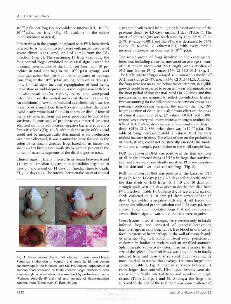
Fifteen frogs in the groups inoculated with FV3, henceforthreferred to as ‘fatally infected’, were euthanized because ofsevere clinical signs (n56) or died (n59) from the FV3infection (Fig. 2). The remaining 19 frogs (including thefour control frogs) exhibited no clinical signs, except forminimal petechiation of the hind feet (less than 10 pe-techiae in total, one frog in the 103.43 p.f.u. group), andmild depression but without loss of posture or reflexes(one frog in the 102.43 p.f.u. group), both on 14 days p.i.only. Clinical signs included regurgitation of food items,dazed stare or mild depression, severe depression with lossof withdrawal and/or righting reflex and widespreadpetechiation on the ventral surface of the skin (Table 1).An additional observation included as a clinical sign was thepresence of a small (less than 0.5 cm in greatest diameter)ovoid pearly white bead found in the water dish of four ofthe fatally infected frogs but never produced by any of thesurvivors. It consisted of proteinaceous material (mucus)admixed with myriads of Gram-negative bacterial rods and afew rafts of cells (Fig. 1d–f). Although the origin of this beadcould not be unequivocally determined as its productionwas never observed, it was assumed to have formed in thecolon of terminally diseased frogs based on its faeces-likeshape and its histological similarity to material present in thelumen of necrotic segments of the distal digestive tract.
Clinical signs in fatally infected frogs began between 6 and14 days p.i. (median 11 days p.i.). Mortalities began at 10days p.i. and ended on 14 days p.i. (median time to death,ST50, 12 days p.i.). The interval between the onset of clinical
signs and death varied from 0 (,12 h based on time of theprevious check) to 4.5 days (median 1 day) (Table 1). Theonset of clinical signs was accelerated by 15 % (95 % CI 5–24 %, P value50.001) and the ST50 was decreased by 16 %(95 % CI 6–25 %, P value50.001) with every tenfoldincrease in dose, when dose was ¢102.43 p.f.u.
The whole group of frogs involved in the experimentalinfection, including controls, measured an average (mean)of 35.0 mm in snout–vent (SV) length, with a median of35.2 mm (range 29–42, mean 95 % CI 33.8–36.2) (Fig. 2).The fatally infected frogs averaged 32.9 mm with a median of33.2 mm (range 29–37, mean 95 % CI 31.5–34.2). Althoughthe frogs were not measured before the experiment, negligiblegrowth would be expected to occur in 1-year-old animals overthe short period of time the trial lasted (10–22 days), and thusmeasurements are assumed to reflect pre-infection lengths.Even accounting for the difference in size between groups as apotential confounding variable, the size of the frog (SVlength) at time of death had a significant effect on the onsetof clinical signs and ST50 (P values ,0.001 and 0.001,respectively): every millimetre increase in length resulted in a6 % (95 % CI 3–9 %) delay in onset of signs and a 5 % delay indeath (95 % CI 2–8 %) when dose was ¢102.43 p.f.u. Theodds of dying increased 23-fold (P value50.017) for everytenfold increase in dose. The effect of size on the probabilityof death, if any, could not be statically assessed (the modelwould not converge), possibly due to the small sample size.
PCR for ranavirus DNA was positive in the skin and liverof all fatally infected frogs (15/15); in frogs that survived,skin and liver were consistently negative. PCR was negativein the skin and liver of all control frogs (Fig. 2).
PCR for ranavirus DNA was positive in the faeces of 3/15frogs (7, 8 and 11 days p.i.; 3–4.5 days before death) and inthe skin sheds of 4/15 frogs (3, 4, 10 and 10 days p.i.;strongly positive 0–1.5 days prior to death) that died fromFV3 infection (Table 1). Collectively, 18 faeces and six skinsheds collected on 1–10 days p.i. from several of the 15dead frogs yielded a negative PCR signal. All faeces andskin sheds collected pre-inoculation and 0–22 days p.i. fromcontrol frogs and inoculated frogs that did not developsevere clinical signs to warrant euthanasia were negative.
Gross lesions noted at necropsy were present only in fatallyinfected frogs and consisted of petechial/ecchymotichaemorrhages in skin (Fig. 1a, b), free blood in oral cavity,focal to extensive haemorrhage in the wall of stomach and/or intestine (Fig. 1c), blood in faecal swab, petechiae incoelomic fat bodies or testicles and an air-filled stomach.Splenomegaly, subjectively determined in reference to thesize of the spleen of control frogs, was noted both in fatallyinfected frogs and those that survived, but it was slightlymore marked in mortalities (average 1.9 times larger thancontrol) (Table 1, Fig. 3) than in survivors (average 1.4times larger than control). Histological lesions were alsorestricted to fatally infected frogs and involved multipletissues (Table 2, Figs 4 and 5). Amongst the frogs thatsurvived to the end of the trial there was some evidence of
(a) (b)
(c) (d) (e) (f)
Fig. 1. Gross lesions due to FV3 infection in adult wood frogs.Petechiae in the skin of ventrum and limbs (a, b) and severehaemorrhage in the intestinal wall (c). Histological appearance ofmucous bead produced by fatally infected frogs: clusters of cells(haematoxylin & eosin stain, d) surrounded by protein-rich mucus(Periodic Acid-Schiff stain, e) and myriads of Gram-negativebacterial rods (Gram stain, f). Bars, 50 mm.
M. J. Forzan and others
1140 Journal of General Virology 96

Downloaded from www.sgmjournals.org by
IP: 160.36.45.170
On: Fri, 31 Jul 2015 13:29:12
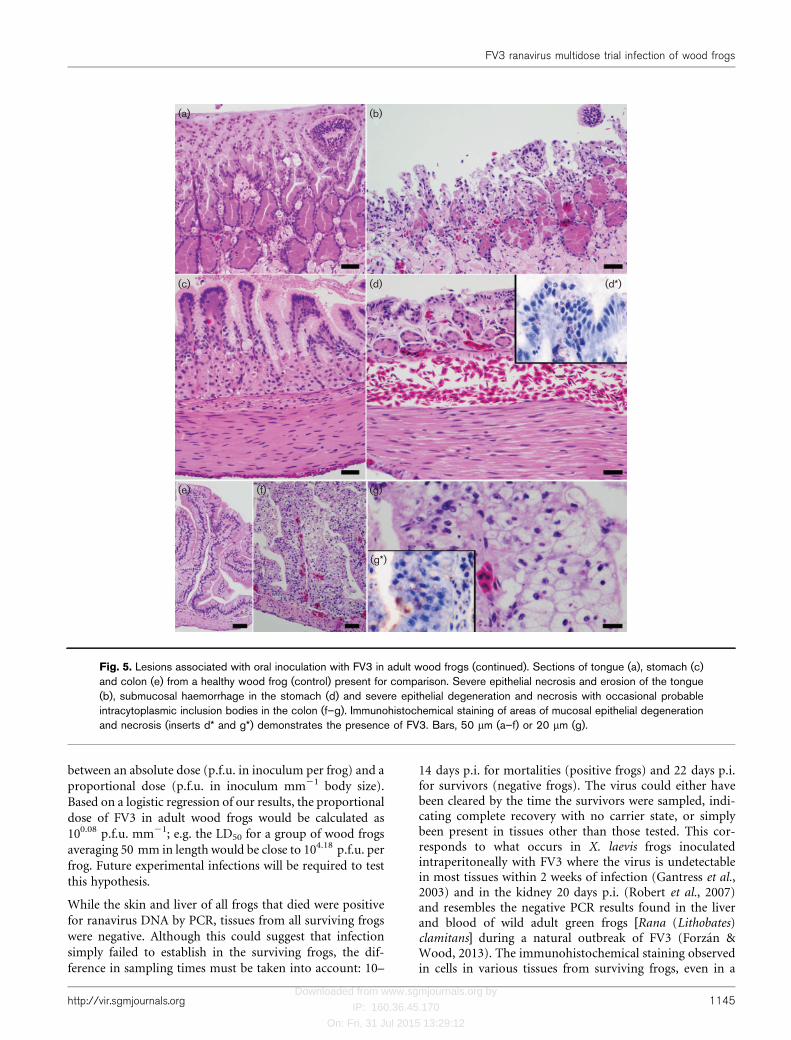
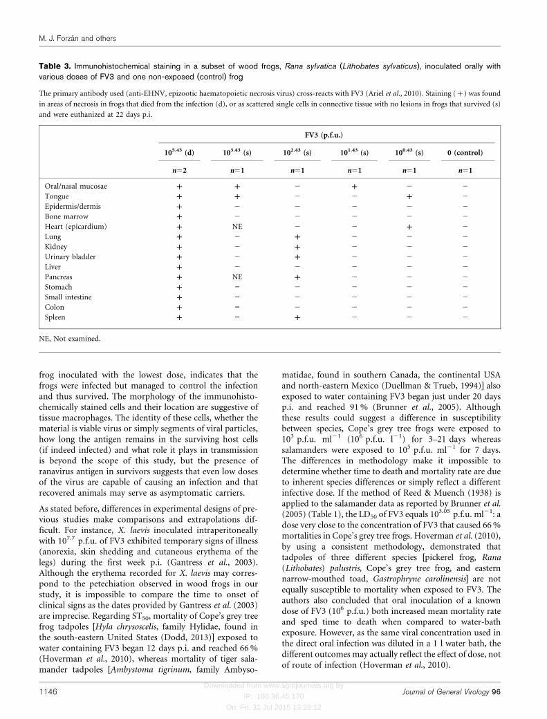
hyperplasia of haematopoietic tissue in the bone marrowand spleen, renal tubular regeneration or hyperplasia andnodular proliferation of lymphocytes in the wall of theurinary bladder and colonic submucosa. Immunohisto-chemical staining was performed in one frog from the non-exposed (control) group, two frogs in the group thatreceived the highest dose (105.43 p.f.u.), the sole survivor inthe group that received 103.43 p.f.u. dose and three frogsthat survived, one from each of the lowest dose groups(102.43, 101.43 and 100.43 p.f.u.) (Table 3). Immunohisto-chemical staining for ranaviral antigen (cytoplasmic,usually as fine to coarse variably abundant granules) ofthe fatally infected frogs that received the highest dosedemonstrated the presence of ranavirus antigen in andaround areas of necrosis in various tissues (Table 3). In thefrogs that survived, staining was observed in scatteredsingle cells in the connective tissue of one or more tissues(Table 3). The morphology of the immunohistochemicallystained cells found in survivors, and their location (rarelyin parenchymal organs like kidney or pancreas andcommonly in the submucosa of a luminal organ or wallof a cavity), suggest tissue macrophages. In nervous tissue(brain and peripheral nerves), testes, ovary, oviduct andabdominal adipose tissue no staining was detected in any
of the frogs. None of the tissues from the non-exposed(control) frog stained immunohistochemically. Non-spe-cific staining was negligible according to the antibody andconjugate internal controls.
DISCUSSION
Our results indicate that the oral LD50 of FV3 in 1-year-oldwood frogs averaging 35 mm of SV length is 102.93 (2.42–3.44)
p.f.u. per frog. Although wood frogs usually begin repro-duction at 2 years of age (Duellman & Trueb, 1994) and ourexperimental subjects were only 1 year old, most weresexually mature (oogenesis or spermatogenesis evident hist-ologically in 19/20 frogs examined) and thus represen-tative of the anatomy and immunophysiology of adultindividuals. Our findings can probably also apply to post-metamorphic juveniles as immune system maturationoccurs at metamorphosis or soon afterwards (Robert &Ohta, 2009). Extrapolations to other experimental or tonatural infections should be made cautiously if envir-onmental conditions are different from those reported heresince habitat characteristics, particularly temperature, areknown to influence the immune function of amphibians
Dose (p.f.u. per frog) Mortality (%)
SV
100
100
80
20
0
0
0
ST50
105.43
104.43
103.43
102.43
101.43
100.43
Control
22 22 22 2241 36 38 38s ––
––
––
––
––
––
––
––
––
––
––
––
––
––
––
––
––
––
––
s s s
22 22 22 2236 35 38 39s s s s
22 22 22 2236 42 34 29s s s s
14 22 22 2231 41 38 32d ++ s s s 22 38 s
22 31 s
22 36 s
22 39 s12 13 13 1432 36 37 34e ++ e ++ e ++ e ++
11 12 14 1034 30 36 34d ++ d ++ e ++ d ++
11.5 10 12 11.533 29 33 31d ++ d ++ e ++ d ++ 11.5 11.334 32
32.8
34.8/39
34.4
36.8
38
31/37.2
d ++
11 11
13
14
n/a
n/a
n/a
30 d ++
Fig. 2. Mortalities (black) used to calculate the LD50 of FV3 inoculated orally to adult wood frogs. Below each frog, from left toright: day of death p.i., snout–vent length [SV (mm)], euthanasia due to severe disease, natural death or survival until end of trial(indicated e, d or s, respectively), and PCR for ranavirus DNA in skin and liver (+, ”). Right column: percentage of mortality,median survival time [ST50 (days)] and mean size [SV (mm)] of mortalities/survivors per group. n/a, Not applicable.
FV3 ranavirus multidose trial infection of wood frogs
http://vir.sgmjournals.org 1141

Downloaded from www.sgmjournals.org by
IP: 160.36.45.170
On: Fri, 31 Jul 2015 13:29:12
Table 1. First appearance of clinical signs, death, interval from first signs to death (ClS–Dth), presence of ranavirus DNA (PCR) in faeces and skin sheds and gross lesions atnecropsy of 15 wood frogs that died or were euthanized following oral inoculation with FV3
FV3
(p.f.u.)
Frog Clinical signs (days p.i.) Death
(days
p.i.)*
ClS–
DthD
PCR (days p.i.) Gross lesions
Faeces Skin
shed
Rgt Brb MDpr SDpr Ptc Onst ” + ” + PH-S B-Or PH-GI B-Fc PH-V AFS Splx
105.43 1 10 11 10 10 11.5 1.5 4, 6, 7 10 2
2 8 10 10 8 10 2 6 y y y 2
3 12 12 12 12 0 2 8 4 y y 3
4 6, 11 11.5 6 11.5 4.5 y y y 1
5 11.5 11.5 11.5 0 4 7 y y y 1
104.43 6 10 11 11 10 11 1 10 y ya y 1.5
7 12 12 12 0 4 9 y 2
8 13 14 14 13 14 1 1, 6, 7, 9, 10 4 y 2.5
9 10 10 10 10 0 1, 6 4 10 y 2
10 10 11 11 10 11 1 1 3d y y 2
103.43 11 12 12 12 0 y y 1
12 11, 13 13 13 11 13 2 6 4d y y y NR
13 13 13 13 13 13 0 y yb y 2.5
14 14 14 14 14 0 7 11 y y 2.5
102.43 16 11 13 13 13 11 14 3 2 5 y ya y 2
Rgt, Regurgitation of food item; Brb, bacteria-rich mucous bead in water dish; MDpr, dazed stare or mild depression; SDpr, severe depression with loss of withdrawal or righting reflex; Ptc,
petechiae in the ventral skin and/or limbs; Onst, first onset of clinical signs; PH-S, petechiae/haemorrhage in skin; B-Or, blood from oral cavity; PH-GI, petechiae/haemorrhage in wall of
gastrointestinal tract; B-Fc, blood in faecal swab; PH-V, petechiae/haemorrhage in parenchymatous organs (a, fat bodies; b, testicles); AFS, air-filled stomach; Splx, proportional increase in size of
spleen compared with age-matched control; NR, not recorded.
*Death was recorded at time of observation except when a frog died overnight, in which case 0.5 of a day was deducted.
DInterval between onset of clinical signs and time of death (ClS–Dth) of 0 indicates no signs detected on the previous check (,12 h earlier).
dIndicates only weak positivity in PCR test for ranavirus DNA.
M.J.F
orzanand
others
11
42
Journal
of
General
Viro
logy
96

Downloaded from www.sgmjournals.org by
IP: 160.36.45.170
On: Fri, 31 Jul 2015 13:29:12
(Maniero & Carey, 1997). Although we conducted theexperimental inoculation in early spring, a time when woodfrogs are most likely to come in contact with infected carriersas they go to the ponds for mating (Brunner et al., 2004), thetemperature maintained during the experiment (average21 uC) was higher than the environmental temperaturewould have been in the wild. Statistical models of disease
incorporating any of our results as parameters must accountfor temperature and humidity differences in the habitat of
the population of interest.
Based on our statistical model, dose of FV3 was the most
important factor in the length of the incubation period, the
survival time and the probability of dying. The odds of
dying increased by 23-fold and both clinical signs and
Control 100.43 p.f.u. 103.43 p.f.u. 105.43 p.f.u.
Fig. 3. Lesions associated with oral inoculation with FV3 in the spleen of adult wood frogs shown at subgross (upper row, 40�)and histological (bottom row, 400�) magnifications. Spleens from control and lowest dose frogs (survivors) have no lesions andprominent melanomacrophages (white arrows). Haemorrhage, multifocal necrosis (black arrows) and loss of melanomacro-phages increase in proportion to the viral dose received. Haematoxylin & eosin stain. Bars, 200 mm (top row) or 20 mm (bottomrow).
Table 2. Histological lesions in a subset (9/15) of wood frogs that died or were euthanized due to oral inoculation with FV3
Skin Digestive tract/coelom Bone
marrow
Kidney Urinary
bladder
Spleen Liver
FV3 (p.f.u.) Frog DH EpdN EpN-Or SH EpN-GI H-Ad HN HN GN TD/N EpN-Ur HN HN
105.43 1 y* y y y y y* y y y
2 y y y y y* y y* y y y
3 y y y* y y y y y* y y y
104.43 6 y* y y y y y y* NR y
8 y y y y* y y y* y* y y
10 y y y y y y y
103.43 12 y y y y y y
13 y y y y y y y y* y y
102.43 16 y y y y y y y* NR y
OverallD (%) 56 22 78(33*) 33 67 44 100 89 89 56(56*) 88(43*) 100 78
DH, Dermal haemorrhage; EpdN, epidermal necrosis; EpN-Or, epithelial necrosis in oral mucosa and/or tongue; SH, submucosal haemorrhage in
gastrointestinal tract; EpN-GI, epithelial necrosis in stomach and/or intestine; H-Ad, haemorrhage in coelomic adipose tissue; HN, haematopoietic
necrosis; GN, glomerular necrosis; TD/N, renal tubular degeneration or necrosis; EpN-Ur, epithelial necrosis in urinary bladder; NR, not recorded,
tissue lost during processing; y, lesion is present; blank space, lesion is absent.
*Probable viral inclusion bodies present.
DProportion of frogs with a lesion out of all those examined (n59).
FV3 ranavirus multidose trial infection of wood frogs
http://vir.sgmjournals.org 1143

Downloaded from www.sgmjournals.org by
IP: 160.36.45.170
On: Fri, 31 Jul 2015 13:29:12
death occurred approximately 15 % sooner per tenfoldincrease in dose. The lone survivor amongst the frogs that
received a dose above the LD50 was the largest of its group,
while the frog that died in the group given a dose below the
LD50 was the smallest (Fig. 2). This is probably a reflection
of the crudeness of the estimate (both mortalities fall
within the 95 % CI for the LD50 dose) and the inherent
variability in susceptibility of live animals to infection more
than an indication that the size of the frog, as estimated by
SV length, could exert an influence on survivability at a
given viral concentration.
While there is no analytical or empirical evidence to sup-port an influence of size on the probability of dying from agiven dose of FV3, the incubation period and survival timemay be lengthened slightly the larger the frog is. At a givendose, the onsets of clinical signs and death were delayedby approximately 6 % and 5 %, respectively, for each
millimetre increase in size. Although the model supportingthis finding is analytically strong and reflects what wasobserved in this trial, its predictive potential is very poor,particularly when applied to frogs outside the range of sizesincluded in this trial. For instance, the model predicts that a51 mm frog would die 31 days p.i. if given 104.43 p.f.u. ofFV3 but, based on infection of adult wood frogs of that size(unpublished data), this overestimates the ST50 by 17–18 days, incorrectly doubling it. The poor predictive abilitycould be due to our small sample size or the occurrence ofthe observations for both events (clinical onset and death)during such a short time interval. The observed effect of sizeon the ST50 and onset of clinical signs is probably an indirectreflection of the effect of dose: when a given dose isadministered to two frogs of different sizes, the larger frognecessarily receives a smaller dose proportionally to its bodysize. This association suggests the need to distinguish
(a) (b) (c)
(d) (e) (f)
(g) (h) (i)
(j) (k) (l)
(m)
Fig. 4. Lesions associated with oral inoculation with FV3 in adult wood frogs. Dermal haemorrhage (a) and epidermal necrosis(b) in skin of limbs. Renal glomerular (d), haematopoietic (e) and tubular necrosis (g) with occasional intraepithelialintracytoplasmic inclusion bodies (h). Haematopoietic necrosis in the bone marrow of an infected frog (k) is presented adjacentto the bone marrow from a healthy frog (control, j); multinucleated cell with eosinophilic intracytoplasmic inclusions suggestiveof viral inclusion bodies (m, arrow). Immunohistochemical staining of skin (c), renal insterstial haematopoietic tissue (f), a renaltubule (i), and bone marrow (l) demonstrates presence of FV3 in lesions. Bars, 50 mm (a–e, g), 20 mm (f, i) and 10 mm (h, j–m).
M. J. Forzan and others
1144 Journal of General Virology 96
Downloaded from www.sgmjournals.org by
IP: 160.36.45.170
On: Fri, 31 Jul 2015 13:29:12
between an absolute dose (p.f.u. in inoculum per frog) and a
proportional dose (p.f.u. in inoculum mm21 body size).
Based on a logistic regression of our results, the proportional
dose of FV3 in adult wood frogs would be calculated as
100.08 p.f.u. mm21; e.g. the LD50 for a group of wood frogs
averaging 50 mm in length would be close to 104.18 p.f.u. per
frog. Future experimental infections will be required to test
this hypothesis.
While the skin and liver of all frogs that died were positive
for ranavirus DNA by PCR, tissues from all surviving frogs
were negative. Although this could suggest that infection
simply failed to establish in the surviving frogs, the dif-
ference in sampling times must be taken into account: 10–
14 days p.i. for mortalities (positive frogs) and 22 days p.i.for survivors (negative frogs). The virus could either havebeen cleared by the time the survivors were sampled, indi-cating complete recovery with no carrier state, or simplybeen present in tissues other than those tested. This cor-responds to what occurs in X. laevis frogs inoculatedintraperitoneally with FV3 where the virus is undetectablein most tissues within 2 weeks of infection (Gantress et al.,2003) and in the kidney 20 days p.i. (Robert et al., 2007)and resembles the negative PCR results found in the liverand blood of wild adult green frogs [Rana (Lithobates)clamitans] during a natural outbreak of FV3 (Forzan &Wood, 2013). The immunohistochemical staining observedin cells in various tissues from surviving frogs, even in a
(a) (b)
(c) (d)
(e) (f) (g)
(g*)
(d*)
Fig. 5. Lesions associated with oral inoculation with FV3 in adult wood frogs (continued). Sections of tongue (a), stomach (c)and colon (e) from a healthy wood frog (control) present for comparison. Severe epithelial necrosis and erosion of the tongue(b), submucosal haemorrhage in the stomach (d) and severe epithelial degeneration and necrosis with occasional probableintracytoplasmic inclusion bodies in the colon (f–g). Immunohistochemical staining of areas of mucosal epithelial degenerationand necrosis (inserts d* and g*) demonstrates the presence of FV3. Bars, 50 mm (a–f) or 20 mm (g).
FV3 ranavirus multidose trial infection of wood frogs
http://vir.sgmjournals.org 1145
Downloaded from www.sgmjournals.org by
IP: 160.36.45.170
On: Fri, 31 Jul 2015 13:29:12
frog inoculated with the lowest dose, indicates that thefrogs were infected but managed to control the infectionand thus survived. The morphology of the immunohisto-chemically stained cells and their location are suggestive oftissue macrophages. The identity of these cells, whether thematerial is viable virus or simply segments of viral particles,how long the antigen remains in the surviving host cells(if indeed infected) and what role it plays in transmissionis beyond the scope of this study, but the presence ofranavirus antigen in survivors suggests that even low dosesof the virus are capable of causing an infection and thatrecovered animals may serve as asymptomatic carriers.
As stated before, differences in experimental designs of pre-vious studies make comparisons and extrapolations dif-ficult. For instance, X. laevis inoculated intraperitoneallywith 107.7 p.f.u. of FV3 exhibited temporary signs of illness(anorexia, skin shedding and cutaneous erythema of thelegs) during the first week p.i. (Gantress et al., 2003).Although the erythema recorded for X. laevis may corres-pond to the petechiation observed in wood frogs in ourstudy, it is impossible to compare the time to onset ofclinical signs as the dates provided by Gantress et al. (2003)are imprecise. Regarding ST50, mortality of Cope’s grey treefrog tadpoles [Hyla chrysoscelis, family Hylidae, found inthe south-eastern United States (Dodd, 2013)] exposed towater containing FV3 began 12 days p.i. and reached 66 %(Hoverman et al., 2010), whereas mortality of tiger sala-mander tadpoles [Ambystoma tigrinum, family Ambyso-
matidae, found in southern Canada, the continental USAand north-eastern Mexico (Duellman & Trueb, 1994)] alsoexposed to water containing FV3 began just under 20 daysp.i. and reached 91 % (Brunner et al., 2005). Althoughthese results could suggest a difference in susceptibilitybetween species, Cope’s grey tree frogs were exposed to103 p.f.u. ml21 (106 p.f.u. l21) for 3–21 days whereassalamanders were exposed to 105 p.f.u. ml21 for 7 days.The differences in methodology make it impossible todetermine whether time to death and mortality rate are dueto inherent species differences or simply reflect a differentinfective dose. If the method of Reed & Muench (1938) isapplied to the salamander data as reported by Brunner et al.(2005) (Table 1), the LD50 of FV3 equals 103.05 p.f.u. ml21: adose very close to the concentration of FV3 that caused 66 %mortalities in Cope’s grey tree frogs. Hoverman et al. (2010),by using a consistent methodology, demonstrated thattadpoles of three different species [pickerel frog, Rana(Lithobates) palustris, Cope’s grey tree frog, and easternnarrow-mouthed toad, Gastrophryne carolinensis] are notequally susceptible to mortality when exposed to FV3. Theauthors also concluded that oral inoculation of a knowndose of FV3 (106 p.f.u.) both increased mean mortality rateand sped time to death when compared to water-bathexposure. However, as the same viral concentration used inthe direct oral infection was diluted in a 1 l water bath, thedifferent outcomes may actually reflect the effect of dose, notof route of infection (Hoverman et al., 2010).
Table 3. Immunohistochemical staining in a subset of wood frogs, Rana sylvatica (Lithobates sylvaticus), inoculated orally withvarious doses of FV3 and one non-exposed (control) frog
The primary antibody used (anti-EHNV, epizootic haematopoietic necrosis virus) cross-reacts with FV3 (Ariel et al., 2010). Staining (+) was found
in areas of necrosis in frogs that died from the infection (d), or as scattered single cells in connective tissue with no lesions in frogs that survived (s)
and were euthanized at 22 days p.i.
FV3 (p.f.u.)
105.43 (d) 103.43 (s) 102.43 (s) 101.43 (s) 100.43 (s) 0 (control)
n52 n51 n51 n51 n51 n51
Oral/nasal mucosae + + 2 + 2 2
Tongue + + 2 2 + 2
Epidermis/dermis + 2 2 2 2 2
Bone marrow + 2 2 2 2 2
Heart (epicardium) + NE 2 2 + 2
Lung + 2 + 2 2 2
Kidney + 2 + 2 2 2
Urinary bladder + 2 + 2 2 2
Liver + 2 2 2 2 2
Pancreas + NE + 2 2 2
Stomach + ” 2 2 2 2
Small intestine + ” 2 2 2 2
Colon + ” 2 2 2 2
Spleen + ” + 2 2 2
NE, Not examined.
M. J. Forzan and others
1146 Journal of General Virology 96

Downloaded from www.sgmjournals.org by
IP: 160.36.45.170
On: Fri, 31 Jul 2015 13:29:12
The use of oral dosing, as opposed to the often usedintraperitoneal injection, is a better approximation of whatoccurs under natural conditions (Gray et al., 2009). Exposureto virus-loaded water is thought to achieve infection viacontact with oral or branchial mucosa (Gray et al., 2009).Therefore, oral administration may be just as relevant inreplicating natural exposure to viral particles in water bodiesas is immersion in a water bath, and possibly more relevantfor terrestrial species like the wood frog. Oral dosing allows foradministration of relatively precise doses, and can be effectedeasily in most post-metamorphic frogs and even tadpoles ofsome species (Wolf et al., 1968; Hoverman et al., 2010).
The positive PCR signal (ranavirus DNA) in faeces andskin sheds of frogs that died from infection suggests thatboth are potential sources of transmission, particularly inthe last few days before death.
Gross and histological lesions present in frogs that died fromFV3 infection resembled those reported in most other speciesinfected with a Ranavirus sp. (Gray et al., 2009; Cullen &Owens, 2002; Cunningham et al., 2007; Kik et al., 2011) andinvolved primarily the haematopoietic cells (bone marrow,spleen, kidney and liver), renal glomeruli, and mucosalepithelium of the oral cavity, gastrointestinal tract and, to alesser degree, urinary bladder. Necrosis of the colonic mucosaseemed to have been associated with an accumulation ofmucus and bacterial overgrowth in the colon of someindividuals, which was shed as a cohesive pearly white massapproximately 24 h prior to death. Whereas the main lesionreported in immunocompromised X. laevis infected with FV3was necrosis of the epithelium of renal proximal tubules(Robert et al., 2005), damage to the epithelium of the renaltubules in wood frogs was observed only in some individualsand it was often mild. Although there may be a true differencein the type of tissue targeted by FV3 in each species, thepublished histopathological images of FV3 infection in X.laevis appear to represent haematopoietic rather than tubularnecrosis (Robert et al., 2005). In some of the frogs thatsurvived the infection, and particularly in the one survivor ofthe group that received 103.43 p.f.u. of FV3, there appeared tohave been hyperplasia of bone marrow haematopoietic tissue,regeneration of renal tubules and formation of small clustersof lymphocytes in the colonic and urinary bladder mucosa,suggesting an activation of the immune system duringinfection. Supporting this interpretation is the lymphocytic(CD8) response to FV3 that occurs in X. laevis (Morales &Robert, 2007), but the precise mechanism of this response inwood frogs require further investigation.
The development and characterization of this ranavirus–wood frog model is an important step to facilitate researchof ranavirus infection in North American frogs. Oral inocu-lation, developed for this study, was easily performed andallowed for the administration of precise doses. Our resultsinclude environmental parameters, clinical signs, mediansurvival time, probability of death at a given dose, viralshedding in faeces and skin sheds, gross and histologicallesions and immunohistochemical staining results under
controlled laboratory conditions. These findings providetransmission, infection and mortality estimates that could beincorporated into ranavirus disease models and facilitate thedesign of experiments to investigate the pathogenesis ofranavirus infection in North American frogs.
METHODS
Origin and housing of experimental subjects. Wood frog
tadpoles were collected from an urban pond in Prince Edward
Island, Canada, 1–2 weeks after hatching (17 May 2012) and housed
in accordance with guidelines of the Canadian Council on Animal
Care (CCAC, 2004; Fig. S1a, b). Tadpoles (later frogs) were maintained
at a fairly constant room temperature both before and during the
experimental infection (overall average 21–22 uC). Humidity varied
considerably and reflected the seasonal ambient temperature (overall
average 41–56 %) (Supplementary Table S1). All mortalities (29/112
frogs that completed metamorphosis) that occurred in the months
prior to the experiment were examined grossly and histologically for
any lesions suggestive of a ranavirus infection since there is no reliable
method to detect subclinical infection in live animals. As none of the
mortalities had any histological evidence of a ranaviral infection, we
assumed that the captive-raised animals were free of the virus. One year
post-hatching (2 May 2013), the 34 frogs used in the experimental trial
were placed in individual tanks and randomly assigned to an infection
(n530, five frogs per dose of inoculum) or control group (n54). After
6 days of acclimation to their individual tanks, the frogs were
inoculated with FV3.
FV3 culture. The virus stock used in this study, originally isolated in
1965 from a Northern leopard frog, Rana (Lithobates) pipiens (Granoff
et al., 1966), was grown by the authors in EPC (one passage) at room
temperature (18–20 uC) (please refer to supplementary Methods).
Titrations of viral stock were performed to determine median TCID50
and p.f.u. following standard methods (Reed & Muench, 1938; Dulbecco
& Vogt, 1954). Calculation of LD50 dose followed the method of Reed &
Muench (1938) corroborated by a logistic regression.
Inoculation, termination and sample collection. On inoculation
day, each frog was orally administered 50 ml of minimum mainten-
ance medium (MEM supplemented with 2 % FBS and 1 % Antibiotic
Antimycotic, Invitrogen) containing 0 (control group, n54), 100.43,
101.43, 102.43, 103.43, 104.43 or 105.43 p.f.u. of FV3 (infection groups,
n55 per dose of inoculum) through a graded pipette (Fig. S1c). The
small volume of inoculum was chosen to avoid any regurgitation.
Frogs were checked two or three times daily to record specific clinical
signs. Faeces and skin sheds found in the water dish of control and
inoculated frogs were collected opportunistically, frozen at 280 uCand later tested for ranavirus DNA by PCR. Upon every collection the
water dish was disinfected and refilled with clean water. All handling
started with the controls and continued through the infection groups
from lowest to highest virus dose; equipment (i.e. plastic gloves, metal
forceps) was disinfected with sodium hypochlorite (5 % bleach
solution) after handling each frog or enclosure.
Euthanasia was performed at a predetermined end point [when frogs
exhibited signs indicative of serious illness (Wright & Whitaker, 2001)
that would have eventually resulted in death] instead of allowing
death to occur naturally. Thus, euthanasia (by immersion in a 10 %
solution of tricaine methanesulfonate, TMS; Syndel Laboratories) was
performed when a frog exhibited two or more of the following signs:
severely depressed appearance (head down and back legs extended
with loss of normal upright posture and of withdrawal reflex), loss of
righting reflex or presence of many petechial haemorrhages in the
skin of the fore or hind feet, inner thighs or ventrum (Fig. 1a, b). The
FV3 ranavirus multidose trial infection of wood frogs
http://vir.sgmjournals.org 1147
Downloaded from www.sgmjournals.org by
IP: 160.36.45.170
On: Fri, 31 Jul 2015 13:29:12
experiment was terminated 22 days p.i., 8 days after the last mortalityoccurred, by euthanizing all remaining frogs. Previous studies haveconsidered 21 days p.i. sufficient for infection and morbidity due toranavirus to occur (Hoverman et al., 2011). Immediately after deaththe snout–vent (SV) length was measured, a necropsy performed andgross lesions recorded. Weight, being extremely variable due tohydration status, food in the stomach and urine in the bladder(Wright & Whitaker, 2001), was intentionally not recorded. Samplesof ventral skin and left liver lobe were collected, frozen at 280 uC andlater tested for ranavirus DNA by PCR. The rest of the carcass waspreserved in 10 % buffered formalin. The formalin-fixed carcasses ofthree frogs from each dose group and two control frogs wereprocessed routinely for histological examination (10 months post-fixation). Tissues, sectioned at 5 mm and stained with haematoxylinand eosin, included one fore foot, one hind foot, a median section ofthe head and jaw, a cross mid-shaft section of the thigh, and sectionsof heart, lungs, abdominal fat body, liver, kidneys, urinary bladder,stomach, intestine, colon, spleen and reproductive organs. Allprocedures followed a protocol approved by the Animal CareCommittee of the University of Prince Edward Island.
Immunohistochemical staining. A subset of the tissues examinedhistologically was stained immunohistochemically using a primaryantibody known to cross-react with FV3 (Ariel et al., 2010) to detect thepresence of viral particles. Briefly, 5 mm sections were deparaffinised byimmersion in two separate baths of xylene (3 min each), three separatebaths of 100 % ethanol (2 min each) and rinsed in running tap water(1 min). After antigen epitope retrieval was achieved by boiling in TE(Tris/EDTA pH 8.5) solution for 20 min using an 850 W microwave,the slides were washed with tap water, carefully dried and a well wascreated around the tissue sections to hold the immunohistochemicalsolutions. Slides were washed three times with TE, blocked with ELISAbuffer containing casein (30 min at room temperature) and incubated(1.5 h at room temperature) with 50 ml of rabbit anti-Bohle Iridovirusantibody diluted 1 : 1 in TE. Slides were then washed three times withTE, incubated in a solution of 0.3 % hydrogen peroxide and 0.1 %sodium azide in TE (15 min) to inactivate endogenous peroxidase,washed three more times with TE and incubated (1.5 h at roomtemperature) with 50 ml of goat anti-rabbit-HRP conjugate antibodydiluted in TE with 1 % BSA. Following another three washes with TE,the slides were developed with the addition of 100 ml of thechromogenic solution (0.005 % 3-amino-9-ethylcarbazole and0.001 % hydrogen peroxide in substrate buffer, 20 min at roomtemperature), then rinsed in running tap water, counter stained withhaematoxylin (5 min) and rinsed again with tap water. Once dried,coverslips were placed on the slides using an aqueous mountingmedium. Non-specific binding and endogenous peroxidase controlslides were produced by omitting the primary antibody and conjugatedantibody, respectively, from the protocol described above (Fig. S3).
PCR for FV3 DNA. Skin and liver samples were individuallytransferred into tissue lysis buffer, total DNA was extracted using aspin-column DNA purification procedure (Qiagen DNeasy 96) andtested for the ranavirus major capsid protein gene with single roundPCR amplification (Mao et al., 1997), using the primers covering thesame region of the MCP gene as the MCP1 assay recommended by theAquatic Animal Health Code (59-GACTTGGCCACTTATGAC-39 and59-GTCTCTGGAGAAGAAGAA-39) (World Organisation for AnimalHealth, 2012). For the faecal samples, lysis buffer was added into thesample tubes and vortexed at 55 uC four times within 1 h, thentransferred to newly labelled microfuge tubes for DNA extraction.
Parameter calculation and statistical analysis. We calculatedmedian time to onset of clinical signs and median time to death(synonym: ST50), and assessed the influence of inoculum dose andbody size (SV length) on: onset of clinical signs and ST50 (time ratios,TRs), probability of infection and probability of death (odds ratio,
OR). Infection was defined as positive PCR amplification from DNA
extracted from a skin or liver sample. TRs were calculated using
parametric survival models with a log-logistic distribution including
only groups where clinical signs or deaths occurred. ORs were
calculated using a logistic regression. Analysis was performed on
STATA 13.1 (Stata statistical software, Stata Corporation LP).
ACKNOWLEDGEMENTS
This work was partly funded by the Alexander Graham Bell Graduate
Scholarship-Doctoral and the Canadian Cooperative Wildlife Health
Centre (now Canadian Wildlife Health Cooperative). The authors
thank Dr Alexandra Reid, who kindly provided the isolate of FV3
used in the infection, Drs Marion Desmarchelier, Shannon Martinson
and Jonathan Spears, Mr Chris MacQuaid and Maciez Zawadzki, and in
particular Dr Jessica Thompson and Ms Sara Vazquez Quiroga, whose
efforts were indispensable to the successful rearing of the wood frogs.
REFERENCES
Ariel, E., Holopainen, R., Olesen, N. J. & Tapiovaara, H. (2010).Comparative study of ranavirus isolates from cod (Gadus morhua)
and turbot (Psetta maxima) with reference to other ranaviruses. Arch
Virol 155, 1261–1271.
Brunner, J. L., Schock, D. M., Davidson, E. W. & Collins, J. P. (2004).Intraspecific reservoirs: complex life history and the persistence of a
lethal ranavirus. Ecology 85, 560–566.
Brunner, J. L., Richards, K. & Collins, J. P. (2005). Dose and host
characteristics influence virulence of ranavirus infections. Oecologia
144, 399–406.
CCAC (2004). Canadian Council on Animal Care Species-specific
Recommendations on: Amphibians and Reptiles. http://www.ccac.ca/
Documents/Standards/Guidelines/Add_PDFs/Wildlife_Amphibians_
Reptiles.pdf
Chinchar, V. G. (2002). Ranaviruses (family Iridoviridae): emerging
cold-blooded killers. Arch Virol 147, 447–470.
Chinchar, V. G., Hyatt, A., Miyazaki, T. & Williams, T. (2009). Family
Iridoviridae: poor viral relations no longer. In Current Topics in
Microbiology and Immunology, vol. 328, pp. 123–170. Edited by J. L.
Van Etten. Berlin: Springer-Verlag.
Cullen, B. R. & Owens, L. (2002). Experimental challenge and clinical
cases of Bohle iridovirus (BIV) in native Australian anurans. Dis
Aquat Organ 49, 83–92.
Cunningham, A. A., Hyatt, A. D., Russell, P. & Bennett, P. M. (2007).Emerging epidemic diseases of frogs in Britain are dependent on the
source of ranavirus agent and the route of exposure. Epidemiol Infect
135, 1200–1212.
Dodd, C. K. (2013). Frogs of the United States and Canada, pp. 250–
262. Baltimore, MD: The Johns Hopkins University Press.
Duellman, W. E. & Trueb, L. (1994). Biology of Amphibians, 2nd edn,
pp. 37, 497, 505, 520–522, 542–544. Baltimore, MD: The Johns
Hopkins University Press.
Dulbecco, R. & Vogt, M. (1954). Plaque formation and isolation of
pure lines with poliomyelitis viruses. J Exp Med 99, 167–182.
Forzan, M. J. & Wood, J. (2013). Low detection of ranavirus DNA in
wild postmetamorphic green frogs, Rana (Lithobates) clamitans, despite
previous or concurrent tadpole mortality. J Wildl Dis 49, 879–886.
Gantress, J., Maniero, G. D., Cohen, N. & Robert, J. (2003).Development and characterization of a model system to study
amphibian immune responses to iridoviruses. Virology 311, 254–262.
M. J. Forzan and others
1148 Journal of General Virology 96

Downloaded from www.sgmjournals.org by
IP: 160.36.45.170
On: Fri, 31 Jul 2015 13:29:12
Granoff, A., Came, P. E. & Breeze, D. C. (1966). Viruses and renal
carcinoma of Rana pipiens. I. The isolation and properties of virus
from normal and tumor tissue. Virology 29, 133–148.
Gray, M. J., Miller, D. L. & Hoverman, J. T. (2009). Ecology and
pathology of amphibian ranaviruses. Dis Aquat Organ 87, 243–266.
Harp, E. M. & Petranka, J. W. (2006). Ranavirus in wood frogs (Rana
sylvatica): potential sources of transmission within and between
ponds. J Wildl Dis 42, 307–318.
Hoverman, J. T., Gray, M. J. & Miller, D. L. (2010). Anuran
susceptibilities to ranaviruses: role of species identity, exposure route,
and a novel virus isolate. Dis Aquat Organ 89, 97–107.
Hoverman, J. T., Gray, M. J., Haislip, N. A. & Miller, D. L. (2011).Phylogeny, life history, and ecology contribute to differences in
amphibian susceptibility to ranaviruses. EcoHealth 8, 301–319.
Kik, M., Martel, A., Sluijs, A. S., Pasmans, F., Wohlsein, P., Grone,A. & Rijks, J. M. (2011). Ranavirus-associated mass mortality in
wild amphibians, the Netherlands, 2010: a first report. Vet J 190,
284–286.
Lesbarreres, D., Balseiro, A., Brunner, J., Chinchar, V. G., Duffus,A., Kerby, J., Miller, D. L., Robert, J., Schock, D. M. & other
authors (2012). Ranavirus: past, present and future. Biol Lett 8,
481–483.
Maniero, G. D. & Carey, C. (1997). Changes in selected aspects of
immune function in the leopard frog, Rana pipiens, associated with
exposure to cold. J Comp Physiol B 167, 256–263.
Mao, J., Hedrick, R. P. & Chinchar, V. G. (1997). Molecular
characterization, sequence analysis, and taxonomic position of newly
isolated fish iridoviruses. Virology 229, 212–220.
Miller, D. L., Gray, M. & Storfer, A. (2011). Ecopathology of
ranaviruses infecting amphibians. Viruses 3, 2351–2373.
Morales, H. D. & Robert, J. (2007). Characterization of primary andmemory CD8 T-cell responses against ranavirus (FV3) in Xenopuslaevis. J Virol 81, 2240–2248.
Reed, L. J. & Muench, H. (1938). A simple method for estimating fiftypercent endpoints. Am J Hyg 27, 493–497.
Robert, J. & Ohta, Y. (2009). Comparative and developmental studyof the immune system in Xenopus. Dev Dyn 238, 1249–1270.
Robert, J., Morales, H., Buck, W., Cohen, N., Marr, S. & Gantress, J.(2005). Adaptive immunity and histopathology in frog virus 3-infected Xenopus. Virology 332, 667–675.
Robert, J., Abramowitz, L., Gantress, J. & Morales, H. D. (2007).Xenopus laevis: a possible vector of Ranavirus infection? J Wildl Dis43, 645–652.
Robert, J., George, E., De Jesus Andino, F. & Chen, G. (2011).Waterborne infectivity of the Ranavirus frog virus 3 in Xenopus laevis.Virology 417, 410–417.
Tweedell, K. & Granoff, A. (1968). Viruses and renal carcinoma ofRana pipiens. V. Effect of frog virus 3 on developing frog embryos andlarvae. J Natl Cancer Inst 40, 407–410.
Wolf, K., Bullock, G. L., Dunbar, C. E. & Quimby, M. C. (1968).Tadpole edema virus: a viscerotropic pathogen for anuran am-phibians. J Infect Dis 118, 253–262.
World Organisation for Animal Health (2012). Manual of diagnostic
tests for aquatic animals. http://www.oie.int/en/international-standard-
setting/aquatic-manual/access-online/
World Organisation for Animal Health (2013). Aquatic Animal Health
Code. http://www.oie.int/en/international-standard-setting/aquatic-code/
access-online/
Wright, K. M. & Whitaker, B. R. (2001). Amphibian Medicine andCaptive Husbandry, pp. 99–101. Malabar, FL: Krieger PublishingCompany.
FV3 ranavirus multidose trial infection of wood frogs
http://vir.sgmjournals.org 1149