clinical study close margins in oral cancers: implication...
TRANSCRIPT

Clinical StudyClose Margins in Oral Cancers: Implication ofClose Margin Status in Recurrence and Survival ofpT1N0 and pT2N0 Oral Cancers
Sandhya Gokavarapu,1 Ravi Chander,1
Nagendra Parvataneni,2 and Sreenivasa Puthamakula1
1 Basavatarakam Indo American Cancer Hospital and Research Centre, Hyderabad, Andhra Pradesh 500034, India2 Seven Hills Hospital, Andheri West, Mumbai, India
Correspondence should be addressed to Sandhya Gokavarapu; [email protected]
Received 12 July 2014; Revised 17 October 2014; Accepted 20 October 2014; Published 11 November 2014
Academic Editor: Steven Heys
Copyright © 2014 Sandhya Gokavarapu et al. This is an open access article distributed under the Creative Commons AttributionLicense, which permits unrestricted use, distribution, and reproduction in any medium, provided the original work is properlycited.
Introduction. Among all prognostic factors, “margin status” is the only factor under clinician’s control. Current guidelines describehistopathologic margin of >5mm as “clear margin” and 1–5mm as “close margin.” Ambiguous description of positive margin inthe published data resulted in comparison of microscopically “involved margin” and “close margin” together with “clear margin”in many publications. Authors attempted to compare the outcome of close and clear margins of stage I and stage II squamouscell carcinoma of oral cavity to investigate the efficacy of description of margin status. Patients and Methods. Historical cohorts ofpatients treated between January 2010 and December 2011 at tertiary cancer hospital were investigated and filtered for stage I andstage II primary squamous cell carcinomas of oral cavity. Patients with margin status of tumor at margin or within 1mm from cutmargin were excluded and analyzed in multivariate logistic regression model for locoregional recurrences and Cox regression foroverall survival. Results. A total of 104 patients fulfilled the abovementioned criteria, of whom 36 were “clear margin” and 68 were“close margin” with median period of follow-up of 39 months. There was no significant difference in locoregional recurrence (Pvalue: 0.0.810) and survival (P value: 0.0.851) among “close margin” and “clear margin” patients.
1. Introduction
Globally lip and oral cancers together comprise of 9.7%of all the cancers [1]. Incidence of oral cancer is muchhigher in developing countries than developed countries.They comprise one-third of all cancers in southeast Asia [2];the higher incidence is attributed to more popular chewableforms of tobacco in this region [3]. About 90% of oral cavitytumors are squamous cell carcinomas [4].
Surgery is primary treatment modality and best choice inoral cancers owing to anatomical considerations of complexbone and soft tissues in this area [5]; moreover morbidityassociated with primary radiotherapy on quality of life andpersistent xerostomia is considerable [6].
Prognostic risk factors of oral cancer include tumor stag-ing and grading, marginal status, lymph vascular invasion,
perineural spread, and perinodal spread of regional disease,of which marginal status is the only factor to a variable extentunder clinician’s control [7].
Although surgeon always aims at a resection with clearmargin, close margins are inevitable; complexity of oralanatomy explains the fact that positive margins are mostfrequent in oral cancer resection in comparison to the cancersof upper aerodigestive tract [8–11].
Intraoperative tools such as use of frozen section maybenefit in some patients to identify involved margin forfurther revision but fail to identify close margins [12]. Closemargins are possible to be evaluated only after completesectional evaluation in histopathology; such processing is notpossible in short intraoperative period. Currently, there isuniform consensus regarding marginal status. Margins are
Hindawi Publishing CorporationInternational Journal of Surgical OncologyVolume 2014, Article ID 545372, 6 pageshttp://dx.doi.org/10.1155/2014/545372

2 International Journal of Surgical Oncology
regarded as clear when histological margin from invasive car-cinoma was more than 5mm, 1–5mm distance was regardedas close, and less than 1mm was regarded as involved [13].
Considerable controversy exists on the criteria for pos-itive margins. Looser et al. [14] have made a classificationof positive margins on four histological criteria: (1) margincloseness (tumor within 0.5 cm), (2) premalignant change inmargin, (3) in situ cancer in the margin, and (4) invasivemicroscopic cancer in the margin. Loree and Strong [15]considered lesion tissue within 0.5mmof the surgical marginwith the exception of laryngeal lesions, dysplastic epitheliumat the margin, carcinoma in situ at the margin, and invasivecarcinoma at the margin to be positive margins.
In patients habituated to tobacco and betel chewing,entire mucosa undergoes premalignant change. Pindborgand Sirsat in their evaluation of 89 OSMF (oral submu-cous fibrosis) patients described coexisting oral cancer anddysplasias [16]; Chaturvedi et al. reported coexisting OSMFin 30% of oral cancer patients in India [17]. Trismus anddifficulty in access to tumors in these patients increase thepossibility of close resection margins; often these patientsundergo extensive resections for low tumor volume in anattempt to gain oncologic safe margin. Considering dysplasiaand premalignant change to be a positivemarginwould resultin excessive reporting of positive margins and comparisonof these patients with patients of involved margin; moreover,this necessitates adjuvant therapy in most of these patients.Anneroth et al. [18] have discussed the reasons behind thevarying results obtained in studies using histomorphologicgrading schemes and the potential errors in clinical researchassociated with oral cancer.
Because of all the abovementioned factors, we investi-gated patients with close margins free of tumor and analyzedthem with clear margin cases for overall outcome.
2. Patients and Materials
A total of 563 primary oral cancer patients were treatedsurgically in this period; after exclusion of patients withnonsquamous cell carcinoma, carcinoma of lip, and recurred,residual, and second primary oral cancers, 457 patients wereisolated: 148 patients were lost to follow-up during this periodand 309 primary oral squamous cell carcinomas were filtered(the demographics of patients such as age, sex, and stage ofdisease were comparable in the patients with and withoutcontinued follow-up; follow-upwas lost due to disconnectionin the contact telephone number inmost of the cases); amongthese patients, cases with verrucous and hybrid carcinomaswere excluded along with patients with tumor within 1mmfrom cut margin; based on pathological staging, stage I andstage II cases (UICC TNM Staging) with close and clearmargins were 104 in number. The median period of follow-up was 37 months.
Patients with margin status of less than or equal to 5mmwere regarded as “close” and greater than 5mm as “clear.” Allthe patients had undergone neck dissections in the selectedsample and pathological staging depended on the same.
Variables were first tested for associations with outcome,that is, “locoregional recurrence” and “death” in univariate
Table 1: Demographic, prognostic factors of the sample.
FactorsTotalsample𝑁
Close margin(group 1)
Clear margin(group 2)
SiteTongue 72 40 32Buccal mucosa 28 24 4Gingiva 4 4 0
Tobacco useNo 44 28 16Yes 60 40 20
PORTNo 52 33 19Yes 52 35 17
HPE diagnosisWDSCC 73 44 29MDSCC 31 24 7
Lymphovascular spreadAbsent 103 67 36Present 1 1 0
Perineural invasionAbsent 100 65 35Present 4 3 1
T stageT1 54 30 24T2 50 38 12
analysis using the chi-square test or Fisher’s exact test and log-rank test as appropriate. Variables associated with outcome(𝑃 < 0.10) were further tested in a multivariable Coxregression model and multivariate logistic regression modeladjusting for potential risk factors and confounders. TheKaplan-Meier graphs were drawn to indicate the survivalprobability. The software SPSS version 17.0 for Windows(SPSS, Chicago, IL, USA) was used for statistical analysis.A two-tailed 𝑃 value of less than 0.05 was consideredstatistically significant.
3. Results
Among a total of 104 patients, 71 (68.2%) were male and 33(31.7%) were female in the sample. The mean age was 49.6years and range of age 21 years to 79 years; 93 (89.4%) survivedto date and eleven patients died.
52 patients received PORT (postoperative radiotherapy);a dose of 56 gray was delivered most often. There was no sig-nificant difference in the outcome (locoregional recurrence:𝑃value = 0.288 [chi-square test] and death 𝑃 value = 0.26 [log-rank test]) among the patients in close margin group whoreceived PORT to that of patients who did not receive PORT.
Demographic and prognostic factors in either of thegroups are illustrated in Table 1.
In the univariate log-rank test, patients graded MDSCC(moderately differentiated squamous cell carcinoma) had

International Journal of Surgical Oncology 3
Table 2: Univariate analysis of factors associated with survival.
Factors Total sample𝑁
Death𝑛 (%) 𝑃 value
SiteTongue 72 8 (11.1) 0.785Buccal mucosa 28 3 (10.7)Gingiva 4 0 (0.0)
Tobacco useNo 44 6 (13.6) 0.432Yes 60 5 (8.3)
PORTNo 52 4 (7.7) 0.336Yes 52 7 (13.5)
HPE DiagnosisWDSCC 73 4 (5.5) 0.008MDSCC 31 7 (22.6)
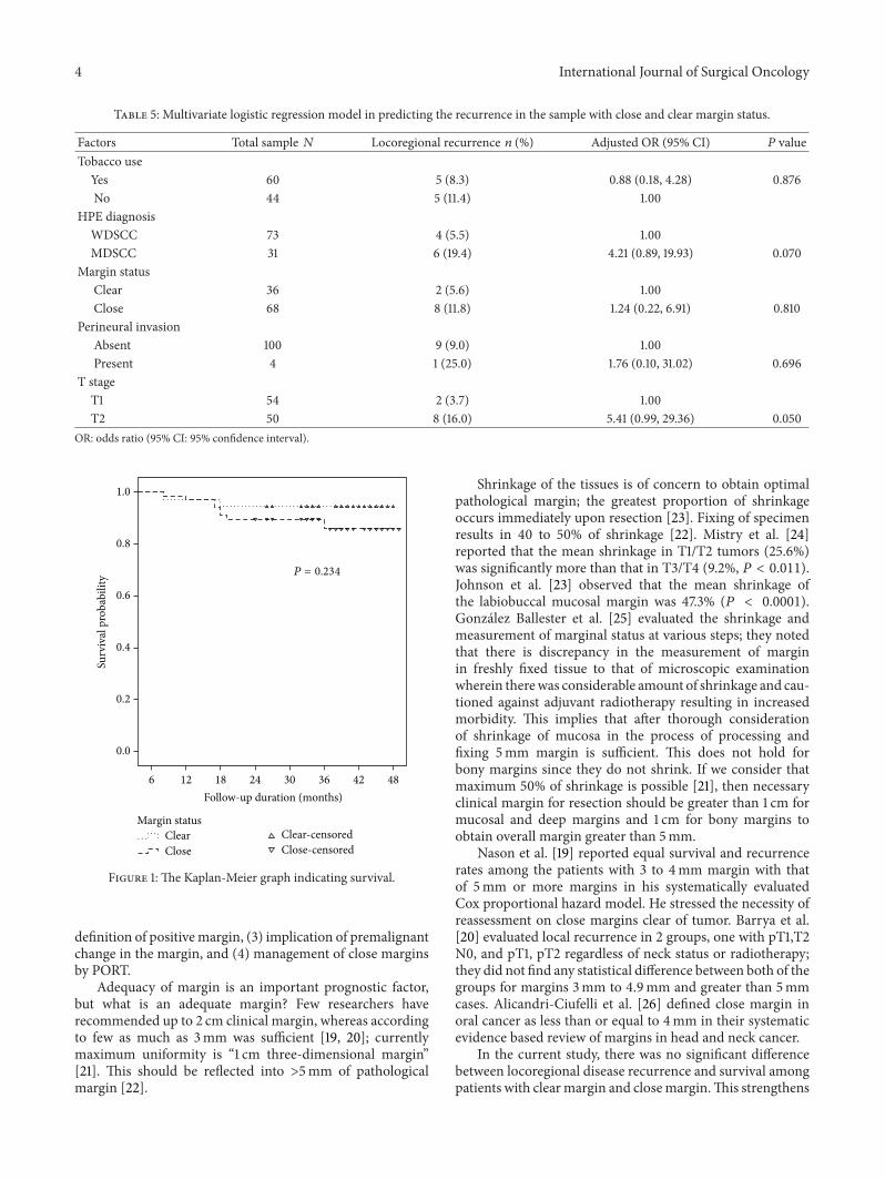
Margin statusClear 36 2 (5.6) 0.234Close 68 9 (13.2)
Lymphovascular SpreadAbsent 103 11 (10.7) 0.762Present 1 0 (0.0)
Perineural InvasionAbsent 100 10 (10.0) 0.264Present 4 1 (25.0)
T StageT1 54 2 (3.7) 0.017T2 50 9 (18.0)
𝛿Based on log-rank test.
higher risk of death (𝑃 = 0.008) to patients with WDSCC(well differentiated squamous cell carcinoma). The patientswith stage II disease had higher risk of death (𝑃 = 0.017,Table 2).
In the multivariable Cox regression model, after adjust-ment for tobacco use, margin status and perineural invasion,patients diagnosed with MDSCC and patients with stage IIdisease had higher risk of death and adjusted hazard ratio(HR): 4.89 (95% CI: 1.19, 20.13) (𝑃 = 0.028) and 6.20 (95%CI: 1.27, 30.21) (𝑃 = 0.024), respectively (Table 3).
In the univariate analysis, patients with stage II diseasesignificantly had higher risk of recurrence when comparedwith stage I disease (𝑃 = 0.046, Table 4). Patients diagnosedwith MDSCC had higher loco recurrence but it was border-line significant risk (𝑃 = 0.062) (Table 4).
In the multivariate logistic regression model, patientswith stage II disease had higher recurrence and adjusted oddsratio (OR): 5.41 (95% CI: 0.99, 29.36) (𝑃 = 0.050, Table 5);however, it was borderline statistically significant. Patientsdiagnosed withMDSCC had higher locoregional recurrence,OR: 4.21 (95% CI: 0.89, 19.93) (𝑃 = 0.070); however,this association was borderline statistically significant. Themultivariate logistic regression model built in this instance
Table 3: Cox regression analysis for risk of mortality in the studiedpatients.
FactorsTotalsample𝑁
Death𝑛 (%)
Hazard ratio(95% CI) 𝑃 value
Tobacco useNo 44 6 (13.6) 1.00 0.842Yes 60 5 (8.3) 0.87 (0.22, 3.51)
HPE diagnosisWDSCC 73 4 (5.5) 1.00MDSCC 31 7 (22.6) 4.89 (1.19, 20.13) 0.028
Margin statusClear 36 2 (5.6) 1.00Close 68 9 (13.2) 1.17 (0.23, 5.88) 0.851
Perineural invasionAbsent 100 10 (10.0) 1.00Present 4 1 (25.0) 1.71 (0.19, 15.20) 0.632
T stageT1 54 2 (3.7) 1.00T2 50 9 (18.0) 6.20 (1.27, 30.21) 0.024
Table 4: Univariate analysis for risk of recurrence in the population.
FactorsTotalsample𝑁
Locoregionalrecurrence𝑛 (%)
𝑃 value𝛿
Tobacco useYes 60 5 (8.3) 0.740No 44 5 (11.4)
HPE diagnosisWDSCC 73 4 (5.5) 0.062MDSCC 31 6 (19.4)
Margin statusClear 36 2 (5.6) 0.488Close 68 8 (11.8)
Perineural invasionAbsent 100 9 (9.0) 0.337Present 4 1 (25.0)
T stageT1 54 2 (3.7) 0.046T2 50 8 (16.0)
𝛿Using either chi-square test or Fisher’s exact test as appropriate.
has best fit according to Hosmer and Lemeshow test (𝜒2 =7.00; 𝑃 = 0.537, Table 5).
The Kaplan-Meier graphs for survival did not show anysignificant difference in either of the groups, Figure 1.
4. Discussion
Margin status has been controversial throughout the liter-ature; various variables regarding margin status include (1)necessary distance of the cut margin from the tumor, (2)
4 International Journal of Surgical Oncology
Table 5: Multivariate logistic regression model in predicting the recurrence in the sample with close and clear margin status.
Factors Total sample 𝑁 Locoregional recurrence 𝑛 (%) Adjusted OR (95% CI) 𝑃 valueTobacco use
Yes 60 5 (8.3) 0.88 (0.18, 4.28) 0.876No 44 5 (11.4) 1.00
HPE diagnosisWDSCC 73 4 (5.5) 1.00MDSCC 31 6 (19.4) 4.21 (0.89, 19.93) 0.070
Margin statusClear 36 2 (5.6) 1.00Close 68 8 (11.8) 1.24 (0.22, 6.91) 0.810
Perineural invasionAbsent 100 9 (9.0) 1.00Present 4 1 (25.0) 1.76 (0.10, 31.02) 0.696
T stageT1 54 2 (3.7) 1.00T2 50 8 (16.0) 5.41 (0.99, 29.36) 0.050
OR: odds ratio (95% CI: 95% confidence interval).
Follow-up duration (months)484236302418126
Surv
ival
pro
babi
lity
1.0
0.8
0.6
0.4
0.2
0.0
Close-censoredClear-censored
CloseClear
Margin status
P = 0.234
Figure 1: The Kaplan-Meier graph indicating survival.
definition of positivemargin, (3) implication of premalignantchange in the margin, and (4) management of close marginsby PORT.
Adequacy of margin is an important prognostic factor,but what is an adequate margin? Few researchers haverecommended up to 2 cm clinical margin, whereas accordingto few as much as 3mm was sufficient [19, 20]; currentlymaximum uniformity is “1 cm three-dimensional margin”[21]. This should be reflected into >5mm of pathologicalmargin [22].
Shrinkage of the tissues is of concern to obtain optimalpathological margin; the greatest proportion of shrinkageoccurs immediately upon resection [23]. Fixing of specimenresults in 40 to 50% of shrinkage [22]. Mistry et al. [24]reported that the mean shrinkage in T1/T2 tumors (25.6%)was significantly more than that in T3/T4 (9.2%, 𝑃 < 0.011).Johnson et al. [23] observed that the mean shrinkage ofthe labiobuccal mucosal margin was 47.3% (𝑃 < 0.0001).Gonzalez Ballester et al. [25] evaluated the shrinkage andmeasurement of marginal status at various steps; they notedthat there is discrepancy in the measurement of marginin freshly fixed tissue to that of microscopic examinationwherein therewas considerable amount of shrinkage and cau-tioned against adjuvant radiotherapy resulting in increasedmorbidity. This implies that after thorough considerationof shrinkage of mucosa in the process of processing andfixing 5mm margin is sufficient. This does not hold forbony margins since they do not shrink. If we consider thatmaximum 50% of shrinkage is possible [21], then necessaryclinical margin for resection should be greater than 1 cm formucosal and deep margins and 1 cm for bony margins toobtain overall margin greater than 5mm.
Nason et al. [19] reported equal survival and recurrencerates among the patients with 3 to 4mm margin with thatof 5mm or more margins in his systematically evaluatedCox proportional hazard model. He stressed the necessity ofreassessment on close margins clear of tumor. Barrya et al.[20] evaluated local recurrence in 2 groups, one with pT1,T2N0, and pT1, pT2 regardless of neck status or radiotherapy;they did not find any statistical difference between both of thegroups for margins 3mm to 4.9mm and greater than 5mmcases. Alicandri-Ciufelli et al. [26] defined close margin inoral cancer as less than or equal to 4mm in their systematicevidence based review of margins in head and neck cancer.
In the current study, there was no significant differencebetween locoregional disease recurrence and survival amongpatients with clearmargin and closemargin.This strengthens

International Journal of Surgical Oncology 5
the observations of Batsakis [22], Nason et al. [19], and Barryaet al. [20] who pointed out the necessity of reevaluation ofdefining 5mm as close margin. Though many of our patientsreceived PORT among “close”margin patients, PORT did notappear to improve survival since 17% (𝑛 = 6) of patients diedin patients of close margin who received PORT (𝑛 = 35)whereas 9% (𝑛 = 3) of patients died in patients of closemargin who did not receive PORT (𝑛 = 33).
It is difficult to compare our results with the earlierresearchers since the results they have mentioned includedpatients with close margin along with the group of patientshaving microscopically involved margin and called them“positive” margin.
Published data on the management of premalignantchange in the margin including dysplasia is complicated.Meier et al. [27] in their survey on current clinical prac-tices in head and neck cancers have reported that 76% ofInternational American Head and Neck Society memberswho participated in the survey considered dysplasia in themargin to be negative. It is clear that there is no uniformityin consensus on what is regarded as a positive margin; thedata on the prognosis of close margins or positive margins isvariable depending on the criteria the researcher has chosen;we did not consider dysplasia in the margin positive sincethe prevalence of premalignant condition such as OSMFis high in patients diagnosed with oral cancer [17]; fieldchanges in the entire mucosa are more frequently observed[16]; considering premalignant change and dysplasia in themargin to be positive would result in justifying PORT inthese patients in whommorbidity associated with RT ismuchsevere due added effect of radiation induced fibrosis [28] topreviously existing submucous fibrosis. Premalignant changein themargin was considered a risk factor in the developmentof second primaries [29] rather than inadequate clearanceof primary tumor. However, carcinoma in situ at marginwas regarded as involved margin and such patient was notincluded in the study.
Published data regarding weather PORT should be givenin patients with close margin is complicated; since initialresearch included these patients in the group of patients withpositive margins, they justified PORT to improve survival.But recent publication by Barrya et al. [20] in which authorcompared recurrences between pT1, T2 N0 and pT1, pT2regardless of neck status or radiotherapy did not find anystatistical difference between patients with margin of 3.0–4.9and >5.0. Wong et al. [30] suggested that surgical marginswithin 2mm should be considered the cut-off for recom-mendation of PORT. Ch’ng et al. [31] also have concludedthat patients with close margins had acceptable local controlwithout PORT in the absence of other risk factors.
52 patients in current study received adjuvant therapy;however, univariate analysis did not show influence of PORTon outcome.
Closemargins did not significantly affect the locoregionalrecurrence or survival; published data was variable regardingthe prognosis of close margins. Selection bias is possiblewherein majority of the patients in a study may be in lowerend of range of 1mm to 5mm or higher end of the samerange. Recent data onmargins distinguishes different survival
among patients below and above 3mm margin [18]. Currentstudy highlights the deficiencies of existing criteria for thedefinition of close margin.
Current study is a retrospective study; all the prognosticfactors may not be evaluated; however, a prospective study inthis regard might not be possible for ethical considerations;thus the information obtained from such data might helpclinician to understand the implications of close marginregarding recurrences and survival. Moreover, this studyincluded primary pT1N0, pT2N0 tumors only and excludedverrucous or hybrid carcinomas. The cases in the study areuncomplicated with regional disease and large surface areasof T3 and T4 tumors.
Conflict of Interests
The authors declare that there is no conflict of interestsregarding the publication of this paper.
Acknowledgment
The authors acknowledge Dr. Sannapaneni Krishnaiah Ph.D.(biostatistics) for professional help in statistics.
References
[1] A. Jemal, F. Bray, M. M. Center, J. Ferlay, E. Ward, and D.Forman, “Global cancer statistics,” CA: Cancer Journal forClinicians, vol. 61, no. 2, pp. 69–90, 2011.
[2] J. Farlay, F. Bray, P. Pisani et al., GLOBOCAN 2002: CancerIncidence, Mortality and Prevalence Worldwide, Version 1.0,IARC Cancer Base no. 5, IARC Press, Lyon, France, 2004.
[3] IARCWorking Group on the Evaluation of Carcinogenic RiskstoHumans, IARCMonographs on the Evaluation of CarcinogenicRisks to Humans, vol. 89, World Health Organization, 2007.
[4] J. Watkinson and R. Gilbert, “Stell & Maran’s textbook of headand neck surgery and oncology,” in Lip and Oral Cavity Cancer,p. 549, CRC Press, 2012.
[5] J. P. Shah and Z. Gil, “Current concepts in management of oralcancer—surgery,” Oral Oncology, vol. 45, no. 4-5, pp. 394–401,2009.
[6] A. P. Jellema, B. J. Slotman, P. Doornaert, C. R. Leemans,and J. A. Langendijk, “Impact of radiation-induced xerostomiaon quality of life after primary radiotherapy among patientswith head and neck cancer,” International Journal of RadiationOncology, Biology, Physics, vol. 69, no. 3, pp. 751–760, 2007.
[7] J. P. Shah, R. A. Cendon, H. W. Farr, and E. W. Strong,“Carcinoma of the oral cavity. Factors affecting treatment failureat the primary site and neck,”The American Journal of Surgery,vol. 132, no. 4, pp. 504–507, 1976.
[8] A. S. Jones, Z. Bin Hanafi, V. Nadapalan, N. J. Roland, A.Kinsella, and T. R. Helliwell, “Do positive resection marginsafter ablative surgery for head and neck cancer adversely affectprognosis? A study of 352 patientswith recurrent carcinoma fol-lowing radiotherapy treated by salvage surgery,” British Journalof Cancer, vol. 74, no. 1, pp. 128–132, 1996.
[9] J. A. Woolgar and A. Triantafyllou, “A histopathologicalappraisal of surgical margins in oral and oropharyngeal cancerresection specimens,” Oral Oncology, vol. 41, no. 10, pp. 1034–1043, 2005.

6 International Journal of Surgical Oncology
[10] J. R. Jacobs, K. Ahmad, R. Casiano et al., “Implications ofpositive surgical margins,”The Laryngoscope, vol. 103, no. 1, pp.64–68, 1993.
[11] J. G. Lee, “Detection of residual carcinoma of the oral cavity,oropharynx, hypopharynx, and larynx: a study of surgical mar-gins,” Transactions of the American Academy of Ophthalmologyand Otolaryngology, vol. 78, no. 1, pp. 49–53, 1974.
[12] J. D. Meier, D. A. Oliver, and M. A. Varvares, “Surgical margindetermination in head and neck oncology: current clinicalpractice. The results of an International American Head andNeck Society member survey,” Head & Neck, vol. 27, no. 11, pp.952–958, 2005.
[13] T. Helliwell and J. A. Woolgar, Standards and MinimumDatasets for Reporting cancers. Dataset for HistopathologicReports on Head and Neck Carcinomas and Salivary Neoplasms,Royal College of Pathologists, London, UK, 2nd edition, 2005.
[14] K. G. Looser, J. P. Shah, and E. W. Strong, “The significance of“positive” margins in surgically resected epidermoid carcino-mas,” Head & Neck Surgery, vol. 1, no. 2, pp. 107–111, 1978.
[15] T. R. Loree and E. W. Strong, “Significance of positive marginsin oral cavity squamous carcinoma,” The American Journal ofSurgery, vol. 160, no. 4, pp. 410–414, 1990.
[16] J. J. Pindborg and S. M. Sirsat, “Oral submucous fibrosis,” OralSurgery, Oral Medicine, Oral Pathology, vol. 22, no. 6, pp. 764–779, 1966.
[17] P. Chaturvedi, S. S. Vaishampayan, S.Nair et al., “Oral squamouscell carcinoma arising in background of oral submucous fibro-sis: a clinicopathologically distinct disease,” Head & Neck, vol.35, no. 10, pp. 1404–1409, 2013.
[18] G. Anneroth, J. Batsakis, andM. Luna, “Review of the literatureand a recommended system of malignancy grading in oralsquamous cell carcinomas,” Scandinavian Journal of DentalResearch, vol. 95, no. 3, pp. 229–249, 1987.
[19] R.W.Nason,A. Binahmed,K.A. Pathak, A.A.Abdoh, andG.K.B. Sandor, “What is the adequate margin of surgical resection inoral cancer?” Oral Surgery, Oral Medicine, Oral Pathology, OralRadiology and Endodontology, vol. 107, no. 5, pp. 625–629, 2009.
[20] C. Barrya, R. Shawa, J. Woolgarb, S. Rogersa, D. Lowea, andJ. Browna, “OP081: evidence to support: a 3mm margin asoncologically safe in early oral SCC,” Oral Oncology, vol. 49,supplement 1, S37 pages, 2013.
[21] J. McMahon, C. J. O’Brien, I. Pathak et al., “Influence ofcondition of surgical margins on local recurrence and disease-specific survival in oral and oropharyngeal cancer,” BritishJournal of Oral andMaxillofacial Surgery, vol. 41, no. 4, pp. 224–231, 2003.
[22] J. G. Batsakis, “Surgical excision margins: A pathologist’s per-spective,”Advances in Anatomic Pathology, vol. 6, no. 3, pp. 140–148, 1999.
[23] R. E. Johnson, J. D. Sigman, G. F. Funk, R. A. Robinson, and H.T. Hoffman, “Quantification of surgical margin shrinkage in theoral cavity,” Head and Neck, vol. 19, no. 4, pp. 281–286, 1997.
[24] R. C. Mistry, S. S. Qureshi, and C. Kumaran, “Post-resectionmucosal margin shrinkage in oral cancer: quantification andsignificance,” Journal of Surgical Oncology, vol. 91, no. 2, pp. 131–133, 2005.
[25] D. Gonzalez Ballester, I. Rubio Correa, C. Hernandez Vila et al.,“OP082: effect of the tissue shrinkage phenomenon on surgicalmargins of resection in patients undergoing cancer oral andoropharynx,” Oral Oncology, vol. 49, p. S37, 2013.
[26] M. Alicandri-Ciufelli, M. Bonali, A. Piccinini et al., “Surgicalmargins in head and neck squamous cell carcinoma: What is“close”?” European Archives of Oto-Rhino-Laryngology, vol. 270,no. 10, pp. 2603–2609, 2013.
[27] J. D. Meier, D. A. Oliver, and M. A. Varvares, “Surgical margindetermination in head and neck oncology: current clinicalpractice. The results of an International American Head andNeck Society member survey,” Head and Neck, vol. 27, no. 11,pp. 952–958, 2005.
[28] J. S. Cooper, K. Fu, J. Marks, and S. Silverman, “Late effects ofradiation therapy in the head and neck region,” InternationalJournal of Radiation Oncology, Biology, Physics, vol. 31, no. 5, pp.1141–1164, 1995.
[29] D. P. Slaughter, H. W. Southwick, and W. Smejkal, “‘Fieldcancerization’ in oral stratified squamous epithelium. Clinicalimplications of multicentric origin,” Cancer, vol. 6, no. 5, pp.963–968, 1953.
[30] L. S. Wong, J. McMahon, J. Devine et al., “Influence of closeresection margins on local recurrence and disease-specificsurvival in oral and oropharyngeal carcinoma,” British Journalof Oral and Maxillofacial Surgery, vol. 50, no. 2, pp. 102–108,2012.
[31] S. Ch’ng, S. Corbett-Burns, N. Stanton et al., “Close marginalone does not warrant postoperative adjuvant radiotherapy inoral squamous cell carcinoma,”Cancer, vol. 119, no. 13, pp. 2427–2437, 2013.

Submit your manuscripts athttp://www.hindawi.com
Stem CellsInternational
Hindawi Publishing Corporationhttp://www.hindawi.com Volume 2014
Hindawi Publishing Corporationhttp://www.hindawi.com Volume 2014
MEDIATORSINFLAMMATION
of
Hindawi Publishing Corporationhttp://www.hindawi.com Volume 2014
Behavioural Neurology
EndocrinologyInternational Journal of
Hindawi Publishing Corporationhttp://www.hindawi.com Volume 2014
Hindawi Publishing Corporationhttp://www.hindawi.com Volume 2014
Disease Markers
Hindawi Publishing Corporationhttp://www.hindawi.com Volume 2014
BioMed Research International
OncologyJournal of
Hindawi Publishing Corporationhttp://www.hindawi.com Volume 2014
Hindawi Publishing Corporationhttp://www.hindawi.com Volume 2014
Oxidative Medicine and Cellular Longevity
Hindawi Publishing Corporationhttp://www.hindawi.com Volume 2014
PPAR Research
The Scientific World JournalHindawi Publishing Corporation http://www.hindawi.com Volume 2014
Immunology ResearchHindawi Publishing Corporationhttp://www.hindawi.com Volume 2014
Journal of
ObesityJournal of
Hindawi Publishing Corporationhttp://www.hindawi.com Volume 2014
Hindawi Publishing Corporationhttp://www.hindawi.com Volume 2014
Computational and Mathematical Methods in Medicine
OphthalmologyJournal of
Hindawi Publishing Corporationhttp://www.hindawi.com Volume 2014
Diabetes ResearchJournal of
Hindawi Publishing Corporationhttp://www.hindawi.com Volume 2014
Hindawi Publishing Corporationhttp://www.hindawi.com Volume 2014
Research and TreatmentAIDS
Hindawi Publishing Corporationhttp://www.hindawi.com Volume 2014
Gastroenterology Research and Practice
Hindawi Publishing Corporationhttp://www.hindawi.com Volume 2014
Parkinson’s Disease
Evidence-Based Complementary and Alternative Medicine
Volume 2014Hindawi Publishing Corporationhttp://www.hindawi.com